Embed Size (px)
Citation preview

Modified SH2 domain to phototrap and identifyphosphotyrosine proteins from subcellular siteswithin cellsAkiyoshi Uezua, Hirokazu Okadaa, Hideji Murakoshib,1, Cosmo D. del Vescovoc, Ryohei Yasudab, Dario Divianic,and Scott H. Soderlinga,2
aDepartment of Cell Biology and bHoward Hughes Medical Institute and Neurobiology Department, Duke University Medical School, Durham, NC 27710;and cDepartment of Pharmacology and Toxicology, University of Lausanne, 1005 Lausanne, Switzerland
Edited by Tony Hunter, Salk Institute for Biological Studies, La Jolla, CA, and approved September 5, 2012 (received for review May 1, 2012)
Spatial regulation of tyrosine phosphorylation is important formany aspects of cell biology. However, phosphotyrosine accountsfor less than 1% of all phosphorylated substrates, and it is typicallya very transient event in vivo. These factors complicate theidentification of key tyrosine kinase substrates, especially in thecontext of their extraordinary spatial organization. Here, we de-scribe an approach to identify tyrosine kinase substrates based ontheir subcellular distribution from within cells. This method uses anunnatural amino acid-modified Src homology 2 (SH2) domain thatis expressed within cells and can covalently trap phosphotyrosineproteins on exposure to light. This SH2 domain-based photoprobewas targeted to cellular structures, such as the actin cytoskeleton,mitochondria, and cellular membranes, to capture tyrosine kinasesubstrates unique to each cellular region. We demonstrate thatRhoA, one of the proteins associated with actin, can be phosphor-ylated on two tyrosine residues within the switch regions, suggest-ing that phosphorylation of these residues might modulate RhoAsignaling to the actin cytoskeleton. We conclude that expressionof SH2 domains within cellular compartments that are capableof covalent phototrapping can reveal the spatial organization oftyrosine kinase substrates that are likely to be important for theregulation of subcellular structures.
protein phosphorylation | proteomics | signal transduction
One of the fundamental goals in cell biology is to identify andreveal the function of molecules in subcellular space and
time to understand mechanisms of cell physiology. However, theanalysis of lysates is often ineffective for the description of manysubcellular proteomes. Fractionation by successive biochemicalpurification steps can be used to identify proteins enriched indistinct organelles, but these approaches likely alter the repre-sentation of low-abundance proteins normally associated withthese organelles in their naive state. Furthermore, some sub-cellular regions are difficult to purify biochemically. Becauseof the difficulty in obtaining organelle structures at high levelsof purity, organelle fractionation usually requires subtractive orcorrelation profile analysis to identify the proteins that areenriched compared with controls (1, 2).Reversible protein phosphorylation is a central signaling mech-
anism that governs important biological functions, such as migra-tion, proliferation, differentiation, and survival. Deregulation ofphosphosignaling pathways, especially tyrosine kinases, is asso-ciated with human diseases, including cancer; thus, clarifyingunregulated kinase signaling pathways is one avenue toward thedevelopment of effective drug therapies and assays for thesediseases (3). One of the critical yet still unresolved issues is theidentity of kinase substrates and how these substrates are func-tionally organized within the cell to regulate physiology. Al-though recent advances in MS technologies have enabled theprofiling of site-specific phosphorylation on a global scale (4–7),several challenges remain. These include the heterogeneity ofphosphorylation sites, the discovery of low-abundance or transientphosphoproteins associated with specific cellular organelles, and
especially the identification of phosphotyrosine due to its lowstoichiometry.In vivo, phosphotyrosine can function as a ligand for docking
domains. The Src homology 2 (SH2) domain is present in 111human proteins and is the prototypic phosphotyrosine bindingdomain (8–11). This phosphobinding property of the SH2 do-main has been used to purify tyrosine-phosphorylated proteinsin vitro by affinity chromatography (12, 13). These SH2-basedaffinity purifications, as well as other in vitro phosphopeptideenrichment strategies, successfully identify phosphorylated proteins,although such approaches may potentially lose low-abundance orlow-stoichiometric phosphotyrosine proteins during extraction.In addition, spurious phosphorylation or dephosphorylation dur-ing the lysis or purification steps likely leads to false-positive orfalse-negative phosphorylation sites, as well as the loss of thespatial organization of phosphoproteome.In this study, we have taken advantage of a modified SH2 do-
main to develop a unique genetically encoded strategy to capturephosphotyrosine proteins in vivo in their native context at distinctsubcellular sites. Using this approach, we have identified a totalof 391 proteins and 189 specifically enriched proteins fromeach subcellular site. We further characterized one of theactin cytoskeletal enriched proteins, RhoA, and showed that itcan be phosphorylated on two tyrosine residues within theswitch regions.
ResultsCharacterization of the Site-Specific Incorporation of p-Benzoyl-L-Phenylalanine in c-Abl SH2. Because protein tyrosine phosphor-ylation status is highly dynamic in living cells, we used the SH2domain, which has a micromolar range of binding affinity towardphosphotyrosine peptides, to capture phosphotyrosine proteinsin living cells combined with an in vivo phototrapping method totrap weak and transient phosphoproteins. The photo–cross-linkingamino acid p-benzoyl-L-phenylalanine (pBpa) can geneticallyincorporate into a desired site of interest within protein domainsin situ by means of orthogonal tRNA/aminoacyl-tRNA synthe-tase pairs (Fig. 1A). We have previously used this approach toinvestigate actin cytoskeletal signaling mediated by Rho familyGTPase Activating Protein (GAP) SH3 domain protein in-teraction (PI) in living cells by trapping them as they occur (14).To test the utility of the approach to identify posttranslational
Author contributions: A.U., D.D., and S.H.S. designed research; A.U., H.O., H.M., and C.D.d.V.performed research; R.Y. contributed new reagents/analytic tools; A.U., H.M., and D.D.analyzed data; and A.U. and S.H.S. wrote the paper.
The authors declare no conflict of interest.
This article is a PNAS Direct Submission.1Present address: Supportive Center for Brain Research, National Institute for Physiolog-ical Science and the Graduate University for Advanced Studies (SOKENDAI), Myodaiji,Okazaki 444-8585, Japan.
2To whom correspondence should be addressed. E-mail: [email protected].
See Author Summary on page 17323 (volume 109, number 43).
This article contains supporting information online at www.pnas.org/lookup/suppl/doi:10.1073/pnas.1207358109/-/DCSupplemental.
www.pnas.org/cgi/doi/10.1073/pnas.1207358109 PNAS | Published online October 1, 2012 | E2929–E2938
CELL
BIOLO
GY
PNASPL
US
Dow
nloa
ded
by g
uest
on
Mar
ch 2
4, 2
020
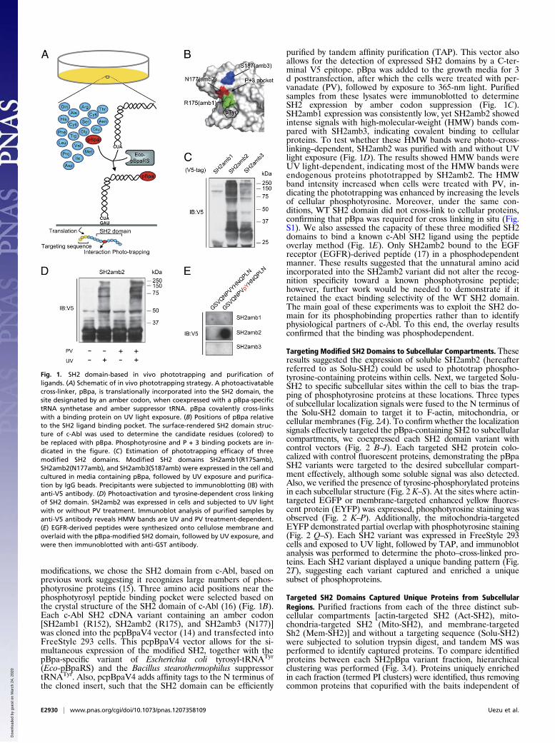
modifications, we chose the SH2 domain from c-Abl, based onprevious work suggesting it recognizes large numbers of phos-photyrosine proteins (15). Three amino acid positions near thephosphotyrosyl peptide binding pocket were selected based onthe crystal structure of the SH2 domain of c-Abl (16) (Fig. 1B).Each c-Abl SH2 cDNA variant containing an amber codon[SH2amb1 (R152), SH2amb2 (R175), and SH2amb3 (N177)]was cloned into the pcpBpaV4 vector (14) and transfected intoFreeStyle 293 cells. This pcpBpaV4 vector allows for the si-multaneous expression of the modified SH2, together with thepBpa-specific variant of Escherichia coli tyrosyl-tRNATyr
(Eco-pBpaRS) and the Bacillus stearothermophilus suppressortRNATyr. Also, pcpBpaV4 adds affinity tags to the N terminus ofthe cloned insert, such that the SH2 domain can be efficiently
purified by tandem affinity purification (TAP). This vector alsoallows for the detection of expressed SH2 domains by a C-ter-minal V5 epitope. pBpa was added to the growth media for 3d posttransfection, after which the cells were treated with per-vanadate (PV), followed by exposure to 365-nm light. Purifiedsamples from these lysates were immunoblotted to determineSH2 expression by amber codon suppression (Fig. 1C).SH2amb1 expression was consistently low, yet SH2amb2 showedintense signals with high-molecular-weight (HMW) bands com-pared with SH2amb3, indicating covalent binding to cellularproteins. To test whether these HMW bands were photo–cross-linking–dependent, SH2amb2 was purified with and without UVlight exposure (Fig. 1D). The results showed HMW bands wereUV light-dependent, indicating most of the HMW bands wereendogenous proteins phototrapped by SH2amb2. The HMWband intensity increased when cells were treated with PV, in-dicating the phototrapping was enhanced by increasing the levelsof cellular phosphotyrosine. Moreover, under the same con-ditions, WT SH2 domain did not cross-link to cellular proteins,confirming that pBpa was required for cross linking in situ (Fig.S1). We also assessed the capacity of these three modified SH2domains to bind a known c-Abl SH2 ligand using the peptideoverlay method (Fig. 1E). Only SH2amb2 bound to the EGFreceptor (EGFR)-derived peptide (17) in a phosphodependentmanner. These results suggested that the unnatural amino acidincorporated into the SH2amb2 variant did not alter the recog-nition specificity toward a known phosphotyrosine peptide;however, further work would be needed to demonstrate if itretained the exact binding selectivity of the WT SH2 domain.The main goal of these experiments was to exploit the SH2 do-main for its phosphobinding properties rather than to identifyphysiological partners of c-Abl. To this end, the overlay resultsconfirmed that the binding was phosphodependent.
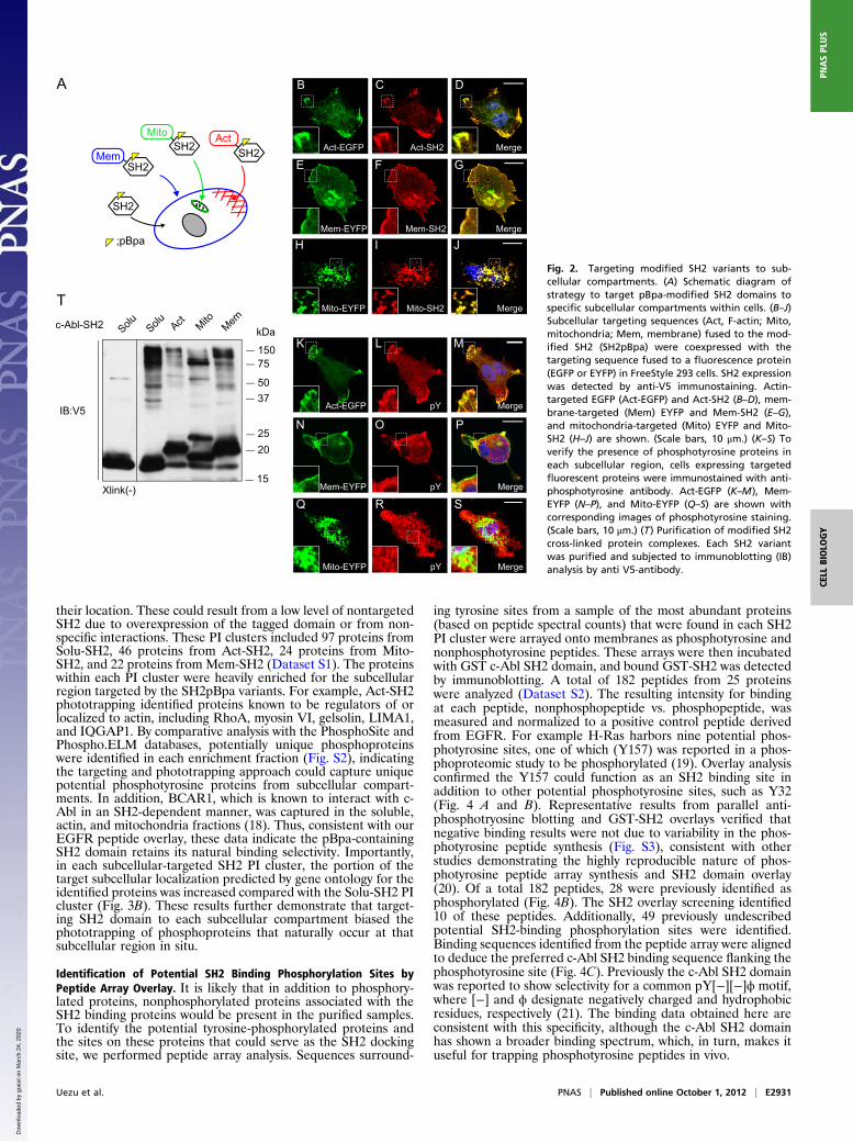
Targeting Modified SH2 Domains to Subcellular Compartments. Theseresults suggested the expression of soluble SH2amb2 (hereafterreferred to as Solu-SH2) could be used to phototrap phospho-tyrosine-containing proteins within cells. Next, we targeted Solu-SH2 to specific subcellular sites within the cell to bias the trap-ping of phosphotyrosine proteins at these locations. Three typesof subcellular localization signals were fused to the N terminus ofthe Solu-SH2 domain to target it to F-actin, mitochondria, orcellular membranes (Fig. 2A). To confirm whether the localizationsignals effectively targeted the pBpa-containing SH2 to subcellularcompartments, we coexpressed each SH2 domain variant withcontrol vectors (Fig. 2 B–J). Each targeted SH2 protein colo-calized with control fluorescent proteins, demonstrating the pBpaSH2 variants were targeted to the desired subcellular compart-ment effectively, although some soluble signal was also detected.Also, we verified the presence of tyrosine-phosphorylated proteinsin each subcellular structure (Fig. 2 K–S). At the sites where actin-targeted EGFP or membrane-targeted enhanced yellow fluores-cent protein (EYFP) was expressed, phosphotyrosine staining wasobserved (Fig. 2 K–P). Additionally, the mitochondria-targetedEYFP demonstrated partial overlap with phosphotyrosine staining(Fig. 2 Q–S). Each SH2 variant was expressed in FreeStyle 293cells and exposed to UV light, followed by TAP, and immunoblotanalysis was performed to determine the photo–cross-linked pro-teins. Each SH2 variant displayed a unique banding pattern (Fig.2T), suggesting each variant captured and enriched a uniquesubset of phosphoproteins.
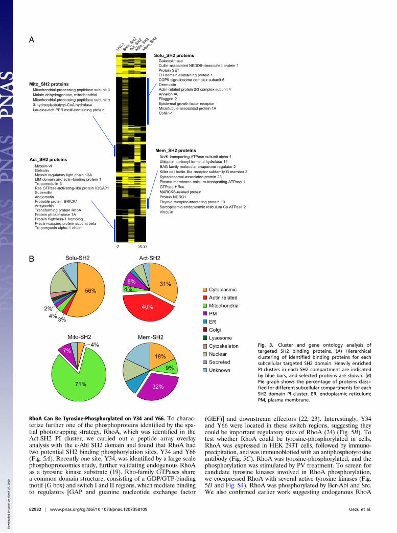
Targeted SH2 Domains Captured Unique Proteins from SubcellularRegions. Purified fractions from each of the three distinct sub-cellular compartments [actin-targeted SH2 (Act-SH2), mito-chondria-targeted SH2 (Mito-SH2), and membrane-targetedSh2 (Mem-SH2)] and without a targeting sequence (Solu-SH2)were subjected to solution trypsin digest, and tandem MS wasperformed to identify captured proteins. To compare identifiedproteins between each SH2pBpa variant fraction, hierarchicalclustering was performed (Fig. 3A). Proteins uniquely enrichedin each fraction (termed PI clusters) were identified, thus removingcommon proteins that copurified with the baits independent of
Fig. 1. SH2 domain-based in vivo phototrapping and purification ofligands. (A) Schematic of in vivo phototrapping strategy. A photoactivatablecross-linker, pBpa, is translationally incorporated into the SH2 domain, thesite designated by an amber codon, when coexpressed with a pBpa-specifictRNA synthetase and amber suppressor tRNA. pBpa covalently cross-linkswith a binding protein on UV light exposure. (B) Positions of pBpa relativeto the SH2 ligand binding pocket. The surface-rendered SH2 domain struc-ture of c-Abl was used to determine the candidate residues (colored) tobe replaced with pBpa. Phosphotyrosine and P + 3 binding pockets are in-dicated in the figure. (C) Estimation of phototrapping efficacy of threemodified SH2 domains. Modified SH2 domains SH2amb1(R175amb),SH2amb2(N177amb), and SH2amb3(S187amb) were expressed in the cell andcultured in media containing pBpa, followed by UV exposure and purifica-tion by IgG beads. Precipitants were subjected to immunoblotting (IB) withanti-V5 antibody. (D) Photoactivation and tyrosine-dependent cross linkingof SH2 domain. SH2amb2 was expressed in cells and subjected to UV lightwith or without PV treatment. Immunoblot analysis of purified samples byanti-V5 antibody reveals HMW bands are UV and PV treatment-dependent.(E) EGFR-derived peptides were synthesized onto cellulose membrane andoverlaid with the pBpa-modified SH2 domain, followed by UV exposure, andwere then immunoblotted with anti-GST antibody.
E2930 | www.pnas.org/cgi/doi/10.1073/pnas.1207358109 Uezu et al.
Dow
nloa
ded
by g
uest
on
Mar
ch 2
4, 2
020
their location. These could result from a low level of nontargetedSH2 due to overexpression of the tagged domain or from non-specific interactions. These PI clusters included 97 proteins fromSolu-SH2, 46 proteins from Act-SH2, 24 proteins from Mito-SH2, and 22 proteins from Mem-SH2 (Dataset S1). The proteinswithin each PI cluster were heavily enriched for the subcellularregion targeted by the SH2pBpa variants. For example, Act-SH2phototrapping identified proteins known to be regulators of orlocalized to actin, including RhoA, myosin VI, gelsolin, LIMA1,and IQGAP1. By comparative analysis with the PhosphoSite andPhospho.ELM databases, potentially unique phosphoproteinswere identified in each enrichment fraction (Fig. S2), indicatingthe targeting and phototrapping approach could capture uniquepotential phosphotyrosine proteins from subcellular compart-ments. In addition, BCAR1, which is known to interact with c-Abl in an SH2-dependent manner, was captured in the soluble,actin, and mitochondria fractions (18). Thus, consistent with ourEGFR peptide overlay, these data indicate the pBpa-containingSH2 domain retains its natural binding selectivity. Importantly,in each subcellular-targeted SH2 PI cluster, the portion of thetarget subcellular localization predicted by gene ontology for theidentified proteins was increased compared with the Solu-SH2 PIcluster (Fig. 3B). These results further demonstrate that target-ing SH2 domain to each subcellular compartment biased thephototrapping of phosphoproteins that naturally occur at thatsubcellular region in situ.
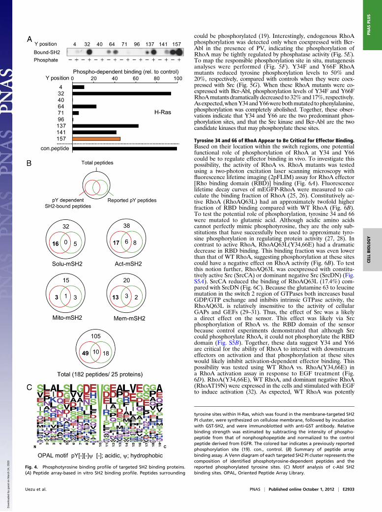
Identification of Potential SH2 Binding Phosphorylation Sites byPeptide Array Overlay. It is likely that in addition to phosphory-lated proteins, nonphosphorylated proteins associated with theSH2 binding proteins would be present in the purified samples.To identify the potential tyrosine-phosphorylated proteins andthe sites on these proteins that could serve as the SH2 dockingsite, we performed peptide array analysis. Sequences surround-
ing tyrosine sites from a sample of the most abundant proteins(based on peptide spectral counts) that were found in each SH2PI cluster were arrayed onto membranes as phosphotyrosine andnonphosphotyrosine peptides. These arrays were then incubatedwith GST c-Abl SH2 domain, and bound GST-SH2 was detectedby immunoblotting. A total of 182 peptides from 25 proteinswere analyzed (Dataset S2). The resulting intensity for bindingat each peptide, nonphosphopeptide vs. phosphopeptide, wasmeasured and normalized to a positive control peptide derivedfrom EGFR. For example H-Ras harbors nine potential phos-photyrosine sites, one of which (Y157) was reported in a phos-phoproteomic study to be phosphorylated (19). Overlay analysisconfirmed the Y157 could function as an SH2 binding site inaddition to other potential phosphotyrosine sites, such as Y32(Fig. 4 A and B). Representative results from parallel anti-phosphotryosine blotting and GST-SH2 overlays verified thatnegative binding results were not due to variability in the phos-photyrosine peptide synthesis (Fig. S3), consistent with otherstudies demonstrating the highly reproducible nature of phos-photyrosine peptide array synthesis and SH2 domain overlay(20). Of a total 182 peptides, 28 were previously identified asphosphorylated (Fig. 4B). The SH2 overlay screening identified10 of these peptides. Additionally, 49 previously undescribedpotential SH2-binding phosphorylation sites were identified.Binding sequences identified from the peptide array were alignedto deduce the preferred c-Abl SH2 binding sequence flanking thephosphotyrosine site (Fig. 4C). Previously the c-Abl SH2 domainwas reported to show selectivity for a common pY[−][−]ϕ motif,where [−] and ϕ designate negatively charged and hydrophobicresidues, respectively (21). The binding data obtained here areconsistent with this specificity, although the c-Abl SH2 domainhas shown a broader binding spectrum, which, in turn, makes ituseful for trapping phosphotyrosine peptides in vivo.
A
T
Act-EGFP pY Merge
K L M
Mito-EYFP pY Merge
Q R SMem-EYFP pY Merge
N O P
Act-EGFP Act-SH2 Merge
B C D
Mito-EYFP Mito-SH2 Merge
H I JMem-EYFP Mem-SH2 Merge
E F G
c-Abl-SH2
IB:V5
75
5037
25
150
20
15
Solu Solu Act MitoMem
Xlink(-)
Mem
Mito Act
;pBpa
SH2
SH2 SH2
SH2
kDa
Fig. 2. Targeting modified SH2 variants to sub-cellular compartments. (A) Schematic diagram ofstrategy to target pBpa-modified SH2 domains tospecific subcellular compartments within cells. (B–J)Subcellular targeting sequences (Act, F-actin; Mito,mitochondria; Mem, membrane) fused to the mod-ified SH2 (SH2pBpa) were coexpressed with thetargeting sequence fused to a fluorescence protein(EGFP or EYFP) in FreeStyle 293 cells. SH2 expressionwas detected by anti-V5 immunostaining. Actin-targeted EGFP (Act-EGFP) and Act-SH2 (B–D), mem-brane-targeted (Mem) EYFP and Mem-SH2 (E–G),and mitochondria-targeted (Mito) EYFP and Mito-SH2 (H–J) are shown. (Scale bars, 10 μm.) (K–S) Toverify the presence of phosphotyrosine proteins ineach subcellular region, cells expressing targetedfluorescent proteins were immunostained with anti-phosphotyrosine antibody. Act-EGFP (K–M), Mem-EYFP (N–P), and Mito-EYFP (Q–S) are shown withcorresponding images of phosphotyrosine staining.(Scale bars, 10 μm.) (T) Purification of modified SH2cross-linked protein complexes. Each SH2 variantwas purified and subjected to immunoblotting (IB)analysis by anti V5-antibody.
Uezu et al. PNAS | Published online October 1, 2012 | E2931
CELL
BIOLO
GY
PNASPL
US
Dow
nloa
ded
by g
uest
on
Mar
ch 2
4, 2
020
RhoA Can Be Tyrosine-Phosphorylated on Y34 and Y66. To charac-terize further one of the phosphoproteins identified by the spa-tial phototrapping strategy, RhoA, which was identified in theAct-SH2 PI cluster, we carried out a peptide array overlayanalysis with the c-Abl SH2 domain and found that RhoA hadtwo potential SH2 binding phosphorylation sites, Y34 and Y66(Fig. 5A). Recently one site, Y34, was identified by a large-scalephosphoproteomics study, further validating endogenous RhoAas a tyrosine kinase substrate (19). Rho-family GTPases sharea common domain structure, consisting of a GDP/GTP-bindingmotif (G box) and switch I and II regions, which mediate bindingto regulators [GAP and guanine nucleotide exchange factor
(GEF)] and downstream effectors (22, 23). Interestingly, Y34and Y66 were located in these switch regions, suggesting theycould be important regulatory sites of RhoA (24) (Fig. 5B). Totest whether RhoA could be tyrosine-phosphorylated in cells,RhoA was expressed in HEK 293T cells, followed by immuno-precipitation, and was immunoblotted with an antiphosphotyrosineantibody (Fig. 5C). RhoA was tyrosine-phosphorylated, and thephosphorylation was stimulated by PV treatment. To screen forcandidate tyrosine kinases involved in RhoA phosphorylation,we coexpressed RhoA with several active tyrosine kinases (Fig.5D and Fig. S4). RhoA was phosphorylated by Bcr-Abl and Src.We also confirmed earlier work suggesting endogenous RhoA
A
B Solu-SH2 Act-SH2
Mito-SH2 Mem-SH2
CytoplasmicActin relatedMitochondriaPMERGolgiLysosomeCytoskeletonNuclearSecretedUnknown
56%
3%4%2%
71%
7%4%
Solu_S
H2
UV(-)
0 ≥0.27
Act_SH2
Mito_S
H2
Mem_S
H2
Solu_SH2 proteinsGalactokinase Cullin-associated NEDD8-dissociated protein 1 Protein SET EH domain-containing protein 1 COP9 signalosome complex subunit 5 Dermcidin
Filaggrin-2
Actin-related protein 2/3 complex subunit 4Annexin A6
Epidermal growth factor receptorMicrotubule-associated protein 1ACofilin-1
Mito_SH2 proteinsMitochondrial-processing peptidase subunit βMalate dehydrogenase, mitochondrial Mitochondrial-processing peptidase subunit α3-hydroxyisobutyryl-CoA hydrolaseLeucine-rich PPR motif-containing protein
Mem_SH2 proteinsNa/K-transporting ATPase subunit alpha-1 Ubiquitin carboxyl-terminal hydrolase 11 BAG family molecular chaperone regulator 2
Synaptosomal-associated protein 23
GTPase HRas
Killer cell lectin-like receptor subfamily G member 2
MARCKS-related protein
Plasma membrane calcium-transporting ATPase 1
Protein NDRG1 Thyroid receptor-interacting protein 13 Sarcoplasmic/endoplasmic reticulum Ca ATPase 2 Vinculin
Act_SH2 proteinsMyosin-VIGelsolin Myosin regulatory light chain 12A LIM domain and actin-binding protein 1 Tropomodulin-3 Ras GTPase-activating-like protein IQGAP1 Supervillin Angiomotin Probable protein BRICK1 Ankycorbin Transforming protein RhoA Protein phosphatase 1A Protein flightless-1 homolog F-actin-capping protein subunit beta Tropomyosin alpha-1 chain
31%
40%
4%8%
18%
9%
32%
Fig. 3. Cluster and gene ontology analysis oftargeted SH2 binding proteins. (A) Hierarchicalclustering of identified binding proteins for eachsubcellular targeted SH2 domain. Heavily enrichedPI clusters in each SH2 compartment are indicatedby blue bars, and selected proteins are shown. (B)Pie graph shows the percentage of proteins classi-fied for different subcellular compartments for eachSH2 domain PI cluster. ER, endoplasmic reticulum;PM, plasma membrane.
E2932 | www.pnas.org/cgi/doi/10.1073/pnas.1207358109 Uezu et al.
Dow
nloa
ded
by g
uest
on
Mar
ch 2
4, 2
020
could be phosphorylated (19). Interestingly, endogenous RhoAphosphorylation was detected only when coexpressed with Bcr-Abl in the presence of PV, indicating the phosphorylation ofRhoA may be tightly regulated by phosphatase activity (Fig. 5E).To map the responsible phosphorylation site in situ, mutagenesisanalyses were performed (Fig. 5F). Y34F and Y66F RhoAmutants reduced tyrosine phosphorylation levels to 50% and20%, respectively, compared with controls when they were coex-pressed with Src (Fig. 5G). When these RhoA mutants were co-expressed with Bcr-Abl, phosphorylation levels of Y34F and Y66FRhoAmutants dramatically decreased to 32%and17%, respectively.Asexpected,whenY34andY66werebothmutated tophenylalanine,phosphorylation was completely abolished. Together, these obser-vations indicate that Y34 and Y66 are the two predominant phos-phorylation sites, and that the Src kinase and Bcr-Abl are the twocandidate kinases that may phosphorylate these sites.
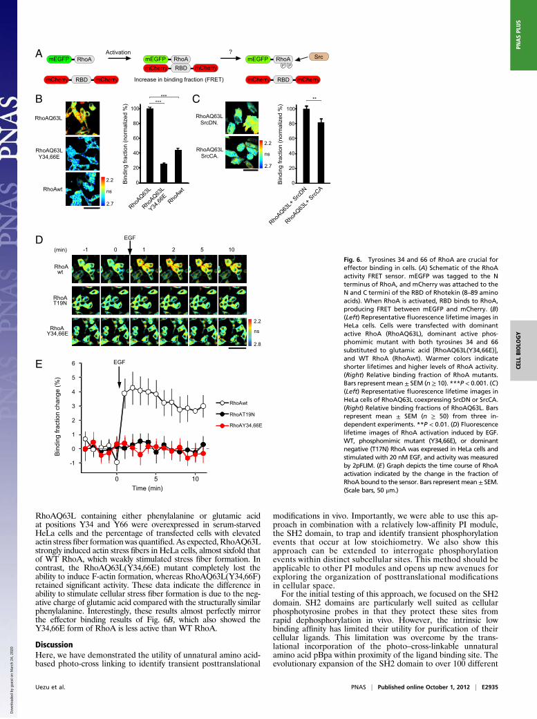
Tyrosine 34 and 66 of RhoA Appear to Be Critical for Effector Binding.Based on their location within the switch regions, one potentialfunctional role of phosphorylation of RhoA at Y34 and Y66could be to regulate effector binding in vivo. To investigate thispossibility, the activity of RhoA vs. RhoA mutants was testedusing a two-photon excitation laser scanning microscopy withfluorescence lifetime imaging (2pFLIM) assay for RhoA effector[Rho binding domain (RBD)] binding (Fig. 6A). Fluorescencelifetime decay curves of mEGFP-RhoA were measured to cal-culate the binding fraction of RhoA (25, 26). Constitutively ac-tive RhoA (RhoAQ63L) had an approximately twofold higherfraction of RBD binding compared with WT RhoA (Fig. 6B).To test the potential role of phosphorylation, tyrosine 34 and 66were mutated to glutamic acid. Although acidic amino acidscannot perfectly mimic phosphotyrosine, they are the only sub-stitutions that have successfully been used to approximate tyro-sine phosphorylation in regulating protein activity (27, 28). Incontrast to active RhoA, RhoAQ63L(Y34,66E) had a dramaticdecrease in RBD binding. This binding fraction was even lowerthan that of WT RhoA, suggesting phosphorylation at these sitescould have a negative effect on RhoA activity (Fig. 6B). To testthis notion further, RhoAQ63L was coexpressed with constitu-tively active Src (SrcCA) or dominant negative Src (SrcDN) (Fig.S5A). SrcCA reduced the binding of RhoAQ63L (17.4%) com-pared with SrcDN (Fig. 6C). Because the glutamine 63 to leucinemutation in the switch 2 region of GTPases both increases basalGDP/GTP exchange and inhibits intrinsic GTPase activity, theRhoAQ63L is relatively insensitive to the activity of cellularGAPs and GEFs (29–31). Thus, the effect of Src was a likelya direct effect on the sensor. This effect was likely via Srcphosphorylation of RhoA vs. the RBD domain of the sensorbecause control experiments demonstrated that although Srccould phosphorylate RhoA, it could not phosphorylate the RBDdomain (Fig. S5B). Together, these data suggest Y34 and Y66are critical for the ability of RhoA to interact with downstreameffectors on activation and that phosphorylation at these siteswould likely inhibit activation-dependent effector binding. Thispossibility was tested using WT RhoA vs. RhoA(Y34,66E) ina RhoA activation assay in response to EGF treatment (Fig.6D). RhoA(Y34,66E), WT RhoA, and dominant negative RhoA(RhoAT19N) were expressed in the cells and stimulated with EGFto induce activation (32). As expected, WT RhoA was potently
Fig. 4. Phosphotyrosine binding profile of targeted SH2 binding proteins.(A) Peptide array-based in vitro SH2 binding profile. Peptides surrounding
tyrosine sites within H-Ras, which was found in the membrane-targeted SH2PI cluster, were synthesized on cellulose membrane, followed by incubationwith GST-SH2, and were immunoblotted with anti-GST antibody. Relativebinding strength was estimated by subtracting the intensity of phospho-peptide from that of nonphosphopeptide and normalized to the controlpeptide derived from EGFR. The colored bar indicates a previously reportedphosphorylation site (19). con., control. (B) Summary of peptide arraybinding assay. A Venn diagram of each targeted SH2 PI cluster represents thecomposition of identified phosphotyrosine-dependent peptides and thereported phosphorylated tyrosine sites. (C) Motif analysis of c-Abl SH2binding sites. OPAL, Oriented Peptide Array Library.
Uezu et al. PNAS | Published online October 1, 2012 | E2933
CELL
BIOLO
GY
PNASPL
US
Dow
nloa
ded
by g
uest
on
Mar
ch 2
4, 2
020

activated by EGF treatment. In contrast, RhoA(Y34,66E) was notresponsive and behaved similar to RhoAT19N (Fig. 6 D and E).
Role of Y34 and Y66 of RhoA in Effector Binding and RhoA-InducedActin Stress Fiber Formation. These results suggested phosphory-lation of RhoA at Y34 and Y66 may inhibit binding to cellulareffectors. To test this possibility, GFP-tagged RhoAQ63L or itsmutant, RhoAQ63L(Y34,66E), expressed in cells was immuno-precipitated to compare interacting partners (Fig. 7A). MS-basedcomparative analysis of the immunoprecipitates showed thatRhoA effectors involved in the regulation of the actin cytoskel-eton, such as DIAPH1, DAAM1, ROCK1/2, and PKN, weredetected only in the RhoAQ63L immunoprecipitations (Fig. 7B).Also, Rhotekin, which was used as an effector binding for 2pFLIMassay, was associated with RhoAQ63L but not with RhoAQ63L
(34,66E). Importantly, RhoGAP1, a GTPase activating proteinfor RhoA and Cdc42 (33, 34), was associated with both RhoAmutants, suggesting that altering tyrosine 34 and 66 does notdisrupt this binding. These observations indicate that Y34 andY66 are critical for effector binding but not for GAP binding.Importantly, it also shows the mutations did not globally disruptRhoA folding or function but did selectively impair its abilityto bind effectors. To determine the role of each tyrosine withregard to effector binding, an RBD pull-down assay was per-formed (Fig. 7 C and D). Both Y34 and Y66 were critical forRhotekin binding, suggesting phosphorylation of either sitecould inhibit RhoA effector functions.Based on these results, the functional consequences to
RhoA-induced cellular actin reorganization were assessed (Fig. 7 Eand F). For this assay, RhoA (wild), RhoAQ63L, and mutants of
A
PV - +
IP: V5-RhoA
IB: pTy 25
25IB: V5
IgG
B
RhoA
Y position
34426674
156
Phospho-dependent binding (rel. to control)0 10 20 30 40 50 60
IP: RhoA
Bcr-Abl
IB: RhoA
IB: pTy
PV - + +
25202520
IP: V5-RhoA
Bcr-Abl
Brk Y44
7F
EGFR L858
R
HER2 rat
Src Y52
7F
contr
ol
wild Y/F wild Y/F wild Y/F wild Y/F wild Y/F wild Y/F
IP: V5-RhoA
+Src
+Bcr-Abl
WT
Y42F
Y66FY74
FY15
6FY34
FY34
,42F
Y34,42
,66F
Y34,66
F
5Y/F
1.6
1.4
1.2
1
0.8
0.6
0.4
0.2
0
WT
Y42F
Y66F
Y74F
Y156F
Y34F
Y34,42
F
Y34,42
,66F
Y34,66
F5Y
/F
+Bcr-Abl+Src
pTy-
Rho
A /
Rho
A (r
el. t
o co
ntro
l)
C D
E
F
Y34
Y74
Y156
Y66
Y42
switch I
switch II
G
- - +
kDa
kDa
IB: pTy
IB: V5
IB: pTy
IB: V5
IB: pTy
IB: V5
Fig. 5. Phosphotyrosine profile of RhoA. (A) Peptides derived from RhoA were synthesized on cellulose membrane, followed by incubation with GST-SH2,and were immunoblotted with anti-GST antibody. Relative binding strength is shown. Bars represent mean ± SEM (n = 3). (B) Surface representations of thestructure of RhoA with switch I and II regions (green and blue, respectively) and five tyrosine sites (red). (C) RhoA is tyrosine-phosphorylated. V5-tagged RhoAwas expressed in 293T cells and treated with or without PV. After lysis, RhoA was immunoprecipitated (IP) by V5 antibody and immunoblotted (IB) withantiphosphotyrosine antibody. (D) Candidate screen of potential RhoA tyrosine kinases. V5-tagged WT RhoA or RhoA with all tyrosines substituted tophenylalanine was coexpressed with the indicated kinases in 293T cells. Immunoprecipitated samples were blotted with antiphosphotyrosine antibody. (E)RhoA is tyrosine-phosphorylated in vivo. Endogenous RhoA was immunoprecipitated from 293T cells transfected with or without Bcr-Abl and treated with PV.Precipitates were immunoblotted with anti-RhoA antibody (Upper) or with antiphosphotyrosine antibody (pTy; Lower). (F) RhoA is tyrosine-phosphorylatedon tyrosines 34 and 66. Various RhoA mutants were coexpressed with Src or Bcr-Abl and treated with PV, followed by immunoprecipitation with anti-V5antibody. Precipitates were blotted with antiphosphotyrosine antibody (Upper) or anti-V5 antibody (Lower). (G) Quantitative analysis of tyrosine-phos-phorylated RhoA mutants was obtained by densitometry. The amount of phospho-RhoA was normalized to the amount of precipitated RhoA. rel., relative.Bars represent mean ± SEM (n = 3).
E2934 | www.pnas.org/cgi/doi/10.1073/pnas.1207358109 Uezu et al.
Dow
nloa
ded
by g
uest
on
Mar
ch 2
4, 2
020
RhoAQ63L containing either phenylalanine or glutamic acidat positions Y34 and Y66 were overexpressed in serum-starvedHeLa cells and the percentage of transfected cells with elevatedactin stress fiber formationwas quantified.As expected,RhoAQ63Lstrongly induced actin stress fibers in HeLa cells, almost sixfold thatof WT RhoA, which weakly stimulated stress fiber formation. Incontrast, the RhoAQ63L(Y34,66E) mutant completely lost theability to induce F-actin formation, whereas RhoAQ63L(Y34,66F)retained significant activity. These data indicate the difference inability to stimulate cellular stress fiber formation is due to the neg-ative charge of glutamic acid compared with the structurally similarphenylalanine. Interestingly, these results almost perfectly mirrorthe effector binding results of Fig. 6B, which also showed theY34,66E form of RhoA is less active than WT RhoA.
DiscussionHere, we have demonstrated the utility of unnatural amino acid-based photo-cross linking to identify transient posttranslational
modifications in vivo. Importantly, we were able to use this ap-proach in combination with a relatively low-affinity PI module,the SH2 domain, to trap and identify transient phosphorylationevents that occur at low stoichiometry. We also show thisapproach can be extended to interrogate phosphorylationevents within distinct subcellular sites. This method should beapplicable to other PI modules and opens up new avenues forexploring the organization of posttranslational modificationsin cellular space.For the initial testing of this approach, we focused on the SH2
domain. SH2 domains are particularly well suited as cellularphosphotyrosine probes in that they protect these sites fromrapid dephosphorylation in vivo. However, the intrinsic lowbinding affinity has limited their utility for purification of theircellular ligands. This limitation was overcome by the trans-lational incorporation of the photo–cross-linkable unnaturalamino acid pBpa within proximity of the ligand binding site. Theevolutionary expansion of the SH2 domain to over 100 different
RBD
P P
-1
0
1
2
3
4
5
6
RhoAwt
RhoAT19N
RhoAY34,66E
Bin
ding
frac
tion
chan
ge (%
)
Time (min)0 5 10
EGF
mEGFP RhoA
mCherry mCherry
mEGFP RhoAmCherry RBD mCherry
Activation
Increase in binding fraction (FRET)
A
D(min) 1 2-1 0 5 10
EGF
RhoA wt
RhoAT19N
RhoAY34,66E
2.2
2.8
ns
B
RhoAQ63L
0
40
60
80
100
RhoAwt
RhoAQ63LY34,66E
Bin
ding
frac
tion
(nor
mal
ized
%)
RhoAQ63
L
RhoAwt
RhoAQ63
L
Y34,66
E
C
E
RhoAQ63
L+ S
rcDN
?mEGFP RhoA
mCherry RBD mCherry
Src
20
0
40
60
80
100
20
Bin
ding
frac
tion
(nor
mal
ized
%)
RhoAQ63
L+ S
rcCA
**
RhoAQ63LSrcDN.
RhoAQ63LSrcCA.
******
2.2
2.7
ns
2.2
2.7
ns
Fig. 6. Tyrosines 34 and 66 of RhoA are crucial foreffector binding in cells. (A) Schematic of the RhoAactivity FRET sensor. mEGFP was tagged to the Nterminus of RhoA, and mCherry was attached to theN and C termini of the RBD of Rhotekin (8–89 aminoacids). When RhoA is activated, RBD binds to RhoA,producing FRET between mEGFP and mCherry. (B)(Left) Representative fluorescence lifetime images inHeLa cells. Cells were transfected with dominantactive RhoA (RhoAQ63L), dominant active phos-phomimic mutant with both tyrosines 34 and 66substituted to glutamic acid [RhoAQ63L(Y34,66E)],and WT RhoA (RhoAwt). Warmer colors indicateshorter lifetimes and higher levels of RhoA activity.(Right) Relative binding fraction of RhoA mutants.Bars represent mean ± SEM (n ≥ 10). ***P < 0.001. (C)(Left) Representative fluorescence lifetime images inHeLa cells of RhoAQ63L coexpressing SrcDN or SrcCA.(Right) Relative binding fractions of RhoAQ63L. Barsrepresent mean ± SEM (n ≥ 50) from three in-dependent experiments. **P < 0.01. (D) Fluorescencelifetime images of RhoA activation induced by EGF.WT, phosphomimic mutant (Y34,66E), or dominantnegative (T17N) RhoA was expressed in HeLa cells andstimulated with 20 nM EGF, and activity was measuredby 2pFLIM. (E) Graph depicts the time course of RhoAactivation indicated by the change in the fraction ofRhoA bound to the sensor. Bars represent mean ± SEM.(Scale bars, 50 μm.)
Uezu et al. PNAS | Published online October 1, 2012 | E2935
CELL
BIOLO
GY
PNASPL
US
Dow
nloa
ded
by g
uest
on
Mar
ch 2
4, 2
020

functional variants in humans infers it is a highly robust struc-tural scaffold capable maintaining phosphotyrosine binding inthe face of mutational pressure. We exploited this property toexpand the physiochemical repertoire of this domain with theincorporation of the unnatural phenylalanine analog. BecausepBpa is translationally incorporated in vivo, this also presentedthe opportunity to add subcellular tags to the recombinant pro-tein to direct the photo–cross-linking events to defined cellularsites. The advantage of this approach is that it allows the bio-chemical probing of subcellular sites in their native state withoutthe need for extensive subcellular fractionation. Furthermore, itmay allow for the interrogation of cellular sites not previouslyamenable to biochemical purification. For the initial testing ofthis approach, we sampled the actin cytoskeleton, mitochondria,and cellular membranes using the c-Abl SH2 domain. Our pri-mary goal was not to identify physiological partners of c-Abl, perse; rather, we used this domain because previous studies hadsuggested it could sample a large number of phosphorylatedproteins when overexpressed (15). Indeed, in each case, we weresignificantly able to enrich the photo–cross-linking reaction to-ward proteins known to exist in each of the targeted sites withincells and to be phosphorylated. In the case of the mitochondria,the SH2 domain was targeted to the intramitochondrial space,where Src family kinases have previously been localized (35).Approximately 40% of the proteins identified in the mitochon-drial targeting experiments have previously been identified asphosphotyrosine substrates (Fig. S2), further confirming thiscellular site can be regulated by tyrosine kinases. Additionally,many of proteins identified are likely be phosphorylated at thecellular sites the SH2 was targeted to. For example, membrane-targeted SH2 identified the multipass membrane protein ATPA1A,a Na+/K+ exchange pump whose activity is regulated by phos-phorylation of tyrosine residue 10 (36). Indeed, this site was alsoidentified in the peptide array as the best binding site for the AblSH2 domain (Dataset S2). Thus, this method can be used to probedefined subcellular spaces, even within organelles in living cells.In this study, we also attempted to identify phosphotyrosine
proteins associated with the actin cytoskeleton directly. This wasimportant, given the known effects of tyrosine kinases on im-portant actin-driven processes, such as adhesion and migration.Our analysis identified several important regulators of actin aspotential phosphotyrosine proteins, including the small GTPaseRhoA. Importantly, both recombinant RhoA and endogenousRhoA were tyrosine-phosphorylated; however, interestingly, thephosphorylation was only apparent on PV treatment to inhibitphosphatase activity. These results highlight that although RhoAmay be a tyrosine kinase substrate, its phosphorylation is likely tobe tightly regulated in vivo or that a small fraction of total cel-lular RhoA is phosphorylated. Often, low stoichiometry ofphosphorylation is interpreted to indicate that the likely impor-tance of phosphorylation is questionable. However, in this case,our approach was specifically designed to identify subcellularpools of phosphorylated proteins. The phototrapping data in-dicated RhoA was only phosphorylated when associated with F-actin; thus, it is possible that the observed low stoichiometry isa reflection of a distinct cellular pool of total RhoA that isphosphoregulated. Further work is needed distinguish among allthese possibilities. Two key phosphotyrosine binding sites, Y34and Y66, were identified by peptide array-based SH2 overlayanalysis, and these sites were confirmed as potential phosphor-ylation sites by mutagenesis. Using an unrelated global phos-phoproteomic approach, RhoA Y34 was also recently detectedin the cancer cell line H1703 (19), further confirming this site asa likely tyrosine kinase substrate. We also screened several ty-rosine kinases to determine which, if any, could target RhoA.From this screen, Src emerged as the best candidate, followed byAbl, which contains an F-actin binding domain (37). Further
GFP
GFP
GAPsEffectors
GAPsEffectors?
A BName FunctionUnique peptides
ROCK1ROCK2DIAPH1DAAM1RhotekinPKN1PKN2
RhoGAP1
Q63L Q63L(Y34,66E)
14 110000000
99
113
151
13
RhoA GTPase activating proteinRhoA-activated kinase
RhoA binding proteinRhoA-activated kinase
RhoA-activated kinaseRhoA-activated ForminRhoA-activated Formin
RhoA-activated kinase
C D
RhoAQ63
L
RhoAQ63
L(Y34
E)
RhoAQ63
L(Y66
E)
RhoAQ63
L(Y34
,66E)
RBD-pulldownIB: anti-V5
Cell extractsIB: anti-V5
kDa
25
20
25
201 2 3 4
RB
D b
indi
ng(%
of c
ontro
l)
RhoAQ63L(Y34,66E)
RhoAQ63L
1 2 3 4
100
80
60
40
20
0
(V5-tag)
E
F
V5 phalloidin
Q63L(34,66E)
Q63L
wild
Q63L(66E)
V5 phalloidin
Q63L(66F)
Q63L(34E)
Q63L(34F)
Q63L(34,66F)
mock
RhoAQ63
L
Fold
indu
ctio
n of
cel
lula
r stre
ssfib
ers
RhoAQ63
L(66F
)
RhoAQ63
L(Y34
,66E)
wild
**
RhoAQ63
L(66E
)
*
RhoAQ63
L(34E
)
*
RhoAQ63
L(34,6
6F)
*
RhoAQ63
L(34F
)
* **
(nor
mal
ized
to R
hoA
wild
)
+++++++
++
+++
+++
Fig. 7. Y34,66E of RhoA abolishes effector binding and stress fiber forma-tion. (A) Schematic figures depict RhoA mutants used to identify endoge-nous GAPs or effectors. Constitutively active RhoA (RhoAQ63L) and itsphosphomimic (Y34,66E) mutant were tagged with GFP and expressed in293T cells, followed by immunoprecipitation. Precipitated samples were sub-jected to MS analyses. (B) Identified proteins in each sample. Actin cytoskele-ton-related proteins identified in constitutively active RhoA (Q63L) orRhoAQ63L phosphomimic mutant are shown with the number of uniquelyidentified peptides in each protein. (C) Representative Western blot of Rho-tekin RBD pull-down assays from cells expressing RhoAQ63L or individualphosphomimic mutants (indicated above). IB, immunoblotting. (Upper)Amounts of each RhoA mutant precipitated by the RBD pull-down are shown.(Lower) Equal levels of expression of each mutant in the lysates are shown. (D)Graph depicts the quantification of the RBD pull-downs as shown in C. Barsrepresent mean ± SEM (n = 3). (E) Induction of F-actin stress fiber formation byRhoA mutants. V5-tagged WT RhoA (wild), constitutive active RhoAQ63L(Q63L), phosphomimic forms of RhoAQ63L (34E, 66E, or 34,66E), or controls(34F, 66F, or 34,66F) were expressed in HeLa cells, followed by immunostainingwith anti-V5 antibody and phalloidin and DAPI labeling. (Scale bars, 10 μm.) (F)Quantitative analysis of RhoA-induced F-actin stress fiber formation. The av-erage fluorescence intensity of F-actin in each V5-RhoA mutant–expressing cellwas measured. The fold induction of cellular stress fiber formation was cal-culated, normalized to RhoA. Bars represent mean ± SEM from three in-
dependent experiments (n ≥ 30). ***P < 0.001; **P < 0.01 in blue are mutantscompared with RhoA. +++P < 0.0001; ++P < 0.01 in red are mutants comparedwith RhoAQ63L(Y34,66E).
E2936 | www.pnas.org/cgi/doi/10.1073/pnas.1207358109 Uezu et al.
Dow
nloa
ded
by g
uest
on
Mar
ch 2
4, 2
020

work is required to identify conclusively the physiological tyro-sine kinases that target RhoA; however, we note evidence insupplementary material of an article by Moritz et al. (19) thatGleevec, an Abl inhibitor, reduces the level of cellular RhoAY34 phosphorylation. Previous reports have also shown that Srcmay, in fact, associate with RhoA and that Src phosphorylationcan inhibit RhoA signaling, in part by phosphorylating and ac-tivating p190RhoGAP during integrin engagement (38–40). Ourdata suggest there could be an additional, direct inhibitory effectof Src on a subfraction of RhoA as a result of phosphorylation ofY34 and Y66. Further work is required to demonstrate that ty-rosine phosphorylation of endogenous RhoA is inhibitory.Several RhoA structures in complex with effectors and mod-
ulators have been solved, and these structures indicate Y34 andY66 are located in important positions for multiple RhoA pro-tein–protein contacts. For example, the structure of RhoA-GTPin complex with the ROCK RBD revealed that Y66 is in theswitch II region and is involved in both electrostatic and hy-drophobic interactions with ROCK1 (41). Also, Y66 is involvedin the binding interaction with the HR1a domain of protein ki-nase C-related kinase 1, suggesting Y66 is critical for RhoAinteractions with multiple types of effector binding domains (42).Additionally, structural analysis of leukemia-associated RhoGEFin complex with RhoA revealed that Y34 is involved in the in-teraction (43). Y34 is also critical for the activation of RhoA bythe GEF AKAP-Lbc (44). Indeed, the Y34 mutant of RhoA(RhoAY34E) abolished binding with AKAP-Lbc (Fig. S6). Thesestudies suggest phosphorylation on tyrosine 34 or 66 may yielddistinct outcomes toward different classes of RhoA binding part-ners, yet the net outcome appears to be inhibition of RhoA-medi-ated signaling to the actin cytoskeleton. Consistent with a potentialregulatory function for Y34, another inhibitory modification hasbeen identified, the AMPlation of Y34, catalyzed by Fic domain-containing proteins, which can also disrupt RhoA signaling (45).Interestingly, Y34 and Y66 are well conserved in Rho pro-
teins, as well as in other small G-protein families (Fig. S7).Consistent with this, we also identified H-Ras as a phosphotyr-osine protein in the membrane-enriched PI cluster, and our SH2overlay mapping identified the homologous tyrosines 32 and 64as possible sites of phosphorylation-dependent binding (Figs. 3and 4). Additionally, Rac1, RalA, and Rab11a contain homol-ogous tyrosine sites that have been reported to be phosphory-lated (46, 19). Furthermore, Y64 of Cdc42 (corresponding to theY66 of RhoA) is also phosphorylated by Src after EGF stimula-tion, and this enhances the interaction between Cdc42 and Rho-GDP dissociation inhibitor (47). Recently, Rac1 was shown to bephosphorylated on Y64 by Src and FAK in vitro (48). Thus, theemerging data suggest that sites homologous to RhoA Y34 andY66 in other small G proteins may function as previously unde-scribed phosphoregulatory sites targeted by tyrosine kinases toregulate cellular functions governed by different GTPase families.In summary, this work describes a unique genetically encoded
approach to discover phosphotyrosine proteins within the con-text of their spatial organization within the cell. This approach isbased on the incorporation of the photoinducible cross-linkingphenylalanine analog pBpa into the phosphotyrosine bindingSH2 domain. Analysis of the identified proteins from each sub-cellular target confirms the overall approach successfully trapsand enriches proteins from these defined compartments. Further-more, this approach reveals unique targets for tyrosine kinasesin different subcellular compartments that could be important forregulation. Additional analysis with SH2 domains with differentbinding specificities compared with Abl will likely expand thecoverage of tyrosine kinase substrates within these subcellular sites.
Materials and MethodsPlasmids. A mammalian expression vector that coexpresses Eco-pBpaRS andB. stearothermophilus suppressor tRNATyr (a gift from Shigeyuki Yokoyama,University of Tokyo, Tokyo, Japan) was modified for TAP (14). A strep-tag,followed by a calmodulin binding peptide (CBP), a V5 epitope, two tobaccoetch virus protease cleavage sites, an HRV3C cleavage site, and a tandemprotein A sequence, was inserted after a mammalian expression promoter.Unique RsrII and SbfI restriction sites were placed between the CBP and
V5 epitope for subcloning SH2 domain coding sequences. The SH2 domain(aa 120–220, Uniprot P00519) derived from c-Abl was inserted, and the tar-geted sequences listed below were placed on the N terminus of SH2. Lifeact,MGVADLIKKFESISKEE (49); membrane (Mem), MLCCMRRTKQVEKNDEDQKI(50) (derived from neuromodulin); and mitochondria (Mito), MSVLTPLLLRGL-TGSARRLPVPRAKIHSL (51) (derived from cytochrome c oxidase subunit 8A)were used. The Lifeact was a gift from Michel Bagnat (Duke University). Otherplasmids used in this study are detailed in SI Materials and Methods.
Photo-Cross Linking. Photo-cross linking was performed as described (14).Briefly, on the day of transfection, a 1-L culture of FreeStyle 293 cells wasgrown in FreeStyle 293 medium (GIBCO) to a density of 1 × 10⁶ cells/mL.Transfection was done following a modified polyethylenimine (PEI) protocol(52). One milligram of the vector encoding TAP-tagged SH2 domain plusEco-pBpaRS and suppressor tRNATyr was mixed with 2 mg of PEI in 50 mL ofFreeStyle 293 medium and added to the cell culture after 10 min of in-cubation. pBpa (270 mg) was dissolved in 1.1 mL of 1 M NaOH and added tothe cell culture following addition of 7.5 mL of 1 M Hepes buffer to theculture. After 3 d of incubation at 37 °C, the cells were treated with 0.5 mMPV for 5 min, followed by exposure to UV light (365 nm) in a custom-builtchamber. After 30 min of photo-cross linking in the chamber at 4 °C, cellswere collected by centrifugation and subjected to TAP purification. Detailedmethods for TAP purification can be found in SI Materials and Methods.
MS. MS was carried out as previously described (14). Briefly, all MS data wereacquired on an LTQ-Orbitrap XL mass spectrometer (Thermo Scientific) op-erating in positive-ion mode with an electrospray voltage of 2.0 kV. Theinstrument was set to acquire a precursor MS scan in the Orbitrap XL massspectrometer from m/z 40–2,000, with r = 60,000 at m/z 400 and a targetautomatic gain control setting of 1e6 ions. In a data-dependent mode ofacquisition, tandem MS spectra of the five most abundant precursor ionswere acquired in the Orbitrap XL mass spectrometer at r = 7,500 at m/z 400with a target AGC setting of 2e5 ions. Maximum fill times were set to 1,000ms for full MS scans and to 500 ms for tandem MS scans with minimumtandem MS triggering thresholds of 5,000 counts. For all experiments,fragmentation occurred in the LTQ linear ion trap, Finnigen LTQ (ThermoElectron Corporation), with a collision-induced dissociation energy setting of35%, and a dynamic exclusion of 60 s was used for previously fragmentedprecursor ions. Details of qualitative identifications from raw liquid chro-matography/tandem MS data can be found in SI Materials and Methods.
Hierarchical Clustering. Relative protein abundance in the sample prepared byTAP was quantified using spectral counting (53). To normalize the relativeprotein abundance, the spectral counts were expressed as a percentage of thetotal spectra observed in the sample. Mean normalized spectral counts wereobtained from multiple independent experiments (n ≥ 2 for each targetedSH2 domain). Hierarchical clustering was performed based on the uncenteredPearson correlation of the mean normalized spectral counts (54, 55).
Peptide Array Synthesis and GST or GST-SH2amb Protein Overlay. Peptides(19mer) were synthesized as previously described (56) using Auto-Spot RobotASP 222 (INTAVIS AG). The GST-SH2 domain was purified as previously de-scribed (57). The peptide arrays were incubated with the GST-SH2 domain ata concentration of 100 nM in 5% (wt/vol) BSA for 2 h. After a wash with Tris-buffered saline/Tween-20, the GST fusion proteins were detected using HRP-conjugated anti-GST antibody (BETHYL). Positive peptide spots were densi-tometrically quantified using ImageJ (National Institutes of Health). Phos-phodependent binding strength was estimated by subtracting the intensityof phosphopeptide from nonphosphopeptide and then normalized to thecontrol peptide. A peptide showing a binding strength over 20 was assessedas a phosphotyrosine-dependent SH2 binding peptide. Amber codon-modifiedSH2 proteins were purified by TAP purification and incubated with the pep-tide arrays derived from EGFR, followed by UV exposure, and bounded SH2was detected with anti-V5 antibody.
Immunoprecipitation Experiments. Immunoprecipitation experiments usingHEK 293T cells were performed as previously described (58). Briefly, trans-fected cells were lysed and centrifuged, and the supernatant was incubatedwith V5 antibody-agarose (Sigma) or GFP trap-agarose (ChromoTek). Afterbeads were washed with lysis buffer, sample buffer was added and sub-jected to immunoblotting. For endogenous RhoA precipitation, cells werelysed with radioimmunoprecipitation assay buffer, followed by sonication,and centrifuged, and the supernatant was incubated with agarose-conjugatedanti-RhoA (26C4; Santa Cruz Biotechnology). Further details can be found inSI Materials and Methods.
Uezu et al. PNAS | Published online October 1, 2012 | E2937
CELL
BIOLO
GY
PNASPL
US
Dow
nloa
ded
by g
uest
on
Mar
ch 2
4, 2
020
Cell Preparation and Image Analysis. Transfected FreeStyle 293 cells wereplated onto poly-D-lysine–coated coverslips, fixed, and stained with anti-V5antibody (1:500; Invitrogen) or antiphosphotyrosine antibody (1:100, 4G10;Millipore). Images were taken on a Zeiss LSM 710 laser scanning confocalmicroscope with a 63×/1.4 N.A. oil-immersion objective or 20×/0.8 N.A. ob-jective. Further details for quantification of phalloidin staining are providedin SI Materials and Methods.
2pFLIM Assay.Details of the 2pFLIM assay have been described previously (26).mEGFP was excited with a Ti/sapphire laser (Mai Tai; Spectraphysics) tuned ata wavelength of 920 nm. Fluorescent images were collected using a 60×/0.9N.A. objective (Olympus). For fluorescence lifetime imaging, a photoelectronmultiplier tube with low transfer time spread (H7422-40p; Hamamatsu) wasused. Fluorescence lifetime images were obtained using a time-correlatedsingle-photon counting board (SPC-140; Becker and Hickl) controlled with
custom software based on Scanimage software (open source software, KarelSvoboda, Janelia Farm, Ashburn, VA).
Statistical Tests. Statistical analysis was done using Excel 2007 (MicrosoftCorporation). Unless otherwise noted, significance was evaluated usinga two-tailed Student t test.
ACKNOWLEDGMENTS. We thank Dr. Shigeyuki Yokoyama (University ofTokyo) for the original pBpa vectors and for advice on pBpa incorporationand cross linking. We also thank Erik Soderblom, Meredith Turner, andArthur Moseley (Duke University Proteomics Core Facility) for help with theMS analysis. This work was supported by National Institutes of Health GrantsR21-CA-140030-01 and R01-NS059957 (to S.H.S.), Fonds National Suisse de laRecherche Scientifique Grant 3100A0-122020 (to D.D), and a grant from theHoward Hughes Medical Institute (to R.Y.).
1. Schirmer EC, Florens L, Guan T, Yates JR, 3rd, Gerace L (2003) Nuclear membrane proteinswith potential disease links found by subtractive proteomics. Science 301:1380–1382.
2. Foster LJ, et al. (2006) A mammalian organelle map by protein correlation profiling.Cell 125:187–199.
3. Blume-Jensen P, Hunter T (2001) Oncogenic kinase signalling. Nature 411:355–365.4. Rikova K, et al. (2007) Global survey of phosphotyrosine signaling identifies
oncogenic kinases in lung cancer. Cell 131:1190–1203.5. Villén J, Beausoleil SA, Gerber SA, Gygi SP (2007) Large-scale phosphorylation analysis
of mouse liver. Proc Natl Acad Sci USA 104:1488–1493.6. Choudhary C, Mann M (2010) Decoding signalling networks by mass spectrometry-
based proteomics. Nat Rev Mol Cell Biol 11:427–439.7. Huttlin EL, et al. (2010) A tissue-specific atlas of mouse protein phosphorylation and
expression. Cell 143:1174–1189.8. Mayer BJ, Jackson PK, Baltimore D (1991) The noncatalytic src homology region 2
segment of abl tyrosine kinase binds to tyrosine-phosphorylated cellular proteins withhigh affinity. Proc Natl Acad Sci USA 88:627–631.
9. Marengere LE, Pawson T (1992) Identification of residues in GTPase-activating proteinSrc homology 2 domains that control binding to tyrosine phosphorylated growthfactor receptors and p62. J Biol Chem 267:22779–22786.
10. Bradshaw JM, Waksman G (2002) Molecular recognition by SH2 domains. Adv ProteinChem 61:161–210.
11. Liu BA, et al. (2011) The SH2 domain-containing proteins in 21 species establish theprovenance and scope of phosphotyrosine signaling in eukaryotes. Sci Signal 4:ra83.
12. Blagoev B, et al. (2003) A proteomics strategy to elucidate functional protein-proteininteractions applied to EGF signaling. Nat Biotechnol 21:315–318.
13. Yang G, et al. (2009) Proteomic, functional and motif-based analysis of C-terminal Srckinase-interacting proteins. Proteomics 9:4944–4961.
14. Okada H, et al. (2011) SH3 domain-based phototrapping in living cells reveals Rhofamily GAP signaling complexes. Sci Signal 4:rs13.
15. Machida K, et al. (2007) High-throughput phosphotyrosine profiling using SH2domains. Mol Cell 26:899–915.
16. Overduin M, Rios CB, Mayer BJ, Baltimore D, Cowburn D (1992) Three-dimensionalsolution structure of the src homology 2 domain of c-abl. Cell 70:697–704.
17. Zhu G, et al. (1994) Sequence specificity in the recognition of the epidermal growthfactor receptor by the abl Src homology 2 domain. Oncogene 9:1379–1385.
18. Mayer BJ, Hirai H, Sakai R (1995) Evidence that SH2 domains promote processivephosphorylation by protein-tyrosine kinases. Curr Biol 5:296–305.
19. Moritz A, et al. (2010) Akt-RSK-S6 kinase signaling networks activated by oncogenicreceptor tyrosine kinases. Sci Signal 3:ra64.
20. Liu BA, et al. (2010) SH2 domains recognize contextual peptide sequence informationto determine selectivity. Mol Cell Proteomics 9:2391–2404.
21. Huang H, et al. (2008) Defining the specificity space of the human SRC homology 2domain. Mol Cell Proteomics 7:768–784.
22. Sprang SR (2000) Conformational display: A role for switch polymorphism in thesuperfamily of regulatory GTPases. Sci STKE 2000:pe1.
23. Vetter IR, Wittinghofer A (2001) The guanine nucleotide-binding switch in threedimensions. Science 294:1299–1304.
24. Wei Y, et al. (1997) Crystal structure of RhoA-GDP and its functional implications. NatStruct Biol 4:699–703.
25. Yasuda R, et al. (2006) Supersensitive Ras activation in dendrites and spines revealedby two-photon fluorescence lifetime imaging. Nat Neurosci 9:283–291.
26. Murakoshi H, Wang H, Yasuda R (2011) Local, persistent activation of Rho GTPasesduring plasticity of single dendritic spines. Nature 472:100–104.
27. Chen Z, et al. (2010) Structure and control of the actin regulatory WAVE complex.Nature 468:533–538.
28. Cuevas BD, et al. (2001) Tyrosine phosphorylation of p85 relieves its inhibitory activityon phosphatidylinositol 3-kinase. J Biol Chem 276:27455–27461.
29. Feig LA, Cooper GM (1988) Relationship among guanine nucleotide exchange, GTPhydrolysis, and transforming potential of mutated ras proteins.Mol Cell Biol 8:2472–2478.
30. Xu X, Barry DC, Settleman J, Schwartz MA, Bokoch GM (1994) Differing structuralrequirements for GTPase-activating protein responsiveness and NADPH oxidaseactivation by Rac. J Biol Chem 269:23569–23574.
31. Longenecker K, et al. (2003) Structure of a constitutively activated RhoA mutant(Q63L) at 1.55 A resolution. Acta Crystallogr D Biol Crystallogr 59:876–880.
32. Kakiashvili E, et al. (2011) The epidermal growth factor receptor mediates tumornecrosis factor-alpha-induced activation of the ERK/GEF-H1/RhoA pathway in tubularepithelium. J Biol Chem 286:9268–9279.
33. Lancaster CA, et al. (1994) Characterization of rhoGAP. A GTPase-activating proteinfor rho-related small GTPases. J Biol Chem 269:1137–1142.
34. Zhou YT, Chew LL, Lin SC, Low BC (2010) The BNIP-2 and Cdc42GAP homology (BCH)domain of p50RhoGAP/Cdc42GAP sequesters RhoA from inactivation by the adjacentGTPase-activating protein domain. Mol Biol Cell 21:3232–3246.
35. Tibaldi E, et al. (2008) Src-Tyrosine kinases are major agents in mitochondrial tyrosinephosphorylation. J Cell Biochem 104:840–849.
36. Féraille E, et al. (1999) Insulin-induced stimulation of Na+,K(+)-ATPase activity inkidney proximal tubule cells depends on phosphorylation of the alpha-subunit at Tyr-10. Mol Biol Cell 10:2847–2859.
37. Hantschel O, et al. (2005) Structural basis for the cytoskeletal association of Bcr-Abl/c-Abl. Mol Cell 19:461–473.
38. Fincham VJ, Chudleigh A, Frame MC (1999) Regulation of p190 Rho-GAP by v-Src islinked to cytoskeletal disruption during transformation. J Cell Sci 112:947–956.
39. Nozu F, Tsunoda Y, Ibitayo AI, Bitar KN, Owyang C (1999) Involvement of RhoA and itsinteraction with protein kinase C and Src in CCK-stimulated pancreatic acini. Am JPhysiol 276:G915–G923.
40. Arthur WT, Petch LA, Burridge K (2000) Integrin engagement suppresses RhoAactivity via a c-Src-dependent mechanism. Curr Biol 10:719–722.
41. Dvorsky R, Blumenstein L, Vetter IR, Ahmadian MR (2004) Structural insights into theinteraction of ROCKI with the switch regions of RhoA. J Biol Chem 279:7098–7104.
42. Hutchinson CL, Lowe PN, McLaughlin SH, Mott HR, Owen D (2011) Mutational analysisreveals a single binding interface between RhoA and its effector, PRK1. Biochemistry50:2860–2869.
43. Kristelly R, Gao G, Tesmer JJG (2004) Structural determinants of RhoA binding andnucleotide exchange in leukemia-associated Rho guanine-nucleotide exchangefactor. J Biol Chem 279:47352–47362.
44. Li R, Zheng Y (1997) Residues of the Rho family GTPases Rho and Cdc42 that specifysensitivity to Dbl-like guanine nucleotide exchange factors. J Biol Chem 272:4671–4679.
45. Worby CA, et al. (2009) The fic domain: Regulation of cell signaling by adenylylation.Mol Cell 34:93–103.
46. Guo A, et al. (2008) Signaling networks assembled by oncogenic EGFR and c-Met. ProcNatl Acad Sci USA 105:692–697.
47. Tu S, WuWJ,Wang J, Cerione RA (2003) Epidermal growth factor-dependent regulationof Cdc42 is mediated by the Src tyrosine kinase. J Biol Chem 278:49293–49300.
48. Chang F, Lemmon C, Lietha D, Eck M, Romer L (2011) Tyrosine phosphorylation ofRac1: A role in regulation of cell spreading. PLoS ONE 6:e28587.
49. Riedl J, et al. (2008) Lifeact: A versatile marker to visualize F-actin. Nat Methods 5:605–607.
50. Skene JH, Virág I (1989) Posttranslational membrane attachment and dynamic fattyacylation of a neuronal growth cone protein, GAP-43. J Cell Biol 108:613–624.
51. von Heijne G, Steppuhn J, Herrmann RG (1989) Domain structure of mitochondrialand chloroplast targeting peptides. Eur J Biochem 180:535–545.
52. Durocher Y, Perret S, Kamen A (2002) High-level and high-throughput recombinantprotein production by transient transfection of suspension-growing human 293-EBNA1 cells. Nucleic Acids Res 30:E9.
53. Liu H, Sadygov RG, Yates JR, 3rd (2004) A model for random sampling and estimationof relative protein abundance in shotgun proteomics. Anal Chem 76:4193–4201.
54. de Hoon MJL, Imoto S, Nolan J, Miyano S (2004) Open source clustering software.Bioinformatics 20:1453–1454.
55. Saldanha AJ (2004) Java Treeview—Extensible visualization of microarray data.Bioinformatics 20:3246–3248.
56. Soderling SH, et al. (2002) The WRP component of the WAVE-1 complex attenuatesRac-mediated signalling. Nat Cell Biol 4:970–975.
57. Westphal RS, Soderling SH, Alto NM, Langeberg LK, Scott JD (2000) Scar/WAVE-1,a Wiskott-Aldrich syndrome protein, assembles an actin-associated multi-kinasescaffold. EMBO J 19:4589–4600.
58. Mason FM, Heimsath EG, Higgs HN, Soderling SH (2011) Bi-modal regulation ofa formin by srGAP2. J Biol Chem 286:6577–6586.
E2938 | www.pnas.org/cgi/doi/10.1073/pnas.1207358109 Uezu et al.
Dow
nloa
ded
by g
uest
on
Mar
ch 2
4, 2
020