Embed Size (px)
Citation preview

Department of Pharmacodynamics and Biopharmacy
University of Szeged, Faculty of Pharmacy
Supervisor: Prof. George Falkay Ph.D., D.Sc. and Róbert Gáspár Ph.D.
Modification of the myometrial relaxing effect of
nifedipine in vitro and in vivo
Ph.D. Thesis
By
Judit Hajagos-Tóth
Szeged
2011
2
Contents
List of abbreviations…………………………………………………………………………………3
1. Introduction……………………………………………………………………………………….4
1.1. Epidemiology and consequences of preterm birth……………………………………...4
1.2. Tocolytic therapy……………………………………………………………………….5
1.3. The role of L-type Ca2+ channels in uterine contractility………………………………7
1.4. The role of BKCa channels, terbutaline and P4 in the regulation of the L-type Ca2+
channels……………………………………………………………………………………..8
2. Aims…………………………………………………………………………………………...…10
3. Materials and methods............................................................................................................…...11
3.1. Animal studies………………………………………………………………………...11
3.1.1. In vitro studies………………………………………………………………………11
3.1.2. In vivo studies…………………………………………………………………….....13
3.2. Human myometrial studies……………………………………………………………14
4. Results……………………………………………………………………………………...……16
4.1. Organ bath studies…………………………………………………………………….16
4.1.1. The effect of nifedipine on isolated pregnant rat uteri on different days (15,
18, 20 and 22) of pregnancy……………………………………………………….16
4.1.2. The effect of nifedipine after P4 pre-treatment on isolated pregnant rat uteri
on pregnancy day 22………………………………………………….…………...19
4.1.3. The effect of nifedipine – terbutaline combination on isolated pregnant rat
uteri on pregnancy day 22…………………………………………………………21
4.1.4. The effect of nifedipine – BKCa-channel blocker combination on the
oxytocin-evoked pregnant rat myometrium on pregnancy day 22 in vitro………..26
4.1.5. The effect of nifedipine – terbutaline combination on the oxytocin-evoked
human myometrial contractions in vitro……………………………………….….27
4.2. In vivo studies…………………………………………………………………………29
4.2.1. The effect of nifedipine, salmeterol and P4 in the hormone-induced PTL
model………………………………………………………………………..…….29
5. Discussion……………………………………………………………………………………….33
6. Conclusion………………………………………………………………………………………37
7. References……………………………………………………………………………………….38
8. Appendix………………………………………………………………………………………...43
8.1 List of publications…………………………………………………………………….43
8.1.1. 8.1.1. Publications related to the Ph.D. thesis……………………...………43
8.1.2. Abstracts………………………………………………………………...….43
Acknowledgments………………………………………………………………………………….45
List of abbreviations
PTB: preterm birth
β2-AR: β2-adrenergic receptor
Ca2+: calcium ion
PGs: prostaglandins
PTL: preterm labour
VGCCs: Voltage-gated calcium channels
MLCK: myosin light chain kinase
DHP: dihydropyridine
BKCa channels: Large-conductance calcium-activated potassium channels
TEA: tetraethyl ammonium
cAMP: cyclic adenosine monophosphate
PK: protein kinase
P4: progesterone
E2: oestrogen
KCl: potassium chloride

1. Introduction
1.1. Epidemiology and consequences of preterm birth
Preterm birth (PTB), defined as childbirth between 20 and 37 weeks of gestation by
WHO, is a major determinant of neonatal mortality and morbidity and has long term
adverse consequences for health (Beck et al., 2010). In the USA, the preterm delivery rate
is 12-13%, in Europe and other developed country, reported rates are generally 5-9%
(Goldenberg et al., 2008). According to the College of Hungarian Obstetricians and
Gynaecologists (Egészségügyi Közlöny, 2008) the incidence of preterm labour is still
around 8% in Hungary. The most common reason of a pregnant woman’s hospitalization
before delivery is threatening PTB.
The exact causes and aetiologies of PTB are not known. Several risk factors for
PTB have been established: previous low birth weight or preterm delivery, repeated second
trimester abortion, uterine and cervical anomalies, in vitro fertilization, multiple
pregnancy, maternal medical complications, gestational bleeding, abnormal placentation,
urogenital infection, low socio-economic status, smoking, and low body mass index (BMI)
before conception (Moutquin, 2003; Pennel et al., 2007; Haas, 2006). Bacterial vaginosis
is also a risk factor and associated with a higher incidence of PTB of infants with low birth
weight (Hillier et al., 1995). Other factors such as maternal age, parity, infertility, drug
abuse, heredity, sexual activities, physical workload, psychosocial stress, inadequate
prenatal care, and maternal weight gain are still under evaluation (Berkowitz et al., 1998).
The incidence of PTB has not decreased over the years despite major improvements in
medical, especially perinatal care facilities and extensive medical research. Children who
are born prematurely have higher rates of cerebral palsy, sensory deficits, learning
disabilities and respiratory illnesses compared with children born at term. The morbidity
associated with PTB often extends to later life, resulting in enormous physical,
psychological and economic costs (Papatsonis, 2005; Petrou, 2005; Petrou et al., 2003).
In view of the enormity of the social, economic and emotional costs of premature
birth, there has been an intensive effort to understand its pathology and to develop
treatment strategies.
5
1.2. Tocolytic therapy
There are no clear first line tocolytic drugs to manage PTB. Despite research directed
to the development of drugs to inhibit myometrial contractions, there has been no reduction
in the incidence of PTB for more than 30 years. Present therapies cannot prevent preterm
delivery, but at best provide sufficient delay in order to attempt treatments that ameliorate
the consequences of prematurity (Mohan and Bennett, 2006). A number of agents are used
clinically as tocolytics, including magnesium sulphate, indometacin, β2-adrenergic receptor
(β2-AR) agonists, atosiban, progesterone (P4), prostaglandin (PG) synthesis inhibitors,
nitric oxide donors and calcium (Ca2+) channel blockers, but the efficacy of the current
modes of pharmacological treatment has been questioned (Kim et al., 2006). The main
rationale for use of these drugs is to delay delivery for at least 48 hours in order to allow
time for the treatment effect of corticosteroids, or transfer of the pregnant mother to a
specialized high-risk obstetrical unit (Husslein et al., 2003).
Intravenous magnesium sulphate has been used in the United States as treatment for
eclamptic convulsions, and for seizure prophylaxis in the setting of suspected
preeclampsia, since 1906 (Chesley, 1978). It has been widely used as a tocolytic in the
USA and for more than 30 years was often the first line tocolytic, but it has been rarely
used in Europe for this purpose. Various mechanisms of action have been proposed, such
as magnesium competes with Ca2+ and thereby affects multiple intracellular pathways, but
the exact role remains controversial (Mohan and Bennett, 2006). According to a 2009
Cochrane review magnesium sulphate is not effective at delaying birth or preventing PTB,
because there was not enough evidence to show any difference between magnesium
maintenance therapy and either placebo or no treatment (Arrowsmith et al., 2010).
PGs play an important role in the onset and maintenance of labour. The use of
indometacin for tocolysis was first reported in 1974 (Zukerman et al., 1974), but despite
the favourable results, most studies have limited the duration of indometacin use because
of the development of oligohydramnios, constriction of the ductus arteriosus and an
increased risk of necrotizing enterocolitis (Giles and Bisits, 2007; Mohan and Bennett,
2006).
Nitric oxide donors have been employed for cervical ripening, labour induction and
tocolysis. NO increases levels of cyclic guanosine monophoshate and protein kinase (PK)
G and can thereby affects several pathway associated with relaxation. According to a 2002
6
intervention review nitric oxide donors did not delay labour or improve neonatal outcome
compared to placebo or alternative tocolytic (Arrowsmith et al., 2010)
Atosiban – a competitive antagonist of oxytocin – has been shown to completely
inhibit the uterotonic action of oxytocin in a competitive and dose-dependent manner and
to downregulate oxytocin receptors and to inhibit oxytocin-mediated PG release. Although
atosiban has been extensively studied in randomized, controlled trials, there is still
controversy about its effectiveness and long-term safety (Kim et al., 2006; Kinsler et al.,
1996).
β2-AR agonists were considered the drugs of choice to treat threatened PTL based on
randomised controlled trials and several subsequent meta-analyses, which showed β-
agonists to delay PTL for at least the required 48 hours. Although there are convincing data
indicating effective prolongation of pregnancy, β-mimetics have the most undesirable side
effect profile of all currently employed tocolytics (Oei, 2006; Pryde et al., 2001).
Supplemental treatment with P4 has been studied to prevent preterm labour (PTL) and
birth (Meis et al., 2003; da Fonseca et al., 2003) and as an adjunct to treat acute PTL
(Noblot et al., 1991). It has been shown to reduce the risk of recurrent PTB when used
prophylactically but has not been thoroughly investigated as an adjunct to tocolytic drugs
on human myometrium. Gálik et al. (2008) investigated the combination of P4 and β2-AR
agonists on rat myometrium. They showed that gestagens can enhance the effect of β2-AR
agonists.
In recent years, Ca2+ channel antagonists have been used increasingly as tocolytic
agents. These agents act to inhibit Ca2+ influx across the cell membrane, thereby
decreasing the tone in the smooth muscle vasculature (Tan et al., 2006). These agents were
originally introduced to treat hypertension. Comparative trials with β-agonists have shown
more favourable neonatal outcomes and better prolongation of gestation (Koks et al., 1998;
Papatsonis et al., 1997), although no placebo-control trials have addressed the acute
management of PTB, and there is uncertainty about the optimal form, dose and route of
administration for Ca channel blockers (Arrowsmith et al., 2010).
With a view to decreasing the potentially maternal and foetal adverse events and
improving the perinatal outcome, there is growing interest in experimental studies of the
possible use of different tocolytic combinations.

7
1.3. The role of L-type Ca2+
channels in uterine contraction
Voltage-gated calcium channels (VGCCs) play a major role both in the normal
functioning and also in various pathological processes that occur in neuronal,
neurosecretory and muscle cells.
VGCCs mediate Ca2+ influx in response to membrane depolarization and regulate
intracellular process such as contraction, secretion, neurotransmission, and gene expression
in many different cell types. Their activity is essential to couple electrical signals in the cell
surface to physiological events in cells (Catterall et al., 2005).
Uterine contractility is generated by contractions of the myometrial smooth muscle
cells that comprise most of the myometrial layer of the uterine wall (Bursztyn et al., 2007).
Depolarization of the cell membrane initiates Ca2+ entry into the cells. Ca2+ binding to
calmodulin activate the myosin light chain kinase (MLCK) and therefore initiate the
phosphorylation and subsequent cross-bridge cycling. There are two sources for the
increase in activator Ca2+: entry across the surface membrane through VG L-type Ca2+
channels and/or release from the sarcoplasmic reticulum. In those phasic smooth muscles,
such as uterus, where action potential occurs, the resulting depolarisation and consequent
opening of L-type Ca2+ channels make this the major source of Ca2+ for contraction. Each
phasic contraction is accompanied by a Ca2+ transient in the uterus, and both the transients
and contractions are abolished if L-type channels are blocked (Noble et al., 2009; Wray,
2007; Dolphin, 2006).
The Ca2+ channels are complex proteins composed of five distinct subunits (α1, α2, β, δ
and γ) encoded by multiple genes (Catterall et al., 2005). The α1 protein was identified as
the component that bound 1,4-dihydropyridines (DHPs). Four of the 10 VGCC α1-subunits
characterized constitute the family of L-type Ca2+ channels, namely Cav1.1, Cav1.2, Cav1.3
and Cav1.4 (Liao et al., 2004). The Cav1.2 L-type Ca2+ channels are localized mainly in the
brain, cardiac and smooth muscles and a large number of alternative splicing sites of the
α1-subunit have been reported in various tissues (Liao et al., 2009).
DHP compounds, such as nifedipine, bind to the inside of the VG L-type channels,
inhibiting the action potential and the contractility. The DHPs are the most potent
inhibitors of uterine tension development among the Ca2+ entry blockers and are therefore
of considerable interest for both therapeutic and experimental purposes (Garfield, 1990).
Nifedipine and its analogs have recently been considered as tools for tocolytic therapy

8
(Moynihan et al., 2008; Oei, 2006). To date, the changes in myometrial contractility to
nifedipine during pregnancy have not been investigated in experimental studies.
1.4. The role of BKCa channels, terbutaline and P4 in the regulation of the L-type Ca2+
channels
The activity of L-type Ca2+ channels is regulated by several factors (Kobayashi,
2007).
Relaxation of the myometrium follows a reversal of the Ca2+-calmodulin - MLCK
pathway. The uterus contains Ca2+-activated potassium channels (BKCa), and their
expression and distribution have been shown to be gestationally regulated (Khan et al.,
2001). BKCa channels are a diverse group of K+ channels participating in the repolarization
and hyperpolarization of action potentials. The increased activity of BKCa channels serves
as a negative feedback mechanism to limit the Ca2+ influx in excitable cells (Wu et al.,
2006). Thus, if Ca2+ sparks or BK channels were to be inhibited, and then Ca2+ transients
and force would be predicted to increase. The BKCa channel antagonists iberiotoxin and
tetraethyl ammonium (TEA) have already been used in combination with nifedipine to
investigate the role of this channel in the effects of nifedipine in the human uterus. It
emerged that the BKCa channel blockers significantly antagonized the relaxant effect of
nifedipine (Moynihan et al., 2008).
The adrenergic system plays an important role in the control of uterine contractility.
Currently, β2-AR agonists are still one of the most frequently used tocolytics, although
their therapeutic significance in PTL is constantly questioned.
β-adrenergic stimulants are known to produce smooth muscle cell relaxation by raising the
intracellular level of cyclic adenosine monophosphate (cAMP), which activates protein
kinase A. This activated form leads to phosphorylation of the Ca2+ channels. This
mechanism in the heart muscle (Kamp and Hell, 2000) might be similar to that in the
pregnant myometrium. Investigation of the effects of combinations of β2-agonists and Ca2+
channel blockers have in the isolated trachea demonstrated that both isradipine and
nifedipine potentiated the relaxant action of terbutaline and salmeterol, respectively
(Thirstrup et al., 1997; Lever et al., 1984). The efficacy of a β2-agonist and a Ca2+ channel
blocker has not been investigated in vitro nor tested in vivo.
Another factor which regulates the L-type Ca2+ channel is the P4/oestrogen (E2)
ratio (Helguera et al., 2002). P4 is a key component in the complex regulation of normal
9
female reproductive function. It plays a central role in the maintenance of pregnancy and
the initiation of parturition by modulating myometrial contractility and excitability. P4
supports pregnancy and prevents parturition by promoting myometrial quiescence (Graham
et al., 1997; Thijssen, 2005).
P4 normally declines at term prior to the development of labour. It was shown, that if P4
levels are maintained by injections of the hormone, animals (rats and rabbits) do not go
into labour (Garfield, 1990). Mackenzie et al. (2006) has found in clinical trials that
progestational agents, initiated in the second trimester of pregnancy, reduce the risk of
delivery less than 37 weeks’ gestation for women at increased risk of spontaneous PTB.
It was shown that mRNA expression of the pore-forming α1C subunit of the L-type channel
is regulated by glucocorticoid hormones but tissue-specific changes may occur (Takimoto
et al., 1997). Biochemical experiments have detected the presence of two forms of the L-
type Ca2+ channel in native tissues: a short form (α1C-short) and a long (α1C-long) form.
Helguera et al. (2002) established that a P4-mediated mechanism favours the expression of
the long form, in the presence of which the channel has lower activity.
10
2. Aims
The main focus of our study was to enhance the tocolytic effect of nifedipine. Since no
extensive experiments have been carried out to investigate the in vivo and in vitro uterus-
relaxing effect of a combination of nifedipine and β2-mimetics or P4 in the rat and also on
human myometrium, the following aims were set:
1. As the changes in myometrial contractility during pregnancy to nifedipine have not
been investigated, the first aim of the study was to investigate the effects of
nifedipine on the potassium chloride (KCl)-evoked rat uterine contractions on
different days (15, 18, 20, and 22) of pregnancy in vitro.
2. It is known, that the activity of the Cav1.2 channel is regulated by several factors. Our
further aim was to alter the effect of nifedipine on rat myometrium by applying a
combination with terbutaline, P4 or BKCa-channel inhibitors, which can have effects
on the Cav1.2 channel.
3. The efficacy of the Cav1.2 channel blocker – β2-agonist combination has not been
investigated on isolated human myometrium. In our study we investigate how
terbutaline and nifedipine modify the contractions of the isolated human
myometrium.
4. And finally, our last aim was to investigate the effects of nifedipine – salmeterol and
nifedipine – P4 combination in hormone-induced PTB model in vivo, whether these
drug interactions apply under in vivo circumstances.
11
3. Materials and methods
3.1. Animal studies
Housing and mating of the animals
The animals were treated according to the European Communities Council
Directives (86/609/ECC) and the Hungarian Act for the Protection of Animals in Research
(XXVIII.tv.32.§). All experiments involving animal subjects were carried out with the
approval of the Hungarian Ethical Committee for Animal Research (registration number:
IV/1758-2/2008). Sprague-Dawley rats (Charles River Laboratories, Hungary) were kept at
22 ± 3ºC; the relative humidity was 30-70% and the light/dark cycle was 12 hours/12
hours. They were maintained on a standard rodent pellet diet (Charles River Laboratories),
with tap water available ad libitum.
Mature female (180-200 g) and male (240-260 g) Sprague-Dawley rats were mated
in a special mating cage. Vaginal smears were taken from the female rats and a sperm
search was performed under a microscope at a magnification of 1200 x. When the smear
provided positive, the female rats were separated as first-day pregnant animals.
3.1.1. In vitro studies
Uterus preparation
Uteri were removed from rats (250-350 g) on day 15, 18, 20 or 22 of pregnancy. Muscle
rings 5 mm long were sliced from the uterine horns and mounted vertically in an organ
bath containing 10 ml de Jongh solution (composition: 137 mM NaCl, 3 mM KCl, 1 mM
CaCl2, 1 mM MgCl2, 12 mM NaHCO3, 4 mM NaH2PO4, 6 mM glucose, pH=7.4). The
organ bath was maintained at 37 °C and carbogen (95% O2 + 5% CO2) was bubbled
through it. After mounting, the rings were equilibrated for about 1 h before experiments
were undertaken, with a solution change every 15 min. The initial tension of the
preparation was set to about 1.25 g, which was relaxed to about 0.5 g at the end of
equilibration. The tension of the myometrial rings was measured with a gauge transducer
(SG-02; Experimetria Ltd., Budapest, Hungary) and recorded with a SPEL Advanced
ISOSYS Data Acquisition System (Experimetria Ltd., Budapest, Hungary).
12
Nifedipine studies
Contractions were elicited with 25 mM or 100 mM KCl, and noncumulative dose–response
curves were constructed in each experiment in the presence of nifedipine (10-11–10-6 M)
(Sigma-Aldrich, Budapest, Hungary). Following the addition of each concentration of
nifedipine, recording was performed for 300 s. Concentration–response curves were fitted,
and areas under curves (AUCs) were evaluated and analyzed statistically with the Prism
4.0 (Graphpad Software Inc. San Diego, CA, USA) computer program. From the AUC
values, the maximal inhibitory effect of nifedipine on a given day of pregnancy (Emax) and
the concentration of nifedipine eliciting 50% of the maximal inhibition of uterine
contraction (EC50) were calculated. For statistical evaluations, data were analyzed by the
ANOVA Neuman-Keuls test.
Investigation the effect of BKCa-channel blockers on nifedipine action
Uteri were removed from rats (250-350g) on day 22 of pregnancy and mounted vertically
in the organ bath as described above. Contractions were elicited with 10-6 M oxytocin and
cumulative nifedipine (10-11 – 10-6 M) dose-response curves were constructed in each
experiment in the presence of paxilline (5 µM) (Sigma-Aldrich, Budapest, Hungary) or
TEA (10-3 M) (Sigma-Aldrich, Budapest, Hungary). For statistical evaluations, data were
analyzed by the ANOVA Neuman-Keuls test.
Nifedipine combination with terbutaline
Uteri were removed from rats (250-350 g) on day 22 of pregnancy and mounted vertically
in the organ bath as described above. Contractions were elicited with 25 mM KCl, and
non-cumulative dose–response curves were constructed in each experiment in the presence
of nifedipine (10-11–10-6 M) and terbutaline (Sigma-Aldrich, Budapest, Hungary) (10-7 M)
or terbutaline (10-10–10-4 M) and nifedipine (10-7 M). The effects of the nifedipine –
terbutaline combination were also investigated in the absence of Ca2+ ion in vitro. De
Jongh solution containing 0.5 mM Ca2+ ion was used to induce a low Ca2+ environment.
After the equilibration period, the normal De Joung solution was changed to the low Ca2+-
containing solution. The Emax and EC50 values of the curves obtained with the
13
combinations were calculated. For statistical evaluations, data were analyzed through the
unpaired t test.
P4 treatment
The P4 treatment of the pregnant animals was started on day 15 of pregnancy. P4 (Sigma
Aldrich, Budapest, Hungary) was dissolved in corn oil and injected subcutaneously every
day up to day 21 in a dose of 0.5 mg/0.1 ml. On day 22, the uterine samples were collected
and the contractility studies (25 mM KCl) were carried out with nifedipine as described
above.
3.1.2. In vivo studies
Induction of PTL
PTL was induced according to Rechberger et al (1996). Briefly, the animals were treated
with mifepristone (3 mg per 0.1 ml) (donated by Richter Gedeon NyRt, Budapest,
Hungary) and PGE2 (0.5 mg/animal) (Sigma Aldrich, Budapest, Hungary) on day 19 of
pregnancy. Mifepristone was suspended in olive oil and given as a subcutaneous injection
at 9:00 A.M. At 4:00 P.M, PGE2 was applied intravaginally. The delivery time of the first
foetus was noted as the duration in hours from the time of mifepristone administration.
Treatments of the animals
Nifedipine was dissolved in a 6:6:4 polyethylene glycol: ethanol: physiologic saline
mixture. Salmeterol xinafoate (Sigma Aldrich, Budapest, Hungary) was dissolved in a 1:1
methanol-water mixture. Alzet osmotic pumps (model 2ml1; DURECT Corp, Cupertino,
CA) were loaded with nifedipine, salmeterol xinofoate solution or vehicle. In combination
studies two different osmotic pumps were inserted subcutaneously into the back skin of
rats on days 16 or 18 of pregnancy (which may correlate to gestation weeks 30-35 in
humans) under isoflurane anesthesia (Burton’s narcotic apparatus). The dose of nifedipine
was 3.89 mg/day per animal and the dose of salmeterol xinofoate was 0.13 mg/day per
animal.
14
The P4 treatment of the pregnant animals was started on day 15 of pregnancy. P4 was
dissolved in corn oil and injected subcutaneously every day up to delivery (days 19 or 20)
in a dose of 0.5 mg/0.1 ml.
Group A was the control group, while group B was treated with vehicle, group C with
nifedipine, group D with the nifedipine–salmeterol combination and group E with the
nifedipine–P4 combination. There were 8 rats in each group. Statistical analyses were
carried out with the analysis of Dunnett's Multiple Comparison Test.
3.2. Human myometrial studies
Tissue collection
Biopsy specimens of human myometrial tissue were obtained at caesarean section in the
third trimester of pregnancy. Uterine smooth muscle tissue samples were collected at 37 to
40 weeks of gestation from 13 women who were undergoing caesarean delivery because of
foetal distress, growth restriction, a previous caesarean delivery, breech presentation, or
suspected cephalopelvic disproportion. None of the women was treated with any tocolytic
agent. The parity of the women varied from 0 to 3, and their mean age was 29.8 years,
range 26-37 years. In all cases, the operation was performed under spinal anaesthesia. The
Ethical Committee of Albert Szent-Györgyi Clinical Center approved the clinical protocol
for the use of human tissue from fully informed and consenting women (registration
number: 114/2009). Each tissue sample (10x10x20 mm) was obtained from the upper edge
of a lower-segment transverse incision, after delivery of the child, but before oxytocin was
given to the mother. Tissues were stored in Krebs-Henseleit solution at 4 °C, and were
used within 12 hours of collection.
Isolated organ studies
Longitudinal myometrial strips (measuring approximately 3x5x10 mm) were mounted
vertically in an organ bath containing 10 mL Krebs-Henseleit (composition: 118 mM
sodium chloride, 5 mM potassium chloride, 2 mM calcium chloride, 0.5 mM magnesium
sulphate, 1 mM potassium sulphate, 25 mM sodium bicarbonate, 10 mM glucose; pH 7.4)
solution. The organ bath was maintained at 37 °C and carbogen (95% O2 + 5% CO2) was
bubbled through it. After mounting, the rings were equilibrated for ~ 2 hours before

15
experiments were undertaken, with a solution change every 15 min. The initial tension of
the preparation was set to ~ 3.00 g, which was relaxed to ~ 1.5 g at the end of equilibration.
The tension of the myometrial rings was measured with an isometric force transducer (SG-
02; Experimetria Ltd., Budapest, Hungary) and recorded with a SPEL Advanced ISOSYS
Data Acquisition System (Experimetria Ltd., Budapest, Hungary).
After eliciting contractions with 10-6 M oxytocin, noncumulative dose–response curves
were constructed in each experiment with nifedipine (10-11–10-5 M) and terbutaline (10-7
M) or terbutaline (10-11–10-5 M) and nifedipine (10-7 M). Drugs were used from stock
solutions and stored at -20 °C. Concentrations-response curves were fitted, and areas under
curves (AUCs) were evaluated and analyzed statistically with the Prism 4.0 (Graphpad
Software Inc. San Diego, CA, USA) computer program. The maximal inhibitory effect
(Emax) and EC50 values in the curves obtained with the combinations were calculated. For
statistical evaluations, data were analyzed by use of the unpaired t test.
16
4. Results
4.1. Organ bath studies
4.1.1. The effect of nifedipine on isolated pregnant rat uteri on different days (15, 18,
20 and 22) of pregnancy
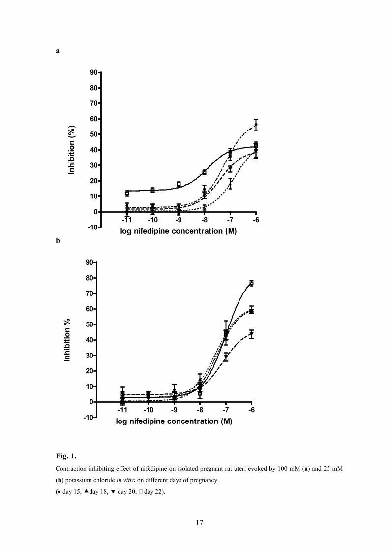
The 25 mM and 100 mM KCl-stimulated uterine contractions were inhibited
concentration-dependently by nifedipine in the range of 10-11−10-6 M (Fig. 1a, b). As
concerns the contractions induced by 100 mM KCl, the calculated EC50 was lower on day
18 than on day 15 (Table 1), but there were no changes on the other days. There were
significant changes in Emax on days 18, 20 and 22 as compared that on day 15. In the
presence of 25 mM KCl, the maximal relaxing effect of nifedipine was significantly
greater on days 20 and 22 than on day 15. There were no significant changes in EC50
(Table 2).
17
a
b
-11 -10 -9 -8 -7 -6-10
0
10
20
30
40
50
60
70
80
90
log nifedipine concentration (M)
Inhibition (%)
-11 -10 -9 -8 -7 -6-10
0
10
20
30
40
50
60
70
80
90
log nifedipine concentration (M)
Inhibition %
Fig. 1.
Contraction inhibiting effect of nifedipine on isolated pregnant rat uteri evoked by 100 mM (a) and 25 mM
(b) potassium chloride in vitro on different days of pregnancy.
(• day 15, �day 18, � day 20, � day 22).
18
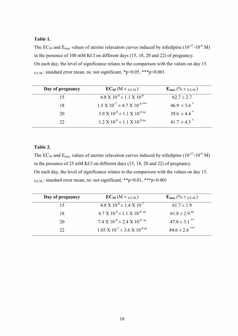
Table 1.
The EC50 and Emax values of uterine relaxation curves induced by nifedipine (10-11-10-6 M)
in the presence of 100 mM KCl on different days (15, 18, 20 and 22) of pregnancy.
On each day, the level of significance relates to the comparison with the values on day 15.
S.E.M.: standard error mean; ns: not significant, *p<0.05, ***p<0.001
Day of pregnancy EC50 (M ± S.E.M.) Emax (% ± S.E.M.)
15 6.8 X 10⁻⁸ ± 1.1 X 10⁻⁸ 62.7 ± 2.7
18 1.5 X 10⁻⁷ ± 4.7 X 10⁻⁸ *** 46.9 ± 3.6 *
20 3.9 X 10⁻⁸ ± 1.1 X 10⁻⁸ ns 39.6 ± 4.4 *
22 1.2 X 10⁻⁸ ± 1.1 X 10⁻⁸ ns 41.7 ± 4.3 *
Table 2.
The EC50 and Emax values of uterine relaxation curves induced by nifedipine (10-11-10-6 M)
in the presence of 25 mM KCl on different days (15, 18, 20 and 22) of pregnancy.
On each day, the level of significance relates to the comparison with the values on day 15.
S.E.M.: standard error mean; ns: not significant, **p<0.01, ***p<0.001
Day of pregnancy EC50 (M ± S.E.M.) Emax (% ± S.E.M.)
15 4.8 X 10⁻⁸ ± 1.4 X 10⁻⁷ 61.7 ± 1.9
18 4.7 X 10⁻⁸ ± 1.1 X 10⁻⁸ ns 61.8 ± 2.9 ns
20 7.4 X 10⁻⁸ ± 2.4 X 10⁻⁸ ns 47.0 ± 3.1 **
22 1.03 X 10⁻⁷ ± 3.6 X 10⁻⁸ ns 84.6 ± 2.6 ***
19
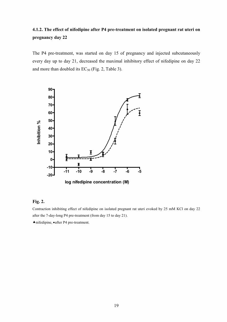
4.1.2. The effect of nifedipine after P4 pre-treatment on isolated pregnant rat uteri on
pregnancy day 22
The P4 pre-treatment, was started on day 15 of pregnancy and injected subcutaneously
every day up to day 21, decreased the maximal inhibitory effect of nifedipine on day 22
and more than doubled its EC50 (Fig. 2, Table 3).
-11 -10 -9 -8 -7 -6 -5-20
-10
0
10
20
30
40
50
60
70
80
90
log nifedipine concentration (M)
Inhibition %
Fig. 2.
Contraction inhibiting effect of nifedipine on isolated pregnant rat uteri evoked by 25 mM KCl on day 22
after the 7-day-long P4 pre-treatment (from day 15 to day 21).
�nifedipine, •after P4 pre-treatment.
20
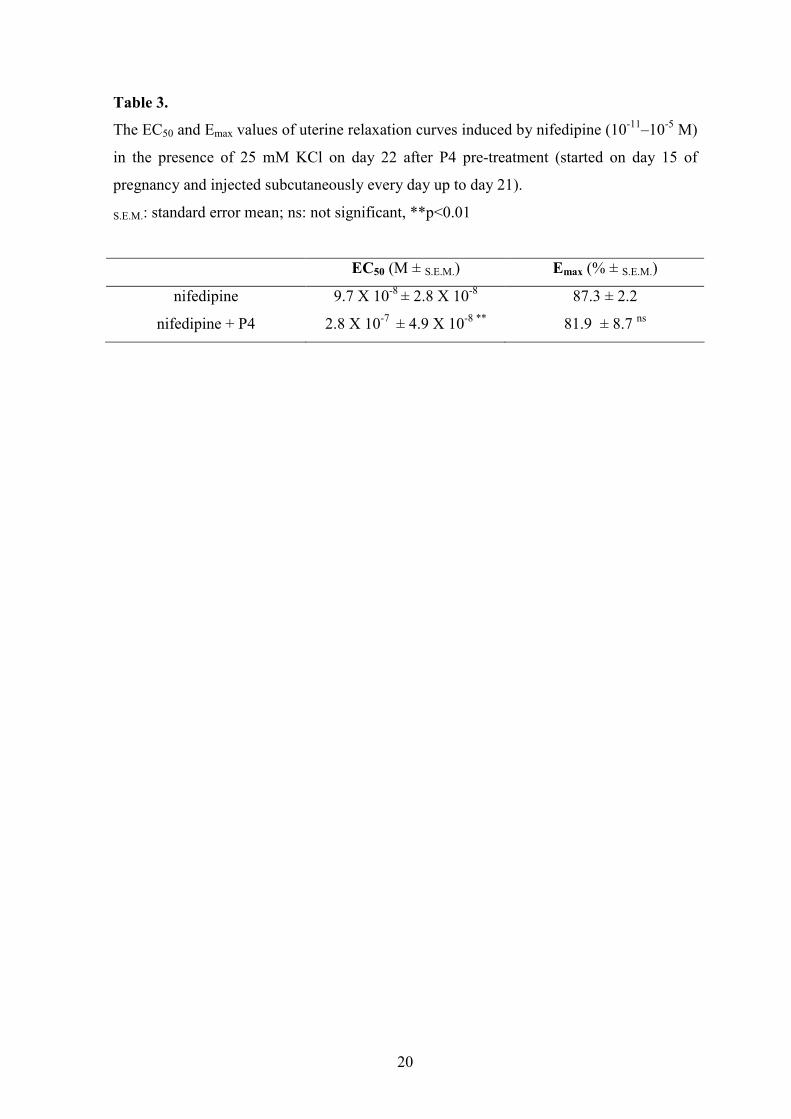
Table 3.
The EC50 and Emax values of uterine relaxation curves induced by nifedipine (10-11–10-5 M)
in the presence of 25 mM KCl on day 22 after P4 pre-treatment (started on day 15 of
pregnancy and injected subcutaneously every day up to day 21).
S.E.M.: standard error mean; ns: not significant, **p<0.01
EC50 (M ± S.E.M.) Emax (% ± S.E.M.)
nifedipine 9.7 X 10⁻⁸ ± 2.8 X 10⁻⁸ 87.3 ± 2.2
nifedipine + P4 2.8 X 10⁻⁷ ± 4.9 X 10⁻⁸ ** 81.9 ± 8.7 ns

21
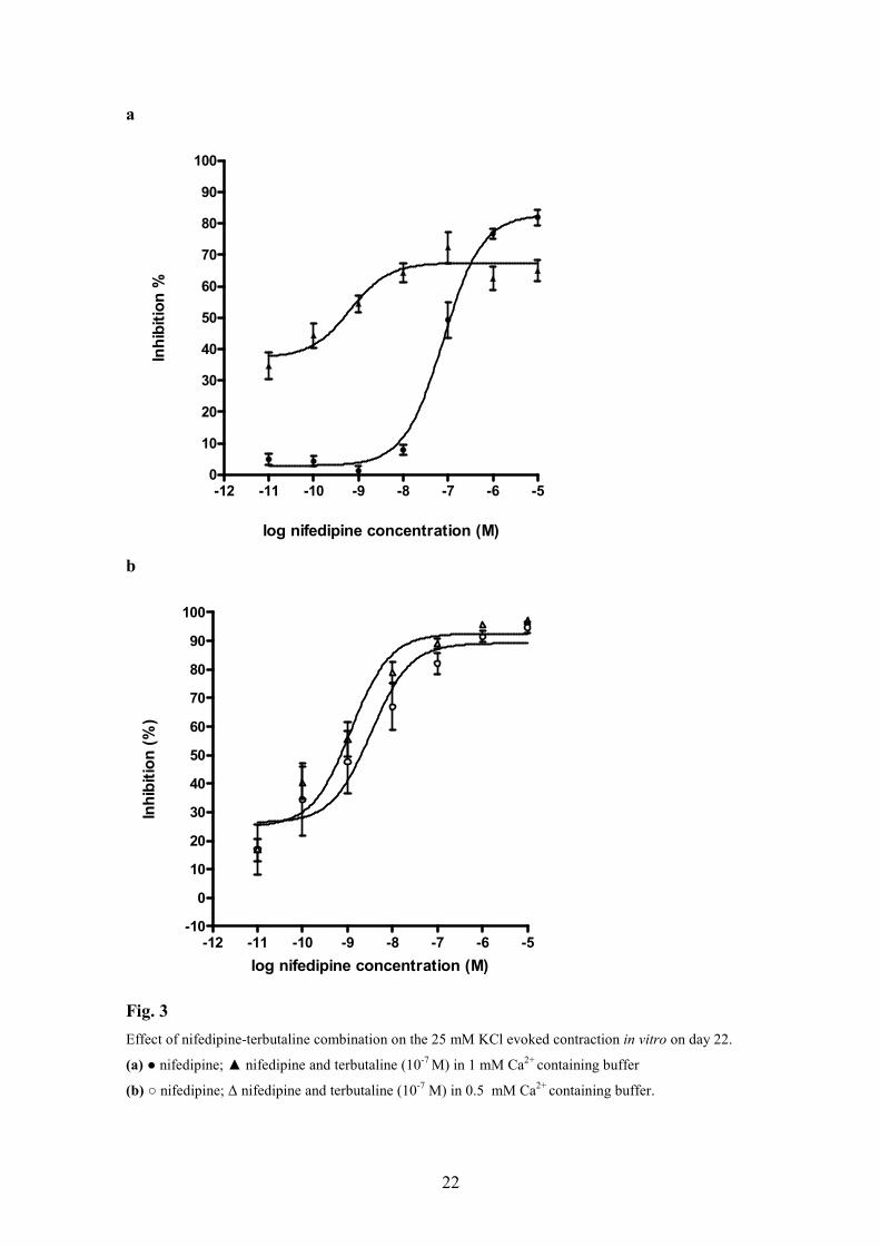
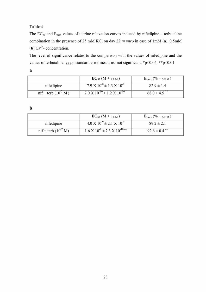
4.1.3. The effect of nifedipine – terbutaline combination on isolated pregnant rat uteri
on pregnancy day 22
The concentration–response curves for nifedipine in the presence of 10-7 M terbutaline
were shifted to the left and a decrease in the maximal inhibitory effect was observed (Fig.
3a, Table 4a). In the presence of 0.5 mM Ca2+ (Ca2+-poor buffer), terbutaline did not alter
the effect of nifedipine (Fig. 3b, Table 4b).
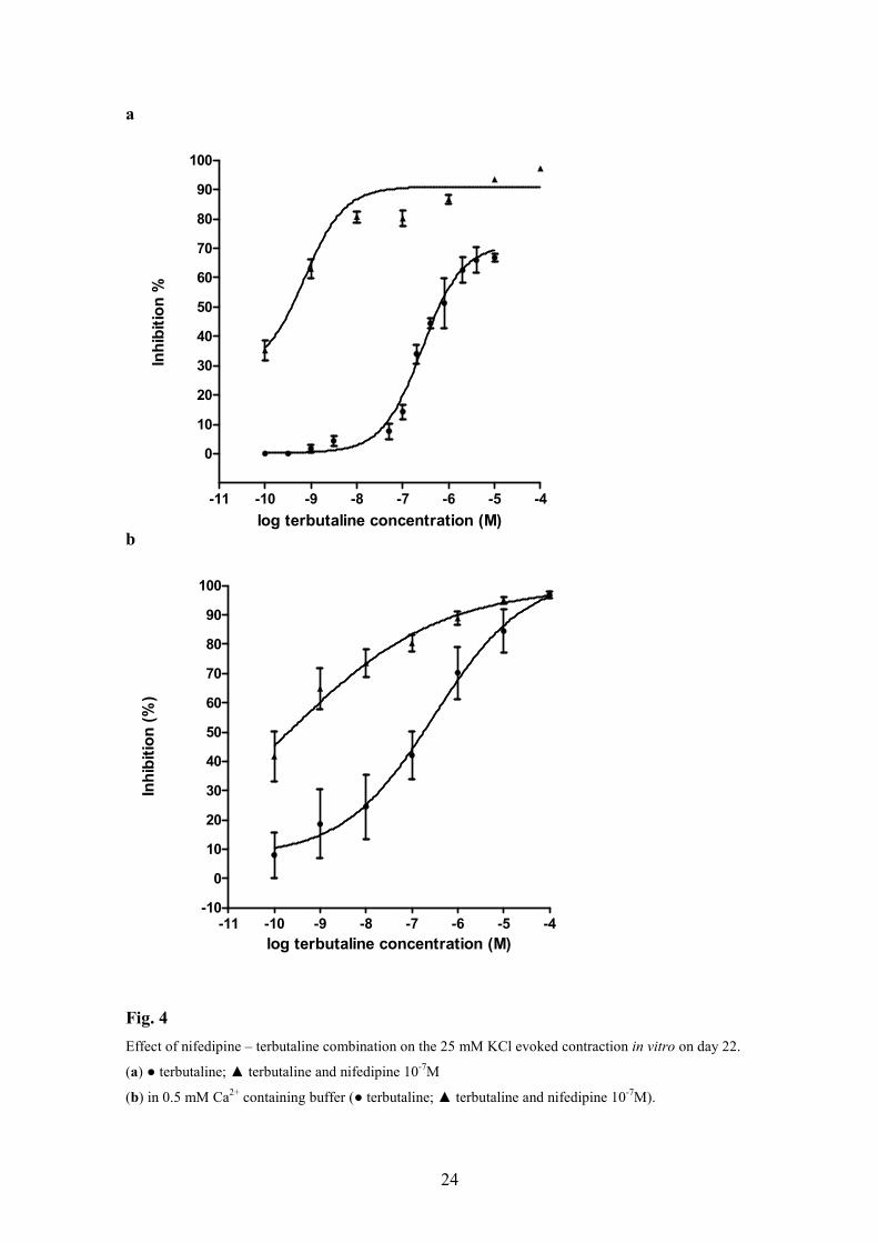
The concentration–response curves for terbutaline in the presence of 10-7 M nifedipine
were also shifted to the left, but this shift was greater than that of the nifedipine curve by
terbutaline. Nifedipine also significantly increased the Emax of terbutaline (Fig. 4a, Table
5a). In the Ca2+-poor buffer, the presence of nifedipine increased the EC50 of terbutaline,
but did not alter its Emax (Fig. 4b, Table 5b).
22
a
b
-12 -11 -10 -9 -8 -7 -6 -50
10
20
30
40
50
60
70
80
90
100
log nifedipine concentration (M)
Inhibition %
-12 -11 -10 -9 -8 -7 -6 -5-10
0
10
20
30
40
50
60
70
80
90
100
log nifedipine concentration (M)
Inhibition (%)
Fig. 3
Effect of nifedipine-terbutaline combination on the 25 mM KCl evoked contraction in vitro on day 22.
(a) ● nifedipine; ▲ nifedipine and terbutaline (10-7 M) in 1 mM Ca2+ containing buffer
(b) ○ nifedipine; ∆ nifedipine and terbutaline (10-7 M) in 0.5 mM Ca2+ containing buffer.
23
Table 4
The EC50 and Emax values of uterine relaxation curves induced by nifedipine – terbutaline
combination in the presence of 25 mM KCl on day 22 in vitro in case of 1mM (a), 0.5mM
(b) Ca2+- concentration.
The level of significance relates to the comparison with the values of nifedipine and the
values of terbutaline. S.E.M.: standard error mean; ns: not significant, *p<0.05, **p<0.01
a
EC50 (M ± S.E.M.) Emax (% ± S.E.M.)
nifedipine 7.9 X 10⁻⁸ ± 1.3 X 10⁻⁸ 82.9 ± 1.4
nif + terb (10-7 M ) 7.0 X 10⁻¹⁰ ± 1.2 X 10-10 * 68.0 ± 4.5 **
b
EC50 (M ± S.E.M.) Emax (% ± S.E.M.)
nifedipine 4.0 X 10⁻⁹ ± 2.1 X 10⁻⁹ 89.2 ± 2.1
nif + terb (10-7 M) 1.6 X 10⁻⁹ ± 7.3 X 10⁻¹⁰ ns 92.6 ± 0.4 ns
24
a
b
-11 -10 -9 -8 -7 -6 -5 -4
0
10
20
30
40
50
60
70
80
90
100
log terbutaline concentration (M)
Inhibition %
-11 -10 -9 -8 -7 -6 -5 -4-10
0
10
20
30
40
50
60
70
80
90
100
log terbutaline concentration (M)
Inhibition (%)
Fig. 4
Effect of nifedipine – terbutaline combination on the 25 mM KCl evoked contraction in vitro on day 22.
(a) ● terbutaline; ▲ terbutaline and nifedipine 10-7M
(b) in 0.5 mM Ca2+ containing buffer (● terbutaline; ▲ terbutaline and nifedipine 10-7M).

25
Table 5
The EC50 and Emax values of uterine relaxation curves induced by nifedipine – terbutaline
combination in the presence of 25 mM KCl on day 22 in vitro in case of 1 mM (a), 0.5 mM
(b) Ca2+- concentration.
The level of significance relates to the comparison with the values of nifedipine and the
values of terbutaline. S.E.M.: standard error mean; ns: not significant, *p<0.05, **p<0.01
***p<0.001
a
EC50 (M ± S.E.M.) Emax (% ± S.E.M.)
terbutaline 6.9 X 10⁻⁷ ± 1.7 X 10⁻⁷ 74.6 ± 2.8
terb + nif (10-7 M) 8.3 X 10⁻¹⁰ ± 1.6 X 10⁻¹⁰ ** 90.8 ± 0.8 ***
b
EC50 (M ± S.E.M.) Emax (% ± S.E.M.)
terbutaline 1.5 X 10⁻⁷ ± 1.6 X 10⁻¹⁰ 97.6 ± 1.1
terb + nif (10-7 M) 5.3 X 10⁻⁹ ± 2.1 X 10⁻⁹ ** 95.4 ± 1.1 ns
26
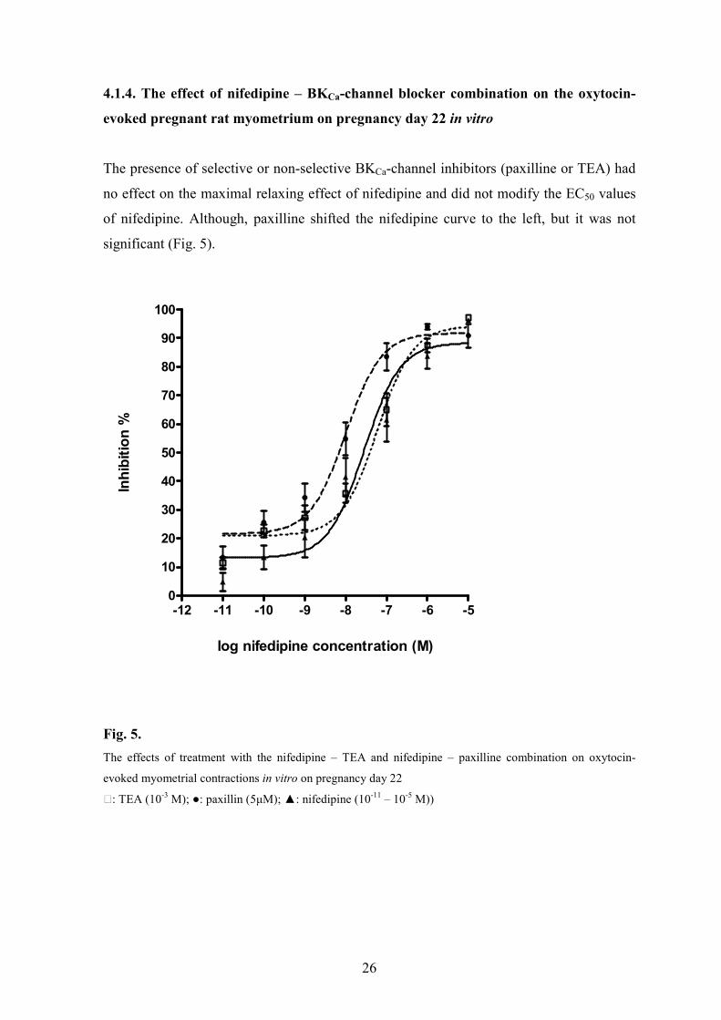
4.1.4. The effect of nifedipine – BKCa-channel blocker combination on the oxytocin-
evoked pregnant rat myometrium on pregnancy day 22 in vitro
The presence of selective or non-selective BKCa-channel inhibitors (paxilline or TEA) had
no effect on the maximal relaxing effect of nifedipine and did not modify the EC50 values
of nifedipine. Although, paxilline shifted the nifedipine curve to the left, but it was not
significant (Fig. 5).
-12 -11 -10 -9 -8 -7 -6 -50
10
20
30
40
50
60
70
80
90
100
log nifedipine concentration (M)
Inhibition %
Fig. 5.
The effects of treatment with the nifedipine – TEA and nifedipine – paxilline combination on oxytocin-
evoked myometrial contractions in vitro on pregnancy day 22
: TEA (10-3 M); ●: paxillin (5µM); ▲: nifedipine (10-11 – 10-5 M))

27
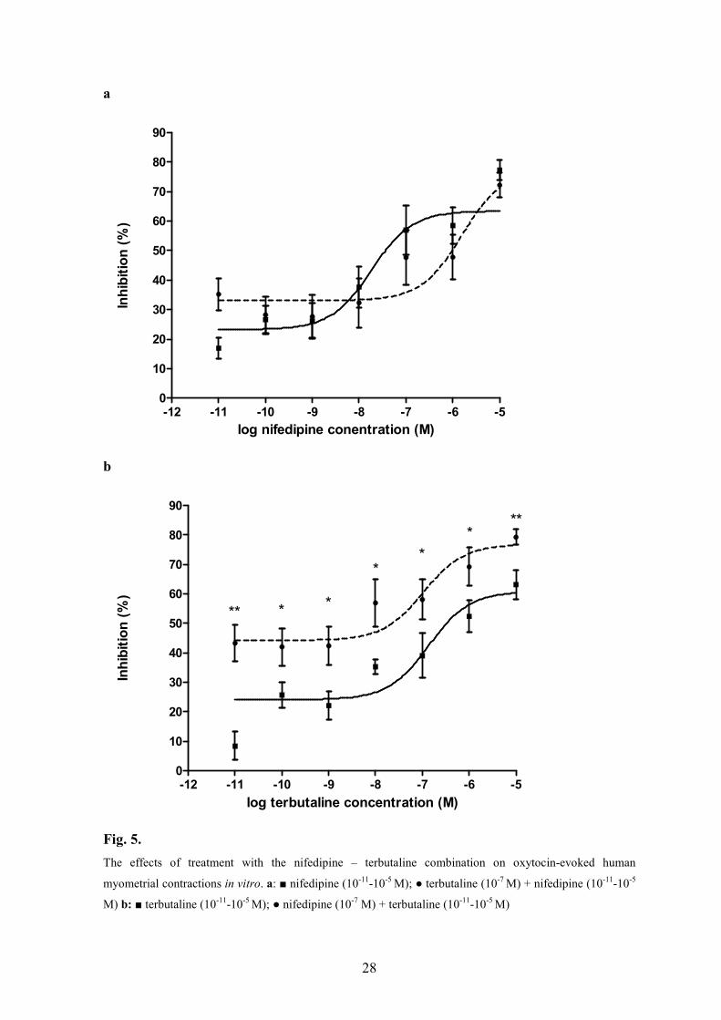
4.1.5. The effect of nifedipine – terbutaline combination on the oxytocin-evoked
human myometrial contractions in vitro
The 10-6 M oxytocin-stimulated human uterine contractions were inhibited concentration-
dependently by nifedipine and terbutaline in the range 10-5−10-11 M (Fig. 6. a, b). The
addition of 10-7 M terbutaline to nifedipine did not alter the Emax of nifedipine, but
decreased EC50 from 1.8 x 10⁻⁸ M to 1.5 x 10⁻⁶ M (p<0.05) (Fig. 6a). The addition of 10-7
M nifedipine to terbutaline increased the Emax of terbutaline (Fig. 6b), from 60.8% to 76.8
% (p<0.01) but EC50 was not changed.
28
a
-12 -11 -10 -9 -8 -7 -6 -50
10
20
30
40
50
60
70
80
90
log nifedipine conentration (M)
Inhibition (%)
b
-12 -11 -10 -9 -8 -7 -6 -50
10
20
30
40
50
60
70
80
90
***
**
****
log terbutaline concentration (M)
Inhibition (%)
Fig. 5.
The effects of treatment with the nifedipine – terbutaline combination on oxytocin-evoked human
myometrial contractions in vitro. a: ■ nifedipine (10-11-10-5 M); ● terbutaline (10-7 M) + nifedipine (10-11-10-5
M) b: ■ terbutaline (10-11-10-5 M); ● nifedipine (10-7 M) + terbutaline (10-11-10-5 M)

29
4.2. In vivo studies
4.2.1. The effect of nifedipine, salmeterol and P4 in the hormone-induced PTL model
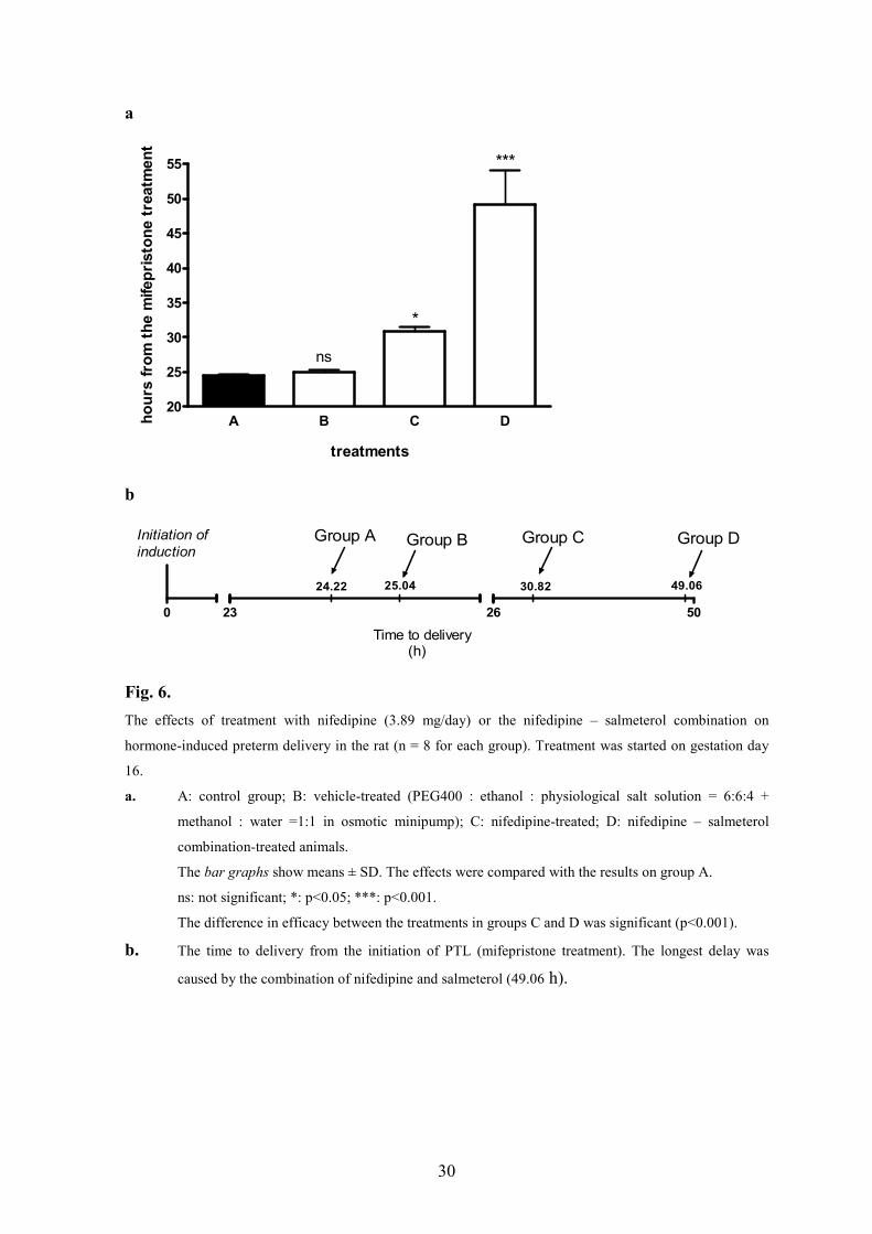
In group A (control), preterm labour occurred within 24 hours after mifepristone treatment,
at about 9:00 a.m. on pregnancy day 20. The vehicle (group B) did not alter the time of
delivery relative to that in group A.
Nifedipine (group C) treatment started on pregnancy day 16 was effective in delaying the
hormone-induced preterm delivery by 6.6 hours. In group D (nifedipine–salmeterol
combination), the treatment was extremely effective; preterm birth was delayed by ~ 24
hours as compared with group A (Fig. 6a, b).
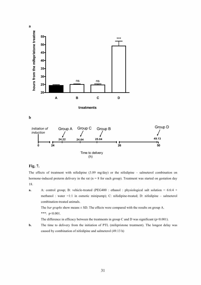
When started on day 18 of pregnancy (Fig. 7a, b), nifedipine treatment (group C) was not
effective. In contrast in group D (combination therapy) the treatment was effective; labour
was delayed by ~ 25 hours. The difference in efficacy between groups C and D was most
expressed for the treatment started on day 18.
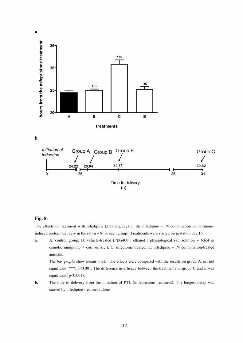
With the nifedipine – P4 combination (Fig. 8a, b), the P4 pre-treatment (group E)
abolished the effect of nifedipine (group C).
30
a
A B C D20
25
30
35
40
45
50
55
ns
***
*
treatments
hours from the mifepristone treatment
b
0 23 26 50
Initiation of
induction
Time to delivery (h)
Group A Group C Group D
25.0424.22 30.82 49.06
Group B
Fig. 6.
The effects of treatment with nifedipine (3.89 mg/day) or the nifedipine – salmeterol combination on
hormone-induced preterm delivery in the rat (n = 8 for each group). Treatment was started on gestation day
16.
a. A: control group; B: vehicle-treated (PEG400 : ethanol : physiological salt solution = 6:6:4 +
methanol : water =1:1 in osmotic minipump); C: nifedipine-treated; D: nifedipine – salmeterol
combination-treated animals.
The bar graphs show means ± SD. The effects were compared with the results on group A.
ns: not significant; *: p<0.05; ***: p<0.001.
The difference in efficacy between the treatments in groups C and D was significant (p<0.001).
b. The time to delivery from the initiation of PTL (mifepristone treatment). The longest delay was
caused by the combination of nifedipine and salmeterol (49.06 h).
31
a
A B C D
20
25
30
35
40
45
50
55***
ns ns
treatments
hours from the mifepristone treatment
b
0 24 26 50
Initiation of
induction
Time to delivery (h)
24.22 25.0424.64 49.13
Group C Group DGroup BGroup A
Fig. 7.
The effects of treatment with nifedipine (3.89 mg/day) or the nifedipine – salmeterol combination on
hormone-induced preterm delivery in the rat (n = 8 for each group). Treatment was started on gestation day
18.
a. A: control group; B: vehicle-treated (PEG400 : ethanol : physiological salt solution = 6:6:4 +
methanol : water =1:1 in osmotic minipump); C: nifedipine-treated; D: nifedipine – salmeterol
combination-treated animals.
The bar graphs show means ± SD. The effects were compared with the results on group A.
***: p<0.001.
The difference in efficacy between the treatments in group C and D was significant (p<0.001).
b. The time to delivery from the initiation of PTL (mifepristone treatment). The longest delay was
caused by combination of nifedipine and salmeterol (49.13 h)
32
a
A B C E20
25
30
35
***
nsns
treatments
hours from the mifepristone treatment
b
0 25 26 31
Time to delivery (h)
Initiation of
inductionGroup A Group B Group E Group C
30.8224.22 25.04 25.21
Fig. 8.
The effects of treatment with nifedipine (3.89 mg/day) or the nifedipine – P4 combination on hormone-
induced preterm delivery in the rat (n = 8 for each group). Treatments were started on gestation day 16.
a. A: control group; B: vehicle-treated (PEG400 : ethanol : physiological salt solution = 6:6:4 in
osmotic minipump + corn oil s.c.); C: nifedipine treated; E: nifedipine – P4 combination-treated
animals.
The bar graphs show means ± SD. The effects were compared with the results on group A. ns: not
significant; ***: p<0.001. The difference in efficacy between the treatments in group C and E was
significant (p<0.001).
b. The time to delivery from the initiation of PTL (mifepristone treatment). The longest delay was
caused by nifedipine-treatment alone.
33
5. Discussion
PTB is the one of the greatest challenges in obstetrical practice. The currently used
medications are not able to stop or sufficiently delay the process of PTB, therefore there is
growing interest in experimental studies of the possible use of different tocolytic
combinations to decrease the potentially maternal and foetal adverse events and improving
the perinatal outcome.
The most important factor controlling force in the myometrium is the concentration
of intracellular Ca2+, and membrane potential is the major factor governing Ca2+ entry into
the cell (Wray et al., 2003). High K+ stimulation, which provokes membrane
depolarization and uterine contractions, is the most common method for the introduction of
Ca2+ into cells without receptor stimulation. There are a number of data relating to the use
of different concentrations of KCl (from low to high K+) to evoke contraction in vitro by
opening VG Ca2+ channels, though it is not clear which of these concentrations causes
rhythmic contractions of the uterus providing an appropriate model for investigation of the
pregnant uterus-relaxing effects.
We found that in the presence of 25 mM KCl the uterine contractions were
rhythmic and the relaxing effect of nifedipine was the highest on the last day of pregnancy.
With 100 mM KCl, however, the contractions became spastic and the inhibitory action of
nifedipine was highest on day 15, but was later quite weak. These results led us to
conclude that stimulation with 25 mM KCl is much more appropriate for investigations of
the action of the Ca2+ channel blocker nifedipine. As the relaxing effect of nifedipine was
highest on the last day of pregnancy, further experiments were carried out on day 22.
It was earlier demonstrated that the myometrial DHP binding is increased more than 3-fold
at the end of pregnancy in the rat (Mershon et al., 1994), and this result was supported by
our findings. However, others found that the protein expressions and mRNA levels of the
pore-forming α1-subunits of the Ca2+ channels were not altered by pregnancy either in rats
or in humans (Helguera et al., 2002; Batra and Popper, 1989). It is known that the Cav1.2
channels possess splice variants in the cardiac muscle, which have different
pharmacological and electrophysiological properties (Liao et al., 2005). So far, such
variants have not been discovered in the myometrium, but the apparent contradictions
observed between the above-mentioned papers and our result might be explained by the
existence of putative channel splice variants in the pregnant uterus.
34
The activity of the Cav1.2 channel is regulated by several factors (Kobayashi et al.,
2007). In our study, three of them (BKCa channels, adrenergic system and P4/E2 ratio)
were investigated with regard to how to influence the uterus-relaxing effect of nifedipine in
vitro.
The studies with the selective BKCa channel blocker paxilline and non-selective blocker
TEA were carried out by oxytocin-induced contractions, because in these cases the
stimulation with KCl was not useful. The results revealed that BKCa channels and any other
K+ channels, contrary to human myometrium (Moynihan et al., 2008), are not involved in
the relaxing effect of nifedipine in pregnant rat myometrium.
P4 is regarded as a preventive drug against PTB, especially in late-preterm birth (Borna
and Sahabi, 2008). Unfortunately, the in vivo P4 pre-treatment decreased the maximal
inhibitory effect of nifedipine and increased its EC50. The uterine smooth muscle possesses
α1C-long and α1C-short isoforms of the L-type Ca2+ channel. In the presence of the long
isoform, the channel has lower activity than with the short isoform. In the pregnant rat
uterus, P4 and E2 enhance the expressions of the α1C-long and α1C-short isoforms,
respectively, of the L-type Ca2+ channels (Helguera et al., 2002). These facts explain why
P4 pre-treatment worsened the relaxing effect of nifedipine.
The other investigated factor which regulates the Cav1.2 channel was the β2-adrenergic
system. We found synergism in the uterus-relaxing effect of nifedipine and the β2-AR
agonist terbutaline, although the extent of potentiation depended on the sequence of
administration of the two compounds. When terbutaline was added first in a single dose,
synergism was found in EC50 (the nifedipine curve was shifted to the left), but the maximal
inhibitory effect of nifedipine was lower. When nifedipine was administered first, the
relaxing effect of terbutaline was obviously stronger.
It is known that stimulation of β2-ARs activates G-proteins and increases the intracellular
cAMP level. cAMP activates PK A the activated form of which phosphorylates the Cav1.2
channels. This mechanism is well known in the heart muscle (Kamp and Hell, 2000) and it
is very probably similar in the pregnant myometrium. The entry of the Ca2+ into the cells
through the VG Ca2+ channel is one of the crucial factors in the generation of smooth
muscle contraction. Terbutaline possibly activates the Cav1.2 channels and decreases the
maximal relaxing effect of nifedipine. The resultant effect of the increase of cAMP level
and activation of Cav1.2 channels causes a weaker smooth muscle relaxation. In the
35
opposite case, when nifedipine is administered first, the Cav1.2 channels are blocked;
hence, there is only a low possibility that terbutaline can activate them.
To check on the above-mentioned hypothesis, the synergism between the two compounds
was investigated in Ca2+-poor buffer. A Ca2+-poor environment theoretically decreases the
terbutaline-induced Ca2+ influx and may alter the extent of the synergism. The Ca2+-poor
environment shifted the nifedipine dose-response curve to the left, and the maximal
inhibitory effect of nifedipine was so high that its effect could not be enhanced by
terbutaline. In contrast, the Ca2+-poor environment shifted the terbutaline dose-response
curve to the left, but nifedipine was able to enhance the shift. However, it could not
increase the maximal uterus-relaxing effect of terbutaline, possibly because of the very
strong blocking effect of the β-mimetic. These results indicate that, in a Ca2+-poor
environment, terbutaline is not able to worsen the maximal effect of nifedipine, which
suggests the role of the Ca2+ inflow in the weakening effect of terbutaline.
Synergism between nifedipine and the β2-agonist terbutaline was also investigated on
human myometrium tissue, which revealed that both nifedipine and terbutaline inhibit the
oxytocin-induced myometrial contractions dose-dependently. When terbutaline was added
first, it decreased the EC50 of nifedipine (the nifedipine curve was shifted to the right),
though it did not alter the Emax of nifedipine. In the opposite case, nifedipine administered
first increased the relaxing effect of terbutaline, though there was no difference in EC50.
The results suggest a parallelism between the human situation and that in the rat
myometrium.
In view of these results, the effects of nifedipine – salmeterol and nifedipine – P4
combination were investigated also in hormone-induced PTB model in vivo, whether these
drug interactions apply under in vivo circumstances.
The effect of combined salmeterol − gestagen treatment in hormone-induced preterm
delivery in rats in vivo was investigated earlier (Gálik et al., 2008): salmeterol treatment
started on pregnancy day 16 delayed preterm birth by 2.8 hours and the salmeterol − P4
combination caused a 4.5-hour delay.
Preterm delivery was induced by the deprivation of P4 using P4 antagonist combined with
PGE2 to accelerate cervical ripening on pregnancy day 19. At this stage of pregnancy the
function of corpus luteum declines which is mainly attributed to the luteolytic effect of
PGF2α (Hernandez et al., 2009). It is also known that P4 induces the metabolism of PGs,
thus substitution of P4 is anticipated to improve relaxation response in late-pregnant rat
uterus (Farina et al., 2004).
36
We used the same in vivo salmeterol dose as was effective in delaying PTB, and the P4
dose that had increased the effect of salmeterol. The in vivo dose for the tocolytic effect
was calculated by using the pharmacokinetic parameters reported for nifedipine in
pregnant rats by Downing et al. (Downing and Hollingsworth, 1998). We planned
administration of a dose regimen via osmotic pumps which provided a plasma nifedipine
concentration of ~1.5 µg/ml (estimated). Downing and Hollingsworth (1998) concluded
that this plasma level of the drug did not cause significant changes in heart rate or blood
pressure, but elicited a well-defined uterus-relaxing effect. We found that treatment with
nifedipine alone started on pregnancy day 16 was more effective in delaying delivery than
salmeterol treatment alone (in the earlier study) (Gálik et al., 2008). Similarly, the
nifedipine − salmeterol combination also had a greater effect than the salmeterol −
gestagen combination (Gálik et al., 2008).
The short-term effects of nifedipine and its combination with salmeterol were also tested in
our experiments. Nifedipine treatment started on day 18 did not delay labour, but the
nifedipine − salmeterol combination was as effective as the combination started on the
pregnancy day 16. The ability of nifedipine to delay labour was tripled by its combination
with the β2-agonist. In our in vivo study two osmotic pumps were implanted
subcutaneously in the rat and the administration of the two agents was simultaneous and
continuous. The results suggested that the parallel administration of the two compounds
may lead to a similar benefit as that of nifedipine-potentiated terbutaline treatment.
P4 pre-treatment also abolished the ability of nifedipine to delay labour in hormone-
induced preterm delivery in rats in vivo. These results correlate with the hypothesis that
progesterone decreases the activity of the L-type Ca2+ channels (Helguera et al., 2002).
Accordingly we presumed that a Ca2+ channel blocker + P4 combination might not have
any benefit in clinical practice.
A weakness of this study is that the experiments do not provide data relating to the prompt
effect of the drugs in the onset of hormone-induced preterm birth, but the investigation of
this effect is almost impossible in rats. The first visual sign of the onset of labour is vaginal
bleeding. From this time on, at most only 10-15 min is available until the delivery of the
first foetus. This short period is not sufficient for the absorption of drugs administered. On
the other hand, intravenous drug administration to delivering rats would be very difficult
and might well cause severe stress for the animal, altering the delivery process. Despite
this weakness, our study reports the first attempt to delay antigestagen – PG-induced PTB
in vivo with a Ca2+ antagonist or a Ca2+ antagonist – gestagen combination. Additionally,
37
the effects of Ca2+ antagonist – β2-agonist combinations were on the pregnant human
myometrium proved.
6. Conclusion
In the light of our results, we can conclude that P4 pre-treatment abolished the ability of
nifedipine to delay labour in hormone-induced preterm delivery in rat. These results
correlate with the hypothesis that P4 decreases the activity of the L-type Ca2+ channels.
There is growing interest in nifedipine as a potentially effective and well-tolerated form of
tocolysis. Nifedipine has been demonstrated (Tsatsaris et al., 2002) to have fewer side-
effects and leads to a better neonatal outcome than β2-mimetics. We presume that the effect
of nifedipine in tocolytic therapy might be intensified through combination with β2-
adrenerg agonists and enhanced by low concentrations of β2-mimetics. However, our
results indicate that the administration of β2-adrenergic agonists can not precede that of
nifedipine. The significance of these experimental findings remains to be validated in
clinical trials, including human side effects.
38
7. References
Arrowsmith S, Kendrick A, Wray S. Drugs acting on the pregnant uterus. Obstet Gynaecol
Reprod Med 2010; 20(8): 241-247
Batra SC, Popper LD. Characterizaion of membrane calcium channels in non-pregnant and
pregnant human uterus. Gynaecol Obstet Invest 1989; 27: 57-61.
Beck S, Wojdyla D, Say L, Betran AP, Merialdi M, Requejo JH, Rubens C, Menon R, Van
Look PF. The worldwide incidence of preterm birth: a systematic review of
maternal mortality and morbidity. Bull World Health Organ 2010; 88:31-38.
Berkowitz GS, Blackmore-Prince C, Lapinski RH, Savitz DA. Risk factors for preterm
birth subtypes. Epidemiol 1998; 9(3):279– 285.
Borna S, Sahabi N. Progesterone for maintance tocolytic therapy after threatened preterm
labour: A randomised controlled trial. Aust N Z J Obstet Gynaecol2008; 48: 58-63.
Bursztyn L, Eytan O, Jaffa AJ, Elad D. Mathematical model of excitation-contraction in a
uterine smooth muscle cell. Am J Physiol Cell Physiol 2007; 292(5):C1816-1829
Catterall WA, Perez-Reyes E, Snutch TP, Striessnig J. International Union of
Pharmacology. XLVIII. Nomenclature and structure-function relaionships of
voltage-gated calcium channels. Pharmacological Reviews 2005; 57:411-425
Chesley LC. A survey of management and case mortality. In: Chesley LC: Hypertensive
disorders in pregnancy. Appleton-Century-Crofts, New York, 1978
Dolphin AC. A short history of voltage-gated calcium channels. Br J Pharmacology 2006;
147:S56–S62
Downing SJ, Hollingsworth M. Nifedipine kinetics in the rat and relationship between its
serum concentrations and uterine and cardiovascular effects. Br J Pharmacol 1998;
95:23-32.
Egészségügyi Közlöny, Az Egészségügyi Minisztérium szakmai protokollja a fenyegetı
koraszülsérıl 2008; LVIII. évfolyam 3:1508-1517
Farina M, Ribeiro ML, Weissmann C, Estevez A, Billi S, Vercelli C, Franchi A.
Biosynthesis and catabolism of prostaglandin F2alpha (PGF2alpha) are controlled
by progesterone in the rat uterus during pregnancy. J Steroid Biochem Mol Biol
2004; 91(4-5):211-8.
da Fonseca EB, Bittar RE, Carvalho MH. Prophyalactic administration of progesterone by
vaginal suppository to reduce the incidence of spontaneous preterm birth in women
39
at increased risk: a randomized placebo-controlled, double-blind study. Am J
Obstet Gynecol 2003; 188:419-424
Garfield RE. Uterine contractility, mechanisms of control, Serono Symposia, Norwell,
Massachusetts, (Chapter 2), 1990
Garfield RE. Uterine contractility, mechanisms of control, Serono Symposia, Norwell,
Massachusetts, (Chapter 3), 1990
Gálik M, Gáspár R, Kolarovszki-Sipiczki Z, Falkay G. Gestagen treatment enhances the
tocolytic effect of salmeterol in hormone-induced preterm labour in the rat in vivo.
Am J Obstet Gynaecol 2008; 198(3): 319. e1-5.
Giles W, Bisits A. The present and future of tocolysis. Clin Obstet Gynaecol 2007;
21(5):857-868
Goldenberg RL, Culhane JF, Iams JD, Romero R. Epidemiology and causes of preterm
birth. Lancet 2008; 371: 75-84.
Graham JD, Clarke CL. Physiological action of progesterone in target tissues. Endocr Rev
1997; 18:502–519
Haas DM. Preterm birth in clinical evidence. London: BMJ Publishing Group; 2006.
Helguera G, Olcese R, Song M, Toro L, Stefani E. Tissue-specific regulation of Ca2+
channel protein expression by sex hormones. Biochim Biophys Acta 2002;
1569:59-66
Hernandez F, Peluffo MC, Bas D, Stouffer RL, Tesone M. Local effects of the sphingosine
1-phosphate on prostaglandin F2alpha-induced luteolysis in the pregnant rat. Mol
Reprod Dev 2009; 76(12):1153-64.
Hillier SL, Nugent RP, Eschenbach DA, Krohn MA, Gibbs RS, Martin DH, Cotch MF,
Edelman R, Pastorek JG 2nd, Rao AV, et al. The Vaginal Infections and
Prematurity study Group. N Engl J Med 1995; 333(26):1737-1742.
Husslein P, Quartorolo JP. Review of clinical experience with atosiban and the Tractocile
Efficacy Assessment Survey in Europe (Treasure) study protocol. Int J Clin Pract
2003, 57:121-127.
Kamp TJ, Hell JW. Regulation of cardiac L-type calcium channels by protein kinase A and
protein kinase C. Circ Res 2000; 87(12):1095-1102.
Khan R, Mathroo-Ball B, Arilkumaran S, Ashford MLJ. Potassium channels in the human
myometrium. Exp Physiol 2001; 86: 255-264.
Kim A, Shim JY. Emerging tocolytics for maintenance therapy of preterm labour: oxytocin
antagonists and calcium channels blockers. Br J Pharmacol 2006; 113:113-115.

40
Kinsler VA, Thornton S, Ashford ML, Melin P, Smith SK. The effect of the oxytocin
antagonists F314 and F792 on the in vitro contractility of human myometrium. Br J
Obstet Gynaecol 1996; 103:373–375
Kobayashi T, Yamada Y, Fukao M, Tsutsuura M, Tohse N. Regulation of Cav1.2 current:
interaction with intracellular molecules. J Pharm Sci 2007; 103: 347-353.
Koks CHB, Kleine M, Manger P. A randomized comparison of nifedipine and ritodrine for
suppression of preterm labor. European J Obstet Gynecol 1998; 77:171-6.
Lever AML, Corris PA, Gibson GJ. Nifedipine enhances the bronchodilator effect of
salbutamol. Thorax 39, 576-578, 1984
Liao P, Yong TF, Liang MC, Yue DT, Soong TW. Splicing for alternative structures of
Cav1.2 Ca2+ channels in cardiac and smooth muscles. Cardiovasc Res 2005; 68:
197-203.
Liao P, Yu D, Lu S, Tang Z, Liang MC, Zeng S, Lin W, Soong TW. Smooth muscle-
selective alternatively spliced exon generates functional variation in Cav1.2 calcium
channels. J Biol Chem. 2004; 279(48):50329-50335.
Liao P, Zhang HY, Soong TW. Alternative splicing of voltage-gated calcium channels:
from molecular biology to disease. Pflugers Arch 2009; 458(3):481-487
Mackenzie R, Walker M, Armson A, Hannah ME. Progesterone for the prevention of
preterm birth among women at increased risk: A systematic review and meta-
analysis of randomized controlled trials. Am J Obstet Gynaecol 2006; 194: 1234-
42.
Meis PJ, Klebanoff M, Thorm E. Prevention of recurrent preterm delivery by 17-alpha
hydroxyprogesterone caproate. N Engl J Med 2003; 349:1299
Mershon JL, Mikala G, Schwartz A. Changes in the expression of the L-type voltage-
cependent calcium channel during pregnancy and parturition in the rat. Biol Rep
1994; 51: 993-999.
Mohan AR, Bennett PR. Drugs acting on the pregnant uterus. Curr Obstet Gynaecol 2006;
16:174–180.
Moynihan AT, Smith TJ, Morrison JJ. The relaxant effect of nifedipine in human uterine
smooth muscle and the BKCa channel. Am J Obstet Gynaecol 2008; 198:237.e1-
237.e8
Moutquin JM. Classification and heterogeneity of preterm birth. BJOG 2003; 110 (20):30-
33.

41
Noble K, Matthew A, Burdyga T, Wray S. A review of recent insights into the role of the
sarcoplasmic reticulum and Ca entry in uterine smooth muscle. Eur J Obstet
Gynecol Reprod Biol 2009; 144S:S11–S19
Noblot G, Audra P, Darquent D. The use of micronized progesterone in the treatment of
menace of preterm delivery. Eur J Obstet Gynecol Reprod Biol 1991; 40:203-209
Oei SG. Calcium channel blockers for tocolysis : A review of their role and safety
following reports of serious adverse events. Eur J Obstet Gynaecol 2006; 126:137–
145
Papatsonis DNM. International Congress Series 2005; 1279:251–270.
Papatsonis DN, Van Geijn HP, Ader HJ, et al. Nifedipine and ritodrine in the management
of preterm labor: a randomized multicenter trial. Obstet Gynecol 1997; 90:230-4.
Pennell CE, Jacobsson B, Williams SM, Buus RM, Muglia LJ, Dolan SM, et al. Genetic
epidemiologic studies of preterm birth: guidelines for research. Am J Obstet
Gynecol 2007; 196: 107-18.
Petrou S. The economic consequences of preterm birth during the first 10 years of life.
BJOG 2005; 112 (1): 10-5.
Petrou S, Mehta Z, Hockley C, Cook-Mozaffari P, Henderson J, Goldacre M. The impact
of preterm birth on hospital inpatient admissions and costs during the first 5 years
of life. Pediatrics 2003; 112: 1290-7.
Pryde PG, Besinger RE, Gianopoulos JG, Mittendorf R. Adverse and Beneficial Effects of
Tocolytic Therapy. Semin Perinatol 2001; 25(5):316-340
Rechberger T, Abramson SR, Woessner JF Jr. Onapristone and prostaglandin E2 induction
of delivery in the rat in late pregnancy: a model for the analysis of cervical
softening. Am J Obstet Gynecol 1996; 175:719-723.
Tan TC, Devendra K, Tan LK, Tan HK. Tocolytic treatment for the management of
preterm labour: a systematic review. Singapore Med J 2006; 47(5) : 361-366.
Takimoto K, Li D, Nerbonne JM, Levitan ES. J Mol Cell Cardiol 1997; 29: 3035-3042.
Thijssen JH. Progesterone receptors in the human uterus and their possible role in
parturition. J Steroid Biochem Mol Biol 2005; 97(5):397-400
Thirstrup S, Nielsen-Kudsk F, Dahl F. In vitro studies on the interactions of β2 –
adrenoreceptor agonists, methylxantienes, Ca2+ - channel blockers, K+ - channel
openers and other airway smooth muscle relaxants in isolated guinea-pig trachea.
Eur J Pharm 1997; 326:191–200.

42
Wray S, Jones K, Kupittayanant S, Li Y, Matthew A, Monir-Bishty E, Noble K, Pierce SJ,
Quenby S, Shmygol AV. Calcium signalling and uterine contractility. J Soc
Gynecol Investig 2003; 10: 252-264.
Wray S. Insight into the uterus. Exp Physiol 2007; 92(4): 621-631
Tsatsaris V, Papatsonis D, Goffinet F, Dekker G, Carbonne B. Tocolysis with nifedipine or
beta-adrenergic agonists: a meta-analysis. Obstet Gynecol 2002; 99(3):518-9.
Wu SN, Wu AZ, Lin MW. Pharmacological roles of the large-conductance calcium-
activated potassium channel. Curr Top Med Chem 2006, 6:1025-1030
Zukerman H, Reiss U, Rubinstein I. Inhibition of human premature labor by indomethacin.
Obstet Gynecol 1974; 44:787-792.

43
8. Appendix
8.1. List of publication
8.1.1. Publications related to the Ph.D. thesis
Hajagos-Tóth J, Falkay G, Gáspár R. Modification of the effect of nifedipine in the
pregnant rat myometrium: The influence of progesterone and terbutaline. Life Sci
2009; 85:568–572. IF: 2.56
Hajagos-Tóth J, Kormányos Zs, Falkay G, Pál A, Gáspár R. Potentiation of the uterus-
relaxing effects of beta-adrenergic agonists with nifedipine: studies on rats and the
human myometrium. Acta Obstetricia et Gynecologica 2010; 89:1284-1289 IF:
1.618
Hajagos-Tóth J, Kormányos Zs, Falkay G, Pál A, Gáspár R. Nifedipin myometrium
relaxáló hatásának vizsgálata terbutaline és K+-csatorna gátlók jelenlétében in vitro.
Acta Pharm Hung 2010;3: 109-114
8.1.2. Abstracts
Hajagos-Tóth J, Gáspár R, Falkay G. Investigation of uterine-relaxing effect of nifedipine
on late-pregnant rat myometrium in vitro. The First Conference of PhD Students in
Medicine and Pharmacy, July 9-11, 2008, Targu Mures, Romania (presentation)
Hajagos-Tóth J, Falkay G, Gáspár R. Nifedipin uterusz kontrakciót gátló hatásának
vizsgálata in vitro. A Magyar Tudomány Ünnepe; Szeged, 2008. November 28.
(presentation)
Hajagos-Tóth J, Falkay G, Gáspár R. Investigation of uterus-relaxing effect of nifedipine
on late pregnant myometrium in vitro. 2nd Meeting of the Egon & Ann Diczfalusy
Foundation; Szeged, 2008.09.30-10.01. (poster)
44
Hajagos-Tóth J, Falkay Gy, Gáspár R. Nifedipin uterusz kontrakciót gátló hatásának
módosítása ptogeszteronnal és terbutalinnal in vitro. XIV. Congressus
Pharmaceuticus Hungaricus; Budapest, 2009. november 13-15. (poster)
Hajagos-Tóth J, Kormányos Zs, Falkay Gy, Gáspár R. A nifedipin tocolyticus hatásának
módosítása béta-mimetikumokkal. A Magyar Élettani Társaság (MÉT) LXXIV.
Vándorgyőlése és a Magyar Kísérletes és Klinikai Farmakológiai Társaság (MFT)
II. Közös tudományos konferenciája; Szeged, 2010. június 16-18. (presentation)
Hajagos-Tóth J, Kormányos Zs, Falkay G, Gáspár R. Potentiation of uterus-relaxing
effects of nifedipine with β-adrenergic agonists: Studies on rats and human
myometrium. 16th World Congress on Basic and Clinical Pharmacology , 17-23
July, 2010, Copenhagen, Denmark (poster)

45
Acknowledgement
I would like to express my thanks to my supervisor, Prof. George Falkay and to István
Zupkó Ph.D. the heads of the Department of Pharmacodynamics and Biopharmacy for the
possibility to take part in the Ph.D. studies. I am also very grateful to Prof. George Falkay
for his guidance, encouragement and management of my work.
I also would like to express my thanks to my tutor, Róbert Gáspár Ph.D. for his generous
help and advice in the experimental work, and for critically reviewing the manuscript.
I also would like to thank my co-authors and colleagues in the Department of
Pharmacodynamics and Biopharmacy for the pleasant co-operation.
I am deeply grateful for my family and friends for their patience and love.