Embed Size (px)
Citation preview

MICROBIOLOGICAL REVIEWS, June 1991, p. 206-224 Vol. 55, No. 20146-0749/91/020206-19$02.00/0Copyright © 1991, American Society for Microbiology
Genetic Basis of Virulence in Shigella SpeciesTHOMAS LARRY HALE
Department of Enteric Infections, Walter Reed Army Institute of Research, Washington, D.C. 20307-5100
BRIEF HISTORICAL PERSPECTIVE ................................................................... 206OVERVIEW OF EPIDEMIOLOGY................................................................... 206OVERVIEW OF PATHOGENESIS ................................................................... 207LABORATORY MODELS OF PATHOGENESIS ................................................................... 208CHROMOSOMAL GENES ASSOCIATED WITH VIRULENCE OF SHIGELLAE ............................208
Siderophores, Somatic Antigens, Superoxide Dismutase, and Cytotoxins ........................................208Aerobactin (iucABCD and iut)................................................................... 208Group-specific somatic antigen ................................................................... 208Superoxide dismutase (sodB)................................................................... 210Shiga toxin (stx) ................................................................... 210
Genes Regulating Expression of the Shigella Virulence Plasmid.............................................. .212Plasmid expression linked to malA (ompR-envZ)................................................................ 212Keratoconjunctivitis provocation (kcpA) ................................................................ 212Plasmid expression linked to galU (virR, osmZ)................................................................ 212Plasmid expression linked to glpK ................................................................ 212
PLASMID GENES ASSOCIATED WITH VIRULENCE ...............................................................213Stb and Rep (Stable Maintenance and Replication)................................................................... 213virF (Positive Regulator of the Plasmid Virulence Regulon) .........................................................213Plasmid Invasion Genes ................................................................... 213
virB (invE, ipaR) ................................................................... 215ipaABCD, ippI, and invGF ................................................................... 215invAKJH (mxiAB) ................................................................... 217
virG (icsA) (Plasmid Gene Associated with Intercellular Bacterial Spread) ......................................217ipaH (Multicopy Invasion Plasmid Antigen Gene)................................................................... 218
DISCUSSION OF VIRULENCE IN SHIGELLA SPECIES .............................................................218Genetic Basis of Virulence in Enteroinvasive Pathogens ..............................................................218Biochemical Basis of Virulence in Enteroinvasive Pathogens ........................................................219Conclusion ................................................................... 220
ACKNOWLEDGMENTS ................................................................... 220REFERENCES ................................................................... 220
BRIEF HISTORICAL PERSPECTIVE
Bacillary dysentery was first differentiated from amoebicdysentery in 1887 and an etiologic agent, Bacillus dysenter-iae, was isolated and described by Shiga in 1898. Thesubsequent painstaking process of epidemiological, physio-logical, and serological characterization of related dysenterybacilli culminated with the recommendations of the 1950Congress of the International Association of MicrobiologistsShigella Commission that Shigella be adopted as the genericname and that species subgroups be designated A (Shigelladysenteriae), B (S. flexneri), C (S. boydii), and D (S. sonnei).The next milestone was the characterization of the basicvirulence mechanism of shigellosis. By the late 1950s, it hadbeen shown that shigellae can infect the corneal epitheliumof guinea pigs (this is the basis of the Sereny test) and it wasalso known that virulent organisms can be grown intracellu-larly in cultured mammalian cells (43, 155). Nonetheless, itwas the prevailing view as late as 1960 that shigellae causedisease by elaborating endotoxin while adhering to thesurface of the intestinal epithelium (155). In 1964, however,it was conclusively demonstrated that S. flexneri causesdisease by penetrating the intestinal mucosa (76, 147).During the late 1960s and early 1970s the pathogenic
mechanism of shigellosis was studied further, and the ge-netic basis of virulence was analyzed by constructing inter-
generic hybrids of S. flexneri and Escherichia coli. Nonethe-less, a puzzling result of the latter work was the finding thatessentially the entire chromosome of S. flexneri could betransferred to E. coli without reconstituting the virulencephenotype of the donor. This enigma was resolved by theseminal work of P. J. Sansonetti and colleagues in the early1980s showing that virulence in Shigella species is depen-dent upon a family of large plasmids (115, 118, 119). Sincethis discovery, genetic analysis carried out in several labo-ratories has shown that a number of plasmid genes arenecessary for virulence, and multiple regulatory loci in theplasmid and in the chromosome have also been identified.Confusing genetic nomenclature has been an unfortunateby-product of this international research effort. This reviewwill describe known chromosomal and plasmid virulence-associated loci in the context of the pathogenic mechanismsof shigellosis and will present an annotated summary ofgenetic nomenclature which may be of assistance in furtherreading of both contemporary and older Shigella literature.
OVERVIEW OF EPIDEMIOLOGYDiarrheal diseases claim the lives of at least five million
children per year in developing countries (109), and shigel-losis or bacillary dysentery is responsible for approximately10% of these deaths (135). Shigella flexneri is the predomi-
206
on February 7, 2020 by guest
http://mm
br.asm.org/
Dow
nloaded from

VIRULENCE IN SHIGELLA SPP. 207
nant species in endemic areas, accounting for approximately50% of culture-positive disease (6). S. dysenteriae 1, theagent of epidemic shigellosis, is responsible for extensiveoutbreaks in Central Africa, Southeast Asia, and the Indiansubcontinent. S. dysenteriae 1 is also isolated from up to30% of dysentery patients in endemic areas (6). In develop-ing countries, shigellosis is most common in children lessthan 5 years old and is usually spread by the excreta ofinfected individuals either directly by the fecal-oral route orby contaminated food, flies, or water. Overcrowded condi-tions and water supplies that are inadequately protectedfrom sewage contamination contribute to the high incidenceof infection.
In developed countries, common-source outbreaks, usu-ally involving S. sonnei, occur sporadically, and the sourceof such outbreaks is often uncooked food such as a salad thatcontains carbohydrates or proteins (9). Homosexual men arealso at risk for direct transmission of Shigella infections, andrecurrent shigellosis complicating human immunodeficiencyvirus infection can occur (10). Direct fecal-oral contamina-tion can contribute to endemic shigellosis in institutionalenvironments such as mental hospitals, day care centers,nursing homes, prisons, and outdoor gatherings. For exam-ple, a recent outbreak of S. sonnei among 12,700 attendees atan outdoor conference was characterized by an attack rate ofgreater than 50% (156). The epidemic strain was unusual fora North American isolate in that it was resistant to tri-methoprim-sulfamethoxazole. It is interesting that 48 labo-ratory-confirmed cases of shigellosis due to this antibiotic-resistant strain were later reported in six states. None ofthese cases could be epidemiologically linked to the originaloutbreak.
OVERVIEW OF PATHOGENESIS
As illustrated by the epidemiological study discussedabove, shigellosis is an unusually contagious infection. Un-der experimental conditions, ingestion of as few as 10organisms can cause disease in 10% of North Americanvolunteers, and ingestion of 500 organisms routinely causesdisease in 50% of these volunteers (28). The reason(s) for thelow 50% infective dose of Shigella species is not readilyapparent, but the relative resistance of shigellae to stomachacid when compared with salmonellae or E. coli may facili-tate the survival of small numbers of ingested organisms andprovide the opportunity for organisms to infect the intestinalmucosa (44a). The clinical signs of shigellosis range frommild diarrhea to severe dysentery with frequent passage ofbloody, mucoid, small-volume stools. Neurologic symptomssuch as lethargy, confusion, severe headache, and convul-sion are the most common extraintestinal manifestations ofshigellosis (2). Shigella infections are usually self-limiting,but bacillary dysentery can be life-threatening in infants as aresult of dehydration or chronic malnutrition (6, 109). Diar-rheal disease is most often associated with S. sonnei infec-tion, whereas S. flexneri and S. dysenteriae may have aprodrome of diarrhea but are usually characterized by dys-entery (135).Although the molecular basis of shigellosis is complex, the
initial step in pathogenesis is clearly bacterial invasion orpenetration of the colonic mucosa (76, 83, 138, 147). Theresulting focus of Shigella infection is characterized bydegeneration of the epithelium and by an acute inflammatorycolitis in the lamina propria. Ultimately, desquamation andulceration of the mucosa cause leakage of blood, inflamma-tory elements, and mucus into the intestinal lumen. Under
these conditions the absorption of water by the colon isinhibited and the volume of stool is dependent upon theileocecal flow (13). As a result, the patient will pass frequent,scanty, dysenteric stools.
In contrast to dysentery, the pathologic basis of Shigelladiarrhea is unclear. In rhesus monkeys, passage of shigellaethrough the small intestine is a prerequisite for diarrhealsymptoms (110), but in humans, this organ is not usuallycolonized by large numbers of organisms (83). S. dysenteriae1 is unique among Shigella species in that high levels of bothenterotoxic and cytotoxic Shiga toxin are present in culturefiltrates (29). S. sonnei and S. flexneri express detectablelevels of cell-bound cytotoxin(s), but this toxic activity ismuch lower than the cytotoxic activity of the S. dysenteriae1 Shiga toxin (2, 96, 107). Weak enterotoxic activity hasrecently been detected in cultural supernatants from shigel-lalike enteroinvasive E. coli (EIEC) strains, and preliminarydata suggest that analogous enterotoxic activity is present inS. flexneri culture supernatants (32).Although uncharacterized Shigella enterotoxin(s) may
play a role in ileal fluid secretion, the fact remains thatdiarrhea is most common in patients suffering extensiveShigella colitis in the transverse colon or cecum (133). Netsecretion of water, as well as impaired colonic absorption,has been observed in these individuals (13). Since indometh-acin (an inhibitor of prostaglandin synthesis) decreases fluidsecretion in ligated rabbit ileal loops injected with invasiveS. flexneri (45), it is possible that prostaglandins elicited bythe inflammatory response to bacterial invasion contribute todiarrhea in patients with Shigella colitis (129).
In addition to the acute symptoms of shigellosis, chronicsequelae have been associated with these infections. Forexample, infections with S. flexneri are associated withreactive arthritis or Reiter syndrome. A strong predisposi-tion for this sequela is found in individuals who express theHLA-B27 histocompatibility antigen. Monoclonal antibody(MAb) raised against B27 epitopes cross-reacts with 36- and23-kDa proteins of S. flexneri (12), and MAb raised against a36-kDa S. flexneri outer membrane protein cross-reacts withlymphocytes expressing HLA-B27 (157). Since these reac-tive proteins are present in many gram-negative bacterialspecies, it has been suggested that additional bacterial char-acteristics such as invasiveness are required to induce anautoimmune response in HLA-B27-expressing individuals(157). In addition, a survey of the plasmids of arthritogenicand nonarthritogenic strains of S. flexneri indicates that a2-MDa plasmid is present only in the former strains. Se-quencing of this plasmid reveals an open reading frame(ORF) encoding a stretch of five amino acids that duplicateresidues 71 to 75 of the polymorphic region of the al domainof HLA-B27 (134), but expression of this epitope by shigel-lae has not been demonstrated. Although demonstration ofimmunodeterminants common to both HLA-B27 and S.flexneri lends support to the concept of molecular mimicry inreactive arthritis, current data do not definitively character-ize this sequela.
Hemolytic-uremic syndrome, a rare sequela characterizedby microangiopathic hemolytic anemia, thrombocytopenia,and acute renal failure, has been observed in childreninfected with S. dysenteriae 1. This complication has beenattributed to circulating immune complexes of lipopolysac-charide (LPS) endotoxin associated with severe colitis (74).However, epidemiological data suggest that Shiga toxinproduced by S. dysenteriae 1 (or by E. coli strains that areagents of hemorrhagic colitis) could be the underlying causeof hemolytic-uremic syndrome (85).
VOL. 55, 1991
on February 7, 2020 by guest
http://mm
br.asm.org/
Dow
nloaded from
208 HALE
LABORATORY MODELS OF PATHOGENESIS
As is the case with most pathogenic microorganisms,genetic analysis of virulence in Shigella species is dependentupon laboratory models that mimic the natural infection.Study of the pathogenic mechanism of shigellosis has beencomplicated by the innate resistance of most animals to oralinfection by shigellae. However, animal models can repro-duce some aspects of human shigellosis. For example, oralchallenge of starved, opiated guinea pigs with S. flexneri 2afirst established the invasive nature of Shigella infections(76), and oral challenge of rhesus monkeys, which aresusceptible to naturally acquired Shigella infections, con-firmed the invasive pathogenic mechanism (138, 147). Injec-tion of shigellae into ligated rabbit ileal loops has been usedas a model of bacterial invasion which elicits fluid secretion(45, 147). Complementing these animal models of intestinalshigellosis is the Sereny test, which assesses the ability ofshigellae to invade the corneal epithelium and to elicitkeratoconjunctivitis in the eyes of rabbits, guinea pig, ormice. Tissue culture monolayers are also invaded by shigel-lae (43, 49, 52, 76), and the spread of shigellae to contiguouscells within the monolayer can be documented by thecytopathic effect of this intercellular spread, i.e., plaqueformation (99, 105, 123).
CHROMOSOMAL GENES ASSOCIATED WITHVIRULENCE OF SHIGELLAE
S. flexneri and E. coli share a common system of matingpolarities and many common gene linkage patterns (86). Infact, the two species are so closely related they cannot bedistinguished on the basis of polynucleotide hybridization(11). Nonetheless, conjugal transfer of chromosomal mate-rial from Hfr E. coli K-12 to S. flexneri 2a can result indecreased virulence (31, 35, 36). For example, S. flexneri 2atransconjugants that had acquired the Xyl marker (80 min)after matings with an E. coli K-12 Hfr strain were the firstintergeneric hybrids demonstrating decreased lethality in theorally challenged guinea pig model (31, 36). Since thesehybrids had consistently incorporated the unselected Rha(88 min) and Arg (90 min) markers in addition to Xyl,virulence determinants located within a 10-min segment ofthe Shigella chromosome were potentially displaced in thesematings. In converse matings, transfer of chromosomalmarkers from Hfr S. flexneri 2a into an E. coli K-12 recipientreconstitutes certain aspects of the virulent phenotype (105,116). For example, conjugal transfer of the Arg, His, Pro,and purE chromosomal regions into a K-12 recipient that hasacquired the S. flexneri 5 virulence plasmid reconstitutes aninvasive hybrid that can elicit fluid in rabbit ileal loops andevoke a positive Sereny test (116).
It is not clear exactly how much of the Hfr genome wastransferred in these classic genetic recombination studies,and the molecular basis of the altered virulence phenotype isnot completely understood. Nonetheless, conjugal matings,transposon mutagenesis, site-directed mutagenesis, and con-struction of cloned gene libraries have allowed the identifi-cation of a number of virulence-associated loci on theShigella chromosome (Table 1). The products of these locican be generally classified as (i) determinants that affect theability of shigellae to survive in the intestinal tract or in themucosal tissues, e.g., siderophores, somatic antigen, andsuperoxide dismutase; (ii) cytotoxins that contribute to theseverity of disease; and (iii) regulatory factors that affect theexpression of plasmid virulence genes.
Siderophores, Somatic Antigens, Superoxide Dismutase,and Cytotoxins
Aerobactin (iucABCD and iutA). Conjugal matings of an S.flexneri 2a Hfr strain with an E. coli K-12 recipient haverevealed that the xyl-rha region of the donor contains theiucABCD locus encoding the hydroxamate aerobactinsiderophore and the iutA gene that encodes a 76-kDa recep-tor protein (46). In E. coli these loci are carried on the pColVplasmid, but in S. flexneri they are linked to the tnaA genelocated at 83 min on the E. coli chromosomal map (26).Biosynthetic and transport genes for aerobactin have beenfound in all Shigella species except S. dysenteriae type 1,but some strains of S. sonnei do not express these genes (77).The last two species usually express the enterobactin sidero-phore that maps at 13 min on the E. coli chromosome (3).Although S. flexneri strains also carry enterobactin genes,the presence of both an IS] element and a deletion mutationwithin this locus precludes expression of the siderophore(126).Transposon mutagenesis of the aerobactin iuc locus in S.
flexneri has allowed construction of aerobactin-negativemutants that were tested for growth in HeLa cells and invarious animal models (78, 95). The ability of these mutantsto invade and multiply within HeLa cells is similar to that ofwild-type strains, and iuc mutants are also able to multiply ina lysate of HeLa cells. These data indicate that an adequatepool of iron is available under the reducing conditionsprevalent in the mammalian cell cytoplasm or that heme-ironis sufficient to satisfy the requirements for intracellularbacterial growth in the absence of siderophore expression(106).Although the tissue culture model fails to differentiate
siderophore phenotypes, subtle differences in the virulenceof aerobactin mutants can be detected in animal models.With a relatively small inoculum (1 x 10⁷ cells), for example,an iuc mutant produces a delayed-positive Sereny test (95).This inoculum also elicits minimal mucosal inflammation andfluid accumulation in rabbit ileal loops. When the inoculumis increased 10- to 100-fold, however, the iuc mutant isnearly as virulent as the parent strain. Thus, it appears thataerobactin facilitates the multiplication of shigellae withinthe tissues, but attenuation associated with the loss ofsiderophore expression can be overcome if the initial chal-lenge includes large numbers of organisms.
Group-specific somatic antigen. A critical role for the LPSsomatic antigen in pathogenesis can be inferred from theavirulence of rough shigellae in animal models such as theSereny test (103). Rough strains of S. flexneri retain theability to invade tissue culture cells (103), and these strainsmultiply within the cells. However, rough strains do notspread to contiguous host cells or form plaques in tissueculture monolayers (102a). The latter observation suggeststhat physical characteristics imparted by a smooth LPS mayaffect the interaction of shigellae with the host cell cytoskel-eton. In addition to defective intercellular spread, roughstrains with exposed lipid A are probably more susceptibleto nonspecific host defense mechanisms such as the LPS-binding protein (142). This bifunctional plasma protein spe-cifically cross-links bacterial lipid A to a receptor on themononuclear phagocytes and stimulates synthesis of tumornecrosis factor alpha, interleukin-1, and interleukin-6 lym-phokines. Intrinsic biochemical characteristics of the rham-nose-rich S. flexneri group 3,4 antigen may also be necessaryfor pathogenicity since intergeneric S. flexneri hybrids ex-pressing the chemically similar E. coli 025 antigen are 90%
MICROBIOL. REV.
on February 7, 2020 by guest
http://mm
br.asm.org/
Dow
nloaded from

VIRULENCE IN SHIGELLA SPP. 209
TABLE 1. Chromosomal loci associated with virulence in S. flexneri
Locus Linkagea (min)b Virulence phenotype of mutants Regulatory or effector function Reference(s)
T-locus pro-lac 7 Y variants expressing only group Integration site for incorporation 10, 34, 131, 132, 1403,4 somatic antigen may ex- of lysogenic phage encodinghibit decreased virulence type-specific somatic antigen
kcpA purE 12 Serdny negative with limited intra- Positive regulation of plasmid 62, 79, 87, 105, 158cellular and intercellular bacte- gene virG (icsA)rial spread in tissue culturemonolayers
virRc galU 27 Invasive in tissue culture when Repression of plasmid invasion 27, 44, 59, 62, 91grown at 30°C loci, e.g., ipaABCD in re-
sponse to temperaturestxd pyrF 28 Decrease in vascular damage in Synthesis of Shiga toxin 67, 75, 96, 127, 136
the colonic epithelium of orallychallenged monkeys
rfb (allele) hisA to hisI 44 Serdny negative, decreased inter- Synthesis of group-specific so- 34, 42, 51, 102a, 116cellular spread in infected tis- matic antigensue culture monolayers
ompR-envZ malA 75 Decreased invasion in tissue cul- Induction of plasmid invasion 7, 38ture; Serdny negative loci, e.g., ipaABCD, in re-
sponse to high osmolarity incolon
rfa (allele) mtl 81 Delayed or negative Ser6ny test, Synthesis of somatic antigen 102adecreased intercellular spread basal corein infected tissue culturemonolayers
iucABCD-iutA tnaA 83 Delayed Ser6ny reaction, de- Synthesis of aerobactin and 76- 25, 46, 77, 78, 95,creased mortality in orally chal- kDa aerobactin receptor protein 106lenged guinea pig; decreasedhistopathology and fluid accu-mulation in ligated rabbit ilealloop; loss of virulence in orallychallenged monkey model
sodB Unknown Unknown Sensitivity to oxygen-dependent Superoxide dismutase inactivates 40killing by phagocytes; Serdny superoxide radicals produced bynegative; greatly decreased his- respiratory burst in phagocytestopathology in ligated rabbitileal loop
a purE, purine; pyrE, pyrimidine; hisA to hisI, histidine; malA, maltose; mtl, mannitol; tnaA, thymine.b Map units as determined by reported time of entry in interrupted-conjugation experiments and by cotransduction of the linked genes in E. coli K-12 (3).c The virR locus may be a homolog of the E. coli osmZ (bglY, cur, pilG, drdX) locus that affects DNA supercoiling.d The stx gene is present only in the chromosome of S. dysenteriae 1.
Sereny positive, whereas other hybrids expressing the chem-ically divergent E. coli 08 antigen are uniformly avirulent(42). S. flexneri somatic antigen facilitates adhesion to guineapig intestinal mucus in vitro (66), and this characteristic mayalso aid in colonization of the colonic epithelium.
Since loci controlling synthesis of the serogroup B somaticantigen integrate into the mtl region (80 min) and his region(44 min) of the chromosome of E. coli K-12 recipients whichhave been mated with S. flexneri Hfr (34, 116), the ShigellaLPS biosynthetic loci are alleles of the mtl-linked rfa locusand his-linked rfb locus of E. coli. The rfa locus is necessaryfor the synthesis of the LPS basal core region, and in S.flexneri this process proceeds from 2-keto-3-deoxyoctonate-lipid A by the sequential addition of L-glycero-D-manno-heptose phosphate, D-glucose, D galactose, and N-acetyl-D-glucosamine (reviewed in reference 151). A TnS insertion inthe rfa locus that increases the mobility of the core constit-uent of smooth S. flexneri 2a LPS results in a mutantexhibiting both delayed plaque formation and delayedSereny reactions (102a). This observation suggests that thestructure of the LPS core affects the virulence of shigellaeeven when 0 repeat units are attached. Changes in thecomposition of the LPS core of E. coli K-12 have pleotropic
effects on the expression of outer membrane proteins, suchas the OmpC and OmpF porins (108). In addition, thetrimerization of OmpA and OmpF in the outer membraneand the conformation of other outer membrane proteins isprofoundly affected by the detergent characteristics of LPScore-lipid A in the bacterial membrane (108). Since OmpCand OmpF expression is necessary for normal growth of S.flexneri within tissue culture cells (7), it is possible thatchanges in core composition affect the virulence of shigellaeby indirectly influencing the expression or conformation ofouter membrane proteins.The rfb locus encodes rhamnose synthetase, rhamnose
transferase, and N-acetylglucosamine transferase, which arerequired for synthesis of the N-acetylglucosamine-rham-nose-rhamnose-rhamnose 0 repeat unit of the group 3,4 or Yvariant antigen of S. flexneri serotypes la, 2a, and 5a (131).In serotypes 4a, 3a, and Sb the primary chain structure hasdifferent sugar linkages, and this structure has the serologi-cally distinct group 7,8 or X variant antigenic specificity. TheS. flexneri serotype antigens consist of secondary side chainsof a-glucosyl or 0-acetylated ot-glucosyl residues attached tothe primary structure. The completed individual repeat unitsare transferred to an antigen carrier lipid and polymerized by
VOL. 55, 1991
on February 7, 2020 by guest
http://mm
br.asm.org/
Dow
nloaded from

210 HALE
the product of an unmapped polymerase gene (rfc). Finally,the completed 0-specific side chain is attached to the basalcore structure by translocase activities encoded by genes ofboth the rfa and the rfb loci (132).Many of the S. flexneri serotype specificities are under the
genetic control of temperate bacteriophages (131) that use
the Y somatic antigen as a receptor for adsorption (41).These lysogenic phages integrate into the Shigella chromo-some at the Type locus or T-locus (126) that is linked to thepro locus (6 min) and to the lac locus (8 min). Expression oftype-specific antigens I, II, IV, V, and 7,8 can be eliminatedby selection for the Lac marker in conjugal matings with an
E. coli Hfr strain (86, 140), and the type II specificity can betransferred to E. coli by selection for the Pro marker (34).The T-locus controls the determinants in which glucose isthe immunodominant sugar, whereas the converting phageswhich encode the type III and type IV antigen (O-acetylglu-cose) map elsewhere on the chromosome (131). Since Yvariants of S. flexneri can cause disease (10), the T-locus isnot technically a virulence locus. Nonetheless, immunityinduced by Shigella infections is serotype specific (37, 39),and these antigens are apparently important protective im-munogens.
In contrast to S. flexneri, S. dysenteriae and S. sonneirequire plasmid-encoded enzymes for somatic antigen bio-synthesis. The galactose transferase gene (rfp) is located on
a 9-kb plasmid in S. dysenteriae (153, 154) (Table 2). Inaddition to this plasmid, His-linked genes from S. dysenter-iae are necessary for core and repeat unit assembly in an E.coli K-12 hybrid (51). The amino sugar disaccharide repeatunit of the form I 0 antigen of S. sonnei is synthesized bygenes located on the 180-kb virulence plasmid of this species(73, 118) (Table 2). The form I antigen locus has recentlybeen cloned and characterized as a 12.6-kb DNA sequence
consisting of four gene clusters. Products of 48, 42, 39, and23 kDa are expressed by two of these gene clusters or
operons, but specific enzymatic activities have not yet beenassigned to the proteins. Since group D antigen was ex-
pressed in E. coli HB101 when the form I locus was clonedinto this genetic background, E. coli rfa and rfb genes
apparently support the biosynthesis of the S. sonnei 0-spe-
cific antigen (158a).Superoxide dismutase (sodB). Allelic exchange of the un-
mapped S. flexneri superoxide dismutase gene has beenaccomplished by P1 transduction of a sodB-kan mutant gene
from E. coli K-12 (40). The resulting sodB Shigella mutant isextremely sensitive to oxygen stress and to killing by mouse
peritoneal macrophages or human polymorphonuclear leu-kocytes. In contrast, 5 to 10% of the wild-type S. flexneriparent cells survive within these phagocytic cells. Since thesodB mutant causes relatively little histopathological dam-age to the mucosa of ligated rabbit ileal loops, the resistanceof sodB+ shigellae to cell-mediated bactericial activity isapparently a decisive factor in the genesis of Shigella colitis.
Shiga toxin (stx). S. dysenteriae 1 is unique among Shigellaspecies in the synthesis of a potent cytotoxin designatedShiga toxin. This toxin binds to Galotl-4Galp (galabiose)glycolipid receptors (84) and inhibits mammalian protein
synthesis by cleaving the N-glycosidic bond at adenine 4324in 28S rRNA. Therefore, the toxic mechanism is identical to
that of the plant toxin ricin (30, 67). Unconcentrated culturefiltrates of S. dysenteriae 1 also have enterotoxigenic activity
that elicits fluid accumulation when injected into ligatedrabbit ileal loops (29, 70).
Mutagenesis of the Shiga toxin A subunit gene by allelicexchange has allowed the construction of isogenic Tox- and
Tox+ S. dysenteriae 1 strains (33). In orally challengedmonkeys, the Tox- strain evokes diarrhea and mucoidstools with pus, but there is little blood in the dysentericstools. Histological analysis indicates that infections withthe Tox- mutant are characterized by decreased capillarydestruction in the colonic mucosa. Since a direct cytotoxiceffect can be demonstrated on cultured human vascularendothelial cells in vitro (101), the vascular damage in thecolonic mucosa of monkeys challenged with the Tox+ strainis probably a cytotoxic manifestation of Shiga toxin.
Expression of Shiga toxin can be transferred to E. coliK-12 by a chromosomal region that integrates with trp-pyrFduring matings with an S. dysenteriae 1 Hfr strain, and thetoxigenic phenotype is cotransduced with pyrF (127). Thus,the approximate map location of the toxin locus (stx) is 28min. Experiments with probes detecting the structural-genesequences of Shiga toxin have confirmed that this locus isthe toxin structural gene (96). On the basis of the sequencesof the cloned stxA and stxB genes, the molecular massescalculated for the processed A and B subunits are 32,225 and7,691 kDa, respectively (75, 136). The sequence of S. dys-enteriae 1 Shiga toxin is identical to that of the Shiga-liketoxin (SLT-I) of E. coli except for a threonine at position 45of the A subunit that is replaced with a serine in SLT-I. BothstxA and stxB are present on a single transcriptional unit(136), but holotoxin consists of one A subunit and five or sixB subunits. Although monocystronic mRNA transcribingonly stxB has been detected in Northern (RNA) blots (75),the smaller RNA fragment could represent breakdown prod-ucts. Since results of P-galactosidase fusions with the A- andB-subunit operons of SLT-I have recently indicated that thesubunits are transcribed at approximately a 1:1 ratio, thebiosynthetic mechanism underlying the 1:5 stoichiometry ofthe holotoxin remains a matter of conjecture (lla). Thepolycystronic stx operator region contains the binding sitefor the Fur protein, and this accounts for the negative effectof high iron levels on the expression of Shiga toxin. Multiplecopies of the stx locus have been detected, and a flankingsequence that is nearly homologous to the IS600 insertionelement may be responsible for gene amplification (75).Although the structural genes for Shiga toxin can be
detected only in S. dysenteriae 1 by colony blot hybridiza-tion with an SLT-1 probe (97), cytotoxic activity that may beneutralized by rabbit antiserum raised against Shiga toxin ispresent in some cultures of S. flexneri 2a (96, 100, 107) andS. sonnei (96, 107) and in S. dysenteriae 1 strains that havesuffered a deletion in the stx locus (96). Although thecytotoxic activity of cell extracts from these strains is 103- to104-fold lower than that of extracts of a fully toxigenic S.dysenteriae 1 strain, infections with cytotoxic strains arecharacterized by higher incidences of fever and occult bloodin the stools (107). Cytotoxic activity that may be neutralizedby Shiga antitoxin can also be detected in extracts of E. coliK-12 strains that have acquired the argE gene cluster fromthe S. flexneri chromosome (141), and the ability of invasiveE. coli-S. flexneri hybrids to evoke fluid accumulation wheninjected into ligated rabbit ileal loops is associated with theincorporation of the Arg or Mtl chromosomal markers froman S. flexneri 2a Hfr strain (116). The putative arg-linkedcytotoxin-enterotoxin locus has been provisionally namedflu (fluid accumulation) (159). Cytotoxic activity that cannotbe neutralized by rabbit antiserum raised against Shiga toxinhas also been found in S. flexneri and S. sonnei strains, andthese non-SLTs have been associated with both occult bloodin stools (107) and neurologic symptoms (2).
MICROBIOL. REV.
on February 7, 2020 by guest
http://mm
br.asm.org/
Dow
nloaded from

VIRULENCE IN SHIGELLA SPP. 211
TABLE 2. Plasmid genes associated with virulence in S. flexneri,a S. sonnei,/ and S. dysenteriae 1
Gene Relative position (kb)C and Regulation Protein product Regulator or effector ReferencesGene 06nonnwdirectionof transcript fon
Stb 0-6 Unknown Unknown Necessary for stable 88, 130
mxiA (may be homo-logous to invK)
invJinvH
invFinvG
ipplipaB
ipaC
ipaD
ipaA
virB (invE, ipaR)
virG (icsA)
Unknown
30 kDa
virB (invE, ipaR)e 38 kDa
virB (invE, ipaR) 76 kDa
virB (invE, ipaR) UnknownUnknown Unknown
Unknown UnknownvirB (invE, ipaR) 24 kDag
virB (invE, ipaR) 18 kDavirB (invE, ipaR) 62 kDa
virB (invE, ipaR) 43 kDa
virB (invE, ipaR) 38 kDa
Unknown (present in Unknownmultiple copies)
Unknown (linked to IS], Unknownmay be homologous tovirF)
Unknown Unknown
78 kDa
33 kDa
120 kDa1
60 kDa
24 kDa
12.5-kb locus encod-ing proteins of 23,39, 42, and 48 kDa
41 kDa
maintenance of 230-kbplasmid
Necessary for replica- 88, 130tion of 230-kb plasmid
Positive regulation of 1, 69, 1]virB (invE, ipaR) and 124, 1virG (icsA)
Necessary for invasion 1, 61, 9((orients ipa gene 149, 1products in outermembrane)
Same as abovef 61, 123,
Same as abovefNecessary for invasion
(role unknownYSame as abovefNot necessary for inva-
sionhSame as abovehNecessary for invasion(may mediate en-docytic uptake ofshigellae)
Same as above
Same as above (maymediate adherence ofshigellae to host cellmembrane)
Not necessary for inva-sion or positiveSereny test (role un-known)
Positive regulation ofipaABCD and invAKJHFG
Associated with intra-and intercellular bac-terial spread
Unknown (may inhibitcoagulation)
Binding of Congo red
Necessary for expres-sion of form I 0 sidechains of S. sonnei
Adds galactose of S.dysenteriae 1 0 sidechain to LPS core
11, 112, 113,125, 149
0, 123, 125,152
,149
61, 123, 149123, 149, 150
123, 149, 150122, 123, 149, 150
4, 5, 122, 1441, 4, 5, 14, 15, 54, 61,
69, 81, 90, 98, 113,122, 123, 144, 146,150
1, 4, 5, 14, 15, 54, 61,69, 81, 90, 98, 113,122, 123, 144, 146,150
1, 4, 5, 14, 15, 54, 61,69, 81, 90, 98, 113,122, 123, 144, 146,150
1, 4, 5, 14, 15, 54, 61,69, 81, 90, 98, 113,122, 123, 143, 144,146, 150
1, 15, 69, 90, 149, 150
8, 35, 79, 87, 98, 105,113, 125
14, 57, 145
22, 23
73, 118, 158a
51, 153, 154
a Genes designated vir, mxi, ipa, ipp, ics, and crb have been identified in S. flexneri.b Genes designated inv have been identified in S. sonnei.c Gene position relative to Sall restriction fragment 0 of the S. flexneri 2a pMYSH600 plasmid (Fig. 2).d The virR locus is an analog of the E. coli osmZ (bglY, cur, pilG, drdX) locus that affects DNA supercoiling.e virB (invE, ipaR) has been shown to positively regulate ipaBC, invG, invJ, and invK. virF has been shown to positively regulate virB (invE, ipaR), ipaBC,
invG, invJ, invK, and virG (icsA). Therefore, it is proposed that invA (mxiB), mxiA (invK), invJ, invG, and ipaBCDA are regulated by virF through the virB (invE,ipaR) intermediary. invH and invF are not regulated by virF or virR (invE, ipaR) (150).f Insertions in this locus may have negative polar effects on expression of the invA gene.8 In S. flexneri 2a, an unnamed ORF that apparently corresponds to the invG locus of S. sonnei encodes a 24-kDa protein. Also, 14 kDa of an analogous ORF
in S. flexneri 5 has been sequenced (144).h Insertions in this locus may have negative polar effects on expression of the ipaABCD locus.Chromosomal gene linked to purE in S. flexneri and in E. coli.
J The virG gene product has been reported to be a 120-, 130-, or 140-kDa protein (based on relative mobility in SDS-PAGE). The virG ORF indicates a transcriptsize of 117 kDa.
k Partially mapped locus on the 180-kb S. sonnei virulence plasmid.'The rfp locus is located on a 9-kb plasmid only in S. dysenteriae 1.
Rep
virF
16-19
68+-69
invA (mxiB)
Unknown
virR (?)d
115.-116
118+-120
122+-124125+-127
127+-128128-129
129-130130- 132
132-+133
133-+134
134-'136
137-)138
160-166
Same as above
virF
kcpA' and virF
ipaH
crb
LPS+k
rfpI 9-kb plasmid Unknown
VOL. 5s, 1991
on February 7, 2020 by guest
http://mm
br.asm.org/
Dow
nloaded from

MICROBIOL. REV.
Genes Regulating Expression of the ShigellaVirulence Plasmid
The discovery that many virulence determinants of Shi-gella species and EIEC strains are encoded by 180- to 230-kbplasmids has lead to a reevaluation of the role of somechromosomal genes in virulence. For example, loci linked topurE (105), malA (38), and galU (91) appear to regulateexpression of the virulence plasmid, and these three loci arepresent in both E. coli and Shigella species.
Plasmid expression linked to malA (ompR-envZ). A Sereny-negative spontaneous colony variant of S. flexneri 5(M90TX) was restored to virulence after conjugal transfer ofthe malA gene of E. coli K-12 (38). Recently it has beenshown that the malA-linked ompR-envZ locus also restoresthe Sereny-positive phenotype to M9OTX (7, 7a). The envZgene product is a transmembrane osmolarity sensor thatphosphorylates the product of the ompR gene, and the latterprotein modulates transcription of porin proteins ompF andompC (21). S. flexneri mutants with TnJO insertions in envZor deletions in ompR are about 75% less invasive than theparent strain in HeLa cell monolayers. Since the residualinvasive organisms are incapable of plaque formation inHeLa cell monolayers, a possible defect in intracellularmultiplication or intercellular spread may also be present inompR-envZ mutants. Expression of a vir::lac operon fusionin an unidentified plasmid virulence gene is enhanced by highosmolarity (300 mOsm), and modulation is not observed inan isogenic ompR deletion mutant (7). Thus, it could bespeculated that products of the two-component ompR-envZregulatory locus modulate the expression of the plasmid-encoded invasive phenotype when shigellae are exposed tothe hypertonic colonic contents of the primate intestine.
Keratoconjunctivitis provocation (kcpA). The marker des-ignated KcpA was originally identified in S. flexneri 2ahybrids that had lost the ability to evoke keratoconjunctivitisafter the purE locus (12 min) was transferred from E. coliK-12 by conjugation or transduction (35). In the cornealepithelium, kcpA mutants invade individual epithelial cells,but the infection remains localized and keratoconjunctivitis(a positive Sereny test) does not develop (158). The kepAlocus is associated with expression of a 120-kDa protein thatis encoded by the virG (icsA) gene on the 230-kb virulenceplasmid (79, 105). S. flexneri strains that are defective ineither kcpA or virG (icsA) expression are fully invasive in thetissue culture model, but they multiply within localized areasof the cytoplasm of infected cells and do not spread tocontiguous cells (87, 105, 158).
It has been suggested that kcpA mutants remain localizedin the host cell cytoplasm because they fail to escape fromendocytic vacuoles (158). However, electron micrographs ofcells infected with nonspreading virG (icsA) mutants of S.flexneri indicate that these organisms can escape endocyticvacuoles (8), and nonspreading E. coli K-12 hybrids carryingthe 230-kb plasmid of S. flexneri also lyse endocytic vacuoles(105). The latter hybrids are usually incapable of the inter-cellular spread and plaque formation in a tissue culturemonolayer, but stable plaque-forming variants of E. coliK-12 that express the 120-kDa virG (icsA) protein productcan be isolated at a low frequency. The plaque-formingphenotype and virG (icsA) expression are linked to the purElocus in these variant E. coli-S. flexneri hybrids (105).Therefore, an allele of the Shigella kcpA locus is apparentlypresent, but rarely expressed, in the E. coli chromosome.By selecting for the ability of an S. flexneri chromosomal
gene library to restore the plaque-forming phenotype to an S.
flexneri strain that has acquired the E. coli K-12 purE region,the putative kcpA gene has been cloned and sequenced (158).A 12-kDa protein product corresponding to the cloned ORFwas identified in minicells. However, it has recently beennoted that the sequence of the cloned kcpA gene is essen-tially identical to the carboxy-terminal sequence of thehistonelike Hi protein product of the osmZ locus (62). Thelatter locus has been mapped at 27.5 min on the E. coli andS. flexneri chromosome. Hybridization studies show thatthere is a single osmZ coding sequence in the chromosome ofE. coli and S. flexneri (27, 59). Thus, it appears that atruncated version ofosmZ rather than kcpA was cloned, andthe product of this multicopy clone may have spuriouslytranscomplemented the kcpA regulatory lesion (62).
Plasmid expression linked to galU (virR, osmZ). The chro-mosomal genes linked to purE (kcpA) and malA (ompR-envZ) are apparently necessary for the positive regulation ofkey plasmid virulence determinants. However, a chromo-somal locus designated virR is necessary for repression ofthese determinants when shigellae are grown at 30°C (91).Since transposon insertions in virR allow constitutiveexpression of the invasive phenotype at 30 and at 37°C, thislocus is not strictly a virulence gene. The virR mutants arefully virulent in animal models. Nonetheless, the virR regu-latory loop may mediate the bacterial response to environ-mental conditions outside the primate host by inhibiting theexpression of virulence determinants and conserving energy(89).The virR locus is linked to galU (27 min) in the S. flexneri
chromosome (91), and an analogous locus is present in E.coli since the transduced galU gene from E. coli K-12 canreconstitute temperature regulation of the invasive pheno-type in an S. flexneri strain that has suffered a deletion invirR (60). The galU gene is also linked to the osmZ locus,which controls DNA supercoiling in E. coli, and similarphenotypic alterations are induced by an osmZ mutation andby a virR::TnJO mutation in an E. coli background. Inaddition, Southern hybridization indicates that the virR::TnJO gene integrates into the E. coli chromosome at theosmZ site. Thus, virR mutants are allelic with a growing listof similar mutants including osmZ, hns, bglY, cur, pilG, anddrdX (27, 62). The product of this gene (protein H1) is ahistonelike protein, and the binding of Hi may change localtopological microdomains of target DNA.
Mutations in drdX negatively regulate two divergentlyoriented pap operon promotors, suggesting independentinteraction between a number of regulatory genes and the Higene product (44). By analogy, virR could mediate plasmidgene expression through changes in DNA supercoiling. Thishypothesis is supported by the observation that small plas-mids in S. flexneri virR::TnlO mutants are relaxed whengrown at 30°C but supercoiled in the virR+ parent. Inaddition, coumermycin A1, a gyrase inhibitor that relaxesDNA, releases the wild-type S. flexneri from temperature-mediated repression of the invasive phenotype (27). Thus itwould appear that virR represses the invasive phenotype inS. flexneri grown at 30°C by inducing supercoiling in inva-sion plasmid DNA.Plasmid expression linked to glpK. An opaque colony
variant of S. flexneri 2a designated 24570 was originally usedto document the avirulence of noninvasive Shigella strains(76). This strain was subsequently found to be deficient inglycerol kinase activity, and selection for transfer of theglpK gene in conjugation or transduction experiments withthe virulent 2457T parent strain restored the Sereny-positivephenotype in approximately half of the recipients (71).
212 HALE
on February 7, 2020 by guest
http://mm
br.asm.org/
Dow
nloaded from
VIRULENCE IN SHIGELLA SPP. 213
However, the linkage of virulence to glpK is weak, and the24570 variant is not restored to the invasive phenotype bytransduction of the glpK locus of E. coli K-12 (71). Subse-quent work has shown that virulence plasmid loci are notexpressed in the 24570 variant (54), and recent observationssuggest that the opaque colony morphology reflects sponta-neous insertions in plasmid DNA rather than in a chromo-somal locus (91a).
PLASMID GENES ASSOCIATED WITH VIRULENCE
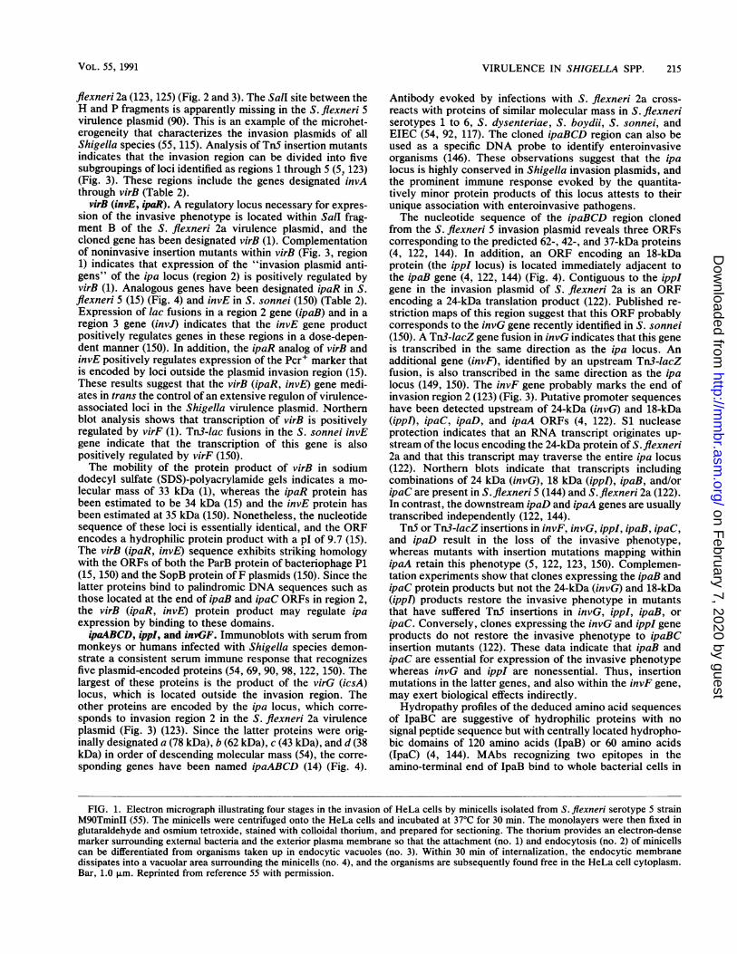
Large plasmids were first detected about 12 years ago in S.flexneri 2a (72), and the essential role of plasmids in viru-lence was established shortly thereafter in both S. sonnei(118) and S. flexneri (119). Subsequently it was shown thatvirulence plasmids are also present in other serotypes of S.flexneri, S. dysenteriae, S. boydii, and EIEC (55, 115, 117).Endonuclease digestion and Southern hybridization indicatethat the virulence plasmids of Shigella species and EIEC areessentially homologous but restriction sites vary with thespecies and serotype (55, 115). The ability to invade tissueculture cells is transferred to E. coli K-12 by the conjugalmobilization of 220- to 240-kb plasmids from S. flexneri s(116). The invasive phenotype can also be transferred by a180-kb plasmid from S. sonnei (151). The characteristicsconferred by these invasion plasmids can be illustrated invitro with S. flexneri s minicells that carry only plasmidDNA (55) (Fig. 1). Plasmid-encoded proteins mediate theattachment and endocytic uptake of minicells by HeLa cells(Fig. 1A). Following ingestion, the endocytic vacuoles dis-sipate in a plasmid-dependent process and the minicells arereleased into the cytoplasm (Fig. 1B).A 230-kb plasmid of S. flexneri 2a has been subjected to
Sall endonuclease digestion, and 23 fragments have beenidentified and mapped (124, 125) (Fig. 2). In the context ofthis Sail map, genetic data from the S.flexneri 2a, S.flexneri5, and S. sonnei plasmids are summarized in Table 2. Therelative position and direction of transcription of virulence-associated loci have been deduced from the literature, andthe loci are listed in a clockwise order starting with the Stbmarker that is located in SalI fragment 0 in S.flexneri 2a (88,125).
Stb and Rep (Stable Maintenance and Replication)
Analysis of plasmid incompatibility has suggested that S.flexneri serotypes 1 to 5 and S. sonnei belong to the IncFIgroup, whereas S. flexneri serotype 6 and S. boydii, S.dysenteriae, and EIEC are compatible with IncFI plasmids(88). The Rep region of SalI fragment C from the S. flexneri2a virulence plasmid shares homology with the RepFIIAfamily of replicons, and this region is responsible for incom-patibility with IncFI and IncHl plasmids (88). Presumably,the Rep region is also responsible for homology seen with aRepFIC probe in clinical isolates of EIEC and Shigellaspecies (130). Along with a contiguous region on Sall frag-ment 0 (Stb, stable maintenance), the Rep locus is necessaryfor replication and maintenance of virulence plasmids.
virF (Positive Regulator of the Plasmid Virulence Regulon)
Located approximately 50 kb from the origin of replica-tion, the virF locus was first identified by a spontaneousdeletion in SalI fragment F that resulted in the simultaneousloss of four virulence-associated phenotypes: Pcr (Congo redbinding), Inv (invasion of tissue culture cells in vitro), Ser
(Sereny test), and Igr (inhibition of growth) (124) (Fig. 2).Expression of these phenotypes is also lost when TnStransposon insertions occur in the F fragment. A plasmidclone containing the Sail F fragment complements thesemutations, and when cloned into E. coli K-12 in a high-copy-number vector, a locus carried on this plasmid fragmentmediates Congo red binding (111). However, the cloned Ffragment does not convey the Pcr+ phenotype upon an S.flexneri recipient that has lost the 230-kb plasmid, and thisfinding indicates that other genes are required for expressionof the Congo red-binding phenotype in the Shigella geneticbackground (111, 124). The virulence-associated gene lo-cated within the F fragment has been designated virF (113)(Fig. 3). A gene designated crb has been independentlycloned from the 240-kb plasmid of S. flexneri 2a in a 9-kbBamHI fragment (22). This cloned locus has the samephenotypic characteristics as virF, and, like virF, it is alsolinked to IS] elements that may mediate spontaneous dele-tions in this region of the plasmid (124).The functional gene product of virF is a 30-kDa protein,
but a 24-amino-acid signal peptide-like sequence may becleaved during passage through the inner membrane, yield-ing a 27-kDa protein that has been detected in minicells(112). The virF gene product plays a central role in positiveregulation of the plasmid virulence regulon. It directlyactivates transcription of the virG gene (113), and it indi-rectly activates ipaABCD (69, 113, 149) and invAKJHF (149)plasmid loci through a second gene variously designatedvirB, ipaR, or invE (Fig. 3). This central regulatory functionaccounts for the concomitant loss of four virulence-associ-ated phenotypes in the virF mutants (124, 125). The clonedvirF locus is temperature regulated in both S. flexneri and E.coli K-12 backgrounds (111), but it is expressed at both 30and 37°C in minicells that do not carry chromosomal regu-latory loci such as virR (osmZ) (112). Therefore, the virRchromosomal gene may regulate plasmid-encoded invasiondeterminants at 30°C by inhibiting virF expression (1).
Since many Shigella genes can transfer the Pcr+ marker toE. coli when cloned on high-copy-number vectors (16, 23,67a, 111), the location of the structural genes encodingCongo red-binding protein(s) in Shigella species is unclear.Nonetheless, a 101-kDa protein (or protein complex) hasrecently been identified as a heme-binding outer membranecomponent that may also be the Congo red-binding protein(137). A 6.1-kb PstI plasmid fragment which confers expres-sion of the 101-kDa protein and the Pcr+ phenotype in aplasmid-cured S. flexneri background does not reconstitutethe invasive phenotype. Conversely, a 37-kb plasmid frag-ment that does reconstitute invasion does not confer thePcr+ phenotype (90). Even though the Inv' and Pcr+phenotypes are expressed independently, they are synergis-tically related in that pretreatment of shigellae with Congored or hemin increases the invasion of HeLa cells (24). SincePcr+ S. flexneri strains are more hydrophobic than isogenicplasmid-cured strains (128), the Pcr+ phenotype may indi-rectly reflect a characteristic of the bacterial surface thatenhances the initial interaction of shigellae with host cells.
Plasmid Invasion Genes
The first successful attempt to clone the invasive pheno-type of S. flexneri 5 into an isogenic strain that had lost the230-kb virulence plasmid indicated that the genes encodingthis phenotype were located within a 37-kb plasmid segment(90). The cloned invasion region corresponds to SalI restric-tion fragments D, H, P, and B in the 230-kb plasmid of S.
VOL. 55, 1991
on February 7, 2020 by guest
http://mm
br.asm.org/
Dow
nloaded from

214 HALE MICROBIOL. REV.
'44
a 4
J j, 9 ,.,,,,~ ~ - 'WY1~~~~~~~~~~~~~~~~~~~~~~~~~~~~~~~~~,4*s-tY.e--- r...\.
A *~~~~~~~~-
_ t #~~.4
S~~~~~~~~~~~~~~~~~~~~~~~~~~~~~~~~~~~~~~~~~~~~~~~~~~~~~~~~~~~~~~~~~~~~~~~~~~~~~~~~~~~~~~~~~~~~~~~~~~~~~
,si i 5 ;;00*'9'
4 4 *1
AL.~~~'
Prwj * USt-V _|iE4#0W - # u. i L e ' t -;S rl r N;.._s *S *BiW0w# i n *W;V,F*'_:. . *M,. -;s .s ; '; 4 {, _p P
AA
.r-
f^¢0t^ ^?lq,* 4.,^: : :,?.j ..e',c
.4',
I.'#7
.1
on February 7, 2020 by guest
http://mm
br.asm.org/
Dow
nloaded from
VIRULENCE IN SHIGELLA SPP. 215
flexneri 2a (123, 125) (Fig. 2 and 3). The Sall site between theH and P fragments is apparently missing in the S. flexneri 5virulence plasmid (90). This is an example of the microhet-erogeneity that characterizes the invasion plasmids of allShigella species (55, 115). Analysis of TnS insertion mutantsindicates that the invasion region can be divided into fivesubgroupings of loci identified as regions 1 through 5 (57 123)(Fig. 3). These regions include the genes designated invAthrough virB (Table 2).
virB (invE, ipaR). A regulatory locus necessary for expres-sion of the invasive phenotype is located within Sall frag-ment B of the S. flexneri 2a virulence plasmid, and thecloned gene has been designated virB (1). Complementationof noninvasive insertion mutants within virB (Fig. 3, region1) indicates that expression of the "invasion plasmid anti-gens" of the ipa locus (region 2) is positively regulated byvirB (1). Analogous genes have been designated ipaR in S.flexneri 5 (15) (Fig. 4) and invE in S. sonnei (150) (Table 2).Expression of lac fusions in a region 2 gene (ipaB) and in aregion 3 gene (invJ) indicates that the invE gene productpositively regulates genes in these regions in a dose-depen-dent manner (150). In addition, the ipaR analog of virB andinvE positively regulates expression of the Pcr+ marker thatis encoded by loci outside the plasmid invasion region (15).These results suggest that the virB (ipaR, invE) gene medi-ates in trans the control of an extensive regulon of virulence-associated loci in the Shigella virulence plasmid. Northernblot analysis shows that transcription of virB is positivelyregulated by virF (1). Tn3-lac fusions in the S. sonnei invEgene indicate that the transcription of this gene is alsopositively regulated by virF (150).The mobility of the protein product of virB in sodium
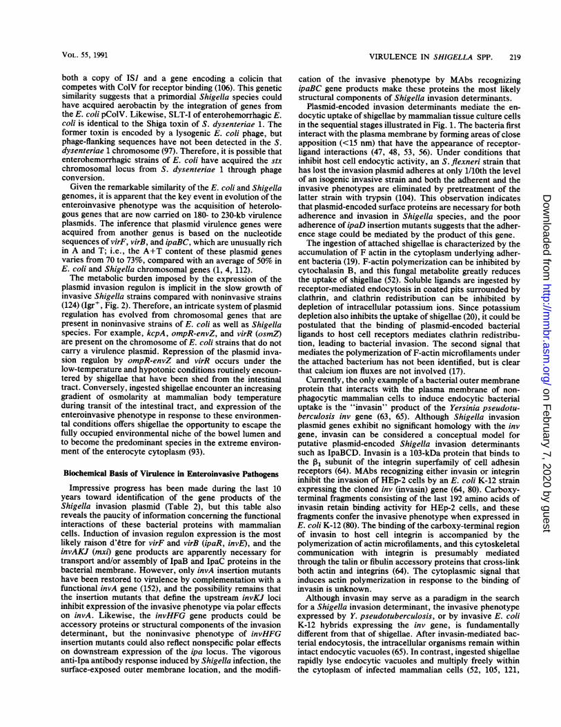
dodecyl sulfate (SDS)-polyacrylamide gels indicates a mo-lecular mass of 33 kDa (1), whereas the ipaR protein hasbeen estimated to be 34 kDa (15) and the invE protein hasbeen estimated at 35 kDa (150). Nonetheless, the nucleotidesequence of these loci is essentially identical, and the ORFencodes a hydrophilic protein product with a pI of 9.7 (15).The virB (ipaR, invE) sequence exhibits striking homologywith the ORFs of both the ParB protein of bacteriophage P1(15, 150) and the SopB protein of F plasmids (150). Since thelatter proteins bind to palindromic DNA sequences such asthose located at the end of ipaB and ipaC ORFs in region 2,the virB (ipaR, invE) protein product may regulate ipaexpression by binding to these domains.ipaABCD, ippI, and invGF. Immunoblots with serum from
monkeys or humans infected with Shigella species demon-strate a consistent serum immune response that recognizesfive plasmid-encoded proteins (54, 69, 90, 98, 122, 150). Thelargest of these proteins is the product of the virG (icsA)locus, which is located outside the invasion region. Theother proteins are encoded by the ipa locus, which corre-sponds to invasion region 2 in the S. flexneri 2a virulenceplasmid (Fig. 3) (123). Since the latter proteins were orig-inally designated a (78 kDa), b (62 kDa), c (43 kDa), and d (38kDa) in order of descending molecular mass (54), the corre-sponding genes have been named ipaABCD (14) (Fig. 4).
Antibody evoked by infections with S. flexneri 2a cross-reacts with proteins of similar molecular mass in S. flexneriserotypes 1 to 6, S. dysenteriae, S. boydii, S. sonnei, andEIEC (54, 92, 117). The cloned ipaBCD region can also beused as a specific DNA probe to identify enteroinvasiveorganisms (146). These observations suggest that the ipalocus is highly conserved in Shigella invasion plasmids, andthe prominent immune response evoked by the quantita-tively minor protein products of this locus attests to theirunique association with enteroinvasive pathogens.The nucleotide sequence of the ipaBCD region cloned
from the S. flexneri 5 invasion plasmid reveals three ORFscorresponding to the predicted 62-, 42-, and 37-kDa proteins(4, 122, 144). In addition, an ORF encoding an 18-kDaprotein (the ippI locus) is located immediately adjacent tothe ipaB gene (4, 122, 144) (Fig. 4). Contiguous to the ippIgene in the invasion plasmid of S. flexneri 2a is an ORFencoding a 24-kDa translation product (122). Published re-striction maps of this region suggest that this ORF probablycorresponds to the invG gene recently identified in S. sonnei(150). A Tn3-lacZ gene fusion in invG indicates that this geneis transcribed in the same direction as the ipa locus. Anadditional gene (invF), identified by an upstream Tn3-lacZfusion, is also transcribed in the same direction as the ipalocus (149, 150). The invF gene probably marks the end ofinvasion region 2 (123) (Fig. 3). Putative promoter sequenceshave been detected upstream of 24-kDa (invG) and 18-kDa(ippl), ipaC, ipaD, and ipaA ORFs (4, 122). Si nucleaseprotection indicates that an RNA transcript originates up-stream of the locus encoding the 24-kDa protein of S. flexneri2a and that this transcript may traverse the entire ipa locus(122). Northern blots indicate that transcripts includingcombinations of 24 kDa (invG), 18 kDa (ippI), ipaB, and/oripaC are present in S. flexneri 5 (144) and S. flexneri 2a (122).In contrast, the downstream ipaD and ipaA genes are usuallytranscribed independently (122, 144).Tn5 or Tn3-lacZ insertions in invF, invG, ippI, ipaB, ipaC,
and ipaD result in the loss of the invasive phenotype,whereas mutants with insertion mutations mapping withinipaA retain this phenotype (5, 122, 123, 150). Complemen-tation experiments show that clones expressing the ipaB andipaC protein products but not the 24-kDa (invG) and 18-kDa(ippl) products restore the invasive phenotype in mutantsthat have suffered TnS insertions in invG, ippI, ipaB, oripaC. Conversely, clones expressing the invG and ippI geneproducts do not restore the invasive phenotype to ipaBCinsertion mutants (122). These data indicate that ipaB andipaC are essential for expression of the invasive phenotypewhereas invG and ippI are nonessential. Thus, insertionmutations in the latter genes, and also within the invF gene,may exert biological effects indirectly.Hydropathy profiles of the deduced amino acid sequences
of IpaBC are suggestive of hydrophilic proteins with nosignal peptide sequence but with centrally located hydropho-bic domains of 120 amino acids (IpaB) or 60 amino acids(IpaC) (4, 144). MAbs recognizing two epitopes in theamino-terminal end of IpaB bind to whole bacterial cells in
FIG. 1. Electron micrograph illustrating four stages in the invasion of HeLa cells by minicells isolated from S. flexneri serotype 5 strainM9OTminII (55). The minicells were centrifuged onto the HeLa cells and incubated at 37°C for 30 min. The monolayers were then fixed inglutaraldehyde and osmium tetroxide, stained with colloidal thorium, and prepared for sectioning. The thorium provides an electron-densemarker surrounding external bacteria and the exterior plasma membrane so that the attachment (no. 1) and endocytosis (no. 2) of minicellscan be differentiated from organisms taken up in endocytic vacuoles (no. 3). Within 30 min of internalization, the endocytic membranedissipates into a vacuolar area surrounding the minicells (no. 4), and the organisms are subsequently found free in the HeLa cell cytoplasm.Bar, 1.0 ,um. Reprinted from reference 55 with permission.
VOL. 55, 1991
on February 7, 2020 by guest
http://mm
br.asm.org/
Dow
nloaded from

MICROBIOL. REV.
230/0 (Kb)
FIG. 2. Circular Sall restriction map of S. flexneri 2a plasmidpMYSH6000. The area indicted by solid segments contains genesresponsible for Ser+ (Sereny test), Inv' (invasion of culturedmammalian cells), Pcr+ (binding of Congo red), and Igr+ (inhibitionof growth). Symbols: M , segment necessary for Ser+ but not forInv', Pcr+, and Igr+; Lii, ISI-like elements and the origin ofreplication (Rep). Reprinted with permission from Sasakawa et al.(125).
an enzyme-linked immunosorbent assay (ELISA) (61, 92).One of these MAbs, designated 2F1, partially inhibits theformation of plaques in baby hamster kidney (BHK) cellmonolayers (92), indicating that the amino-terminal domainof IpaB is necessary for bacterial invasion or for intercellularbacterial spread. In contrast, three MAbs recognizingepitopes closer to the central hydrophobic domain of IpaBare much less reactive in the whole-cell ELISA (92), sug-gesting that this is a transmembrane protein region. Theseweakly reactive MAbs have no effect upon plaque formationin infected BHK monolayers. Three MAbs recognizingepitopes near the amino-terminal end of IpaC are stronglyreactive in the whole-cell ELISA, but these reagents have noeffect upon plaque formation. One MAb, designated 2G2,recognizes an epitope on the carboxy-terminal side of thecentral hydrophobic domain of IpaC, and pretreatment of S.
G
I IIFS SS
lI
C=virG
10 kb
flexneri with this MAb enhances plaque formation (92). Thispositive effect suggests that the carboxy-terminal portion ofIpaC affects the expression of the invasive phenotype, and itshould be noted that the hydropathy profile of the carboxy-terminal half of IpaC mirrors the hydropathy profile of theamino-terminal half of IpaB.
Results of immunofluorescence studies with polyclonalrabbit antiserum recognizing IpaC indicate that this proteinis detectable on the bacterial surface only if shigellae aregrown in the presence of Congo red (114), and pretreatmentof bacteria with this dye also enhances expression of theinvasive phenotype (24). In addition, shigellae that haveinvaded tissue culture cells are recognized by anti-IpaCantiserum (114). These data suggest that the transport and/orsynthesis of Ipa proteins is enhanced by the binding ofhydrophobic dyes and by growth within the host cell cyto-plasm. However, a recent report indicates that growth of S.flexneri within tissue culture cells actually inhibits de novoexpression of Ipa proteins (58). The latter experimentsinvolved immunoprecipitation of radiolabeled Shigella pro-teins from monolayers that had been solubilized by relativelygentle detergent treatment, and interpretation of the resultswas complicated by the precipitation of many radiolabeledproteins in addition to the Ipa proteins. Nonetheless, it ispossible that IpaC protein is transported to the outer mem-brane of S. flexneri growing in the host cell cytoplasm at thesame time that de novo synthesis of Ipa proteins is re-pressed. Additional studies are required to fully characterizethe regulatory patterns of Ipa protein expression duringbacterial invasion and intracellular growth.The hydropathy profile of the deduced IpaD amino acid
sequence reveals the periodicity of a helical molecule withno extensive hydrophobic domains suggestive of a signalsequence or a transmembrane region. A critical role for IpaDin the invasion process can be inferred from observationsobtained with noninvasive TnS insertion mutants with mu-tations that map within the ipaD gene (5, 122). Thesemutants are less adherent to tissue culture cells than arewild-type shigellae; this may indicate that IpaD facilitatesthe initial interaction of shigellae with the surface of hostcells (59a).Although the molecular mobility of IpaA in SDS-poly-
acrylamide gel electrophoresis (PAGE) suggests a mass of 78kDa (54), the recently sequenced ipaA ORF is indicative ofa hydrophilic protein of only 70 kDa (143). TnS insertionsinto the ipaA gene retain the invasive phenotype in the tissueculture model (5) and elicit a delayed-positive Sereny reac-
B
EI
ipaR ipaJ
R2 =
RI
=
ADCB invA region virF
R3 =R4 invasion-associated regions
R5 =J
FIG. 3. Restriction map of Sail fragments G, B, P, H, D, and F containing the known plasmid virulence genes of S. flexneri. The fiveinvasion-associated regions were defined by TnS insertions that eliminate expression of the invasive phenotype (123). The virG gene has alsobeen designated icsA, and the ipaR gene has been designated virB or invE. Restriction sites: S, Sall; E, EcoRI; H, Hindlll. Figure courtesyof J. M. Buysse.
I I I I
216 HALE
on February 7, 2020 by guest
http://mm
br.asm.org/
Dow
nloaded from

VIRULENCE IN SHIGELLA SPP. 217
virG
27,28 kDa
/
38K43K 62K 78K I
ipaR ipaA ipaD ipaC ipaB ippI1. --: -I...I: ---- '-1-- "-'T-;:,-.----,,
Th5
~~ ~ ~ ~ ~ ~P
0 1 2 3 4 5 6
pH
P BgA \
H H E IP SS E IE 1 P7 II9f1 l
7 8 9 10 11 12 13 14 kb
FIG. 4. Composite figure with an immunoblot showing the protein products of the invasion plasmid antigen (ipa) locus imposed above arestriction map of the encoding region of the S. flexneri 5 invasion plasmid. Also detectable in the immunoblot is the protein product of thevirG (icsA) locus. The immunoblot was developed with a convalescent-phase serum sample from a rhesus monkey infected with S. flexneri2a. The TnS insertion mutant pHS1042 (90), which defines the ipaR (virB, invE) regulatory gene (15), is included in the mapped region.Restriction sites: E, EcoRI; P, PuvII; A, AccI; B, BamHI; H, Hindlll; Bg, BgII. Figure courtesy of J. M. Buysse.
tion. Therefore, the role of IpaA in virulence has not yetbeen ascertained.invAAJH (mxiAB). Tn3-lac fusion inserts within the S.
sonnei invasion plasmid have defined four transcribed genes,designated invAKJH, that are necessary for expression ofthe invasive phenotype (149, 150) (Table 2). Restrictionmaps suggest that these genes correspond to invasion re-gions 3 and 5 of the S. flexneri 2a plasmid (123) (Fig. 3).Expression of invAKJH is under the positive control of thevirF-virB (invE-ipaR) system (1, 61, 149, 150), but this locusdiffers from the genes of invasion regions 1 and 2 in thedirection of transcription (Table 2) (61, 149, 150). Publishedrestriction maps suggest that invA should map within inva-sion region 5 and invKJ should map within invasion region 3on the S. flexneri 2a plasmid. Restriction analysis alsoindicates that an S. sonnei gene designated invH should mapat the junction of invasion region 3 and region 2 in the S.flexneri 2a virulence plasmid (123, 149, 150). Since results ofprecise mapping and sequencing of invasion regions 3, 4, and5 have yet to be published, the molecular mass of invKJHgene products is unknown. However, invA insertion mutantsare complemented by a cloned fragment from the S. sonneiplasmid which expresses a 38-kDa protein (152).
Deletion mutants eliminating invasion region 3, 4, or 5 arenoninvasive even though they express the gene products ofinvasion regions 1 and 2 (15). A noninvasive, inv::lacZ S.flexneri 2a fusion mutant designated BS226 has been foundto bind 80% less 2F1 MAb (anti-IpaB) and 66% less 2G2MAb (anti-IpaC) in a whole-cell ELISA (61). Restrictionmaps indicate that the BS226 fusion is homologous to invAmutants. Similar inhibition of MAb binding was observed ina whole-cell ELISA with inv::lacZ fusion mutants BS232and BS230 that map within an 8-kb EcoRI-SalI fragment ofinvasion region 3 (61). It is possible that the genes defined bythe latter fusions are homologous to invKJ. Quantitativeimmunoblots of isolated membrane fractions have recentlyshown that neither IpaB nor IpaC is transported to the outermembrane in BS226 or in another inv::lacZ fusion mutant(BS260) that is probably analogous to BS232. The invasionplasmid antigens accumulate within the inner membrane inthese mutants (la). Therefore, the S. flexneri 2a gene defined
by the inv::lacZ insertion BS226 has been designated mxiBand the gene defined by the BS260 or BS232 fusions has beendesignated mxiA (mxi, membrane expression of Ipa) (61)(Table 2). Rabbit antiserum raised against the BS260 fusionprotein recognizes a 76-kDa protein in immunoblots of an S.flexneri 5 whole-cell lysate (la).
virG (icsA) (Plasmid Gene Associated with IntercellularBacterial Spread)
The virG locus was first identified by transposon insertionswithin SalI fragment G, located approximately 20 kb frominvasion region 1 in the S. flexneri 2a plasmid (Fig. 2 and 3).virG insertion mutants are Inv', Pcr+, and Igr+, but Ser(125). As was explained in the description of the chromo-somal kepA locus, the Ser- phenotype of virG mutantsreflects an inability of intracellular organisms to spreadwithin the cytoplasm of infected cells. Since this geneticdefect precludes the infection of contiguous cells in tissueculture monolayers or within the corneal epithelium (79, 87,105, 158), the homologous locus in S. flexneri 5 has also beendesignated icsA (intercellular spread) (8).The virG (icsA) gene product was originally identified as
the fifth invasion plasmid antigen of S. flexneri 5 (98), andextrinsic radioiodination of whole cells has shown that thisprotein is exposed on the bacterial surface (79). A uniqueproperty of strains that express the virG protein is a polardeposition of F actin (filamentous actin) around organisms inthe cytoplasm of infected HeLa cells. This F actin some-times gives the appearance of an elongated tail, and theseactin tails are often found associated with bacteria in protru-sions of the host cell plasma membrane (8). These deforma-tions may represent the first stage in intercellular bacterialspread. Since the intracellular movement of organisms ex-pressing virG (icsA) is reversibly inhibited by cytochalasin D(8, 105), the deposition of F actin apparently provides amotive force. The deduced amino acid sequence of virG isnot indicative of a signal sequences or hydrophobic regionsthat might be associated with the outer membrane (79), but aremarkable feature of the sequence is a series of repeatingmotifs in the amino-terminal one-third of the molecule.
VOL. 55, 1991
on February 7, 2020 by guest
http://mm
br.asm.org/
Dow
nloaded from

MICROBIOL. REV.
The virG (icsA) gene product has been reported to have amolecular mobility in SDS-polyacrylamide gels of 140 kDa(98, 105), 130 kDa (79), or 120 kDa (8). The ORF of thecloned virG gene of S. flexneri 2a suggests a protein ofapproximately 117 kDa (79). However, minicell analysis ofthe product(s) of the cloned virG (icsA) gene reveals at leastnine nonvector polypeptides of 130 kDa or less (8, 79), and ithas been suggested that these polypeptides are the productsof internal initiation codons (79). Nonetheless, immunoblotswith convalescent-phase monkey antiserum detect only asingle large icsA product in a plasmid-cured Shigella straincarrying the cloned gene (8), and radioiodination of eitherwild-type S. flexneri 2a or a plasmid-cured strain carryingonly virG also labels only a single large virG product (79).Since the putative internal initiation products are not recog-nized by convalescent-phase serum and are not radioiodi-nated in whole cells, perhaps only the amino-terminal por-tion of the largest protein product is exposed on the bacterialsurface. Alternatively, it has recently been suggested thatmultiple virG polypeptides seen in minicells are aberranttranslation products of the virG mRNA in that they aretranslated only in the absence of the kcpA chromosomalgene product. Thus, the kcpA protein has been described asa posttranscriptional activator that favors the transcriptionof a functional 120-kDa virG product (160).
ipaH (Multicopy Invasion Plasmid Antigen Gene)When the ipaBCD genes were cloned from S. flexneri 5
into a Xgtll expression vector, the cloned genes wereidentified by colony lifts and immunoblots with antibodyfrom rabbits that had been immunized with crude fractionsof plasmid-encoded proteins (14). A subset of clones thatexpressed a 60-kDa protein was recognized by this rabbitserum, but affinity-purified antibody reacting with this pro-tein did not cross-react with the 62-kDa IpaB protein. Inaddition, these clones did not hybridize with Agtll: :ipaB (14,15a, 57). Since a new invasion plasmid antigen had appar-ently been cloned in these experiments, the gene was desig-nated ipaH (57).The ipaH gene is unique in that five complete or partial
copies are present on the invasion plasmids of the various S.flexneri serotypes and multiple copies are also found on theinvasion plasmids of other Shigella species and EIEC (57).In addition, multiple copies of ipaH are present on theShigella and EIEC chromosome but not on the chromosomeof nonpathogenic E. coli strains (145). The copies of ipaHhave been characterized by Southern hybridization ofHindIII digests of S. flexneri 5 plasmid DNA with aAgtll::ipaH probe. These genes have been designatedipaH9.8, ipaH7.8, ipaH4.5, ipaH2.5, and ipaH.4 on the basisof the size of the hybridizing DNA fragment (57). ipaH7.8and ipaH45 have been mapped to SalI fragment B betweenvirG and ipaR (within 10 kb of the latter gene). Northern blotanalysis indicates that both ipaH7.8 and ipaH4.5 are tran-scribed in vitro whereas ipaH25 and ipaH,.4 are unex-pressed, truncated sequences (143a). It is clear that one ormore copies of ipaH is expressed during Shigella infectionsbecause specific antibody has been detected in convalescent-phase human serum (99a). Cloning of ipaH7.8 in an E. colibackground does not confer Congo red binding or theinvasive phenotype, and, unlike other plasmid virulencegenes, expression of ipaH is independent of temperatureregulation or induction by virF (57).The DNA sequence of the complete ipaH7 8 gene and the
amino-terminal half of ipaH4.5 has been published, and the
former ORF encodes a 60.8-kDa protein with a pl of 5.9 (57).Recent DNA sequencing and immunoblotting indicate thatipaH45 encodes a 65.3-kDa protein. Both IpaH productshave a predominantly hydrophilic structure with no charac-teristic signal peptide. The amino-terminal sequence of bothipaH78 and ipaH45 reveals six or eight evenly spaced,14-residue, leucine-rich glycoprotein (LRG) motifs consist-ing of Leu-X2-Leu-Pro-X-Leu-Pro-X2-Leu-X2-Leu (where Xrepresents any amino acid). The regular spacing of leucineresidues in this motif suggests that the amino-terminal half ofthe IpaH protein is configured as an amphipathic helixpresenting a uniform hydrophobic surface on one side. Thecarboxy-terminal half of both genes is identical except for ashort terminal sequence, and this conserved portion elicitscross-reacting antibody recognizing the other ipaH geneproducts (143a).The conservation of ipaH-like genes in at least two com-
plete plasmid copies suggests a powerful selection for theexpression of these proteins. A possible explanation of therole of these proteins in virulence involves the amino-terminal, LRG-like motifs. These leucine-rich repeats aresimilar to the LRG-like motifs of the platelet glycoproteinGPIba that binds von Willebrand factor, triggering conver-sion of prothrombin to thrombin in the process of fibrin clotformation and in the initiation of inflammatory processes.The only other example of LRG-like motifs in a prokaryoticprotein is found in the YopM protein of Yersinia pestis (82).Culture supernatants of Y. pestis containing YopM inhibitcoagulation, and yopM mutants of Y. pestis exhibit greatlydecreased virulence in mice (81). Therefore, it has beensuggested that YopM competitively inhibits the normalinteraction of von Willebrand factor and/or thrombin withplatelets. This competitive effect should prevent plateletadhesion to the exposed subendothelium of injured vesselsand inhibit normal thrombus formation and inflammation.The similarity of the LRG-like motifs of YopM and IpaH
(50% identity in a 93-amino-acid overlap) indicates that IpaHmay also act to modify the host response to Shigella infec-tion. For example, the IpaH protein could inhibit boththrombus formation and the recruitment of inflammatorycells into the lamina propria during the early stages ofinfection. Thus, dissemination of shigellae within the mu-cosa could be facilitated, and inhibition of thrombin-inducedcoagulation could also exacerbate the loss of blood into thestools of dysenteric patients. Final characterization of therole of IpaH in virulence awaits the construction of IpaH-mutants, and the recent sequencing of ipaH7.8 and ipaH4.5should allow the elimination of these plasmid genes bysite-directed mutagenesis through gene replacement.
DISCUSSION OF VIRULENCE IN SHIGELLA SPECIES
Genetic Basis of Virulence in Enteroinvasive PathogensWith the possible exceptions of theflu (fluid accumulation)
locus (32, 116, 141, 159) and the chromosomal copies of ipaH(145), there are presently no identified chromosomal viru-lence-associated loci in Shigella species that do not havecounterparts in the genome of E. coli. In the latter species,for example, the iucABCD and iutA genes of the aerobactinsystem are present in the E. coli pColV plasmid as atransposonlike element flanked by two inverted IS) repeatelements, and this configuration has been shown to promotecointegrate formation and transposition of the entire aero-bactin system to other replicons (25). Flanking regions of thechromosomal aerobactin locus in S. flexneri also include
218 HALE
on February 7, 2020 by guest
http://mm
br.asm.org/
Dow
nloaded from
VIRULENCE IN SHIGELLA SPP. 219
both a copy of IS1 and a gene encoding a colicin thatcompetes with ColV for receptor binding (106). This geneticsimilarity suggests that a primordial Shigella species couldhave acquired aerobactin by the integration of genes fromthe E. coli pColV. Likewise, SLT-I of enterohemorrhagic E.coli is identical to the Shiga toxin of S. dysenteriae 1. Theformer toxin is encoded by a lysogenic E. coli phage, butphage-flanking sequences have not been detected in the S.dysenteriae 1 chromosome (97). Therefore, it is possible thatenterohemorrhagic strains of E. coli have acquired the stxchromosomal locus from S. dysenteriae 1 through phageconversion.Given the remarkable similarity of the E. coli and Shigella
genomes, it is apparent that the key event in evolution of theenteroinvasive phenotype was the acquisition of heterolo-gous genes that are now carried on 180- to 230-kb virulenceplasmids. The inference that plasmid virulence genes wereacquired from another genus is based on the nucleotidesequences of virF, virB, and ipaBC, which are unusually richin A and T; i.e., the A+T content of these plasmid genesvaries from 70 to 73%, compared with an average of 50% inE. coli and Shigella chromosomal genes (1, 4, 112).The metabolic burden imposed by the expression of the
plasmid invasion regulon is implicit in the slow growth ofinvasive Shigella strains compared with noninvasive strains(124) (Igr+, Fig. 2). Therefore, an intricate system of plasmidregulation has evolved from chromosomal genes that arepresent in noninvasive strains of E. coli as well as Shigellaspecies. For example, kcpA, ompR-envZ, and virR (osmZ)are present on the chromosome of E. coli strains that do notcarry a virulence plasmid. Repression of the plasmid inva-sion regulon by ompR-envZ and virR occurs under thelow-temperature and hypotonic conditions routinely encoun-tered by shigellae that have been shed from the intestinaltract. Conversely, ingested shigellae encounter an increasinggradient of osmolarity at mammalian body temperatureduring transit of the intestinal tract, and expression of theenteroinvasive phenotype in response to these environmen-tal conditions offers shigellae the opportunity to escape thefully occupied environmental niche of the bowel lumen andto become the predominant species in the extreme environ-ment of the enterocyte cytoplasm (93).
Biochemical Basis of Virulence in Enteroinvasive PathogensImpressive progress has been made during the last 10
years toward identification of the gene products of theShigella invasion plasmid (Table 2), but this table alsoreveals the paucity of information concerning the functionalinteractions of these bacterial proteins with mammaliancells. Induction of invasion regulon expression is the mostlikely raison d'etre for virF and virB (ipaR, invE), and theinvAKJ (mxi) gene products are apparently necessary fortransport and/or assembly of IpaB and IpaC proteins in thebacterial membrane. However, only invA insertion mutantshave been restored to virulence by complementation with afunctional invA gene (152), and the possibility remains thatthe insertion mutants that define the upstream invKJ lociinhibit expression of the invasive phenotype via polar effectson invA. Likewise, the invHFG gene products could beaccessory proteins or structural components of the invasiondeterminant, but the noninvasive phenotype of invHFGinsertion mutants could also reflect nonspecific polar effectson downstream expression of the ipa locus. The vigorousanti-Ipa antibody response induced by Shigella infection, thesurface-exposed outer membrane location, and the modifi-
cation of the invasive phenotype by MAbs recognizingipaBC gene products make these proteins the most likelystructural components of Shigella invasion determinants.Plasmid-encoded invasion determinants mediate the en-
docytic uptake of shigellae by mammalian tissue culture cellsin the sequential stages illustrated in Fig. 1. The bacteria firstinteract with the plasma membrane by forming areas of closeapposition (<15 nm) that have the appearance of receptor-ligand interactions (47, 48, 53, 56). Under conditions thatinhibit host cell endocytic activity, an S. flexneri strain thathas lost the invasion plasmid adheres at only 1/10th the levelof an isogenic invasive strain and both the adherent and theinvasive phenotypes are eliminated by pretreatment of thelatter strain with trypsin (104). This observation indicatesthat plasmid-encoded surface proteins are necessary for bothadherence and invasion in Shigella species, and the pooradherence of ipaD insertion mutants suggests that the adher-ence stage could be mediated by the product of this gene.The ingestion of attached shigellae is characterized by the
accumulation of F actin in the cytoplasm underlying adher-ent bacteria (19). F-actin polymerization can be inhibited bycytochalasin B, and this fungal metabolite greatly reducesthe uptake of shigellae (52). Soluble ligands are ingested byreceptor-mediated endocytosis in coated pits surrounded byclathrin, and clathrin redistribution can be inhibited bydepletion of intracellular potassium ions. Since potassiumdepletion also inhibits the uptake of shigellae (20), it could bepostulated that the binding of plasmid-encoded bacterialligands to host cell receptors mediates clathrin redistribu-tion, leading to bacterial invasion. The second signal thatmediates the polymerization of F-actin microfilaments underthe attached bacterium has not been identified, but is clearthat calcium ion fluxes are not involved (17).
Currently, the only example of a bacterial outer membraneprotein that interacts with the plasma membrane of non-phagocytic mammalian cells to induce endocytic bacterialuptake is the "invasin" product of the Yersinia pseudotu-berculosis inv gene (63, 65). Although Shigella invasionplasmid genes exhibit no significant homology with the invgene, invasin can be considered a conceptual model forputative plasmid-encoded Shigella invasion determinantssuch as IpaBCD. Invasin is a 103-kDa protein that binds tothe P1 subunit of the integrin superfamily of cell adhesinreceptors (64). MAbs recognizing either invasin or integrininhibit the invasion of HEp-2 cells by an E. coli K-12 strainexpressing the cloned inv (invasin) gene (64, 80). Carboxy-terminal fragments consisting of the last 192 amino acids ofinvasin retain binding activity for HEp-2 cells, and thesefragments confer the invasive phenotype when expressed inE. coli K-12 (80). The binding of the carboxy-terminal regionof invasin to host cell integrin is accompanied by thepolymerization of actin microfilaments, and this cytoskeletalcommunication with integrin is presumably mediatedthrough the talin or fibulin accessory proteins that cross-linkboth actin and integrins (64). The cytoplasmic signal thatinduces actin polymerization in response to the binding ofinvasin is unknown.Although invasin may serve as a paradigm in the search
for a Shigella invasion determinant, the invasive phenotypeexpressed by Y. pseudotuberculosis, or by invasive E. coliK-12 hybrids expressing the inv gene, is fundamentallydifferent from that of shigellae. After invasin-mediated bac-terial endocytosis, the intracellular organisms remain withinintact endocytic vacuoles (65). In contrast, ingested shigellaerapidly lyse endocytic vacuoles and multiply freely withinthe cytoplasm of infected mammalian cells (52, 105, 121,
VOL. 55, 1991
on February 7, 2020 by guest
http://mm
br.asm.org/
Dow
nloaded from

MICROBIOL. REV.
138). The latter phenomenon is apparently mediated by theShigella invasion plasmid because it occurs in HeLa cellsthat have ingested either plasmid-carrying minicells (55)(Fig. 1) or plasmid-carrying E. coli K-12 (121). The lysis ofendocytic vacuoles is correlated with the ability to lysesheep erythrocytes, but this hemolytic assay requires bacte-rial contact with the erythrocyte membrane (121). Theinvasive phenotype cannot be differentiated from this hemo-lytic activity (5, 69), and these phenomena are probablymanifestations of binding of the same plasmid-encoded de-terminant to plasma or erythrocyte membrane receptors.For epithelial cells, this binding induces bacterial endocyto-sis and eventually causes the lysis of endocytic vacuoles,whereas erythrocytes are lysed from without because theycannot ingest the attached organisms.Concomitant with the lysis of endocytic vacuoles by
invasive S. flexneri or E. coli K-12 carrying the Shigellainvasion plasmid, host cell protein synthesis ceases (18, 50,120) and intracellular ATP levels decrease sharply, reflectingthe arrest of both mitochondrial respiration and fermentativemetabolic pathways (120). In J774 macrophages that haveingested a noninvasive S. flexneri mutant that does not lyseendocytic vacuoles, the inhibition of host cell respiration,fermentation, and protein synthesis is much less pro-
nounced. Although host cell protein synthesis is inhibited bythe presence of invasive shigellae in the cytoplasm, transportof amino acids continues in infected cells, and these nutri-ents are incorporated into the growing bacterial cells (50).Once the lysis of endocytic vacuoles has occurred, shigel-
lae elicit the localized deposition of F actin in a polarorientation (8). As a result, the organisms begin to move
through the cytoplasm of the host cell, leaving a trail of Factin (8), and they infect contiguous cells (79, 87, 105). Thisprocess requires bacterial protein synthesis (102), and theplasmid-encoded 120-kDa VirG (IcsA) protein is probablythe mediating determinant (8, 79, 87, 105). Another exampleof actin-mediated intracellular bacterial mobility is found incultured cells infected with Listeria monocytogenes. Thisgram-positive organism also induces the localized cytoplas-mic deposition of F actin in a tail formation that is associatedwith intracellular bacterial mobility and intercellular bacte-rial spread (94, 139).The ability of invasive shigellae to lyse endocytic vacuoles
and to spread to contiguous epithelial cells takes on addedpotential significance in view of histological analysis ofrabbit ileal loops showing that, during the initial stages ofinfection, S. flexneri preferentially invade and multiplywithin M cells that overlie the Peyer patch lymphoid follicles(148). These data suggest that shigellae destroy the intestinalmucosa by lateral spread to the villous epithelium fromlymphoid follicles. Since rabbits are not a natural host forshigellae, it could be argued that the villous brush border inrabbits lacks receptors for human enteroinvasive pathogens.However, results of recent studies of rhesus monkeys in-fected with an icsA (virG) deletion mutant of S. flexnerisuggest that the follicle-associated epithelium is also theprimary portal of invasion in the primate colon (114a).Endoscopic and histopathological analysis of the colonicmucosa of monkeys infected with this nonspreading mutant
revealed small abscesses and bloody ulcerations over lym-phoid nodules. Although the general architecture of theinflamed tissue was not typical of a Peyer patch, it isprobable that M cells are enriched in these mucosal areas.
Results of similar endoscopic and histopathological anal-ysis of monkey colon 3 days after infection with wild-type S.
flexneri strains do not suggest a proclivity for infection of
follicle-associated epithelium (68), but rapid spread of organ-isms from M cells to contiguous epithelial cells may obscurethe origin of the observed foci of infection. It should benoted that monkeys are relatively resistant to Shigella infec-tion compared with humans, and this resistance may alsoreflect a limited uptake of shigellae by colonic enterocytes.Although it cannot be assumed at this time that theseorganisms are taken up predominantly by M cells in thehuman colon, data suggesting that this is case in animalmodels should stimulate additional research to test thisprovocative hypothesis in humans.
Conclusion
In a historical perspective, the characterization of thepathogenic mechanism of shigellosis and the discovery of theShigella invasion plasmid must be considered milestones ofmicrobiological research. The next milestone may be thecharacterization of plasmid and chromosomal gene productsin biochemical terms that explain the cell biology of Shigellainfection. Success in this and in other basic research endeav-ors should lead to a greater understanding of the biologicrelationships of eucaryotic and procaryotic cells, and possi-bly to vaccines that protect against Shigella infections.
ACKNOWLEDGMENTS
I acknowledge the indispensable research contributions of myfriend and mentor S. B. Formal in the field of Shigella research overthe past 40 years. I also thank J. M. Buysse and J. W. Newland forinformative discussions of molecular genetics and E. V. Oaks forinformative discussions of immunochemistry. Finally, I thankM. M. Venkatesan, J. A. Mills, M. Yoshikawa, H. Watanabe, A. T.Maurelli, and P. J. Sansonetti for the generous contribution ofunpublished or in press manuscripts so that this review could be ascurrent as possible.
REFERENCES1. Adler, B., C. Sasakawa, T. Tobe, S. Makino, K. Komatsu, and
M. Yoshikawa. 1989. A dual transcriptional activation systemfor the 230kb plasmid genes coding for virulence-associatedantigens of Shigella flexneri. Mol. Microbiol. 3:627-635.
la.Andrews, G. P., A. E. Hromockyj, C. Coker, and A. T.Maurelli. Submitted for publication.
2. Ashkenazi, S., K. R. Cleary, L. K. Pickering, B. E. Murray,and T. G. Cleary. 1990. The association of Shiga toxin andother cytotoxins with the neurologic manifestations of shigel-losis. J. Infect. Dis. 161:961-965.
3. Bachmann, B. J. 1990. Linkage map of Escherichia coli K-12,edition 8. Microbiol. Rev. 54:130-197.
4. Baudry, B., M. Kaczorek, and P. J. Sansonettl. 1988. Nucleo-tide sequence of the invasion plasmid antigen B and C genes(ipaB and ipaC) of Shigella flexneri. Microb. Pathog. 4:345-357.
5. Baudry, B., A. T. Maurelli, P. Clerc, J. C. Sadoff, and P. J.Sansonetti. 1987. Localization of plasmid loci necessary for theentry of Shigellaflexneri into HeLa cells, and characterizationof one locus encoding four immunogenic polypeptides. J. Gen.Microbiol. 133:3403-3413.
6. Bennish, M. L., J. R. Harris, B. J. Wojtyniak, and M. Stru-elens. 1990. Death in shigellosis: incidence and risk factors inhospitalized patients. J. Infect. Dis. 161:500-506.
7. Bernardini, M. L., A. Fontaine, and P. J. Sansonetti. 1990. Thetwo-component regulatory system OmpR-EnvZ controls thevirulence of Shigella flexneri. J. Bacteriol. 172:6274-6281.
7a.Bernardini, M. L., A. Fontaine, and P. J. Sansonetti. 1990.Abstr. Annu. Meet. Am. Soc. Microbiol. 1990, B 242, p. 67.
8. Bernardini, M. L., J. Mounier, H. D'Hauteville, M. Coquis-Rondon, and P. J. Sansonetti. 1989. Identification of icsA, aplasmid locus of Shigella flexneri that governs bacterial intra-
220 HALE
on February 7, 2020 by guest
http://mm
br.asm.org/
Dow
nloaded from

VIRULENCE IN SHIGELLA SPP. 221
and intercellular spread through interaction with F-actin. Proc.Natl. Acad. Sci. USA 86:3867-3871.
9. Black, R. E., G. F. Craun, and P. A. Blake. 1978. Epidemiologyof common-source outbreaks of shigellosis in the UnitedStates, 1961-1975. Am. J. Epidemiol. 108:47-52.
10. Blaser, M. J., T. L. Hale, and S. B. Formal. 1989. Recurrentshigellosis complicating human immunodeficiency virus infec-tion: failure of pre-existing antibodies to confer protection.Am. J. Med. 86:105-107.
11. Brenner, D. J., G. R. Fanning, G. V. Miklos, and A. G.Steigerwalt. 1973. Polynucleotide sequence relatedness amongShigella species. Int. J. Syst. Bacteriol. 23:1-7.
11a.Brunton, J. L., and S. L. Lee. 1990. Abstr. Annu. Meet. Am.Soc. Microbiol. 1990, H 117, p. 174.
12. Bunning, V. K., R. B. Raybourne, and D. L. Archer. 1988.Foodborne enterobacterial pathogens and rheumatoid disease.J. Appl. Bacteriol. Symp. Suppl. 1988:87S-107S.
13. Butler, T., P. Speelman, I. Kabir, and J. Banweil. 1986. Colonicdysfunction during shigellosis. J. Infect. Dis. 154:817-824.
14. Buysse, J. M., C. K. Stover, E. V. Oaks, M. Venkatesan, andD. J. Kopecko. 1987. Molecular cloning of invasion plasmidantigen (ipa) genes from Shigella flexneri: analysis of ipagene products and genetic mapping. J. Bacteriol. 169:2561-2569.
15. Buysse, J. M., M. Venkatesan, J. A. Mills, and E. V. Oaks.1990. Molecular characterization of a trans-acting, positiveeffector (ipaR) of invasion plasmid antigen synthesis in Shi-gella flexneri serotype 5. Microb. Pathog. 8:197-211.
15a.Buysse, J. M., M. Venkatesan, C. K. Stover, E. V. Oaks, and S.Palchaudhuri. 1987. Abstr. Annu. Meet. Am. Soc. Microbiol.1987, D 42, p. 79.
16. Chambers, C. E., S. L. Stockman, and D. W. Niesel. 1985.Thermoregulated expression of a cloned congo red bindingactivity gene of Shigella flexneri. FEMS Microbiol. Lett.28:281-286.
17. Clerc, P. L., B. Berthon, M. Claret, and P. J. Sansonetti. 1989.Internalization of Shigella flexneri into HeLa cells occurswithout an increase in cytosolic Ca2' concentration. Infect.Immun. 57:2919-2922.
18. Clerc, P. L., A. Ryter, J. Mounier, and P. J. Sansonetti. 1987.Plasmid-mediated early killing of eucaryotic cells by Shigellaflexneri as studied by infection of J774 macrophages. Infect.Immun. 55:521-527.
19. Clerc, P. L., and P. J. Sansonetti. 1987. Entry of Shigellaflexneri into HeLa cells: evidence for directed phagocytosisinvolving actin polymerization and myosin accumulation. In-fect. Immun. 55:2681-2688.
20. Clerc, P. L., and P. J. Sansonetti. 1989. Evidence for clathrinmobilization during directed phagocytosis of Shigella flexneriby HEp2 cells. Microb. Pathog. 7:329-336.
21. Csonka, L. N. 1989. Physiological and genetic responses ofbacteria to osmotic stress. Microbiol. Rev. 53:124-147.
22. Daskaleros, P. A., and S. M. Payne. 1985. Cloning the gene forCongo red binding in Shigellaflexneri. Infect. Immun. 48:165-168.
23. Daskaleros, P. A., and S. M. Payne. 1986. Characterization ofShigella flexneri sequences encoding Congo red binding (crb):observation of multiple crb sequences and role of IS] in loss ofCrb+ phenotype. Infect. Immun. 54:434 443.
24. Daskaleros, P. A., and S. M. Payne. 1987. Congo red bindingphenotype is associated with hemin binding and increasedinfectivity of Shigella flexneri in the HeLa cell model. Infect.Immun. 55:1393-1398.
25. deLorenzo, V., M. Herrero, and J. B. Neilands. 1988. ISI-mediated mobility of the aerobactin system of pColV-K30 inEscherichia coli. Mol. Gen. Genet. 213:487-490.
26. Derbyshire, P., P. Stevenson, E. Griffiths, M. Roberts, P.Williams, T. L. Hale, and S. B. Formal. 1989. Expression inEscherichia coli K-12 of the 76,000-dalton iron-regulated outermembrane protein of Shigella flexneri confers sensitivity tocloacin DF13 in the absence of Shigella 0 antigen. Infect.Immun. 57:2794-2798.
27. Dorman, C. J., N. N. Bhriain, and C. F. Higgins. 1990. DNA
supercoiling and environmental regulation of virulence geneexpression in Shigella flexneri. Nature (London) 344:789-792.
28. DuPont, H. L., M. M. Levine, R. B. Hornick, and S. B. Formal.1989. Inoculum size in shigellosis and implications for ex-pected mode of transmission. J. Infect. Dis. 159:1126-1128.
29. Eiklid, K., and S. Olsnes. 1983. Animal toxicity for Shigelladysenteriae cytotoxin: evidence that the neurotoxic, entero-toxic, and cytotoxic activities are due to one toxin. J. Immu-nol. 130:380-384.
30. Endo, Y., K. Tsurugi, T. Yutsudo, Y. Takeda, T. Ogasawara,and K. Igarashi. 1988. Site of action of a vero toxin (VT2) fromEscherichia coli 0157:H7 and of Shiga toxin on eukaryoticribosomes. RNA N-glycosidase activity of the toxins. Eur. J.Biochem. 171:45-50.
31. Falkow, S., H. Schneider, L. S. Baron, and S. B. Formal. 1963.Virulence of Escherichia-Shigella genetic hybrids in the guineapig. J. Bacteriol. 86:1251-1258.
32. Fasano, A., B. K. Kay, R. G. Russell, D. R. Maneval, Jr., andM. M. Levine. 1990. Enterotoxin and cytotoxin productionby enteroinvasive Escherichia coli. Infect. Immun. 58:3713-3723.
33. Fontaine, A., J. Arondel, and P. J. Sansonetti. 1988. Role ofShiga toxin in the pathogenesis of bacillary dysentery, studiedby using a Tox- mutant of Shigella dysenteriae 1. Infect.Immun. 56:3099-3109.
34. Formal, S. B., P. Gemski, Jr., L. S. Baron, and E. H. LaBrec.1970. Genetic transfer of Shigella flexneri antigens to Esche-richia coli K-12. Infect. Immun. 1:279-287.
35. Formal, S. B., P. Gemski, Jr., L. S. Baron, and E. H. LaBrec.1971. A chromosomal locus which controls the ability ofShigellaflexneri to evoke keratoconjunctivitis. Infect. Immun.3:73-79.
36. Formal, S. B., E. H. LaBrec, T. H. Kent, and S. Falkow. 1965.Abortive intestinal infection with an Escherichia coli-Shigellaflexneri hybrid strain. J. Bacteriol. 89:1374-1382.
37. Formal, S. B., E. H. LaBrec, A. Palmer, and S. Falkow. 1965.Protection of monkeys against experimental shigellosis withattenuated vaccines. J. Bacteriol. 90:63-68.
38. Formal, S. B., E. H. LaBrec, H. Schneider, and S. Falkow.1965. Restoration of virulence to a strain of Shigellaflexneri bymating with Escherichia coli. J. Bacteriol. 80:835-838.
39. Formal, S. B., and M. M. Levine. 1984. Shigellosis, p. 167-186.In R. Germanier (ed.), Bacterial vaccines. Academic Press,Inc., New York.
40. Franzon, V. L., J. Arondel, and P. J. Sansonetti. 1990. Contri-bution of superoxide dismutase and catalase activities toShigella flexneri pathogenesis. Infect. Immun. 58:529-535.
41. Gemski, P., Jr., D. E. Koeltzow, and S. B. Formal. 1975. Phageconversion of Shigellaflexneri group antigens. Infect. Immun.11:685-691.
42. Gemski, P., D. G. Sheahan, 0. Washington, and S. B. Formal.1972. Virulence of Shigella flexneri hybrids expressing Esche-richia coli somatic antigens. Infect. Immun. 6:104-111.
43. Gerber, D. F., and H. M. S. Watkins. 1961. Growth of shigellaein monolayer tissue cultures. J. Bacteriol. 82:815-822.
44. Goransson, M., B. Sonden, P. Nilsson, B. Dagberg, K. Fors-man, K. Emanuelsson, and B. E. Uhlin. 1990. Transcriptionalsilencing and thermoregulation of gene expression in Esche-richia coli. Nature (London) 344:682-685.
44a.Gordon, J., and P. Small. 1990. Abstr. Annu. Meet. Am. Soc.Microbiol. 1990, B 224, p. 64.
45. Gots, R. E., S. B. Formal, and R. A. Giannella. 1974. Indo-methacin inhibition of Salmonella typhimurium, Shigella flex-neri, and cholera-mediated rabbit ileal secretion. J. Infect. Dis.130:280-284.
46. Griffiths, E., P. Stevenson, T. L. Hale, and S. B. Formal. 1985.Synthesis of aerobactin and a 76,000-dalton iron-regulatedouter membrane protein by Escherichia coli K-12-Shigellaflexneri hybrids and by enteroinvasive strains of Escherichiacoli. Infect. Immun. 49:67-71.
47. Hale, T. L. 1986. Invasion of epithelial cells by shigellae. Ann.Inst. Pasteur Microbiol. 137A:307-356.
VOL. 55, 1991
on February 7, 2020 by guest
http://mm
br.asm.org/
Dow
nloaded from

MICROBIOL. REV.222 HALE
48. Hale, T. L. 1989. Intercellular parasitism of shigellae, p.
157-168. In J. W. Moulder (ed.), Intercellular parasitism. CRCPress, Inc., Boca Raton, Fla.
49. Hale, T. L., and P. F. Bonventre. 1979. Shigella infection ofHenle intestinal epithelial cells: role of the bacterium. Infect.Immun. 24:879-886.
50. Hale, T. L., and S. B. Formal. 1981. Protein synthesis in HeLaor Henle 407 cells infected with Shigella dysenteriae 1, Shi-gella flexneri 2a, or Salmonella typhimurium W118. Infect.Immun. 32:137-144.
51. Hale, T. L., P. Guerry, R. C. Seid, Jr., C. Kapfer, M. E.Wingfield, C. B. Reaves, L. S. Baron, and S. B. Formal. 1984.Expression of lipopolysaccharide 0 antigen in Escherichia coliK-12 hybrids containing plasmid and chromosomal genes fromShigella dysenteriae 1. Infect. Immun. 46:470-475.
52. Hale, T. L., R. E. Morris, and P. F. Bonventre. 1979. Shigellainfection of Henle intestinal epithelial cells: role of the hostcell. Infect. Immun. 24:887-894.
53. Hale, T. L., E. V. Oaks, G. Dinari, and S. B. Formal. 1986.Virulence associated characteristics of Shigella flexneri andthe immune response to shigella infection, p. 311-315. In D. L.Lark (ed.), Protein-carbohydrate interactions in biological sys-
tems. Academic Press, Inc. (London), Ltd., London.54. Hale, T. L., E. V. Oaks, and S. B. Formal. 1985. Identification
and antigenic characterization of virulence-associated, plas-mid-coded proteins of Shigella spp. and enteroinvasive Esch-erichia coli. Infect. Immun. 50:620-626.
55. Hale, T. L., P. J. Sansonetti, P. A. Schad, S. Austin, and S. B.Formal. 1983. Characterization of virulence plasmids and plas-mid-associated outer membrane proteins in Shigella flexneri,Shigella sonnei, and Escherichia coli. Infect. Immun. 40:340-350.
56. Hale, T. L., P. A. Schad, and S. B. Formal. 1983. The envelopeand tissue invasion, p. 87-108. In C. S. F. Easmon, J.Jelijaszewicz, M. R. W. Brown, and P. A. Lambert (ed.),Medical microbiology. Academic Press, Inc. (London), Ltd.,London.
57. Hartman, A. B., M. Venkatesan, E. V. Oaks, and J. M. Buysse.1990. Sequence and molecular characterization of a multicopyinvasion plasmid antigen gene, ipaH, of Shigella flexneri. J.Bacteriol. 172:1905-1915.
58. Headley, V. L., and S. M. Payne. 1990. Differential proteinexpression by Shigellaflexneri in intracellular and extracellularenvironments. Proc. Natl. Acad. Sci. USA 87:4179-4183.
59. Higgins, C. F., C. J. Dorman, D. A. Stirling, L. Waddell, I. R.
Booth, G. May, and E. Bremer. 1988. A physiological role forDNA supercoiling in the osmotic regulation of gene expressionin S. typhimurium and E. coli. Cell 52:569-584.
59a.High, N., and P. J. Sansonetti. Unpublished data.
60. Hromockyj, A. E., and A. T. Maurelli. 1989. Identification ofan Escherichia coli gene homologous to virR, a regulator of
Shigella virulence. J. Bacteriol. 171:2879-2881.61. Hromockyj, A. E., and A. T. Maurelli. 1989. Identification of
Shigella invasion genes by inoculation of temperature-regu-
lated inv: :lacZ operon fusions. Infect. Immun. 57:2963-2970.
62. Hulton, C. S. J., A. Seirafi, J. C. D. Hinton, J. M. Sidebotham,
L. Waddell, G. D. Pavitt, T. Owen-Hughes, A. Spassky, H. Buc,
and C. F. Higgins. 1990. Histone-like protein Hi (H-NS), DNA
supercoiling, and gene expression in bacteria. Cell 63:631-
642.63. Isberg, R. R., and J. M. Leong. 1988. Cultured mammalian
cells attach to the invasin protein of Yersinia pseudotubercu-
losis. Proc. Natl. Acad. Sci. USA 85:6682-6686.
64. Isberg, R. R., and J. M. Leong. 1990. Multiple B1 chain
integrins are receptors for invasin, a protein that promotes
bacterial penetration into mammalian cells. Cell 60:861-871.
65. Isberg, R. R., D. L. Voorhis, and S. Falkow. 1987. Identifica-
tion of invasin: a protein that allows enteric bacteria to
penetrate cultured mammalian cells. Cell 50:769-778.
66. Izhar, M., Y. Nuchamowitz, and D. Mirelman. 1982. Adher-
ence of Shigella flexneri to guinea pig intestinal cells is medi-
ated by a mucosal adhesin. Infect. Immun. 35:1110-1118.
67. Jackson, M. P. 1990. Structure-function analyses of Shiga toxin
and the Shiga-like toxins. Microb. Pathog. 8:235-242.67a.Janik, J., L. Chaahda, and C. W. Shuster. 1988. Abstr. Annu.
Meet. Am. Soc. Microbiol. 1988, D 167, p. 99.68. Katakura, S., F. P. Reinholt, A. Karnel, P. T. Huan, D. D.
Trach, and A. A. Lindberg. 1989. The pathology of Shigellaflexneri infection in Rhesus monkeys: an endoscopic andhistopathological study of colonic lesions. Acta Pathol. Micro-biol. Immunol. Scand. 98:313-319.
69. Kato, J.-I., K. Ito, A. Nakamura, and H. Watanabe. 1989.Cloning of regions required for contact hemolysis and entryinto LLC-MK2 cells from Shigella sonnei form I plasmid: virFis a positive regulator for these phenotypes. Infect. Immun.57:1391-1398.
70. Keusch, G. T., G. F. Grady, I. J. Mata, and J. McIver. 1972.The pathogenesis of shigella diarrhea. I. Enterotoxin produc-tion by Shigella dysenteriae 1. J. Clin. Invest. 51:1212-1218.
71. Kim, R., and L. M. Corwin. 1974. Mutation in Shigellaflexneriresulting in loss of ability to penetrate HeLa cells and loss ofglycerol kinase activity. Infect. Immun. 9:916-923.
72. Kopecko, D., J. Holcombe, and S. B. Formal. 1979. Molecularcharacterization of plasmids from virulent and spontaneouslyoccurring avirulent colonial variants of Shigella flexneri. In-fect. Immun. 24:580-582.
73. Kopecko, D. J., 0. Washington, and S. B. Formal. 1980.Genetic and physical evidence for plasmid control of Shigellasonnei form I cell surface antigen. Infect. Immun. 29:207-214.
74. Koster, F., J. Levin, L. Walker, K. S. K. Tung, R. H. Gilman,M. M. Rahaman, M. A. Majid, S. Islam, and R. C. Williams,Jr. 1978. Hemolytic-uremic syndrome after shigellosis. Rela-tion to endotoxemia and circulating immune complexes. N.Engl. J. Med. 298:927-933.
75. Kozlov, Y. V., A. A. Kabishev, E. V. Lukyanov, and A. A.Bayev. 1988. The primary structure of the operons coding forShigella dysenteriae toxin and temperate phage H30 shiga-liketoxin. Gene 67:213-221.
76. LaBrec, E. H., H. Schneider, T. J. Magnani, and S. B. Formal.1964. Epithelial cell penetration as an essential step in thepathogenesis of bacillary dysentery. J. Bacteriol. 88:1503-1518.
77. Lawlor, K., and S. M. Payne. 1984. Aerobactin genes inShigella spp. J. Bacteriol. 160:266-272.
78. Lawlor, K. M., P. A. Daskaleros, R. E. Robinson, and S. M.Payne. 1987. Virulence of iron transport mutants of Shigellaflexneri and utilization of host iron compounds. Infect. Immun.55:594-599.
79. Lett, M., C. Sasakawa, N. Okada, T. Sakai, S. Makino, M.Yamada, K. Komatsu, and M. Yoshikawa. 1989. virG, a plas-mid-coded virulence gene of Shigellaflexneri: identification ofthe virG protein and determination of the complete codingsequence. J. Bacteriol. 171:353-359.
80. Leong, J. M., R. S. Fournier, and R. R. Isberg. 1990. Identifi-cation of the integrin binding domain of the Yersinia pseudo-tuberculosis invasin protein. EMBO J. 9:1979-1989.
81. Leung, K. Y., B. S. Reisner, and S. Straley. 1990. YopMinhibits platelet aggregation and is necessary for virulence ofYersinia pestis in mice. Infect. Immun. 58:3262-3271.
82. Leung, K. Y., and S. C. Straley. 1989. The yopM gene ofYersinia pestis encodes a release protein having homology withthe human platelet surface protein GPIb. J. Bacteriol. 171:4623-4632.
83. Levine, M. M., H. L. DuPont, S. B. Formal, R. B. Hornick, A.Takeuchi, E. J. Gangarosa, M. J. Snyder, and J. P. Libonati.1973. Pathogenesis of Shigella dysenteriae 1 (Shiga) dysentery.J. Infect. Dis. 127:261-270.
84. Lindberg, A. A., J. E. Brown, N. Stromberg, M. Westling-Ryd,J. E. Schultz, and K.-A. Karlsson. 1987. Identification of thecarbohydrate receptor for Shiga toxin produced by Shigelladysenteriae type 1. J. Biol. Chem. 262:1779-1786.
85. Lopez, E. L., M. Diaz, S. Grinstein, S. Dovoto, F. Mendila-harzu, B. E. Murray, S. Ashkenazi, E. Rubeglio, M. Woloj, M.Vasquez, M. Turco, L. K. Pickering, and T. G. Cleary. 1989.Hemolytic uremic syndrome and diarrhea in Argentine chil-dren: the role of Shiga-like toxins. J. Infect. Dis. 160:469-475.
on February 7, 2020 by guest
http://mm
br.asm.org/
Dow
nloaded from

VIRULENCE IN SHIGELLA SPP. 223
86. Luria, S. E., and J. K. Burrous. 1957. Hybridization betweenEscherichia coli and Shigella. J. Bacteriol. 74:461-476.
87. Makino, S., C. Sasakawa, K. Kamata, T. Kurata, and M.Yoshikawa. 1986. A genetic determinant required for continu-ous reinfection of adjacent cells on large plasmid in Shigellaflexneri 2a. Cell 46:551-555.
88. Makino, S., C. Sasakawa, and M. Yoshikawa. 1988. Geneticrelatedness of the basic replicon of the virulence plasmid inshigellae and enteroinvasive Escherichia coli. Microb. Pathog.5:267-274.
89. Maurelli, A. T. 1990. Regulation of virulence genes in shigella.Mol. Biol. Med. 6:425-432.
90. Maurelli, A. T., B. Baudry, H. d'Hauteville, T. L. Hale, andP. J. Sansonetti. 1985. Cloning of plasmid DNA sequencesinvolved in invasion of HeLa cells by Shigellaflexneri. Infect.Immun. 49:164-171.
91. Maurelli, A. T., and P. J. Sansonetti. 1988. Identification of achromosomal gene controlling temperature-regulated expres-sion of Shigella virulence. Proc. Natl. Acad. Sci. USA 85:2820-2824.
91a.Mills, J. A., and J. M. Buysse. Unpublished data.92. Mills, J. A., J. M. Buysse, and E. V. Oaks. 1988. Shigella
flexneri invasion plasmid antigens B and C: epitope locationand characterization by monoclonal antibodies. Infect. Im-mun. 56:2933-2941.
93. Moulder, J. W. 1974. Intracellular parasitism: life in an ex-treme environment. J. Infect. Dis. 130:300-306.
94. Mounier, J., A. Ryter, M. Coquis-Rondon, and P. J. Sansonetti.1990. Intracellular and cell-to-cell spread of Listeria monocy-togenes involves interaction with F-actin in the enterocytelikecell line Caco-2. Infect. Immun. 58:1048-1058.
95. Nassif, X., M. C. Mazert, J. Mounier, and P. J. Sansonetti.1987. Evaluation with an iuc::TnJO mutant of the role ofaerobactin production in the virulence of Shigella flexneri.Infect. Immun. 55:1%3-1969.
96. Neill, R. J., P. Gemski, S. B. Formal, and J. W. Newland. 1988.Deletion of the Shiga toxin gene in a chlorate-resistant deriv-ative of Shigella dysenteriae type 1 that retains virulence. J.Infect. Dis. 158:737-741.
97. Newland, J. W., and R. J. Neill. 1988. DNA probes forShiga-like toxins I and II and for toxin-converting bacterio-phages. J. Clin. Microbiol. 26:1292-1297.
98. Oaks, E. V., T. L. Hale, and S. B. Formal. 1986. Serumimmune response to Shigella protein antigens in rhesus mon-keys and humans infected with Shigella spp. Infect. Immun.53:57-63.
99. Oaks, E. V., M. E. Wingfield, and S. B. Formal. 1985. Plaqueformation by virulent Shigellaflexneri. Infect. Immun. 48:124-129.
99a.Oberhelman, R. A., J. M. Buysse, R. Leon-Barua, and R. B.Sack. 1987. Abstr. U.S.-Jpn. Coop. Med. Sci. Prog. 1987, p.77.
100. O'Brien, A. D., G. D. LaVeck, M. R. Thompson, and S. B.Formal. 1982. Production of Shigella dysenteriae type 1-likecytotoxin by Escherichia coli. J. Infect. Dis. 146:763-769.
101. Obrig, T. G., P. J. DelVecchi, J. E. Brown, T. P. Morgan,B. M. Rowland, T. K. Judge, and S. W. Rothman. 1988. Directcytotoxic action of Shiga toxin on human vascular endothelialcells. Infect. Immun. 56:2373-2378.
102. Ogawa, H., A. Nakamura, and R. Nakaya. 1968. Cinemicro-graphic study of tissue cell cultures infected with Shigellaflexneri. Jpn. J. Med. Sci. Biol. 21:259-273.
102a.Okada, N., C. Sasakawa, T. Tobe, M. Yamada, S. Nagai, K. A.Talukder, K. Komatsu, S. Kanegasaki, and M. Yoshikawa.1991. Virulence-associated chromosomal loci of Shigella flex-neri identified by random TnS insertion mutagenesis. Mol.Microbiol. 5:187-195.
103. Okamura, N., T. Nagai, R. Nakaya, S. Kondo, M. Murakami,and K. Hisatsune. 1983. HeLa cell invasiveness and 0 antigenof Shigella flexneri as separate and prerequisite attributes ofvirulence to evoke keratoconjunctivitis in guinea pigs. Infect.Immun. 39:505-513.
104. Pal, T., and T. L. Hale. 1989. Plasmid-associated adherence of
Shigellaflexneri in a HeLa cell model. Infect. Immun. 57:2580-2582.
105. Pal, T., J. W. Newland, B. D. Tall, S. B. Formal, and T. L.Hale. 1989. Intracellular spread of Shigella flexneri associatedwith the kcpA locus and a 140-kilodalton protein. Infect.Immun. 57:477-486.
106. Payne, S. M. 1989. Iron and virulence. Mol. Microbiol. 3:1301-1306.
107. Prado, D., T. G. Cleary, L. K. Pickering, C. D. Ericsson, A. V.Bartlett Ill, H. L. DuPont, and P. C. Johnson. 1986. Therelation between production of cytotoxin and clinical featuresin shigellosis. J. Infect. Dis. 154:149-155.
108. Reid, G., I. Hindennach, and U. Henning. 1990. Role oflipopolysaccharide in assembly of Escherichia coli outer mem-brane proteins OmpA, OmpC, and OmpF. J. Bacteriol. 172:6048-6053.
109. Rohde, J. E. 1984. Selective primary health care: strategies forcontrol of disease in the developing world. XV. Acute diar-rhea. Rev. Infect. Dis. 6:840-854.
110. Rout, W. R., S. B. Formal, R. A. Giannella, and G. J. Dammin.1975. Pathophysiology of shigella diarrhea in the rhesus mon-key: intestinal transport, morphological, and bacteriologicalstudies. Gastroenterology 68:270-278.
111. Sakai, T., C. Sasakawa, S. Makino, K. Kamata, and M.Yoshikawa. 1986. Molecular cloning of a genetic determinantfor Congo red binding ability which is essential for the viru-lence of Shigella flexneri. Infect. Immun. 51:476-482.
112. Sakai, T., C. Sasakawa, S. Makino, and M. Yoshikawa. 1986.DNA sequence and product analysis of the virF locus respon-sible for Congo red binding and cell invasion in Shigellaflexneri 2a. Infect. Immun. 54:395-402.
113. Sakai, T., C. Sasakawa, and M. Yoshikawa. 1988. Expressionof four virulence antigens of Shigella flexneri is positivelyregulated at the transcriptional level by the 30 kilodalton virFprotein. Mol. Microbiol. 2:589-597.
114. Sankaran, K., V. Ramachandran, Y. B. K. Subrahamanyam, S.Rajarathnam, S. Elango, and R. K. Roy. 1989. Congo red-mediated regulation of levels of Shigellaflexneri 2a membraneproteins. Infect. Immun. 57:2364-2371.
114a.Sansonetti, P. J., J. Arondel, A. Fontaine, H. D'Hauteville, andM. L. Bernardini. Submitted for publication.
115. Sansonetti, P. J., H. d'Hauteville, C. Ecobichon, and C. Pour-cel. 1983. Molecular comparison of virulence plasmids inShigella and enteroinvasive Escherichia coli. Ann. Microbiol.(Paris) 134A:295-318.
116. Sansonetti, P. J., T. L. Hale, G. J. Dammin, C. Kapfer, H. H.Collins, Jr., and S. B. Formal. 1983. Alterations in the patho-genicity of Escherichia coli K-12 after transfer of plasmid andchromosomal genes from Shigella flexneri. Infect. Immun.39:1392-1402.
117. Sansonetti, P. J., T. L. Hale, and E. V. Oaks. 1985. Genetics ofvirulence in enteroinvasive Escherichia coli, p. 74-77. In D.Schlessinger (ed.), Microbiology-1985. American Society forMicrobiology, Washington, D.C.
118. Sansonetti, P. J., D. J. Kopecko, and S. B. Formal. 1981.Shigella sonnei plasmids: evidence that a large plasmid isnecessary for virulence. Infect. Immun. 34:75-83.
119. Sansonetti, P. J., D. J. Kopecko, and S. B. Formal. 1982.Involvement of a plasmid in the invasive ability of Shigellaflexneri. Infect. Immun. 35:852-860.
120. Sansonetti, P. J., and J. Mounier. 1987. Metabolic eventsmediating early killing of host cells infected by Shigella flex-neri. Microb. Pathog. 3:53-61.
121. Sansonetti, P. J., A. Ryter, P. Clerc, A. T. Maurelli, and J.Mounier. 1986. Multiplication of Shigellaflexneri within HeLacells: lysis of the phagocytic vacuole and plasmid-mediatedcontact hemolysis. Infect. Immun. 51:461-469.
122. Sasakawa, C., B. Adler, T. Tobe, N. Okada, S. Nagai, K.Komatsu, and M. Yoshikawa. 1989. Functional organizationand nucleotide sequence of virulence region-2 on the largevirulence plasmid of Shigella flexneri 2a. Mol. Microbiol.3:1191-1201.
123. Sasakawa, C., K. Kamata, T. Sakai, S. Makino, M. Yamada, N.
VOL. 55, 1991
on February 7, 2020 by guest
http://mm
br.asm.org/
Dow
nloaded from

MICROBIOL. REV.
Okada, and M. Yoshikawa. 1988. Virulence-associated geneticregions comprising 31 kilobases of the 230-kilobase plasmid inShigella flexneri 2a. J. Bacteriol. 170:2480-2484.
124. Sasakawa, C., K. Kamata, T. Sakai, S. Y. Murayama, S.Makino, and M. Yoshikawa. 1986. Molecular alteration of the140-megadalton plasmid associated with loss of virulence andCongo red binding activity in Shigellaflexneri. Infect. Immun.51:470-475.
125. Sasakawa, C., S. Makino, K. Kamata, and M. Yoshikawa. 1986.Isolation, characterization, and mapping of TnS insertionsinto the 140-megadalton invasion plasmid defective in themouse Sereny test in Shigella flexneri 2a. Infect. Immun.54:32-36.
126. Schmitt, M. P., and S. M. Payne. 1988. Genetics and regulationof enterobactin genes in Shigella flexneri. J. Bacteriol. 170:5579-5587.
127. Sekizaki, T., S. Harayama, G. M. Brazil, and K. N. Timmis.1987. Localization of stx, a determinant essential for high-levelproduction of Shiga toxin by Shigella dysenteriae serotype 1,near pyrF and generation of stx transposon mutants. Infect.Immun. 55:2208-2214.
128. Seltmann, G., T. Pal, and H. Tschape. 1986. Surface hydro-phobicity of plasmid-carrying virulent Shigella flexneri andtheir avirulent variants. J. Basic Microbiol. 26:283-287.
129. Sharon, P., M. Ligumsky, D. Rachmilewitz, and U. Zor. 1978.Role of prostaglandins in ulcerative colitis. Enhanced produc-tion during active disease and inhibition by sulfasalazine.Gastroenterology 75:638-640.
130. Silva, R. M., S. Saadi, and W. K. Maas. 1988. A basic repliconfor virulence-associated plasmids of Shigella spp. and entero-invasive Escherichia coli is homologous with a basic repliconin plasmids of IncF groups. Infect. Immun. 56:836-842.
131. Simmons, D. A. R. 1971. Immunochemistry of Shigellaflexneri0-antigens: a study of structural and genetic aspects of thebiosynthesis of cell-surface antigens. Bacteriol. Rev. 35:117-148.
132. Simmons, D. A. R., and E. Romanowska. 1987. Structure andbiology of Shigella flexneri 0 antigens. J. Med. Microbiol.23:289-302.
133. Speelman, P., I. Kabir, and M. Islam. 1984. Distribution andspread of colonic lesions in shigellosis: a colonoscopic study.J. Infect. Dis. 150:899-903.
134. Stieglitz, H., S. Fosmire, and P. Lipsky. 1989. Identification ofa 2-Md plasmid from Shigellaflexneri associated with reactivearthritis. Arthritis Rheum. 32:937-946.
135. Stoll, B. J., R. I. Glass, M. I. Hug, M. U. Khan, H. Banu, andJ. Holt. 1982. Epidemiologic and clinical features of patientsinfected with Shigella who attended a diarrheal diseases hos-pital in Bangladesh. J. Infect. Dis. 146:177-183.
136. Strockbine, N. A., M. P. Jackson, L. M. Sung, R. K. Holmes,and A. D. O'Brien. 1988. Cloning and sequencing of the genesfor Shiga toxin from Shigella dysenteriae type 1. J. Bacteriol.170:1116-1122.
137. Stugard, C. E., P. A. Daskaleros, and S. M. Payne. 1989. A101-kilodalton heme-binding protein associated with Congo redbinding and virulence of Shigella flexneri and enteroinvasiveEscherichia coli strains. Infect. Immun. 57:3534-3539.
138. Takeuchi, A., S. B. Formal, and H. Sprinz. 1968. Experimentalacute colitis in the rhesus monkey following peroral infectionwith Shigella flexneri. Am. J. Pathol. 52:503-520.
139. Tilney, L., and D. A. Portnoy. 1989. Actin filaments and thegrowth, movement, and spread of the intracellular bacterialparasite, Listeria monocytogenes. J. Cell Biol. 109:1597-1608.
140. Timakov, V. D., V. G. Petrovskaya, and V. M. Bondarenko.1970. Studies of the genetic control of Shigella subgroup B typespecific antigens. Ann. Inst. Pasteur (Paris) 118:3-9.
141. Timmis, K. N., C. L. Clayton, and T. Sekizaki. 1985. Localiza-tion of Shiga toxin gene in the region of Shigella dysenteriae 1chromosome specifying virulence functions. FEMS Microbiol.Lett. 30:301-305.
142. Tobias, P. S., K. Soldau, and R. J. Ulevitch. 1989. Identificationof a lipid A binding site in the acute phase reactant lipopoly-saccharide binding protein. J. Biol. Chem. 264:10867-10871.
143. Venkatesan, M. M., and J. M. Buysse. 1990. Nucleotidesequence of invasion plasmid antigen gene ipaA from Shigellaflexneri 5. Nucleic Acids Res. 18:1648.
143a.Venkatesan, M. M., J. M. Buysse, and A. B. Hartman. Submit-ted for publication.
144. Venkatesan, M. M., J. M. Buysse, and D. J. Kopecko. 1988.Characterization of invasion plasmid antigen genes (ipaBCD)from Shigella flexneri. Proc. Natl. Acad. Sci. USA 85:9317-9321.
145. Venkatesan, M. M., J. M. Buysse, and D. J. Kopecko. 1989.Use of Shigella flexneri ipaC and ipaH gene sequences for thegeneral identification of Shigella spp. and enteroinvasive Esch-erichia coli. J. Clin. Microbiol. 27:2687-2691.
146. Venkatesan, M. M., J. M. Buysse, E. Vandendries, and D. J.Kopecko. 1988. Development and testing of invasion-associ-ated DNA probes for detection of Shigella spp. and enteroin-vasive Escherichia coli. J. Clin. Microbiol. 26:261-266.
147. Voino-Tasenetsky, M. V., and T. N. Khavkin. 1964. A study ofintraepithelial localization of dysentery causative agents withthe aid of fluorescent antibodies. J. Microbiol. 12:98-100.
148. Wassef, J. S., D. F. Keren, and J. L. Mailloux. 1989. Role ofMcells in initial antigen uptake and in ulcer formation in therabbit intestinal loop model of shigellosis. Infect. Immun.57:858-863.
149. Watanabe, H. 1988. Genetics of virulence of Shigella species.Microbiol. Sci. 5:307-310.
150. Watanabe, H., E. Arakawa, K. Ito, J.-I. Kato, and A. Naka-mura. 1990. Genetic analysis of an invasion region by use of aTn3-lac transposon and identification of a second positiveregulator gene, invE, for cell invasion of Shigella sonnei:significant homology of InvE with ParB of plasmid P1. J.Bacteriol. 172:619-629.
151. Watanabe, H., and A. Nakamura. 1985. Large plasmids asso-ciated with virulence in Shigella species have a commonfunction necessary for epithelial cell penetration. Infect. Im-mun. 48:260-262.
152. Watanabe, H., and A. Nakamura. 1986. Identification of Shi-gella sonnei form I plasmid genes necessary for cell invasionand their conservation among Shigella species and enteroinva-sive Escherichia coli. Infect. Immun. 53:352-358.
153. Watanabe, H., A. Nakamura, and K. N. Timmis. 1984. Smallvirulence plasmid of Shigella dysenteriae 1 strain W30864encodes a 41,000-dalton protein involved in formation ofspecific lipopolysaccharide side chains of serotype 1 isolates.Infect. Immun. 46:55-63.
154. Watanabe, H., and K. N. Timnmis. 1984. A small plasmid inShigella dysenteriae 1 specifies one or more functions essentialfor 0 antigen production and bacterial virulence. Infect. Im-mun. 43:391-396.
155. Watkins, H. M. S. 1960. Some attributes of virulence inShigella. Ann. N.Y. Acad. Sci. 88:1167-1186.
156. Wharton, M., R. A. Spiegel, J. M. Horan, R. V. Tauxe, J. G.Wells, N. Barg, J. Herndon, R. A. Meriwether, J. NewtonMacCormack, and R. H. Levine. 1990. A large outbreak ofantibiotic-resistant shigellosis at a mass gathering. J. Infect.Dis. 162:1324-1328.
157. Williams, K. M., and R. B. Raybourne. 1990. Demonstration ofcross-reactivity between bacterial antigens and class I humanleukocyte antigens by using monoclonal antibodies to Shigellaflexneri. Infect. Immun. 58:1774-1781.
158. Yamada, M., C. Sasakawa, N. Okada, S.-I. Makino, and M.Yoshikawa. 1989. Molecular cloning and characterization ofchromosomal virulence region kcpA of Shigella flexneri. Mol.Microbiol. 3:207-213.
158a.Yoshida, Y., N. Okamura, J. Kato, and H. Watanabe. J. Gen.Microbiol., in press.
159. Yoshikawa, M., C. Sasakawa, S. Makino, N. Okada, M.-C.Lett, T. Sakai, M. Yamada, K. Komatsu, K. Kamata, T.Kurata, and T. Sata. 1988. Molecular genetic approaches to thepathogenesis of bacillary dysentery. Microbiol. Sci. 5:333-339.
160. Yoshikawa, M., C. Sasakawa, M. Yamada, T. Tobe, B. Adler,N. Okada, S. Nagai, M.-C. Lett, and K. Komatsu. 1989. Abstr.25th Jt. Conf. Cholera, 1989, p. 48.
224 HALE
on February 7, 2020 by guest
http://mm
br.asm.org/
Dow
nloaded from