Embed Size (px)
Citation preview

Mitral cerclage annuloplasty, a novel transcatheter treatment forsecondary mitral valve regurgitation: Initial results in swine
June-Hong Kim, MD, Ozgur Kocaturk, MSc, Cengizhan Ozturk, PhD, MD, Anthony Z.Faranesh, PhD, Merdim Sonmez, MSc, Smita Sampath, PhD, Christina E. Saikus, BS, AnnH. Kim, BS, Venkatesh K. Raman, MD, FACC, J. Andrew Derbyshire, PhD, William H.Schenke, BS, Victor J. Wright, BS, Colin Berry, PhD, MD, Elliot R. McVeigh, PhD, andRobert J. Lederman, MD, FACCFrom the Translational Medicine Branch, Division of Intramural Research, National Heart Lungand Blood Institute, National Institutes of Health, Bethesda, MD, USA (all); Cardiology Division,Pusan National University, Busan, Korea (JHK); Department of Biomedical Engineering, BogaziciUniversity, Istanbul, Turkey (CO, MS)
Structured AbstractObjectives—We developed and tested a novel transcatheter circumferential annuloplastytechnique to reduce mitral regurgitation in porcine ischemic cardiomyopathy.
Background—Catheter-based annuloplasty for secondary mitral regurgitation exploits theproximity of the coronary sinus to the mitral annulus, but is limited by anatomic variants andcoronary artery entrapment.
Methods—The procedure, “cerclage annuloplasty,” is guided by MRI roadmaps fused with liveX-ray. A coronary sinus guidewire traverses a short segment of basal septal myocardium to reenterthe right heart where it is exchanged for a suture. Tension is applied interactively during imagingand secured with a locking device.
Results—We found two feasible suture pathways from the great cardiac vein across theinterventricular septum to create cerclage. Right-ventricular septal reentry required shorterfluoroscopy times than right atrial reentry, which entailed a longer intramyocardial traversal butdid not cross the tricuspid valve. Graded tension progressively reduced septal-lateral annulardiameter but not end-systolic elastance or regional myocardial function. A simple arch-like deviceprotected entrapped coronary arteries from compression even during supra-therapeutic tension.
Cerclage reduced mitral regurgitation fraction (from 22.8 ± 12.7% to 7.2 ± 4.4%, p=0.04) by slice-tracking velocity-encoded MRI. Flexible cerclage reduced annular size but preserved annularmotion. Cerclage also displaced the posterior annulus towards the papillary muscles. Cerclageintroduced reciprocal constraint to the left ventricular outflow tract and mitral annulus thatenhanced leaflet coaptation.
A sample of human coronary venograms and CT angiograms suggested that most have suitablevenous anatomy for cerclage.
Address for Correspondence: Robert J. Lederman, MD, Translational Medicine Branch, Division of Intramural Research, NationalHeart Lung, and Blood Institute, National Institutes of Health, Building 10, Room 2c713, Bethesda, MD 20892-1538, USA.Telephone: +1-301-402-6769. Email: [email protected] of Interest and Relationship with industry: JHK, OK, RJL are co-inventors in patent applications related to cerclage andcoronary artery protection assigned to NIH.MS was a student employee of Siemens Corporate Research.
NIH Public AccessAuthor ManuscriptJ Am Coll Cardiol. Author manuscript; available in PMC 2011 February 6.
Published in final edited form as:J Am Coll Cardiol. 2009 August 11; 54(7): 638–651. doi:10.1016/j.jacc.2009.03.071.
NIH
-PA Author Manuscript
NIH
-PA Author Manuscript
NIH
-PA Author Manuscript

Conclusions—Transcatheter mitral cerclage annuloplasty acutely reduces mitral regurgitation inporcine ischemic cardiomyopathy. Entrapped coronary arteries can be protected. MRI providedinsight into the mechanism of cerclage action.
KeywordsImage guided intervention; Catheter-based intervention, non-coronary; Magnetic resonanceimaging; Multimodality image fusion
IntroductionMitral valve regurgitation aggravates symptoms and prognosis in ischemic cardiomyopathy(1,2). Secondary mitral regurgitation caused by annular dilation and subvalvular traction incardiomyopathy can be corrected by surgical annuloplasty and adjunctive leaflet andsubvalvular repair(3,4).
Investigational catheter-based procedures improve valve function in this setting(2). Theseinclude devices that shorten or displace the coronary sinus(5–11), trans-cameral fixtures(12–14), endoventricular annular plication(15), sub-papillary interstitial polymer injections(16),and direct leaflet stapling(17–20). Most suffer important limitations compared with surgicalrepair. Coronary sinus approaches are limited by discordant anatomic planes of the sinus andmitral annulus(21–23); by devices that shorten only a small arc of annular circumference notnecessarily encompassing or in-phase with the mitral commissures; and by compression ofentrapped left circumflex coronary arteries(21,22,24). Leaflet-stapling procedures constrainleaflet excursion, do not reduce annular dilation, and may exacerbate subvalvulartraction(25).
Inspired by epicardial pursestring annuloplasty before the cardiopulmonary bypass era (26),we have developed a novel catheter-based technique to reduce mitral annular circumference.A circumferential “cerclage” suture traverses the coronary sinus and basal septalmyocardium and is secured within the right atrium. Cerclage introduces centripetal forceuniformly, irrespective of the rotational orientation of the commissures, to enhance mitralleaflet coaptation.
In this paper we address whether cerclage is feasible in swine, whether entrapped coronaryarteries can be protected from extrinsic compression, whether cerclage affects mitralregurgitation and mitral annular dynamics in ischemic cardiomyopathy, and whatmechanisms appear to underlie cerclage action.
MethodsAnimals
Animal experiments were approved by the NHLBI Animal Care and Use Committee. Naïve(healthy) Yorkshire and Yucatan swine (62±17 kg) were used for technical development,and Yucatans (66±23 kg) for cardiomyopathy models. Anesthesia was induced withatropine, butorphanol, ketamine, and xylazine and maintained with inhaled isoflurane andmechanical ventilation.
Ischemic cardiomyopathy was created with serial transcatheter myocardial infarction over6–8 weeks. Animals were pretreated with amiodarone, atenolol, and aspirin. Target coronaryarteries underwent transfemoral balloon preconditioning (10 cycles of 1–2 minutes)followed by undiluted ethanol 1–2 mL through inflated balloons. Ethanol injection wasrepeated into additional posterolateral coronary branches up to three cycles every 2–4 weeks
Kim et al. Page 2
J Am Coll Cardiol. Author manuscript; available in PMC 2011 February 6.
NIH
-PA Author Manuscript
NIH
-PA Author Manuscript
NIH
-PA Author Manuscript

until mitral regurgitation was evident on MRI. The average number of infarction procedureswas 1.6 over 66 days, with a cumulative mortality of 45% before cerclage. After infarction,animals received topical fentanyl and butorphanol as needed. Animals exhibiting signs ofheart failure (57%) received lisinopril and furosemide daily (33%) as needed.
For cerclage, 9Fr introducer sheaths were placed percutaneously into the right jugular andfemoral veins, 6Fr introducer sheaths into a femoral artery, and heparin 150 units/kg wasadministered. After endpoint assessment, animals were euthanized under general anesthesia.
The concept of mitral cerclage annuloplastyA guidewire loop is created around the mitral annulus and left ventricular outflow tract andthen exchanged for a suture [Figure 1]. The guidewire traverses the coronary sinus and theproximal great cardiac vein into the first septal perforator vein towards the basalinterventricular septum. It is then directed across a short segment of myocardium underimaging guidance (see supplement) to reenter a right heart chamber where it is ensnared andexchanged for a suture and tension fixation device.
To conduct cerclage, a transjugular balloon-tipped guiding catheter (Vueport 8Fr, Cardima)is introduced into the coronary sinus beyond the hemiazygous branch (common in pigs(27)),the occlusion balloon inflated, and a retrograde venogram pressurizes and opacifies the greatcardiac vein and septal perforator veins. A 0.014″ guidewire (MiracleBros 3 or 4.5 orConfianza, Asahi) is steered using a deflectable microcatheter (Venture, St Jude) into thefirst basal septal perforator vein. Once the targeted right heart chamber is entered, theguidewire is ensnared and replaced with a braided non-absorbable suture. To fix the tension,both suture free ends were externalized and tied beyond a short catheter, which wasimplanted in a subcutaneous pocket.
Coronary artery protection deviceCircumflex coronary artery branches underlie the coronary sinus in most human(21,22,24)and porcine hearts, and are susceptible to compression during coronary sinus annuloplasty.We developed a rigid arch protection device to displace compressive forces away from anentrapped coronary artery [Figure 2]. The device is positioned over the cerclage suturewhere it crosses the coronary artery, identified by selective coronary arteriography. Suturetension anchors and orients the arch away from the underlying artery. The prototype consistsof a length of seamless, annealed, and sinter-polished nitinol alloy hypotube. It has an innerdiameter of 1.1 mm, an outer diameter of 1.5mm, a length of 12 mm, and an arch-to-baseheight of 4mm.
Protection efficacy was measured during graded annuloplasty tension, recordingsimultaneous aortic and distal coronary artery pressure, using a 0.014″ guidewire pressuretransducer (PressureWire, Radi Medical Systems). Cerclage annular tension was measuredwith a digital force meter (ZPH, Imada).
Imaging and Imaging GuidanceCerclage procedures were guided by X-ray Fused with MRI (XFM), in which MRI-derivedroadmaps are registered, with millimeter precision, onto live X-ray using external fiducialmarkers(28,29). Briefly, 16 multi-modality fiducial marker beads are affixed to the thorax.The markers are identified using three-dimensional T1-weighted gradient echo MRI.Corresponding fiducial markers are identified on MRI and two or three different baseline X-ray projections and registered using closed-form rigid body registration(29).
Kim et al. Page 3
J Am Coll Cardiol. Author manuscript; available in PMC 2011 February 6.
NIH
-PA Author Manuscript
NIH
-PA Author Manuscript
NIH
-PA Author Manuscript

XFM regions of interest were segmented manually from cardiac MRI, including theinterventricular septum, the planned right ventricular endocardial or right atrial endocardialre-entry target for the cerclage guidewire, and right ventricular outflow tract [Figure 1C–E].These were intended to help the operator maintain an intra-septal course and target suitableright-sided chamber re-entry.
The regions of interest are contoured on an external workstation (Leonardo, Siemens) frommultiple, breath-held, electrocardiogram (ECG)-gated cine steady-state free precession(SSFP) MRI at end-diastole, described below. X-ray images are captured at 8 frames/s usinga video frame grabber (Accustream 170, Foresight Imaging) in a separate workstation.Custom fusion software operates in MATLAB (Mathworks). After baseline registration,fused images are displayed to the operator alongside live X-ray.
Magnetic resonance imagingImpact of cerclage on global and regional ventricular function—MRI wasconducted at 1.5T (Sonata or Espree, Siemens, Erlangen) using standard 4–8 channel Torsoarray coils. Function was assessed using ECG-gated, segmented, breath-held, balancedSSFP pulse sequences. For geometry and function, typical acquisition parameters were: TR/TE, 3.6/1.8 ms; flip angle, 65°; FOV, 300 mm×244 mm; matrix, 256×127 pixels; slicethickness, 8 mm; bandwidth, 1085 Hz/pixel). Septal-lateral annular and cerclage diameter,and the angle and offset between the annular and cerclage planes were measured in thethree-chamber view. The offset, or distance between the coronary sinus and the mitralannulus, was measured orthogonal to the annulus plane(21). Left ventricular wall motionscore index was determined using a 16-segment model(30).
Mitral annular dynamics—A basal short axis slice was obtained at the level of the mitralvalve annulus after which a stack of long-axis (typically 15) slices 2mm apart were obtainedperpendicular to the line of mitral valve coaptation. Typical parameters were voxels,1.6×1.6×4 mm; flip angle, 50°; views per cardiac cycle, 5; TR/TE, 5/2.5 ms. The annulusand mitral leaflets from the annulus to the line of coaptation were segmented for each frameusing semiautomatic tools (Argus, Siemens) and time-resolved three-dimensional leafletsurfaces and least-squares annular paths were reconstructed and further analyzed inMATLAB (Mathworks, Inc). Mitral leaflet configuration and traction measurements wererecorded as described for echocardiography by Zhu et al(31), [Supplementary figure 1].Mitral leaflet tenting area was recorded as described by Yiu et al (32). Annular geometrywas recorded as described by Kaji et al(33) including commissural and septal-lateral annularwidth. Annular height was measured from the peak to nadir with relation to the meanannular plane, and normalized by commissural width. Annular area was measured as thethree-dimensional surface connecting annular points to the annular centroid. For analysis ofannular motion, time-resolved measurements were fitted using a moving-average splineinterpolation using the five nearest neighbor points. Reconstructed annulus and leaflets werealso overlaid on time-resolved images for visual reference. Displays of time-resolvedparameters were normalized for duration of cardiac cycle beginning with theelectrocardiogram QRS gating signal.
Velocity encoded MRI—Our laboratory is not consistently successful in measuring mitralvalve regurgitation using closed-chest, transthoracic or transesophageal echocardiography.Therefore, we developed custom tools accurately to measure small changes in mitralregurgitation using velocity-encoded MRI with correction for through-plane annular motionin a two-step process.
Kim et al. Page 4
J Am Coll Cardiol. Author manuscript; available in PMC 2011 February 6.
NIH
-PA Author Manuscript
NIH
-PA Author Manuscript
NIH
-PA Author Manuscript

The time-resolved location and orientation of the mitral valve scan plane was determinedfrom a set of two-dimensional long-axis breathheld, ECG-gated, cine SSFP acquisitionsprescribed in a radial arrangement through the mitral valve. Typical imaging parameterswere: 320×260mm (81.25% phase) FOV; 1.67×1.67mm (100% phase) resolution; 6mmslice thickness; 80° flip angle, TR/TE=3.02/1.51ms and 930Hz/pixel acquisition bandwidth.An acquisition matrix of 192×160 with 16 views per segment was employed with 48mstemporal resolution and a scan time of 10 R-R intervals. Flow sensitive, two-dimensionalbreathheld, ECG-gated cine phase-contrast, gradient echo imaging was performed to acquireshort axis slices spanning the full-extent of the range of motion of the mitral valve plane thatwas determined from the previously scanned series. Typical imaging parameters were:320×220mm (68.75% phase) FOV; 1.67×2.22mm resolution (interpolated to 1.67×1.67mmpixels); 6mm slice thickness incrementally offset each 3mm; 30° flip angle; TR/TE = 6.2/2.8ms and 440Hz/pixel bandwidth. An acquisition matrix of 192×100 with 5 views per segmenteach for reference and flow encoding, yielded 62ms temporal resolution. Flow sensitivity(Venc) was optimized for each study, ranging from± 150cm/s to 200cm/s.
Custom semiautomatic analysis software (MATLAB, The MathWorks, Natick, MA, USA)was employed to identify the position and orientation of the mitral valve plane as a functionof cardiac phase from the two-dimensional SSFP (anatomical) images, and interpolate thephase-contrast velocity data to obtain a velocity map from a two-dimensional virtual slicethat dynamically tracks the mitral valve plane. We also graded mitral regurgitation byradiocontrast ventriculography according to a standard 4-point scale(34).
Color velocity maps with simultaneous tagged MRI were obtained in single breath-heldacquisitions using spatial modulation of magnetization with encoded gradients for gaugingspeed (“SPAMM n’EGGS”)(35).
Impact of cerclage on pressure-volume relationshipsEnd-systolic and end-diastolic pressure-volume relationships were recorded during preloadreduction as cerclage tension was varied. Continuous high-fidelity left ventricular pressureand conductance-derived volume signals were obtained using a dedicated instrument(CFL-512, CD Leycom, Zoetermeer, Netherlands) during ventilator-apnea at end-expiration.We calibrated volumes by injecting 10% saline into the pulmonary artery (36). For eachtension, data were obtained at steady state and during balloon occlusion of the inferioratriocaval junction (Coda 32mm, Cook Medical). Surface electrocardiograms were recordedduring cerclage.
Human Imaging for SuitabilityWe sought evidence from two clinical datasets that humans have septal perforator veinssuitable for cerclage. In the first, we reviewed ten anonymized and de-linked coronaryvenograms obtained during left ventricular pacemaker lead implantation for congestive heartfailure (Courtesy of MS Lee and RR Makkar, Cedars-Sinai Medical Center). In the secondwe analyzed 24 sequential anonymized coronary computed tomography datasets (64 ×0.625mm detector rows (Brilliance, Philips)) from ECG-gated breath-held acquisitions(Courtesy of DE Bush and EP Shapiro, Johns Hopkins Bayview Medical Center). These donot constitute human subjects research under US 45CFR§46.102(f).
StatisticsContinuous parameters were compared using a Student t-test (paired when appropriate) andtheir correlation measured using a Pearson product-moment correlation coefficient r.Regurgitation grades were compared using a Wilcoxon signed rank test. Parameters are
Kim et al. Page 5
J Am Coll Cardiol. Author manuscript; available in PMC 2011 February 6.
NIH
-PA Author Manuscript
NIH
-PA Author Manuscript
NIH
-PA Author Manuscript

reported as mean ± standard deviation. Right atrial and ventricular cerclage success werecompared using a Fisher exact test. A p value < 0.05 is considered significant.
ResultsCerclage creation
We identified two suitable cerclage guidewire trajectories [Figure 1A, B]. A “simple,” orright ventricular, cerclage traverses a short distance of myocardium into the nearby rightventricular outflow tract. The wire re-enters the right atrium along the septal tricuspid valvecommissure. A “right atrial” cerclage trajectory extends from the septal vein further insidethe basal septal myocardium into the right atrium near the coronary sinus ostium. Uponreentry the guidewire moves freely, as it does after successful recanalization of arterialocclusion. To avoid trabecular entrapment, the guidewire is directed into the pulmonaryartery, ensnared, externalized, and replaced with a suture. Graded cerclage tension can beapplied to both ends of the cerclage suture, interactively during MRI until further tension didnot further abate regurgitation. Finally a fixation device secures the tension near the originof the coronary sinus.
Technical success and complicationsFigure 1-C–E illustrate XFM-guided trajectory planning for myocardial guidewire traversal.
The first consecutive 12 cerclage attempts failed. In the next 13 consecutive technicaldevelopment experiments, cerclage was successful in 8 (62%) naïve swine. Of these, onesuffered coronary sinus thrombosis attributed to prolonged guiding catheter manipulation,another suffered tricuspid chordal transection by an entrapped cerclage guidewire, andanother suffered intraventricular conduction delay.
In the third phase in 16 consecutive swine with ischemic cardiomyopathy, cerclage wassuccessful in 14 (88%). Both failures were nonfatal exit (perforation) into the pericardium,once from the right ventricular free wall, and another from the left ventricular septum. Theseare attributed to inadequate use of imaging guidance to distinguish the septum and free wall.There were two major complications, including one pericardial tamponade during jugularvein access, and high degree atrioventricular block after right atrial cerclage. One animaldeveloped reversible tricuspid valve regurgitation after right ventricular cerclage tensionentrapped a variant septal tricuspid valve leaflet.
Combining naïve and myopathic animals, right ventricular cerclage succeeded in 15/16animals (94%) compared with right atrial cerclage (7/10 animals, 70% success, p=0.26).Right ventricular cerclage required less fluoroscopy (55±20 minutes versus 144±84 minutes,p=0.02).
Necropsy examination confirmed the predicted guidewire trajectory for right ventricular[Figure 1-G] and right atrial [Figure 1-H] cerclage. In right ventricular cerclage, the suturetraversed a 28 ± 4.8mm intraseptal path measured from the epicardium includingapproximately 15mm of septal vein, passed near tricuspid chordae and reentered the rightatrium along the septal tricuspid commissure.
A minority of animals had unsuitable coronary venous anatomy by coronary venography. Agreat cardiac vein was not contiguous with the coronary sinus in 4 of 45 animals, whichwere excluded from analysis. One animal had an absent basal septal perforating vein butunderwent successful right ventricular cerclage using an enhanced-stiffness guidewire (atransected Confianza).
Kim et al. Page 6
J Am Coll Cardiol. Author manuscript; available in PMC 2011 February 6.
NIH
-PA Author Manuscript
NIH
-PA Author Manuscript
NIH
-PA Author Manuscript
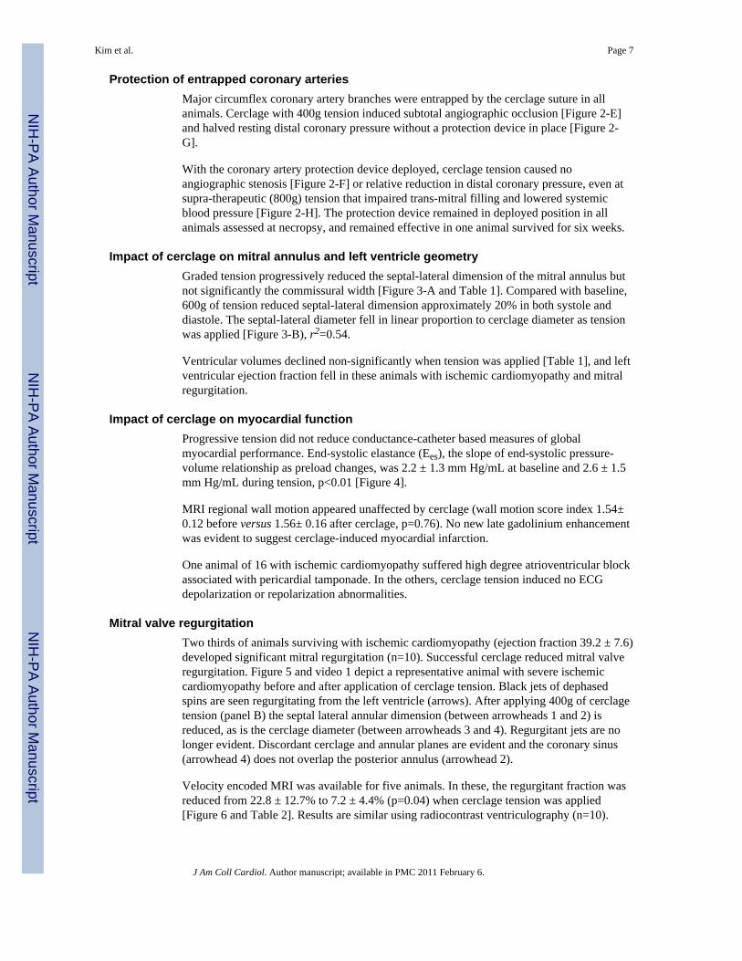
Protection of entrapped coronary arteriesMajor circumflex coronary artery branches were entrapped by the cerclage suture in allanimals. Cerclage with 400g tension induced subtotal angiographic occlusion [Figure 2-E]and halved resting distal coronary pressure without a protection device in place [Figure 2-G].
With the coronary artery protection device deployed, cerclage tension caused noangiographic stenosis [Figure 2-F] or relative reduction in distal coronary pressure, even atsupra-therapeutic (800g) tension that impaired trans-mitral filling and lowered systemicblood pressure [Figure 2-H]. The protection device remained in deployed position in allanimals assessed at necropsy, and remained effective in one animal survived for six weeks.
Impact of cerclage on mitral annulus and left ventricle geometryGraded tension progressively reduced the septal-lateral dimension of the mitral annulus butnot significantly the commissural width [Figure 3-A and Table 1]. Compared with baseline,600g of tension reduced septal-lateral dimension approximately 20% in both systole anddiastole. The septal-lateral diameter fell in linear proportion to cerclage diameter as tensionwas applied [Figure 3-B), r2=0.54.
Ventricular volumes declined non-significantly when tension was applied [Table 1], and leftventricular ejection fraction fell in these animals with ischemic cardiomyopathy and mitralregurgitation.
Impact of cerclage on myocardial functionProgressive tension did not reduce conductance-catheter based measures of globalmyocardial performance. End-systolic elastance (Ees), the slope of end-systolic pressure-volume relationship as preload changes, was 2.2 ± 1.3 mm Hg/mL at baseline and 2.6 ± 1.5mm Hg/mL during tension, p<0.01 [Figure 4].
MRI regional wall motion appeared unaffected by cerclage (wall motion score index 1.54±0.12 before versus 1.56± 0.16 after cerclage, p=0.76). No new late gadolinium enhancementwas evident to suggest cerclage-induced myocardial infarction.
One animal of 16 with ischemic cardiomyopathy suffered high degree atrioventricular blockassociated with pericardial tamponade. In the others, cerclage tension induced no ECGdepolarization or repolarization abnormalities.
Mitral valve regurgitationTwo thirds of animals surviving with ischemic cardiomyopathy (ejection fraction 39.2 ± 7.6)developed significant mitral regurgitation (n=10). Successful cerclage reduced mitral valveregurgitation. Figure 5 and video 1 depict a representative animal with severe ischemiccardiomyopathy before and after application of cerclage tension. Black jets of dephasedspins are seen regurgitating from the left ventricle (arrows). After applying 400g of cerclagetension (panel B) the septal lateral annular dimension (between arrowheads 1 and 2) isreduced, as is the cerclage diameter (between arrowheads 3 and 4). Regurgitant jets are nolonger evident. Discordant cerclage and annular planes are evident and the coronary sinus(arrowhead 4) does not overlap the posterior annulus (arrowhead 2).
Velocity encoded MRI was available for five animals. In these, the regurgitant fraction wasreduced from 22.8 ± 12.7% to 7.2 ± 4.4% (p=0.04) when cerclage tension was applied[Figure 6 and Table 2]. Results are similar using radiocontrast ventriculography (n=10).
Kim et al. Page 7
J Am Coll Cardiol. Author manuscript; available in PMC 2011 February 6.
NIH
-PA Author Manuscript
NIH
-PA Author Manuscript
NIH
-PA Author Manuscript

In preliminary experiments, three animals were survived for three weeks or more aftercerclage without recurrent mitral regurgitation or evident myocardial erosion.
Figure 5-C–D and video 2 show simultaneous tagged and color-flow MRI in an animal withmitral regurgitation that is reduced by cerclage tension. Posterobasal myocardial thinning,late enhancement, and severe hypokinesis also are evident as expected in this ischemiccardiomyopathy model.
Mitral valve tenting area, a measure of annular dilation and subvalvular traction, wasreduced after application of cerclage tension [Table 2]. The degree of tenting was reduced inproportion to the reduction in cerclage diameter [Figure 3-C]. By virtue of reducing septallateral distance, the posterior displacement of the line of coaptation was reduced by cerclage.
Cerclage tension did not significantly change two-dimensional measures of mitral leafletcurvature and angulation with regard to the annulus (Table 3), which reflect leaflet traction.
Discordant Annular and Cerclage PlanesVariable coronary sinus anatomy contributes to discordant cerclage and mitral annularplanes [Figure 1-F]. In our pig model, the average maximum distance of the coronary sinusto the mitral annulus was 6.6 ± 2.0 mm and the angle of the two planes was 21.8 ± 6.4°.These did not vary significantly throughout the cardiac cycle or during application oftension.
Because the posterior coronary sinus may pass along the atrial aspect of the mitral valveannulus, the vector difference of these discordant planes contributes an apical displacementforce to the cerclage annuloplasty.
Mitral annular geometry and motionCerclage induced conformational changes in the mitral valve annulus (Table 3). Mitralannular area (measured as a three-dimensional surface) fell with application of tension.Mitral annular geometry varied throughout the cardiac cycle [Figure 7]. This cyclicalannular contraction is preserved despite application of cerclage tension, which neverthelessreduced circumference and septal-lateral width. Annular height to commissure width ratio isincreased by cerclage, which alters the annular saddle morphology. Tension acted todisplace the posterior annulus caudally, towards the posterior papillary muscle [Figure 7-E–F, videos 3–4].
Reciprocal constraint of mitral annulus and left ventricular outflow tractCerclage annuloplasty encircles both the mitral annulus and the left ventricular outflow tract[Figure 8] and Table 4. We observed reciprocal constraint of these two structures during thecardiac cycle when cerclage tension was applied. Whereas the cerclage diameter remainsfixed throughout the cardiac cycle, the left ventricular outflow tract diameter enlarges duringsystole and contracts during diastole. Conversely, the constrained septal-lateral dimension ofthe mitral annulus is reduced by the left ventricular outflow tract during systole but notconstrained during diastole. There was no gradient induced across the left ventricularoutflow tract using conventional fluid-filled catheters, nor was aortic regurgitation induced.
Anatomic suitability of humansWe reviewed pressurized coronary venous angiograms in 10 patients with congestive heartfailure undergoing cardiac resynchronization therapy. Eight had evident proximal septalcoronary veins arising from the great cardiac vein that appear suitable for cerclage [Figure9]. The other two had inadequate angulation to view septal perforator veins.
Kim et al. Page 8
J Am Coll Cardiol. Author manuscript; available in PMC 2011 February 6.
NIH
-PA Author Manuscript
NIH
-PA Author Manuscript
NIH
-PA Author Manuscript
We also reviewed 29 sequential CT angiograms in a coronary teaching file. The mean agewas 53.2 ± 12.3 years, 79% were male. Of these, 24 (83%) had proximal septal perforatorveins that appear suitable for cerclage.
DiscussionWe have developed a novel procedure for catheter-based mitral cerclage annuloplasty. Wehave shown circumferential tension can be introduced near the mitral annulus plane viabasal septal perforator veins in swine; that attainable tension reduces annular circumferenceand septal-lateral diameter; that annuloplasty does not appear to alter measures of globalmyocardial performance; that a simple device protects entrapped coronary artery brancheseven during supra-therapeutic annuloplasty tension; and in a preliminary review that humansappear to have comparable coronary septal perforator vein anatomy.
We have also shown that cerclage annuloplasty immediately attenuates functional mitralvalve regurgitation in a clinically-relevant animal model of ischemic cardiomyopathycharacterized by left ventricular dysfunction, mitral annular dilation and posterior papillarymuscle traction. Qualitative and quantitative measures of mitral regurgitation are reducedimmediately after cerclage. Magnetic resonance imaging has provided insight into themechanism of cerclage action. First, flexible cerclage reduces annular size while preservingannular motion. Second, in this model, the cerclage suture folds the annulus and displacesthe posterior annulus caudally, apparently to accommodate posterior papillary traction.Third, cerclage introduces reciprocal constraint to the left ventricular outflow tract andmitral annulus such that systolic ejection enhances anterior mitral leaflet coaptation.
Anatomic and geometric factors appear to limit innovative approaches to transcathetercoronary-sinus-based annuloplasty reported previously. First, the coronary sinus oftencourses along the left atrial wall far from the mitral annular plane. Second, the coronarysinus usually entraps underlying major coronary artery branches, so annular compressioncan induce myocardial ischemia. Third, non-circumferential coronary sinus annuloplastydevices may limit annular shortening to only a small arc of annular circumference.
Cerclage annuloplasty addresses these shortcomings. The crossing planes of the mitralannulus and the cerclage trajectory [Figure 1-F] permit effective reduction of the septal-lateral dimension even though the coronary sinus plane does not parallel the annulus. Ourprotection device averts coronary artery compression, and may have value in other coronary-sinus based annuloplasty approaches. Circumferential cerclage constrains the annulusincluding the fibrous trigones, unlike other coronary sinus-based devices.
AnnuloplastyFunctional mitral regurgitation is a manifestation of severe myocardial dysfunction(1)associated with worse symptoms and prognosis. Global and regional myocardial dysfunctionand remodeling contribute to functional mitral regurgitation through annular dilation,traction by displaced and elongated chordal-papillary apparatus (usually posterior), regionalleft ventricular dyssynchrony, and deleterious compensatory loading conditions. Non-surgical treatment may include cardiac resynchronization therapy, beta adrenergic blockade,and angiotensin converting enzyme inhibitors. Surgical repair for organic mitralregurgitation has been extended to patients with cardiomyopathy and normal mitral leaflets.Bolling and colleagues(37) advocate “undersized” annuloplasty. Additional surgical optionsinclude papillary-chordal repair, reinforcement, relocation, ligation, or bowtie constraint ofthe leaflets (38,39). Myocardial reduction procedures or constraining devices also are underdevelopment (2,4).
Kim et al. Page 9
J Am Coll Cardiol. Author manuscript; available in PMC 2011 February 6.
NIH
-PA Author Manuscript
NIH
-PA Author Manuscript
NIH
-PA Author Manuscript

The choice of rigid or flexible annuloplasty devices remains controversial. Normal heartsexhibit cyclic contraction of the mitral valve annulus which may enhance leafletcoaptation(40,41). Preserved annular contraction is associated with a smaller effectiveregurgitant orifice (32,40). Rigid annuloplasty rings impede mitral annular contraction andtilting. Partial bands, flexible rings, or open suture annuloplasty(42) preserve annulardynamics and have been advocated as a more “physiologic” annuloplasty associated withlower trans-valvular gradients. However, flexible annuloplasty rings may contribute to laterecurrence of mitral regurgitation compared with rigid rings (43).
In some reports(31) rigid annuloplasty exacerbates leaflet tethering by displacing theposterior annulus further away from the papillary muscles. Rigid rings have been developedto compensate for the P2–P3 leaflet tethering by enforcing an apical “dip” in the posteriorannulus(44) much like we observe after cerclage. The saddle morphology of the normalmitral annulus may confer a mechanical advantage of reduced leaflet stress (45,46) that maybe reduced by annular flattening in functional mitral regurgitation(33,47). Like flexibleannuloplasty and suture annuloplasty(42), cerclage preserves annular dynamics and restoresannular height. Like newer rigid annuloplasty devices, cerclage serendipitously redistributesthe posterior segment to accommodate apical tethering. However, cerclage appears to inducea more complex shape than the natural annular hyperbolic paraboloid.
Our measures of leaflet configuration and leaflet traction are similar to those reported inpatients undergoing surgical repair of ischemic mitral valve regurgitation(31). In that report,persistent posterior leaflet traction (wide α2) corresponded with failed surgical repair. Aftercerclage, we found the angular relation of the posterior leaflet with the annulus to becomparably narrow. In our experiments, the length of the coaptation line increasednonsignificantly.
Initially we were concerned that entrapment of the left ventricular outflow tract, inevitablewith a trans-septal venous cerclage trajectory, might induce dynamic left ventricular outflowtract obstruction. On the contrary, we found reciprocal enlargement of the entrained leftventricular outflow tract and reduction of septal-lateral distance by cerclage during systole.This interaction improved mitral leaflet coaptation during systole and relaxed the mitralorifice during diastolic filling. Similar aorto-mitral interaction has been reported in healthysheep(48), but was not evident in our experiments until cerclage tension was applied.
Mild-moderate mitral regurgitation is hard to measure, especially using magnetic resonanceimaging (49), which for clinically relevant examinations tends to have inferior timeresolution and leaflet visualization. The severity of mitral regurgitation varies with loadingconditions(50), further confounding measurement. Echocardiography in swine has provenunsuitable in our laboratories because of inadequate imaging windows. We therefore usedvelocity-encoded MRI with slice-tracking to measure through-plane regurgitation across themoving mitral valve plane(51).
Imaging GuidanceBecause neither conventional X-ray nor ultrasound alone satisfactorily depicts myocardialstructures and devices during cerclage, we used fusion image guidance. We superimposedMRI-derived roadmaps onto live X-ray, using a system that compensates automatically forchanges in table position or gantry orientation(28).
Failure modes and limitationsThis is an early experience with a new procedure using largely off-the-shelf catheter tools.We considered several potential failure modes.
Kim et al. Page 10
J Am Coll Cardiol. Author manuscript; available in PMC 2011 February 6.
NIH
-PA Author Manuscript
NIH
-PA Author Manuscript
NIH
-PA Author Manuscript

Cerclage suture applies non-physiological centripetal force to multiple structures and mayrisk chronic erosion. Erosion does not appear limiting after other tangential myocardialsutures including “Fontan” endoventricular pursestring and Paneth-Burr mitral annuloplasty.In general erosion is related to suture tension and tissue friability and inversely related tosuture diameter and time to scar formation. To mitigate the local force and erosion risk, weselected a cerclage suture significantly larger (0.8mm versus 0.3mm) than 2-0 suturecommonly used in myocardial applications with- or without pledgets. No erosion wasevident in a small number of animals survived after cerclage. These considerations must befurther tested in survival experiments. Guidewire-based cerclage traversal of theinterventricular septum is a “blunt dissection” that has not evidently entered or interruptedvisible septal perforator arteries to date. Right ventricular cerclage reenters the coronarysinus ostium across the tricuspid commissure and may encroach on the Koch triangle butgenerally not its apex, where the compact atrioventricular nodal is located.
Right ventricular cerclage requires care to avoid tricuspid valve injury. Chordal entrapmentcan be avoided by deflecting the guidewire immediately into the pulmonary artery uponright ventricular reentry. Leaflet entrapment was observed in one animal with variant septalleaflet anatomy and ventricular septal defect. There was no leaflet entrapment when cerclagesuture crossed “normal” septal tricuspid commissures, which should be identifiable oncardiac CT or MRI. There is precedent for chronic implantation of pacemaker anddefibrillator leads across the tricuspid valve, albeit not under intentional tension. Tricuspidvalve function does not appear affected by right ventricular cerclage (data not shown) withtension across the septal tricuspid commissure.
By contrast, right atrial cerclage avoids the tricuspid valve entirely. Right atrial cerclage ismore technically demanding, and was associated with conduction block in one animal.Further data are necessary to understand the interaction of right atrial cerclage and normaland pathologic conduction tissue(52).
While cerclage does not appear possible if the great cardiac vein is not connected to thecoronary sinus, in one case we were able to complete cerclage even without an evident basalseptal perforator vein. Incorrect guidewire traversal of, for example, a right ventricular freewall, likely can be avoided with procedure experience and enhanced image guidance.
We have shown that compression of entrapped circumflex coronary artery branches can beaverted by implanting a simple protection device along the cerclage suture. We aredeveloping a more sophisticated low-profile tension-fixation device.
We are not able to generate severe functional mitral regurgitation in our porcine model ofischemic cardiomyopathy. Regurgitation severity may have been reduced by our use ofisoflurane anesthesia(53).
The durability of our short term findings is not known. The coronary sinus is typicallyconnected to the mitral annulus by non-fibrous atrial myocardium(54). Chronic remodelingof this sino-annular tissue might contribute to late failure of cerclage annuloplasty.Conversely, intertrigonal remodeling may be less likely after circumferential cerclage,compared with flexible annuloplasty bands, which are attached to the trigones only.
Mitral valve repair may not benefit patients with cardiomyopathy(55–58), even though theperioperative mortality may be low in experienced hands. Surgical repair is usuallyperformed with concomitant myocardial revascularization. Whether primary mitral valverepair improves symptoms or outcome remains to be tested(59). Moreover, we cannotpredict where cerclage annuloplasty, if it proves technically successful in humans, would fitinto a therapeutic armamentarium against heart failure with mitral regurgitation.
Kim et al. Page 11
J Am Coll Cardiol. Author manuscript; available in PMC 2011 February 6.
NIH
-PA Author Manuscript
NIH
-PA Author Manuscript
NIH
-PA Author Manuscript

Conceivably it could be combined with effective pharmacologic and cardiacresynchronization treatments, and it would not likely interfere with subsequent surgicalvalve or myocardial procedures. Cerclage might be used as an adjunct to catheter-basedAlfieri-type bowtie repair which, without annuloplasty, may generate excessive stitch andleaflet tension(60). Early repair of ischemic mitral regurgitation may reverse deleteriousremodeling in some animal models(61) but not others(62).
ConclusionTranscatheter cerclage annuloplasty delivers circumferential annular tension around themitral annulus and left ventricular outflow tract. Cerclage shrinks the mitral annulus andimmediately abates mitral regurgitation in an animal model of chronic ischemiccardiomyopathy. MRI of annular configuration and dynamics suggests cerclage acts bymore than reducing annular size. Cyclical annular motion is preserved and height restored.The posterior annulus is displaced toward the posterior papillary apparatus. Reciprocalconstraint of the mitral annulus and the left ventricular outflow tract appears to contribute tomitral leaflet coaptation. Entrapped coronary arteries can be protected from extrinsiccompression using a simple arch-like device implanted over the cerclage suture. Otherpotential failure modes appear non-limiting. A preliminary review of human venograms andCT angiograms suggest a majority have suitable venous anatomy. Further experiments willtest the longer-term impact of this approach.
Supplementary MaterialRefer to Web version on PubMed Central for supplementary material.
AcknowledgmentsWe are grateful to Katherine Lucas and Joni Taylor for animal experiments, to David Bush and Edward Shapiro ofJohns Hopkins Bayview for anonymized CT images, to Michael Lee and Raj Makkar of Cedars-Sinai foranonymized coronary venograms, and to Lydia Kibiuk of NIH Medical Arts for illustrations. Siemens CorporateResearch supported and Christine Lorenz mentored two graduate students (MS and Akin Yucetas) who contributedto MRI-X-ray registration and investigational velocity-encoded MRI techniques.
Abbreviations
CT Computed tomography
MRI Magnetic resonance imaging
SSFP Steady state free precession MRI
TE MRI echo time
TR MRI repetition time
XFM X-ray fused with MRI
References1. Levine RA, Schwammenthal E. Ischemic mitral regurgitation on the threshold of a solution: from
paradoxes to unifying concepts. Circulation 2005;112:745–58. [PubMed: 16061756]2. Fedak PW, McCarthy PM, Bonow RO. Evolving concepts and technologies in mitral valve repair.
Circulation 2008;117:963–74. [PubMed: 18285577]3. Chen FY, Adams DH, Aranki SF, et al. Mitral valve repair in cardiomyopathy. Circulation
1998;98:II124–7. [PubMed: 9852893]
Kim et al. Page 12
J Am Coll Cardiol. Author manuscript; available in PMC 2011 February 6.
NIH
-PA Author Manuscript
NIH
-PA Author Manuscript
NIH
-PA Author Manuscript

4. Acker MA, Bolling S, Shemin R, et al. Mitral valve surgery in heart failure: insights from the AcornClinical Trial. J Thorac Cardiovasc Surg 2006;132:568–77. 577, e1–4. [PubMed: 16935112]
5. Liddicoat JR, Mac Neill BD, Gillinov AM, et al. Percutaneous mitral valve repair: a feasibility studyin an ovine model of acute ischemic mitral regurgitation. Catheter Cardiovasc Interv 2003;60:410–6. [PubMed: 14571496]
6. Kaye DM, Byrne M, Alferness C, Power J. Feasibility and short-term efficacy of percutaneousmitral annular reduction for the therapy of heart failure-induced mitral regurgitation. Circulation2003;108:1795–7. [PubMed: 14530194]
7. Maniu CV, Patel JB, Reuter DG, et al. Acute and chronic reduction of functional mitralregurgitation in experimental heart failure by percutaneous mitral annuloplasty. J Am Coll Cardiol2004;44:1652–61. [PubMed: 15489099]
8. Daimon M, Shiota T, Gillinov AM, et al. Percutaneous mitral valve repair for chronic ischemicmitral regurgitation: a real-time three-dimensional echocardiographic study in an ovine model.Circulation 2005;111:2183–9. [PubMed: 15851597]
9. Duffy SJ, Federman J, Farrington C, Reuter DG, Richardson M, Kaye DM. Feasibility and short-term efficacy of percutaneous mitral annular reduction for the therapy of functional mitralregurgitation in patients with heart failure. Catheter Cardiovasc Interv 2006;68:205–10. [PubMed:16817176]
10. Webb JG, Harnek J, Munt BI, et al. Percutaneous transvenous mitral annuloplasty: initial humanexperience with device implantation in the coronary sinus. Circulation 2006;113:851–5. [PubMed:16461812]
11. Dubreuil O, Basmadjian A, Ducharme A, et al. Percutaneous mitral valve annuloplasty forischemic mitral regurgitation: first in man experience with a temporary implant. CatheterCardiovasc Interv 2007;69:1053–61. [PubMed: 17525965]
12. Rogers JH, Macoviak JA, Rahdert DA, Takeda PA, Palacios IF, Low RI. Percutaneous septal sinusshortening: a novel procedure for the treatment of functional mitral regurgitation. Circulation2006;113:2329–34. [PubMed: 16682615]
13. Palacios IF, Condado JA, Brandi S, et al. Safety and feasibility of acute percutaneous septal sinusshortening: First-in-human experience. Catheter Cardiovasc Interv 2007;69:513–8. [PubMed:17323357]
14. Pedersen WR, Block P, Leon M, et al. iCoapsys mitral valve repair system: Percutaneousimplantation in an animal model. Catheter Cardiovasc Interv 2008;72:125–31. [PubMed:18561162]
15. Hlavka, EJ.; Podmore, JL.; Spence, PA., inventors. Method and apparatus for catheter-basedannuloplasty using local plications. US patent. 6718985. 2004 Apr 13. Mitralign, assignee
16. Hung J, Solis J, Guerrero JL, et al. A novel approach for reducing ischemic mitral regurgitation byinjection of a polymer to reverse remodel and reposition displaced papillary muscles. Circulation2008;118:S263–9. [PubMed: 18824765]
17. St Goar FG, Fann JI, Komtebedde J, et al. Endovascular edge-to-edge mitral valve repair: short-term results in a porcine model. Circulation 2003;108:1990–3. [PubMed: 14530193]
18. Fann JI, St Goar FG, Komtebedde J, et al. Beating heart catheter-based edge-to-edge mitral valveprocedure in a porcine model: efficacy and healing response. Circulation 2004;110:988–93.[PubMed: 15302782]
19. Feldman T, Wasserman HS, Herrmann HC, et al. Percutaneous mitral valve repair using the edge-to-edge technique: six-month results of the EVEREST Phase I Clinical Trial. J Am Coll Cardiol2005;46:2134–40. [PubMed: 16325053]
20. Naqvi TZ, Buchbinder M, Zarbatany D, et al. Beating-heart percutaneous mitral valve repair usinga transcatheter endovascular suturing device in an animal model. Catheter Cardiovasc Interv2007;69:525–31. [PubMed: 17323355]
21. Choure AJ, Garcia MJ, Hesse B, et al. In vivo analysis of the anatomical relationship of coronarysinus to mitral annulus and left circumflex coronary artery using cardiac multidetector computedtomography: implications for percutaneous coronary sinus mitral annuloplasty. J Am Coll Cardiol2006;48:1938–45. [PubMed: 17112981]
Kim et al. Page 13
J Am Coll Cardiol. Author manuscript; available in PMC 2011 February 6.
NIH
-PA Author Manuscript
NIH
-PA Author Manuscript
NIH
-PA Author Manuscript

22. Maselli D, Guarracino F, Chiaramonti F, Mangia F, Borelli G, Minzioni G. Percutaneous mitralannuloplasty: an anatomic study of human coronary sinus and its relation with mitral valveannulus and coronary arteries. Circulation 2006;114:377–80. [PubMed: 16864726]
23. Tops LF, Van de Veire NR, Schuijf JD, et al. Noninvasive evaluation of coronary sinus anatomyand its relation to the mitral valve annulus: implications for percutaneous mitral annuloplasty.Circulation 2007;115:1426–32. [PubMed: 17353434]
24. Tops LF, Van de Veire NR, Schuijf JD, et al. Noninvasive evaluation of coronary sinus anatomyand Its relation to the mitral valve annulus. Implications for percutaneous mitral annuloplasty.Circulation. 2007
25. Croft LR, Jimenez JH, Gorman RC, Gorman JH 3rd, Yoganathan AP. Efficacy of the edge-to-edgerepair in the setting of a dilated ventricle: an in vitro study. Ann Thorac Surg 2007;84:1578–84.[PubMed: 17954065]
26. Glover RP, Davila JC. Surgical treatment of mitral insufficiency by total circumferential purse-string suture of the mitral ring. Circulation 1957;15:661–81. [PubMed: 13427121]
27. Swindle MM, Horneffer PJ, Gardner TJ, et al. Anatomic and anesthetic considerations inexperimental cardiopulmonary surgery in swine. Lab Anim Sci 1986;36:357–61. [PubMed:3534438]
28. de Silva R, Gutierrez LF, Raval AN, McVeigh ER, Ozturk C, Lederman RJ. X-ray fused withmagnetic resonance imaging (XFM) to target endomyocardial injections: validation in a swinemodel of myocardial infarction. Circulation 2006;114:2342–50. [PubMed: 17101858]
29. Gutiérrez LF, Silva R, Ozturk C, et al. Technology preview: X-ray fused with magnetic resonanceduring invasive cardiovascular procedures. Catheter Cardiovasc Interv 2007;70:773–82. [PubMed:18022851]
30. Schiller NB, Shah PM, Crawford M, et al. Recommendations for quantitation of the left ventricleby two-dimensional echocardiography. American Society of Echocardiography Committee onStandards, Subcommittee on Quantitation of Two-Dimensional Echocardiograms. J Am SocEchocardiogr 1989;2:358–67. [PubMed: 2698218]
31. Zhu F, Otsuji Y, Yotsumoto G, et al. Mechanism of persistent ischemic mitral regurgitation afterannuloplasty: importance of augmented posterior mitral leaflet tethering. Circulation2005;112:I396–401. [PubMed: 16159853]
32. Yiu SF, Enriquez-Sarano M, Tribouilloy C, Seward JB, Tajik AJ. Determinants of the degree offunctional mitral regurgitation in patients with systolic left ventricular dysfunction: A quantitativeclinical study. Circulation 2000;102:1400–6. [PubMed: 10993859]
33. Kaji S, Nasu M, Yamamuro A, et al. Annular geometry in patients with chronic ischemic mitralregurgitation: three-dimensional magnetic resonance imaging study. Circulation 2005;112:I409–14. [PubMed: 16159855]
34. Dujardin KS, Enriquez-Sarano M, Bailey KR, Nishimura RA, Seward JB, Tajik AJ. Grading ofmitral regurgitation by quantitative Doppler echocardiography: calibration by left ventricularangiography in routine clinical practice. Circulation 1997;96:3409–15. [PubMed: 9396435]
35. Sampath S, Kim JH, Lederman RJ, McVeigh ER. Simultaneous imaging of myocardial motion andchamber blood flow with SPAMM n’ EGGS (spatial modulation of magnetization with encodedgradients for gauging speed). Journal of Magnetic Resonance Imaging 2008;27:809–817.[PubMed: 18383258]
36. Steendijk P, Staal E, Jukema JW, Baan J. Hypertonic saline method accurately determines parallelconductance for dual-field conductance catheter. Am J Physiol Heart Circ Physiol2001;281:H755–63. [PubMed: 11454580]
37. Bolling SF, Pagani FD, Deeb GM, Bach DS. Intermediate-term outcome of mitral reconstruction incardiomyopathy. J Thorac Cardiovasc Surg 1998;115:381–6. [PubMed: 9475533]
38. Savage, EB.; Bolling, SF. Atlas of mitral valve repair. Philadelphia: Lippincott Williams &Wilkins; 2006.
39. Borger MA, Alam A, Murphy PM, Doenst T, David TE. Chronic ischemic mitral regurgitation:repair, replace or rethink? Ann Thorac Surg 2006;81:1153–61. [PubMed: 16488757]
Kim et al. Page 14
J Am Coll Cardiol. Author manuscript; available in PMC 2011 February 6.
NIH
-PA Author Manuscript
NIH
-PA Author Manuscript
NIH
-PA Author Manuscript

40. Tsakiris AG, Von Bernuth G, Rastelli GC, Bourgeois MJ, Titus JL, Wood EH. Size and motion ofthe mitral valve annulus in anesthetized intact dogs. J Appl Physiol 1971;30:611–8. [PubMed:5576071]
41. Dent JM, Spotnitz WD, Nolan SP, Jayaweera AR, Glasheen WP, Kaul S. Mechanism of mitralleaflet excursion. Am J Physiol 1995;269:H2100–8. [PubMed: 8594922]
42. Tibayan FA, Rodriguez F, Liang D, Daughters GT, Ingels NB Jr, Miller DC. Paneth sutureannuloplasty abolishes acute ischemic mitral regurgitation but preserves annular and leafletdynamics. Circulation 2003;108(Suppl 1):II128–33. [PubMed: 12970221]
43. Spoor MT, Geltz A, Bolling SF. Flexible versus nonflexible mitral valve rings for congestive heartfailure: differential durability of repair. Circulation 2006;114:I67–71. [PubMed: 16820648]
44. Daimon M, Fukuda S, Adams DH, et al. Mitral valve repair with Carpentier-McCarthy-AdamsIMR ETlogix annuloplasty ring for ischemic mitral regurgitation: early echocardiographic resultsfrom a multi-center study. Circulation 2006;114:I588–93. [PubMed: 16820643]
45. Salgo IS, Gorman JH 3rd, Gorman RC, et al. Effect of annular shape on leaflet curvature inreducing mitral leaflet stress. Circulation 2002;106:711–7. [PubMed: 12163432]
46. Jimenez JH, Liou SW, Padala M, et al. A saddle-shaped annulus reduces systolic strain on thecentral region of the mitral valve anterior leaflet. J Thorac Cardiovasc Surg 2007;134:1562–8.[PubMed: 18023684]
47. Tibayan FA, Rodriguez F, Langer F, et al. Annular remodeling in chronic ischemic mitralregurgitation: ring selection implications. Ann Thorac Surg 2003;76:1549–54. discussion 1554–5.[PubMed: 14602284]
48. Timek TA, Green GR, Tibayan FA, et al. Aorto-mitral annular dynamics. Ann Thorac Surg2003;76:1944–50. [PubMed: 14667619]
49. Kilner PJ, Gatehouse PD, Firmin DN. Flow measurement by magnetic resonance: a unique assetworth optimising. J Cardiovasc Magn Reson 2007;9:723–8. [PubMed: 17613655]
50. Yoran C, Yellin EL, Becker RM, Gabbay S, Frater RW, Sonnenblick EH. Dynamic aspects ofacute mitral regurgitation: effects of ventricular volume, pressure and contractility on the effectiveregurgitant orifice area. Circulation 1979;60:170–6. [PubMed: 445720]
51. Kozerke S, Schwitter J, Pedersen EM, Boesiger P. Aortic and mitral regurgitation: quantificationusing moving slice velocity mapping. J Magn Reson Imaging 2001;14:106–12. [PubMed:11477667]
52. Anderson RH, Ho SY, Becker AE. The surgical anatomy of the conduction tissues. Thorax1983;38:408–20. [PubMed: 6348996]
53. Grewal KS, Malkowski MJ, Piracha AR, et al. Effect of general anesthesia on the severity of mitralregurgitation by transesophageal echocardiography. Am J Cardiol 2000;85:199–203. [PubMed:10955377]
54. Hueb AC, Jatene FB, Moreira LF, Pomerantzeff PM, Kallas E, de Oliveira SA. Ventricularremodeling and mitral valve modifications in dilated cardiomyopathy: new insights from anatomicstudy. J Thorac Cardiovasc Surg 2002;124:1216–24. [PubMed: 12447190]
55. Gorman JH 3rd, Gorman RC. Mitral valve surgery for heart failure: a failed innovation? SeminThorac Cardiovasc Surg 2006;18:135–8. [PubMed: 17157234]
56. Wu AH, Aaronson KD, Bolling SF, Pagani FD, Welch K, Koelling TM. Impact of mitral valveannuloplasty on mortality risk in patients with mitral regurgitation and left ventricular systolicdysfunction. J Am Coll Cardiol 2005;45:381–7. [PubMed: 15680716]
57. Mihaljevic T, Lam BK, Rajeswaran J, et al. Impact of mitral valve annuloplasty combined withrevascularization in patients with functional ischemic mitral regurgitation. J Am Coll Cardiol2007;49:2191–201. [PubMed: 17543639]
58. Braun J, van de Veire NR, Klautz RJ, et al. Restrictive mitral annuloplasty cures ischemic mitralregurgitation and heart failure. Ann Thorac Surg 2008;85:430–6. [PubMed: 18222238]
59. Adams DH, Anyanwu A. Pitfalls and limitations in measuring and interpreting the outcomes ofmitral valve repair. J Thorac Cardiovasc Surg 2006;131:523–9. [PubMed: 16515900]
60. Timek TA, Nielsen SL, Lai DT, et al. Effect of chronotropy and inotropy on stitch tension in theedge-to-edge mitral repair. Circulation 2007;116:I276–81. [PubMed: 17846317]
Kim et al. Page 15
J Am Coll Cardiol. Author manuscript; available in PMC 2011 February 6.
NIH
-PA Author Manuscript
NIH
-PA Author Manuscript
NIH
-PA Author Manuscript

61. Beeri R, Yosefy C, Guerrero JL, et al. Early repair of moderate ischemic mitral regurgitationreverses left ventricular remodeling: a functional and molecular study. Circulation 2007;116:I288–93. [PubMed: 17846319]
62. Guy TS 4th, Moainie SL, Gorman JH 3rd, et al. Prevention of ischemic mitral regurgitation doesnot influence the outcome of remodeling after posterolateral myocardial infarction. J Am CollCardiol 2004;43:377–83. [PubMed: 15013117]
Kim et al. Page 16
J Am Coll Cardiol. Author manuscript; available in PMC 2011 February 6.
NIH
-PA Author Manuscript
NIH
-PA Author Manuscript
NIH
-PA Author Manuscript

Figure 1.Schematic, imaging guidance, and necropsy of cerclage annuloplasty. (A) shows the mitralannulus from the cardiac apex and (B) with free walls of right atrium and ventricle removed.A guidewire through the coronary sinus enters a basal septal perforator vein and traverses ashort distance of septal myocardium. Wire 1 follows a right ventricular cerclage trajectoryinto the right ventricular outflow tract, and wire 2 a longer trajectory to reenter the rightatrium directly. The guidewire is replaced with a suture and tension applied to both ends andfixed near the coronary sinus ostium. (C–E) shows XFM procedure guidance. MRI-derivedcontours include LV and RV endocardium (blue and yellow), LV epicardium (green), andaortic root (red). (D) shows live X-ray fluoroscopy and (E) the corresponding XFM display.The guidewire tip (white arrow) crosses septal myocardium and reenters the right ventricle.Registration is maintained even when the table or gantry move. (F) shows the discordantplanes of the mitral annulus (blue) and cerclage annuloplasty (red). Necropsy findings areshown immediately after right ventricular (G) and right atrial (H) cerclage with the RV freewall removed. In (G) the suture (arrow) emerges from the septum and returns to the rightatrium across a tricuspid commissure. In (H) a suture emerges (arrow) near the cavo-tricuspid isthmus, alongside the coronary sinus end of the same suture (dotted arrow). PanelsA,B,F courtesy of Lydia Kibiuk, NIH Medical Arts.
Kim et al. Page 17
J Am Coll Cardiol. Author manuscript; available in PMC 2011 February 6.
NIH
-PA Author Manuscript
NIH
-PA Author Manuscript
NIH
-PA Author Manuscript

Figure 2.Coronary artery entrapment and protection. (A,B) A typical great cardiac vein configurationpassing outside a circumflex artery branch. (C) Cerclage would compress the underlyingartery. (D) A protection device along the cerclage suture redistributes compressive forcesaway from coronary artery. (E–H) circumflex coronary artery pressure during cerclagetension without (E,G) and with (F,H) a protection device in place. (E) Angiographicstenosis (arrow) induced by cerclage and (F) the same segment during cerclage tension witha protection device (dashed arrow) in place. (G,H) Distal coronary artery pressure (Pd,depicted in green, axis on left, mm), the aortic pressure (Pa) in red, and their ratio in yellow(axis on right, displayed as fractional flow reserve). Without a protection device (G), thedistal coronary pressure falls by more than half when cerclage tension (400g) is applied. (H)With the protection device in place, there is no distal pressure drop after cerclage tension isintroduced (dotted arrow) until tension is sufficiently high (solid arrow) to impede mitralinflow. Panels A–D courtesy of Lydia Kibiuk, NIH Medical Arts.
Kim et al. Page 18
J Am Coll Cardiol. Author manuscript; available in PMC 2011 February 6.
NIH
-PA Author Manuscript
NIH
-PA Author Manuscript
NIH
-PA Author Manuscript

Figure 3.Effect of graded tension on annular dimensions and leaflet tenting. (A) Progressivelyincreased cerclage tension reduces the annular septal-lateral dimension, perpendicular to theline of mitral coaptation. (B) With progressive tension, the decline in cerclage diameter isdirectly related to the decline in septal-lateral dimension. (C) Reduced cerclage diameter isdirectly related to the reduction in mitral valve tenting area, an index of mitral regurgitation.
Kim et al. Page 19
J Am Coll Cardiol. Author manuscript; available in PMC 2011 February 6.
NIH
-PA Author Manuscript
NIH
-PA Author Manuscript
NIH
-PA Author Manuscript

Figure 4.Representative dynamic pressure-volume loops before (A) and after (B–D) progressiveapplication of cerclage tension in naïve swine. There is no significant change in the end-diastolic (upper slope) and end-systolic (lower slope) pressure-volume relationships astension is introduced. 600g tension was found to reduce annular circumference sufficientlyto impede transmitral inflow. In this animal cerclage does not acutely alter ventricularvolumes.
Kim et al. Page 20
J Am Coll Cardiol. Author manuscript; available in PMC 2011 February 6.
NIH
-PA Author Manuscript
NIH
-PA Author Manuscript
NIH
-PA Author Manuscript

Figure 5.Mitral regurgitation before (A,C) and after (B,D) application of cerclage tension.Arrowheads 1 and 2 indicate the anterior and posterior mitral annulus, respectively.Arrowheads 3 and 4 indicate the anterior and posterior course of the cerclage annuloplasty.Arrows indicate the twin jets of mitral regurgitation in this MRI in an animal with aregurgitant fraction of 0.43. After tension is applied, the regurgitant fraction fell to 0.08, andjets are no longer visible. Note the anterior displacement of point 4 and its alteredconfiguration in relation to point 2 (animated in video 1). Note also that regurgitant jets ofdephased spins in steady state free precession MRI under-represent mitral regurgitationcompared with echocardiography. (C,D) show combined motion (tagged) and velocity-encoded MRI during systole before (C) and after (D) application of cerclage tension inanother animal, animated in video 2. Mitral regurgitation is evident as a blue jet in (C, blackarrow) and nearly extinguished in (D). The posterior cerclage wire (black spot indicated bywhite arrow) is displaced toward the septum when tension is applied. Late gadoliniumenhancement and reduced myocardial contraction are evident from prior posterobasalinfarction.
Kim et al. Page 21
J Am Coll Cardiol. Author manuscript; available in PMC 2011 February 6.
NIH
-PA Author Manuscript
NIH
-PA Author Manuscript
NIH
-PA Author Manuscript

Figure 6.Quantitative (A) and qualitative (B) measures of mitral regurgitation before and afterapplication of cerclage tension.
Kim et al. Page 22
J Am Coll Cardiol. Author manuscript; available in PMC 2011 February 6.
NIH
-PA Author Manuscript
NIH
-PA Author Manuscript
NIH
-PA Author Manuscript
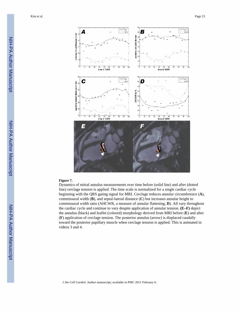
Figure 7.Dynamics of mitral annulus measurements over time before (solid line) and after (dottedline) cerclage tension is applied. The time scale is normalized for a single cardiac cyclebeginning with the QRS gating signal for MRI. Cerclage reduces annular circumference (A),commissural width (B), and septal-lateral distance (C) but increases annular height tocommissural width ratio (AHCWR, a measure of annular flattening, D). All vary throughoutthe cardiac cycle and continue to vary despite application of annular tension. (E–F) depictthe annulus (black) and leaflet (colored) morphology derived from MRI before (E) and after(F) application of cerclage tension. The posterior annulus (arrow) is displaced caudallytoward the posterior papillary muscle when cerclage tension is applied. This is animated invideos 3 and 4.
Kim et al. Page 23
J Am Coll Cardiol. Author manuscript; available in PMC 2011 February 6.
NIH
-PA Author Manuscript
NIH
-PA Author Manuscript
NIH
-PA Author Manuscript

Figure 8.Reciprocal constraint of the left ventricular outflow tract and mitral annulus after cerclageannuloplasty. The combined diameter of the two structures remains constant throughout thecardiac cycle. During diastole, the anterior mitral leaflet is relatively unconstrained. Duringsystole, the outflow tract enlarges and displaces the anterior mitral valve leaflet posteriorly(* = p<0.01 vs diastole). This appears to enhance leaflet coaptation and valve function.
Kim et al. Page 24
J Am Coll Cardiol. Author manuscript; available in PMC 2011 February 6.
NIH
-PA Author Manuscript
NIH
-PA Author Manuscript
NIH
-PA Author Manuscript

Figure 9.Representative human venograms. (A) A pressurized venogram in a patient undergoingcardiac resynchronization therapy. A basal septal perforator vein was evident (arrow) in all 8patients with evaluable angiograms. (B) A CT angiogram showing a basal septal perforatorvein (arrows) apparently suitable for cerclage.
Kim et al. Page 25
J Am Coll Cardiol. Author manuscript; available in PMC 2011 February 6.
NIH
-PA Author Manuscript
NIH
-PA Author Manuscript
NIH
-PA Author Manuscript

NIH
-PA Author Manuscript
NIH
-PA Author Manuscript
NIH
-PA Author Manuscript
Kim et al. Page 26
Table 1
Impact of cerclage tension on annular and left ventricular dimensions, n=16.
Baseline Tension p value
Mitral annulus
Septal-lateral annular diameter (cm)
systole 3.1 ± 0.4 2.5 ± 0.4 <0.001
diastole 3.4 ± 0.6 2.9 ± 0.5 <0.001
Commissural Width (cm)
systole 2.6 ± 0.3 2.2 ± 0.8 0.41
diastole 2.6 ± 0.4 2.2 ± 1.0 0.28
Cerclage diameter
Cerclage diameter (cm) 6.2 ± 0.8 4.5 ± 0.8 < 0.001
Left ventricle
diastolic LVID (cm) 5.4 ± 0.6 5.3 ± 0.6 0.40
systolic LVID (cm) 4.7 ± 0.5 4.6 ± 0.7 0.45
End-systolic volume (ml) 106 ± 43 98 ± 37 0.22
End-diastolic volume (ml) 172 ± 55 158 ± 52 0.09
Ejection fraction 0.39 ± 0.08 0.34 ± 0.04 0.04
J Am Coll Cardiol. Author manuscript; available in PMC 2011 February 6.

NIH
-PA Author Manuscript
NIH
-PA Author Manuscript
NIH
-PA Author Manuscript
Kim et al. Page 27
Table 2
Measures of mitral valve regurgitation and leaflet function before and after application of cerclage tension(n=10 except for velocity-encoded MRI, n=5).
Baseline Tension p
Mitral regurgitation
Regurgitant fraction by slice-tracking velocity-encoded MRI 22.8 ± 12.7 7.2 ± 4.4 0.04
Categorical mitral regurgitation total (Grade 1–4) 1.7 ± 0.8 0.7 ± 0.5 0.01
Mitral valve coaptation
Mitral valve tenting area (cm2) 2.1 ± 0.5 1.5 ± 0.5 0.01
Posterior displacement of line of coaptation (mm) 25.7 ± 4.0 21.2 ± 4.8 0.01
Leaflet coaptation length (mm) 3.0 ± 2.4 4.8 ± 1.6 0.12
J Am Coll Cardiol. Author manuscript; available in PMC 2011 February 6.

NIH
-PA Author Manuscript
NIH
-PA Author Manuscript
NIH
-PA Author Manuscript
Kim et al. Page 28
Table 3
Leaflet function and annular motion before and after application of cerclage tension in pigs with secondarymitral regurgitation, n=5. Supplemental figure 1 depicts the angles measured.
Baseline Tension p
Mitral leaflet configuration and traction
Anterior leaflet angle with annulus (α1, degrees) 36.6 ± 6.4 34.0 ± 7.9 0.66
Posterior leaflet angle with annulus (α2, degrees) 58.6 ± 21.5 72.2 ± 35.0 0.32
Anterior leaflet curvature (β, degrees) 145 ± 11 150 ± 9.0 0.14
Mitral annular dynamics
Mitral annular area (systolic, cm2) 7.0 ± 1.5 3.7 ± 1.2 0.01
Mitral annular systolic contraction (%) 13.1 ± 11.2 13.3 ± 7.2 0.98
Annular height to commissure width ratio (%) 27.0 ± 2.2 36.8 ± 3.9 0.02
J Am Coll Cardiol. Author manuscript; available in PMC 2011 February 6.

NIH
-PA Author Manuscript
NIH
-PA Author Manuscript
NIH
-PA Author Manuscript
Kim et al. Page 29
Table 4
Enhanced reciprocal constraint of left ventricular outflow tract and mitral valve annulus during application ofcerclage tension, n=10.
Baseline Tension p
Left ventricular outflow tract (LVOT) diameter (cm) Systole 2.1 ± 0.4 2.0 ± 0.4 0.047
Diastole 1.9 ± 0.4 1.5 ± 0.4 <0.001
Difference 11% 30% <0.001
Mitral valve annulus (MVA) diameter (cm) Systole 3.1 ± 0.5 2.5 ± 0.4 <0.001
Diastole 3.4 ± 0.6 2.9 ± 0.5 <0.001
Difference −10% −14% <0.001
Combined LVOT and MVA (cm) Systole 5.2 ± 0.7 4.5 ± 0.6 NS
Diastole 5.3 ± 0.9 4.4 ± 0.7 NS
Difference −3% 1% NS
J Am Coll Cardiol. Author manuscript; available in PMC 2011 February 6.