Embed Size (px)
Citation preview

UNIVERSIDADE DA BEIRA INTERIOR Ciências
miRNA-29 bioseparation and target delivery strategies for Alzheimer’s disease
Patrícia Alexandra Nunes Pereira
Tese para obtenção do Grau de Doutor em
Bioquímica (3º ciclo de estudos)
Orientadora: Prof.ª Doutora Fani Pereira de Sousa Co-orientador(a): Prof.ª Doutora Ana Rita Figueiras
Prof. Doutor Ilídio Correia
Covilhã, abril de 2016

ii

iii
"Whether or not your efforts are smiled upon by fate,
what really matters in the end is to be able to say,
I did what I was able to"
Louis Pasteur

iv

v
Aos meus Pais…
Por todo o amor, apoio incondicional e compreensão
Amo-vos

vi

vii
This work was financed by the Portuguese Foundation for Science and Technology
(SFRH/BD/81914/2011) under the program QREN - POPH - Type 4.1 – Advanced
Training, cofounded by the European Social Fund and by national funds from the
MCTES. Moreover, this work was also supported in the framework of research and
development projects (PTDC/EBB-BIO/114320/2009 and EXPL/BBB-BIO/1056/2012)
and strategic programs (Pest-C/SAU/UI0709/2011 and Pest-OE/SAU/UI0709/2014) co-
founded by the operational program factors of competitiveness – COMPETE (FCOMP-
01-0124-FEDER-041068 – EXPL/QEQ-MED/1068/2013) and co-founded by FEDER funds
(FCOMP-01-0124-FEDER-027560) of COMPETE.
Final version printed in April of 2016

viii

ix
Acknowledgments
First and foremost, I would like to express my sincere gratitude to my supervisors Professor
Fani Sousa, Professor Ana Rita Figueiras and Professor Ilídio Correia, for all the careful
guidance, valuable advice and trust. To Professor Fani Sousa my deepest appreciation for her
valuable scientific knowledge and experiences shared during the last years. I would like to
thanks for encouraging my research and for allowing me to grow as a research scientist. In
addition, I am also grateful for believing in me, for her availability, dedication, help,
motivation, unconditional support in difficult times, by the endless hours of conversation and
especially for her friendship and patience, my very thanks to my " scientific mother "! To my
co-supervisor Professor Ana Rita Figueiras my sincerely appreciation for her invaluable
support, for the continuous guidance, help, availability, dedication, for all encouragement
and especially for her friendship. It was a huge privilege working with you.
I would like to express my sincere gratitude to Professor João Queiroz for all the vast and
valuable scientific knowledge that he transmitted me. I am really thankful for the
availability, dedication and interest that he demonstrated for my work through the
constructive scientific discussions and suggestions made during the development of the same,
which were crucial for their success.
I would like to thanks to Professor Carla Cruz for her friendship, availability, help, support
and interest demonstrated for my work through the scientific expertise provided, as well as
for the trust in me through participation in her scientific projects. I would also like to thanks
to Professor Cláudio Maia for the scientific expertise and valuable contribution throughout
this thesis. To Professor Luís Passarinha, for his friendship, motivation, help and support,
between a coffee and a laugh.
I’m deeply grateful to Fundação para a Ciência e Tecnologia (FCT) for the financial support
through my PhD fellowship (SFRH/BD/81914/2011).
I also thank to all the people involved in Health Sciences Research Centre of the University of
Beira Interior (CICS-UBI), especially to the Biotechnology and Biomolecular Sciences research
group which cooperated in the development of this work. A special acknowledgment to the
laboratory technicians Margarida Carrilho and Sofia Duarte that really cooperated for the
successful development of this work and, especially for all the friendliness, availability,
helpfulness and excellent moments of good humor.
To all my laboratory colleagues, specially Ângela Sousa, Eduarda Coutinho, Rita Martins,
Margarida Almeida, Rita Videira, Helena Marcelino, Ana Martinho, Susana Ferreira and
Filomena Silva, I would like to acknowledge all these people by their teachings, valuable

x
technical help, feedback, unspeakable patience, friendship, advice, support on the not so
good times and willingness that contributed for this work to move forward. Thanks for all the
great moments. Moreover, I am extremely thankful to Joana Tomás, who with her special
expertise provided me with invaluable advice and encouragement. Your patience, support,
friendship and collaboration were crucial for the success of this work, I sincerely thank you.
A very special thanks to Augusto Pedro, companion and friend, who supported me in good
times and bad times, by their critical thinking that contributed significantly to the quality of
this work, as well as, by your availability and help whenever I needed. For all your love,
affection and friendship, and all the confidence placed in me, by the patience, advices and
motivation throughout all the days of intensive work. We managed to overcome more this
stage of our lives together, thank you my love.
Por último, e não menos importante, gostaria de agradecer de uma forma muito especial à
minha família, por todo o amor, confiança e apoio nos tempos mais difíceis durante a minha
formação acadêmica. Aos meus pais, por tornarem isto possível, por toda a vossa bondade e
paciência, por me apoiarem sempre sem nunca duvidar e por entenderem as minhas inúmeras
ausências. Estou-vos eternamente grata. Esta tese tem o vosso apoio incondicional, amor e
carinho. Amo-vos.

xi

xii

xiii
Resumo Alargado
A recente descoberta da tecnologia do RNA de interferência tornou-se numa nova ferramenta
que permite regular seletivamente o padrão de expressão de um ou mais genes, o que pode
ser explorado no âmbito de aplicação terapêutica. Deste modo, os resultados promissores
desta nova abordagem têm vindo a reforçar a investigação relacionada com o RNA, avaliando
e compreendendo os mecanismos celulares em que está envolvido, com o objetivo de o usar
como uma nova classe de produtos bioterapêuticos, de fácil translação e implementação para
o âmbito clínico. Na verdade, o mecanismo celular responsável pelo silenciamento da
expressão génica apresenta um enorme potencial terapêutico que poderá alterar os
tratamentos atualmente disponíveis para diversas patologias, como por exemplo as doenças
neurológicas.
Os microRNAs (miRNAs) constituem uma classe de RNAs de baixo peso molecular e, nos
últimos anos, têm sido cada vez mais reconhecidos como moléculas endógenas reguladoras
em numerosos processos biológicos. Devido à sua especificidade e eficiência, os miRNAs
tornaram-se uma das ferramentas mais utilizadas no silenciamento de genes pelo mecanismo
de RNA de interferência, uma vez que podem bloquear a síntese de proteínas através da
indução da degradação do RNA mensageiro. Assim, os miRNAs podem ser considerados agentes
terapêuticos promissores.
Durante a última década, a hipótese de que os miRNAs podem ser usados como biofármacos
para regular e controlar várias vias envolvidas no desenvolvimento e progressão da doença de
Alzheimer (DA) ganhou consistência, uma vez que estes apresentam um papel crucial em
muitas funções neuronais, tais como diferenciação, plasticidade sináptica, formação da
memória, e normalmente estão sub-expressos em doentes com esta patologia. A doença de
Alzheimer é a forma mais comum de demência e caracteriza-se por uma perda neuronal e
sináptica generalizada, que causa um declínio progressivo e irreversível em diversas funções
cognitivas. Embora, as causas ainda não sejam completamente compreendidas, sabe-se que
esta doença neurodegenerativa está associada ao aparecimento de dois tipos de agregados
proteicos, nomeadamente placas extracelulares beta-amiloides (Aβ) e complexos
neurofibrilares intraneuronais.
A formação das placas de Aβ resulta da clivagem sequencial da proteína percursora amiloide
(APP) pela BACE1 e, posteriormente, pelo complexo gama-secretase dando origem às espécies
Aβ tóxicas. Alguns estudos têm descrito uma relação causal entre a expressão do miR-29 e a
DA, uma vez que a diminuição dos níveis de expressão da família miR-29 tem sido associada
ao aumento da expressão da BACE1 e, consequentemente da formação dos péptidos Aβ em
doentes com DA. Desta forma, a utilização do miR-29 pode proporcionar uma estratégia
terapêutica eficaz na prevenção, controlo da progressão e tratamento desta patologia.

xiv
As terapêuticas baseadas no uso de miRNAs recorrem, na sua maioria, à utilização de miRNAs
sintéticos. Embora a síntese de miRNAs possa ser muito eficiente, verifica-se normalmente a
presença de contaminantes nas amostras de RNA sintetizado o pode levar à inespecificidade
do silenciamento génico. Além disso, por norma, no processo de preparação do RNA sintético
é necessário recorrer à utilização de solventes tóxicos, solventes orgânicos e condições
desnaturantes, que podem comprometer a qualidade e integridade do produto alvo. Por todos
estes motivos, e considerando o objetivo de aplicação terapêutica destes novos produtos
biofarmacêuticos, torna-se evidente a necessidade de desenvolver novos processos eficientes
ou melhorar as metodologias atualmente empregues para a sua preparação. Assim, um dos
desafios mais importantes no desenvolvimento destas estratégias terapêuticas surge com a
necessidade de produzir miRNA com elevado grau de pureza e atividade biológica, de modo a
satisfazer os requisitos necessários à sua aplicação. Deste modo, uma das estratégias que
pode ser aplicada consiste na produção recombinante de biomoléculas utilizando hospedeiros
procarióticos.
Desta forma, o objetivo principal do presente trabalho consiste no desenvolvimento e
implementação de uma plataforma biotecnológica para a produção e purificação de
precursores de miRNAs, em particular o pre-miR-29b (percursor do miR-29), cuja aplicação
visa o silenciamento seletivo de vias endógenas diretamente relacionados com DA,
nomeadamente a BACE1 e os péptidos Aβ. Paralelamente, serão desenvolvidas e
caracterizadas nanopartículas poliméricas para a entrega do pre-miR-29b no citoplasma de
células neuronais de forma seletiva e eficaz, de modo a assegurar o sucesso destas
administrações.
O sistema de expressão recombinante utilizado permitirá, pela primeira vez, a produção e o
isolamento do pre-miR-29b humano na bactéria Rhodovulum sulfidophilum (R. sulfidophilum)
DSM 1374, mantendo a sua atividade biológica. A utilização deste hospedeiro bacteriano é
inovadora e vantajosa, devido à sua capacidade invulgar de secretar os ácidos nucleicos para
o meio de cultura, bem como devido à ausência de ribonucleases no mesmo, permitindo a
obtenção do miRNA de interesse com baixo conteúdo de impurezas bacterianas. A fim de
otimizar a produção e acumulação de miRNAs no espaço extracelular, as condições de
crescimento foram estudadas, nomeadamente no que diz respeito ao efeito da temperatura e
concentração de cloreto de sódio. Os ensaios realizados demonstraram ser possível
desenvolver um protocolo para o crescimento aeróbio da bactéria R. sulfidophilum, na
ausência de luz, a 30ºC, o que resulta num melhoramento do crescimento das células, seguido
de um aumento da produção do pre-miR-29b humano. Neste trabalho foi possível atingir uma
concentração de pre-miR-29b no meio extracelular de aproximadamente 182 µg/L, após 40
horas de crescimento bacteriano e uma concentração de 358 µg/L de pre-miR-29b
intracelular, após 32 horas de fermentação.

xv
Para que seja possível a aplicação terapêutica do pre-miR-29b é necessário ter em conta as
exigências estabelecidas pelas agências reguladoras internacionais, que requerem a produção
de miRNA com elevada qualidade e atividade biológica. Para assegurar esta condição, foram
desenvolvidas novas estratégias de purificação para o pre-miR-29b, baseadas em
cromatografia de afinidade. A fim de alcançar a máxima seletividade e especificidade na
separação do pre-miRNA de outras biomoléculas do hospedeiro (outras espécies de RNA e
proteínas), foram desenvolvidos suportes de afinidade baseados nas interações biológicas que
são estabelecidas a nível celular, usando como ligandos de afinidade, aminoácidos básicos
como a L-lisina e a L-arginina. Na estratégia de purificação com o aminoácido de L-lisina foi
demonstrada pela primeira vez a purificação de pre-miRNA utilizando um gradiente
decrescente de concentração de sulfato de amónio em três passos, o que permitiu explorar
maioritariamente interações hidrofóbicas. Contudo, a necessidade de aplicar elevadas
concentrações de sal pode ser visto como uma desvantagem devido aos custos e ao impacto
ambiental associado ao processo, principalmente no que diz respeito à aplicação ao nível
industrial. De modo a ultrapassar estas limitações foi usada L-arginina como aminoácido
imobilizado. Este estudo demonstrou a possibilidade de purificar o pre-miR-29b utilizando
três estratégias diferentes de eluição, nomeadamente concentrações decrescentes de sulfato
de amónio e duas condições de eluição moderadas, tais como usando um gradiente crescente
de cloreto de sódio e a adição de um agente de competição (arginina) ao tampão de eluição.
A versatilidade da matriz de arginina na purificação do pre-miR-29b sugeriu que o mecanismo
de interação envolveria uma multiplicidade de interações não-covalentes, que globalmente
resultam no bioreconhecimento do RNA de interesse. O reconhecimento bioespecífico e
seletivo do pre-miR-29b por estes suportes cromatográficos permitiu a sua purificação e
recuperação de forma eficiente, com elevados rendimentos, grau de pureza e integridade, a
partir de uma mistura complexa. Além disso, a utilização da cromatografia de afinidade com
arginina resultou na eliminação das impurezas associadas à produção recombinante,
nomeadamente proteínas e endotoxinas, respeitando os critérios estabelecidos pelas agências
reguladoras (por exemplo “Food and Drug Administration” – FDA). Considerando que esta
estratégia cromatográfica requer condições suaves de eluição, torna-se um método de
purificação mais económico do que a lisina-agarose.
Em paralelo, a ligação do pre-miR-29b aos aminoácidos em estudo foi avaliada por biosensor.
Este estudo também permitiu compreender as interações envolvidas entre o pre-miR-29b e as
matrizes de lisina- e arginina-agarose, assim como determinar as melhores condições que
favorecem o bioreconhecimento e a especificidade de ligação dos aminoácidos ao pre-miR-
29b, preservando a sua estabilidade e integridade. Os resultados obtidos neste estudo
mostraram a existência de várias interações entre o pre-miR-29b e as matrizes de afinidade
com os aminoácidos imobilizados, tais como interações hidrofóbicas, eletrostáticas, catião-π,
pontes de hidrogénio e forças de “van der Waals”.

xvi
Tendo em conta que a estrutura dos suportes cromatográficos está em contínuo
desenvolvimento de modo a proporcionar separações rápidas e eficientes, nomeadamente
para a purificação de ácidos nucleicos, foi também testado um suporte monolítico na
purificação do pre-miR-29b. Esta estratégia que associa a alta capacidade destes suportes
com a especificidade e seletividade conferida pelo ligando de agmatina (um derivado da L-
arginina), permitiu a recuperação do pre-miR-29b com alta eficiência (95%) e com elevado
grau de pureza (90%) para posterior aplicação nos ensaios in vitro. Além disso, este suporte
monolítico revelou elevada capacidade de ligação para o RNA, permitindo uma rápida e
eficiente separação do pre-miR-29b, independentemente da taxa de fluxo aplicada. No geral,
foi possível desenvolver métodos de purificação simples, robustos, versáteis e de elevada
reprodutibilidade, que permitiram minimizar o manuseamento das amostras e evitar o uso de
condições desnaturantes e solventes orgânicos, contribuindo para o sucesso das aplicações
terapêuticas do RNA.
No entanto, o sucesso das terapêuticas baseadas em miRNAs depende também da capacidade
de entrega do miRNA, de forma seletiva e eficiente, aos órgãos-alvo, com a mínima
toxicidade. De facto, a entrega cerebral de fármacos é limitada por diversos fatores
intrínsecos, nomeadamente a sua rápida degradação quando em contato com os fluidos
corporais e a reduzida permeabilidade ao longo da barreira hematoencefálica (BHE). Para
ultrapassar estas limitações, vários sistemas de entrega de fármacos não-virais têm sido
desenvolvidos e caracterizados, nomeadamente os sistemas poliméricos (poliplexos) que
possuem características intrínsecas ideais para a transfecção, proteção e libertação
controlada e direcionada de RNA. No presente trabalho, as formulações foram preparadas
com polímeros comerciais, tais como quitosano e polietilenimina e demonstraram elevada
capacidade de transporte de RNA, apresentando pequenas dimensões e uma forte carga
superficial positiva. Além disso, e considerando o campo de aplicação do presente trabalho,
estes sistemas devem também ter a capacidade de penetrar a BHE, levando a um aumento da
concentração do pre-miRNA no cérebro e, consequentemente uma melhoria da sua ação
terapêutica. Deste modo, a fim de potenciar o efeito terapêutico das abordagens baseadas no
RNAi no sistema nervoso central, os poliplexos desenvolvidos foram funcionalizados com
ligandos específicos, tais como a lactoferrina e o ácido esteárico, os quais são reconhecidos
pelos recetores localizados à superfície da BHE. Este estudo revelou que os sistemas de
entrega desenvolvidos conseguem penetrar a BHE e assim entregar o pre-miR-29b no cérebro.
Finalmente, avaliou-se a atividade biológica do pre-miR-29b recombinante através da
verificação da sua eficiência na regulação dos níveis de expressão dos genes relacionados com
a DA, em particular no silenciamento da BACE1 humana, utilizando modelos in vitro (linhas de
células neuronais). O efeito da administração do pre-miR-29b recombinante foi verificado
tanto ao nível da expressão do RNA mensageiro como ao nível da expressão da proteína
BACE1, através de RT-qPCR, Western blot e Imunocitoquímica. Os resultados sugerem que o

xvii
pre-miR-29b recombinante pode funcionar como biofármaco para a modulação terapêutica
dos níveis de BACE1, uma vez que foram atingidos elevados níveis de inibição, ou seja 80% de
redução para a expressão da proteína BACE1 e 45% para os níveis dos péptidos beta amiloides,
quando comparados com as células não transfectadas e células transfectadas como um RNA
não relacionado bem como um miR-29b sintético.
Em suma, a implementação destas metodologias terá um grande impacto na indústria
farmacêutica e biotecnológica, fornecendo a base para a utilização de novas formas
terapêuticas baseadas na utilização de miRNAs, não apenas para aplicação em doenças
neurológicas, mas também para futuros alvos terapêuticos que possam ser de interesse.
Palavras-chave
Doença de Alzheimer; Cromatografia de Afinidade; Direcionamento ao cérebro; Silenciamento
Génico; Pre-miR-29b humano; Produção Recombinante; Eficiência de Transfecção; Poliplexos

xviii
xix
Abstract
The possibility of selectively alter the expression pattern of a particular gene has been sought
by scientists and clinicians for a long time. Nowadays, RNA interference (RNAi)-based
technology has become a novel tool for silencing gene expression in cells. In addition, this
strategy encloses an enormous therapeutic potential that could change the course of the
currently applied treatments in several life threatening pathologies and it is expected that
this technology can be translated onto clinical applications in a near future. MicroRNA
(miRNA) has become a commonly employed tool for gene silencing, since it prevents protein
synthesis by inducing the messenger RNA (mRNA) degradation, with a high specificity degree.
Consequently, in the last years, the miRNAs have emerged as biopharmaceuticals to regulate
several pathways involved in the insurgence and progression of the Alzheimer’s disease (AD),
since they might have key regulatory roles in many neuronal functions, such as
differentiation, synaptic plasticity and memory formation, and typically they are down-
regulated in disease conditions. In the literature there are some studies describing a causal
relationship between miR-29 expression and AD, since a loss of miR-29 cluster can contribute
to increased beta-amyloid precursor protein-converting enzyme 1 (BACE1) and Amyloid-β (Aβ)
levels in sporadic AD patients. Thus, this evidence supports the possibility to use miR-29 as a
potential therapeutic target for AD therapy.
In general, miRNA-based therapy relies on the use of synthetic microRNAs. However, these
synthesized formulations typically present contaminants that can lead to non-targeted gene
silencing, which still restricts the pre-clinical or clinical application of these RNAs. Thus,
considering this therapeutic purpose and the global distribution of novel biopharmaceuticals
it is necessary to develop efficient processes for their preparation. The development of new
strategies for microRNA production with high purity degree and biologically active is
extremely required. One of the strategies might be the use of the recombinant production of
biomolecules using prokaryotic hosts.
Hence, the present work intends to develop and establish an integrative biotechnological
platform to biosynthesize and purify a recombinant miRNA precursor (pre-miR-29b) to act in
the selective silencing of endogenous pathways directly related with AD, in particular BACE1
and Aβ. In addition, the success of these therapies also depends upon the ability to
selectively and efficiently deliver the pre-miR-29b in the cytoplasmic compartment of
neuronal cells, the location where their function is exerted; therefore the development of
miRNA delivery systems was also envisioned.
The expression system Rhodovulum sulfidophilum (R. sulfidophilum) DSM 1374 allowed, for
the first time, the production of human pre-miR-29b with a straightforward recuperation of

xx
pre-miR-29b in a single step, maintaining its biological active form. The application of this
recombinant bacterial microorganism is innovative and is supported by the unusual capacity
of secreting the nucleic acids to the extracellular space and the absence of host ribonucleases
in the culture medium. Therefore, it is expected that the secreted miRNA will be devoid of
main bacterial associated impurities. Regarding the growth conditions, and conversely to
what was previously described for this bacterium, our results showed to be possible to
develop an original approach for the aerobic growth of the R. sulfidophilum, which results in
a cell growth improvement followed by an enhanced production of human pre-miR-29b. The
extracellular pre-miR-29b concentration was approximately 182 µg/L, after 40 hours of
bacterial growth and the total intracellular pre-miR-29b was of about 358 μg/L, at 32 hours of
cell growth.
To further develop a potential therapeutic application, the major interest is not only to
produce high quantities of RNA but also to obtain and preserve its biological active form,
fulfilling the requirements of regulatory agencies. Hence, to assure that this prerequisite is
met it was used a novel and effective purification strategy, based on affinity
chromatography, to purify the pre-miR-29b. Therefore, in order to achieve the selectivity
towards the target pre-miRNA and the maximum resolution between the pre-miR-29b and
other host biomolecules (transfer RNAs and proteins) it was used an affinity support that
exploits the same biological interactions that are established within the cell, by using
immobilized amino acids (L-lysine and L-arginine), as specific ligands. The recognition of the
pre-miR-29b achieved with these supports, allowed its selective recovery from a complex
mixture with high efficiency and high purity. In parallel, the binding of pre-miRNA to these
different amino acids was studied by Surface Plasmon Resonance. This information brings
important insights concerning the characterization of the pre-miRNA binding onto
chromatographic supports. Moreover, it was possible to determine some particular conditions
enabling the improvement of the binding specificity of the amino acid ligands used to purify
miRNA, preserving the RNA integrity. Taking into account that the structure of the
chromatographic supports has been continuously developed to afford rapid and efficient
separations, namely for the purification of nucleic acids, it was also tested a monolithic
support to purify the pre-miR-29b. The association of the high capacity of these supports with
the specificity conferred by the agmatine ligand (a derivative of L-arginine) represented a
novelty and an advantage to obtain highly pure pre-miR-29b (90%) with a high recovery yield
(95%).
The establishment of an effective application of miRNAs is usually constrained by different
phenomena, namely their easy degradation when in contact with the body fluids. To
overcome this limitation, delivery systems, such as polymeric systems (polyplexes), were
developed and characterized in order to encapsulate and protect the pre-miR-29b
biopharmaceuticals from degradation, allowing their sustained and targeted release. The
formulations prepared with chitosan and polyethylenimine demonstrated high loading
xxi
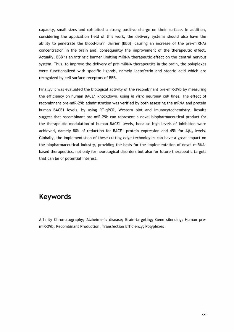
capacity, small sizes and exhibited a strong positive charge on their surface. In addition,
considering the application field of this work, the delivery systems should also have the
ability to penetrate the Blood-Brain Barrier (BBB), causing an increase of the pre-miRNAs
concentration in the brain and, consequently the improvement of the therapeutic effect.
Actually, BBB is an intrinsic barrier limiting miRNA therapeutic effect on the central nervous
system. Thus, to improve the delivery of pre-miRNA therapeutics in the brain, the polyplexes
were functionalized with specific ligands, namely lactoferrin and stearic acid which are
recognized by cell surface receptors of BBB.
Finally, it was evaluated the biological activity of the recombinant pre-miR-29b by measuring
the efficiency on human BACE1 knockdown, using in vitro neuronal cell lines. The effect of
recombinant pre-miR-29b administration was verified by both assessing the mRNA and protein
human BACE1 levels, by using RT-qPCR, Western blot and Imunocytochemistry. Results
suggest that recombinant pre-miR-29b can represent a novel biopharmaceutical product for
the therapeutic modulation of human BACE1 levels, because high levels of inhibition were
achieved, namely 80% of reduction for BACE1 protein expression and 45% for Aβ42 levels.
Globally, the implementation of these cutting-edge technologies can have a great impact on
the biopharmaceutical industry, providing the basis for the implementation of novel miRNA-
based therapeutics, not only for neurological disorders but also for future therapeutic targets
that can be of potential interest.
Keywords
Affinity Chromatography; Alzheimer’s disease; Brain-targeting; Gene silencing; Human pre-
miR-29b; Recombinant Production; Transfection Efficiency; Polyplexes

xxii

xxiii
Thesis Overview
This thesis is structured in four main chapters. The first chapter is divided into three
sections. The first section of chapter 1 provides a brief explanation concerning the
importance and interest to apply microRNAs as biopharmaceuticals, in order to regulate
endogenous pathways involved in the emergence and progression of the Alzheimer’s disease.
This section presents a general introduction to the main issues detailed in sections 2 and 3.
The second section consists in a succinct literature review about the relevance of developing
and establishing new and integrative approaches to biosynthesize, isolate, purify and deliver
the pre-miRNAs molecules, also discussing the main challenges and future directions for the
development of miRNA-based therapies for neurodegenerative diseases (Paper I. Current
progress on microRNAs-based therapeutics). Lastly, the third section discusses the main issues
related with non-coding RNAs purification, the advantages and limitations associated to the
current chromatographic methods, as well as the improvements that have been done lately,
in order to obtain pure and biologically active samples. This third section is also presented in
a paper review form (Paper II. Affinity approaches in RNAi-based therapeutics purification).
The second chapter includes the main, as well as, the specific goals established for the
development of the present research project.
Afterwards, the third chapter consists in the presentation and discussion of the results
obtained during this research work, in the form of original research papers organized as
follows:
Paper III. Advances in time-course extracellular production of human pre-miR-29b from
Rhodovulum sulfidophilum
Paper IV. Analysis of pre-miR-29b binding conditions to amino acids by Surface Plasmon
Resonance Biosensor
Paper V. New approach for purification of pre-miR-29 using lysine-affinity chromatography
Paper VI. Purification of pre-miR-29 by arginine-affinity chromatography
Paper VII. Pharmaceutical-grade pre-miR-29 purification using an agmatine monolithic support
Paper VIII. Characterization of polyplexes involving small RNA
Paper IX. Recombinant pre-miR-29b for Alzheimer´s disease therapeutics

xxiv
Paper X. Brain-targeting study of lactoferrin-stearic acid–Chitosan/Polyethylenimine pre-miR-
29b-delivery system
To finish, the fourth chapter summarizes the concluding remarks obtained during this
research work, regarding the development and implementation of cutting-edge technologies
to the production, bioseparation and delivery of pre-miRNAs. The impact of the findings here
reported on biopharmaceutical industry will also be addressed, discussing the application of
these novel approaches based on microRNAs, not only for neurological disorders but also for
future therapeutic targets that can be of potential interest. In addition, the future trends of
this thesis and additional research work will be also suggested to complement the important
findings achieved with this project.

xxv
Table of Contents
List of Figures xxvii
List of Tables xxix
List of Abbreviations xxxi
List of Scientific Publications xxxiii
List of Scientific Communications xxxv
Chapter 1
1. Alzheimer’s disease and microRNAs 3
1.1. Introduction 3
1.2. Alzheimer’s disease 4
1.3. MicroRNAs as therapeutic products 11
1.4. miR-29 in Alzheimer’s disease 15
1.5. References 19
2. Current progress on microRNAs-based therapeutics (Paper I) 25
3. Affinity approaches in RNAi-based therapeutics purification (Paper II) 65
Chapter 2
Aims of the thesis 101
Chapter 3
Paper III. Advances in time-course extracellular production of human pre-miR-
29b from Rhodovulum sulfidophilum 107
Paper IV. Analysis of pre-miR-29b binding conditions to amino acids by Surface
Plasmon Resonance Biosensor 135
Paper V. New approach for purification of pre-miR-29 using lysine-affinity
chromatography 159
Paper VI. Purification of pre-miR-29 by arginine-affinity chromatography 165
Paper VII. Pharmaceutical-grade pre-miR-29 purification using an agmatine
monolithic support 175
Paper VIII. Characterization of polyplexes involving small RNA 187
Paper IX. Recombinant pre-miR-29b for Alzheimer´s disease therapeutics 201
Paper X. Brain-targeting study of lactoferrin-stearic acid–
Chitosan/Polyethylenimine pre-miR-29b-delivery system 227
Chapter 4
Concluding remarks 255
Future trends 259

xxvi

xxvii
List of Figures
Figure 1. Amyloidogenic and Non-amyloidogenic pathways for proteolytic processing of
APP by α-secretase, BACE1, and γ-secretase 7
Figure 2. Alzheimer's disease pathways. 8
Figure 3. Biogenesis of microRNAs. 13
Figure 4. Pre-miR-29b-1 regulation of BACE1 in Alzheimer’s disease. 18

xxviii

xxix
List of Tables
Table 1. Dysregulated microRNAs in Alzheimer’s disease. 16

xxx

xxxi
List of Abbreviations
3′UTR 3’-Untranslated Regions
5′UTR 5’-Untranslated Regions
AAA poly(A) tail
Aβ Amyloid-beta
AD Alzheimer's Disease
Ago Argonaute
APH-1 Anterior Pharynx-defective 1
APP Amyloid Precursor Protein
APOE Apolipoprotein E
BACE1 β-site Amyloid Precursor Protein-Cleaving Enzyme 1
BBB Blood-Brain Barrier
C. elegans Caenorhabditis elegans
cDNA complementary DNA
Cdk5 Cell division protein kinase 5
CircRNAs Circular RNAs
CS Chitosan
CSF Cerebrospinal Fluid
CNS Central Nervous System
DGCR8 DiGeorge Syndrome Critical Region Gene 8
DNA Deoxyribonucleic Acid
dsRNA(s) Double-stranded RNA(s)
E. coli Escherichia coli
EE Encapsulation Efficiency
EMA European Medicines Agency
eRNA Enhancer associated RNAs
FAD Familial Alzheimer’s disease
FDA Food and Drug Administration
gDNA Genomic DNA
GSK3 Glycogen Synthase Kinase 3
GTP Guanosine Triphosphate
Lf Lactoferrin
lincRNA Large Intervening Non-coding RNA
LNA Locked Nucleic Acid
lncRNA(s) Long Non-coding RNA(s)
lpaRNA Promoter-associated long RNAs
MAPT Microtubule-Associated Protein Tau
miRISC miRNA-Induced Silencing Complex
miRNA(s) / miR(s) MicroRNA(s)
MRE MicroRNA Response Elements
mRNA Messenger RNA
NATs Natural Antisense Transcripts
NcRNA(s) Non-coding RNA(s)
NCSTN Nicastrin
ND(s) Neurodegenerative Disease(s)

xxxii
NFTs Neurofibrillary Tangles
NMDA receptor N-Methyl-D-Aspartate receptor
nt Nucleotide
OD600 Optical Density at 600 nm
ORF Open Reading Frame
PACT Protein kinase R-activating Protein
PEG Polyethylene Glycol
PEI Polyethylenimine
pDNA Plasmid DNA
PEN 2 Presenilin Enhancer 2
piRNAs PIWI-Interacting RNAs
PKR Protein Kinase Receptor
pre-miRNA(s) Precursor MicroRNA(s)
pri-miRNA(s) Primary MicroRNA(s)
PSEN Presenilin
RBP(s) RNA Binding Protein(s)
R. sulfidophilum Rhodovulum sulfidophilum
RISC RNA-Induced Silencing Complex
RNA Ribonucleic Acid
RNAi RNA Interference
RNAt Total RNA
RNase(s) Ribonuclease(s)
RNP Ribonucleoprotein
rRNA(s) Ribosomal RNA(s)
ROS Reactive Oxygen Species
SA Stearic Acid
SAD Sporadic Alzheimer’s Disease
SELEX Systematic Evolution of Ligands by Exponential
shRNA Short Hairpin RNAs
siRNA(s) Small Interfering RNA(s)
SIRT1 Sirtuin 1
sncRNA(s) Small Non-coding RNA(s)
snRNA(s) Small Nuclear RNA(s)
snoRNA(s) Small Nucleolar RNA(s)
sRNA(s) Small RNA(s)
SPE Solid-Phase Extraction
TLRs Toll-like Receptors
TRBP Tar RNA-binding Protein
tRNA(s) Transfer RNA(s)
WHO World Health Organization
xxxiii
List of Scientific Publications Papers related with this Thesis
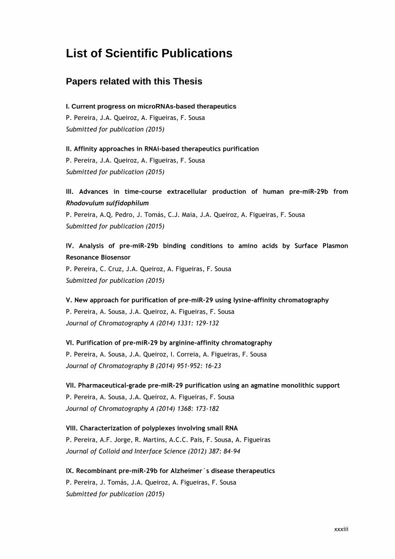
I. Current progress on microRNAs-based therapeutics
P. Pereira, J.A. Queiroz, A. Figueiras, F. Sousa
Submitted for publication (2015)
II. Affinity approaches in RNAi-based therapeutics purification
P. Pereira, J.A. Queiroz, A. Figueiras, F. Sousa
Submitted for publication (2015)
III. Advances in time-course extracellular production of human pre-miR-29b from
Rhodovulum sulfidophilum
P. Pereira, A.Q. Pedro, J. Tomás, C.J. Maia, J.A. Queiroz, A. Figueiras, F. Sousa
Submitted for publication (2015)
IV. Analysis of pre-miR-29b binding conditions to amino acids by Surface Plasmon
Resonance Biosensor
P. Pereira, C. Cruz, J.A. Queiroz, A. Figueiras, F. Sousa
Submitted for publication (2015)
V. New approach for purification of pre-miR-29 using lysine-affinity chromatography
P. Pereira, A. Sousa, J.A. Queiroz, A. Figueiras, F. Sousa
Journal of Chromatography A (2014) 1331: 129-132
VI. Purification of pre-miR-29 by arginine-affinity chromatography
P. Pereira, A. Sousa, J.A. Queiroz, I. Correia, A. Figueiras, F. Sousa
Journal of Chromatography B (2014) 951-952: 16-23
VII. Pharmaceutical-grade pre-miR-29 purification using an agmatine monolithic support
P. Pereira, A. Sousa, J.A. Queiroz, A. Figueiras, F. Sousa
Journal of Chromatography A (2014) 1368: 173-182
VIII. Characterization of polyplexes involving small RNA
P. Pereira, A.F. Jorge, R. Martins, A.C.C. Pais, F. Sousa, A. Figueiras
Journal of Colloid and Interface Science (2012) 387: 84-94
IX. Recombinant pre-miR-29b for Alzheimer´s disease therapeutics
P. Pereira, J. Tomás, J.A. Queiroz, A. Figueiras, F. Sousa
Submitted for publication (2015)

xxxiv
X. Brain-targeting study of lactoferrin-stearic acid–Chitosan/Polyethylenimine pre-miR-
29b-delivery system
P. Pereira, J. Tomás, C. Cruz, C. Santos, J.A. Queiroz, A. Figueiras, F. Sousa
In preparation (2015)
Papers not related with this Thesis
I. Polyplexes as Nanovectors for Gene Therapy - Chapter 9
Patrícia Pereira, Fani Sousa, Ana Figueiras
Biotechnology, Nanobiotechnology (2013) Volume 10: 179-223
Published by Studium Press LLC, Houston, USA; ISBN: 1-62699-025-5
II. Purification of pre-miR-29 by a new O-phospho-L-tyrosine affinity chromatographic
strategy optimized using design of experiments
A. Afonso, P. Pereira, J.A. Queiroz, A. Sousa, F. Sousa
Journal of Chromatography A (2014)1343: 119-127
III. Binding mechanisms for histamine and agmatine ligands in plasmid deoxyribonucleic
acid purifications
A. Sousa, P. Pereira, F. Sousa, J.A. Queiroz
Journal of Chromatography A (2014)1366: 110-119
IV. Purification of Membrane-Bound Catechol-O-Methyltransferase by Arginine-Affinity
Chromatography
A.Q. Pedro, P. Pereira, M.J. Bonifácio, J.A. Queiroz, L.A. Passarinha
Chromatographia (2015) DOI: 10.1007/s10337-015-2970-3
V. Purification of supercoiled G-quadruplex pDNA for in vitro transcription
T. Santos, P. Pereira, F. Sousa, J.A. Queiroz, C. Cruz
Submitted for publication (2015)
VI. Stabilization of novel immunoglobulin switch regions G-quadruplexes by naphthalene
and quinoline-based ligands
J. Carvalho, J. Ferreira, P. Pereira, E. Coutinho, A. Guédin, P. Nottelet, G.F. Salgado, J.L.
Mergny, J.A. Queiroz, F. Sousa, E.J. Cabrita, C. Cruz
Submitted for publication (2015)
VII. Síntese e caracterização do suporte cromatográfico de L-tirosina para purificação de
plasmídeos
T. Santos, P. Pereira, F. Sousa, J.A. Queiroz, C. Cruz
Portuguese Patent 108360. Submitted (2015)

xxxv
List of Scientific Communications Oral Communications related with this Thesis
I. A biotechnological platform for pre-miRNAs biosynthesis, purification and brain-
targeted delivery for Alzheimer’s disease
P. Pereira, A.Q. Pedro, J. Tomás, A. Sousa, J.A. Queiroz, A. Figueiras, F. Sousa
MicroBiotec’15 – Congress of Microbiology and Biotechnology 2015. Évora, Portugal
II. Biosynthesis, purification and brain-targeted delivery of pre-miR-29b
biopharmaceuticals for Alzheimer’s disease therapy
P. Pereira, A.Q. Pedro, J. Tomás, C. Cruz, A. Sousa, C. Maia, J.A. Queiroz, A. Figueiras, F.
Sousa
X Annual CICS-UBI Symposium 2015. Covilhã, Portugal
III. Pre-miR-29 biosynthesis and purification for polyplexes-mediated delivery to in vitro
Alzheimer’s disease model
P. Pereira, J. Tomás, A. Sousa, J.A. Queiroz, A. Figueiras, F. Sousa
IX Annual CICS-UBI Symposium 2014. Covilhã, Portugal
IV. Pre-miR-29 purification by amino acids-affinity chromatography
P. Pereira, A. Sousa, J.A. Queiroz, A. Figueiras, F. Sousa
8º Encontro Nacional de Cromatografia 2013. Covilhã, Portugal
Invited Oral Communications related with this Thesis
I. Pre-miR-29b biopharmaceutical for Alzheimer’s disease therapy: biosynthesis,
purification and targeted delivery
P. Pereira
III Jornadas de Química e Bioquímica 2015. Covilhã, Portugal
II. Pre-miR-29 bioseparation and target delivery strategies for Alzheimer’s disease
P. Pereira
V Jornadas Nacionais de Ciências Biomédicas 2014. Covilhã, Portugal
III. Development of new therapeutic strategies for miRNA delivery
P. Pereira, A. Jorge, A.C.C. Pais, F. Sousa, A. Figueiras
I Workshop on Pharmaceutical Technology - “New Drug Delivery Systems” 2012. Coimbra,
Portugal

xxxvi
Poster Communications related with this Thesis
I. Biosynthesis of pre-miR-29 for application in neurodegenerative disorders
P. Pereira, C.J. Maia, I.J. Correia, A. Figueiras, F. Sousa
ECCE9 – ECAB2 (9th European Congress of Chemical Engineering and 2nd European Congress of
Applied Biotechnology) 2013. The Hague, Netherlands
II. Pre-miR-29 biosynthesis and purification: possible implications in Alzheimer’s disease
P. Pereira, A. Sousa, C.J. Maia, J.A. Queiroz, I.J. Correia, A. Figueiras, F. Sousa
MicroBiotec’13 – Congress of Microbiology and Biotechnology 2013. Aveiro, Portugal
III. Exploiting multiple interactions in pre-miR-29 purification by arginine-affinity
chromatography
P. Pereira, A. Sousa, I.J. Correia, A. Figueiras, F. Sousa
19th International Symposium on Separation Sciences – New achievements in Chromatography
2013. Porec, Croatia
IV. Improved native pre-miR-29 purification with lysine-affinity chromatography
P. Pereira, A. Sousa, I.J. Correia, A. Figueiras, F. Sousa
19th International Symposium on Separation Sciences – New achievements in Chromatography
2013. Porec, Croatia
V. Structural and functional characterization of polyplexes for small RNA delivery
P. Pereira, A. Jorge, R. Martins, A.C.C. Pais, F. Sousa, A. Figueiras
8th World Meeting on Pharmaceutics, Biopharmaceutics and Pharmaceutical Technology 2012.
Istanbul, Turkey
VI. miRNA-29 bioseparation and target delivery strategies for Alzheimer disease
P. Pereira, I.J. Correia, A. Figueiras, F. Sousa
V Jornadas sobre Tecnologia e Saúde 2012. Guarda, Portugal
VII. Characterization of polyplexes containing small RNA
P. Pereira, A. Jorge, A.C.C. Pais, F. Sousa, A. Figueiras
Conference CEF (Center for Pharmaceutical Studies) 2012. Coimbra, Portugal

xxxvii
Oral Communications not related with this Thesis
I. DNA vaccine development for HPV treatment: biotechnological production, purification
and in vitro application
A.M. Almeida, P. Pereira, J. Tomás, V. Figueiredo, J.A. Queiroz, F. Sousa, A. Sousa
MicroBiotec’15 – Congress of Microbiology and Biotechnology 2015. Évora, Portugal
II. Production and purification of DNA G-quadruplex using pPH600 plasmid
T.A. Santos, P. Pereira, F. Sousa, J.A. Queiroz, C. Cruz
X Annual CICS-UBI Symposium 2015. Covilhã, Portugal
III. Biosynthesis of a G-quadruplex-forming sequence and its stabilization by naphthalene-
based ligands
J. Carvalho, J. Ferreira, P. Pereira, E. Coutinho, F. Sousa, E. Cabrita, J.A. Queiroz, C. Cruz
X Annual CICS-UBI Symposium 2015. Covilhã, Portugal
IV. Supercoiled HPV16 E6/E7 plasmid DNA-based vaccine purified by affinity
chromatography for in vitro transfection studies
A.M. Almeida, P. Pereira, J. Tomás, V. Figueiredo, J.A. Queiroz, F. Sousa, A. Sousa
VIII Jornadas sobre Tecnologia e Saúde 2015. Guarda, Portugal
V. Optimized purification strategy of supercoiled HPV16 E6/E7 plasmid DNA-based vaccine
for in vitro transfection studies
A.M. Almeida, P. Pereira, J. Tomás, J.A. Queiroz, F. Sousa, A. Sousa
IX Annual CICS-UBI Symposium 2014. Covilhã, Portugal
VI. Pre-miR-29b-1 isolation using a new chromatographic strategy with tyrosine-agarose
A. Afonso, P. Pereira, A. Sousa, F. Sousa
VIII Annual CICS-UBI Symposium 2013. Covilhã, Portugal

xxxviii
Poster Communications not related with this Thesis
I. Biotechnological approach to prepare a DNA vaccine for HPV induced-cervical cancer
treatment
A.M. Almeida, P. Pereira, J. Tomás, V. Figueiredo, J.A. Queiroz, F. Sousa, A. Sousa
30th International Papillomavirus Conference 2015. Lisboa, Portugal
II. A breakthrough optimization of O-phospho-L-tyrosine affinity chromatography to purify
pre-miR-29
A. Afonso, P. Pereira, J.A. Queiroz, A. Sousa, F. Sousa
IX Annual CICS-UBI Symposium 2014. Covilhã, Portugal
III. Pre-miR-29 isolation using a new chromatographic strategy with tyrosine-agarose
A. Afonso, P. Pereira, A. Sousa, F. Sousa
ECCE – ECAB (9th European Congress of Chemical Engineering and 2nd European Congress of
Applied Biotechnology) 2013. The Hague, Netherlands
IV. Purification of pre-miR-29 using a new affinity chromatographic strategy optimized by
Design of Experiments
A. Afonso, P. Pereira, A. Sousa, J.A. Queiroz, F. Sousa
8º Encontro Nacional de Cromatografia 2013. Covilhã, Portugal
V. Development of an O-phospho-L-tyrosine affinity chromatography to purify pre-miR-29
using Design of Experiments
A. Afonso, P. Pereira, J.A. Queiroz, A. Sousa, F. Sousa
V Symposium on Bioengineering 2013. Porto, Portugal
VI. Biosynthesis and purification of siNOX1 for application in neurodegenerative disorders
P. Pereira, C. Maia, I.J. Correia, A. Figueiras, F. Sousa
ESBES - ISPPP (9th European Symposium on Biochemical Engineering Science – 32nd
International Symposium on the Separation of Proteins, Peptides and Polynucleotides) 2012.
Istanbul, Turkey

xxxix

xl

1
Chapter 1

2

3
1. Alzheimer’s disease and microRNAs
1.1. Introduction
The discovery of RNA interference (RNAi) by Andrew Fire and Craig Mello in the late 1990s,
allowed them to win the Nobel Prize in Physiology or Medicine in 2006, and revolutionized the
contemporary understanding of gene regulation (Fire et al., 1998). Exogenous double-
stranded RNA (dsRNA) was identified in the nematode Caenorhabditis elegans (C. elegans) as
an effector molecule of the RNAi pathway, since this RNA silenced the expression of a
homologous target gene by directing degradation of its messenger RNA (mRNA). Moreover, the
study demonstrated that the gene silencing was achieved with the administration of only few
molecules of dsRNAs and that these molecules were more efficient than the corresponding
antisense RNA (Fire et al., 1998). Since then, several studies have demonstrated that RNAi is
a well-conserved, endogenous mechanism in several species, including mammals and plants
(Chu and Rana, 2007; Lehman, 2010). In mammalian cells, the inhibitory capability of RNA
was initially demonstrated by the introduction of shorter dsRNAs, named small interfering
RNAs (siRNAs), with perfect sequence complementarity to target mRNA transcripts, which had
already been discovered in plants (Chu and Rana, 2007; Dykxhoorn and Lieberman, 2006;
Filipowicz et al., 2005; Zamore et al., 2000).
Accordingly, the discovery that genes could be silenced by RNAs allowed the biological
understanding of other roles of RNA, which changed from a simple intermediate molecule in
the information flux between DNA and proteins to a dynamic and versatile molecule,
fundamental in the regulation of genes expression, involved in essential cellular processes of
all living systems (Burnett and Rossi, 2012; Ramachandran and Ignacimuthu, 2013).
Currently, RNAi regulates the expression and function of individual genes, involved in
pathways or known to be associated with diseases (harmful or unwanted genes). This
regulation occurs at the post-transcriptional level through non-coding RNAs molecules
(ncRNAs), via sequence-specific degradation or blocking the translation of their target mRNA
in most eukaryotic species (Bumcrot et al., 2006; Burnett and Rossi, 2012; Deng et al., 2014;
Ramachandran and Ignacimuthu, 2013). RNAi-based technology possesses attractive
characteristics to be considered for therapeutic proposes, such as high specificity, efficiency,
ability to induce a robust and potent knockdown of the targeted genes and the possibility to
promote a long-lasting therapy (the therapeutic effect lasts from days up to weeks), reducing
the expenses of medical treatments (Bumcrot et al., 2006; Burnett and Rossi, 2012; Deng et
al., 2014; Milhavet et al., 2003). On the other hand, usually, the dosage required of ncRNA
therapeutics is low, which can reduce or eliminate the occurrence of undesirable adverse

4
effects in the patients (Guzman-Villanueva et al., 2012). These breakthroughs clearly
encouraged a variety of studies on the mechanisms of gene expression regulation, which led
to the emergence of numerous and different types of RNA-based therapeutics that extends
the range of targets of the existing pharmacological drugs (Burnett and Rossi, 2012).
NcRNAs are a class of transcripts which, as the name implies, are not translated into proteins.
Presently, ncRNAs are recognized as key regulatory molecules in a variety of cellular
processes as diverse as development, cell viability, cell cycle regulation, stem cell self-
renewal, transposon activity control, differentiation, heterochromatin formation and
maintenance of cell integrity by gene silencing pathways, through translational repression or
mRNA degradation (Kim, 2005; Svoboda, 2014). A very simplistic classification based on
ncRNAs length arbitrarily separates these biomolecules into two groups according to their
sizes: the small (<200 nucleotides (nt)) and the long (>200 nt) ncRNAs (Dogini et al., 2014;
Gomes et al., 2013). In turn, the small group comprises infrastructural RNAs and regulatory
RNAs. The infrastructural RNAs include ribosomal RNA (rRNA), transfer RNA (tRNA), small
nuclear RNA (snRNA) and small nucleolar RNA (snoRNA) that are involved in the spliceosomal
and translational machinery. On the other hand, regulatory RNAs include microRNAs
(miRNAs), siRNAs delivered as synthetic duplexes to the cells, short hairpin RNA (shRNA) and
Piwi-interacting RNAs (piRNAs), among others (Chu and Rana, 2007; Dogini et al., 2014;
Gomes et al., 2013; Matera et al., 2007). These differ significantly in their biogenesis and
mode of action (Kim, 2005; Kim et al., 2009).
Presently, RNAi-based therapies have major implications for basic and biomedical research
that can lead to a number of clinical applications, for treating the vast number of human
diseases caused by one or few genes, such as viral infections, metabolic diseases,
cardiovascular disease, hypertension and stroke, immune dysfunction and autoimmune
disorders, distinct types of cancers and neurodegenerative and psychiatric diseases (Gomes et
al., 2013; Ramachandran and Ignacimuthu, 2013; Sullenger and Gilboa, 2002).
1.2. Alzheimer’s disease
Neurodegenerative diseases (NDs) are a family of disorders of central nervous system (CNS)
characterized by a progressive loss of neuronal structure and function, resulting in a motor
and cognitive dysfunction (Goodall et al., 2013; Maciotta et al., 2013). For that reason, these
disorders do not have cure, once the neurons cannot regenerate on their own after cell death
or damage. Some of the most studied NDs include Alzheimer’s disease (AD), Parkinson’s
disease, Huntington’s disease, Amyotrophic Lateral Sclerosis and Prion diseases (Junn and
Mouradian, 2012; Tabares-Seisdedos et al., 2011).

5
NDs constitute a serious and growing health problem for our societies because they
collectively represent one of the leading causes of disability and mortality. Currently, it is
estimated that NDs affect over 30 million people worldwide and that by 2030, this number
can reach 66 million people and increase to 115 million by 2050 (Schonrock et al., 2012). As a
result, these diseases are associated to higher health care costs, as well as, being a source of
severe human suffering (Dunkel et al., 2012). The increased prevalence of these diseases is
intimately related with the current trends: the aging of the population due to increased life
expectancy which cause more people living long enough to be affected and the continuing
lack of progress in identifying effective diagnosis and treatment modalities. All these reasons
emphasize the necessity to study novel therapeutic interventions for these incurable diseases
(Bertram et al., 2010; Qiu et al., 2009).
Among all dementing disorders, AD is the most prevalent (45-60%) and devastating form of
dementia in the elderly and leads to death within 3 to 9 years after appearance of symptoms
(Isik, 2010). According to the National Institute on Aging (www.nia.nih.gov/alzheimers), the
AD is clinically characterized by loss of memory and eventually by disturbances in reasoning
(e.g. trouble handling money), planning and performing activities of daily living, language,
and perception (e.g. loss of orientation to time and place), as well as, changes of mood,
behavior and personality. Common behavioral symptoms of Alzheimer’s include sleeplessness,
agitation, anxiety, and depression (Buchman et al., 2013; Sperling et al., 2011; Wilson et al.,
2013).
In 2014, it was estimated that more than 5.4 million individuals in the USA have AD and that
this number can nearly triplicate, from 5 million to as many as 16 million by 2050, thus
significantly increasing social and economical burdens in industrialized countries, where the
aging population is increasing every year (Provost, 2010b; Suehs et al., 2013). AD is commonly
classified into two types: Sporadic Alzheimer’s disease (SAD) or Familial Alzheimer’s disease
(FAD) (Blennow et al., 2006; Piaceri et al., 2013). SAD reports most AD cases (99%), occurs
late in life (65 years), and it can take up to 20 years for the disease to develop. Until now,
SAD was only associated with a genetic mutation in the ε4 allele of the apolipoprotein E
(APOE) gene, which promotes amyloid-beta (Aβ) deposition and impairs Aβ clearance
(Bertram et al., 2010; Bu, 2009; Genin et al., 2011; Hardy and Selkoe, 2002; Piaceri et al.,
2013). Presently, sporadic type affects 11% of the population over the age 65 and older, 32%
of those over the age 85 and, the vast majority, 82% of the population aged 75 or older. In
contrast, FAD typically develops before the age 65 (40-50 years), is a very rare condition (1%)
and it is caused by autosomal, dominant mutations in three genes, namely in the amyloid
precursor protein (APP) and in the genes of presenilin (PSEN1 and PSEN2), which are linked to
Aβ processing by γ-secretase complexes (Blennow et al., 2006; Chen et al., 2013; Citron,
2010; Piaceri et al., 2013). Generally, individuals are diagnosed with FAD when more than
one member is affected in more than one generation (Piaceri et al., 2013). Hence, it is
fundamental to found a solution to this problem as soon as possible.

6
AD involves various regions of the brain that control learning, language, thought and memory
and results from progressive neuronal loss (death of neurons), arising from often unknown or
insufficiently characterized risk factors and causes. Thereby, AD is a highly complex disease
that involves hundreds of defective genes distributed across the human genome (genetic
factors), in close interaction with other factors such as age (atrophy of certain parts of the
brain, inflammation, production of free radicals and mitochondrial dysfunction), family
history (heredity), lifestyle factors (education, diet, smoking, substance abuse) and
environmental risk factors (chemical exposure) contributing to its development and
progression (Bertram et al., 2010; Maes et al., 2009).
Neuropathological hallmarks of AD include extracellular senile plaques (known as amyloid
plaques) formed of aggregates of toxic Aβ peptides and intracellular neurofibrillary tangles
(NFTs) composed by insoluble hyperphosphorylated tau, a microtubule-associated protein
(Blennow et al., 2006; Goedert et al., 1989; Hardy and Selkoe, 2002; Mullane and Williams,
2013). Although the mechanisms of AD remain unclear, it has been hypothesized that it can
be associated with abnormal protein metabolism, particularly in the formation and
accumulation of the Aβ peptides (Mullane and Williams, 2013; Provost, 2010a; Schonrock et
al., 2012).
Amyloid peptide is composed by 40 or 42 amino acids and is generated upon sequential
proteolytic cleavage of APP, a transmembrane protein expressed in many tissues and involved
in the synaptogenesis, axonal transport, transmembrane signal transduction, cell adhesion
and calcium metabolism (Chen et al., 2013; Glenner, 1989a; Glenner, 1989b). The initial step
in the “amyloidogenic pathway” is catalyzed by β-secretase (also known as β-site APP-
cleaving enzyme 1 (BACE1)), producing a carboxyl-terminal fragment of 99 amino acids
(CTF99) and simultaneously the ectodomain of APP is released as soluble APPβ (sAPPβ) (see
Figure 1) (Kim et al., 2014; De Strooper, 2010). Posteriorly, the resulting fragment of APP
(CTF99) is cleaved by the γ-secretase complex (that is composed by at least four components,
namely PSEN1, Nicastrin (NCSTN), Presenilin enhancer 2 (PEN2) and Anterior Pharynx-
defective 1 (APH-1)), releasing into the cytoplasm an intracellular domain of APP of
approximately 50 amino acids (AICD), and a variety of potentially amyloidogenic Aβ peptides
(39-42 amino acids) (Figure 1) (Chen et al., 2013; Ghiso and Frangione, 2002; Kim et al.,
2014; Provost, 2010a). In alternative, in the "non-amyloidogenic pathway", APP is initially
cleaved by the metalloprotease α-secretase, releasing a soluble APPα ectodomain (sAPPα),
and generating at the same time, a carboxyl-terminal fragment of 83 residues (CTF83), which
is further processed by γ-secretase, liberating extracellular p3 and the AICD (Ghiso and
Frangione, 2002).

7
Figure 1 - Amyloidogenic and Non-amyloidogenic pathways for proteolytic processing of APP
by α-secretase, BACE1, and γ-secretase.
Abbreviation: APP, amyloid precursor protein; BACE1, β-site APP-cleaving enzyme 1; CTF83/99,
carboxyl-terminal fragment of 83/99 amino acids; sAPPα/β, soluble APPα/β.
While γ-secretase produces several Aβ peptides with heterogeneous carboxyl-terminus
ranging from 38-43 amino acids, BACE1 is a site-specific protease that cleaves exactly
between specific amino-acids of APP. In addition, BACE1 catalyses the first and rate-limiting
step in the pathway (Provost, 2010a; Schonrock et al., 2012; Vassar et al., 2009). Consistent
with this hypothesis, several research groups have demonstrated that the levels and the
activity of BACE1 are elevated in sporadic AD brain, suggesting the possibility that BACE1
overexpression might initiate or accelerate AD. For these reasons, BACE1 has been recognized
as a prime drug target for therapeutic inhibition of Aβ production in AD, once the BACE1
inhibition can decrease production of all forms of Aβ peptides (Vassar et al., 2009). Within
the variety of Aβ peptide isoforms produced in the CNS, Aβ1-40 is the most predominant form
contributing for more than 90% of the total Aβ, whereas Aβ1-42 is the most neurotoxic form and
is considered to be primarily responsible for neuronal damage. As mentioned above, amyloid
plaques are formed from the gradual accumulation and aggregation of secreted Aβ peptides in
oligomers and fibrils in the extracellular space. However, recent studies demonstrated that
the accumulation of Aβ occur also within neurons with AD pathogenesis (De Strooper, 2010;
Dong et al., 2012).

8
Although several molecular mechanisms have been implicated in the AD pathology, most
evidences indicate that the intraneuronal Aβ accumulation and the amyloid plaques play a
pivotal role in the synaptic dysfunction, neuronal death and cognitive dysfunction (Figure 2),
which cause neurodegeneration and dementia in a first instance and then leads to AD (Delay
et al., 2012; Feng and Feng, 2011; Iyengar et al., 2014; Maciotta et al., 2013; Tan et al.,
2013). Many processes are common features in neurodegeneration, namely cellular oxidative
stress (overproduction of reactive oxygen species (ROS) and activation of cell death
pathways), protein oligomerization and aggregation, deregulation of calcium homeostasis,
mitochondrial dysfunction, neuroinflammation, DNA damage and aberrant RNA processing
(Borgesius et al., 2011; Goodall et al., 2013; Jeppesen et al., 2011; Qiu et al., 2009). These
events initiate pro-apoptotic cascades that activate members of the B-cell lymphoma 2 (Bcl-
2) family and lead to the rapid activation of caspases (Feng and Feng, 2011; Maciotta et al.,
2013; Van den Hove et al., 2014). In addition to plaques and tangles as histopathological
hallmarks, other pathological changes include impaired axonal transport, neuron-glial
interactions and reduced synaptic plasticity (Delay et al., 2012). As shown in Figure 2,
mechanisms that affect AD pathogenesis involve dysregulation of multiple fundamental
cellular pathways, including protein folding and clearance processes. Concluding, in order to
develop an AD therapeutic it is essential to understand the principal features of this disease.
Figure 2 - Alzheimer's disease pathways.
Abbreviations: Aβ, Amyloid-beta; APH-1, Anterior Pharynx-defective 1; APP, Amyloid Precursor Protein;
APOE, Apolipoprotein E; Cdk5, Cell division protein kinase 5; GSK3, Glycogen Synthase Kinase 3; MAPT,
Microtubule-Associated Protein Tau; NCSTN, Nicastrin; PEN2, Presenilin Enhancer 2; PSEN1, Presenilin 1;
ROS, Reactive Oxygen Species.

9
Many other disorders can be confused with AD, once no specific blood test or imaging test
exists for the diagnosis of AD. Thus, AD is currently being diagnosed using a combination of
several methods and clinical tools to reach a correct diagnosis, which includes a thorough
general medical workup, a neurological exam based in mental status tests (such as tests of
memory, problem solving, attention and language) and psychiatric evaluation (to assess
mood, anxiety and clarity of thought) (Jack et al., 2011). Furthermore, the diagnosis includes
structural imaging of the brain with computed tomography, magnetic resonance imaging,
single-photon emission computed tomography or positron emission tomography (Albert et al.,
2011; Barthel et al., 2011; McKhann et al., 2011). Nonetheless, brain imaging is not a
standard part of the assessment for possible AD since although these methods are good
biomarkers, they are also quite expensive. The clinical diagnosis of AD remains difficult and,
sometimes the definitive diagnostic is only established by post-mortem confirmation.
In the last few years, several studies have been made to identify the neuropathological,
biochemical, and genetic biomarkers of the AD so that the diagnosis could be done as early as
possible, even in the absence of obvious symptoms, once AD patients are known to have
neuropathology in their brains for over 10 to 20 years before any symptoms occur. Aβ
peptides (Aβ40 and Aβ42, which are more prone to aggregation) and tau/phospho-tau (Thr 181,
a common phospho-epitope), in cerebrospinal fluid (CSF), have been widely accepted as
robust biomarkers in AD diagnosis (Holtzman, 2011; Tarawneh and Holtzman, 2010). However,
the wide variability and inconsistency that exists among different studies and the invasive
nature of the procedures required to obtain CSF have been delaying the use of more accurate
and efficient biomarkers as a diagnostic tool for AD in the clinical setting (Holtzman, 2011;
Ingelson et al., 1999). Finally, the biomarkers also allow following the patient response to the
treatment, enabling to elaborate specific regimen changes if a drug is not providing the
desired benefit.
Despite the efforts to identify optimal treatment schemes containing one or more drugs, the
majority of the currently available options are still largely ineffective due to several reasons.
In particular, the molecular pathology of dementia is still poorly understood, the genetic and
epigenetic component of dementia is poorly defined, and the understanding of genome-drug
interactions is very limited (Cacabelos and Torrellas, 2014). In addition, the drug targets are
inappropriated and most treatments are symptomatic, for instance, although patients treated
with antipsychotic drugs experience moderate benefits in reducing aggression and psychosis,
they are accompanied of serious adverse effects, such as sedation, stroke, movement
difficulties (Parkinsonism), thus limiting their routinely use (Cacabelos and Torrellas, 2014).
Presently, there are only four drugs approved by the Food and Drug Administration (FDA)
available to treat the cognitive problems of Alzheimer's: three are acetylcholinesterase
inhibitors (Donepezil (Aricept®), Rivastigmine (Exelon®), Galantamine (Razadyne®)
commonly prescribed for mild-to-moderate AD) and the other one (Memantine (Namenda®))

10
is an N-Methyl-D-Aspartate (NMDA) receptor antagonist prescribed for moderate-to-severe AD
(Cacabelos and Torrellas, 2014). These drugs can help maintain thinking, memory, and
communication skills and help with certain behavioral problems but they do not delay or
interrupt AD progression. The most common adverse effects are nausea, hallucinations,
confusion, dizziness, headache, fatigue and vomiting.
Although the precise mechanisms responsible for the pathology of AD are still unclear, several
convincing evidences suggest that the generation and accumulation of Aβ peptides, in the
brain, plays the central role in initiating and development of a complex neurodegenerative
cascade, which results in progressive cognitive impairment and dementia (Glenner, 1989a).
Hence, the "amyloid cascade" is the most widely discussed and investigated pathway, to find
the cause of AD. So the regulation of protein expression levels involved in the Aβ generation
process has demonstrated to be important in AD progression. Consequently, the current drug
discovery approaches have focused on preventing the formation and aggregation of the Aβ or
to facilitate their clearance.
During the past 30 years, the understanding of the genetic and molecular basis of the
pathophysiology of this dementia has advanced dramatically; however, limited knowledge of
the basic pathogenic mechanisms is a major hurdle in the identification of drug targets and
development of therapeutic strategies for this yet incurable disorder. All these reasons
highlight the need for the development of innovative and alternative therapeutic strategies to
prevent, slow or stop the progression of this disease.
At present, alternative therapeutic strategies focused on the specific gene expression control
(mRNA and protein levels) can contribute to the regulation of several neuronal functions,
such as differentiation, synaptic plasticity and memory formation (Cacabelos and Torrellas,
2014). Gene therapy is a highly promising therapeutic method for the treatment or prevention
of AD, using genetic information. In addition, gene therapy can be more economical and
convenient because it provides higher targeting and prolonged action. A possible regulatory
mechanism of gene expression in AD is via ncRNAs, designed RNAi mechanism (Cacabelos and
Torrellas, 2014). One important class of gene expression regulators is miRNAs. A number of
studies, in the last decade, focused on the hypothesis of using miRNAs as biopharmaceuticals
to regulate several pathways involved in the insurgence and progression of AD. This idea has
gained support, because some miRNAs are differentially expressed in the human brain
(including the spinal cord, cerebellum, hypothalamus, hippocampus and cortex) and regulate
the expression of genes associated with specific neurodegenerative disorders (Delay et al.,
2012; Feng and Feng, 2011; Maciotta et al., 2013; Satoh, 2012; Tan et al., 2013; Tan et al.,
2015). Thus, the use of these RNAs can provide an effective therapeutic strategy for AD
treatment and diagnosis.

11
1.3. MicroRNAs as therapeutic products
MiRNAs (miRNAs or miRs) were discovered approximately two decades ago by Ambros and
colleagues, when their studies revealed that a small non-coding RNA (sncRNA), lineage-4 (lin-
4) regulated the larval development timing of the C. elegans (Lee et al., 1993; Wightman et
al., 1993). This regulation is accomplished by inhibition of the expression of a protein-coding
gene through recognition of complementarity sites in the 3' untranslated region (3'UTR) of lin-
14 mRNA (Lee et al., 1993). Accordingly, the discovery of miRNAs has been one of the most
fascinating breakthroughs of recent times and, now, miRNAs represent the most extensively
studied class of evolutionarily conserved, endogenous, sncRNAs (Satoh, 2010). At present,
1881 miRNAs have been identified in humans and its sequences are available in the official
miRNA database (miRBase, www.mirbase.org), a repository of miRNAs from many organisms.
MiRNAs have emerged as pivotal modulators that are directly involved in the regulation of
gene expression at the post-transcriptional level via base-pairing interactions between the 5′
end of the miRNA and the 3′UTR of their target mRNA, which results in mRNA translational
repression or degradation, depending on the degree of sequence complementarity (Bartel,
2004; Filipowicz et al., 2005; Selbach et al., 2008).
MiRNAs are formed in a multistep biological process involving critical endonucleases. Figure 3
shows the miRNA biogenesis pathway. The great majority of miRNA genes are commonly
transcribed as a long primary transcript (usually 1–4 kilobases), known as primary-miRNAs (pri-
miRNAs) by RNA polymerase II (Bartel, 2004; Lee et al., 2004). The genomic location of
miRNAs genes varies and can be located in intra- and/or inter-genetic regions of the genome
and at least 42% are estimated to be expressed in clusters. Like mRNAs, these pri-miRNAs are
capped at the 5′ end and polyadenylated at the 3′ end, forming double stranded stem-loop
structures, and can be transcribed as autonomous transcription units, or as clusters from a
polycistronic transcription unit (Bartel, 2004; Gomes et al., 2013; Junn and Mouradian, 2012).
In the nucleus, these primary transcripts, containing the mature miRNA sequence, are
recognized by a microprocessor complex (composed by an enzyme ribonuclease III Drosha
along with its regulatory subunit, the double-stranded RNA-binding protein called DiGeorge
syndrome critical region gene 8 (DGCR8)) that cleaves the base of the stem-loop (~11 nt) (Han
et al., 2006). This step produces a long stem-loop precursor of miRNA, with a hairpin
secondary structure of approximately 70 to 100 nucleotides, named pre-miRNAs, with two
nucleotides overhang at its 3′ end. After nuclear processing by a series of enzymes, the pre-
miRNAs are exported to the cytoplasm by a nuclear transport receptor complex, Exportin-5
and its co-factor Ran (the GTP-bound form) that acts by recognizing a 2-3 base pair overhang
of the pre-miRNA stem-loop structure (Lund et al., 2004).
Once in the cytoplasm, the pre-miRNAs are further processed and its terminal loop is removed
by a complex that contains the cytoplasmic protein Dicer (a ribonuclease III) and its co-
12
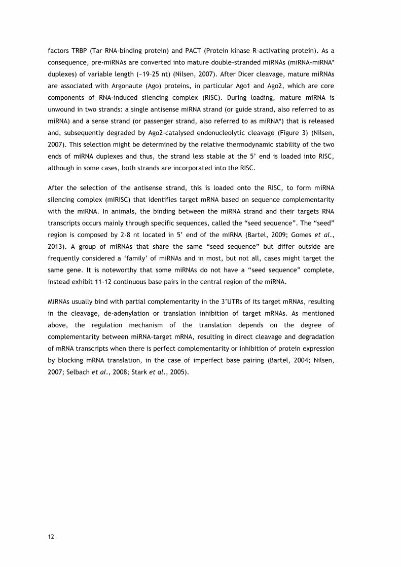
factors TRBP (Tar RNA-binding protein) and PACT (Protein kinase R-activating protein). As a
consequence, pre-miRNAs are converted into mature double-stranded miRNAs (miRNA-miRNA*
duplexes) of variable length (~19–25 nt) (Nilsen, 2007). After Dicer cleavage, mature miRNAs
are associated with Argonaute (Ago) proteins, in particular Ago1 and Ago2, which are core
components of RNA-induced silencing complex (RISC). During loading, mature miRNA is
unwound in two strands: a single antisense miRNA strand (or guide strand, also referred to as
miRNA) and a sense strand (or passenger strand, also referred to as miRNA*) that is released
and, subsequently degraded by Ago2-catalysed endonucleolytic cleavage (Figure 3) (Nilsen,
2007). This selection might be determined by the relative thermodynamic stability of the two
ends of miRNA duplexes and thus, the strand less stable at the 5’ end is loaded into RISC,
although in some cases, both strands are incorporated into the RISC.
After the selection of the antisense strand, this is loaded onto the RISC, to form miRNA
silencing complex (miRISC) that identifies target mRNA based on sequence complementarity
with the miRNA. In animals, the binding between the miRNA strand and their targets RNA
transcripts occurs mainly through specific sequences, called the “seed sequence”. The “seed”
region is composed by 2-8 nt located in 5’ end of the miRNA (Bartel, 2009; Gomes et al.,
2013). A group of miRNAs that share the same “seed sequence” but differ outside are
frequently considered a ‘family’ of miRNAs and in most, but not all, cases might target the
same gene. It is noteworthy that some miRNAs do not have a “seed sequence” complete,
instead exhibit 11-12 continuous base pairs in the central region of the miRNA.
MiRNAs usually bind with partial complementarity in the 3’UTRs of its target mRNAs, resulting
in the cleavage, de-adenylation or translation inhibition of target mRNAs. As mentioned
above, the regulation mechanism of the translation depends on the degree of
complementarity between miRNA-target mRNA, resulting in direct cleavage and degradation
of mRNA transcripts when there is perfect complementarity or inhibition of protein expression
by blocking mRNA translation, in the case of imperfect base pairing (Bartel, 2004; Nilsen,
2007; Selbach et al., 2008; Stark et al., 2005).

13
Figure 3 - Biogenesis of microRNAs.
Abbreviations: 3/5′-UTR, 3/5′ Untranslated Region; AAA, poly(A) tail; Ago, Argonaute; DGCR8, DiGeorge
syndrome Critical Region gene 8; GTP, Guanosine Triphosphate; miRISC, miRNA-Induced Silencing
Complex; mRNA, messenger RNA; miRNA, microRNA; ORF, Open Reading Frame; PACT, Protein kinase R-
Activating Protein; pre-miRNA, precursor miRNA; pri-miRNA, primary miRNA; TRBP, Tar RNA Binding
Protein.

14
The target mRNA is enzymatically cleaved, leading to a decrease in the corresponding protein
expression. Regulatory subunits within the RISC complex and/or present in the mRNAs (i.e.
cytoplasmic ribonucleoprotein complexes, mRNPs) play decisive roles in miRNA-mRNA
localization, miRNA responsiveness to cellular signaling and mode of action. These complexes
can be directed to polyribosomes in sub-cellular compartments, where miRNA regulates
translation, resulting, sometimes, in truncation of the protein (Nilsen, 2007). Alternatively,
the resulting translationally blocked mRNAs and miRNA complex can then be sequestered in
cytoplasmic processing bodies (termed P-bodies), where untranslated mRNAs are stored and
eventually degraded through exonuclease enzymes (Figure 3). On the other hand, specific
miRNAs have been shown to destabilize targets by recruiting de-adenylating enzymes, poly(A)
nucleases, to help modulate de-adenylation of mRNA and thereby prevent translation.
Therefore, the regulation of gene expression mediated by miRNA is a complex science in
eukaryotes and is still an evolving concept, once several molecular mechanisms have been
reported, including translational repression, increased mRNA de-adenylation and degradation
of the target mRNAs (Bartel, 2004; Gomes et al., 2013; Nilsen, 2007). Although these
mechanisms are quite different, the main mode of action of mature miRNA is based on the
recognition of specific sites (known as microRNA response elements (MRE)), typically present
in the 3’UTRs of their target mRNAs, leading to a decrease in the production of the proteins
by cells. Despite, of this binding occurs typically in the 3’UTR of mRNA, some miRNAs can
bind specific sites in the 5’UTRs of the mRNA with less frequency and efficiency compared to
3’UTR targeting (Stark et al., 2005).
Overall, the miRNAs can regulate a large proportion of the human transcriptome (up to 60% of
all human protein-coding genes) by targeting multiple transcripts. An important feature of
these RNAs is that an individual miRNA can regulate up to a few hundred of different mRNA
targets and, consequently, can induce multiple effects on the expression of several related
gene families, suppressing the expression of multiple and diverse proteins implicated in
biological processes (Selbach et al., 2008). On the other hand, multiple miRNAs can regulate
the expression of a single target mRNA, and most likely synergistically acting. Despite
advances in miRNA discovery, the role of miRNAs in pathophysiologic processes is still in early
stages of research. The extensive research in the last decade has implicated miRNAs as
master regulators of a variety of physiological and biological processes, including
developmental timing, cell cycle, cell proliferation, cell adhesion, apoptosis, cell
differentiation, stress response, metabolism, stem cell self-renewal, organ development,
hematopoiesis, embryogenesis and neurogenesis (Alvarez-Garcia and Miska, 2005; Bartel,
2004; Bartel, 2009; Kloosterman and Plasterk, 2006). As a result of its efficacy and potential
to inhibit the expression of a specific mRNA, coupled with its versatility in several cellular
processes, miRNAs might be explored as new therapeutic agents in diseases that have genetic
origins, as is the case of AD. In fact, it is expected that miRNAs-based therapeutics will be
one of the major classes of therapeutic molecules in the future (Delay et al., 2012; Dessy and

15
Gorman, 2011; Kocerha et al., 2009; Kosik, 2006; Maes et al., 2009). Not surprisingly, miRNA-
targeting therapies are an area of intense interest to pharmaceutical companies, and many of
such compounds are now being evaluated in preclinical and clinical trials for a variety of
indications, such as cancer and cardiovascular diseases.
1.4. miR-29 in Alzheimer’s disease
As mentioned above, over the last years, a growing number of studies have been
concentrated in the identification of deregulated proteins and protein-encoding genes
involved in the pathogenesis of AD, and, as a result, numerous genes have been identified as
targets for therapeutic approaches (see Table 1) (Cacabelos and Torrellas, 2014). Despite
their relatively recent discovery, it is already clear that miRNAs might play important
regulatory roles in the molecular control of neuronal development, synaptic plasticity (forms
the basis of learning and memory), neurotransmission, aging of the brain, and in a variety of
physiologically relevant processes in neurodegeneration (Fineberg et al., 2009; Grasso et al.,
2014; Klein et al., 2005; Kosik, 2006; Schratt et al., 2006). Consequently, in the past decade,
tremendous efforts have been put mainly into identifying miRNAs that are changed in AD,
demonstrating that an abundant and particularly diverse source of miRNAs is present in the
brain and helping in the characterization of molecular pathways that are deregulated
concomitantly to the formation of Tau and amyloid aggregates (Delay et al., 2012; Feng and
Feng, 2011; Maciotta et al., 2013; Maes et al., 2009; Satoh, 2012; Tan et al., 2013; Tan et
al., 2015; Van den Hove et al., 2014). Nevertheless, it remains difficult to predict whether
these changes, like the abnormal expression profiles and dysfunction of brain-enriched
miRNAs linked to AD, are a cause or a consequence of the neurodegenerative process and
dementia (Du and Pertsemlidis, 2011; Grasso et al., 2014; Kocerha et al., 2009; Nelson et al.,
2008; Ullah et al., 2014).
Thus, miRNAs have been intensively investigated as potential diagnostic and prognostic
biomarkers, solely or in combination with other AD biomarkers, or also as novel therapeutic
agents for AD (Cogswell et al., 2008; Grasso et al., 2014; Kumar et al., 2013). In Table 1 are
described some of the de-regulated miRNAs that have been recognized as being involved in
the pathological features of AD, as well as their target genes.

16
Table 1 - Dysregulated microRNAs in Alzheimer’s disease (adapted from (Cacabelos and Torrellas, 2014; Delay et al., 2012; Feng and Feng, 2011;
Maciotta et al., 2013; Satoh, 2012; Tan et al., 2013; Tan et al., 2015; van den Hove et al., 2014)).
miRNAs Trend Status Validated targets
miR-15a, -16, -34 Up Bcl-2 (anti-apoptotic factor)
miR-124 Down APP alternative splicing via PTPB1
miR-9, -15a, -19b, -29a/b-1/c, -107, -124, -195,
-298, -328, -485-5p Down BACE1
Let-7, miR-15a, -16, -17-5p, -20a, -101, -106a/b,
-147, -153, -323, -520c, -644, -655 Down APP
miR-29a/b-1 Down NAV3 (Neuron navigator 3), Bim, Bmf, Hrk, Puma
miR-9, -181c Down TRIM2, BTBD3, TGFBI
miR-9, -34c, -128 Down/Up SIRT1 (Sirtuin 1, a de-acetylase that interacts with Tau)
miR-146a Up CFH (Complement factor H) and IRAK1 (IL-1 receptor-associated kinase-1)
miR-9 Down REST (RE-1 silencing transcription factor), BAF53a, Tlx,
NF-H (Neurofilament H), Aβ
miR-9, -15, -29a/b-1, -137, -181c Down SPT (Serine palmitoyletransferase)
miR-9 Down PSEN1
miR-34 Up Cdk4, cyclin D1, Tau
miR-128 ROS
miR-107 Down CDK6, CFL1, CDK5R1, HIF-1b
miR-132 Down FOXO3a, Tau via PTBP2 (Polypyrimidine tract-binding protein 2)
miR-9, -15a
miR-181c Down
Hyperphosphorylation of tau via ERK1(Extracellular signal regulated kinase 1)
Tau acetylation

17
Interestingly, several studies showed that there is a set of miRNAs (include miR-9, miR-15,
miR-29, miR-106, miR-107 and miR-124) that seems to be specifically altered in the AD brain
and they have been associated to different molecular pathways in AD pathogenesis. In
particular, the regulation of these miRNAs was correlated with the modulation of the
expression and/or processing of AD-related genes, such as APP, BACE1, Aβ peptides
deposition, tau protein phosphorylation, SIRT1 and MAPT signaling pathway (which encodes
for tau) (see Table 1) (Delay et al., 2012). Indeed, this can represent a significant advance for
AD diagnosis and treatment.
The miR-29 remains as one of the most interesting and intriguing miRNA families, once this
miRNA has a strong impact on many processes involved in the pathogenesis of the AD
(Cogswell et al., 2008). The miR-29 family includes four members: miR-29a, miR-29b-1, miR-
29b-2 and miR-29c, and all share identical sequences at nucleotide positions 2-8 of 5’ end and
the same seed region (Kriegel et al., 2012; Liston et al., 2012).
As mentioned above, the BACE1 protein levels are elevated in AD brains, suggesting that
BACE1 dysregulation is directly implicated in AD pathogenesis through the formation of toxic
Aβ peptides. Thus, it has been established that reducing BACE1 expression may delay or even
reduce AD progression. On the other hand, significant alterations in the expression levels of
miR-29 family were found in brains of AD patients, being significantly decreased and
displaying abnormally high levels of BACE1 protein (Hebert et al., 2008; Zong et al., 2011). It
is tempting to speculate that miR-29 might play an important role in the adult brain.
Particularly, the work of Hebert and co-workers, in 2008, showed that the decreased levels of
the miR-29a/b-1 cluster were correlated with increased levels of BACE1 expression in patients
with SAD (Figure 4) and that the introduction of synthetic miR-29 reduces secretion of Aβ
peptides (Hebert et al., 2008).
In addition to its potential role as a major element to silence BACE1 protein expression, it
was recently shown that miR-29b can also be used as an inhibitor of neuronal apoptosis.
However, the miR-29b is decreased in the AD brain and therefore it loses its ability to inhibit
apoptosis. MiR-29b has been identified to target a family of pro-apoptotic regulators,
including Bim, Bmf, Hrk and Puma (see Table 1) (Kole et al., 2011; Zhu et al., 2006).
These findings raised the opportunity to use pre-miR-29 in the implementation of a possible
protective therapeutic strategy for AD and suggest that BACE1 can be a promising target in
the therapeutic of this disease. The use of pre-miRNA instead of the mature miRNA could
represent an advantage to the therapeutic process, because, in cellular environment the pre-
miRNA is more easily recognized and processing within the cell is more efficient (Tsutsumi et
al., 2011). Moreover, the pre-miR-29b structural characteristics, specifically the single chain
with approximately 80 nucleotides of which some are unpaired in the 3’overhang, will
possibly make easier its production and purification.

18
Figure 4 - Pre-miR-29b-1 regulation of BACE1 in Alzheimer’s disease.

19
1.5. References
Albert MS, DeKosky ST, Dickson D, Dubois B, Feldman HH, et al. (2011) The diagnosis of mild
cognitive impairment due to Alzheimer's disease: recommendations from the National
Institute on Aging-Alzheimer's Association workgroups on diagnostic guidelines for Alzheimer's
disease. Alzheimer's & dementia: the journal of the Alzheimer's Association 7, 270-279.
Alvarez-Garcia I and Miska EA (2005) MicroRNA functions in animal development and human
disease. Development 132, 4653-4662.
Bartel DP (2004) MicroRNAs: genomics, biogenesis, mechanism, and function. Cell 116, 281-
297.
Bartel DP (2009) MicroRNAs: target recognition and regulatory functions. Cell 136, 215-233.
Barthel H, Gertz HJ, Dresel S, Peters O, Bartenstein P, et al. (2011) Cerebral amyloid-beta
PET with florbetaben (18F) in patients with Alzheimer's disease and healthy controls: a
multicentre phase 2 diagnostic study. The Lancet. Neurology 10, 424-435.
Bertram L, Lill CM, Tanzi RE (2010) The genetics of Alzheimer disease: back to the future.
Neuron 68, 270-281.
Blennow K, de Leon MJ, Zetterberg H (2006) Alzheimer's disease. Lancet 368, 387-403.
Borgesius NZ, de Waard MC, van der Pluijm I, Omrani A, Zondag GC, et al. (2011) Accelerated
age-related cognitive decline and neurodegeneration, caused by deficient DNA repair. The
Journal of neuroscience: the official journal of the Society for Neuroscience 31, 12543-
12553.
Bu G (2009) Apolipoprotein E and its receptors in Alzheimer's disease: pathways, pathogenesis
and therapy. Nature reviews. Neuroscience 10, 333-344.
Buchman AS, Yu L, Wilson RS, Schneider JA, Bennett DA (2013) Association of brain pathology
with the progression of frailty in older adults. Neurology 80, 2055-2061.
Bumcrot D, Manoharan M, Koteliansky V, Sah DW (2006) RNAi therapeutics: a potential new
class of pharmaceutical drugs. Nature chemical biology 2, 711-719.
Burnett JC and Rossi JJ (2012) RNA-based therapeutics: current progress and future
prospects. Chemistry Biology 19, 60-71.
Cacabelos R and Torrellas C (2014) Epigenetic drug discovery for Alzheimer's disease. Expert
opinion on drug discovery 9, 1059-1086.
Chen S, Ge X, Chen Y, Lv N, Liu Z, et al. (2013) Advances with RNA interference in
Alzheimer's disease research. Drug design, development and therapy 7, 117-125.
Chu CY and Rana TM (2007) Small RNAs: regulators and guardians of the genome. Journal of
Cellular Physiology 213, 412-419.
Citron M (2010) Alzheimer's disease: strategies for disease modification. Nature reviews. Drug
discovery 9, 387-398.

20
Cogswell JP, Ward J, Taylor IA, Waters M, Shi Y, et al. (2008) Identification of miRNA changes
in Alzheimer's disease brain and CSF yields putative biomarkers and insights into disease
pathways. Journal of Alzheimer's disease: JAD 14, 27-41.
Delay C, Mandemakers W, Hebert SS (2012) MicroRNAs in Alzheimer's disease. Neurobiology of
disease 46, 285-290.
Deng Y, Wang CC, Choy KW, Du Q, Chen J, et al. (2014) Therapeutic potentials of gene
silencing by RNA interference: principles, challenges, and new strategies. Gene 538, 217-227.
Dessy A and Gorman JM (2011) The emerging therapeutic role of RNA interference in disorders
of the central nervous system. Clinical Pharmacology Therapeutics 89, 450-454.
De Strooper B (2010) Proteases and Proteolysis in Alzheimer Disease: A Multifactorial View on
the Disease Process. Physiological Reviews 90, 465-494
Dogini DB, Pascoal VD, Avansini SH, Vieira AS, Pereira TC, et al. (2014) The new world of
RNAs. Genetics and molecular biology 37, 285-293.
Dong S, Duan Y, Hu Y, Zhao Z (2012) Advances in the pathogenesis of Alzheimer's disease: a
re-evaluation of amyloid cascade hypothesis. Translational Neurodegeneration 1, 18
Du L and Pertsemlidis A (2011) Cancer and neurodegenerative disorders: pathogenic
convergence through microRNA regulation. Journal of molecular cell biology 3, 176-180.
Dunkel P, Chai CL, Sperlagh B, Huleatt PB, Matyus P (2012) Clinical utility of neuroprotective
agents in neurodegenerative diseases: current status of drug development for Alzheimer's,
Parkinson's and Huntington's diseases, and amyotrophic lateral sclerosis. Expert opinion on
investigational drugs 21, 1267-1308.
Dykxhoorn DM and Lieberman J (2006) Running interference: prospects and obstacles to using
small interfering RNAs as small molecule drugs. Annual Review of Biomedical Engineering 8,
377-402.
Feng W and Feng Y (2011) MicroRNAs in neural cell development and brain diseases. Science
China. Life sciences 54, 1103-1112.
Filipowicz W, Jaskiewicz L, Kolb FA, Pillai RS (2005) Post-transcriptional gene silencing by
siRNAs and miRNAs. Current Opinion in Structural Biology 15, 331-341.
Fineberg SK, Kosik KS, Davidson BL (2009) MicroRNAs potentiate neural development. Neuron
64, 303-309.
Fire A, Xu S, Montgomery MK, Kostas SA, Driver SE, et al. (1998) Potent and specific genetic
interference by double-stranded RNA in Caenorhabditis elegans. Nature 391, 806-811.
Genin E, Hannequin D, Wallon D, Sleegers K, Hiltunen M, et al. (2011) APOE and Alzheimer
disease: a major gene with semi-dominant inheritance. Molecular psychiatry 16, 903-907.
Ghiso J and Frangione B (2002) Amyloidosis and Alzheimer's disease. Advanced drug delivery
reviews 54, 1539-1551.
Glenner GG (1989a) Amyloid beta protein and the basis for Alzheimer's disease. Progress in
clinical and biological research 317, 857-868.

21
Glenner GG (1989b) The pathobiology of Alzheimer's disease. Annual review of medicine 40,
45-51.
Goedert M, Spillantini MG, Jakes R, Rutherford D, Crowther RA (1989) Multiple isoforms of
human microtubule-associated protein tau: sequences and localization in neurofibrillary
tangles of Alzheimer's disease. Neuron 3, 519-526.
Gomes AQ, Nolasco S, Soares H (2013) Non-coding RNAs: multi-tasking molecules in the cell.
International journal of molecular sciences 14, 16010-16039.
Goodall EF, Heath PR, Bandmann O, Kirby J, Shaw PJ (2013) Neuronal dark matter: the
emerging role of microRNAs in neurodegeneration. Frontiers in cellular neuroscience 7, 178.
Grasso M, Piscopo P, Confaloni A, Denti MA (2014) Circulating miRNAs as biomarkers for
neurodegenerative disorders. Molecules 19, 6891-6910.
Guzman-Villanueva D, El-Sherbiny IM, Herrera-Ruiz D, Vlassov AV, Smyth HD (2012)
Formulation approaches to short interfering RNA and MicroRNA: challenges and implications.
Journal of pharmaceutical sciences 101, 4046-4066.
Han J, Lee Y, Yeom KH, Nam JW, Heo I, et al. (2006) Molecular basis for the recognition of
primary microRNAs by the Drosha-DGCR8 complex. Cell 125, 887-901.
Hardy J and Selkoe DJ (2002) The amyloid hypothesis of Alzheimer's disease: progress and
problems on the road to therapeutics. Science 297, 353-356.
Hebert SS, Horre K, Nicolai L, Papadopoulou AS, Mandemakers W, et al. (2008) Loss of
microRNA cluster miR-29a/b-1 in sporadic Alzheimer's disease correlates with increased
BACE1/beta-secretase expression. Proceedings of the National Academy of Sciences of the
United States of America 105, 6415-6420.
Holtzman DM (2011) CSF biomarkers for Alzheimer's disease: current utility and potential
future use. Neurobiology of aging 32 Suppl 1, S4-9.
Ingelson M, Blomberg M, Benedikz E, Wahlund LO, Karlsson E, et al. (1999) Tau
immunoreactivity detected in human plasma, but no obvious increase in dementia. Dementia
and geriatric cognitive disorders 10, 442-445.
Isik AT (2010) Late onset Alzheimer's disease in older people. Clinical interventions in aging 5,
307-311.
Iyengar BR, Choudhary A, Sarangdhar MA, Venkatesh KV, Gadgil CJ, et al. (2014) Non-coding
RNA interact to regulate neuronal development and function. Frontiers in cellular
neuroscience 8, 47.
Jack CR Jr, Albert MS, Knopman DS, McKhann GM, Sperling RA, et al. (2011) Introduction to
the recommendations from the National Institute on Aging-Alzheimer's Association workgroups
on diagnostic guidelines for Alzheimer's disease. Alzheimer's & dementia: the journal of the
Alzheimer's Association 7, 257-262.
Jeppesen DK, Bohr VA, Stevnsner T (2011) DNA repair deficiency in neurodegeneration.
Progress in neurobiology 94, 166-200.
Junn E and Mouradian MM (2012) MicroRNAs in neurodegenerative diseases and their
therapeutic potential. Pharmacology & therapeutics 133, 142-150.

22
Kim DH, Yeo SH, Park JM, Choi JY, Lee TH, et al. (2014) Genetic markers for diagnosis and
pathogenesis of Alzheimer's disease. Gene 545, 185-193.
Kim VN (2005) Small RNAs: classification, biogenesis, and function. Molecules and Cells 19, 1-
15.
Kim VN, Han J, Siomi MC (2009) Biogenesis of small RNAs in animals. Nature reviews.
Molecular cell biology 10, 126-139.
Klein ME, Impey S, Goodman RH (2005) Role reversal: the regulation of neuronal gene
expression by microRNAs. Current opinion in neurobiology 15, 507-513.
Kloosterman WP and Plasterk RH (2006) The diverse functions of microRNAs in animal
development and disease. Developmental cell 11, 441-450.
Kocerha J, Kauppinen S, Wahlestedt C (2009) microRNAs in CNS disorders. Neuromolecular
medicine 11, 162-172.
Kole AJ, Swahari V, Hammond SM, Deshmukh M (2011) miR-29b is activated during neuronal
maturation and targets BH3-only genes to restrict apoptosis. Genes & development 25, 125-
130.
Kosik KS (2006) The neuronal microRNA system. Nature reviews. Neuroscience 7, 911-920.
Kriegel AJ, Liu Y, Fang Y, Ding X, Liang M (2012) The miR-29 family: genomics, cell biology,
and relevance to renal and cardiovascular injury. Physiological genomics 44, 237-244.
Kumar P, Dezso Z, MacKenzie C, Oestreicher J, Agoulnik S, et al. (2013) Circulating miRNA
biomarkers for Alzheimer's disease. PloS one 8, e69807.
Lee RC, Feinbaum RL, Ambros V (1993) The C. elegans heterochronic gene lin-4 encodes small
RNAs with antisense complementarity to lin-14. Cell 75, 843-854.
Lee Y, Kim M, Han J, Yeom KH, Lee S, et al. (2004) MicroRNA genes are transcribed by RNA
polymerase II. The EMBO journal 23, 4051-4060.
Lehman N (2010) RNA in evolution. Wiley interdisciplinary reviews. RNA 1, 202-213.
Liston A, Papadopoulou AS, Danso-Abeam D, Dooley J (2012) MicroRNA-29 in the adaptive
immune system: setting the threshold. Cellular and molecular life sciences: CMLS 69, 3533-
3541.
Lund E, Guttinger S, Calado A, Dahlberg JE, Kutay U (2004) Nuclear export of microRNA
precursors. Science 303, 95-98.
Maciotta S, Meregalli M, Torrente Y (2013) The involvement of microRNAs in
neurodegenerative diseases. Frontiers in cellular neuroscience 7, 265.
Maes OC, Chertkow HM, Wang E, Schipper HM (2009) MicroRNA: Implications for Alzheimer
Disease and other Human CNS Disorders. Current genomics 10, 154-168.
Matera AG, Terns RM, Terns MP (2007) Non-coding RNAs: lessons from the small nuclear and
small nucleolar RNAs. Nature reviews. Molecular cell biology 8, 209-220.
McKhann GM, Knopman DS, Chertkow H, Hyman BT, Jack CR Jr, et al. (2011) The diagnosis of
dementia due to Alzheimer's disease: recommendations from the National Institute on Aging-

23
Alzheimer's Association workgroups on diagnostic guidelines for Alzheimer's disease.
Alzheimer's & dementia: the journal of the Alzheimer's Association 7, 263-269.
Milhavet O, Gary DS, Mattson MP (2003) RNA interference in biology and medicine.
Pharmacological Reviews 55, 629-648.
Mullane K and Williams M (2013) Alzheimer's therapeutics: continued clinical failures question
the validity of the amyloid hypothesis-but what lies beyond? Biochemical pharmacology 85,
289-305.
Nelson PT, Wang WX, Rajeev BW (2008) MicroRNAs (miRNAs) in neurodegenerative diseases.
Brain pathology 18, 130-138.
Nilsen TW (2007) Mechanisms of microRNA-mediated gene regulation in animal cells. Trends
in genetics: TIG 23, 243-249.
Piaceri I, Nacmias B, Sorbi S (2013) Genetics of familial and sporadic Alzheimer's disease.
Frontiers in bioscience 5, 167-177.
Provost P (2010a) Interpretation and applicability of microRNA data to the context of
Alzheimer's and age-related diseases. Aging 2, 166-169.
Provost P (2010b) MicroRNAs as a molecular basis for mental retardation, Alzheimer's and
prion diseases. Brain research 1338, 58-66.
Qiu C, Kivipelto M, von Strauss E (2009) Epidemiology of Alzheimer's disease: occurrence,
determinants, and strategies toward intervention. Dialogues in clinical neuroscience 11, 111-
128.
Ramachandran PV and Ignacimuthu S (2013) RNA interference-a silent but an efficient
therapeutic tool. Applied biochemistry and biotechnology 169, 1774-1789.
Satoh J (2010) MicroRNAs and their therapeutic potential for human diseases: aberrant
microRNA expression in Alzheimer's disease brains. Journal of Pharmacological Sciences 114,
269-275.
Satoh J (2012) Molecular network of microRNA targets in Alzheimer's disease brains.
Experimental neurology 235, 436-446.
Schonrock N, Matamales M, Ittner LM, Gotz J (2012) MicroRNA networks surrounding APP and
amyloid-beta metabolism-implications for Alzheimer's disease. Experimental neurology 235,
447-454.
Schratt GM, Tuebing F, Nigh EA, Kane CG, Sabatini ME, et al. (2006) A brain-specific microRNA
regulates dendritic spine development. Nature 439, 283-289.
Selbach M, Schwanhausser B, Thierfelder N, Fang Z, Khanin R, et al. (2008) Widespread
changes in protein synthesis induced by microRNAs. Nature 455, 58-63.
Sperling RA, Aisen PS, Beckett LA, Bennett DA, Craft S, et al. (2011) Toward defining the
preclinical stages of Alzheimer's disease: recommendations from the National Institute on
Aging-Alzheimer's Association workgroups on diagnostic guidelines for Alzheimer's disease.
Alzheimer's & dementia: the journal of the Alzheimer's Association 7, 280-292.

24
Stark A, Brennecke J, Bushati N, Russell RB, Cohen SM (2005) Animal MicroRNAs confer
robustness to gene expression and have a significant impact on 3'UTR evolution. Cell 123,
1133-1146.
Suehs BT, Davis CD, Alvir J, van Amerongen D, Pharmd NC, et al. (2013) The clinical and
economic burden of newly diagnosed Alzheimer's disease in a medicare advantage population.
American journal of Alzheimer's disease and other dementias 28, 384-392.
Sullenger BA and Gilboa E (2002) Emerging clinical applications of RNA. Nature 418, 252-258.
Svoboda P (2014) Renaissance of mammalian endogenous RNAi. FEBS letters 588, 2550-2556.
Tabares-Seisdedos R, Dumont N, Baudot A, Valderas JM, Climent J, et al. (2011) No paradox,
no progress: inverse cancer comorbidity in people with other complex diseases. The Lancet.
Oncology 12, 604-608.
Tan L, Yu JT, Hu N, Tan L (2013) Non-coding RNAs in Alzheimer's disease. Molecular
neurobiology 47, 382-393.
Tan L, Yu JT, Tan L (2015) Causes and Consequences of MicroRNA Dysregulation in
Neurodegenerative Diseases. Molecular neurobiology 51, 1249-1262.
Tarawneh R and Holtzman DM (2010) Biomarkers in translational research of Alzheimer's
disease. Neuropharmacology 59, 310-322.
Tsutsumi A, Kawamata T, Izumi N, Seitz H, Tomari Y (2011) Recognition of the pre-miRNA
structure by Drosophila Dicer-1. Nature structural & molecular biology 18, 1153-1158.
Ullah S, John P, Bhatti A (2014) MicroRNAs with a role in gene regulation and in human
diseases. Molecular biology reports 41, 225-232.
Van den Hove DL, Kompotis K, Lardenoije R, Kenis G, Mill J, et al. (2014) Epigenetically
regulated microRNAs in Alzheimer's disease. Neurobiology of aging 35, 731-745.
Vassar R, Kovacs DM, Yan R, Wong PC (2009) The beta-secretase enzyme BACE in health and
Alzheimer's disease: regulation, cell biology, function, and therapeutic potential. The Journal
of neuroscience: the official journal of the Society for Neuroscience 29, 12787-12794.
Wightman B, Ha I, Ruvkun G (1993) Posttranscriptional regulation of the heterochronic gene
lin-14 by lin-4 mediates temporal pattern formation in C. elegans. Cell 75, 855-862.
Wilson RS, Nag S, Boyle PA, Hizel LP, Yu L, et al. (2013) Brainstem aminergic nuclei and late-
life depressive symptoms. JAMA psychiatry 70, 1320-1328.
Zamore PD, Tuschl T, Sharp PA, Bartel DP (2000) RNAi: double-stranded RNA directs the ATP-
dependent cleavage of mRNA at 21 to 23 nucleotide intervals. Cell 101, 25-33.
Zhu X, Raina AK, Perry G, Smith MA (2006) Apoptosis in Alzheimer disease: a mathematical
improbability. Current Alzheimer research 3, 393-396.
Zong Y, Wang H, Dong W, Quan X, Zhu H, et al. (2011) miR-29c regulates BACE1 protein
expression. Brain research 1395, 108-115.

25
Paper I
2. Current progress on microRNAs-based therapeutics
P. Pereira, J. A. Queiroz, A. Figueiras, F. Sousa
(Submitted for publication)
Short description: This review provides a general idea of the currently available microRNAs-
based therapeutics. Initially, it is presented a comparative overview of the different
strategies applied for obtaining microRNAs, either through recombinant biosynthesis from
different biological sources or by chemical and enzymatic methods. It is also discussed the
methodologies applied for the extraction and isolation of the produced microRNAs.
Afterwards, it is summarized the main chromatographic strategies that have been used to
purify microRNAs obtained from different sources. Then, the methods currently employed to
assess the quality of the recombinant and synthetic microRNAs are also elucidated, as well as
general guidelines to obtain microRNAs with clinical-grade quality, intactness and biological
activity. Finally, it is also presented and discussed the latest technology for the delivery of
microRNAs in the brain, as well as advantages and limitations for their application.

26

27
Current progress on microRNAs-based therapeutics
P. Pereira1, J. A. Queiroz1, A. Figueiras1,2, F. Sousa1*
1 CICS-UBI – Health Sciences Research Centre, University of Beira Interior, Avenida Infante D.
Henrique, 6200-506 Covilhã, Portugal;
2 CNC - Center of Neuroscience and Cell Biology, University of Coimbra, Largo Marquês de
Pombal, 3004-517 Coimbra, Portugal
*Corresponding author:
Dr. Fani Sousa
Phone: +351 275 329 074
Fax: +351 275 329 099
E-mail address: [email protected]
Postal address: Health Sciences Research Centre, Universidade da Beira Interior, Avenida
Infante D. Henrique, 6200-506 Covilhã, Portugal

28
Contents
1. Introduction
2. MicroRNAs as therapeutic products
2.1. MicroRNA manufacturing
2.2. MicroRNA isolation
2.3. MicroRNA purification
2.4. MicroRNA quality control
3. Obstacles to effective microRNA therapeutics
3.1. Biological instability
3.2. Off-target effects
3.3. Immune response
4. Delivery Strategies
4.1. Targeting
5. Conclusions and Future trends
6. Acknowledgments
7. References

29
Abstract
MicroRNAs-based therapy has recently emerged as a promising strategy in the treatments of
neurodegenerative diseases. Thus, in this review, the most recent and important challenges
and advances on the development of miRNA therapeutics for brain-targeting are discussed. In
particular, this review highlights current knowledge and progress in the field of
manufacturing, recovery, isolation, purification and analysis of these therapeutic
oligonucleotides. Finally, the available miRNA delivery systems are reviewed and an analysis
is presented in what concerns to the current challenges that have to be addressed to ensure
their specificity and efficacy. Overall, it is intended to provide a perspective on the future of
miRNA-based therapeutics, focusing the biotechnological approach to obtain miRNAs.
Keywords
Biotechnological platform, Delivery systems, Manufacturing, microRNAs-based therapeutics,
Neurodegenerative diseases, Purification, Recovery, RNA interference

30

31
1. Introduction
Nowadays, microRNAs (miRNAs or miRs) represent the most extensively studied class of non-
coding RNAs due their great potential for therapeutic knockdown of disease-causing genes
(Satoh, 2010; Soifer et al., 2007). On the other hand, several evidences indicate that some
endogenous miRNAs circulate extracellularly steadily and stable in bodily fluids (such as blood
serum, plasma, urine, saliva and cerebrospinal fluid (CSF)) and therefore they have also been
reported as promising biomarkers (Cogswell et al., 2008; Grasso et al., 2014; Sheinerman and
Umansky, 2013).
These RNAs are small, highly-conserved and act as endogenous modulators of gene expression
at the post-transcriptional level (reviewed in (Bartel, 2004; Filipowicz et al., 2008; Guo et
al., 2010; Selbach et al., 2008)). In a few words, miRNAs mediate the regulation of gene
expression through a sequential process involving a long primary miRNA (pri-miRNA) that is
cleaved producing a hairpin secondary structure called precursor of miRNA (pre-miRNA).
Afterwards, subjected to cytoplasmic processing by a series of enzymes, the pre-miRNA is
converted into a mature double-stranded miRNA and, subsequently, the mature miRNA is
incorporated into RNA-induced silencing complex (RISC) to regulate the messenger RNA
(mRNA) (Bartel, 2004; Gomes et al., 2013; He and Hannon, 2004). The main mechanism of
action of mature miRNA is the recognition of specific sites (“seed sequence”, 2-8
nucleotides), typically present in the 3’untranslated region (3’UTR) of their target mRNAs.
This recognition results in the degradation of mRNA transcripts, when there is perfect
complementarity between the 5′ end of the miRNA and selected target mRNA, or translational
repression of protein expression by blocking mRNA translation or inducing de-adenylation of
mRNA, if imperfect base pairing prevails (Nilsen, 2007; Selbach et al., 2008; Stark et al.,
2005). The complexity of the regulation induced by miRNA is also the result of multiple
interactions established between miRNAs and their targets, once a single mRNA is targeted by
multiple miRNAs, while a single miRNA (or miRNA family) can have effects in the expression
of several hundreds of target mRNAs and consequently, is able to regulate the production of
multiple and diverse proteins involved in a biological process (Selbach et al., 2008).
As result of the recent and extensive research, miRNAs have been implicated as master
regulators of a variety of physiological and cellular processes, such as cell proliferation and
differentiation, cell cycle regulation, cellular physiology, cell adhesion, apoptosis, stress
response, regulation of signaling pathways and metabolism (Alvarez-Garcia and Miska, 2005;
Bartel, 2004, 2009; Kloosterman and Plasterk, 2006). Furthermore, since some miRNAs are
expressed in high abundance in the human brain, they are also involved in the regulation of
neurobiological functions, namely neurogenesis, aging of the brain, neuronal and dendritic
spine development, neurotransmission, synaptic plasticity and self-renewal of neural stem
cells (Fineberg et al., 2009; Grasso et al., 2014; Klein et al., 2005; Kosik et al., 2006; Schratt
et al., 2006).

32
As demonstrated by a large number of studies in the last decade, dysregulation of expression
levels of miRNAs, as well as their dysfunction in the brain have been associated with the
pathogenesis of various diseases of the nervous system, including Alzheimer’s disease,
Parkinson’s disease, Huntington’s disease and Amyotrophic Lateral Sclerosis (Du and
Pertsemlidis, 2011; Grasso et al., 2014; Kocerha et al., 2009; Nelson et al., 2008; Ullah et al.,
2014). The neurodegenerative diseases (NDs) constitute a serious and growing health problem
for our societies, being one of the main causes of morbidity and healthcare costs to society.
In general, this family of disorders is characterized by a profound and progressive loss of
neuronal structure and function in specific parts of the nervous system (such as, cognitive,
motor or sensory areas), culminating in a final common pathway of neuronal cell death,
arising from often unknown or insufficiently characterized risk factors and causes (Goodall et
al., 2013; Maciotta et al., 2013). In fact, there are increasing evidences supporting that NDs
are caused by genetic and/or epigenetic alterations to protein-coding genes (Morris et al.,
2010; Serretti et al., 2007). The implications of this knowledge for the understanding of the
contribution of miRNAs in NDs are the eventual generation of novel targets for therapeutic
intervention, which are based on the specific genetic alterations which are directly involved
in neurodegenerative pathogenesis, by inhibition of the activity of neurotoxic genes or by
molecular activation of neuroprotective pathways (Grasso et al., 2014; Junn and Mouradian,
2012). Thus, the characteristics of miRNAs, as well as, their expression profiling in the NDs
has led the advance towards new diagnosis, staging, progression, prognosis/prevention, drug
development and response to the treatments of these disorders (Kocerha et al., 2009).
Accordingly, miRNAs have recently gained much attention for their roles in neurodegenerative
pathogenesis, supporting their potential as targets/drug candidates for the development of
novel therapies for application in specific NDs that currently remain very challenging and
difficult to overcome. However, the success of these emerging therapeutic strategies depends
on the miRNA integrity, the absence of contaminants and its biological activity. Therefore, it
is highlighted the importance of the purification procedures in obtaining the final RNA
products with quality and quantity compatible with the administration in humans
In this review, it is briefly discussed the key aspects and research trends related to the use of
miRNAs in the treatment of NDs, followed by a detailed analysis of the methodologies for
their production, isolation, recovery, purification and delivery. Thus, the focus it will be the
strategies presently used for miRNA therapy; it will be discussed their use and drawbacks, and
the challenges and future directions for development of miRNA-based therapy for NDs.
2. MicroRNAs as therapeutic products
To understand the role of miRNAs in neurological processes, it is mandatory to develop tools
to regulate their levels, once NDs may be caused by the loss- or gain-of-function of an
individual or family miRNA. The normal function of a miRNA can be efficiently and specifically
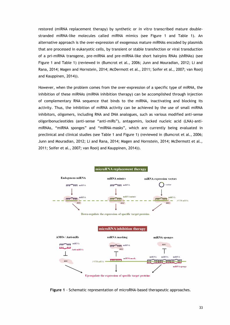
33
restored (miRNA replacement therapy) by synthetic or in vitro transcribed mature double-
stranded miRNA-like molecules called miRNA mimics (see Figure 1 and Table 1). An
alternative approach is the over-expression of exogenous mature miRNAs encoded by plasmids
that are processed in eukaryotic cells, by transient or stable transfection or viral transduction
of a pri-miRNA transgene, pre-miRNA and pre-miRNA-like short hairpins RNAs (shRNAs) (see
Figure 1 and Table 1) (reviewed in (Bumcrot et al., 2006; Junn and Mouradian, 2012; Li and
Rana, 2014; Magen and Hornstein, 2014; McDermott et al., 2011; Soifer et al., 2007; van Rooij
and Kauppinen, 2014)).
However, when the problem comes from the over-expression of a specific type of miRNA, the
inhibition of these miRNAs (miRNA inhibition therapy) can be accomplished through injection
of complementary RNA sequence that binds to the miRNA, inactivating and blocking its
activity. Thus, the inhibition of miRNA activity can be achieved by the use of small miRNA
inhibitors, oligomers, including RNA and DNA analogues, such as various modified anti-sense
oligoribonucleotides (anti-sense “anti-miRs”), antagomirs, locked nucleic acid (LNA)-anti-
miRNAs, “miRNA sponges” and “miRNA-masks”, which are currently being evaluated in
preclinical and clinical studies (see Table 1 and Figure 1) (reviewed in (Bumcrot et al., 2006;
Junn and Mouradian, 2012; Li and Rana, 2014; Magen and Hornstein, 2014; McDermott et al.,
2011; Soifer et al., 2007; van Rooij and Kauppinen, 2014)).
Figure 1 - Schematic representation of microRNA-based therapeutic approaches.

34
miRNA reinstatement Advantages Limitations
miRNA mimics
-Small, synthetic and chemically modified double-stranded miRNA that are processed into single-strand form inside the cells
-They can be loaded into RISC to achieve the downstream inhibition of the target mRNAs
-Mimics the function of endogenous mature or precursor miRNA molecules by direct re-introduction in the cells or tissues
-Increases the levels of a miRNA that is lost during disease progression
-Usually conjugated or encapsulated with different in vivo delivery system, in order to influence the stability and uptake
-Systemic delivery can result in uptake by non-target tissues that normally do not express the miRNA of interest, resulting in potential off-target effects
-Mimics can potentially induce a non-specific interferon response through Toll-like receptors
miRNA expression
vectors
-Plasmid or viral expression vectors constructed with strong promoters that enable the expression of various species of miRNAs in specific tissues
-Restoration of the expression and function of a specific miRNA
-Viral delivery of miRNAs can be optimized to achieve a specific and continuous expression level
-Evidences of transduction efficiency and minimal toxicity
-The vector-based miRNA expression systems are generally less efficient because it is necessary to transcribe the DNA to miRNA precursors by the action of RNA polymerases, which implies the delivery to the nucleus
-Over-expression of shRNA in rats caused hepatotoxicity, organ failure and death
-Argonaute proteins and the pre-miRNA export protein (exportin-5) limit the amount of exogenous miRNA that a cell can tolerate
miRNA inhibiting drugs Advantages Limitations
Antisense oligonucleotides (AMOs), known
as anti-miRs
-Synthetic, single-stranded antisense RNA molecules designed to be complementary to the target miRNA -Competitive inhibition between mature miRNAs and their mRNA targets by Watson-Crick binding
-The targeted inhibition of a specific miRNA and subsequent upregulation of target mRNAs can be achieved, as they function by binding to miRNAs inside the RISC -Under physiological conditions, they are generally unable to distinguish between miRNAs within the same family, especially those with identical “seed regions” -Used in vitro and in vivo to discover gene function, and some AMOs are being tested in clinical trials
-Unmodified anti-miRs are poorly suited to in vivo applications because they are incapable of penetrating negatively charged cell membranes and are susceptible to degradation by serum nucleases -Chemical modifications are required to increase resistance to serum nucleases, to enhance their binding affinity, biostability, specificity for the target miRNAs and to improve their entry into the cell -Most of the chemically modified AMOs show limited tissue distribution when administered in the absence of a carrier, and are taken up by the liver and kidney and rapidly excreted in urine
Table 1 - Overview of the available therapeutic strategies for reinstatement or inhibition of microRNAs.
(reviewed in (Bumcrot et al., 2006; Junn and Mouradian, 2012; Li and Rana, 2014; Magen and Hornstein, 2014, McDermott et al., 2011; Soifer et al., 2007; Zhang et al., 2013))

35
Antagomirs
-A class of chemically modified oligonucleotides (AMOs) which are able to silence endogenous miRNAs -Modified to have a phosphorothioate backbone and a cholesterol group at the 3’-position of the nucleic acid
-Antagomirs are fully complementary to mature miRNAs – competitive inhibition
-Antagomirs are unlikely to be used clinically because the dose required for in vivo inhibition is often high (~80 mg per kg) to achieve the same efficacy as other AMO strategies, which increases the risk of off-target effects -Cannot cross the blood–brain barrier, but can penetrate brain cells if injected directly into the brain
Locked Nucleic
Acid (LNA) anti-
miRs
-Short, single-stranded LNA-modified oligonucleotides -The extraordinary properties of LNA-oligonucleotides enable a reduction in anti-miR size, from 15-16 nucleotides to 8 nucleotides with comparable effects -Addition of an extra methylene bridge connecting the 2’-O atom and the 4’-C atom and ‘locks’ the ribose ring in a C3’-endo or C2’-endo conformation
-Their small size together with their potency/stability/specificity provided by the LNA modifications, makes delivery possible without vehicle-systems (in vivo naked) -Create and enhance high-affinity Watson-Crick binding to target mRNAs and thus exhibit higher thermal stability and superior hybridization with their RNA target molecules
-Only moderate efficiency for miRNA inhibition, possibly because of the tendency of LNA oligonucleotides to form dimers with exceptional thermal stability
“tiny LNAs” -Modified LNA anti-miRs that are 8 nucleotides long and specifically bind to the 5’-seed sequence
-Can block all members of the same miRNA family or of several miRNAs families that share the same seed region, inducing a consequent upregulation of their direct targets
‘miRNA
sponges’
-RNA transcripts with multiple tandem-repeats of miRNA binding sites to block the function of a given endogenous miRNA or a miRNA family -To reproduce the miRNA-mRNA binding, like in the natural setting, a bulge is introduced at position 9-12, which achieve better activity due to increased miRNA retention
-Stably interact with the corresponding miRNA and prevent its interaction with its endogenous target mRNAs -Ability to affect all closely related miRNAs within a family that share overlapping targets -Can be stably integrated into chromosomes, designed to be drug inducible or controlled by promoters whose expression is restricted to a desired cell type, tissue or developmental stage
-Although sponges have been widely used to investigate miRNA function in vitro, their utility in vivo has been limited to transgenic animals in which the sponge mRNA is over-expressed in target tissues. Interestingly, it seems that some large non-coding RNAs could serve as natural sponges to regulate cellular miRNA availability and lead to upregulation of downstream target genes
miR-Mask -Single-stranded 2’-O’methyl-modified antisense oligonucleotides with locked 5’ and 3’ ends that are entirely complementary to the miRNA binding sites in the 3’-UTR of the target mRNA
-‘Mask’ the target mRNA from the endogenous miRNA and thus prevent its suppression -This specific mechanism reduces the off-target effects and is highly target specific

36
2.1. MicroRNA manufacturing
As mentioned above, most miRNAs employed in the development of new therapeutic
approaches, are manufactured either by enzymatic (in vitro transcription) or chemical (via
phophoramidite solid-phase chemical synthesis) methods (Beckert and Masquida, 2011; Ling
et al., 2013; Milligan et al., 1987; Ponchon and Dardel, 2011; Sherlin et al., 2001). In the last
years, new advances in synthetic RNA chemistry were accomplished. Thereafter, the synthesis
process has become more efficient, highly effective, and affordable for large scale production
of RNA, however, the fidelity of the process is inversely correlated with the length of the
oligonucleotide being synthesized (El-Sagheer and Brown, 2010; Ponchon and Dardel, 2007).
In addition, the introduction of chemical modifications (see Table 2) in these strategies allows
reducing the instability of RNA, as well as their degradation by cellular ribonucleases,
increasing the RNA half-life. In turn, this advantage makes difficult to obtain the RNA highly
pure and intact (Broderick and Zamore, 2011; Bumcrot et al., 2006; El-Sagheer and Brown,
2010; Li and Rana, 2014; van Rooij and Kauppinen, 2014). Thus, additional purification
protocols to remove the contaminants (linearized plasmid DNA template, enzymes, free
nucleotides, salts or buffer, short aberrant transcripts, failure in the sequences, among
others) derived from the synthesis process have to be employed in the final RNA product
(Martins et al., 2014; Milligan et al., 1987). Actually, the presence of these impurities can
lead to non-targeted gene silencing, what is commonly associated with a decrease in
therapeutic effectiveness and still restrict the implementation of these RNAs onto pre-clinical
or clinical applications.
On the other hand, miRNA therapeutics can also be obtained via expression systems of
miRNAs encoded by eukaryotic vectors. However, this alternative approach is generally less
efficient because it is necessary to transcribe the DNA to RNA (Chen et al., 2007; Huang et
al., 2011; Li et al., 2014; Ponchon and Dardel, 2007, 2011) and it is dependent of the action
of the RNA polymerase. Thus, considering the rapidly growing interest on these novel
biopharmaceuticals, as a result of its potential therapeutic application, novel technologies to
improve their manufacturing are currently being pursued. Moreover, the success of any
therapeutic will depend on the easy production at large scale, while maintaining maximal
product quality and biological activity. For all these reasons, it is required the development
of economic and efficient methods for large-scale production of miRNAs.

37
Chemical Modifications Features
Phosphorothioate (P=S)
-Exchanges an oxygen atom by a sulfur atom in the phosphate backbone -Inhibits uptake of the passenger strand into the RISC -Promotes binding to serum/plasma proteins, leading to longer serum retention times (half-life), which may induce unwanted side effects, due to reduced clearance by glomerular filtration and urinary excretion -Enhances the resistance to nucleases and cellular uptake -Keeps miRNA silencing function intact -Inhibits coagulation -Can cause cytotoxicity -Activates complement -Improves pharmacokinetic properties, facilitating their delivery into many peripheral tissues in vivo -Perhaps a better choice is modification boranophosphate (P=B) instead P=S substitution, which enhances nuclease resistance without causing cytotoxicity or damage to miRNA silencing function
2´-O-methyl (2’-O-Me)
-Adds a methyl group (-CH3) to the second carbon of the ribose -Occurs naturally in rRNA and tRNA and is safe -Improves in vivo stability in the cytoplasm and increases the resistance to serum nucleases (enhancing half-life) by the closer proximity between the 2´group and 3´phospho-group -Enhances cellular uptake -Minimizes or inhibits the innate immune system activation, as it is a competitive inhibitor of TLR7 -Their introduction into nucleotides within the seed region, can reduce seed region mediated off-target effects without compromising silencing of the intended mRNA -When the sugars of both strands are replaced with 2′-O-methyl groups, the duplex loses its silencing ability
2´-Fluoro
-Adds a fluorine atom to the second carbon of the ribose -Increases in vivo stability and the resistance to exonucleases by the closer proximity between the 2´group and 3´phospho-group -Decreases nonspecific immune stimulation and off-target effects -Can be introduced through endogenous transcription as opposed to chemical synthesis
2´-O-(methoxyethyl) (2’-MOE) -Adds a methoxy-ethyl group (-C3H7O) to the second carbon of the ribose -Increases in vivo stability and the resistance against nucleases in the serum by the closer proximity between the 2´group and 3´phospho-group
Cholesterol conjugation
-Addition of cholesterol functionality at the 3’-position of the nucleic acid to generate ‘antagomirs’ -Enhances serum half-life by binding to serum albumin, improving stability and availability -Enhances the cellular uptake of the modified oligonucleotide by promoting its association with high-density lipoproteins (HDLs) and Low-density lipoproteins (LDL)-that can bind cell surface membrane receptors -Incorporation into HDLs can direct miRNAs to the liver, gut, kidney and steroidogenic organs and LDL-incorporated RNAs are primarily targeted to the liver
Table 2 – Chemical modifications used in miRNA modulators.
(reviewed in (Broderick and Zamore, 2011; Bumcrot et al., 2006; El-Sagheer and Brown, 2010; Li and Rana, 2014; van Rooij and Kauppinen, 2014; Zhang et al., 2013)

38
Note: P=S, 2’-F and 2’-OMe modifications have been extensively used in clinically tested and FDA approved oligonucleotide drugs.
2’-O-allylation -Inhibits activity -Enhances serum half-life
Antibody-conjugated
-The high affinity and binding specificity of antibodies make them attractive vehicles for cell- or tissue-type-specific delivery -Bind multiple miRNAs, creating a particle that bypasses kidney filtration and targets the miRNAs only into cells bearing the cell surface receptor recognized by the antibody -A common approach is to link an RNA-binding protein (e.g. antibody fragment-protamine fusion protein) or domain to Fab fragments isolated from the cell- or tissue-targeting antibody
Locked nucleic acid (LNA) modifications
-LNA is a bicyclic nucleic acid that uses an extra methylene bridge between the 2´-O atom and 4´-C atom also referred to as 2´O-4´C-methylene ‘locks’ the ribose ring in a C3’-endo or C2’-endo conformation -Prolongs half-life in serum -Inhibits nuclease activity -Without adverse affects on the gene-silencing activity -Displays higher aqueous solubility and increased metabolic stability for in vivo delivery and lower toxicity -Reduces the immunostimulatory effects
Folate, various peptides and aptamers
-Help in transport across cellular barriers -Target to specific cells and organs -Improve stability and availability -Frequently associated with impaired function and severe toxicities

39
These issues have spurred a novel interest for recombinant methods using prokaryotic hosts,
which allow the production of large amounts of RNA stable structured. So far, only few
strategies have emerged to produce stable recombinant miRNAs, namely for the biosynthesis
of human mir-27b and human pre-miR-29b, using recombinant bacteria, such as Escherichia
coli (E. coli) and Rhodovulum Sulfidophilum (R. sulfidophilum) as preferential hosts,
respectively (Li et al., 2014; Pereira et al., 2014a). In general, the recombinant approaches
comprise several steps, starting with the selection and construction of appropriate expression
vectors (Nagao et al., 2014; Ponchon and Dardel, 2011; Suzuki et al., 2010). After the
establishment of this step, it is possible to follow to the optimization and selection of the
best culture conditions (dissolved oxygen, temperature, stirring speed, pH and medium
formulation) for miRNAs production by fermentation (Ponchon and Dardel, 2011). This
strategy is usually more cost-effective and simple than the synthesis methods, considering the
large-scale production, allowing the costs reduction, but remaining highly efficient.
2.2. MicroRNA isolation
The RNA recovery, either from the recombinant production strategies using prokaryotic hosts
or from a biological matrix (e.g. tissue samples, whole organisms, cell cultures, bodily fluids),
requires the employment of suitable methodologies for the extraction and isolation of target
RNAs. In general, the procedure includes harvesting of cells by centrifugation and ultimately
cell lysis to release the molecule of interest, since miRNAs are accumulated in cytoplasmic
space (Bernardo et al., 2012; Tan and Yiap, 2009).
Up until now, the most commonly used methods to isolate RNA are the guanidinium
thiocyanate-phenol-chloroform extraction and the solid-phase extraction (SPE) (Bernardo et
al., 2012; Chomczynski and Sacchi, 2006; Tan and Yiap, 2009; Vomelova et al., 2009). The
first method, based on chemical extraction, consists in the disruption of cells using a solution
composed by guanidinium thiocyanate, phenol and chloroform (see Figure 2), followed by
precipitation (Chomczynski and Sacchi, 2006). This organic extraction can also be carried out
using commercial available reagents, such as TRIzol® (Ambion), TRI (Sigma-Aldrich), RiboZol™
(AMRESCO’s) and NZYol (Nzytech), which are recommended to isolate intact and pure total
RNA with high recovery yield, once endogenous ribonucleases are inactivated by the
chaotropic agent used (cationic detergent guanidinium thiocyanate) (Bernardo et al., 2012;
Chomczynski and Sacchi, 2006). However, this chemical extraction is extremely toxic,
biologically hazardous and conveys health risks, which may strongly compromise the success
of several RNA based-procedures in basic and clinical research, due to the use of denaturing
agents (guanidinium thiocyanate and β-mercaptoethanol) and organic solvents (alcohols)
(Chomczynski and Sacchi, 2006; Tan and Yiap, 2009; Vomelova et al., 2009). Furthermore,
this RNA extraction involves multiple and complex processing steps that makes it a time-
consuming and laborious protocol.

40
Figure 2 – Flowchart of the main unit operations to be considered during manufacturing
process of microRNAs. It is shown a comparison between the most common methodologies for
obtaining miRNA from cells, tissues or E. coli with the recombinant production methodology
in R. sulfidophilum. It is also indicated which are the impurities eliminated in each step.
The SPE is an adsorption method based on the ability of RNA to create a hydrogen-binding
interaction with specific surfaces, namely silica gel matrix or glass fiber filter (Tan and Yiap,
2009; Vomelova et al., 2009). In general, in the solid-phase extraction, four steps are
involved: i) cell lysis employing chemical extraction; ii) RNA adsorption to solid support (with
the aid of high pH and salt concentration in the binding buffer); iii) washing; and iv) elution
of the desired RNA from the column (Bernardo et al., 2012; Tan and Yiap, 2009; Vomelova et
al., 2009). Nowadays, there is a variety of commercially available extraction kits (e.g.,
Qiagen (miRNeasy Micro Kit), Roche (High Pure miRNA Isolation Kit), Sigma–Aldrich
(mirPremier microRNA Isolation Kit), Invitrogen (Ambion® PureLink® miRNA Isolation Kit) and
Ambion (mirVana miRNA Isolation kit)), based on SPE or exploiting a magnetic-bead based
extraction technology, in order to isolate RNA molecules from different biological sources
with high quality, intactness and purity for further analytical or laboratorial applications

41
(Mraz et al., 2009). These methods significantly reduce sample preparation time, need less
manipulation and offer safer and simpler operations, allowing a quick and efficient
purification of RNA compared to chemical extraction methods (Bernardo et al., 2012; Tan and
Yiap, 2009; Vomelova et al., 2009). However, some limitations are also associated with these
procedures, such as, low RNA yields due to the low binding capacity of the cartridges, and
sometimes, the RNA isolation is achieved through the use of enzymes, namely DNases that are
usually animal-derived and therefore are not accepted by the regulatory agencies due to the
adverse effects of their pharmaceutical administrations. It is noted that throughout these
extraction processes, several impurities are removed, such as cell debris, genomic DNA
(gDNA) and some proteins, but it still remains a large quantity of impurities mixed with the
target RNA, namely other small RNAs (sRNAs).
In what concerns to the recovery of RNAs from recombinant hosts, and along with the
aforementioned disadvantages, another major issue associated with the cell lysis of the
prokaryotic host, E. coli, is the release of endotoxins, which can lead to the contamination of
the RNA sample (Wei et al., 2007). The importance of removing endotoxins is related to the
possibility of these foreign components to originate adverse effects and inflammatory
responses to the patients (e.g. symptoms of toxic shock syndrome), if present in quantities
higher than those acceptable (Wei et al., 2007). In addition, as E. coli expresses several
endonucleases, in particular RNases, it can be difficult to maintain the integrity of the target
RNA, being always necessary to establish methods to avoid degradation (Wassarman et al.,
1999). For all these reasons, an ideal bioprocess must be designed to maximize recovery
yield, to reduce the unwanted contaminants (sRNAs, gDNA, proteins, endotoxins, salts,
denaturing agents and organic solvents) and to minimize sample transfers. Nonetheless, and
considering the therapeutic applications of these products, the major interest is not only to
produce high quantities of the target RNA but also to assure its quality, stability, integrity and
biological activity, fulfilling the requirements of regulatory agencies.
To accomplish this, an innovative recombinant bacteria, R. sulfidophilum, has been recently
proposed and used to produce a pre-miRNA. It is non-pathogenic and presents several
advantages over E. coli, such as the unusual capacity of secretion of the heterologous nucleic
acids directly into the culture medium and the absence of detectable host ribonucleases in
the culture medium (Ando et al., 2004, 2006; Suzuki et al., 2010). This prokaryotic host can
integrate a promising alternative technology, since it allows the recombinant biosynthesis and
recovery from the extracellular medium of biologically active miRNAs, circumventing the
need of cell lysis that frequently induces RNA denaturation. Besides, this strategy can be
highly advantageous regarding the RNA product safety, as it will not stimulate innate
immunity because the secreted miRNAs will be devoid of main bacterial associated impurities.
Moreover, it is possible to minimize the contamination of the RNA fraction, by recovering the
enriched-miRNA extracellular medium, in a specific stage of cell growth (see Figure 2).
Indeed, it is expected that the application of this strategy allows suppressing the use of

42
organic and toxic solvents, and results in an increased RNA recovery yield, drastically
reducing the amount and type of impurities present in the RNA extract, prior to purification.
Thus, this integrative approach of recombinant production and pre-miRNAs extracellular
recovery, is not only innovative but also presents more advantages than the conventional RNA
isolation methods, being more economical, efficient, simple and fast in obtaining functional
RNA. The recovery and isolation of miRNAs from fermentation broths can be performed using
membrane filtration or concentration techniques, designed to eliminate media components
and metabolites and recover the target RNAs (Morão et al., 2011; Nunes et al., 2014; Nunes
et al., 2012). Figure 2 schematizes the most important steps to be considered during miRNAs
manufacturing. It is essential to optimize and improve all unit operations involved in the
biotechnological production and recovery of miRNAs, which will comprise a great benefit to
the implementation of a new platform on the biopharmaceutical industry, since the primary
isolation conditions will also greatly influence the following purification strategy.
2.3. MicroRNA purification
During the last decades, different methods and protocols have been extensively employed for
the purification of RNAs obtained either by synthesis or from biological sources, such as
preparative denaturing polyacrylamide gel electrophoresis and different chromatographic
strategies namely reversed-phase ion-pair liquid chromatography, anion-exchange and size
exclusion chromatography, which can be applied either as an isolated or integrated steps
(Easton et al., 2010; Kim et al., 2007; Koubek et al., 2013; McCarthy et al., 2009; Noll et al.,
2011). Although these methods can be very efficient for the purification of RNA with high
resolution, some of these still require time-consuming preparatory steps and are expensive on
large scale (Martins et al., 2014). On the other hand, these purification methods can cause
structural modifications and degradation of the RNA molecules and may even introduce some
contaminants, e.g. acrylamide, lithium metal, ion-pairing agents and organic solvents,
related with the method (Easton et al., 2010; Kim et al., 2007; Koubek et al., 2013; Martins
et al., 2014; McCarthy et al., 2009; Noll et al., 2011). For these reasons, the RNA product
purified by these strategies may require additional treatments to be suitable for the use in
analytical and clinical applications, and therefore it is difficult to maintain the stability and
biological activity of the target RNA. These challenges associated with RNA purification
process, motivate the improvement of the already existing chromatographic techniques or the
development of new purification approaches.
In the last years, the downstream strategies based on affinity chromatography have been
developed in an attempt to circumvent several challenges in RNA purification (reviewed in
(Martins et al., 2014)). Most approaches described in affinity chromatography make use of
tags (small sequences that are introduced in the RNA molecules) that bind with high affinity
to specific molecules, used as ligands in chromatographic matrices for the selective
purification of RNAs from cellular extracts (Di Tomasso et al., 2011, 2012; Pestourie et al.,

43
2005; Ponchon and Dardel, 2011; Srisawat and Engelke, 2001, Srisawat et al., 2001). In most
cases, the tag-RNA complexes can be released from the chromatographic support either by
competitive elution or cleavage by a protease that recognizes a specific site that is
incorporated along with the affinity tag (Martins et al., 2014; Srisawat and Engelke, 2001;
Srisawat et al., 2001). Overall, although these chromatographic methods are rapid, reliable
and efficient in obtaining the target RNA with high recovery yields and stability, they still are
expensive on large scale and labor intensive (Martins et al., 2014). In addition, the separation
procedures with RNA affinity tags make use of enzymes and may induce structural
modifications in RNA by the introduction of the tag sequences, which can affect the overall
RNA topology (Srisawat and Engelke, 2002; Walker et al., 2008).
A promissory approach to overcome these limitations includes the recent developed affinity
chromatographic method using amino acids as specific ligands, which simulates and exploits
biological and reversible interactions that occur at the cellular level between amino acids and
nucleic acids. The major advantage of this technique relies on the selectivity achieved,
resultant from the combination of multiple non-covalent interactions (including electrostatic
and hydrophobic interactions, van der Waals forces, cation–π interactions, dipole–dipole
forces and hydrogen bonding) between the biospecific ligands (amino acids and derivatives,
namely O-Phospho-L-Tyrosine, L-Arginine, L-Lysine and Agmatine) and the target RNAs. The
interactions can be favored or disfavored by manipulating the binding/elution conditions,
such as the temperature, pH and buffer composition (type of salt, ionic strength or presence
of competitive agents) (Sousa et al., 2008). The use of amino acids in this chromatographic
strategy has the singular ability of purifying nucleic acids with high-selectivity, -specificity, -
efficiency, robustness and durability (Lowe et al., 2001; Martins et al., 2014; Sousa et al.,
2008). The potential of amino acids-based affinity chromatography to obtain miRNA
molecules, in particular the recombinant human pre-miR-29b, from a complex extract, was
recently proved (Afonso et al., 2014; Pereira et al., 2014a, 2014b, 2014c).
Although, amino acids immobilized in bead-packed columns showed high selectivity in the
purification of miRNAs, these supports present certain restrictions, such as low capacity and
flow rates (Arrua and Alvarez Igarzabal, 2011; Pfaunmiller et al., 2013; Sousa et al., 2012).
Considering this limitation, an easier, reliable and more robust procedure for miRNA
purification was developed, using a monolithic-based strategy, to improve the performance of
the biotechnological strategy, while maintaining the efficiency of the method (Pereira et al.,
2014c). Overall, the separation of the target miRNA from a crude sample was achieved, with
a good recovery yield, high purity level, good integrity and biological activity, due to the fast
separation and consequent short contact time with the support (Pereira et al., 2014c).
Amongst the several chromatographic strategies implemented for miRNA purification, amino
acids-based affinity chromatography allowed an improvement of many technical issues,
including the analysis speed, the sensitivity and the separation conditions (use of mild salt
conditions instead of organic compounds and enzymes). Furthermore, miRNA is separated in

44
one single step, improving process economics over other less-selective and time-consuming
multi-step procedures (Afonso et al., 2014; Pereira et al., 2014a, 2014b, 2014c).
In accordance, different affinity ligands (amino acids and its derivatives, peptides,
complementary oligonucleotides sequences to the target miRNAs, among others) can be
characterized and immobilized onto different chromatographic matrices (agarose-based,
monoliths, superporous matrices), in order to purify the RNA of interest from host
contaminants, thus minimizing non-targeted gene silencing and immunologic responses.
2.4. MicroRNA quality control
RNA purification is a critical step to obtain the product with suitable quality to be applied in
several biomedical fields. At present, none regulatory authority (such as Food and Drug
Administration (FDA) centre for Drug Evaluation and Research, European Agency for the
Evaluation of Medical Products (EMEA) or World Health Organization (WHO)) has formal
guidelines available for RNA oligonucleotide products (Martins et al., 2014). Over the years,
the scientific community is providing the guidance in the regulatory framework for RNA-based
therapies based in the guidelines for plasmid DNA (pDNA) (Ferreira et al., 2000; Stadler et al.,
2004).
As previously mentioned, miRNA quality depends on the manufacturing process, the cellular
lysis step and the subsequent processes of isolation and purification, which must be carefully
designed in order to reduce or eliminate the host impurities without loss or damage of the
biological activity of desired RNA. To accomplish this, a strict control of the process and
assessment of the remaining impurities on the final sample is crucial, by using several
complementary methods (Batkai and Thum, 2014; Chen et al., 2005; McGinnis et al., 2012;
Moreno-Moya et al., 2014; Schleef and Schmidt, 2004; Smith et al., 1999; Stadler et al.,
2004). Table 3 describes several effective techniques that are commonly applied to evaluate
the quality and quantity of purified miRNA, and to verify if the requirements of regulatory
authorities are fulfilled. There are also commercially available analyzers to quantify and
simultaneously check the integrity of RNA samples (e.g. Bio-Rad Experion, Hercules, CA, or
Agilent Bioanalyzer 2100, Santa Clara, CA). The implementation of methodologies able to
control RNA quality will be increasingly necessary, especially when the RNA products are
finally released to the market. To overcome this issue, convenient, sensitive and
straightforward analytical methods have been developed and validated to detect and quantify
specific miRNAs in unfractionated total RNA from different biological samples, namely whole
blood, plasma, cells and some organs (such as liver, heart, brain, spleen, lungs and kidneys)
(reviewed in (Batkai and Thum, 2014)).

45
Table 3 - Accepted levels of impurities in final pDNA preparation for clinical applications, as
recommended by regulatory agencies (adapted from (Batkai and Thum, 2014; Chen et al.,
2005; Ferreira et al., 2000; McGinnis et al., 2012; Moreno-Moya et al., 2014; Schleef and
Schmidt, 2004; Smith et al., 1999; Stadler et al., 2004)).
3. Obstacles to effective microRNA therapeutics
As discussed earlier in this review, the therapeutic application of miRNAs is extremely
promising due to the specific and efficient silencing of gene expression, as well as for its
simplicity, safety profile and ease of manufacturing. However, some problems must be
addressed, namely the extracellular barriers, low bioavailability, rapid enzymatic degradation
of miRNA by ubiquitous nucleases found in human serum, rapid renal clearance and
phagocytosis by macrophages (Al-Dosari and Gao, 2009; Pathak et al., 2009; Tokatlian and
Segura, 2010). Also, the intracellular barriers, such as inefficient cellular uptake and
intracellular processing of endosome-targeted RNAs (escape from the endosome, vector
unpacking and processing by the RNAi machinery) need to be surpassed before the
therapeutic gene arrives at the cytoplasm, allowing an improvement on miRNA
pharmacokinetic and pharmacodynamic properties (Al-Dosari and Gao, 2009; Pathak et al.,
2009; Tokatlian and Segura, 2010).
Component Accepted level in final
product Recommended Assay
Host RNA Undetectable
Agarose gel electrophoresis Polyacrylamide electrophoresis Analytical High-Performance Liquid Chromatography Spectrophotometric Method
Proteins Undetectable Micro-BCA (bicinchoninic acid) protein assay Bradford assay SDS-PAGE
Genomic DNA <2 µg/mg pDNA Real-time Polymerase Chain Reaction Southern blot Hybridization
Endotoxins <0.1 EU/µg pDNA Limulus Amoebocyte Lysate (LAL) assay
miRNA
Polyacrylamide electrophoresis Electrospray ionization mass spectrometry TaqMan-based arrays Reverse-transcriptase polymerase chain reaction Northern blot analysis Nuclease protection analysis Primer extension assay Microarray analysis In situ hybridization assay Complementary DNA (cDNA) library construction High-Performance Liquid Chromatography In Vitro Translation LC-MS

46
3.1. Biological instability
As previously discussed, for the systemic delivery, unmodified naked miRNAs are highly
susceptible, due to their rapid degradation by cellular and serum nucleases, resulting in a
short half-life in the circulatory system (from several minutes to an hour) (Deng et al., 2014;
Gavrilov and Saltzman, 2012). Chemical modifications can increase the RNA stability but are
also associated with certain adverse effects, such as decrease on mRNA hybridization,
inhibition of coagulation, activation of the complement cascade and cytotoxicity (see Table
2) (reviewed in (Broderick and Zamore, 2011; Bumcrot et al., 2006; Deng et al., 2014; El-
Sagheer and Brown, 2010; Li and Rana, 2014; van Rooij and Kauppinen, 2014). For all these
reasons, it is still necessary to customize, optimize and/or minimize the chemical
modifications, through the identification and modification only of the sites susceptible to
degradation.
3.2. Off-target effects
The in vivo specificity of miRNAs is based on sequence homology between miRNA and target
mRNA. Thus a major challenge in developing miRNAs-based therapeutics remains in the
assurance of RNA purity to minimize off-target gene silencing (Deng et al., 2014).
Nonetheless, miRNA-based therapeutics can sometimes induce non-specific side effects, i.e.,
suppression of other non-target genes, which are as critical as effectiveness and duration of
miRNA expression or inhibition (as reviewed in (Singh et al., 2011)). The off-target effects can
arise through the partial homology of the nucleotides involved in the “seed region” of the
endogenous miRNAs, and by effects related with competition of exogenous vectors and/or
RNA species and endogenous miRNAs by the incorporation and retention into RISC, resulting in
the non-desirable gene silencing (Gavrilov and Saltzman, 2012). Another possible side effect
is the saturation of the cellular machinery by miRNA processing, leading to aberrant
processing of endogenous miRNAs by the accumulation of toxic precursors (pri-miRNAs, pre-
miRNAs, shRNA, stem-loop structures and high expression of miRNA mimics) (Singh et al.,
2011; Wang et al., 2011). This saturation interferes with the normal functioning of the cell,
not allowing access of the miRNAs in the natural pathway (Ballarin-Gonzalez and Howard,
2012). Most of these undesirable and unspecific effects can be minimized without impairing
their silencing capability, with small changes in miRNA sequences (chemical modifications) or
by the use of miRNA mimics that are specific enough to distinguish between similar miRNAs.
Despite of all the progress that has been made in the miRNAs-based therapeutics, it is still
required more basic research on miRNAs to increase the efficacy and significantly reduce and
eventually eradicate off-target effects.
3.2. Immune response
Long double-stranded RNA (e.g. vector-mediated expression of shRNAs, viral dsRNAs or other
dsRNAs), as well as, siRNAs synthesized in vitro can, in some cases, trigger an innate immune

47
response via interaction with RNA-binding proteins such as Toll-like receptors (TLRs) and
protein kinase receptor (PKR) (Ballarin-Gonzalez and Howard, 2012; Deng et al., 2014). The
innate immune response is mediated mainly by activation of interferon and pro-inflammatory
cytokines, after recognition of immunostimulatory sequence in the RNAs, such as specific GU-
rich sequences (“danger motif”), in particular 5’-GUCCUUCAA-3’ and 5’-UGUGU-3’ (Deng et
al., 2014; Gavrilov and Saltzman, 2012). In this manner, there are several parameters
determining the stimulation of innate immunity, namely structure and sequence of RNAs,
delivery vehicle, delivery route, cell type and species. To minimize these issues, appropriate
chemical modifications as well as, the possibility to use miRNA delivery strategies can
prevent, decrease and avoid the immunostimulatory properties (see Table 2).
Understandably, immunogenicity and toxicity are grounds for concern that must be addressed
in developing RNAi for therapeutic use.
4. Delivery Strategies
The major challenge in the development of miRNAs-based therapeutics for clinical
applications is the establishment of an effective mode to deliver these molecules to their
target cells, specific tissues or organs, in order to exert their function, preventing unwanted
side effects. The physicochemical properties of naked miRNAs interfere with their ability to
cross the cell membrane by passive diffusion due to their negative charge, hydrophilicity and
size (7-20kDa) (Zabner et al., 1995; Zhang et al., 2013). In addition, naked miRNAs are
preferentially accumulated by the kidneys and rapidly eliminated into urine within one hour
because they are smaller than the size threshold for glomerular filtration.
In the last years, a variety of different and efficient delivery approaches have been
developed and applied in gene therapy trials to promote cellular uptake and delivery of RNAs
into the cytoplasm of target cells, increasing the accumulation of the therapeutics in the site
of interest, increasing the silencing potency, thereby making a given treatment dose more
effective. In a simple way, delivery systems can be divided into two categories, viral vectors
and non-viral vectors. However, each of these approaches has distinct advantages and
disadvantages, which require careful consideration (see Table 4). Initial research focused on
the use of viral vectors because these vectors displayed high efficiency at delivering miRNAs,
both in vitro and in vivo, taking advantage of their favorable cell uptake and intracellular
trafficking machineries, allowing long-term gene expression (see Table 4) (Mintzer and
Simanek, 2009; Robbins et al., 1998; Walther and Stein, 2000). However, the focus has
recently changed to the non-viral approaches because of the advantages over viral vectors,
such as ability to deliver the exogenous biopharmaceuticals into the cell/tissue-specific, non-
immunogenicity, high biocompatibility and low cytotoxicity (De Smedt et al., 2000; Nimesh
and Chandra, 2009; Park et al., 2006). In addition, these non-viral vectors offer other
advantages such as relatively low production costs, high flexibility and easy quality control,

48
allowing designing carriers with well-defined structural and chemical properties on a large
scale with high reproducibility and simplicity. Thus the potential for large-scale production
makes these non-viral vectors increasingly attractive for gene therapy (see Table 4) (Ahn et
al., 2002). These vectors are also relatively stable to storage, they can be administered
repeatedly with no or little immune response and the dimension of the genetic material they
can carry is virtually unlimited. However, they also present some limitations such as the
reduced transfection efficiency, due to the cellular barriers and immune defense
mechanisms, poor oral bioavailability and instability in circulation (see Table 4).
The non-viral delivery systems include liposomes, lipoplexes, lipid-based delivery systems,
polymers, cyclodextrins, dendrimers, polymeric micelles, pluronic block copolymers,
exosomes and metallic core nanoparticles, allowing for RNA compacting for systemic delivery
(see Table 4) (Chen and Xie, 2012; Cho et al., 2008; Gao and Huang, 2009; Hart, 2010;
Kabanov et al., 2002; Mintzer and Simanek, 2009; Morille et al., 2008; Patil and Panyam,
2009; Yin et al., 2014; Zhang et al., 2013).
Consequently, during the last decade a number of polymeric delivery systems, nanoparticles
with sizes between 10 and 100 nm, have been developed. These systems should contain a
positively charged domain to efficiently bind the miRNAs molecules and facilitate the
progression of the transmembrane transport (Al-Dosari and Gao, 2009; Gupta et al., 2005;
Hart, 2010; Zabner et al., 1995). In these systems, the miRNAs can be loaded, adsorbed or
chemically linked to their surface. The nanoparticle size is a critical factor for effective drug
delivery in vivo, since nanoparticles should be big enough to avoid fast clearance through the
kidney excretion system but small enough to penetrate the target tissue. As mentioned, to be
effective and to be considered ideal, there are several aspects to be carefully considered
when developing the delivery vehicles, such as, safety, specificity and efficiency of gene
transfer; magnitude and duration of expression; immunogenicity and manufacturing (Pathak
et al., 2009).
After entry into the target cells, miRNAs still face a number of hurdles before they can exert
their gene silencing activity, since non-viral delivery systems are immediately transported
into the endocytic vesicles, initially, to the early endosomes where the pH drops to 6
followed by trafficking to late endosomes which are acidified to pH 5-6 (see Figure 3)
(Caracciolo et al., 2009; Dominska and Dykxhoorn, 2010; Medina-Kauwe et al., 2005). Once
taken up into the endosome, the RNA has to escape in order to avoid subsequent degradation
by lysosome (Caracciolo et al., 2009; Dominska and Dykxhoorn, 2010; Medina-Kauwe et al.,
2005). Even in the cell cytoplasm, miRNAs remain vulnerable to degradation by intracellular
nucleases and still need to be incorporated into RISC with high efficiency, to exert the
silencing function (see Figure 3) (Nguyen et al., 2009; Suh et al., 2003).

49
Non-Viral Delivery (Cho et al., 2008; Gao and Huang, 2009; Hart, 2010; Mintzer and Simanek, 2009; Morille et al., 2008; Patil and Panyam, 2009; Zhang et al., 2013)
Lipid-based delivery systems
Liposome
-Consisting of a phospholipid bilayer with an inner aqueous core that contains RNA -Versatility and flexibility in structure -It is an attractive delivery approach because of the biocompatibility of the constituents and facile assembly of the complexes, which requires only mixing and incubation of components -Higher stability in biological fluids and against the enzymatic metabolism -Increase retention in the blood stream -The elimination half-life increased from ∼2 min to 6.5 h -Excellent RNA delivery efficiency in multiple cell lines in vitro as well as in mice, rats and monkeys -Facilitate efficient internalization of RNA via membrane fusion with the host cell -Are usually heterogeneous in size owing to interactions between water molecules and the hydrophobic groups of lipids -Traditional liposomes have low transfection efficiency into cells due to their lack of surface charges -Highly toxic for in vivo applications, nonspecific uptake, and unwanted immune response
Lipoplexes
-Liposomes that contain cationic lipids driving the interaction between the lipid bilayer and the negatively charged RNA molecules, resulting in a net positive charge that enable the binding to anionic cell surface molecules -The composition of these lipid particles can be tailored to facilitate fusion with the cytoplasm, as well as to promote endosomal release once inside the cell
Neutral Lipids1 DOPC; DOPE; DSPC Cationic lipids2 DOTMA; DOGS; DC-Chol; DDAB; DMRIE and DOTAP
-Lipid vectors are presumed to achieve endosome release of RNA through a membrane fusion mechanism -Neutral lipids are non-toxic and do not activate an immune response -Cationic lipids can complex electrostatically with RNAs, enhancing cell entry and protection against serum enzymes -Incorporation of positive charge to increase transfection efficiency must be carefully balanced because it can induce inflammatory effects and unwanted interaction with negatively charged serum proteins, which can lead to opsonization and clearance of the lipocomplex
Biodegradable polymers
Polyethylenimine (PEI)
Poly(lactic acid)
Poly(lactic-co-glycolic acid)
Poly(butylcyanoacrylate)
Chitosan (CS)
Polylysine
Poly(alkylcyanoacrylate)
Poly(butylcyanoacrylate)
-High delivery efficacy, biodegradable, biocompatible and non-toxic -Higher stability in biological fluids and protection of the RNA against degradation by RNases -Reduced non-specific biodistribution -Have capacity to encapsulate large amounts of genetic material and co-delivery -Facilitate the cellular uptake via endocytosis -Polymeric vectors commonly use the proton sponge effect to facilitate endosome escape -Can readily be surface-modified to enhance stability, transport properties, targeting or uptake and can be produced in relatively homogeneous sizes (up to 100 nm) -Successfully used to deliver poorly diffused drugs into the brain -Low toxicity and low immunogenicity -Successfully administered to silence target genes in vitro and in vivo, such as p-glycoprotein in brain endothelial cells -PEI act as a proton sponge that induces the release of RNA to the cytoplasm by osmolysis from the endosome due its high content of protonable amino groups -PEI is a highly efficient cationic polymer since it has numerous amine groups in its structure -PEI showed its dose-dependent cytotoxicity due to non-biodegradability inside cells that results in the formation of aggregates with negatively
Table 4 – Barriers to successful in vivo delivery of microRNAs using non-viral vectors.

50
charged intracellular proteins -Use of a high-molecular-weight branched PEI leads to high transfection efficiency but to unfavorable adverse effects such as cellular toxicity -CS is a natural, nonallergic and has mucoadhesive properties, and nuclease resistance -The degradation time of PLGA can be altered from days to years by varying the molecular weight and the lactic acid to glycolic acid ratio of the copolymers -Increase the duration of drug circulation in the blood, which facilitates drug ability to interact with specific molecules expressed on the luminal side of BBB endothelial cells, and consequently to cross the BBB, increasing the uptake of appropriate drugs in the brain safely and effective
Cyclodextrin (CD)
Naturally cyclic oligomers of glucose (oligosaccharides) that are complexed with
transferrin
-CD possesses unique structural features, with hydrophilic outer surface and hydrophobic inner cavity -CD containing polymers (CDPs) consist of cationic polymer complexed with RNA and interdigitated with funnel-like cyclodextrin molecules, which, in turn, can be linked to functionalized molecules -CDPs have been investigated intensely in targeted delivery of small therapeutic molecules due to its non-toxicity and not producing immune stimulation even if the RNA cargo contains an immune stimulatory sequence
Polymeric micelles
Polymeric micelles
-Easy to formulate and therefore exist with variable well-defined geometry where insoluble/sparingly soluble bioactive molecules can be incorporated at different sites in micelles -An ideal candidate for brain-targeting delivery -Small particle size that allows escaping from the reticuloendothelial system -Offer protective stability within their hydrophobic core -Flexibility of modification with targeting ligands that can effectively enhance drug solubility and improve drug pharmacokinetics and biodistribution -Require less excipients -High physical stability -Sustained drug release -Self-assembled nanostructures composed of amphiphilic block copolymers can be tuned for RNA delivery by grafting them with amines that can complex RNA -Can act as nanosized structures to solubilize and release hydrophobic drugs
Dendrimers PAMAM Dendrimers
Polypropylenimine Dendrimers
-Ability to control their well-defined structure and molecular weight, shape, degree of branching, flexibility, as well as the “multivalency effect” -Easily modified surface groups -Low polydispersity and high functionality -Improved solubility, pharmacokinetics and biodistribution -High loading capacity and high transfection efficiency -Low toxicity and low immunogenicity -Can also act as a proton sponge, triggering endosomal escape and release RNA into the cytoplasm due its tertiary amine groups inside the repeating backbone structure -Offer a new length scale (~5–10 nm), with significant versatility to incorporate multiple active molecules -Display the ability to cross cell membrane -A number of biologically and pharmacokinetically desirable properties, however, controlled drug release and high drug loading still remain challenge -PAMAM dendrimers require further modifications of their peripheral end groups since the intact structure shows low transfection efficiency -They are cleared rapidly by the bloodstream, preventing ‘long-term’ accumulation in non-targeted organs, such as kidney, lung and liver,

51
1Neutral Lipids: 1,2-Oleoyl-sn-Glycero-3-phosphocholine (DOPC); 1,2-Dioleoyl-sn-Glycero-3-phosphoethanolamine (DOPE); 1, 2-distearoyl-sn-glycero-3-phosphocholine (DSPC); 2Cationic lipids: N-[1-(2, 3-dioleoyloxy) propyl]-N,N,Ntrimethylammonium chloride (DOTMA), dioctadecylamido glycylspermine (DOGS); 3b[N-(N′,N′-dimethylaminoethane)-carbamoyl) cholesterol (DC-Chol); dimethyldioctadecyl-ammonium bromide (DDAB), dimyristyloxypropyl-3-dimethylhydroxyethyl ammonium bromide (DMRIE) and 1-oleoyl-2-[6-[(7-nitro-2-1, 3-benzoxadiazol-4-yl) amino]hexanoyl]-3-trimethylammonium propane (DOTAP)
reducing the potential for side effects -The cytotoxicity of PAMAM increases proportionally with the generation number
Pluronic block copolymer
-Enhance the transport of drugs across in vitro models of the BBB, which it is related to the inhibition of P-glycoprotein-mediated efflux -The polymer may have potential as a CNS-targeted delivery system for drugs that are substrates for the P-glycoprotein efflux pump
Exosomes
-Endogenous nanovesicles secreted by several cells and tissues that transport the desired RNAs -The specific targeting of exosomes to the brain following systemic delivery -Ability to escape an immune response (reduced immunogenicity), thus presenting reduced toxicity -The possible effects of nucleic acids and proteins derived from dendritic cells and carried with the exosomes on the target cell need to be further explored
Metallic core nanoparticles
Gold nanoparticles (GNPs)
-Metal cores, such as iron cobalt, iron gold, or iron nickel are coated with various peptides, proteins, antibodies and other biomolecules generating a core-shell structure to which RNA can be externally conjugated through linking molecules such as thiols dextran, cationic polymers or biotin-streptavadin -Ability to be synthesized at diverse sizes, their chemical stability and their unique optical properties -Allow the study of biodistribution upon injection using magnetic resonance imaging or targeting to specific tissues by applying external magnets -In vivo toxicity may limit their application
Viral Delivery Systems Reviewed in (El-Aneed, 2004; Mintzer and Simanek, 2009; Robbins et al., 1998; Walther and Stein, 2000)
Viral
Retrovirus vectors
Lentivirus vectors
Adenovirus vectors
Adeno-associated viruses
Herpes simplex viruses
-To express the virulent genome, the virus needs to infect the host cells, thereby inducing prolonged gene silencing -A single administration could lead to durable down-modulation of the targeted pathological protein -High efficiency in in vivo delivery because the ability to deliver the gene into the cell is an inherent capacity that they possess -Transient gene silencing -LV and AVV were the most used, as they have the best safety records -Easily eliminated by existing bloodstream antibodies -Can activate coagulation or complement factors to induce mutation in the host genome, which may lead to strong inflammatory reactions -Allergy and infection caused by viruses can be fatal -Their immunogenicity, cytotoxicity and the possibility of mutagenic effects make them risky -The use of these delivery vehicles is mostly moderated in what concerns to their manufacturing and scale-up procedures

52
4.1. Targeting
In the development of miRNAs-based therapies, delivery of miRNAs for the treatment of NDs
is intrinsically limited because the delivery of all substances into the brain is tightly regulated
by the Blood-Brain Barrier (BBB) (Edwards, 2001). Physiologically, the BBB is a unique
structure that separates brain from the rest of the body and their barrier function is due to
anatomical features, namely the tight junctions formed between adjacent brain capillary
endothelial cells in the brain tissues (see Figure 3) (Alam et al., 2010).
Figure 3 – Main barriers to successful in vivo delivery of nucleic acids using non-viral vectors.
This barrier prevents the transport of many harmful substances (foreign organisms, toxic
chemicals, hydrophilic drugs and large molecules) present in the systemic blood circulation
into the brain and restricts the movement of ions and fluid into the brain (Alam et al., 2010).
However, the BBB allows selective access of necessary nutrients (small gaseous agents,
glucose, amino acids and small lipophilic molecules) and chemical signaling molecules to the
nervous system (Egleton and Davis, 1997). Thus, the BBB controls substance flow in and out of
the brain with precision and strictness, ensuring an optimal environment for brain function,
making the brain a site of poor permeability to various drugs as well as delivery systems.
Another important factor that limits and control the uptake, distribution and efflux of many

53
drugs in the nervous system are the proteins presents in a variety of drug efflux transporters,
such as P-glycoprotein (Kusuhara and Sugiyama, 2001), glucose-related transporters,
nucleoside transporters, receptors for transferrin (Descamps et al., 1996, Visser et al., 2004),
lactoferrin (Fillebeen et al., 1999), insulin (Frank et al., 1986), leptin, lectins, epidermal
growth factor, insulin-like growth factors (Duffy and Pardridge, 1987; Reinhardt and Bondy,
1994), as well as, low-density lipoprotein receptor-related protein (LRP) (Candela et al.,
2008; Dehouck et al., 1997), reinforcing BBB function by effectively removing drugs from the
brain and pumping them back into blood. Because of this BBB transport restriction
mechanism, more than 98% of candidate drugs have been abandoned during their
development due to their poor permeability to cross the BBB, in sufficient quantities to
produce a therapeutic effect (Pangalos et al., 2007; Pardridge, 2005). These findings are
attracting the attention of the pharmaceutical and biotechnology industries.
To accomplish this purpose of targeted delivery, nanoparticles have been modified on the
surface with targeting molecules to improve their interactions with the BBB. As mentioned,
the surface of the brain endothelial cells of the BBB possess several transport mechanisms
mediated by various specific receptors that potentially can be exploited as a means to target
drugs to the brain. Small molecules, peptides and proteins, such as angiopep-2 (van Rooy et
al., 2011), certain apolipoproteins and lipoproteins (Candela et al., 2008; Kreuter et al.,
2002, 2007; Michaelis et al., 2006) directed to low-density lipoprotein receptor (Dehouck et
al., 1997) and RVG (rabies virus glycoprotein) (Kim et al., 2013; Son et al., 2011) to GABA
receptor have been extensively used as brain-targeting ligands for the development of drug
delivery systems to the brain, trying the BBB crossing after intravenous administration. A
common drug delivery strategy for targeting cells of interest is the conjugation with proteins,
including lactoferrin (Fillebeen et al., 1999), TAT (transactivator of transcription) peptides
(Santra et al., 2004) and transferrin (Descamps et al., 1996) that can effectively facilitate the
passage of the BBB (Zhang et al., 2012). An alternative strategy to enhance drug delivery to
the CNS is the co-administration of a pharmacological modulator or a formulation component,
in order to enhance brain penetration of various P-glycoprotein substrates through inhibiting
the P-glycoprotein-mediated efflux transport system of a desired therapeutic agent out of the
brain as, for example, Pluronic P85 (Batrakova et al., 2001; Yi and Kabanov, 2013).

54
5. Conclusion and Future trends
MiRNAs are of particular interest in understanding complex disorders, such as NDs, because
they can potentially regulate several pathways involved in the insurgence and progression of
the disease. Recent developments have suggested that the use of miRNAs as
biopharmaceuticals will be possible in a near future and they will become not only important
to treat ND, but may also be employed as biomarkers. The biopharmaceutical sector is an
important and fast growing part of the wider global pharmaceutical industry, developing
healthcare products. However, the development and preparation of biopharmaceuticals poses
many technical challenges, including their production, recovery, purification and delivery into
the brain, which need to be addressed in order to carry these molecules into clinical trials.
Thus, it is necessary the development of effective technologies for miRNAs recombinant
production and purification with the intent of obtaining large amounts of highly pure and
thus, clinically suitable miRNAs, as an alternative to in vitro transcription or chemical
synthesis. Within the biotechnological domain, we believe that the integrative approach of
extracellular production of stable structured RNA in vivo coupled to affinity-based
purification using amino acids as ligands will enable the target pre-miRNAs isolation with high
efficiency, selectivity, throughput, purity and integrity. The major gain in this field is the
possibility to use these methodologies for the preparation of other non-coding RNAs, proving
the wide application of the technology, which may have a great impact on biopharmaceutical
industry. Furthermore, these approaches could contribute for the establishment of reliable
and cost-effective processes, easily adopted by biopharmaceutical industries. In general, this
review reports the most recent improvements achieved with the preparation of recombinant
miRNAs that meet the required criteria established for clinical application. In addition,
another major bottleneck is to achieve a highly targeted delivery of biopharmaceuticals
because only then it will be possible to create a significant opportunity to generate new
health products useful for diagnosis and treatment of NDs. It is necessary to develop effective
brain drug delivery systems that must also be non-invasive, safe, with a low-cost and easy
route of administration, for increasing treatment efficacy and patient compliance, reducing
societal service burden. The success of these targeted delivery systems preparation will be
really impacting since it will open the way for the delivery of numerous drugs to the brain,
with relevance not only in AD but also transversal to other neurodegenerative diseases. In
fact, the surpass of all issues herein discussed, will lead to the establishment of more cost
efficient processes that would reduce the time to application.

55
6. Acknowledgments
Patrícia Pereira acknowledges the fellowship, SFRH/BD/81914/2011, from FCT (Portuguese
Foundation for Science and Technology). The authors also acknowledge the program COMPETE
(FCOMP-01-0124-FEDER-041068 – EXPL/QEQ-MED/1068/2013), the FCT project (Pest-
OE/SAU/UI0709/2014) and the program Fundo Europeu de Desenvolvimento Regional (FEDER)
[COMPETE [FCOMP-01-0124-FEDER-027560]].

56
7. References
Afonso A, Pereira P, Queiroz JA, Sousa A, Sousa F. Purification of pre-miR-29 by a new O-
phospho-l-tyrosine affinity chromatographic strategy optimized using design of experiments. J
Chromatogr A 2014;1343:119-27.
Ahn CH, Chae SY, Bae YH, Kim SW. Biodegradable poly(ethylenimine) for plasmid DNA
delivery. J Control Release 2002;80:273-82.
Al-Dosari MS, Gao X. Nonviral gene delivery: principle, limitations, and recent progress. AAPS
J 2009;11:671-81.
Alam MI, Beg S, Samad A, Baboota S, Kohli K, et al. Strategy for effective brain drug delivery.
Eur J Pharm Sci 2010;40:385-403.
Alvarez-Garcia I, Miska EA. MicroRNA functions in animal development and human disease.
Development 2005;132:4653-62.
Ando T, Suzuki H, Komura K, Tanaka T, Hiraishi A, et al. Extracellular RNAs produced by a
marine photosynthetic bacterium Rhodovulum sulfidophilum. Nucleic Acids Symp Ser (Oxf)
2004:165-6.
Ando T, Suzuki H, Nishimura S, Tanaka T, Hiraishi A, et al. Characterization of extracellular
RNAs produced by the marine photosynthetic bacterium Rhodovulum sulfidophilum. J
Biochem 2006;139:805-11.
Arrua RD, Alvarez Igarzabal CI. Macroporous monolithic supports for affinity chromatography.
J Sep Sci 2011;34:1974-87.
Ballarin-Gonzalez B, Howard KA. Polycation-based nanoparticle delivery of RNAi therapeutics:
adverse effects and solutions. Adv Drug Deliv Revi 2012;64:1717-29.
Bartel DP. MicroRNAs: genomics, biogenesis, mechanism, and function. Cell. 2004;116:281-97.
Bartel DP. MicroRNAs: target recognition and regulatory functions. Cell. 2009;136:215-33.
Batkai S, Thum T. Analytical approaches in microRNA therapeutics. J Chromatogr B
2014;964:146-52.
Batrakova EV, Miller DW, Li S, Alakhov VY, Kabanov AV, et al. Pluronic P85 enhances the
delivery of digoxin to the brain: in vitro and in vivo studies. J Pharmacol Exp Ther
2001;296:551-7.
Beckert B, Masquida B. Synthesis of RNA by in vitro transcription. Methods Mol Biol
2011;703:29-41.
Bernardo BC, Charchar FJ, Lin RC, McMullen JR. A microRNA guide for clinicians and basic
scientists: background and experimental techniques. Heart Lung Circ 2012;21:131-42.
Broderick JA, Zamore PD. MicroRNA therapeutics. Gene therapy. 2011;18:1104-10.
Bumcrot D, Manoharan M, Koteliansky V, Sah DW. RNAi therapeutics: a potential new class of
pharmaceutical drugs. Nat Chem Biol 2006;2:711-9.

57
Candela P, Gosselet F, Miller F, Buee-Scherrer V, Torpier G, et al. Physiological pathway for
low-density lipoproteins across the blood-brain barrier: transcytosis through brain capillary
endothelial cells in vitro. Endothelium 2008;15:254-64.
Caracciolo G, Caminiti R, Digman MA, Gratton E, Sanchez S. Efficient Escape from Endosomes
Determines the Superior Efficiency of Multicomponent Lipoplexes. J Phys Chem B
2009;113:4995-7.
Chen C, Ridzon DA, Broomer AJ, Zhou Z, Lee DH, et al. Real-time quantification of microRNAs
by stem-loop RT-PCR. Nucleic acids Res 2005;33:e179.
Chen J, Xie J. Progress on RNAi-based molecular medicines. Int J Nanomedicine. 2012;7:3971-
80.
Chen S, Ni M, Yu B, Lv T, Lu M, et al. Construction and identification of a human liver specific
microRNA eukaryotic expression vector. Cell Mol Immunol 2007;4:473-7.
Cho K, Wang X, Nie S, Chen ZG, Shin DM. Therapeutic nanoparticles for drug delivery in
cancer. Clin Cancer Res 2008;14:1310-6.
Chomczynski P, Sacchi N. The single-step method of RNA isolation by acid guanidinium
thiocyanate-phenol-chloroform extraction: twenty-something years on. Nat Protoc
2006;1:581-5.
Cogswell JP, Ward J, Taylor IA, Waters M, Shi Y, et al. Identification of miRNA changes in
Alzheimer's disease brain and CSF yields putative biomarkers and insights into disease
pathways. J Alzheimers Dis 2008;14:27-41.
De Smedt SC, Demeester J, Hennink WE. Cationic polymer based gene delivery systems.
Pharm Res 2000;17:113-26.
Dehouck B, Fenart L, Dehouck MP, Pierce A, Torpier G, et al. A new function for the LDL
receptor: transcytosis of LDL across the blood-brain barrier. J Cell Biol 1997;138:877-89.
Deng Y, Wang CC, Choy KW, Du Q, Chen J, et al. Therapeutic potentials of gene silencing by
RNA interference: principles, challenges, and new strategies. Gene. 2014;538:217-27.
Descamps L, Dehouck MP, Torpier G, Cecchelli R. Receptor-mediated transcytosis of
transferrin through blood-brain barrier endothelial cells. Am J Physiol 1996;270:H1149-58.
Di Tomasso G, Dagenais P, Desjardins A, Rompre-Brodeur A, Delfosse V, et al. Affinity
purification of RNA using an ARiBo tag. Methods Mol Biol 2012;941:137-55.
Di Tomasso G, Lampron P, Dagenais P, Omichinski JG, Legault P. The ARiBo tag: a reliable
tool for affinity purification of RNAs under native conditions. Nucleic acids Res 2011;39:e18.
Dominska M, Dykxhoorn DM. Breaking down the barriers: siRNA delivery and endosome
escape. J Cell Sci. 2010;123:1183-9.
Du L, Pertsemlidis A. Cancer and neurodegenerative disorders: pathogenic convergence
through microRNA regulation. J Mol Cell Biol 2011;3:176-80.
Duffy KR, Pardridge WM. Blood-brain barrier transcytosis of insulin in developing rabbits.
Brain Res 1987;420:32-8.

58
Easton LE, Shibata Y, Lukavsky PJ. Rapid, nondenaturing RNA purification using weak anion-
exchange fast performance liquid chromatography. RNA. 2010;16:647-53.
Edwards RH. Drug delivery via the blood-brain barrier. Nat Neurosci. 2001;4:221-2.
Egleton RD, Davis TP. Bioavailability and transport of peptides and peptide drugs into the
brain. Peptides. 1997;18:1431-9.
El-Aneed A. An overview of current delivery systems in cancer gene therapy. J Control
Release 2004;94:1-14.
El-Sagheer AH, Brown T. New strategy for the synthesis of chemically modified RNA constructs
exemplified by hairpin and hammerhead ribozymes. Proc Natl Acad Sci U S A 2010;107:15329-
34.
Ferreira GN, Monteiro GA, Prazeres DM, Cabral JM. Downstream processing of plasmid DNA for
gene therapy and DNA vaccine applications. Trends Biotechnol. 2000;18:380-8.
Filipowicz W, Bhattacharyya SN, Sonenberg N. Mechanisms of post-transcriptional regulation
by microRNAs: are the answers in sight? Nat Rev Genet 2008;9:102-14.
Fillebeen C, Descamps L, Dehouck MP, Fenart L, Benaissa M, et al. Receptor-mediated
transcytosis of lactoferrin through the blood-brain barrier. J Biol Chem 1999;274:7011-7.
Fineberg SK, Kosik KS, Davidson BL. MicroRNAs potentiate neural development. Neuron
2009;64:303-9.
Frank HJ, Pardridge WM, Jankovic-Vokes T, Vinters HV, Morris WL. Insulin binding to the
blood-brain barrier in the streptozotocin diabetic rat. J Neurochem 1986;47:405-11.
Gao K, Huang L. Nonviral Methods for siRNA Delivery. Mol Pharm 2009;6:651-8.
Gavrilov K, Saltzman WM. Therapeutic siRNA: principles, challenges, and strategies. Yale J
Biol Med 2012;85:187-200.
Gomes AQ, Nolasco S, Soares H. Non-coding RNAs: multi-tasking molecules in the cell. Int J
Mol Sci 2013;14:16010-39.
Goodall EF, Heath PR, Bandmann O, Kirby J, Shaw PJ. Neuronal dark matter: the emerging
role of microRNAs in neurodegeneration. Front Cell Neurosci 2013;7:178.
Grasso M, Piscopo P, Confaloni A, Denti MA. Circulating miRNAs as biomarkers for
neurodegenerative disorders. Molecules. 2014;19:6891-910.
Guo H, Ingolia NT, Weissman JS, Bartel DP. Mammalian microRNAs predominantly act to
decrease target mRNA levels. Nature. 2010;466:835-40.
Gupta B, Levchenko TS, Torchilin VP. Intracellular delivery of large molecules and small
particles by cell-penetrating proteins and peptides. Adv Drug Deliv Rev 2005;57:637-51.
Hart SL. Multifunctional nanocomplexes for gene transfer and gene therapy. Cell Biol Toxicol
2010;26:69-81.
He L, Hannon GJ. MicroRNAs: small RNAs with a big role in gene regulation. Nat Rev Genet
2004;5:522-31.

59
Huang Y, Zou Q, Wang SP, Tang SM, Zhang GZ, et al. Construction and detection of expression
vectors of microRNA-9a in BmN cells. J Zhejiang Univ Sci B 2011;12:527-33.
Junn E, Mouradian MM. MicroRNAs in neurodegenerative diseases and their therapeutic
potential. Pharmacol Ther 2012;133:142-50.
Kabanov AV, Batrakova EV, Alakhov VY. Pluronic block copolymers as novel polymer
therapeutics for drug and gene delivery. J Control Release 2002;82:189-212.
Kim I, McKenna SA, Viani Puglisi E, Puglisi JD. Rapid purification of RNAs using fast
performance liquid chromatography (FPLC). RNA. 2007;13:289-94.
Kim JY, Choi WI, Kim YH, Tae G. Brain-targeted delivery of protein using chitosan- and RVG
peptide-conjugated, pluronic-based nano-carrier. Biomaterials. 2013;34:1170-8.
Klein ME, Impey S, Goodman RH. Role reversal: the regulation of neuronal gene expression by
microRNAs. Curr Opin Neurobiol 2005;15:507-13.
Kloosterman WP, Plasterk RH. The diverse functions of microRNAs in animal development and
disease. Dev Cell. 2006;11:441-50.
Kocerha J, Kauppinen S, Wahlestedt C. microRNAs in CNS disorders. Neuromolecular Med
2009;11:162-72.
Kosik KS. The neuronal microRNA system. Nat Rev Neurosci 2006;7:911-20.
Koubek J, Lin KF, Chen YR, Cheng RP, Huang JJ. Strong anion-exchange fast performance
liquid chromatography as a versatile tool for preparation and purification of RNA produced by
in vitro transcription. RNA. 2013;19:1449-59.
Kreuter J, Hekmatara T, Dreis S, Vogel T, Gelperina S, et al. Covalent attachment of
apolipoprotein A-I and apolipoprotein B-100 to albumin nanoparticles enables drug transport
into the brain. J Control Release 2007;118:54-8.
Kreuter J, Shamenkov D, Petrov V, Ramge P, Cychutek K, et al. Apolipoprotein-mediated
transport of nanoparticle-bound drugs across the blood-brain barrier. J Drug Target
2002;10:317-25.
Kusuhara H, Sugiyama Y. Efflux transport systems for drugs at the blood-brain barrier and
blood-cerebrospinal fluid barrier (Part 1). Drug Discov Today. 2001;6:150-6.
Li MM, Wang WP, Wu WJ, Huang M, Yu AM. Rapid production of novel pre-microRNA agent
hsa-mir-27b in Escherichia coli using recombinant RNA technology for functional studies in
mammalian cells. Drug Metab Dispos 2014;42:1791-5.
Li Z, Rana TM. Therapeutic targeting of microRNAs: current status and future challenges. Nat
Rev Drug Discov 2014;13:622-38.
Ling H, Fabbri M, Calin GA. MicroRNAs and other non-coding RNAs as targets for anticancer
drug development. Nat Rev Drug Discov 2013;12:847-65.
Lowe CR, Lowe AR, Gupta G. New developments in affinity chromatography with potential
application in the production of biopharmaceuticals. J Biochem Biophys Methods.
2001;49:561-74.

60
Maciotta S, Meregalli M, Torrente Y. The involvement of microRNAs in neurodegenerative
diseases. Front Cell Neurosci 2013;7:265.
Magen I, Hornstein E. Oligonucleotide-based therapy for neurodegenerative diseases. Brain
Res 2014;1584:116-28.
Maitani Y, Aso Y, Yamada A, Yoshioka S. Effect of sugars on storage stability of lyophilized
liposome/DNA complexes with high transfection efficiency. Int J Pharm 2008;356:69-75.
Martins R, Queiroz JA, Sousa F. Ribonucleic acid purification. J Chromatogr A 2014;1355:1-14.
McCarthy SM, Gilar M, Gebler J. Reversed-phase ion-pair liquid chromatography analysis and
purification of small interfering RNA. Anal Biochem 2009;390:181-8.
McDermott AM, Heneghan HM, Miller N, Kerin MJ. The therapeutic potential of microRNAs:
disease modulators and drug targets. Pharm Res 2011;28:3016-29.
McGinnis AC, Chen B, Bartlett MG. Chromatographic methods for the determination of
therapeutic oligonucleotides. J Chromatogr B 2012;883-884:76-94.
Medina-Kauwe LK, Xie J, Hamm-Alvarez S. Intracellular trafficking of nonviral vectors. Gene
Ther 2005;12:1734-51.
Michaelis K, Hoffmann MM, Dreis S, Herbert E, Alyautdin RN, et al. Covalent linkage of
apolipoprotein e to albumin nanoparticles strongly enhances drug transport into the brain. J
Pharmacol Exp Ther 2006;317:1246-53.
Milligan JF, Groebe DR, Witherell GW, Uhlenbeck OC. Oligoribonucleotide synthesis using T7
RNA polymerase and synthetic DNA templates. Nucleic Acids Res 1987;15:8783-98.
Mintzer MA, Simanek EE. Nonviral Vectors for Gene Delivery. Chem Rev 2009;109:259-302.
Morão A, Nunes JC, Sousa F, Pessoa de Amorim MT, Escobar IC, et al. Ultrafiltration of
supercoiled plasmid DNA: Modeling and application. J Mem Sci 2011;378:280-289
Moreno-Moya JM, Vilella F, Simon C. MicroRNA: key gene expression regulators. Fertil Steril
2014;101:1516-23.
Morille M, Passirani C, Vonarbourg A, Clavreul A, Benoit JP. Progress in developing cationic
vectors for non-viral systemic gene therapy against cancer. Biomaterials 2008;29:3477-96.
Morris LG, Veeriah S, Chan TA. Genetic determinants at the interface of cancer and
neurodegenerative disease. Oncogene 2010;29:3453-64.
Mraz M, Malinova K, Mayer J, Pospisilova S. MicroRNA isolation and stability in stored RNA
samples. Biochem Biophys Res Commun 2009;390:1-4.
Nagao N, Suzuki H, Numano R, Umekage S, Kikuchi Y. Short hairpin RNAs of designed
sequences can be extracellularly produced by the marine bacterium Rhodovulum
sulfidophilum. J Gen Appl Microbiol 2014;60:222-6.
Nelson PT, Wang WX, Rajeev BW. MicroRNAs (miRNAs) in neurodegenerative diseases. Brain
Pathol 2008;18:130-8.
Nguyen DN, Green JJ, Chan JM, Longer R, Anderson DG. Polymeric Materials for Gene Delivery
and DNA Vaccination. Adv Mater 2009;21:847-67.

61
Nilsen TW. Mechanisms of microRNA-mediated gene regulation in animal cells. Trends Genet
2007;23:243-9.
Nimesh S, Chandra R. Polyethylenimine nanoparticles as an efficient in vitro siRNA delivery
system. Eur J Pharm Biopharm 2009;73:43-9.
Noll B, Seiffert S, Hertel F, Debelak H, Hadwiger P, et al. Purification of small interfering RNA
using nondenaturing anion-exchange chromatography. Nucleic Acid Ther 2011;21:383-93.
Nunes C, Sousa A, Nunes JC, Morão A, Sousa F, et al. Supercoiled plasmid DNA purification by
integrating membrane technology with a monolithic chromatography. J Sep Sci 2014;37:11.
Nunes JC, Morão A, Nunes C, Pessoa de Amorim MT, Escobar IC, et al. Plasmid DNA recovery
from ferementation broths by a combined process of micro- and ultrafiltration: Modeling and
application. J Membr Sci 2012;415-416:24-35.
Pangalos MN, Schechter LE, Hurko O. Drug development for CNS disorders: strategies for
balancing risk and reducing attrition. Nat Rev Drug Discov 2007;6:521-32.
Pardridge WM. The blood-brain barrier: bottleneck in brain drug development. NeuroRx
2005;2:3-14.
Park TG, Jeong JH, Kim SW. Current status of polymeric gene delivery systems. Adv Drug
Deliv Rev 2006;58:467-86.
Pathak A, Patnaik S, Gupta KC. Recent trends in non-viral vector-mediated gene delivery.
Biotechnol J. 2009;4:1559-72.
Patil Y, Panyam J. Polymeric nanoparticles for siRNA delivery and gene silencing. Int J Pharm.
2009;367:195-203.
Pereira P, Sousa A, Queiroz J, Correia I, Figueiras A, et al. Purification of pre-miR-29 by
arginine-affinity chromatography. J Chromatogr B 2014a;951-952:16-23.
Pereira P, Sousa A, Queiroz J, Figueiras A, Sousa F. New approach for purification of pre-miR-
29 using lysine-affinity chromatography. J Chromatogr A 2014b;1331:129-32.
Pereira P, Sousa A, Queiroz JA, Figueiras A, Sousa F. Pharmaceutical-grade pre-miR-29
purification using an agmatine monolithic support. J Chromatogr A 2014c;1368:173-82.
Pestourie C, Tavitian B, Duconge F. Aptamers against extracellular targets for in vivo
applications. Biochimie. 2005;87:921-30.
Pfaunmiller EL, Paulemond ML, Dupper CM, Hage DS. Affinity monolith chromatography: a
review of principles and recent analytical applications. Anal Bioanal Chem 2013;405:2133-45.
Ponchon L, Dardel F. Recombinant RNA technology: the tRNA scaffold. Nat Methods
2007;4:571-6.
Ponchon L, Dardel F. Large scale expression and purification of recombinant RNA in
Escherichia coli. Methods. 2011;54:267-73.
Reinhardt RR, Bondy CA. Insulin-like growth factors cross the blood-brain barrier.
Endocrinology. 1994;135:1753-61.
Robbins PD, Tahara H, Ghivizzani SC. Viral vectors for gene therapy. Trends Biotechnol
1998;16:35-40.

62
Santra S, Yang H, Dutta D, Stanley JT, Holloway PH, et al. TAT conjugated, FITC doped silica
nanoparticles for bioimaging applications. Chem commun 2004:2810-1.
Satoh J. MicroRNAs and their therapeutic potential for human diseases: aberrant microRNA
expression in Alzheimer's disease brains. J Pharmacol Sci 2010;114:269-75.
Schleef M, Schmidt T. Animal-free production of ccc-supercoiled plasmids for research and
clinical applications. J Gene Med 2004;6 Suppl 1:S45-53.
Schratt GM, Tuebing F, Nigh EA, Kane CG, Sabatini ME, et al. A brain-specific microRNA
regulates dendritic spine development. Nature 2006;439:283-9.
Selbach M, Schwanhausser B, Thierfelder N, Fang Z, Khanin R, et al. Widespread changes in
protein synthesis induced by microRNAs. Nature 2008;455:58-63.
Serretti A, Olgiati P, De Ronchi D. Genetics of Alzheimer's disease. A rapidly evolving field. J
Alzheimers Dis 2007;12:73-92.
Sheinerman KS, Umansky SR. Circulating cell-free microRNA as biomarkers for screening,
diagnosis and monitoring of neurodegenerative diseases and other neurologic pathologies.
Front Cell Neurosci 2013;7:150.
Sherlin LD, Bullock TL, Nissan TA, Perona JJ, Lariviere FJ, et al. Chemical and enzymatic
synthesis of tRNAs for high-throughput crystallization. RNA. 2001;7:1671-8.
Singh S, Narang AS, Mahato RI. Subcellular fate and off-target effects of siRNA, shRNA, and
miRNA. Pharm Res 2011;28:2996-3015.
Smith GJ, Helf M, Nesbet C, Betita HA, Meek J, et al. Fast and accurate method for
quantitating E. coli host-cell DNA contamination in plasmid DNA preparations. BioTechniques.
1999;26:518-22, 24, 26.
Soifer HS, Rossi JJ, Saetrom P. MicroRNAs in disease and potential therapeutic applications.
Mol Ther 2007;15:2070-9.
Son S, Hwang do W, Singha K, Jeong JH, Park TG, et al. RVG peptide tethered bioreducible
polyethylenimine for gene delivery to brain. J Control Release 2011;155:18-25.
Sousa A, Sousa F, Queiroz JA. Advances in chromatographic supports for pharmaceutical-grade
plasmid DNA purification. J Sep Sci 2012;35:3046-58.
Sousa F, Prazeres DM, Queiroz JA. Affinity chromatography approaches to overcome the
challenges of purifying plasmid DNA. Trends Biotechnol 2008;26:518-25.
Srisawat C, Engelke DR. Streptavidin aptamers: affinity tags for the study of RNAs and
ribonucleoproteins. RNA 2001;7:632-41.
Srisawat C, Engelke DR. RNA affinity tags for purification of RNAs and ribonucleoprotein
complexes. Methods 2002;26:156-61.
Srisawat C, Goldstein IJ, Engelke DR. Sephadex-binding RNA ligands: rapid affinity purification
of RNA from complex RNA mixtures. Nucleic Acids Res 2001;29:E4.
Stadler J, Lemmens R, Nyhammar T. Plasmid DNA purification. J Gene Med 2004;6 Suppl
1:S54-66.

63
Stark A, Brennecke J, Bushati N, Russell RB, Cohen SM. Animal MicroRNAs confer robustness to
gene expression and have a significant impact on 3'UTR evolution. Cell 2005;123:1133-46.
Suh J, Wirtz D, Hanes J. Efficient active transport of gene nanocarriers to the cell nucleus.
Proc Natl Acad Sci U S A 2003;100:3878-82.
Suzuki H, Ando T, Umekage S, Tanaka T, Kikuchi Y. Extracellular production of an RNA
aptamer by ribonuclease-free marine bacteria harboring engineered plasmids: a proposal for
industrial RNA drug production. Appl Environ Microbiol 2010;76:786-93.
Talsma H, Cherng J, Lehrmann H, Kursa M, Ogris M, et al. Stabilization of gene delivery
systems by freeze-drying. Int J Pharm 1997;157:233-8.
Tan SC, Yiap BC. DNA, RNA, and protein extraction: the past and the present. J Biomed
Biotechnol. 2009;2009:574398.
Tokatlian T, Segura T. siRNA applications in nanomedicine. Wiley Interdiscip Rev Nanomed
Nanobiotechnol 2010;2:305-15.
Ullah S, John P, Bhatti A. MicroRNAs with a role in gene regulation and in human diseases. Mol
Biol Rep 2014;41:225-32.
van Rooij E, Kauppinen S. Development of microRNA therapeutics is coming of age. EMBO Mol
Med 2014;6:851-64.
van Rooy I, Mastrobattista E, Storm G, Hennink WE, Schiffelers RM. Comparison of five
different targeting ligands to enhance accumulation of liposomes into the brain. J Control
Release 2011;150:30-6.
Visser CC, Stevanovic S, Heleen Voorwinden L, Gaillard PJ, Crommelin DJ, et al. Validation of
the transferrin receptor for drug targeting to brain capillary endothelial cells in vitro. J Drug
Target 2004;12:145-50.
Vomelova I, Vanickova Z, Sedo A. Methods of RNA purification. All ways (should) lead to
Rome. Folia Biol 2009;55:243-51.
Walker SC, Scott FH, Srisawat C, Engelke DR. RNA affinity tags for the rapid purification and
investigation of RNAs and RNA-protein complexes. Methods Mol Biol 2008;488:23-40.
Walther W, Stein U. Viral vectors for gene transfer: a review of their use in the treatment of
human diseases. Drugs 2000;60:249-71.
Wang Z, Rao DD, Senzer N, Nemunaitis J. RNA interference and cancer therapy. Pharm Res
2011;28:2983-95.
Wassarman KM, Zhang A, Storz G. Small RNAs in Escherichia coli. Trends Microbiol 1999;7:37-
45.
Wei Z, Huang W, Li J, Hou G, Fang J, et al. Studies on endotoxin removal mechanism of
adsorbents with amino acid ligands. J Chromatogr B 2007;852:288-92.
Yi X, Kabanov AV. Brain delivery of proteins via their fatty acid and block copolymer
modifications. J Drug Target 2013;21:940-55.
Yin H, Kanasty RL, Eltoukhy AA, Vegas AJ, Dorkin JR, et al. Non-viral vectors for gene-based
therapy. Nat Rev Genet 2014;15:541-55.

64
Zabner J, Fasbender AJ, Moninger T, Poellinger KA, Welsh MJ. Cellular and Molecular Barriers
to Gene-Transfer by a Cationic Lipid. J Biol Chem 1995;270:18997-9007.
Zhang P, Hu L, Yin Q, Feng L, Li Y. Transferrin-modified c[RGDfK]-paclitaxel loaded hybrid
micelle for sequential blood-brain barrier penetration and glioma targeting therapy. Mol
Pharm 2012;9:1590-8.
Zhang Y, Wang Z, Gemeinhart RA. Progress in microRNA delivery. J Control Release
2013;172:962-74.

65
Paper II
3. Affinity approaches in RNAi-based therapeutics purification
P. Pereira, J. A. Queiroz, A. Figueiras, F. Sousa
(Submitted for publication)
Short description: This paper is a review that summarizes the most recent and important
achievements concerning the application of affinity procedures for the purification of non-
coding RNAs (small interfering RNA (siRNAs), PIWI-interacting RNAs (piRNAs), microRNAs
(miRNAs) and long non-coding RNAs), considering their therapeutic application. Initially, it is
presented an overview of the biogenesis pathways of non-coding RNAs, as well as their
therapeutic potential. Then, it is focused the application of immobilized metal ion-affinity
chromatography and RNA affinity tags-based chromatography. Moreover, recent investigations
using affinity approaches based on chromatographic purification exploiting the biorecognition
between amino acids ligands and RNA molecules are discussed. Finally, it is highlighted the
potential contribution of these strategies to the future development of new and more robust
bioseparation methods.

66

67
Affinity approaches in RNAi-based therapeutics purification
P. Pereira1, J. A. Queiroz1, A. Figueiras1,2, F. Sousa1*
1 CICS-UBI – Health Sciences Research Centre, University of Beira Interior, Avenida Infante D.
Henrique, 6200-506 Covilhã, Portugal;
2 CNC - Center of Neuroscience and Cell Biology, University of Coimbra, Largo Marquês de
Pombal, 3004-517 Coimbra, Portugal
*Corresponding author:
Dr. Fani Sousa
Phone: +351 275 329 074
Fax: +351 275 329 099
E-mail address: [email protected]
Postal address: Health Sciences Research Centre, Universidade da Beira Interior, Avenida
Infante D. Henrique, 6200-506 Covilhã, Portugal

68
Contents
1. Introduction
2. Biogenesis of regulatory non-coding RNAs
2.1. Small interfering RNAs
2.2. MicroRNAs
2.3. PIWI-RNAs
2.4. Long non-coding RNAs
3. Therapeutic potential of regulatory non-coding RNAs
4. RNAi affinity chromatography
4.1. Boronate affinity chromatography
4.2. RNA affinity tags
4.3. Amino acid-based affinity chromatography
5. Conclusions and Future perspectives
6. Acknowledgments
7. References

69
Abstract
The recent investigation on RNA interference (RNAi) related mechanisms and applications led
to an increased awareness of the importance of RNA in biology. Nowadays, RNAi-based
technology has emerged as a potentially powerful tool for silencing gene expression, being
exploited to develop new therapeutics for treating a vast number of human disease
conditions, as it is expected that this technology can be translated onto clinical applications
in a near future. This approach makes use of a large number of small (namely short
interfering RNAs, microRNAs and PIWI-interacting RNAs) and long non-coding RNAs (ncRNAs),
which are likely to have a crucial role as the next generation therapeutics. The commercial
and biomedical interest in these RNAi-based therapy applications have fostered the need to
develop innovative procedures to easily and efficiently purify RNA, aiming to obtain the final
product with high purity degree, good quality and biological activity. Recently, affinity
chromatography has been applied to ncRNAs purification, in view of the high specificity.
Therefore, this article intends to review the biogenesis pathways of regulatory ncRNAs and
also to discuss the most significant and recent developments as well as applications of affinity
chromatography in the challenging task of purifying ncRNAs. In addition, the importance of
affinity chromatography in ncRNAs purification is addressed and prospects for what is
forthcoming are presented.
Keywords
Affinity chromatography; Non-coding RNAs; RNA purification

70
71
1. Introduction
In the late 1990s there have been significant advances in biology, in which one of the most
important and remarkable discoveries was the new world of RNA interference (RNAi), leading
to the use of RNA-based strategies for therapy [1,2]. Accordingly, the discovery that genes
could be silenced by RNAs allowed the biological understanding of other roles of RNA, which
changed from a simple intermediate molecule between DNA and proteins to a dynamic and
versatile molecule, fundamental in the regulation of genes expression, involved in numerous
cellular processes of all living organisms [1,3]. RNAi is defined as a highly conserved
intracellular mechanism, involving the recognition and post-transcriptional control of specific
messenger RNA (mRNA), mediated by non-coding RNAs (ncRNAs), that can result in the
silencing gene [3]. NcRNAs are a class of transcripts which, as the name implies, are not
translated into proteins, but play important roles in the cellular function. A very simplistic
classification based on transcript size, arbitrarily separates these biomolecules into two
major groups: the small (sncRNAs, <200 nucleotides (nt) in length) and the long (lncRNAs,
from 200 nt to approximately 100 kilobases) non-coding RNAs [4,5]. In turn, the small group
comprises infrastructural RNAs and regulatory RNAs. The infrastructural ncRNAs include
ribosomal, transfer, small nuclear and small nucleolar RNAs with well-known functions that
are involved in the spliceosomal and translational machinery [4]. On the other hand, the
regulatory ncRNAs include small interfering RNA (siRNAs), PIWI-interacting RNAs (piRNAs) and
microRNAs (miRNAs), among others [4,5]. In the last decade, several studies showed that
many of these small ncRNAs have important regulatory functions influencing almost all areas
of cellular biology with a wide variety of molecular mechanisms. Another group of ncRNAs
comprises the lncRNAs, a highly heterogeneous group of transcripts that are the least well
understood ncRNAs [6,7]. They have only recently emerged as a major category of regulatory
eukaryotic transcripts and are mostly involved in trafficking of protein complexes, genes and
chromosomes to appropriate locations.
Considering the functions attributed to ncRNAs, presently they constitute the most promising
class of next generation therapeutics with a great potential to exert a revolutionary effect on
modern medicine [8]. However, enthusiasm vanished when it was realized that getting
ncRNAs to work as drugs was not as straightforward as originally thought. In addition, the
mRNA molecules are also an attractive option in the development of new therapeutic
approaches and, therefore, they have emerged as a new class of therapeutics due to the
extremely important and fundamental roles that they play in all living cells. The ongoing
research in this field includes the creation of vaccines for the treatment of cancer or
infectious diseases [9,10].
Synthesized RNAs have become an indispensable tool for biological sciences, namely in
structural, biochemical and biophysical studies, as well as in the development of new
therapeutic approaches using RNAi technology [11-13]. To date, ncRNAs used for therapeutic

72
purposes have mostly been produced by chemical or enzymatic (in vitro transcription)
synthesis [14]. Chemical synthesis is normally used for the generation of short
oligoribonucleotides (<50 nt) while in vitro transcription can produce longer RNAs. Although
these processes can be very efficient, the final RNA products typically present a maximum
purity of 90%, what means that, depending on the application objective, additional
purification protocols to remove the impurities derived from the synthesis process have to be
employed. Most impurities, often referred to as failure sequences, may lead to non-targeted
gene silencing, what is commonly associated with a decrease in therapeutic effectiveness and
still restrict the implementation of these oligonucleotides onto pre-clinical and clinical
applications [14,15].
On the other hand, along with the RNAs obtained from chemical or enzymatic synthesis, the
RNAi therapy can also be based in recombinant RNAs obtained after biosynthesis using
prokaryotic hosts such as Escherichia coli or Rhodovulum Sulfidophilum which is usually more
cost-effective than the chemical or enzymatic synthesis, considering the large-scale
production [14,16-20]. Moreover, most of the biological RNAs have post-transcriptional
modifications that may not be reproduced under chemical or enzymatic synthesis and some of
the modifications are quite important for their function and structure [21]. However, in this
strategy, together with the target RNA that is being heterologous produced, other host RNA
species are also produced, highlighting the need for an effective purification protocol to
isolate the RNA of interest from other contaminants, thus minimizing non-targeted gene
silencing and immunologic responses [14].
Thus, a major challenge in developing RNAi-based therapeutics remains the assurance of RNA
purity to minimize off-target gene silencing. During the last decades, different methods have
been extensively employed to isolate and/or purify of small RNAs obtained either by in vitro
transcription or from biological sources, such as denaturing polyacrylamide gel
electrophoresis, and some chromatographic techniques namely ion-pairing, reversed-phase,
anion-exchange as well as size exclusion chromatography [22-28]. Overall, with these
methods, the purification of RNA still remains rather time-consuming, expensive, tedious,
difficult to scale up and can cause degradation of the RNA molecules (requirement of toxic
solvents and denaturing conditions) [22]. On the other hand, affinity chromatography
purification represents a highly efficient strategy for RNA purification because it is very rapid,
facilitates the maintenance of RNA stability and can be easily adapted to any molecular
weight RNA as well as high-throughput applications [22].
Therefore, the aim of this review is an overview of the current knowledge in the field of
affinity purification techniques for ncRNAs purification. Finally, it will bring to discussion the
importance to recover pure ncRNAs, in accordance to the guidelines of the regulatory
agencies for therapeutic application, with an emphasis on new experimental approaches,
using different affinity ligands, such as amino acids and amino acids derivatives.
73
2. Biogenesis of regulatory non-coding RNAs
The most important mechanistic features of RNAi will be briefly presented here to provide
information regarding the physicochemical and molecular characteristics of RNA molecules
involved and the type of interaction that is established along the processing.
2.1. Small interfering RNAs
siRNAs are short mediators of RNAi that are originated from repetitive sequences, either
sense-antisense pairs or long double-stranded RNAs (dsRNAs) precursors designed specifically
to silence expression of target genes (see Table 1) [29]. They can be introduced exogenously
into the cell in short form (e.g., synthetic siRNAs) or in the form of long dsRNA molecules
[3,30]. After introduction into the cytoplasm, long dsRNAs are recognized and processed by a
combination of proteins including Dicer enzyme, ribonuclease (RNase) III, Tar RNA binding
protein (TRBP), and PACT into a short dsRNA (siRNA), with phosphate groups at both the 5′
ends along with hydroxyl groups and two nucleotide overhangs at both the 3′ ends [3,5,30,31].
After processing, the siRNA consists of a sense strand and a complementary antisense strand.
siRNA duplex is then incorporated into the holoenzymatic complex, RISC (RNA-induced
silencing complex), where the strands are separated into the complementary antisense strand
(or the guide strand) and the sense strand (or the passenger strand). The sense strand is
subsequently degraded and expelled by Argonaute-2 (AGO2) protein, a component of the RISC
(as shown in Figure 1) [29,32,33]. Posteriorly, the antisense strand of the siRNA molecule,
which is in the RISC complex, serves as a template to recognize a complementary region of
the target mRNA sequence, with a fully perfect complementarity. The resulting interaction
suppresses the gene expression, leading to the endonucleolytic cleavage of the mRNA, which
in-turn inhibits translation to the corresponding protein. After the silencing process, the
fragments of the mRNA are released and the RISC is free to bind to another mRNA target and
to start a new round of cleavage [5,31,32,34].
As previously mentioned, it is also possible to silence gene expression via the siRNA pathway
by direct introduction of chemically synthesized siRNAs or dsRNAs (see Figure 1). When
synthetic siRNAs are introduced into cells, they can directly engage with the RNAi machinery,
without any processing steps, thus avoiding the Dicer catalyzed reaction [3,30]. However,
since the half-life of siRNA is short, a number of methodologies exist to take advantage of
using the Dicer machinery to produce siRNA within the cell from precursor siRNA molecules
[29,31]. In fact, alternative RNA molecules such as plasmid expressed short hairpin RNAs
(shRNA) are the most common (see Table 1) [29,30]. Generally, shRNA is transcribed in the
nucleus from an external expression DNA vector (see Figure 1). The hairpin loop is
spontaneously formed due to the complementary region, by RNA polymerase II or III. Then,
the shRNA transcript is processed by Drosha, an RNase III endonuclease.[3] The resulting pre-
shRNA is exported to cytoplasm, where it is recognized by the cellular RNAi machinery and is

74
processed to form active siRNA by Dicer and incorporated into RISC [30]. In comparison to
siRNA, these shRNAs are constantly synthesized in host cells and have many advantages like
long-lasting silencing effects as well as easy delivery methods. Moreover, the preparation of a
shRNA expression vector costs less than the bulk manufacturing of siRNA [3,29].
Figure 1 - RNA silencing pathways and their essential components: siRNA; miRNA; piRNA.

75
2.2. MicroRNAs
MiRNAs are a class of RNAi inducers, codified in the genome that can regulate gene expression
at post-transcriptional level by mRNA degradation, translational repression or both
mechanisms (see Figure 1) [4,35]. This regulation occurs through a sequential process
involving a miRNA precursor (pre-miRNA) and, subsequently, a mature miRNA that is
incorporated into RISC to regulate the target mRNA. Thus, most miRNAs are processed from
endogenously expressed transcripts [34,36]. RNA polymerase II produces long primary miRNAs
transcripts (pri-miRNAs, approximately >100 nt) containing the mature miRNA sequence,
which possess a characteristic stem-loop structure and are afterward subjected to processing
mechanisms [3,5,33,34]. In the nucleus, these pri-miRNAs transcripts are cleaved by a
complex containing both the RNase III endonucleases Drosha and DiGeorge syndrome critical
region gene 8 (DGCR8), leading to generation of a corresponding precursor of miRNA (pre-
miRNA) [5,36]. The resulting product is RNA of 70-100 ntucleotides in length containing the
long hairpin like structure and a two nucleotide overhang at its 3′end [32,33]. The pre-miRNA
is then actively transported to the cytoplasm by Exportin-5, a nuclear transport receptor
complex. Once in the cytoplasm, the pre-miRNA is further processed, where its loop is
removed by a complex that contains the enzyme Dicer, TRBP and PACT [34]. As a
consequence, pre-miRNAs are converted into mature miRNAs duplexes (miRNA-miRNA*
duplexes, where miRNA is the antisense, or guide strand, and miRNA* is the sense, or
passenger strand) (see Table 1) [3,5,32,36,37].
Subsequently, mature miRNAs are associated to Argonaute proteins, in particular AGO1 and
AGO2, within the RISC complex. During loading, the sense strand, termed miRNA* is stripped
away and targeted for degradation by Argonaute protein [34-36]. In turn, the miRNA antisense
strand, called mature miRNA strand, binds to the 3' untranslated regions (3’UTR) of the target
mRNA. Hence, the main mode of action of mature miRNA strand is the recognition of specific
sites, typically present in the 3’UTR of target mRNA [32,34,35]. This binding results in
degradation of mRNA transcripts when perfect base-pairing is present or induction of
translational repression by blocking mRNA translation, when not perfectly base pairing
prevails [3,5,35,36]. The general processing miRNA pathway is shown in Figure 1.
Indeed, siRNAs and miRNAs are both processed by essentially the same RNAi machinery
(Figure 1) due to their similarity in terms of the physicochemical and molecular
characteristics (see Table 1). Nevertheless, they differ in the way that they recognize their
targets and interact to trigger RNAi. Typically, miRNAs have internal mismatches, creating so-
called bulges in the secondary structure, whereas siRNAs usually have complete internal base
pairing and no bulging. On the other hand, siRNAs are directed to a particular mRNA target
via perfect complementarity, whereas single miRNA can regulate multiple mRNA targets via
binding with a number of mismatches [5].

76
Table 1 - General properties of non-coding RNAs therapeutics.
Class Mean Size (bp) Origin Structure Biogenesis Action mechanism [Ref.]
miRNA 21-25 Endogenous
Double stranded
Two-step cleavage of hairpin precursors
by Drosha and Dicer
Induce translation
repression or mRNA
degradation
[3,5,33,34]
siRNA 19-25 Exogenous or
endogenous
Double stranded
Cleavage of long endogenous dsRNAs by
Dicer Induce mRNA degradation [3,5,30,31]
shRNA 19-29 Exogenous
Single stranded
Processed by Dicer to produce siRNA,
transcribed by RNA polymerase III Induce mRNA degradation [29,30]
piRNA 24-31 Endogenous
Single stranded
Transcribed from specific genomic loci
in primary piRNAs that associate with
the PIWI proteins
Can lead to the target RNA
molecules degradation [5,32,34]
lncRNAs 200 to >100 Kb Endogenous
Double stranded
Transcribed of primary long non-coding
transcripts by RNA polymerase II into
smaller non-coding RNAs
Induce translation
repression or mRNA
degradation
[5,7,38]

77
2.3. PIWI-interacting RNAs
The piRNAs are a class of sncRNAs widely expressed in different tissues and cell types, in
particular in germ line cells of mammals (see Table 1). The name of these ncRNAs results
from the fact they associate to the PIWI family of proteins [5,32,34]. However, the
intervening factors involved in the piRNAs biogenesis and transcription regulation mechanisms
remain to be discovered [5]. Until now, it is only well known that the piRNA biogenesis
pathway is very different from that of miRNA and siRNA since it is generated by RNase III-
independent pathways that do not involve dsRNA precursors, neither Drosha nor Dicer (Figure
1) [5,32,34]. Mostly, the piRNAs arise from long and single-stranded RNA precursors, derived
from intergenic repetitive elements, transposons or large piRNA clusters, which are
transported via an unknown mechanism to the cytoplasm [30]. Once in the cytoplasm, they
are associated with PIWI subfamily proteins, forming RNA-protein complexes (piRNP
complexes) [32,34]. PIWI proteins are responsible for generating the 5′ end of the piRNA, but
the nuclease that creates the 3′ end has yet to be identified (Figure 1) [5,32]. On the other
hand, these antisense piRNAs can undergo a cycle of amplification, called ping-pong cycle, to
generate secondary piRNAs in the cytoplasm [34]. Specifically, in mouse, piRNA-mediated
transposon repression, predominantly, happens via DNA methylation [5,32].
2.4. Long non-coding RNAs
LncRNAs represent the most recent breakthrough in the field of RNAi, as pivotal molecules for
the regulation of gene expression, since they bind to the mRNA with little or no protein-
coding capacity (see Table 1). LncRNAs form a significant part of the eukaryotic
transcriptome, regulating the expression of up to 70% of genes, although they are among the
least well understood ncRNAs. Many identified lncRNAs are transcribed of primary long non-
coding transcripts by RNA polymerase II into sncRNAs [5-7]. Nonetheless, according to their
positional location with relation to protein-coding genes, the lncRNAs can be placed into
diverse categories, namely sense, antisense, bidirectional, intronic and intergenic. In
addition, some studies demonstrated that lncRNAs might also be produced by transcriptional
active pseudogenes [5,6,38]. Despite all the efforts, the origins of lncRNAs are not yet clearly
defined and understood. Thus, another classification have been used to establish the classes
of lncRNA, according with the transcriptional origins, such as promoter-associated long RNAs
(lpaRNAs), natural antisense transcripts (NATs), large intervening non-coding RNA (lincRNA),
and enhancer associated RNAs (eRNA) [5,7]. The cellular location of lncRNA is also varied and
may imply its function, since they can be found in nucleus, cytoplasm, or into specific sub-
cellular compartments (p.e. mitochondria) [5,7,38].
The majority of lncRNAs are transcribed from the nuclear genome but some can be generated
from mitochondrial genomes. LncRNAs typically have the same structural features as mRNA,
including 5’capping, polyadenylated 3’ tails and undergo alternative splicing to give rise to
the final product. In general, the lncRNAs may be located within nuclear or cytosolic fractions
78
and may be polyadenylated or non-polyadenylated [5-7,38]. In 2013, it was demonstrated
that several lncRNAs can be spliced at their 5’ and 3’-ends to give stable circular RNAs
(circRNAs) [39]. These circRNAs form an important class of post-transcriptional regulators
with unknown function. However, recent data indicated that circRNAs compete with other
RNAs for binding by RNA binding proteins (RBPs) or miRNAs, and have function as negative
regulators of miRNAs [39]. Therefore, their targets can be either coding or non-coding RNAs,
and can positively (concordant regulation) or negatively (discordant regulation) modify the
expression or processing of their target genes.
3. Therapeutic potential of regulatory non-coding RNAs
Over the past ten years, a growing number of studies have shown that many of the ncRNAs
play important roles in the cellular function, establishing ncRNAs as a promising next-
generation therapeutic drug [5,8]. In fact, RNAi-based technology possesses attractive
characteristics such as high specificity, efficiency and ability to induce a robust and potent
knockdown of the targeted genes. In addition, another advantage of RNAi over other
therapies is the possibility to promote a long-lasting therapy (the therapeutic effect lasts
from days up to weeks), reducing the expenses of medical treatments. Usually, the dosage
required of ncRNA therapeutics is low, which can reduce or eliminate the occurrence of
undesirable adverse effects in the patient [33].
For all these reasons, RNAi has become one of the most powerful and widely used tools for
the study of gene function/expression. Furthermore, RNAi has also been used to develop new
alternative approaches, in order to regulate several pathways involved in the origin and
progression of many gene-related diseases [3,5,7,8]. Presently, this biomedical approach is
being considered in numerous clinical trials focusing distinct types of cancers, viral infections,
metabolic diseases, cardiovascular disease, hypertension and stroke, immune dysfunction and
auto-immunity, neurodegenerative and psychiatric diseases [3,5,7,29]. These novel therapies
focused on the specific gene expression control can contribute to the regulation of a variety
of physiological and biological/cellular processes as diverse as development, cell cycle
regulation (DNA repair), proliferation, differentiation, cell adhesion, apoptosis, stress
response, metabolism, stem cell self-renewal, embryogenesis and neurogenesis [5-7,38]. The
main functions of ncRNAs are summarized in Table 2.

79
Table 2 – Biological Functions regulated by regulatory non-coding RNAs [5-7,38].
Function lncRNAs siRNAs miRNAs piRNAs
Genetic Imprinting
Genome Rearrangement
Alternative Splicing
RNA Editing
Nuclear and cytoplasmic trafficking
Formation of endogenous siRNAs by NATs
Protection of the cell/genome against viruses
mRNA deadenylation / sequestration
Heterochromatin Formation
Genome Integrity and Stability
Drug resistance
Developmental Robustness
Evolution
Transposon activity Regulation
Genome Reorganization
Telomere Protection Complex
Germ line development
Epigenetic Regulation
Chromatin Remodeling
Histone and DNA methylation
Protein synthesis inhibition
RNA stability
Biomarkers
Maintenance of cell integrity
Transcriptional inhibition (mRNA decay)
Post-transcriptional RNA processing

80
4. RNAi affinity-chromatography
The promising and successful results of these novel therapeutic approaches reinforced the
interest on RNA investigation, which led to an increase in the number of structural,
biochemical, biophysical and biomedical studies, allowing the emergence of RNA as a new
class of biopharmaceuticals [8,40]. Nowadays, and considering the therapeutic application of
these biomolecules, the major interest is not only to produce high quantities of ncRNAs but
also to obtain and preserve its good quality and biological activity, fulfilling the requirements
of regulatory agencies, such as Food and Drug Administration (FDA), European Medicines
Agency (EMA) and World Health Organization (WHO) [22].
In general, purification protocols used for RNA isolation have a number of common
requirements, due to the high risk of RNA degradation during the procedure. RNA is an
unstable molecule and has a very short half-life once extracted due to the ubiquitous
presence of RNA-degrading enzymes (RNases) which are present in biological samples,
aqueous buffers, on labware and can be introduced via human handling [41]. Consequently,
one of the most important concerns when working on purification of RNA is the adjustment of
all working conditions in order to improve the performance of the biotechnological strategy,
but always guaranteeing the structural and functional stability of ncRNAs to maintain their
biological activity.
Thus, in the last years, the downstream strategies based on affinity chromatography evolved
and now represent a highly efficient strategy for RNA purification and have been
implemented in order to increase the yields and the selectivity, contributing for the
establishment of reliable and economical processes to assure a strict quality in production of
therapeutic biomolecules [22,42,43]. In general, these strategies are time-efficient, occur
under non-denaturing conditions and can be easily adapted to any molecular weight RNA as
well as high-throughput applications. However, they also do present some limitations,
particularly in regard to the biological origin of the ligands [44] (see Table 3).
Affinity chromatography is a powerful technique in the development of therapeutically useful
products that uses biological agents as specific ligands to analyze, identify and purify RNA on
the basis of their biological function or individual chemical structure [22,42,45]. The binding
between the target biomolecules and the affinity ligand is specific, selective and reversible.
Thereby, this approach simulates and exploits natural biological multiple non-covalent
interactions occurring at the cellular level and involved in the molecular recognition of many
biological phenomena, for the selective and efficient purification of the target biomolecules
[45,46]. Hence, the binding mechanism inherent on the specific biorecognition of the target
biomolecule by the specific ligand results from the combination of multiple intermolecular
forces, such as electrostatic and hydrophobic interactions, hydrogen bonds and cation–π
interactions, as well as van der Waals forces and dipole–dipole forces [43,45,47]. However,
81
depending on the environmental conditions used, some interactions can be preferentially
established than other, becoming more evidenced. This distinctive capacity to establish
different types of interactions is a major advantage of this kind of chromatography, resulting
in a technique with extremely high selectivity and resolution [22,43,45]. On the other hand,
the exploitation of the affinity interactions can trigger new insights not only in isolation
strategies but also in many other RNA research fields owing to its implication in molecular
recognition phenomena.
In affinity chromatography, the choice of the matrix and conditions to be used will depend on
the molecular properties of the target biomolecules and the physicochemical and
thermodynamic nature of their molecular interactions. Moreover, the elution of a target
biomolecule that is bound to the affinity ligands can be achieved either specifically, using a
competitive ligand, or non-specifically, by changing the pH, ionic strength or polarity
depending on the matrix used and the chemical characteristics of the biomolecules [22,48].
The addition of a competing agent in the elution buffer, known as biospecific elution, is
commonly employed in order to improve the selectivity as well as to get higher elution
efficiency [16,49]. The competing agent can bind to the retained target or to the immobilized
ligand, depending on their characteristics, thus allowing predicting the interactions that can
be involved.
Most ligands used in affinity chromatography such as amino acids, peptides and proteins are
of biological origin, although they tend to be fragile, showing low binding capacities.
Currently, to address this issue, new strategies have been developed focusing on the search
and design of new synthetic molecules to be further employed as affinity ligands [44]. This
approach intends to combine the selectivity of natural ligands, with high capacity,
robustness, durability and reproducibility of synthetic systems for the purification of
biomolecules. Another issue to take into account is the materials typically employed into
chromatographic matrices, namely beads, gels or membranes manufactured from raw
materials [48,50]. Therefore, the ideal support for the separation and purification of RNA
must present diverse characteristics namely high selectivity/specificity and binding capacity,
mechanical and chemical stability and low cost. The design of selective ligands and matrices
for RNA purification can be complex, time-consuming and expensive, though their
implementation into affinity chromatography processes result in significant economic
benefits, such as reduction of downstream steps and the improvement of the product quality,
therefore justifying the initial investments [43,45,51].
Affinity chromatography can have a widespread application in ncRNAs preparation in view of
RNA structural properties (size, charge, hydrophobicity) and versatility in biological function
that includes interaction with many molecules in the cell. The stranded nature of RNA (single
and double), which is normally involved in RNA recognition due to the high nucleotide bases
exposure and accessibility for interactions also influences the purification process [52]. These

82
properties and features continue to make affinity chromatography a popular method and a
useful and important tool in the development of biopharmaceuticals and/or clinical testing.
Below it will be discussed different types of affinity chromatography that have been utilized
for the purification of ncRNAs. The advantages and limitations of these methodologies applied
for the affinity isolation of RNA molecules are discussed in Table 3.
4.1. Boronate affinity chromatography
Since the 1970s, one of the most commonly used affinity techniques in the isolation of RNA
molecules is the boronate affinity chromatography (Table 3) [53]. In this affinity technique is
exploited one of the few chemical differences between RNA and DNA, the presence of cis-diol
groups of ribose sugar at the 3' end of the RNA molecule and their absence in the deoxyribose
backbone of DNA [54-57]. Thus, RNA molecules are specifically purified from the mixture of
nucleic acids (DNA and RNA) and other biomolecules due to the specific and reversible
interaction between diol moieties which are in a cis configuration and the boronate ligands
[54-57]. This interaction is dependent on acidic and basic experimental conditions. However,
other interactions can also play an important role in the separation of nucleic acids in the
boronate affinity methods, namely hydrophobic (due to the phenyl ring present in aromatic
boronate ligands), π-π (can also occur with phenyl groups), ionic (due to the negative charge
of the active tetrahedral boronate), hydrogen bonding (due to the presence of two hydroxyl
groups) and coordination (because the boron atom has an empty orbital, it can serve as an
electron receptor for charge transfer interaction) interactions [58]. Several boronate
functionalized packed column systems have been developed for the selective separation of
RNA from the nucleic acids mixture, including dextran and cellulose derived bipolar 2-[(4-
boromophenyl)-methyl]-ethylammonium] ethyl solution polymer system [59], thermo-
responsive poly(N-isopropylacrylamide)-co-(vinylphenylboronic acid) [poly(NiPAAm-co-VPBA)]
latex particles [60], aminophenyl boronic acid (APBA) modified hydrogel beads [61],
monolithic ‘cryogel’ matrices [62] and monolithic silica columns [63].
4.2. RNA affinity tags
In last years, RNA affinity tags have emerged as useful tools for the isolation of RNAs,
allowing the development of easy, reliable, fast and more robust procedures for native RNA
purification of long constructs produced by in vitro transcription methods, providing highly
pure RNA preparations (Table 3) [64]. The selection of several oligoribonucleotides sequences
functioning as tags for RNA affinity purification (such as, RNA aptamers) with the desired
properties and ability to bind specifically and with high affinity to a specific ligand has been
performed using the Systematic Evolution of Ligands by Exponential enrichment (SELEX)
method [64,65].
To date, a limited number of highly specific RNA affinity tags have already been successfully
used for the purification and selective recovery of the tagged RNAs. RNA affinity tags are

83
based on RNA-protein interactions found in nature and are inserted in the target RNAs
molecules, during in vitro transcription [14]. Moreover, the tags should bind with high affinity
to a ligand that can either be immobilized on a chromatographic matrix or be conjugated to
an antibody detection system [14,64]. Thus, the purification of RNAs from cellular extracts is
then achieved using an affinity column with a specific RNA-binding protein immobilized, in
such a way that the formed complex can be selectively and gently dissociated afterwards (see
Figure 2). The protein affinity tags include polyhistidine [66], myc epitope [67], the tandem
affinity protein (TAP) tag [68], protein A [69], MS2 coat protein [70], glutathione S-
transferase [71], Streptavidin-tag [72-73] and the FLAG epitope [74]. In these strategies, in
order to recover the bound RNA of interest it can be used competitive elution (with biotin or
dextran) or cleavage by a protease with a recognition site that is incorporated along with the
affinity tag [75-78]. In addition, some tagged RNAs do not cleave even after prolonged
incubation, presumably due to alternate and unpredictable secondary structures at the
cleavage site. Thus, in general, these methods can induce structural modifications in RNA
molecules, which can affect the overall topology of the RNA construct, as well as significantly
degrade the target biomolecule and compromise the final purification. Generally, the limiting
step when is used a known RNA-protein interaction as part of the affinity purification is the
inability to efficiently elute or release the purified complex under native conditions since the
binding affinity of the known RNA-protein interaction is usually high [73,79,80].
In line with previous expectations, several alternative schemes have been developed for fast
non-denaturing purification of RNA transcribed in vitro. In 2007, Batey and Kieft developed an
affinity-based method that utilizes a MS2 coat protein binding stem–loops as affinity tags at
the 3’end of the RNA transcript, which bind to the nickel-affinity matrix via interaction with
the hexahistidine-tagged MBP(maltose binding protein)-MS2 coat protein fusion for native RNA
purification produced by in vitro transcription methods [81]. This RNA tag is preceded by the
glmS ribozyme sequence which is activated by glucosamine-6-phosphate, removing the tag
used at the end of the RNA transcript, to induce the specific elution of the desired RNA [81].
This affinity purification methodology for native RNA purification is robust, rapid and broadly
applicable to any RNA of interest, being easily accessible to a wide range of researchers with
minimal difficulty. On the other hand, in a recent report, a quick affinity purification
procedure was developed for RNAs transcribed with a 3¢-ARiBo tag and shown to provide RNA
with exceptionally high purity and yield [82-85]. The ARiBo tag contains the λ boxB RNA and
the glmS ribozyme, allowing immobilization on glutathione-Sepharose resin via a λ N-GST
fusion protein and elution by activation of the glmS ribozyme with glucosamine-6-phosphate
[84]. The use of RNA tags containing ribozymes has been shown to substantially simplify the
procedure, since no additional purification steps other than a buffer exchange are required
after the RNA elution. After several matrix washes to remove impurities from the
transcription reaction, the RNA of interest is eluted by activation of the self-cleaving
ribozyme (see Figure 2) [82-85].

84
Overall, although these robust affinity purification methodologies can be very efficient for
recovering RNA with high quality and quantity, they still are expensive on large scale and
labor intensive. Besides, the purification of RNA produced by in vitro transcription requires in
some cases more than one purification step or the application of denaturing conditions in
order to achieve higher RNA enrichments (Table 3) [64,76,86]. Therefore, the challenges
associated with RNA purification motivate the improvement of the already existing
chromatographic techniques or the development of new purification approaches.
Figure 2 – The general scheme for the native purification of the desired sequence (RNA X)
and the amino acids-based affinity chromatography of the recombinant RNA.
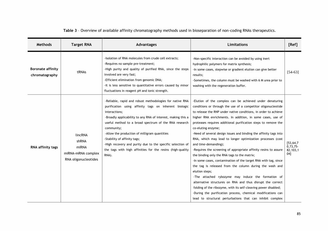
85
Table 3 – Overview of available affinity chromatography methods used in bioseparation of non-coding RNAs therapeutics.
Methods Target RNA Advantages Limitations [Ref]
Boronate affinity
chromatography tRNAs
-Isolation of RNA molecules from crude cell extracts;
-Requires no sample pre-treatment;
-High purity and quality of purified RNA, since the steps
involved are very fast;
-Efficient elimination from genomic DNA;
-It is less sensitive to quantitative errors caused by minor
fluctuations in reagent pH and ionic strength.
-Non-specific interaction can be avoided by using inert
hydrophilic polymers for matrix synthesis;
-In some cases, stepwise or gradient elution can give better
results;
-Sometimes, the column must be washed with 6 M urea prior to
washing with the regeneration buffer.
[54-63]
RNA affinity tags
lincRNA
shRNA
miRNA
miRNA-mRNA complex
RNA oligonucleotides
-Reliable, rapid and robust methodologies for native RNA
purification using affinity tags on inherent biologic
interactions;
-Broadly applicability to any RNA of interest, making this a
useful method to a broad spectrum of the RNA research
community;
-Allow the production of milligram quantities
-Stability of affinity tags;
-High recovery and purity due to the specific selection of
the tags with high affinities for the resins (high-quality
RNA).
-Elution of the complex can be achieved under denaturing
conditions or through the use of a competitor oligonucleotide
to release the RNP under native conditions, in order to achieve
higher RNA enrichments. In addition, in some cases, use of
proteases requires additional purification steps to remove the
co-eluting enzyme;
-Need of several design issues and binding the affinity tags into
RNA, which may lead to longer optimization processes (cost
and time-demanding);
-Requires the screening of appropriate affinity resins to assure
the binding only the RNA tags to the matrix;
-In some cases, contamination of the target RNA with tag, since
the tag is released from the column during the wash and
elution steps;
-The attached rybozyme may induce the formation of
alternative structures on RNA and thus disrupt the correct
folding of the ribozyme, with its self-cleaving power disabled;
-During the purification process, chemical modifications can
lead to structural perturbations that can inhibit complex
[53,64,70,73,75-82,103,104]

86
formation or can significantly degrade RNA, an undesirable
feature.
Amino acid-based
affinity
chromatography
Precursor miRNA
rRNAs
tRNAs
6S RNA
Total RNA
-Isolation of target RNA from crude cell extracts in a single
chromatographic step;
-Robust methodologies using natural small molecular
ligands;
-High purity and quality of purified RNA, since the steps
involved are very fast;
-Purification with high reproducibility;
-Efficient elimination of endotoxin, proteins, genomic DNA
and single/double-stranded RNAs;
-Simplified the workflow integration and miniaturizes
sample handling process;
-Occur under non-denaturing conditions and can be easily
adapted to any molecular weight RNA as well as high-
throughput applications.
-The high salt concentrations (namely tyrosine, histidine and
lysine amino acids) required for elution of the targets RNAs are
the main drawbacks of this approach, especially with regard to
biotechnological application since the use of salt is associated
with higher costs and environmental impact;
-Low recovery yields;
-Lower durability of supports.
[16,17,2
2,87-91]

87
4.3. Amino acid-based affinity chromatography
In the past few years, a new affinity methodology called amino acid-based affinity
chromatography has been successfully developed to efficiently purify different RNA species
[22]. Indeed, several studies are already available demonstrating the applicability of amino
acids (namely L-Arginine, L-Histidine, L-Lysine and O-Phospho-L-Tyrosine), as specific affinity
chromatographic ligands, in the purification/separation of diverse RNA molecules, such as
total RNA, ribosomal RNA, tRNA, 6S RNA and pre-miRNAs (in particular, pre-miR-29b), using
conventional particle-based columns (amino acids-agarose matrices) [16,17,87-91]. These
results demonstrated the versatility, exceptional ability and the potential of the amino acids
to strongly interact with all functional classes of RNA, despite their structural diversity and
different folding states, as they are in their native state. Amino acid-based affinity
chromatography is a powerful technique that has been used as an improved, very specific and
effective bioseparation methodology for the purification of RNA molecules that exploits
natural affinity interactions occurring between amino acids and RNA [22,45]. These studies
were based on the fact that many several specific interactions exist between proteins and
nucleic acids in biological systems, involving particularly basic amino acids [22,45,92,93]. For
example, Calnan and co-workers demonstrated that arginine residues are important for
specific RNA recognition, even with different conformational rearrangements and that
arginine-base interactions have been recognized as the most prevalent interactions in several
protein-RNA complexes [93]. Thus, this type of selectivity can be explained on the basis of
the biological function or chemical structure of the RNA, which favors the interaction.
In general, the RNA structural features (namely, negative charge of the RNA backbone and
single-stranded nature) seem to be relevant for the specific interactions established between
the RNA and the amino acids used as immobilized ligands [92,94]. Nonetheless, the high base
exposure on RNA species was also suggested to have a crucial role in the RNA recognition and
on its distinct retention behavior. Recently, the structural diversity of RNAs was described to
be of significant importance in RNA-protein interactions because RNA can exhibit different
moieties according to its folding state, for example RNA hairpins and bulges form stable
tertiary structures that contributes substantially to their recognition [47,95,96]. On the other
hand, the salts used in the chromatographic conditions can also have a profound positive
effect on RNA folding as well as on the stability of non-covalent RNA-amino acid interactions
during the course of purification, once the salt allows that single-stranded nucleic acid
molecules form stable hydrogen bonds between their complementary bases [96,97].
Additionally, this type of selectivity can also be explained in light of the atomic and
molecular recognition studies performed on RNA-protein complex structures have predicted
preferential interactions/contacts occurring between particular amino acids side chains and
specific nucleotides either involving the RNA bases or through a defined conformation of the
RNA backbone phosphates (e.g. arginine fork is defined as an interaction between a arginine

88
and a pair of adjacent phosphates, which mediates specific recognition of RNA structure)
[93,98-100]. For instance, several studies have shown that some amino acids, in particular
arginine has strongly favored interaction with guanines bases along the bulge and through the
continuous stem sequences of the RNA, involving H-bonding [92,93]. In general, these
evidences showed that the specific interaction that lead to a biorecognition of different RNA
molecules by the amino acids seem to be dependent on the base composition, RNA backbone
and RNA conformational rearrangement. Globally, the development of this isolation
methodology allowed recovery and efficiently separate of the target RNA while the quality
control analysis showed a high efficiency, selectivity and integrity in RNA preparations as well
as good purity in what concerns to host RNAs, proteins, genomic DNA and endotoxins (Table 3)
[16,17,87-91]. Overall, in a single step, affinity purification can offer several advantages over
other less-selective and time-consuming multi-step procedures (see Figure 2).
Although, amino acid ligands immobilized in conventional chromatographic columns of
agarose showed high selectivity for RNA purification but a faster and more robust purification
method will benefit ncRNAs isolation since it will possibly have important implications in RNA
stability. In addition, available conventional columns (particle-based supports) present
certain restrictions, namely limitations with the mass transfer, low capacity, gel
compressibility and poor pore diffusion leading to high pressure drops and low flow rates
application - all of which incur process time and cost [50]. These disadvantages have driven
researchers to develop alternative chromatographic supports capable of maintaining the
efficiency of the established processes while improving their limitations. One of these new
generations is the monolithic supports whose application has been increasing owing to their
structural properties [48,50,101,102].
Indeed, monolithic supports present several advantages, namely a huge quantity of accessible
binding sites (high binding capacity), high external porosity, high sample distribution and
excellent mass transfer properties [48,101]. Moreover, monoliths allow the application of
higher flow rates, a very fast separation, short analysis times and purification with high
reproducibility both at small and large scales, which reduces the biomolecules degradation
due to the shorter contact time with the chromatographic support. Thus, due to their
excellent morphological, physicochemical and mechanical properties, monolithic supports
have attracted attention for the use in high-throughput separations, coupled to a variety of
affinity ligands, both at research and industrial scales. In 2014, our research group showed
the possibility of exploring and characterizing the interactions occurring between pre-miR-
29b and an agmatine amino acid derivative immobilized into a monolithic disk [49]. As a
matter of fact, for the first time, the selectivity, specificity and biorecognition of agmatine
ligands was combined with the structural versatility and capacity provided by monolithic
supports, as a promising strategy for miRNA purification [49]. Additionally, this monolithic-
based strategy represents an advantageous alternative to conventional supports due to the
fast separation and consequent short contact time with the chromatographic matrix, ensuring

89
structural stability of the target molecule (reduces RNA degradation). The successful results
obtained in the isolation of several RNA species revealed an efficient affinity methodology to
obtain RNA with high purity and good integrity, with potential applicability in several RNA
based-procedures in basic and clinical research.
5. Conclusions and Future perspectives
In this review, it was emphasized the potential of affinity chromatography in the RNA
purification. As discussed above, in the RNA affinity tags-based chromatography, different
tags are introduced into RNA molecules and since they need to be removed before further
RNA application, conformational changes can be induced in the target RNAs. In general, these
methods are mostly time-consuming, expensive, tedious and the usage of synthetic RNAs also
represents a limitation. Thus, there is still an urgent need for high-selectivity, -specificity, -
capacity, -reliability and –throughput purification processes, to obtain high quantities of
ncRNAs with the demand quality.
From our point of view, amino acid-based affinity chromatography emerges as a particularly
promising approach for RNA purification, once it combines the selectivity of a naturally
occurring biological interaction with the simplicity of RNA molecule. Thus, in order to achieve
the maximum resolution and selectivity between the target RNAs and other host sRNA
species, it should be used an affinity support that relies on the same biological interactions
that are established within the cell, i.e., by using immobilized amino acids, its derivates,
peptides or complementary nucleotide sequences to the target RNAs, as specific ligands. The
nucleotide bases complementarity can also be effectively used for ncRNAs trapping onto the
column which facilitates their separation. Indeed, since synthetic oligonucleotides can be
covalently bound to chromatographic matrices, ncRNAs can be easily captured when the
appropriated oligonucleotides is attached to the matrix. Therefore, these new affinity
protocols for miRNA isolation can offer advantages over other less-selective and time-
consuming multistep procedures and can improve process economics. Moreover, some
improvements over other chromatographic techniques are expected because RNA recovery
can be achieved under mild elution conditions, rather than using organic or toxic compounds,
and the use of enzymes is not necessary. In addition, exploiting the specific and natural
affinity interactions between miRNA and the amino acids/peptides/sequences of nucleotides,
it is not necessary to modify the recombinant ncRNA in order to bind it to the matrix. Indeed,
it is expected that this strategy will allow the recovery of RNAs under their native state and
with high integrity, efficiency and purity level. Furthermore, coupling an affinity based
bioseparation methodology to its recombinant biosynthesis onto prokaryotic hosts represents
an interesting integrated strategy that can be used for ncRNA biosynthesis and purification for
further medical or biotechnological applications without the need of using in vitro transcribed
RNAs. Actually, there are several tools available with some success for producing a large
90
variety of recombinant RNA using Escherichia coli as a host, including cloning vectors and
fusion tags (RNA is masked within a stable RNA scaffold, such as tRNA fusions and 5S
ribosomal RNA) [14,105]. In order to further develop a therapeutic product based on this
approach, the ncRNAs must possess unquestionable biocompatibility features for the potential
application in future studies in several RNA research fields, namely in RNA structural and
functional studies as well as for application in medical therapies.
6. Acknowledgments
This work was supported by the Portuguese Foundation for Science and Technology (FCT),
through the projects EXPL/BBB-BIO/1056/2012, Pest-OE/SAU/UI0709/2014 and FCOMP-01-
0124-FEDER-041068 – EXPL/QEQ-MED/1068/2013. Patrícia Pereira acknowledges the
fellowship (Ph.D. Grant, Ref SFRH/BD/81914/2011) from FCT. The authors also acknowledge
the program Fundo Europeu de Desenvolvimento Regional (FEDER) [COMPETE [FCOMP-01-
0124-FEDER-027560]].

91
7. References
[1] A. Fire, S. Xu, M.K. Montgomery, S.A. Kostas, S.E. Driver, C.C. Mello, Potent and specific
genetic interference by double-stranded RNA in Caenorhabditis elegans, Nature. 391
(1998) 806-811.
[2] N. Lehman, RNA in evolution, Wiley Interdiscip Rev RNA. 1 (2010) 202-213.
[3] P.V. Ramachandran, S. Ignacimuthu, RNA interference - a silent but an efficient
therapeutic tool, Appl Biochem Biotechnol. 169 (2013) 1774-1789.
[4] D.B. Dogini, V.D. Pascoal, S.H. Avansini, A.S. Vieira, T.C. Pereira, I. Lopes-Cendes, The
new world of RNAs, Genet Mol Biol. 37 (2014) 285-293.
[5] A.Q. Gomes, S. Nolasco, H. Soares, Non-coding RNAs: multi-tasking molecules in the cell,
Int J Mol Sci. 14 (2013) 16010-16039.
[6] X. Shi, M. Sun, H. Liu, Y. Yao, Y. Song, Long non-coding RNAs: a new frontier in the study
of human diseases, Cancer Lett. 339 (2013) 159-166.
[7] L.W. Harries, Long non-coding RNAs and human disease, Biochem Soc Trans. 40 (2012)
902-906.
[8] B.A. Sullenger, E. Gilboa, Emerging clinical applications of RNA, Nature. 418 (2002) 252-
258.
[9] S. Kreiter, M. Diken, A. Selmi, O. Tureci, U. Sahin, Tumor vaccination using messenger
RNA: prospects of a future therapy, Curr Opin Immunol. 23 (2011) 399-406.
[10] S. Pascolo, Vaccination with messenger RNA, Methods Mol Med. 127 (2006) 23-40.
[11] L.G. Scott, M. Hennig, RNA structure determination by NMR, Methods Mol Biol. 452 (2008)
29-61.
[12] J.A. Doudna, Preparation of homogeneous ribozyme RNA for crystallization, Methods Mol
Biol. 74 (1997) 365-370.
[13] A. Wittmann, B. Suess, Engineered riboswitches: Expanding researchers' toolbox with
synthetic RNA regulators, FEBS Lett. 586 (2012) 2076-2083.
[14] L. Ponchon, F. Dardel, Large scale expression and purification of recombinant RNA in
Escherichia coli, Methods. 54 (2011) 267-273.
[15] L.D. Sherlin, T.L. Bullock, T.A. Nissan, J.J. Perona, F.J. Lariviere, O.C. Uhlenbeck, S.A.
Scaringe, Chemical and enzymatic synthesis of tRNAs for high-throughput crystallization,
RNA. 7 (2001) 1671-1678.
[16] P. Pereira, A. Sousa, J. Queiroz, I. Correia, A. Figueiras, F. Sousa, Purification of pre-
miR-29 by arginine-affinity chromatography, J Chromatogr B Analyt Technol Biomed Life
Sci. 951-952 (2014) 16-23.
[17] R. Martins, J.A. Queiroz, F. Sousa, A new affinity approach to isolate Escherichia coli 6S
RNA with histidine-chromatography, J Mol Recognit. 23 (2010) 519-524.
[18] L. Huang, J. Lieberman, Production of highly potent recombinant siRNAs in Escherichia
coli, Nat Protoc. 8 (2013) 2325-2336.

92
[19] M.M. Li, W.P. Wang, W.J. Wu, M. Huang, A.M. Yu, Rapid production of novel pre-
microRNA agent hsa-mir-27b in Escherichia coli using recombinant RNA technology for
functional studies in mammalian cells, Drug Metab Dispos. 42 (2014) 1791-1795.
[20] N. Nagao, H. Suzuki, R. Numano, S. Umekage, Y. Kikuchi, Short hairpin RNAs of designed
sequences can be extracellularly produced by the marine bacterium Rhodovulum
sulfidophilum, J Gen Appl Microbiol. 60 (2014) 222-226.
[21] S. Dunin-Horkawicz, A. Czerwoniec, M.J. Gajda, M. Feder, H. Grosjean, J.M. Bujnicki,
MODOMICS: a database of RNA modification pathways, Nucleic Acids Res. 34 (2006) D145-
149.
[22] R. Martins, J.A. Queiroz, F. Sousa, Ribonucleic acid purification, J Chromatogr A. 1355
(2014) 1-14.
[23] L.E. Easton, Y. Shibata, P.J. Lukavsky, Rapid, nondenaturing RNA purification using weak
anion-exchange fast performance liquid chromatography, RNA. 16 (2010) 647-653.
[24] S.M. McCarthy, M. Gilar, J. Gebler, Reversed-phase ion-pair liquid chromatography
analysis and purification of small interfering RNA, Anal Biochem. 390 (2009) 181-188.
[25] B. Noll, S. Seiffert, F. Hertel, H. Debelak, P. Hadwiger, H.P. Vornlocher, I. Roehl,
Purification of small interfering RNA using nondenaturing anion-exchange
chromatography, Nucleic Acid Ther. 21 (2011) 383-393.
[26] J. Koubek, K.F. Lin, Y.R. Chen, R.P. Cheng, J.J. Huang, Strong anion-exchange fast
performance liquid chromatography as a versatile tool for preparation and purification of
RNA produced by in vitro transcription, RNA. 19 (2013) 1449-1459.
[27] F.S. Hagen, E.T. Young, Preparative polyacrylamide gel electrophoresis of ribonucleic
acid. Identification of multiple molecular species of bacteriophage T7 lysozyme
messenger ribonucleic acid, Biochemistry. 13 (1974) 3394-3400.
[28] I. Kim, S.A. McKenna, E. Viani Puglisi, J.D. Puglisi, Rapid purification of RNAs using fast
performance liquid chromatography (FPLC), RNA. 13 (2007) 289-294.
[29] A. Gallas, C. Alexander, M.C. Davies, S. Puri, S. Allen, Chemistry and formulations for
siRNA therapeutics, Chem Soc Rev. 42 (2013) 7983-7997.
[30] V.N. Kim, Small RNAs: classification, biogenesis, and function, Mol Cells. 19 (2005) 1-15.
[31] K. Gavrilov, W.M. Saltzman, Therapeutic siRNA: principles, challenges, and strategies,
Yale J Biol Med. 85 (2012) 187-200.
[32] Y. Huang, J.L. Zhang, X.L. Yu, T.S. Xu, Z.B. Wang, X.C. Cheng, Molecular functions of
small regulatory noncoding RNA, Biochemistry (Mosc). 78 (2013) 221-230.
[33] D. Guzman-Villanueva, I.M. El-Sherbiny, D. Herrera-Ruiz, A.V. Vlassov, H.D. Smyth,
Formulation approaches to short interfering RNA and MicroRNA: challenges and
implications, J Pharm Sci. 101 (2012) 4046-4066.
[34] A. Qurashi, P. Jin, Small RNA-mediated gene regulation in neurodevelopmental disorders,
Curr Psychiatry Rep. 12 (2010) 154-161.
[35] T.W. Nilsen, Mechanisms of microRNA-mediated gene regulation in animal cells, Trends
Genet. 23 (2007) 243-249.

93
[36] Y. Zhang, Z. Wang, R.A. Gemeinhart, Progress in microRNA delivery, J Control Release.
172 (2013) 962-974.
[37] J.A. Broderick, P.D. Zamore, MicroRNA therapeutics, Gene Ther. 18 (2011) 1104-1110.
[38] J. Zhu, H. Fu, Y. Wu, X. Zheng, Function of lncRNAs and approaches to lncRNA-protein
interactions, Sci China Life Sci. 56 (2013) 876-885.
[39] S. Memczak, M. Jens, A. Elefsinioti, F. Torti, J. Krueger, A. Rybak, L. Maier, et al.,
Circular RNAs are a large class of animal RNAs with regulatory potency, Nature. 495
(2013) 333-338.
[40] J.C. Burnett, J.J. Rossi, RNA-based therapeutics: current progress and future prospects,
Chem Biol. 19 (2012) 60-71.
[41] F. RE, RNA methodologies - Laboratory Guide for Isolation and Characterization,
Academic Press, York, USA, 2005.
[42] M. Uhlen, Affinity as a tool in life science, Biotechniques. 44 (2008) 649-654.
[43] A.C. Roque, C.R. Lowe, Affinity chromatography: history, perspectives, limitations and
prospects, Methods Mol Biol. 421 (2008) 1-21.
[44] N.E. Labrou, Design and selection of ligands for affinity chromatography, J Chromatogr B
Analyt Technol Biomed Life Sci. 790 (2003) 67-78.
[45] F. Sousa, D.M. Prazeres, J.A. Queiroz, Affinity chromatography approaches to overcome
the challenges of purifying plasmid DNA, Trends Biotechnol. 26 (2008) 518-525.
[46] C.R. Lowe, A.R. Lowe, G. Gupta, New developments in affinity chromatography with
potential application in the production of biopharmaceuticals, J Biochem Biophys
Methods. 49 (2001) 561-574.
[47] M. Yarus, J.J. Widmann, R. Knight, RNA-amino acid binding: a stereochemical era for the
genetic code, J Mol Evol. 69 (2009) 406-429.
[48] R. Mallik, D.S. Hage, Affinity monolith chromatography, J Sep Sci. 29 (2006) 1686-1704.
[49] P. Pereira, A. Sousa, J.A. Queiroz, A. Figueiras, F. Sousa, Pharmaceutical-grade pre-miR-
29 purification using an agmatine monolithic support, J Chromatogr A. 1368 (2014) 173-
182.
[50] A. Sousa, F. Sousa, J.A. Queiroz, Advances in chromatographic supports for
pharmaceutical-grade plasmid DNA purification, J Sep Sci. 35 (2012) 3046-3058.
[51] B.V. Ayyar, S. Arora, C. Murphy, R. O'Kennedy, Affinity chromatography as a tool for
antibody purification, Methods. 56 (2012) 116-129.
[52] P.B. Moore, Structural motifs in RNA, Annu Rev Biochem. 68 (1999) 287-300.
[53] H.L. Weith, J.L. Wiebers, P.T. Gilham, Synthesis of cellulose derivatives containing the
dihydroxyboryl group and a study of their capacity to form specific complexes with
sugars and nucleic acid components, Biochemistry. 9 (1970) 4396–401.
[54] N. Singh, R.C. Willson, Boronate affinity adsorption of RNA: possible role of
conformational changes, J Chromatogr A. 840 (1999) 205–213.

94
[55] H. Schott, E. Rudloff, P. Schmidt, R. Roychoudhury, H. Koessel, A
dihydroxyborylsubstituted methacrylic polymer for the column chromatographic
separation of mononucleotides, oligonucleotides, and transfer ribonucleic acid,
Biochemistry. 12 (1973) 932-8.
[56] S. Ackerman, B. Cool, J. Furth, Removal of DNA from RNA by chromatography on
acetylated N-[N'-(m-dihydroxylborylphenyl)succinamyl]aminoethyl cellulose, Anal
Biochem. 100 (1979) 174-8.
[57] M. Rosenberg, P.T. Gilham, The isolation of 3'-terminal polynucleotides from RNA
molecules, Biochim Biophys Acta. 246 (1971) 337-40.
[58] X. Liu, W.H. Scouten, Part 1: Boronic Acids as Selective Ligands for Affinity
Chromatography, Miscellaneous Methods in Affinity Chromatography. Florida: Taylor and
Francis Group (2002).
[59] A. Yurkevich, I. Kolodkina, E. Ivanova, E. Pichuzhkina, Study of the interaction of polyols
with polymers containing N-substituted [(4-boronophenyl)methylammonio] groups,
Carbohydr Res. 43 (1975) 215–24.
[60] B. Elmas, M.A. Onur, S. Senel, A. Tuncel, Thermosensitive N-
isopropylacrylamidevinylphenylboronic acid copolymer latex particles for nucleotide
isolation, Colloids Surf A Physicochem Eng Asp. 232 (2004) 253–9.
[61] H. Clcek, Nucleotide isolation by boronic acid functionalized hydrogel beads, J Bioact
Compat Polym. 20 (2005) 245–57.
[62] N. Tanaka, H. Kobayashi, H. Ishizuka, H. Minakuchi, K. Nakanishi, K. Hosoya, T. Ikegami,
Monolithic silica columns for high efficiency chromatographic separations, J Chromatogr
A. 965 (2002) 35–49.
[63] A. Srivastava, A.K. Shakya, A. Kumar, Boronate affinity chromatography of cells and
biomacromolecules using cryogel matrices, Enzyme Microb Technol. 51 (2012) 373– 381.
[64] C. Srisawat, D.R. Engelke, RNA affinity tags for purification of RNAs and
ribonucleoprotein complexes, Methods. 26 (2002) 156-161.
[65] C. Pestourie, B. Tavitian, F. Duconge, Aptamers against extracellular targets for in vivo
applications, Biochimie. 87 (2005) 921-930.
[66] J. Porath, Immobilized metal ion affinity chromatography, Protein Expr Purif. 3 (1992)
263–281.
[67] G.I. Evan, G.K. Lewis, G. Ramsay, J.M. Bishop, Isolation of monoclonal antibodies
specific for human cmyc proto-oncogene product, Mol Cell Biol. 5 (1985) 3610–3616.
[68] G. Rigaut, A. Shevchenko, B. Rutz, M. Wilm, M. Mann, B. Séraphin, A generic protein
purification method for protein complex characterization and proteome exploration, Nat
Biotechnol. 17 (1999) 1030–1032.
[69] M. Uhlen, B. Nilsson, B.M. Guss, M. Lindberg, S. Gatenbeck, L. Philipson, Gene fusion
vectors based on the gene for staphylococcal protein A, Gene. 23 (1983) 369–378.
[70] J.H. Yoon, S. Srikantan, M. Gorospe, MS2-TRAP (MS2-tagged RNA affinity purification):
tagging RNA to identify associated miRNAs, Methods. 58 (2012) 81-87.

95
[71] P.C. Simons, D.L. Vander Jagt, Purification of glutathione S-transferases by glutathione-
affinity chromatography, Methods Enzymol. 77 (1981) 235–237.
[72] T.G. Schmidt, J. Koepke, R. Frank, A. Skerra, Molecular interaction between the Strep-
tag affinity peptide and its cognate target, streptavidin, J Mol Biol. 255 (1996) 753–766.
[73] K. Leppek, G. Stoecklin, An optimized streptavidin-binding RNA aptamer for purification
of ribonucleoprotein complexes identifies novel ARE-binding proteins, Nucleic Acids Res.
42 (2014) e13.
[74] K.S. Prickett, D.C. Amberg, T.P. Hopp, A calcium-dependent antibody for identification
and purification of recombinant proteins, Biotechniques. 7 (1989) 580–589.
[75] C. Srisawat, D.R. Engelke, Streptavidin aptamers: affinity tags for the study of RNAs and
ribonucleoproteins, RNA. 7 (2001) 632-641.
[76] C. Srisawat, I.J. Goldstein, D.R. Engelke, Sephadex-binding RNA ligands: rapid affinity
purification of RNA from complex RNA mixtures, Nucleic Acids Res. 29 (2001) E4.
[77] K. Wei, F. Yan, H. Xiao, X. Yang, G. Xie, Y. Xiao, T. Wang, et al., Affinity purification of
binding miRNAs for messenger RNA fused with a common tag, Int J Mol Sci. 15 (2014)
14753-14765.
[78] U.A. Orom, A.H. Lund, Isolation of microRNA targets using biotinylated synthetic
microRNAs, Methods. 43 (2007) 162-165.
[79] C. Gong, M.W. Popp, L.E. Maquat, Biochemical analysis of long non-coding RNA-
containing ribonucleoprotein complexes, Methods. 58 (2012) 88-93.
[80] C.F. Flores-Jasso, W.E. Salomon, P.D. Zamore, Rapid and specific purification of
Argonaute-small RNA complexes from crude cell lysates, RNA. 19 (2013) 271-279.
[81] R.T. Batey, J.S. Kieft, Improved native affinity purification of RNA, RNA. 13 (2007) 1384-
1389.
[82] G. Di Tomasso, P. Lampron, P. Dagenais, J.G. Omichinski, P. Legault, The ARiBo tag: a
reliable tool for affinity purification of RNAs under native conditions, Nucleic Acids Res.
39 (2011) e18.
[83] G. Di Tomasso, P. Dagenais, A. Desjardins, A. Rompre-Brodeur, V. Delfosse, P. Legault,
Affinity purification of RNA using an ARiBo tag, Methods Mol Biol. 941 (2012) 137-155.
[84] G. Di Tomasso, A. Salvail-Lacoste, J. Bouvette, J.G. Omichinski, P. Legault, Affinity
purification of in vitro transcribed RNA with homogeneous ends using a 3'-ARiBo tag,
Methods Enzymol. 549 (2014) 49-84.
[85] A. Salvail-Lacoste, G. Di Tomasso, B.L. Piette, P. Legault, Affinity purification of T7 RNA
transcripts with homogeneous ends using ARiBo and CRISPR tags, RNA. 19 (2013) 1003-
1014.
[86] S.C. Walker, F.H. Scott, C. Srisawat, D.R. Engelke, RNA affinity tags for the rapid
purification and investigation of RNAs and RNA-protein complexes, Methods Mol Biol. 488
(2008) 23-40.
96
[87] A. Afonso, P. Pereira, J.A. Queiroz, A. Sousa, F. Sousa, Purification of pre-miR-29 by a
new O-phospho-l-tyrosine affinity chromatographic strategy optimized using design of
experiments, J Chromatogr A. 1343 (2014) 119-127.
[88] P. Pereira, A. Sousa, J. Queiroz, A. Figueiras, F. Sousa, New approach for purification of
pre-miR-29 using lysine-affinity chromatography, J Chromatogr A. 1331 (2014) 129-132.
[89] R. Martins, C.J. Maia, J.A. Queiroz, F. Sousa, A new strategy for RNA isolation from
eukaryotic cells using arginine affinity chromatography, J Sep Sci. 35 (2012) 3217-3226.
[90] R. Martins, J.A. Queiroz, F. Sousa, Histidine affinity chromatography-based methodology
for the simultaneous isolation of Escherichia coli small and ribosomal RNA, Biomed
Chromatogr. 26(7) 2011 781-8.
[91] D.S. Jones, H.K. Lundgren, F.T. Jay, The separation of ribonucleic acids from Escherichia
coli on lysin-agarose, Nucleic Acids Res. 3 (1976) 1569-1576.
[92] F. Sousa, C. Cruz, J.A. Queiroz, Amino acids-nucleotides biomolecular recognition: from
biological occurrence to affinity chromatography, J Mol Recognit. 23 (2010) 505-518.
[93] B.J. Calnan, B. Tidor, S. Biancalana, D. Hudson, A.D. Frankel, Arginine-mediated RNA
recognition: the arginine fork, Science. 252 (1991) 1167-1171.
[94] P. Pereira, C. Cruz, J.A. Queiroz, A. Figueiras, F. Sousa, Analysis of pre-miR-29b binding
conditions to amino acids by using a surface plasmon resonance biosensor, Anal Methods.
8 (2016) 205.
[95] J.J. Ellis, M. Broom, S. Jones, Protein-RNA interactions: structural analysis and
functional classes, Proteins. 66 (2007) 903-911.
[96] M. Treger, E. Westhof, Statistical analysis of atomic contacts at RNA-protein interfaces, J
Mol Recognit. 14 (2001) 199-214.
[97] M.M. Hoffman, M.A. Khrapov, J.C. Cox, J. Yao, L. Tong, A.D. Ellington, AANT: the Amino
Acid-Nucleotide Interaction Database, Nucleic Acids Res. 32 (2004) D174-181.
[98] N.M. Luscombe, R.A. Laskowski, J.M. Thornton, Amino acid-base interactions: a three-
dimensional analysis of protein-DNA interactions at an atomic level, Nucleic Acids Res. 29
(2001) 2860-2874.
[99] J.D. Puglisi, J.R. Wyatt, I. Tinoco, Jr., Solution conformation of an RNA hairpin loop,
Biochemistry. 29 (1990) 4215-4226.
[100] T. Janas, J.J. Widmann, R. Knight, M. Yarus, Simple, recurring RNA binding sites for L-
arginine, RNA. 16 (2010) 805-816.
[101] E.L. Pfaunmiller, M.L. Paulemond, C.M. Dupper, D.S. Hage, Affinity monolith
chromatography: a review of principles and recent analytical applications, Anal Bioanal
Chem. 405 (2013) 2133-2145.
[102] R.D. Arrua, C.I. Alvarez Igarzabal, Macroporous monolithic supports for affinity
chromatography, J Sep Sci. 34 (2011) 1974-1987.
[103] L. Ponchon, M. Catala, B. Seijo, M. El Khouri, F. Dardel, S. Nonin-Lecomte, C. Tisne, Co-
expression of RNA-protein complexes in Escherichia coli and applications to RNA biology,
Nucleic Acids Res. 41 (2013) e150.

97
[104] L. Ponchon, F. Dardel, Recombinant RNA technology: the tRNA scaffold, Nat Methods. 4
(2007) 571-6.
[105] J. Jin, M. Cid, C.B. Poole, L.A. McReynolds, Protein mediated miRNA detection and
siRNA enrichment using p19, Biotechniques. 48 (2010) xvii-xxiii.

98

99
Chapter 2

100

101
Aims of the thesis
Up until now, microRNAs-based gene therapy applications are mainly performed through
enzymatically and chemically synthesized microRNAs. However, the presence of contaminants
in these synthesized formulations can lead to non-targeted gene silencing, frequently
associated with a decrease in therapeutic effectiveness and can still restrict the
implementation of these oligoribonucleotides onto pre-clinical and clinical applications. In
addition, these current techniques present various limitations in the preparation, isolation
and purification of microRNAs, namely by the use of chemical solvents and denaturing
conditions.
Thus, the global aim of this work is the development and establishment of an innovative
biotechnological platform for the biosynthesis, recovery and purification of the recombinant
human pre-miR-29b, to be delivered to neuronal cells using suitable non-viral delivery
systems, in order to assure a more effective biological effect in the selective silencing of
endogenous pathways involved in the insurgence and progression of the Alzheimer’s disease.
In particular, this work intends to explore pre-miR-29b as a novel biopharmaceutical to
decrease the APP, BACE1 and Aβ expression levels, which could represent a significant
advance in terms of treatment of AD. With this work, it is believed that the integrative
approach of extracellular production, using Rhodovulum sulfidophilum host, and affinity-
based purification will enable the preparation of the target pre-miRNA with high efficiency,
selectivity, throughput, purity, integrity and biological activity, to be applied in RNA research
or RNA therapeutic areas. The high gain in this field is the possibility to use these
methodologies for the preparation of other ncRNAs, proving the wideness application of the
technology, which may have a great impact on industry, improving workload, process time
and economics.
To accomplish the main scope of this thesis, the experimental work will be developed
concerning the following tasks:
1. Cloning the human pre-miR-29b sequence in a prokaryotic expression vector, pBHSR1-
RM and transformation of the Rhodovulum sulfidophilum DSM 1374 host. Development
and establishment of the innovative biosynthesis strategy of extracellular pre-miR-29b,
testing different growth conditions. Characterization of the intra and extracellular pre-
miR-29b production.
2. Characterization of the specificity of the interactions occurring between the amino
acids-affinity ligands and microRNA molecules, as well as determination of the
influence of several chromatographic conditions in the structure and stability of RNA.

102
3. Study of the ability of amino acids-based affinity supports, such as lysine-agarose,
arginine-agarose and agmatine monolith, in the specific recognition of the intracellular
pre-miR-29b. The adequate binding/elution conditions will result from the
manipulation of temperature, flow rate and buffer composition (pH, ionic strength,
type of salt and presence of competitive agents). These studies will allow the
implementation of new methodologies for miRNA purification.
4. Development and characterization of non-viral delivery systems composed by
biodegradable polymers, for human pre-miR-29b protection and stabilization, allowing
their sustained delivery to the cells. It will be also performed the functionalization of
these delivery systems with specific ligands, to be recognized by cell surface receptors
of Blood-Brain Barrier, and mediate the delivery of pre-miR-29b to the target cells.
5. Evaluation of the biological activity and efficiency of the pure recombinant pre-miR-29b
to regulate the expression of the AD related-genes, namely by inducing the BACE1
knockdown, using in vitro neuronal cells lines. These results will be compared with
those obtained with the commercially available miR-29b.

103

104

105
Chapter 3

106

107
Paper III
Advances in time-course extracellular production of human
pre-miR-29b from Rhodovulum sulfidophilum
P. Pereira, A. Q. Pedro, J. Tomás, C. J. Maia, J. A. Queiroz, A. Figueiras,
F. Sousa
(Submitted for publication)
Short description: This paper introduces a novel prokaryotic system for biosynthesis and
recovery of recombinant human pre-miR-29b. First, in order to transfer the pre-miR-29b
encoding plasmid to Rhodovulum sulfidophilum host, the transformation conditions were
established. The expression of the human pre-miR-29b was carried out at small-scale in
shake-flasks by Rhodovulum sulfidophilum cultures. Then, in order to attain high quantities of
pre-miR-29b, several fermentation variables (culture media, sodium chloride concentration,
aerobic conditions and temperature) were optimized, allowing the establishment of improved
growth conditions. Finally, the expression of both intracellular and extracellular recombinant
pre-miR-29b was monitored during different fermentation periods, as well as genomic DNA
and proteins, major host contaminants that influence the purity of the target biomolecule.

108

109
Advances in time-course extracellular production of human pre-
miR-29b from Rhodovulum sulfidophilum
P. Pereira1, A. Q. Pedro1, J. Tomás1, C. J. Maia1, J. A. Queiroz1, A.
Figueiras1,2, F. Sousa1*
1 CICS-UBI – Health Sciences Research Centre, University of Beira Interior, Avenida Infante D.
Henrique, 6200-506 Covilhã, Portugal;
2 CNC - Center of Neuroscience and Cell Biology, University of Coimbra, Largo Marquês de
Pombal, 3004-517 Coimbra, Portugal
*Corresponding author:
Dr. Fani Sousa
Phone: +351 275 329 074
Fax: +351 275 329 099
E-mail address: [email protected]
Postal address: Health Sciences Research Centre, Universidade da Beira Interior, Avenida
Infante D. Henrique, 6200-506 Covilhã, Portugal

110

111
Abstract
The present study reports the successful production of human pre-miR-29b both intra and
extracellularly in the marine phototrophic bacterium Rhodovulum sulfidophilum using
recombinant RNA technology. In a first stage, the optimal transformation conditions (0.1 µg of
plasmid DNA, with a heat-shock of 2 min at 35ºC) were established, in order to transfer the
pre-miR-29b encoding plasmid to Rhodovulum sulfidophilum host. Furthermore, the
extracellular recovery of this RNA product from the culture medium was greatly improved,
achieving quantities that are compatible with the majority of applications, namely for in vitro
or in vivo studies. Using this system, the extracellular human pre-miR-29b concentration was
approximately 182 µg/L, after 40 h of bacterial growth, and the total intracellular pre-miR-
29b was of about 358 μg/L, at 32 h. At the end of the fermentation it was verified that almost
87% of cells were viable, indicating that cell lysis is minimized and that the extracellular
medium is not highly contaminated with the host intracellular RNases and endotoxins, which
is a critical parameter to guarantee the miRNA integrity. These findings demonstrate that
pre-miRNAs can be produced by recombinant RNA technology, offering novel clues for the
production of natural pre-miRNA agents for functional studies and RNAi-based therapeutics.
Keywords
Extracellular production; Recombinant human pre-miR-29b; Rhodovulum sulfidophilum; RNAi
technology; Transformation efficiency

112

113
Introduction
In the last couple of decades, RNA interference (RNAi)-based technology has become a novel
tool for regulating gene expression in eukaryotic cells, since many small RNAs (sRNAs) act as
regulators of messenger RNA (mRNA) expression and have shown great potential for
therapeutic knockdown of disease-causing genes (Fire et al. 1998; Ramachandran and
Ignacimuthu 2013). This discovery led to the expectation that RNA molecules, such as
ribozymes, small interfering RNAs (siRNAs), short hairpin RNAs (shRNAs), microRNAs (miRNAs)
and long non-coding RNAs (lncRNAs) will be major classes of new therapeutic molecules in the
near future (Dogini et al. 2014; Gomes et al. 2013). Amongst RNAi-based therapeutics,
miRNAs have recently gained much attention, being investigated as potential drug candidates
for application in several disease scenarios that currently remain very challenging and
difficult to overcome (Sullenger and Gilboa 2002). In particular, their therapeutic potential
can be exploited in gene expression regulation at post-transcriptional level, through
degradation/de-adenylation of selected target mRNAs, translational repression or both
mechanisms (Nilsen 2007). Briefly, miRNAs mediate post-transcriptional regulation through a
sequential process involving a miRNA precursor (pre-miRNA) and, subsequently, a mature
miRNA that is incorporated into RNA-induced silencing complex (RISC) to regulate the mRNA
translation. Due to the rapidly growing demand for these biopharmaceuticals, various
strategies are currently being pursued to improve production in order to achieve higher
product titers while maintaining maximal product quality (Ponchon and Dardel 2007; Ponchon
and Dardel 2011). Furthermore, economically efficient methods for large-scale production are
also required.
Currently, miRNAs-mediated gene silencing is mainly performed through chemically
synthesized RNAs, by phosphoamidite chemistry, normally used for the generation of short
oligoribonucleotides (Ling et al. 2013). In addition to chemical synthesis, RNAs can also be
obtained by enzymatic synthesis, for example longer RNAs can be produced by in vitro
transcription using T7 RNA polymerase (Beckert and Masquida 2011; Milligan et al. 1987).
Although these methods can be very efficient in producing miRNA, in general, several
purification protocols to remove the contaminants (impurities of plasmid DNA template,
enzymes, nucleotides, chemicals, salts or buffers, which are indispensable for in vitro
transcription or chemical synthesis of RNAs) have to be employed (Martins et al. 2014;
Milligan et al. 1987). The presence of these impurities can lead to non-targeted gene
silencing, what is commonly associated with a decrease in therapeutic effectiveness and still
restrict the implementation of these RNAs onto pre-clinical or clinical trials. On the other
hand, the purification methods usually employed can cause degradation of RNA molecules due
to the requirement of toxic solvents and the use of denaturing conditions (Martins et al.
2014). MiRNA-based therapeutic applications can also be performed via the endogenous
expression of miRNAs encoded by viral vectors or plasmids in eukaryotic cells. However, this

114
alternative approach is generally less efficient because it is necessary to transcribe the DNA
to miRNA precursors (Chen et al. 2007; Huang et al. 2011; Li et al. 2014; Ponchon and Dardel
2007; Ponchon and Dardel 2011). Considering the potential therapeutic applications and the
mandatory global distribution of these novel therapeutics, it is crucial to develop efficient
methods for their preparation. One of the most promising strategies might be the use of
recombinant microorganisms to produce the target biomolecules. This approach can reduce
the costs and remains highly efficient.
At present, few strategies have emerged to produce stable RNAs, namely a circular RNA
aptamer (Umekage and Kikuchi 2009), a “tRNA scaffold” (Ponchon and Dardel 2007) and hsa-
mir-27b, using recombinant bacteria, mainly Escherichia coli (E. coli) as preferential host (Li
et al. 2014). Occasionally, modifications of RNA products are required to efficiently obtain
the product, such as the addition of flanking sequences in the case of tRNA scaffold, which
are then removed by ribonuclease (RNase) H treatment, making the process unsuitable for
industrial RNA drug production (Ponchon and Dardel 2007). Moreover, in what concerns to the
use of E. coli for RNA production, it demands the completion of cell lysis and extraction of
nucleic acids, which can be laborious and time-consuming due to the number and complexity
of the processing steps (Wassarman et al. 1999). Along with the aforementioned
disadvantages, another major issue associated with the cell lysis is the release of endotoxins,
which can lead to the contamination of the target RNA sample, and, in a last analysis,
produce symptoms of toxic shock syndrome if present in sufficient quantities in vivo (Wei et
al. 2007). In addition, as E. coli expresses several endonucleases, it can be difficult to
maintain the integrity of the target RNA, being always necessary to establish methods to
avoid its degradation.
Very recently, the marine phototrophic bacterium Rhodovulum sulfidophilum (R.
sulfidophilum) was also described as an attractive host for the recombinant production of an
artificial RNA model (streptavidin RNA aptamer) (Suzuki et al. 2010; Suzuki et al. 2011) and
shRNAs (Nagao et al. 2014). In particular, this bacterium is non-pathogenic and presents
several advantages over E. coli, such as the capacity of secretion of nucleic acids directly to
the culture medium and the absence of detectable host RNases in the extracellular medium
(Suzuki et al. 2009a; Ando et al. 2004; Ando et al. 2006). The endogenous RNAs released by
R. sulfidophilum to the extracellular medium are mainly nonaminoacylated fully mature
transfer RNAs (tRNAs) and fragments of 16S and 23S ribosomal RNAs (rRNAs) (Ando et al.
2006; Suzuki et al. 2009b). In fact, previous works described that both recombinant RNAs
(streptavidin RNA aptamer and shRNAs) produced in R. sulfidophilum were found not only
inside the cells but also in the culture medium (Nagao et al. 2014; Suzuki et al. 2010; Suzuki
et al. 2011).
In the present study, for the first time, an alternative strategy for the recombinant
biosynthesis and extracellular recovery of biologically active human pre-miR-29b (hsa-pre-

115
mir-29b) is proposed. The pre-miR-29 target was chosen because it belongs to one of the most
interesting miRNA families in humans to date, once this miRNA is involved in several
regulatory pathways associated with neurodegenerative diseases and also presenting tumor-
suppressing and immune-modulating properties (Alvarez-Garcia and Miska 2005; Du and
Pertsemlidis 2011; Hebert et al. 2008; Nelson et al. 2008). It is expected that this strategy
can be highly advantageous regarding the RNA product safety, since it will not stimulate
innate immunity, as the secreted miRNAs will be devoid of main bacterial associated
impurities. Moreover, after the application of an appropriated purification flowsheet (Fig. 1),
the isolation and purification of the target pre-miRNA will be easier and successful. The
recovery of miRNAs from the fermentation broth will be performed using membrane filtration
or concentration techniques. In fact, the application of this approach can be a promising
technology since it suppresses the use of salts and solvents typically used in conventional RNA
isolation processes. Overall, an economical and fast method for the production of functional
RNA molecules with clinical-grade quality and biological activity is described, which can be
applied in basic studies or in therapeutic strategies based on RNAi technology. From this
stand point, it seems clear that the establishment of a robust and reliable miRNA
extracellular production technology can indeed unlock the possibility of further developing
effective therapeutic strategies that rely on the intrinsic gene silencing capacity of these
oligonucleotides, presenting major advantages over the currently existing practices.
Figure 1 - Schematics of the integrative platform that allows the biosynthesis and
extracellular recovery of the recombinant hsa-pre-miR-29b.

116
Materials and Methods
Materials and Chemicals
Restriction enzymes including EcoRI, EcoT22I, AatI and StuI were purchased from Thermo
Fisher Scientific Inc. (Waltham, MA) and XbaI was purchased from New England BioLabs
(Ipswich, MA). T4 DNA ligase and pGEM-T easy vector were used as recommended by the
manufactures (Promega). DreamTaq DNA polymerase was purchased from Fermentas (Beijing,
China). Hyper Ladder I (Bioline, London, UK) was used as DNA molecular weight marker.
Primers were synthesized by StabVida (Caparica, Lisbon). The sequence of pre-miRNA was
obtained from the miRBase (http://www.mirbase.org/index.shtml).
Bacterial strains and growth conditions
The R. sulfidophilum DSM 1374 strain (BCCM/LMG, Belgium) modified with plasmid pBHSR1-
RM containing the sequence of human pre-miR-29b was used in this study for pre-miR-29b
production. One Shot® TOP10 Chemically Competent E. coli (Invitrogen) was used for the
propagation of the recombinant plasmid DNA (pDNA). The pGEM-T easy vector was applied for
DNA manipulations and the plasmid pBHSR1-RM (a generous gift from Dr. Yo Kikuchi) was used
as the expression vector (Nagao et al. 2014; Suzuki et al. 2011). E. coli TOP10 was grown in
Luria-Bertani (LB) medium (10.0 g/L tryptone, 5.0 g/L yeast extract and 5.0 g/L sodium
chloride (NaCl)) and LB-Agar. In the preliminary results, R. sulfidophilum was tested in
Nutrient Agar (g per liter of deionized water: beef extract, 1.5; yeast extract, 1.5; peptidic
digest of animal tissue, 5.0; NaCl, 5.0 and agar, 15.0), Tryptone Soya Agar (g per liter of
deionized water: casein peptone (pancreatic), 15.0; soya peptone, 5.0; NaCl, 5.0 and agar,
15.0) and Mueller Hinton Agar (g per liter of deionized water: beef, dehydrated infusion from,
30.0; casein hydrolysate, 17.5; starch, 1.5 and agar 17.0). R. sulfidophilum cells were
cultivated in a semi-defined medium, developed in this work, containing (per L of water)
tryptone, 10.0 g; polypeptone, 5.0 g; yeast extract, 0.5 g; NaCl, 30.0 g; K2HPO4, 4.0 g;
KH2PO4, 1.0 g; glucose, 50.0 g; MgSO4.7H2O, 0.2 g; CaCl2.2H2O, 0.05 g; (NH4)2SO4, 1.0 g and 1
mL of trace elements solution (TES). The TES composition was as follows: (5.56 g/L
FeSO4.7H2O, 3.96 g/L MnCl2.4H2O, 5.62 g/L CoSO4.7H2O, 0.34 g/L CuCl2.2H2O, 0.58 g/L
ZnSO4.7H2O, 0.6 g/L H3BO3, 0.04 g/L NiCl2.6H2O and 0.06 g/L Na2MoO4.2H2O in 0.5 N HCl) (Kim
et al. 2000; Silva et al. 2009). For solid media, the MT medium was used (10.0 g/L glucose,
10.0 g/L peptone, 5.0 g/L yeast extract, 20.0 g/L NaCl, 10.0 mg/L FeSO4.7H2O, 10.0 mg/L
MnSO4.4H2O, 1.0 mg/L ZnSO4.7H2O, 4.1 g/L MgCl2 and 15.0 g/L agar) (Pedro et al. 2011). The
medium pH was adjusted to 7.0, with 0.1 M NaOH before autoclaving. When appropriated,
media were supplemented with the following antibiotics: for E. coli, ampicillin and
kanamycin were used with final concentrations of 100 and 20 µg/mL, respectively, while for
R. sulfidophilum, kanamycin was used at a concentration of 30 µg/mL. Unless otherwise
stated, E. coli cells were grown at 37ºC and R. sulfidophilum growth was carried out at 30ºC
under dark-aerobic conditions. Cell growth was monitored by measuring the optical density
117
(OD) of the culture medium at a wavelength of 600 nm (OD600). Posteriorly, cells were
recovered by centrifugation and stored at -20ºC.
Construction of pBHSR1-RM-pre-miR-29b plasmid
Using the plasmid pBHSR1-RM as template (Suzuki et al. 2011), a DNA fragment containing the
rrn promoter, the streptavidin RNA aptamer sequence with flanker ribozyme sequences and
puf terminator region was amplified by PCR. The sequence primers pBHSR1_Fw and
pBHSR1_Rv are shown in Table S1, Supporting Information. The amplified DNA fragment of the
pBHSR1-RM was purified and cloned into pGEM-T easy vector. Then, the vector was digested
with StuI and XbaI, releasing a fragment containing the streptavidin RNA aptamer sequence
with flanker ribozyme sequences, and the pGEM-T easy vector was purified from agarose gel
with NucleoSpin Gel and PCR Clean-up (Macherey-Nagel). Using the pBHSR1-RM plasmid as
template in a PCR reaction, specific primers (pBHSR1-RM_StuI_premiR_511Fw and pBHSR1-
RM_XbaI820Rv) flanking the second sequence of hammer head ribozyme and the puf
terminator region were used to insert and amplify the pre-miR-29b sequence, which was
digested by StuI and XbaI and cloned into pGEM-T easy vector previously digested. The StuI
and XbaI sites in the primers are underlined and the pre-miR-29b sequence is a bold
(Supporting Information, Table S1). In order to obtain the sequence of pre-miR-29b flanked by
hammer ribozymes and puf terminator region, the pGEM-T easy vector was digested with
EcoRI and XbaI restriction enzymes and cloned into pBHSR-RM previously digested with the
same restriction enzymes. After E. coli TOP10 transformation with the vector, several
colonies were screened by PCR for the presence of the recombinant plasmid pBHSR1-RM
containing the pre-miR-29b sequence, being designated by pBHSR1-RM-pre-miR-29b
(Supporting Information, Fig. S1). Therefore, some colonies were inoculated in LB medium
and grown at 37ºC and 250 rpm, overnight. From these bacterial cultures, the plasmid
pBHSR1-RM-pre-miR-29b was isolated and purified using the NZYMiniprep Kit (NZYtech,
Lisbon, Portugal) and digested with EcoT22I and AatI to confirm the presence of the pre-miR-
29b sequence. Plasmid DNA concentrations were determined by NanoPhotometer (Implen)
and sequenced in order to confirm the identity and orientation of the amplicon.
Preparation and Transformation of competent Rhodovulum sulfidophilum cells
Growth of R. sulfidophilum strain was carried out in 500 mL shake flasks containing 100 mL of
semi-defined medium, in a rotary shaker at 30ºC and 250 rpm under aerobic-dark conditions,
until the cell suspension reached an OD600 of 0.4 and 0.8. These cells were used to prepare
competent cells for heat shock method. For this purpose, 50 mL aliquots of cell suspension
were centrifuged at 5000 rpm for 10 min at 4ºC. The medium was decanted from the cell
pellets and each one was ressuspended in 12.5 mL of ice-cold 100 mM MgCl2. The cells were
recovered again at 4000 rpm for 10 min at 4ºC and ressuspended in 50 mL of ice-cold 100 mM
CaCl2. The cell suspension was maintained on ice for at least 20-30 min. After this, the
bacterial culture was centrifuged at 4000 rpm for 10 min at 4ºC and the pellet was

118
ressuspended in 1 mL of ice-cold 85 mM CaCl2 containing ice-cold 15% (w/v) glycerol for each
50 mL of original culture. Then, the plasmid pBHSR1-RM-pre-miR-29b was introduced into R.
sulfidophilum cells by the method of heat shock. To this end, different quantities of plasmid
pBHSR1-RM-pre-miR-29b (0.1, 0.5, 1 and 5 µg) were immediately added to 0.1 mL of
competent cells (the suspension was left on ice for 10 min), and the heat shock was
performed at different temperatures (35 and 42ºC) and incubation periods (1 and 2 min), in
order to determine the best conditions. After the heat shock, 200 µL of semi-defined medium
was added to the transformation mixture and it was incubated at 30ºC, 250 rpm for 2 h under
aerobic-dark conditions to allow the expression of antibiotic resistance gene. The culture was
then plated on MT agar plates with kanamycin and incubated aerobically at 30ºC for at least 2
days until the appearance of colonies. For each different condition tested, three independent
fermentation runs were performed.
Production of pre-miR-29b in Rhodovulum sulfidophilum
Unless otherwise stated, recombinant pre-miR-29b biosynthesis was carried out according to
the following protocol. Cells containing the expression construct were grown at 30ºC in MT
plates containing kanamycin. Then, colonies were used to inoculate 100 mL of the semi-
defined medium, on 500 mL shake flasks. Cells were grown at 30ºC and 250 rpm under dark-
aerobic conditions, until the OD600 typically reached 2.6. Subsequently, in order to start the
fermentation with an initial OD600 of 0.3, an aliquot of the pre-fermentation medium was
added to 500 mL shake flasks to a total volume of 100 mL. The fermentations were carried
out during 72 h. Finally, the cells were harvested by centrifugation (1500 g, 10 min, 4ºC) and
stored at -20ºC until use.
Isolation of intracellular and extracellular human pre-miR-29b
The isolation of extracellular nucleic acids was based on the method described by Martins and
co-workers (2010) with some modifications (Martins et al. 2010). Cultivated cells of R.
sulfidophilum DSM 1374 were recovered by centrifugation at 10000 g for 10 min at 4ºC, and
the supernatant was carefully transferred to an autoclaved tube. The nucleic acids fraction of
the supernatant was precipitated by adding 3 volumes of 100% ethanol, 1/20th of 3 M sodium
acetate (pH 5.0) and 20 µL of glycogen (30 µg/mL). After incubation overnight, nucleic acids
were recovered by centrifugation at 10000 g for 10 min at 4ºC and washed with 75% ethanol.
The nucleic acids pellets were air-dried for 10 min at room temperature and solubilized in
0.05% DEPC-treated water and incubated with DNase I (Sigma-Aldrich, St Louis, MO, USA). For
the intracellular RNAs recovery, the total RNA was extracted from the collected cells using
TRIzol reagent (Invitrogen, Carlsbad, CA, USA) and chloroform, according to the protocol
provided by the manufacturer. After centrifugation, the aqueous phase was transferred to
new tubes, and the RNA was precipitated with isopropanol and washed with 75% of ethanol.
The air-dried total RNA pellet was solubilized in 0.05% DEPC-treated water.

119
Assessment of recombinant human pre-miR-29b production
RNA samples were quantified and evaluated regarding their purity (260/280 nm ratio) using a
NANOPhotometer. The integrity and quality of RNA was assessed by agarose gel
electrophoresis. A total of 1 µg of RNA was reverse transcribed into complementary DNA
(cDNA) in 20 µL final volume using RevertAid First Strand cDNA Synthesis Kit (Thermo Fisher
Scientific Inc.), according to the protocol provided by the manufacturer. For quantitative
analysis, the amount of pre-miR-29b produced was determined by reverse transcription-
quantitative real-time polymerase chain reaction (RT-qPCR) through amplification of cDNA
using a specific probe (Roche, Table 1) on a CFX Connect™ Real-Time PCR Detection System
from BioRad. RT-qPCR efficiencies were calculated from the given slopes with MyIQ 2.0
software (BioRad). The calibration curve to determine the pre-miR-29b concentration was
constructed by serial dilutions of the pre-miR-29b synthetic sample (Stabvida) in the range of
0.00001 to 0.01 pmol/µL. The quantification of the pre-miR-29b expression was calculated by
applying the comparative threshold cycle (CT) method. A list of primers used for RT-qPCR
analysis can be seen in Table 1. RT-qPCRs were performed in a 20 μL mixture containing 5 μL
cDNA, 400 nM Fw and Rv primers (Roche), 1× FastStart TaqMan® Probe Master (Roche), 200 nM
of UPL Probe (Roche, Table S1, Supporting Information) and water, under the following
conditions: 95ºC for 10 min for initial denaturation followed by 40 cycles at 95ºC for 10 s,
60ºC for 30 s and 72ºC for 15 s. Due to the small space between the primers it is only possible
to design a probe with a sufficient melting temperature if few Locked Nucleic Acid (LNA)
bases are added into the probes sequence. Each sample was run in triplicate, and CT values
were averaged from the triplicates. The final data were averaged from three independent
experiments.
Protein analysis
Protein content in each sample was measured by the Pierce BCA Protein Assay Kit (Thermo
Fisher Scientific Inc.), according to manufacturer’s instructions. Reducing sodium dodecyl
sulfate-polyacrylamide gel electrophoresis (SDS-PAGE) was performed, according to the
method of Laemmli on a 10% polyacrylamide gel (Laemmli 1970). Samples were denatured by
the addition of loading dye followed by incubation at 95ºC for 10 min. Gel was stained by
BlueSafe (NZYtech, Lisbon, Portugal). Low molecular weight protein marker (NZYtech, Lisbon,
Portugal) was used as a molecular weight standard.
Genomic DNA quantification
The concentration of genomic DNA (gDNA) was obtained by real-time qPCR in an iQ5
Multicolor Real-Time PCR Detection System (BioRad). The primers used in these experiments
were 5’-ACACGGTCCAGAACTCCTACG-3’ (forward) and 5’-
CCGGTGCTTCTTCTGCGGGTAACGTCA-3’ (reverse) for the amplification of a 181 bp fragment
of the 16S rRNA gene. PCR amplicons were quantified by following changes in fluorescence of
the DNA binding dye Maxima® SYBR Green/Fluorescein qPCR Master Mix (Thermo Fisher
120
Scientific Inc.). The calibration curve to achieve the gDNA concentration was constructed by
serial dilutions of the R. sulfidophilum gDNA sample (purified with the Wizard® Genomic DNA
Purification kit, Promega) in the range of 0.005 to 50 ng/mL. Each sample was run in
triplicate, and CT values were averaged from the triplicate. The final data were averaged
from three independent experiments.
Cell viability
At different time points of the culture growth, the cellular cytotoxicity was assessed by the in
vitro toxicology assay kit using the resazurin dye (Sigma-Aldrich, St Louis, MO, USA),
conducted according to the manufacturer’s instructions. Briefly, cells were incubated at 37ºC
during 2 h with a resazurin dye solution. Following incubation, the resulting pink product was
transferred to a 96-well black clear bottom fluorescence plates (Corning® Costar®) and
quantified in a spectrofluorometer (Spectramax Gemini XS, Molecular Devices LLC, US), using
an excitation/emission wavelength of λex = 560 nm and λem = 590 nm. As a positive control for
cytotoxicity, death cells were used after incubation at 75ºC during 30 min, while a blank
analysis was performed with complete medium without cells. Each value represents the mean
of three independent experiments.

121
Results
pBHSR1-RM-pre-miR-29b expression vector construction
Hsa-pre-miR-29b was already identified as a target in several diseases, and its sequence was
selected in the miRBase, which is the official repository of miRNAs. In this study, the
production of the pre-miRNA instead of the mature miRNA is described, since the recognition
and processing in the cell are more efficient. Moreover, its structural characteristics,
specifically the single chain with ~80 nucleotides of which some are unpaired in the
3’overhang, will possibly facilitate the purification of the target miRNAs (Tsutsumi et al.
2011). The hsa-pre-miR-29b sequence was amplified by PCR using specific primers (Supporting
Information, Table S1) containing two restrictions sites for StuI and XbaI and cloned into the
plasmid pBHSR1-RM, previously digested with the same restriction enzymes. For the
construction of pBHSR1-RM-pre-miR-29b, the target sequence was cloned in the pBHSR1-RM
vector, as it presents two self-cleaving hammerhead ribozyme sequences flanking the target
pre-miRNA, allowing its release. As can be observed in Figure S1A, Supporting Information,
the electrophoretic analysis of the PCR products obtained from the amplification with the
specific primers for hsa-pre-miR-29b shows one band corresponding to the size of the gene of
interest cloned into pBHSR1-RM-pre-miR-29b. The recombinant plasmid pBHSR1-RM-pre-miR-
29b was also digested with restriction enzymes, namely EcoT22I and AatI, to confirm the
presence of the cloned hsa-pre-miR-29b sequence. In this electrophoresis, one band
corresponding to the size of the gene of interest it is also visible (Supporting Information, Fig.
S1B). In addition, the vector was sequenced to confirm the identity and orientation of the
target gene produced, being identified as the hsa-pre-miR-29b (Supporting Information, Fig.
S1C). After confirming that the isolated positive clone contained the hsa-pre-miR-29b, it was
introduced into freshly made competent R. sulfidophilum cells by heat shock. Thus, in this
work, the recombinant hsa-pre-miR-29b was produced in a genetically modified organism, R.
sulfidophilum DSM 1374, harboring the plasmid pBHSR1-RM-pre-miR-29b.
Improvement of Rhodovulum sulfidophilum transformation conditions
Bacterial transformation is of utmost importance in all aspects of genetic engineering. A small
number of chemical and physical methods have been established to introduce exogenous DNA
into R. sulfidophilum cells, including heat shock with polyethylene glycol 6000 and
conjugation with the mobilizing strain E. coli S17-1 (Donohue and Kaplan 1991; Nagao et al.
2014; Suzuki et al. 2010; Suzuki et al. 2011). Although these two methods have been tested
for R. sulfidophilum transformation, so far the Rhodovulum genus has not been characterized
concerning its optimal transformation conditions. In laboratory, the most widely used
methods for artificial bacterial transformation are based on the treatment of the cells with
CaCl2 or, alternatively, the electroporation (Donohue and Kaplan 1991). Therefore, in this
study, an effective method using CaCl2 for the preparation of competent R. sulfidophilum
cells it is described. As it is known, there are several factors that affect the bacterial

122
transformation efficiency, namely the incubation time (1 and 2 min) of bacteria with DNA,
the plasmid DNA concentration (0.1 to 5 µg/mL) used in the transformation mixture, the
temperature of incubation (35 and 42ºC), and growth phase (OD=0.4 and 0.8). To estimate
the effect of the growth phase on the transformation efficiency, R. sulfidophilum cells
cultured to mid-lag (OD=0.4) and late-lag (OD=0.8) phases were subjected to heat shock. As
depicted in Figure 2, when the cells were collected in the mid-lag phase for preparation of
competent cells, higher transformation efficiencies were obtained, in comparison to that
achieved with the cells collected in late-lag phase. Therefore, as the transformation
efficiency of mid-lag recovered bacteria are higher than late-lag phase, this was the condition
used for the preparation of competent R. sulfidophilum cells. The effect of the plasmid
concentration was also evaluated, since it also influences the process transformation
efficiency. Thus, different plasmid DNA amounts (from 0.1 up to 5 µg/mL) were employed and
the transformation efficiency was determined, as represented in Figure 2. In fact, it is
possible to observe that the optimum amount of plasmid DNA was 0.1 µg, since it corresponds
to the higher transformation efficiency achieved, 8.22x10³ transformants/µg plasmid (Fig.
2A). When the plasmid DNA was increased to 5 µg, the transformation efficiency drastically
decreased, because this concentration can be toxic for the bacteria. In general, from all the
conditions tested, the highest R. sulfidophilum transformation efficiency was obtained using
the following conditions: 0.1 µg of plasmid DNA, with a heat-shock of 2 min at 35ºC (Fig. 2).
The increase in temperature from 35 to 42ºC also played an important role in the
transformation efficiency resulting in a decrease of the transformation efficiency from
8.22x10³ to 3.85x10³ transformants/µg plasmid (Fig. 2B).
Figure 2 – Effects of growth phase, time incubation, amount of plasmid DNA (A) at 35ºC and
(B) at 42ºC on the transformation efficiency of R. sulfidophilum cells. Data shown are mean ±
s.d. from triplicate runs.
123
The effect of temperature and sodium chloride concentration on Rhodovulum
sulfidophilum growth
In general, the media formulation for culturing R. sulfidophilum cells seems to require
specific components for the development of a sustainable and successfully upstream stage.
Initially, R. sulfidophilum growth was tested in aerobic conditions, using different solid
culture media commonly applied for bacterial growth, namely Nutrient Agar, Tryptone Soya
Agar and Mueller Hinton Agar (see Materials and Methods section). R. sulfidophilum growth
was only detected with Nutrient Agar medium, indicating that specific components such as
sodium chloride and yeast extract are required for R. sulfidophilum growth and recombinant
hsa-pre-miR-29b biosynthesis (data not shown). Then, in order to define the optimum
conditions for bacterial growth and over-expression of the target RNA, several experiments
varying the temperature (25 and 30ºC) and sodium chloride concentration (10, 20, 30 and 50
g/L), were carried out, under aerobic conditions. In what concerns to the effect of sodium
chloride concentration using Nutrient Broth medium, our results demonstrated that the
bacteria hardly grow with 10 g/L NaCl (Fig. 3A), highlighting the requirement of a higher salt
concentration for R. sulfidophilum growth. On the other hand, using sodium chloride
concentrations of 20, 30 and 50 g/L, higher growth rates were achieved. In general, the ideal
sodium chloride concentration for higher R. sulfidophilum growth was 30 g/L NaCl (Fig. 3A).
Although the optimized concentration of sodium chloride improved the R. sulfidophilum
growth profile, under aerobic conditions, its growth was not yet optimal, indicating that
other compounds such as glucose or polypeptone can be required. Thus, several experiments
were performed and the composition of the semi-defined medium adapted from Sankhla and
co-workers (Sankhla et al. 2010) was evaluated for maximizing R. sulfidophilum growth rates
(see Materials and Methods section). Considering that the main objective of this work is to
produce a microRNA, it is important to mention that the yeast extract, one of the minor
components added to the medium, presents RNA traces, which can constitute a contaminant
of the target product. Furthermore, after the optimization of the culture medium, it was also
evaluated the effect of the temperature (25 and 30ºC) on R. sulfidophilum DSM 1374 growth
profile, using the semi-defined medium supplemented with 30 g/L of NaCl under aerobic
conditions. The time course profiles are shown in Figure 3B, where it is possible to observe
that bacterial growth was slightly lower at 25ºC than at 30ºC. Furthermore, Figure 3B shows
an initial lag phase of 18 h, followed by a rapid increase in the growth rates at 30ºC where
6.86±0.09 units of OD600 was obtained. The temperature selected is within the range of
optimum R. sulfidophilum growth (30 to 35ºC) as previously reported by other authors
(Hansen and Veldkamp 1973). In general, the experiments performed with the aim of
understanding the effect of temperature and NaCl concentration on the growth profile of DSM
1374 strain demonstrated that the optimal conditions are 30ºC in semi-defined medium, pH
6.8 (Fig. 3B). In addition, our results suggest that these conditions enable a shorter
cultivation time and higher optical densities are achieved (Fig. 3B).

124
Figure 3 – (A) Effect of sodium chloride concentrations (10, 20, 30 and 50 g/L) on growth of
the photosynthetic bacterium Rhodovulum sulfidophilum DSM 1374 in Nutrient Broth medium
under aerobic conditions, in the dark. The data are the average of three repeats of duplicate
cultivations. Bars indicate standard deviation. (B) Effect of temperature (25 and 30ºC) on
growth of the photosynthetic bacterium Rhodovulum sulfidophilum DSM 1374. R.
sulfidophilum was cultivated in semi-defined medium supplemented with 30 g/L NaCl under
aerobic conditions, in the dark. The data are the average of three repeats of duplicate
cultivations. Bars indicate standard deviation.
Time course profile of hsa-pre-miR-29b biosynthesis
The cells transformed with the recombinant plasmid expressing the hsa-pre-miR-29b were
cultivated under aerobic dark conditions, to clarify the relationship between cell growth and
the production of intra and extracellular nucleic acids. First of all, the presence of pre-miR-
29b inside the cells was confirmed. Therefore, at various time points of the culture, the cells
were recovered by centrifugation and the total RNA was extracted and collected using the
phenol/chloroform extraction. The concentration of the intracellular total RNA was estimated
by spectrophotometric analysis, measuring the absorbance at 260 and 280 nm. The 260/280
ratio was also determined, which is an indication of the purity of each sample, being
achieved a ratio of 2.0, which is often characteristic of a pure RNA preparation (RE 2005). By
RT-qPCR using a specific probe, the maximum amount of intracellular pre-miR-29b
determined was 358.47±5.48 µg/L of culture, at 32 h of cultivation. On the other hand, to
elucidate the relationship between cell growth and the production of extracellular pre-miR-
29b, the total extracellular nucleic acids were isolated from the culture medium and
analyzed by agarose gel electrophoresis during different periods of cultivation. Figure 4A
shows that the nucleic acids profile of the clarified precipitates recovered along the R.
sulfidophilum cultivation contained different RNA species and fragments of gDNA. The
analysis of the electrophoresis (Fig. 4A) revealed the presence of bands with high molecular
weight, corresponding to fragments of gDNA (higher than 10000 bp). The 23S and 16S rRNA

125
bands are also present, with molecular weights between 750 and 1500 bp. In addition, sRNAs
are present in sharp bands of low molecular weight (about 200 bp). The results show different
contents of extracellular nucleic acids during bacteria growth (Fig. 4A). In order to identify
and quantify specifically the pre-miR-29, during 72 h of cultivation (until the stationary
phase), the nucleic acids in the culture medium were collected by ethanol precipitation and
the resulting fraction was treated with DNase I. The cDNA of the RNA fraction was prepared
and subjected to RT-qPCR analysis to detect the pre-miR-29b sequence at various time points
of the bacterial growth (Fig. 4B). These results indicated that in fact the self-processed
precursor form of the miR-29 (pre-miR-29b) was present in the culture medium. In particular,
it was found that the maximum level of extracellular pre-miR-29b obtained in the present
study was 181.64±35.78 µg/L of culture (Fig. 4B), and the maximum level of production was
observed at 40 h of cultivation (late log phase).
Figure 4 – Extracellular production and release of the human pre-miR-29b, gDNA and proteins
into the culture medium. (A) Electrophoretic analysis of extracellular nucleic acids. Lanes 0–
72 h, cultivation times. Lane M, DNA molecular weight marker. (B) The time course of
extracellular production of the hsa-pre-miR-29b from the bacteria harboring pBHSR1-RM-pre-
miR-29b is shown. The amounts of hsa-pre-miR-29b were measured by quantitative RT-PCR
using a specific probe, as described in Materials and Methods (see Table S1, Supporting
Information). Error bars indicate the respective standard deviations which were calculated
from the results of three independent experiments. (C) Production of extracellular gDNA and
proteins. (D) Electrophoretic analysis of extracellular proteins. Lanes 0–72 h, cultivation
times. Lane M, protein molecular weight marker.

126
As can be seen by Figure 4B, the extracellular pre-miR-29b production increased until 40 h,
after which a significant decrease in yield was observed. The time course profile obtained is
very similar to the curve of the streptavidin RNA aptamer production which was published by
Suzuki and collaborators (Nagao et al. 2014; Suzuki et al. 2010; Suzuki et al. 2011).
Detection of extracellular genomic DNA and proteins
Regarding the potential applicability of this method to obtain recombinant pre-miRNAs, along
with the time-course profile of the target RNA biosynthesis, it is also important to evaluate
its purity during the fermentation time. Therefore, the levels of gDNA and proteins in the
extracellular medium were evaluated during the fermentation period. As discussed above, the
purity of the sample is commonly evaluated by the 260/280 nm ratio because it is an indirect
method to verify the contamination of nucleic acids with proteins. Nevertheless, a more
accurate method was used to verify the protein contamination level. Thus, the protein level
in RNA samples was quantified by the micro-BCA method during the cultivation because
contaminating protein can be overlooked by spectrophotometric analysis. Figure 4C shows the
results for protein quantification in the extracellular RNA fraction, resultant from a culture at
30ºC, 30 g/L NaCl concentration assay. The findings revealed that the proteins level in the
extracellular medium was high during the first 32 h of cultivation, with a maximum value of
114.65±6.64 µg/mL at 16h (Fig. 4C). Considering this profile, it is suggested that the initial
proteins content can be associated to the presence of complex components in the medium,
which can contain residual proteins. Therefore, the proteins of the medium (without bacterial
inoculation) were also quantified using the BCA method and a concentration of 93.04±1.43
µg/mL was obtained. As the fermentation time increases, the medium components start to be
metabolized by the bacterium and the contribution of the medium to the global protein
content slows down. In addition, to identify the profile of the proteins quantified by BCA
assay, a SDS-PAGE electrophoresis was also performed (Fig. 4D). These results obtained with
SDS-PAGE are in agreement with the results obtained with BCA assay because in the lanes
corresponding to 24 h of cultivation, there are no protein bands, indicating that the secretion
of proteins by the bacteria is not significant at this point (Fig. 4D). However, it is possible to
visualize faint protein bands with molecular weights between 48 and 63 KDa in the samples
obtained after 32 h of growth, where the secretion starts to be more significant (Fig. 4D). The
time course profile of extracellular gDNA production and secretion to the culture medium at
various time points in culture was also determined by quantitative real-time PCR (Fig. 4C).
The production of extracellular gDNA showed a similar pattern to bacterial cell growth except
that the rapid increase of gDNA production is noticed after 24 h (Fig. 4C).
Cytotoxic profile
Finally, the cellular cytotoxicity during the R. sulfidophilum growth was evaluated by
resazurin dye and compared with cells treated with heat (positive control). As presented in
Figure 5, at 72 h, end of the fermentation, more than 87% of viable cells were obtained, a

127
value that is acceptable. Thus, as the majority of the cells remain viable, they are
contributing to the newly synthesized product and as the cell lysis is minimized, the
extracellular medium is not significantly contaminated with the host RNases and other host
components.
Figure 5 – Cell viability assays during the fermentation time. Heat treated cells were used as
positive controls to induce toxicity. Mean percentage values relative to the untreated cells
and standard error of the mean in three independent experiments are shown.
Discussion
MicroRNAs have been produced using either prokaryotic or eukaryotic hosts, with eukaryotic
expression vectors (Chen et al. 2007; Huang et al. 2011; Li et al. 2014). Although these
systems have had complete success, the development of a system capable of producing
recombinant miRNAs with increased stability and quality for further applications, such as
structural studies or clinical applications, remains a hurdle to overcome. Recent studies have
pointed out that R. sulfidophilum DSM 1374 has great potential in the production of
extracellular RNAs, which can be further applied in structural and biophysical studies and as
therapeutic agents in a broad range of diseases (Nagao et al. 2014; Suzuki et al. 2011). In
fact, the use of R. sulfidophilum as an alternative host presents a number of advantages over
other prokaryotic hosts for obtaining RNAs, namely the unusual ability to secrete heterologous
nucleic acids during cell growth and the characteristic of not secrete host RNases to the
culture medium, maintaining the integrity and activity of the RNA product. (Ando et al. 2004;
Ando et al. 2006; Nagao et al. 2014; Suzuki et al. 2010; Suzuki et al. 2009b; Suzuki et al.
2011). As a result, this strategy allows avoiding time-consuming and laborious RNA extraction
methods.
Hence, in this work and for the first time, R. sulfidophilum DSM 1374 was successfully applied
for the recombinant biosynthesis and isolation of hsa-pre-miR-29b from both the intra and
extracellular species. Initially, the optimal conditions (0.1 µg of plasmid DNA, with a heat-
shock of 2 min at 35ºC) for transforming R. sulfidophilum were determined. The

128
transformation method developed is advantageous because of its simplicity, low cost, high
transformation efficiency (103 transformants per µg of plasmid DNA) achieved and the
possibility to transfer relatively large segments of DNA. In addition, bacteria prepared by this
method can be frozen and stored for future use.
Additionally, the influence of the temperature and sodium chloride concentration in the
growth profile of R. sulfidophilum cells under aerobic conditions was also addressed. In
general, according to the results described in this study, the R. sulfidophilum DSM 1374
aerobic cultivation should be performed at 30ºC using semi-defined medium, supplemented
with 30 g/L NaCl, under dark-aerobic conditions, since these conditions allowed relatively
rapid growth with higher optical densities. In fact, our results showed that cultivation under
aerobic conditions was similar to that achieved in anaerobic conditions, previously described
in other works (Ando et al. 2004; Ando et al. 2006; Suzuki et al. 2010; Suzuki et al. 2009a;
Suzuki et al. 2009b; Suzuki et al. 2011), being possible to reach higher growth rates, higher
optical densities and a shorter cultivation time. These results can be explained by the fact
that R. sulfidophilum DSM 1374 being a facultative phototroph, which is able to grow
anaerobically in the presence of light or aerobically in the dark. Despite this feature, this is
the first description of RNA production using this bacterium on aerobic conditions.
Therefore, the main purpose of this study was to evaluate the time-course profile of the
production of intra and extracellular hsa-pre-miR-29b using R. sulfidophilum DSM 1374. The
total yield of the pre-miR-29b inside the cells was 358 µg/L of culture at 32 h, which is a
quantity almost 17.9 times higher than the value obtained (about 20 μg from 1 L of culture)
by Suzuki and co-workers for streptavidin RNA aptamer (Suzuki et al. 2010; Suzuki et al.
2011). On the other hand, the maximum production of extracellular pre-miR-29b achieved
was approximately 182 µg/L of culture. Furthermore, in order to obtain a high production of
the extracellular human pre-miR-29b while minimizing the impurities (genomic DNA and
proteins), R. sulfidophilum DSM 1374 should be cultivated up to 40 h.
The value obtained (182 µg/L) is more than 100 fold higher than those reported in the
literature for the production of RNA aptamer and shRNA using this bacterium (Nagao et al.
2014; Suzuki et al. 2010; Suzuki et al. 2011). In accordance to what has been described by
other authors, the yield obtained for extracellular streptavidin RNA aptamer was 195 ng/L of
culture and 200 ng/L of shRNA, after 70 h of cultivation (stationary phase), using the
mutated-rrn promoter (Table 1). In another study, a yield of 100 ng/L of culture was obtained
under aerobic conditions in the dark. This result can be explained, because the mutated-rrn
promoter is independent of the FIS protein activation, which is expressed only in early log
phase, and allows the transcription even during the late log and stationary phases (Dryden
and Kaplan 1993; Suzuki et al. 2011). In addition, because the production was observed in the
log phase but not in the stationary or the post-stationary phase, it is suggested that the
nucleic acids do not originate from the cell autolysis but from active production.

129
Table 1 - Comparison of growth rates and RNA production yields in R. sulfidophilum DSM 1374
under different growth conditions.
Globally, the bioprocess optimization here described using R. sulfidophilum allows the
production and isolation of recombinant extracellular pre-miRNA molecules with high yields
as a way to simplify the current extraction methodologies, because this method requires only
the cultivation of the bacterium for the product biosynthesis. Thus, the RNAs secretion
already represents a significant advantage for the recovery and purification processes,
circumventing the need of the lysis procedure that frequently induces RNA denaturation. In
addition, it is expected that the application of this strategy allows suppressing the use of
organic solvents and extremely toxic chemicals which are employed in conventional RNA
isolation techniques and that are biologically hazardous, representing a simplification of the
process and allowing an increase in the RNA recovery yield and stability (Fig. 1). The
extracellular production is thought to be very beneficial for the future development of an
efficient process engineering system for industrial production of RNA drugs. Moreover, the
culture volume can be scaled up easily, which is necessary for large-scale industrial
preparation. Therefore, it was described a novel, fast and simple process, economically
feasible and highly efficient for pre-miRNA isolation. For all these reasons, the approach here
proposed for recombinant production of extracellular pre-miRNAs is not only innovative but
also presents more advantages in the point-of-view of the potential biological application of
miRNAs. Furthermore, it is believed that the pre-miRNAs obtained using this protocol, can be
applied in many molecular biology subjects and RNAi-based therapeutics.
Acknowledgments
This work was supported by the Portuguese Foundation for Science and Technology (FCT)
through the following projects: EXPL/BBB-BIO/1056/2012 and Pest-OE/SAU/UI0709/2014.
Patrícia Pereira and Augusto Pedro acknowledge the fellowships, SFRH/BD/81914/2011 and
SFRH/BD/81222/2011, respectively from FCT. The authors would like to thank Prof. Yo
Kikuchi (Division of Life Science and Biotechnology, Department of Ecological Engineering,
Toyohashi University of Technology) for kindly provide the pBHSR1-RM plasmid.
Growth Conditions Intracellular RNA
(µg/L)
Extracellular RNA (µg/L)
Target RNA Reference
Anaerobically (25ºC) 7 0.045 RNA aptamer (Suzuki et al. 2010)
Anaerobically (25ºC) 20 0.195 RNA aptamer (Suzuki et al. 2011)
Anaerobically (30ºC) 0.2 shRNA (Nagao et al. 2014)
Aerobically (30ºC) 358 182 pre-miRNA This study

130
References
Alvarez-Garcia I, Miska EA (2005) MicroRNA functions in animal development and human
disease. Development 132(21):4653-62
Ando T, Suzuki H, Komura K, Tanaka T, Hiraishi A, Kikuchi Y (2004) Extracellular RNAs
produced by a marine photosynthetic bacterium Rhodovulum sulfidophilum. Nucleic Acids
Symp Ser (Oxf)(48):165-6
Ando T, Suzuki H, Nishimura S, Tanaka T, Hiraishi A, Kikuchi Y (2006) Characterization of
extracellular RNAs produced by the marine photosynthetic bacterium Rhodovulum
sulfidophilum. J Biochem 139(4):805-11
Beckert B, Masquida B (2011) Synthesis of RNA by in vitro transcription. Methods Mol Biol
703:29-41
Chen S, Ni M, Yu B, Lv T, Lu M, Gong F (2007) Construction and identification of a human liver
specific microRNA eukaryotic expression vector. Cell Mol Immunol 4(6):473-7
Dogini DB, Pascoal VD, Avansini SH, Vieira AS, Pereira TC, Lopes-Cendes I (2014) The new
world of RNAs. Genet Mol Biol 37(1 Suppl):285-93
Donohue TJ, Kaplan S (1991) Genetic techniques in rhodospirillaceae. Methods Enzymol
204:459-85
Dryden SC, Kaplan S (1993) Identification of cis-acting regulatory regions upstream of the
rRNA operons of Rhodobacter sphaeroides. J Bacteriol 175(20):6392-402
Du L, Pertsemlidis A (2011) Cancer and neurodegenerative disorders: pathogenic convergence
through microRNA regulation. J Mol Cell Biol 3(3):176-80
Fire A, Xu S, Montgomery MK, Kostas SA, Driver SE, Mello CC (1998) Potent and specific
genetic interference by double-stranded RNA in Caenorhabditis elegans. Nature
391(6669):806-11
Gomes AQ, Nolasco S, Soares H (2013) Non-coding RNAs: multi-tasking molecules in the cell.
Int J Mol Sci 14(8):16010-39
Hansen TA, Veldkamp H (1973) Rhodopseudomonas sulfidophila, nov. spec., a new species of
the purple nonsulfur bacteria. Arch Mikrob 92(1):45-58
Hebert SS, Horre K, Nicolai L, Papadopoulou AS, Mandemakers W, Silahtaroglu AN, Kauppinen
S, Delacourte A, De Strooper B (2008) Loss of microRNA cluster miR-29a/b-1 in sporadic
Alzheimer's disease correlates with increased BACE1/beta-secretase expression. Proc Natl
Acad Sci U S A 105(17):6415-20
Huang Y, Zou Q, Wang SP, Tang SM, Zhang GZ, Shen XJ (2011) Construction and detection of
expression vectors of microRNA-9a in BmN cells. J Zhejiang Univ Sci B 12(7):527-33
Kim DY, Kim YB, Rhee YH (2000) Evaluation of various carbon substrates for the biosynthesis
of polyhydroxyalkanoates bearing functional groups by Pseudomonas putida. Int J Biol
Macromol 28(1):23-9
Laemmli UK (1970) Cleavage of structural proteins during the assembly of the head of
bacteriophage T4. Nature 227(5259):680-5

131
Li MM, Wang WP, Wu WJ, Huang M, Yu AM (2014) Rapid production of novel pre-microRNA
agent hsa-mir-27b in Escherichia coli using recombinant RNA technology for functional studies
in mammalian cells. Drug Metab Dispos 42(11):1791-5
Ling H, Fabbri M, Calin GA (2013) MicroRNAs and other non-coding RNAs as targets for
anticancer drug development. Nat Rev Drug Discov 12(11):847-65
Martins R, Queiroz JA, Sousa F (2010) A new affinity approach to isolate Escherichia coli 6S
RNA with histidine-chromatography. J Mol Recognit 23(6):519-24
Martins R, Queiroz JA, Sousa F (2014) Ribonucleic acid purification. J Chromatogr A 1355:1-14
Milligan JF, Groebe DR, Witherell GW, Uhlenbeck OC (1987) Oligoribonucleotide synthesis
using T7 RNA polymerase and synthetic DNA templates. Nucleic Acids Res 15(21):8783-98
Nagao N, Suzuki H, Numano R, Umekage S, Kikuchi Y (2014) Short hairpin RNAs of designed
sequences can be extracellularly produced by the marine bacterium Rhodovulum
sulfidophilum. J Gen Appl Microbiol 60(6):222-6
Nelson PT, Wang WX, Rajeev BW (2008) MicroRNAs (miRNAs) in neurodegenerative diseases.
Brain Pathol 18(1):130-8
Nilsen TW (2007) Mechanisms of microRNA-mediated gene regulation in animal cells. Trends
Genet 23(5):243-9
Pedro AQ, Bonifacio MJ, Queiroz JA, Maia CJ, Passarinha LA (2011) A novel prokaryotic
expression system for biosynthesis of recombinant human membrane-bound catechol-O-
methyltransferase. J Biotechnol 156(2):141-6
Ponchon L, Dardel F (2007) Recombinant RNA technology: the tRNA scaffold. Nat Methods
4(7):571-6
Ponchon L, Dardel F (2011) Large scale expression and purification of recombinant RNA in
Escherichia coli. Methods 54(2):267-73
Ramachandran PV, Ignacimuthu S (2013) RNA interference-a silent but an efficient
therapeutic tool. Appl Biochem Biotechnol 169(6):1774-89
RE F (2005) RNA methodologies - Laboratory Guide for Isolation and Characterization, 4th
edn. Academic Press, York, USA
Sankhla IS, Bhati R, Singh AK, Mallick N (2010) Poly(3-hydroxybutyrate-co-3-hydroxyvalerate)
co-polymer production from a local isolate, Brevibacillus invocatus MTCC 9039. Bioresour
Technol 101(6):1947-53
Silva F, Passarinha L, Sousa F, Queiroz JA, Domingues FC (2009) Influence of growth
conditions on plasmid DNA production. J Microbiol Biotechnol 19(11):1408-14
Sullenger BA, Gilboa E (2002) Emerging clinical applications of RNA. Nature 418(6894):252-8
Suzuki H, Ando T, Umekage S, Tanaka T, Kikuchi Y (2010) Extracellular production of an RNA
aptamer by ribonuclease-free marine bacteria harboring engineered plasmids: a proposal for
industrial RNA drug production. Appl Environ Microbiol 76(3):786-93

132
Suzuki H, Daimon M, Awano T, Umekage S, Tanaka T, Kikuchi Y (2009a) Characterization of
extracellular DNA production and flocculation of the marine photosynthetic bacterium
Rhodovulum sulfidophilum. Appl Microbiol Biotechnol 84(2):349-56
Suzuki H, Umekage S, Tanaka T, Kikuchi Y (2009b) Extracellular tRNAs of the marine
photosynthetic bacterium Rhodovulum sulfidophilum are not aminoacylated. Biosci
Biotechnol Biochem 73(2):425-7
Suzuki H, Umekage S, Tanaka T, Kikuchi Y (2011) Artificial RNA aptamer production by the
marine bacterium Rhodovulum sulfidophilum: improvement of the aptamer yield using a
mutated transcriptional promoter. J Biosci Bioeng 112(5):458-61
Tsutsumi A, Kawamata T, Izumi N, Seitz H, Tomari Y (2011) Recognition of the pre-miRNA
structure by Drosophila Dicer-1. Nat Struct Mol Biol 18(10):1153-8
Umekage S, Kikuchi Y (2009) In vitro and in vivo production and purification of circular RNA
aptamer. J Biotechnol 139(4):265-72
Wassarman KM, Zhang A, Storz G (1999) Small RNAs in Escherichia coli. Trends Microbiol
7(1):37-45
Wei Z, Huang W, Li J, Hou G, Fang J, Yuan Z (2007) Studies on endotoxin removal mechanism
of adsorbents with amino acid ligands. J Chromatogr B Analyt Technol Biomed Life Sci 852(1-
2):288-92

133
Supporting Information
Figure S1 – (A) Amplification of the hsa-pre-miR-29b DNA sequence from R. sulfidophilum
harboring pBHSR1-RM-pre-miR-29b, using specific primers (M - DNA maker; Lane 1 – Negative
control and Lane 2 - PCR product); (B) Characterization of the recombinant plasmid after
double digestion with restriction enzymes (M - DNA maker; Lane 1 - pBHSR1-RM-pre-miR-29b
digested by AatI and EcoT22I); (C) Sequencing of the recombinant plasmid pBHSR1-RM-pre-
miR-29b.

134
Table S1 – Primers used in this study.
Note: The underlined letters indicate the restriction enzyme recognition sites.
Primer Sequence (5'–3')
pBHSR1-RM_Fw TGT GTG TTC TTT GGG CGT AA
pBHSR1-RM_Rv TGT GGA ATT GTG AGC GGA TA
pBHSR1-RM_StuI_premiR_511 Fw
CGA GGC CTA GGA AGC TGG TTT CAT ATG GTG GTT TAG ATT TAA
ATA GTG ATT GTC TAG CAC CAT TTG AAA TCA GTG TTC TTG GGG
GAT CAT ACC GCT GTC AGC CGT GCA AG
pBHSR1-RM_XbaI_820Rv CAT CTA GAG CTC ATG CCC TTG AGA TCG GCC
pre-miR-29b_Fw GGA AGC TGG TTT CAT ATG GTG
pre-miR-29b_Rv CCC CCA AGA ACA CTG ATT TC
Pre-miR-29b_synthetic CTT CAG GAA GCT GGT TTC ATA TGG TGG TTT AGA TTT AAA TAG
TGA TTG TCT AGC ACC ATT TGA AAT CAG TGT TCT TGG GGG
Probe Sequence (5'–3')
pre-miR-29b_Fw AGG AAG CTG GTT TCA TAT GGT GG
pre-miR-29b_Rv CAA GAA CAC TGA TTT CAA ATG GTG C
miR-29b_TM FAM - AGA CAA TCA CTA TTT AAA - BBQ

135
Paper IV
Analysis of pre-miR-29b binding conditions to amino acids by
Surface Plasmon Resonance Biosensor
P. Pereira, C. Cruz, J. A. Queiroz, A. Figueiras, F. Sousa
(Submitted for publication)
Short description: This paper characterizes the binding affinity of pre-miR-29b to L-arginine
and L-lysine by Surface Plasmon Resonance. Indeed, this information was obtained under
different experimental conditions, in order to mimic the experimental conditions used in
chromatographic assays using these amino acids as specific ligands. These results provide
relevant information regarding the set of buffers as well as temperature conditions that
induce pre-miRNA binding, thereby increasing the selectivity and improving efficient
microRNA separation. In addition, in order to verify the effect of these conditions on the
interactions and the maintenance of the integrity and stability of the pre-miR-29b molecules,
circular dichroism analysis was performed. This study showed the importance of Surface
Plasmon Resonance as a technique to assess the most appropriated experimental conditions in
future chromatographic experiments.

136

137
Analysis of pre-miR-29b binding conditions to amino acids by
Surface Plasmon Resonance Biosensor
P. Pereira1, C. Cruz1, J. A. Queiroz1, A. Figueiras1,2, F. Sousa1*
1 CICS-UBI – Health Sciences Research Centre, University of Beira Interior, Avenida Infante D.
Henrique, 6200-506 Covilhã, Portugal;
2 CNC - Center of Neuroscience and Cell Biology, University of Coimbra, Largo Marquês de
Pombal, 3004-517 Coimbra, Portugal
*Corresponding author:
Dr. Fani Sousa
Phone: +351 275 329 074
Fax: +351 275 329 099
E-mail address: [email protected]
Postal address: Health Sciences Research Centre, Universidade da Beira Interior, Avenida
Infante D. Henrique, 6200-506 Covilhã, Portugal

138

139
Abstract
The aim of this work was to provide binding information between the recombinant pre-miR-
29b and L-arginine/L-lysine by surface plasmon resonance (SPR) and circular dichroism (CD).
This information brings important insights concerning the characterization of the microRNA
binding onto chromatographic supports using these amino acids as specific ligands. Moreover,
it is possible to determine some particular conditions enabling the improvement of the
binding specificity of the amino acid ligands used to purify microRNA, preserving their
integrity. The binding responses were detectable and reproducible and allowed the
determination of the equilibrium dissociation constant (KD). Considering the binding affinities,
it was verified that the pre-miR-29b binds more strongly to L-arginine (KD between 10-6 and
10-7M) than to L-lysine (KD between 10-5 and 10-7M). Remarkably, the results revealed that the
ligands possess high affinity to RNA molecules using buffers with low salt concentration and
no binding responses were detected with high salt concentrations, suggesting that
electrostatic interactions are mainly responsible for ligand-analyte interaction. Above all, this
study showed the importance of SPR for future screening of other ligands that, like the ones
described herein, can be used to design novel microRNA purification platforms which will
have a significant impact in biopharmaceutical-based therapeutics.
Keywords
Amino acid-microRNA interactions; Binding affinities; Circular Dichroism; Recombinant pre-
miR-29b; Surface Plasmon Resonance

140

141
Introduction
Over the past decade, several hundred small non-coding RNA molecules, known as microRNAs
(miRNAs) have emerged as potential biomarkers and drug targets, appearing to be promising
to the next generation of diagnostic and therapeutic tools.1-3 These RNA molecules play
important roles in modulating various biological and cellular functions and have potential to
treat several pathologies such as cancer, cardiovascular and neurodegenerative diseases. This
suggests the use of miRNA as a promising approach in a therapeutic point of view.4-6 Human
pre-miR-29b was chosen for this work because it belongs to one of the most interesting miRNA
families identified in humans, being involved in several regulatory actions, namely in tumor-
suppressing and immune-modulating processes, as well as in the regulation of pathways of
neurodegenerative diseases.5-7
Hence, considering the potential therapeutic application and the mandatory global
distribution of these novel therapeutics, it is crucial to develop efficient methods for isolation
of pharmaceutical grade miRNAs, with high purity degree and biological activity, envisioning
their application in gene therapy. Several strategies are available to isolate and purify miRNA
molecules chemically synthesized or derived from various biological sources.8,9 However,
these methods make use of denaturing conditions and structural modifications in the RNA
molecule can occur, by introduction of tag-sequences. Consequently, these changes can
induce RNA conformational alterations which may compromise their activity, in addition,
these procedures are expensive and time-consuming.10,11 More recently, our research group
developed an affinity chromatographic method with amino acids as specific ligands, named
amino acid–affinity chromatography, applied to efficiently purify different RNA species (total
RNA, ribosomal RNA, small RNA, 6S RNA and recombinant pre-miR-29b), on the basis of their
biological function or individual chemical structure.12-17 For these reasons, this
chromatographic method can be an attractive approach for the miRNA purification, since it
can be successfully employed in the purification of a wide range of biomolecules, based on
specific and reversible interactions, simulating natural biological processes that are
established within the cell. In addition, affinity chromatography is a selective and reliable
technique, offering reduced processing time, and capable of preparing therapeutic
biomolecules with the strict quality assurance.18 However, the problem of finding a suitable
ligand in affinity chromatography is not restricted to specificity, but concerns also to the
binding strength and the kinetics of the ligand–miRNA interaction. This implies that a ligand
with an optimized dissociation constant will facilitate the successful operation of affinity
chromatography for miRNA purification.
In the past recent years, Cruz and collaborators applied surface plasmon resonance (SPR) for
the analysis of the interaction between plasmid DNA and amino acids ligands.19-22 This
technique provides both equilibrium and kinetic information about intermolecular
interactions and is a powerful tool to study the dynamics of nucleic acids–amino acids

142
interactions and, consequently, to apply this important structural data in the optimization of
chromatographic systems.23-26 According to these considerations, a comparative study was
performed to characterize and understand the interactions established between pre-miR-29b
and L-arginine and L-lysine amino acids. For this purpose, we have studied the affinity and the
binding responses between pre-miR-29b and L-arginine and L-lysine immobilized on a surface
to mimic the affinity support under different experimental conditions, such as, the
composition and ionic strength of the elution, buffer and temperature. This study revealed
several interesting characteristics of RNA molecules, including natural affinity interactions
that can occur between L-arginine and L-lysine and pre-miRNA, providing important
information that can be further applied in unveiling the miRNA chromatographic behavior in
these systems. Furthermore, the experimental data allowed to get binding information to
improve pre-miR-29b purification with low sample consumption, using a rapid and automated
analysis. Moreover, circular dichroism (CD) was used to evaluate the conformational changes
that pre-miR-29b may suffer when exposed to certain experimental conditions. In particular,
experimental conditions can influence the interactions established between pre-miRNA and
amino acids, as well as the structure of RNA, leading repulsion or neutralization of the
biomolecules, or even inducing stabilization or denaturation of the target RNA.
The present work evaluates the conformational changes of pre-miR-29b when exposed to
different experimental conditions by circular dichroism (CD) and the binding between the
recombinant pre-miR-29b and L-arginine/L-lysine by surface plasmon resonance (SPR) in
different buffers, salt conditions, pH and temperature to achieve the best binding/elution
conditions.

143
Materials and Methods
Materials
L-arginine–Sepharose 4B gel was acquired from Amersham Biosciences (Uppsala, Sweden) and
Lysine-Sepharose 4B was obtained from GE Healthcare Biosciences (Uppsala, Sweden). The
guanidinium salt and all the chemicals used in the lysis buffer were obtained from Sigma-
Aldrich (St Louis, MO, USA). All buffers used for the chromatographic experiments were
freshly prepared with sterilized water pre-treated with 0.05% diethyl pyrocarbonate (DEPC;
Sigma-Aldrich, St Louis, MO, USA), filtered through a 0.20 µm pore size membrane (Schleicher
Schuell, Dassel, Germany) and degassed ultrasonically. The sodium chloride (NaCl) used was
purchased from Panreac (Barcelona, Spain), tris(hydroxymethyl) aminomethane (Tris), HEPES
sodium salt and L-arginine were from Sigma-Aldrich (St Louis, MO, USA). L-lysine
Hydrochloride was from USB, Acide morpholinopropanesulfonique (MOPS) from Fisher
Scientific and, finally, potassium dihydrogen phosphate from Chem-Lab NV (Zedelgem,
Belgium). All the materials used in the experiments were RNase-free.
Pre-miR-29b biosynthesis and isolation
The pre-miR-29b used in the experiments was produced by a cell culture of Rhodovulum
sulfidophilum DSM 1374 strain (BCCM/LMG, Belgium) modified with the pBHSR1-RM plasmid
containing the sequence of pre-miR-29b.27 Growth was carried out at 30ºC under dark-aerobic
conditions, using Nutrient Broth medium (1 g/L beef extract; 2 g/L yeast extract; 5 g/L
peptone and 30 g/L sodium chloride) supplemented with 30 µg/mL kanamycin. Cells were
recovered by centrifugation and stored at -20ºC. Small RNA was extracted using the acid
guanidinium thiocyanate-phenol-chloroform extraction method based on the protocol
described by Chomczynski and co-workers.28 Briefly, cells were lysed by adding 5 mL of
denaturing cell lysis solution (4 M guanidinium thiocyanate; 25 mM sodium citrate, pH 4.0;
0.5% N-laurosylsarcosine and 0.1 M β-mercaptoethanol). After incubating on ice for 10 min,
cellular debris, genomic DNA and proteins were precipitated by gently adding and mixing 5
mL of water-saturated phenol and 0.5 mL of 2 M sodium acetate (pH 4.0). The RNA isolation
was achieved by adding 1 mL of chloroform/isoamyl alcohol (49:1), and by mixing vigorously
until two immiscible phases were obtained. The upper aqueous phase, which contained
mostly RNA, was recovered and concentrated by the addition of 5 mL of ice-cold isopropanol.
Precipitated molecules were recovered by centrifugation at 10 000 g for 20 min at 4ºC and
resuspended in 1.5 mL of lysis solution. It was concentrated again with 1.5 mL of ice-cold
isopropanol. After centrifuging for 10 min at 10 000 g (4ºC), the RNA pellet was washed with
7.5 mL of 75% ethanol and incubated at room temperature for 10 min, followed by a 5 min
centrifugation at 10 000 g (4ºC). The air-dried RNA pellet was solubilized in 1 mL of 0.05%
DEPC-treated water. Finally, 260 and 280 nm absorbance of the samples was measured using
Nanodrop spectrophotometer in order to assess RNA quantity and an agarose gel
electrophoresis was performed to assess purity.
144
Pre-miR-29b Purification
Chromatographic experiments were performed in an ÄKTA Avant system with UNICORN 6.1
software (GE Healthcare). A 10 mm diameter x 20 mm long (about 2 mL) column was packed
with commercial L-arginine–Sepharose 4B gel. This support is characterized by the
manufacturer as a cross-linked 4% beaded agarose matrix with a 12-atom spacer and an
extent of labeling between 14 and 20 µmoL/mL. The column was first equilibrated with 0.28
M of NaCl in 10 mM Tris-HCl buffer (pH 8.0). Small RNAs samples (30 µg) were applied onto
the column using a 100 µL loop at a flow-rate of 1 mL/min. After the elution of unbound
species with 0.28 M of NaCl in 10 mM Tris-HCl buffer (pH 8.0), the ionic strength of the buffer
was increased to 0.36 M NaCl in order to elute the pre-miR-29b. The most retained species
were finally eluted with 0.5 M NaCl in 10 mM Tris-HCl buffer (pH 8.0), as previously
described.14 The absorbance of the eluate was continuously monitored at 260 nm. All
chromatographic runs were performed at 20ºC, by using a specific column containing a water
jacket tube connected to a circulating water bath to maintain the temperature. Pre-miR-29b
fractions were pooled according to the chromatograms obtained, and were concentrated and
desalted with Vivaspin concentrators. Posteriorly, the integrity and purity of pre-miR-29b
samples isolated from R. sulfidophilum were also analyzed by vertical electrophoresis using
an Amersham Biosciences system (GE Healthcare) with 10% polyacrylamide gel supplemented
with 8 M urea. Electrophoresis was performed at a running voltage of 125 V for 90 min in TBE
buffer, pH 8.3, and samples were previously denatured with 97.5% formamide. Gels were
visualized using a UV transilluminator (UVItec, Cambridge) after staining with GreenSafe
Premium (NZYTech, Lisbon, Portugal).
SPR measurements
All SPR experiments were performed on a Biacore T200 Biosensor software v 1.0 (Biacore, GE
Healthcare, Sweden) using carboxymethylated dextran-coated sensor chips (CM5 research
grade). L-arginine and L-lysine (0.2 M in 0.1 M borate buffer, pH 8.5) were immobilized on the
CM5 sensor chip surface, to mimic immobilized ligands in affinity chromatographic supports,
based on the protocol described by Cruz and co-workers.19 Briefly, the immobilization of L-
arginine and L-lysine was performed through amine coupling chemistry using HBS and 1-ethyl-
3-(3-dimethylaminopropyl) carbodiimide (EDC) as a running buffer (7 min, 5 μL/min) in the
flow cells 2 and 3, respectively. Flow cell 1 was used as reference control for all experiments,
which was treated as the other cells, without amino acids. After immobilization, the surfaces
of all flow cells were blocked with 1 M ethanolamine-HCl (pH 8.5), followed by HBS-EP
injection to stabilize the baseline. The immobilized density averaging for L-arginine was 245.6
RU and for L-lysine was 242.2 RU. Human pre-miR-29b samples used to collect affinity binding
data were prepared by serial dilutions to obtain the desired concentration range (2.5 to
0.0195 μM) with adequate buffers, for each experiment. Duplicate injections of each pre-miR-
29b sample were analyzed in random order and a buffer blank was flowed over the L-arginine
and L-lysine surface, as well as over the reference surface, at a flow rate of 2 μL/min for 7

145
min. No regeneration solution was required since all pre-miR-29b solutions were removed
from the surface chip during the dissociation time. Thus, equilibrium binding experiments
were performed under similar conditions to those used in the affinity chromatography assays
(temperature, pH, ionic strength of the buffer and competitive ligand), in order to
understand the biomolecular interactions occurring between ligands and pre-miR-29b. Several
buffers were studied to determine which are suitable to be used with RNA allowing the
establishment of interactions involved in miRNA purification, namely (i) 10 mM Tris-HCl, pH
8.0; (ii) 10 mM and 100 mM HEPES buffer, pH 7.4; (iii) 10 mM and 50 mM phosphate buffer, pH
8.0; (iv) 10 mM MOPS, pH 8.0; (v) 280 mM and 500 mM NaCl in 10 mM Tris-HCl, pH 8.0; (vi)
200 mM NaCl supplemented with 8 mM L-arginine in 10 mM Tris-HCl, pH 8.0 and, finally, (vii)
200 mM NaCl supplemented with 10 mM L-lysine in 10 mM Tris-HCl, pH 8.0. As previously
mentioned, two different temperatures, 4 and 20ºC, were also investigated to address their
influence on the interactions. To correct the bulk refractive index background, the signal
obtained for the reference surface was subtracted from sensorgrams for the amino acid
immobilized on cell surfaces. In addition, subtraction of the blank injection responses
(running buffer without pre-miR-29b) was performed prior to each collection of binding data
following exactly the same time course as the runs with pre-miR-29b. The equilibrium
dissociation constants (KD) were determined by averaging of the resonance unit values (RU) in
the plateau region of the sensorgrams (300 to 400 sec) to an affinity model, [Req]=Rmax−(1/(1+
KD/[A]), where Req represents the amount of analyte complexed with the ligand on the
surface, [A] is the analyte concentration, and Rmax is the maximum binding capacity of the
surface. Rmax was also determined by the previous mathematical model. All data processing
and analyses were performed in BIAevaluation software v.4.1.
Structural analysis of pre-miR-29b samples by Circular Dichroism (CD)
CD was used to monitor the structural behavior of pre-miR-29b (50 μg/mL) when subjected at
different buffer conditions. CD spectra were obtained using a quartz rectangular cell with an
optical path length of 0.1 cm at a constant temperature of 25ºC, in a Jasco J-1850
Spectrophotometer (Jasco, Easton, MD, USA). Spectra were recorded from 320 to 215 nm at a
scan speed of 10 nm/min and a spectral bandwidth of 1 nm. All measurements were
conducted under a constant nitrogen gas flow, to purge the ozone generated by the light
source of the instrument. The data were collected in triplicate and the average spectra are
presented for each sample after subtraction of the buffer contribution. The CD signal was
converted to molar ellipticity. Noise in the data was smoothed using Jasco J-1850 software,
including the Fast Fourier transform algorithm, which allows enhancement of most noisy
spectra without distorting their peak shape.
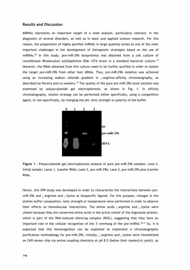
146
Results and Discussion
MiRNAs represents an important target of a wide analysis, particularly relevant, in the
diagnostic of several disorders, as well as in basic and applied science research. For this
reason, the preparation of highly purified miRNAs in large quantity arises as one of the most
important challenges in the development of therapeutic strategies based on the use of
miRNAs.29 In this study, pre-miR-29b biosynthesis was obtained from a cell culture of
recombinant Rhodovulum sulfidophilum DSM 1374 strain in a standard bacterial culture.15
However, the RNAs obtained from this culture need to be further purified in order to isolate
the target pre-miR-29b from other host sRNAs. Thus, pre-miR-29b isolation was achieved
using an increasing sodium chloride gradient in L-arginine-affinity chromatography, as
described by Pereira and co-workers.14 The quality of the pure pre-miR-29b stock solution was
examined by polyacrylamide gel electrophoresis, as shown in Fig. 1. In affinity
chromatography, elution strategy can be performed either specifically, using a competitive
agent, or non-specifically, by changing the pH, ionic strength or polarity of the buffer.
Figure 1 – Polyacrylamide gel electrophoresis analysis of pure pre-miR-29b samples. Lane S,
Initial sample; Lanes 1, transfer RNAs; Lane 2, pre-miR-29b; Lane 3, pre-miR-29b plus transfer
RNAs.
Hence, this SPR study was developed in order to characterize the interactions between pre-
miR-29b and L-arginine and L-lysine as biospecific ligands. For this purpose, changes in the
elution buffer composition, ionic strength or temperature were performed in order to observe
their effects on biomolecular interactions. The amino acids L-arginine and L-lysine were
chosen because they are conserved amino acids in the active center of the Argonaute protein,
which is part of the RNA-induced silencing complex (RISC), suggesting that they have an
important role in the cellular recognition of the 3' overhang of the pre-miRNA.30,31 So, it is
expected that this biorecognition can be exploited to implement a chromatographic
purification methodology for pre-miR-29b. Initially, L-arginine and L-lysine were immobilized
on CM5 sensor chip via amine coupling chemistry at pH 8.5 (below their isoelectric point), as

147
described by Cruz and collaborators.19 This protocol of immobilization ensures that the
guanidine functional groups (ε-amino group) of L-arginine and L-lysine are positively charged
and the α-NH2 group is attached to the reactive ester groups of the chip. All SPR equilibrium
data were fitted to a steady state bimolecular interaction model. The SPR binding profile
(square-shaped) was identical for all complexes and some were exemplified in Fig. 2.
Figure 2 - SPR analysis of pre-miR-29b interaction with L-lysine ligands in different buffers, at
4 and 20ºC. Sensorgrams and equilibrium-binding analysis: (a) 10 mM Tris-HCl buffer (4ºC), (b)
10 mM MOPS (20ºC) and (c) 10 mM HEPES (20ºC).
Biomolecular Interactions between pre-miR-29b and L-arginine
Recently, our research group has demonstrated the potential of the arginine amino acid in
the purification of RNA, suggesting that the binding mechanism involves multiple non-
covalent interactions with RNA, due to its ability to interact in different conformation
arrangements, the length of its side chain, the guanidine functional group and its ability to
produce hydrogen bond geometries.12,17,32 Thereby, since RNA is negatively charged due to the
phosphate groups in the backbone, it is easy to predict favorable electrostatic interactions
between RNA phosphate groups and L-arginine.
Interestingly, the SPR sensorgrams obtained with L-arginine ligand, present response profiles
in shaped of square, indicating rapid dissociation (450 sec), i.e., the binding assays suggest
that pre-miR-29b biomolecules interact with the immobilized L-arginine, and that these
interactions are reversible. The KD values are listed in Table 1. At 20ºC, in the range of
concentrations tested, SPR responses for L-arginine-pre-miR-29b were obtained in Tris-HCl,
NaCl and NaCl supplemented with L-lysine buffers. The equilibrium dissociation constants
demonstrate that Tris-HCl (10 mM) promote lower ligand−analyte affinity (KD =

148
5.11x10⁻⁶±4.4x10⁻⁷ M) when compared with the values obtained for NaCl (KD = 4.06x10-
7±3.5x10⁻⁸ M). In fact, in chromatography, the Tris-HCl buffer can be used to promote the
retention of total small RNAs onto the L-arginine support, but the application of NaCl in the
binding buffer is responsible for the higher selectivity towards the target miRNA. On the other
hand, negligible (≤ 5 RU) non-specific binding was detected when using the following buffers:
phosphate (10 and 50 mM), HEPES (10 and 100 mM) and MOPS (10 mM), regardless of their
concentration, indicating that L-arginine do not interact with pre-miR-29b under these
conditions (data not shown).
In contrast, the ionic strength of buffer (10 mM Tris-HCl supplemented with NaCl) has a
significant effect on the binding of pre-miR-29b. The SPR response to the pre-miR-29b
solution in 500 mM NaCl was very low (< 5 RU), implying that the chromatographic binding
and elution of pre-miR-29b may be manipulated with this parameter. In the sensorgrams was
notorious the dissociation in the first 100 sec using 500 mM NaCl (data not shown), indicating
that the high content of NaCl results in the loss of pre-miR-29b binding. Our results also
revealed that when a moderate salt concentration (280 mM NaCl) is used, high binding
responses were obtained (KD = 4.06x10⁻⁷±3.5x10⁻⁸ M). In general, the affinity of the pre-miR-
29b with the L-arginine surface was high (KD ≥ 10−6 M) at 20ºC. These results may be
correlated with the chromatographic experiments, where the total retention of pre-miR-29b
could be achieved using a low concentration of NaCl (280 mM) and high concentrations (500
mM NaCl) lead to its elution.14 Besides, the salt concentration can also have a positive effect
on RNA folding as well as on the stabilization of non-covalent RNA–amino acid interactions
during the purification, once the salt allows the single-stranded nucleic acid molecules to
form stable hydrogen bonds between their complementary bases. In this case, the
chromatographic interaction was promoted by using a low ionic strength buffer and the
selective elution of the bound species was achieved by increasing the NaCl concentration.14
Moreover, it was verified that the pre-miR-29b was eluted at a higher ionic strength,
indicating a stronger interaction than other contaminants. We also evaluated the influence of
a competing agent (L-arginine and L-lysine) in the buffer, as a way to conclude about the use
of these amino acids to promote binding or elution of the pre-miR-29b to the L-arginine
ligand. The experiments were carried out using different concentrations, from 8 to 19 mM of
L-arginine and 10 to 16 mM of L-lysine in the 200 mM NaCl buffer. However, for the highest
concentrations it was not verified the binding of pre-miR-29b to the L-arginine ligand (results
not shown). In these experiments, it was only obtained a binding response for the condition of
10 mM of L-lysine in 200 mM NaCl (KD = 3.53x10⁻⁶±1.9x10⁻⁷ M), while 8 mM of L-arginine
resulted in a negligible (≤ 5 RU) sensor response to the pre-miR-29b solution. This result is in
accordance with some evidences obtained in L-arginine-affinity chromatography, once this
condition can be used to the pre-miR-29b elution, as it functions as a competitive agent. This
behavior can be due to the positive character of these amino acids that promotes the binding
of pre-miR-29b to the free amino acid present in the buffer by electrostatic interactions,

149
inducing the elution of bound pre-miR-29b. Moreover, the results suggest that the elution of
bound pre-miR-29b can be more effective when using L-lysine in the buffer. As demonstrated
in Table 1, the KD value for 10 mM L-lysine is higher than the value obtained at 280 mM NaCl,
indicating that the affinity of pre-miR-29b to the L-arginine ligand is higher for the 280 mM
NaCl condition, and lysine is expected to weaken the interaction.
Considering that RNA is an unstable molecule and has a very short half-life once extracted,
the effect of temperature in the miRNA-arginine binding is a critical parameter in RNA
purification.33 In contrary to the results achieved for the temperature of 20ºC, at 4ºC it was
only found SPR response for L-arginine and pre-miR-29b using 10 mM Tris-HCl buffer. The
dissociation constants for Tris-HCl are of the same order of magnitude at 20 and 4ºC (KD =
1.34x10⁻⁶±2.3x10⁻⁷ M), but a slightly higher affinity was found for 4ºC, suggesting stronger
interactions at lower temperatures. Non-specific binding (≤ 5 RU) was detected when using
the other buffers, regardless their concentration, indicating that no significant interactions
occur between pre-miR-29b and L-arginine ligand in these conditions (data not shown). The KD
values obtained are consistent with the chromatographic results previously reported by
Pereira and collaborators14, where the pre-miR-29b retention in the L-arginine-agarose
support was higher for the lowest temperature studied (4ºC), suggesting that the temperature
is a critical factor in the binding pre-miR-29b.
Summarizing, the affinity order of pre-miR-29b to L-arginine was 280 mM NaCl > 10 mM Tris-
HCl ≈ 10 mM L-lysine in 200 mM NaCl, with NaCl promoting the highest affinity for the pre-
miR-29b to the L-arginine surface. Hence, this comparative study reinforces the idea that the
binding mechanism inherent to the biorecognition between the L-arginine amino acid and pre-
miR-29b molecules can result from the combination of several phenomenological interactions
such as electrostatic interactions, multiple hydrogen bond and van der Waals forces.17 Thus,
it is reasonable to suggest that the retention of RNA in L-arginine is due to its side chain,
which can promote multi contacts with RNA backbone or bases, according to RNA
folding.32,34,35 Additionally, recent studies with saturation transfer difference-nuclear
magnetic resonance spectroscopy and SPR reported that guanine polynucleotides interact
preferably with L-arginine through the sugar-phosphate backbone.18,19 Thus, another
explanation for these findings is related with pre-miR-29 structural features that seem to be
relevant on its distinct retention behavior in the arginine ligand. Pre-miR-29b is a small RNA
molecule with a stem-loop shape consisting of two long irregular double-stranded stem
regions, which are interrupted by a largely single-stranded internal loop. In addition, along
the bulge of pre-miR-29b and through the continuous stem sequences there are several
guanines, which were described to interact preferably with arginine. Hence, the nucleotide
bases composition and exposure can also favor other biomolecular interactions, responsible
for the specificity and biorecognition found in the L-arginine ligand.

150
Table 1 – Equilibrium analysis of the pre-miR-29.
Buffers
Tris-HCl (10 mM)
MOPS (10 mM)
HEPES (10 mM)
NaCl (280 mM)
L-arginine (8 mM)
L-lysine (10 mM)
L-arginine
20ºC 5.11×10⁻⁶±4.4×10⁻⁷ - - 4.06×10⁻⁷±3.5×10⁻⁸ - 3.53×10⁻⁶±1.9×10⁻⁷
4ºC 1.34×10⁻⁶±2.3×10⁻⁷ - - - - -
L-lysine
20ºC 3.24×10⁻⁷±4.6×10⁻⁸ 1.74×10⁻⁵±4.1×10⁻⁶ 2.47×10⁻⁵±6.6×10⁻⁶ 9.96×10⁻⁷±3.5×10⁻⁸ 2.08×10⁻⁶±4.4×10⁻⁷ 2.28×10⁻⁶±6.3×10⁻⁷
4ºC 8.76×10⁻⁶±6.8×10⁻⁷ 2.31×10⁻⁵±3.1×10⁻⁶ 3.95×10⁻⁶±1.5×10⁻⁷ - - -

151
Biomolecular Interactions between pre-miR-29b and L-lysine
Previous studies have shown that the main interactions occurring between nucleic acids and
the L-lysine amino acid are electrostatic, because of its amine groups.36-38 Moreover, L-lysine
has also some characteristics, such as the lateral side chain with significant length and ability
to interact with several RNA classes even in different conformational rearrangements, which
may produce hydrogen bond geometries, reinforcing the establishment of multiple non-
covalent interactions involved in the specific recognition mechanism of nucleic acids.38
Indeed, besides the multiple interactions (electrostatic interactions, van de Waals forces and
hydrogen bonds) that L-lysine can establish with the RNA backbone or with the more exposed
bases, several sRNAs can also interact and be recognized by the presence of hydrophobic
interactions with the aliphatic portion of L-lysine side chain.15 Considering this complex
biorecognition mechanism, it is important to study the influence of experimental conditions
on the interaction occurring between pre-miR-29b and L-lysine amino acid, resulting in
different binding affinities. It was verified that pre-miR-29b binds more strongly to L-lysine in
10 mM Tris-HCl and 280 mM NaCl at 20ºC (KD = 3.24x10⁻⁷±4.6x10⁻⁸ M and KD = 9.96x10⁻⁷±3.5x10⁻⁸
M, respectively) than in 10 mM HEPES (KD = 2.47×10⁻⁵±6.6×10⁻⁶ M) or 10 mM MOPS buffers (KD =
1.74×10⁻⁵±4.1×10⁻⁶ M). In fact, these findings are in agreement with the results reported in
the literature, where Tris-HCl buffer is commonly used to promote the binding of RNA to L-
lysine. Negligible binding (≤ 5 RU) was detected when the following buffers were used:
phosphate (10 mM and 50 mM) and HEPES (100 mM), indicating that pre-miR-29b has no
affinity to L-lysine under these conditions. Similarly to the results obtained to L-arginine, the
ionic strength of buffer (10 mM Tris-HCl supplemented with NaCl or with amino acids) has an
effect on the binding of pre-miR-29b to L-lysine. Our findings demonstrate that when high
concentrations of salt (500 mM NaCl) or amino acids are used, no binding responses of pre-
miR-29b are observed and, consequently, these conditions can be used for the elution of the
target biomolecule in chromatography. The addition of 10 mM of L-lysine in 200 mM NaCl
resulted in a binding response characterized by KD = 2.28x10⁻⁶±6.3×10⁻⁷ M and the
supplementation with 8 mM of L-arginine in 200 mM NaCl resulted in a KD = 2.08x10⁻⁶±4.4×10⁻⁷
M. Although these concentrations of competing agents induce binding of pre-miR-29b, the
interaction is weaker when compared with Tris-HCl. Briefly, the affinity order for pre-miR-
29b binding to L-lysine was 10 mM Tris-HCl ≈ 280 mM NaCl > 10 mM L-lysine ≈ 8 mM L-arginine
in 200 mM NaCl > 10 mM HEPES ≈ 10 mM MOPS, thus Tris-HCl and NaCl buffers induced the
greater affinity to L-lysine surface, followed by HEPES and MOPS (Table 1).
In this study it was also evaluated the effect of decreasing the temperature to 4ºC on the
binding of pre-miR-29b to the L-lysine surface, comparing with the binding at 20ºC. The
binding responses is similar to that obtained at 20ºC, illustrating that the interactions
between L-lysine and pre-miR-29b also occur under these conditions. However, some
differences were found in KD values determined at 4ºC (3.24x10⁻⁷ M to 8.76x10⁻⁶ M for 10 mM
Tris-HCl; 2.47x10⁻⁵ M to 3.95x10⁻⁶ M for 10 mM HEPES and 1.74x10⁻⁵ to 2.31x10⁻⁵ M for 10 mM

152
MOPS, at 20 and 4ºC, respectively). In general, the KD values obtained are consistent with the
chromatographic results, which the lowest temperature studied induced higher pre-miR-29b
retention onto the L-lysine-agarose support (data not shown). Globally, these results can also
be related with the pre-miRNA structure, which presents continuous stem sequences mostly
containing guanines that are described to preferentially interact with L-lysine due to hydrogen
bonds that are established with one of the donor atoms.39,40
Briefly, to compare the salt influence on the binding of pre-miR-29b to the L-arginine and L-
lysine surfaces, several buffers were analyzed. In general, the maximum RU signal was higher
for the L-lysine than for the L-arginine surface (Fig. 3).
Figure 3 - Relative resonance response (RU) with different running buffers at 450 s after
injection of pre-miR-29b at a concentration of 1.25 μM, at 20ºC, in L-arginine and L-lysine
surfaces.
The experiments showed a low resonance response level with both surfaces (normalized
signal). It was found that the maximum binding between pre-miR-29b and immobilized L-
arginine and L-lysine was observed in 280 mM NaCl, followed by 10 mM Tris-HCl, whereas the
lowest binding was observed in 200 mM NaCl supplemented with 8 mM L-arginine or 10 mM L-
lysine, 10 mM HEPES and 10 mM MOPS (Fig. 3). Thus, our affinity data show that Tris-HCl and
NaCl are optimal buffers for interaction assays and distinct buffer environments such as MOPS
or HEPES did not enhanced the binding to L-arginine and L-lysine. Overall, these results
provide suitable conditions to induce pre-miRNA binding, thereby increasing the selectivity
and improving efficient microRNA separation. It was also proved that under these conditions
(ionic strength and buffer composition) the electrostatic interactions are favored between
the amino positive groups of the L-arginine and L-lysine and the negative phosphate groups of
the pre-miR-29b, mainly with guanosine.
These results demonstrated that SPR allows making a screening of ligands that, like the ones
described herein, can be used to design and implement novel miRNA purification methods.

153
Pre-miR-29b structural analysis
Both ligands promoted the biorecognition of pre-miR-29b as well as an effective interaction
for most conditions studied. To select the buffer that is able to enhance the pre-miR-29b
binding to the amino acids ligands and to evaluate the effect of the different conditions in
the pre-miR-29b structural stability, four buffers were analyzed (Tris-HCl, HEPES buffer,
MOPS, phosphate buffer) by circular dichroism (CD) spectroscopy. Thus, structural
characterization of the pre-miR-29b under different experimental conditions give additional
information about the interactions with the ligands and also about the suitable conditions
that must be used in chromatographic procedures to purify the target biomolecules while
maintaining its stability and biological activity. CD technique offers good resolution, provides
sensitive spectra and it has been widely used in structural determination of biomolecules
(proteins, nucleic acids, etc.). Quantitative analysis of CD spectra is possible, but, as with any
assay, the correct and proper interpretation of the results depends on an accurate
understanding of the procedure. The spectra in Figure 4 are given in terms of molar ellipticity
thus making it possible to directly compare the intensity of the bands obtained for different
samples. CD spectra of the pre-miR-29b show characteristic peaks around 210 nm (negative
maximum signal) and 265 nm (positive maximum signal). The comparison with the CD
spectrum of established nucleic acids structures confirms that the pre-miRNA possesses a
stem-loop structure because the CD spectral pattern typically indicates the B-form RNA
duplex.15,41 Although, CD is not sensible to detect the contamination of other cytosol
containing miRNAs, it is sensible to detect the contamination of other biomolecules.
However, the pre-miR-29 subjected to this analysis has a purity of 98%, and it is unlikely that
the sample is contaminated, being not expected changes in the CD spectra resulting from
contamination.
In Figure 4 are shown the CD spectra resulting from the evaluation of pre-miR-29b in four
buffers, after an initial 15 min incubation in Tris-HCl buffer (10 mM) and HEPES (10 mM). The
CD spectra of the pre-miRNA sample in Tris-HCl and HEPES show typical CD spectrum of RNA,
with a positive band at 265 nm and a negative band at 210 nm, similar to the obtained with
DEPC water. However, a significant difference in the bands magnitude of the pre-miR-29b
spectra was found (Figure 4).

154
Figure 4 - Circular Dichroism spectra of the pre-miR-29b.
These results are consistent with the responses obtained with SPR, suggesting that Tris-HCl is
a valuable choice for promoting pre-miRNA−L-arginine/lysine binding interactions in future
applications. In the presence of HEPES or MOPS, the electrostatic interaction between the L-
arginine and L-lysine surface and the pre-miR-29b backbone could be more favored, since
these buffers at neutral pH are a zwitterionic molecule with a positive charge on one of the
protonated nitrogens. This can reduce the repulsive electrostatic interaction between
charged phosphate groups of pre-miR-29b, stabilizing the pre-miRNA molecule and favoring
the interaction with the amino acids. By analyzing the CD spectra of pre-miR-29b in MOPS (10
mM), it is visible a decrease of intensity of the positive band as well as the negative band. By
the CD spectra analysis of Figure 4, it is evident a strong modification in the RNA structure for
the phosphate buffer. This data can also justify why pre-miR-29b molecules don’t interact
with the L-arginine or L-lysine supports in the presence of phosphate buffer due to the
increase of the repulsions with the pre-miR-29b backbone. In contrast, in the presence of salt
(280 mM NaCl), phosphate groups of the pre-miR-29b backbone are effectively screened by
the salt counterions resulting in a more compact and less regular structure. The data
obtained from SPR can play an important role in the optimization of chromatographic-based
purification processes since it allows the determination of which buffers favor interactions of
the target biomolecule with the chromatographic support. Moreover, the conjugation with
the CD technique allows the confirmation of which buffers are able to maintain the stability
of the miRNA.

155
Conclusions
The binding affinity quantification between pre-miR-29b and L-arginine and L-lysine surfaces
was examined by SPR technique, under different conditions, in order to mimic the
immobilized ligand onto an affinity chromatographic support, and to verify the effect of these
conditions on the interactions and stability of the molecule. Therefore, several experimental
conditions for amino acids ligands-pre-miR-29b binding were characterized, and an optimal
set of buffers as well as temperature conditions were defined. Distinct buffers affected the
interaction strength with an increased response in presence of Tris-HCl and NaCl. These
results provide suitable conditions to induce pre-miRNA binding, thereby increasing the
selectivity and improving efficient miRNA separation. The predominant interactions of pre-
miR-29b with L-arginine and L-lysine ligands are ionic between the amino positive groups of
the L-arginine and L-lysine and the negative phosphate groups of the pre-miR-29b and
hydrogen bonds with the more exposed bases, mainly with guanosine. Overall, the present
study showed that SPR can be used as a first technique to screen, manipulate, determine and
establish the best binding/elution conditions that can lead to an improved performance and
selectivity of the chromatographic process. In addition, it was demonstrated that although
some conditions used in chromatographic experiments favor multiple interactions between
the pre-miR-29b and the respective ligand (L-arginine and L-lysine), these conditions can
affect the structural stability of the target molecule. From CD, it was possible to conclude
that the biorecognition observed by the pre-miR-29b can be related to the higher availability
and base exposition, which confers a different conformational structure of this molecule.
Moreover, the highest equilibrium dissociation constants determined by SPR ranged from 10−6
to 10−7 M to L-arginine and from 10−5 to 10−7 M to L-lysine, indicating that pre-miR-29b showed
high binding affinity on these surfaces. In addition, the results obtained in this study, provide
valuable and relevant information that can be further applied in unveiling the miRNA
chromatographic behavior in these systems, in order to improve the binding of the pre-miR-
29b onto the amino acids-based supports, preserving their structural stability, integrity and
activity. In conclusion, it was proved that the association of these two techniques might be a
powerful strategy to define the best conditions to be applied in the miRNA purification.
Acknowledgments
This work was supported by the Portuguese Foundation for Science and Technology (FCT),
through the projects EXPL/BBB-BIO/1056/2012, Pest-OE/SAU/UI0709/2014 and FCOMP-01-
0124-FEDER-041068 – EXPL/QEQ-MED/1068/2013. Patrícia Pereira and Carla Cruz acknowledge
the fellowships, SFRH/BD/81914/2011 and SFRH/BPD/100015/2014, respectively from FCT.
The authors also acknowledge the program Fundo Europeu de Desenvolvimento Regional
(FEDER) [COMPETE [FCOMP-01-0124-FEDER-027560]].

156
References
1. D. P. Bartel, Cell, 2004, 116, 281-297.
2. J. A. Broderick and P. D. Zamore, Gene therapy, 2011, 18, 1104-1110.
3. A. Eulalio and M. Mano, Journal of biomolecular screening, 2015, DOI:
10.1177/1087057115578837.
4. C. L. Bartels and G. J. Tsongalis, Clinical chemistry, 2009, 55, 623-631.
5. M. V. Latronico and G. Condorelli, Nature reviews. Cardiology, 2009, 6, 419-429.
6. P. T. Nelson, W. X. Wang and B. W. Rajeev, Brain Pathol, 2008, 18, 130-138.
7. L. Du and A. Pertsemlidis, J Mol Cell Biol, 2011, 3, 176-180.
8. I. Vomelova, Z. Vanickova and A. Sedo, Folia biologica, 2009, 55, 243-251.
9. M. A. Dineva, L. MahiLum-Tapay and H. Lee, The Analyst, 2007, 132, 1193-1199.
10. L. Ponchon and F. Dardel, Methods, 2011, 54, 267-273.
11. S. C. Walker, F. H. Scott, C. Srisawat and D. R. Engelke, Methods Mol Biol, 2008, 488,
23-40.
12. R. Martins, C. J. Maia, J. A. Queiroz and F. Sousa, J Sep Sci, 2012, 35, 3217-3226.
13. R. Martins, J. A. Queiroz and F. Sousa, Journal of molecular recognition : JMR, 2010, 23,
519-524.
14. P. Pereira, A. Sousa, J. Queiroz, I. Correia, A. Figueiras and F. Sousa, J Chromatogr B
Analyt Technol Biomed Life Sci, 2014, 951-952, 16-23.
15. P. Pereira, A. Sousa, J. Queiroz, A. Figueiras and F. Sousa, J Chromatogr A, 2014, 1331,
129-132.
16. R. Martins, J. A. Queiroz and F. Sousa, Biomedical chromatography : BMC, 2012, 26, 781-
788.
17. R. Martins, J. A. Queiroz and F. Sousa, Journal of chromatography. A, 2014, 1355, 1-14.
18. F. Sousa, D. M. Prazeres and J. A. Queiroz, Trends Biotechnol, 2008, 26, 518-525.
19. C. Cruz, E. J. Cabrita and J. A. Queiroz, Anal Bioanal Chem, 2011, 401, 983-993.
20. C. Cruz, S. D. Santos, E. J. Cabrita and J. A. Queiroz, International journal of biological
macromolecules, 2013, 56, 175-180.
21. C. Cruz, A. Sousa, E. Mota, F. Sousa and J. A. Queiroz, Int J Biol Macromol, 2014, 70,
131-137.
22. C. Cruz, A. Sousa,F. Sousa, J.A. Queiroz, Anal Methods, 2013, 5, 1682-1686.
23. C. T. Campbell and G. Kim, Biomaterials, 2007, 28, 2380-2392.
24. P. S. Katsamba, S. Park and I. A. Laird-Offringa, Methods, 2002, 26, 95-104.
25. B. Nguyen, F. A. Tanious and W. D. Wilson, Methods, 2007, 42, 150-161.
26. J. Hwang and S. Nishikawa, Journal of biomolecular screening, 2006, 11, 599-605.
27. H. Suzuki, S. Umekage, T. Tanaka and Y. Kikuchi, J Biosci Bioeng, 2011, 112, 458-461.
28. P. Chomczynski and N. Sacchi, Nature protocols, 2006, 1, 581-585.
29. D. P. Bartel, Cell, 2009, 136, 215-233.

157
30. F. V. Rivas, N. H. Tolia, J. J. Song, J. P. Aragon, J. Liu, G. J. Hannon and L. Joshua-Tor,
Nat Struct Mol Biol, 2005, 12, 340-349.
31. S. Griffiths-Jones, Methods Mol Biol, 2006, 342, 129-138.
32. J. D. Puglisi, J. R. Wyatt and I. Tinoco, Jr., Biochemistry, 1990, 29, 4215-4226.
33. F. RE, RNA methodologies - Laboratory Guide for Isolation and Characterization,
Academic Press, York, USA, 4th edn., 2005.
34. B. J. Calnan, B. Tidor, S. Biancalana, D. Hudson and A. D. Frankel, Science, 1991, 252,
1167-1171.
35. T. Janas, J. J. Widmann, R. Knight and M. Yarus, RNA, 2010, 16, 805-816.
36. A. Sousa, F. Sousa and J. A. Queiroz, J Chromatogr B Analyt Technol Biomed Life Sci,
2011, 879, 3507-3515.
37. A. Sousa, F. Sousa and J. A. Queiroz, J Chromatogr B Analyt Technol Biomed Life Sci,
2009, 877, 3257-3260.
38. D. S. Jones, H. K. Lundgren and F. T. Jay, Nucleic Acids Res, 1976, 3, 1569-1576.
39. J. J. Ellis, M. Broom and S. Jones, Proteins, 2007, 66, 903-911.
40. M. M. Hoffman, M. A. Khrapov, J. C. Cox, J. Yao, L. Tong and A. D. Ellington, Nucleic
Acids Res, 2004, 32, D174-181.
41. B. Ranjbar and P. Gill, Chem Biol Drug Des, 2009, 74, 101-120.

158

159
Paper V
New approach for purification of pre-miR-29 using lysine-
affinity chromatography
P. Pereira, A. Sousa, J. A. Queiroz, A. Figueiras, F. Sousa
Journal of Chromatography A. 2014. 1331: 129-132
Short description: This article describes a new strategy employing a lysine-agarose support to
efficiently separate recombinant pre-miR-29b from a complex mixture containing small RNAs.
Besides this novelty, it is further described for the first time the use of ammonium sulfate in
this matrix for the purification of nucleic acids. Overall, it was verified that lysine ligand can
promote a specific interaction with the pre-miR-29b, thus favoring its isolation and
purification.

160

Journal of Chromatography A, 1331 (2014) 129– 132
Contents lists available at ScienceDirect
Journal of Chromatography A
jou rn al hom epage: www.elsev ier .com/ locate /chroma
Short communication, Col Liquid Chromatography
New approach for purification of pre-miR-29 using lysine-affinitychromatography
Patrícia Pereiraa, Ângela Sousaa, João Queiroza, Ana Figueirasa,b, Fani Sousaa,∗
a CICS-UBI – Health Sciences Research Centre, University of Beira Interior, Avenida Infante D. Henrique, 6200-506 Covilhã, Portugalb CEF-FFUC – Pharmaceutical Studies Center, Faculty of Pharmacy, University of Coimbra, Pólo das Ciências da Saúde, Azinhaga de Santa Comba, 3000-548Coimbra, Portugal
a r t i c l e i n f o
Article history:Received 18 November 2013Received in revised form 6 January 2014Accepted 10 January 2014Available online 17 January 2014
Keywords:Affinity chromatographyLysine-agarosemicroRNApre-miR-29RNA purification
a b s t r a c t
miRNA-based gene therapy applications require microRNA with high purity degree, good quality andbiologically active. Owing to the commercial interest in these approaches, there is a growing interest inthe development of innovative procedures to easily and efficiently purify the RNA. Thus, several chro-matographic and non-chromatographic methods have been reported to accomplish this purpose, but notall of these strategies allow the efficient separation of miRNAs. The present study describes a new strategythat uses a lysine ligand in affinity chromatography to efficiently separate pre-miR-29 from a small RNAsmixture. The interest on this biomolecule is related to the fact that pre-miR-29 deficiencies or excesseshave been associated to a number of clinically important diseases. The retention behaviour of pre-miR-29was characterized and adjusted to achieve higher specificity in this chromatographic operation, using anammonium sulfate stepwise gradient. Overall, it was verified that lysine-agarose support can promote aspecific interaction with the pre-miR-29 favouring its total separation. The results also suggest that theunderlying mechanism involves biorecognition of pre-miR-29 by the lysine ligands, resulting from theoccurrence of different elementary interactions, including hydrogen and hydrophobic interactions.
© 2014 Elsevier B.V. All rights reserved.
1. Introduction
Currently, microRNA (miRNA) is considered a powerful biolog-ical material that is directly involved in performing or controllingmany biological functions [1]. Thus, understanding the role of sev-eral miRNAs present in cellular processes is critical to developnew diagnosis methods or drugs for comprising and treating ill-ness. Recently, differential expression of miRNAs, in patients withAlzheimer’s disease (AD) suggests that they might have key reg-ulatory roles in this neurodegenerative disease [2]. Hebert andcollaborators showed that miR-29 is potentially involved in theregulation of �-amyloid precursor protein-converting enzyme 1(BACE1) expression because in vitro studies revealed that miR-29 cluster was significantly decreased in AD patients displayingabnormally high BACE1 protein levels, leading the generation andsubsequent accumulation of �-amyloid [3]. These findings raisedthe possibility to use miR-29 as a potential therapeutic target forAD therapy. In fact, miRNAs play a prominent role in the posttran-scriptional regulation of gene expression, since it prevents proteinsynthesis by inducing the messenger RNA degradation, with a highspecificity degree [3]. Nevertheless, to get pre-miR-29 as a suitable
∗ Corresponding author. Tel.: +351 275 329 074; fax: +351 275 329 099.E-mail address: [email protected] (F. Sousa).
product for therapeutic applications it is necessary to guaranteeits purity, stability and integrity. Regardless of the principle ofRNA isolation used, purification protocols have a number of com-mon requirements, because it is a critical process due to the highrisk of RNA degradation during the procedure, by the presence ofRNA degrading enzymes, once RNA molecule is very susceptible tochemical and enzymatic degradation [4].
Hence, several kits are commercially available for miRNA iso-lation from a diverse variety of samples; these methods allow theseparation of miRNAs from high molecular weight RNA [4]. Further-more, there are several chromatographic methods reported in theliterature for the purification of the RNAs [5,6]. In these methods,different tags are introduced into RNA molecules, which subse-quently have to be released from the chromatographic support bycompetitive elution or cleaved off by a protease [5,6], which mayinduce conformational changes in RNA and are expensive proce-dures. The approach to overcome these limitations includes therecent development, by our research group, of an affinity chro-matographic (AC) method with amino acids as specific ligands,applied for plasmid DNA [7–9] and RNA [10] purification allowingan improvement in the purification technologies since this tech-nique exploits natural biological processes of specific recognitionof the target biomolecules [11].
In this study, a lysine-agarose support was tested to purify pre-miR-29 from a complex mixture containing small RNAs (sRNAs)
0021-9673/$ – see front matter © 2014 Elsevier B.V. All rights reserved.http://dx.doi.org/10.1016/j.chroma.2014.01.033
161

130 P. Pereira et al. / J. Chromatogr. A 1331 (2014) 129– 132
of Rhodovulum sulfidophilum. In 1978, Jones et al. [12] employedthe lysine ligand to separate RNA species with different molecularweights, using a linear gradient of sodium chloride (NaCl). Buildingon these considerations, it is interesting to verify if the lysine matrixmay contribute to the separation of pre-miR-29 and to understandthe interaction mechanism underlying the specificity of the sup-port that allowed a biorecognition of the pre-miR-29. Therefore,this matrix allowed the efficient isolation and purification of pre-miR-29 from the other sRNAs. These findings will strengthen thepossibility of using lysine-affinity chromatography as a potentialenabling technology in the downstream process of pre-miRNA fortherapeutic applications.
2. Materials and methods
2.1. Pre-miR-29 production and purification
The pre-miR-29 used in the experiments was produced by a cellculture of Rhodovulum sulfidophilum DSM 1374 strain (BCCM/LMG)modified with the plasmid pBHSR1-RM [13] containing thesequence of pre-miR-29. Growth was carried out at 30 ◦C, usingNutrient Broth medium (1 g/L beef extract; 2 g/L yeast extract; 5 g/Lpeptone and 5 g/L NaCl) supplemented with 30 �g/mL kanamycin.sRNA was extracted using the acid guanidinium thiocyanate-phenol-chloroform method based on the protocol described byMartins et al. [10]. Chromatographic experiments were performedin an ÄKTA Avant system with UNICORN 6 software (GE Health-care). A 10 mm diameter × 20 mm long (about 2 mL) column waspacked with Lysine-agarose matrix (GE Healthcare). All solutionswere freshly prepared using 0.05% diethyl pyrocarbonate (DEPC)-treated water and were filtered through a 0.20 �m pore sizemembrane. Chromatographic runs were performed at 20 ◦C. Thecolumn was first equilibrated with 2.0 M of (NH4)2SO4 in 10 mMTris buffer (pH 8.0). sRNAs samples (40 �g) were applied onto thecolumn using a 100 �L loop at a flow-rate of 1 mL/min. After theelution of unbound species, the ionic strength of the buffer wasdecreased to 1.5 and 1.1 M of (NH4)2SO4 and then to 10 mM Trisbuffer (pH 8.0). The absorbance of the eluate was continuouslymonitored at 260 nm. Fractions were pooled according to the chro-matograms obtained, and following concentration and desaltingwith Vivaspin concentrators. The integrity of pre-miR-29 sampleswas analyzed by vertical electrophoresis with 10% polyacrylamidegel [10].
2.2. PCR analysis
Pre-miR-29 identification was assessed using polymerase chainreaction (PCR). cDNAs synthesis was performed using RevertAidFirst Strand cDNA Synthesis Kit (Fermentas), according to manu-facturer’s instructions. A total of 0.5 �g of RNA samples collectedfrom the chromatographic purification process with lysine–agarosecolumn was used to initiate cDNA synthesis, which was denaturedfor 5 min at 65 ◦C with 20 pmol of gene-specific primer (5′ GACAGC GGT ATG ATC CCC CAA 3′). Then, PCR reactions were carriedout using 1 �L of cDNA in a 25 �L reaction containing 0.125 U ofSupreme DNA polymerase (NZYtech) and 150 nM of each primer.Specific primers for cDNA, designed from RNA database informa-tion, were used to amplify a fragment of 145 bp (Fw – GGA AGCTGG TTT CAT ATG GTG and Rv – CCC CCA AGA ACA CTG ATT TC).PCR was conducted as follows: denaturation at 95 ◦C for 5 min, fol-lowed by 40 cycles at 95 ◦C for 30 s, 63 ◦C for 30 s and 72 ◦C for 15 s,and a final elongation step at 72 ◦C for 5 min. To confirm the pres-ence and purity of amplicons, PCR products were analyzed using 1%agarose gel electrophoresis [14]. The DNA molecular weight markerwas obtained from Vivantis Technologies.
3. Results and discussion
In the present study, the applicability of lysine-affinity chro-matography in the RNA downstream process was exploited, topurify pre-miR-29 from a sRNA complex mixture. The lysine aminoacid was studied due to the fact of being conserved in the active cen-tre of the PAZ domains of the Argonaute proteins, suggesting that ithas an important role in the specific recognition of the pre-miRNAafter which occurs the degradation of mRNA [15]. In this work, itwas produced a pre-miRNA instead of the mature miRNA consid-ering their structural characteristics, specifically the single chainwith ∼100 nucleotides, being some unpaired in the 3′ overhang,which will possibly facilitate the purification, due to the higherbases exposure [16]. Previous published studies, have shown thatthe main interactions between nucleic acids [12,17] and the lysineamino acid are ionic, because of its amine groups. Thus, initialexperiments were performed to choose the best binding/elutionstrategy for pre-miR-29 using a gradient of NaCl. However, underthese conditions the RNA binding was not effective and the selec-tive purification of pre-miRNA was not possible to be achieved (datanot shown). Therefore, a different strategy was used to promote theRNA binding. In a first step, lysine-agarose column was equilibratedwith 2 M (NH4)2SO4 in 10 mM Tris buffer (pH 8.0). After injection ofthe 40 �g of pre-miR-29 containing mixture, was obtained a peak(Fig. 1A) where pre-miR-29 was not detected (data not shown).This can result either from the elution of unbound material thatpresents a lower affinity to the matrix or by the alteration in theionic strength since the injected RNA sample is dissolved in H2O.The ionic strength of the buffer was decreased to 1.5 and 1.1 M(NH4)2SO4 in 10 mM Tris buffer (pH 8.0) in order to elute specificmolecules in a peak 2. The chromatographic run was concludedwith a final elution step, using 10 mM Tris buffer (pH 8.0), to recoverthe strongly bound species. The polyacrylamide gel electrophore-sis was used to detect and identify different sRNAs species elutedin each peak and assess of RNA integrity and purity (Fig. 1B). Theanalysis of electrophoresis revealed that the peak 1 mostly cor-responds to the transfer RNA (tRNA) (lane 1), while pre-miR-29remained bound under the same conditions, being identified in thepeak 2 (lane 2). The electrophoretic evaluation of fractions recov-ered from the peak 3 revealed the presence of other tRNAs (lane3). Thus, the electrophoresis of all fractions pooled from the chro-matographic experiment indicated that the pre-miR-29 eluted inthe peak 2 is pure. These findings suggest that the lysine liganddistinguishes and differentially interacts with several RNA species(Fig. 1) further suggesting a specific recognition of the pre-miR-29.Moreover, the pores of the lysine-support are characterized with anexclusion limit of >2 × 10⁷ (∼872 base pairs), allowing the accessof sRNAs molecules (<150 nucleotides and M.W. ∼4.8 × 10⁴) to theimmobilized ligands at the beads surface and inside the pores. pre-miR-29 presents the shape of a stem-loop consisting of two longirregular double-stranded stem regions, which are interrupted bya largely single-stranded internal loop. Along the bulge and throughthe continuous stem sequences there are mostly guanines, whichare described to preferentially interact with lysine [18–20] due tohydrogen bonds that are established with one of the donor atomsof the lysine [19]. Thus, an increased retention of pre-miRNA withinserts mainly constituted by guanine will be expected. Likewise,the lysine amino acid has some characteristics, such as the lat-eral side chain with significant length and ability to interact withseveral RNA classes even with different conformational rearrange-ments producing good hydrogen bond geometries, which reinforcethe specific recognition mechanism [19]. Between multiple interac-tions (electrostatic interactions, van de Waals forces and hydrogenbonds) that the lysine can promote with the RNA backbone or withthe more exposed bases, several sRNAs can also be recognized bythe presence of hydrophobic interactions [18] with the aliphatic
162

P. Pereira et al. / J. Chromatogr. A 1331 (2014) 129– 132 131
Fig. 1. (A) Chromatographic profile of the pre-miR-29 purification from a sRNA mixture by lysine–agarose chromatography. Elution was performed at 1.0 mL/min bydecreasing (NH4)2SO4 stepwise gradient from 2 M (NH4)2SO4 to 10 mM Tris buffer (pH 8.0), as represented by the arrows. (B) Polyacrylamide gel electrophoresis of samplescollected at the column outlet. Fractions corresponding to peaks (1), (2) and (3) are shown in lanes 1-3, respectively. Lane S, sRNA sample injected onto the column.
portion of side chain [17], which can be intensified by the pres-ence of (NH4)2SO4. Moreover, the salt concentration can also havea profound positive effect on RNA folding as well as on the stabil-ity of non-covalent RNA–amino acid interactions during the courseof purification [19,20], once the salt allows that single-strandednucleic acid molecules form stable hydrogen bonds between theircomplementary bases.
The conformational stability of the pre-miR-29 secondary struc-ture was assessed using Circular Dichroism (CD) protocol describedby Pereira et al. [14] (data not shown). CD spectra of the pre-miR-29 shows characteristic peaks around 210 (negative signal)and 265 nm (positive signal), which, when compared with theCD spectrum of established nucleic acids structures confirm thatthe pre-miRNA possesses a stem-loop structure that is an intra-molecular variant of the B-form and also contains a single-stranded[21]. While the CD spectra of the sRNA shows characteristic fea-tures of the A-RNA, a positive band around 260 nm was observed,and a negative signal at 220 nm, with approximately the sameintensity. Due to single-stranded, the duplex to stem-loop tran-sition is usually accompanied by CD amplitude reduction whencompared with sRNA spectrum. Our results demonstrate thatisolation steps involved in the affinity procedure allowed the main-tenance of the pre-miRNA stability. Considering the relevance ofobtaining pharmaceutical-grade pre-miR-29 is required furthercharacterization of quality parameters by spectrophotometry anal-ysis, estimating the pre-miR-29 concentration (absorbance valuesat 260 nm) and purity (calculating the 260/280 ratio). The chro-matographic process demonstrates an excellent performance in thepre-miR-29 isolation, attaining a significant yield (11.80 ± 0.50 �g)in the peak 2. In addition, pre-miR-29 pure has shown 260/280ratio of 2.00 ± 0.01, which is often characteristic of a pure RNApreparation [10]. The protein contamination level was assessed bymicro-BCA assay (Thermo Fisher Scientific Inc.) in the RNA fractionscollected from the chromatographic peaks. This chromatographicstrategy provides a reduction of protein level present in the finalpre-miR-29 samples (2.613 ± 0.006 ng/�L). As it can be observedfrom the electrophoretic analysis of the PCR products (Fig. 2), thePCR reaction allowed the amplification of pre-miR-29 cDNA frag-ments. No further amplification was seen in the control (lane C) orin the peaks 1 (lane 1). The control was made of PCR reaction solu-tions without cDNA. Therefore, it was verified that the identity ofthe sRNA isolated from the original population was the pre-miR-29.
Fig. 2. pre-miR-29 identification by PCR. The agarose gel electrophoretic analysisof PCR products shows amplification of pre-miR-29 cDNA fragments. Lane M, DNAmolecular weight marker; lanes C, negative control; lane 1, 2 and 3 correspondingto the three chromatographic peaks, respectively.
These analyses allowed the assessment of RNA quality, indi-cating that the developed pre-miR-29 isolation methodology canpurify the target RNA from different RNA species with high yield,purity and good integrity. Thus, the present data show the develop-ment of an AC approach based on natural occurrence interactions,between lysine and RNA species, resulting in the selective purifi-cation of pre-miR-29 molecules, in the required conditions fortherapeutic applications. These results also demonstrate the ver-satility of the lysine ligand in the RNA purification, since thepre-miRNA can be purified with (NH4)2SO4 but classes of RNAs canalso be separated with NaCl [12]. The successful isolation of miRNAspecies with high purity and quality is a step forward in RNA isola-tion methodologies and can widely contribute to studies in severalRNA research fields.
Acknowledgments
This work was supported by the Portuguese Foundation forScience and Technology (PTDC/EBB-BIO/114320/2009, EXPL/BBB-BIO/1056/2012, PEst-C/SAU/UI0709/2011 and COMPETE:
163

132 P. Pereira et al. / J. Chromatogr. A 1331 (2014) 129– 132
FCOMP-01-0124-FEDER-027560). Patrícia Pereira and ÂngelaSousa acknowledge Ph.D. and Postdoctoral fellowships(SFRH/BD/81914/2011 and SFRH/BPD/79106/2011, respectively).The authors thank Prof. Yo Kikuchi (Toyohashi University ofTechnology) for kindly provided of pBHSR1-RM plasmid.
References
[1] D.P. Bartel, Cell 116 (2004) 281.[2] P. Provost, Brain Res. 1338 (2010) 58.[3] S.S. Hebert, K. Horre, L. Nicolai, A.S. Papadopoulou, W. Mandemakers, A.N.
Silahtaroglu, S. Kauppinen, A. Delacourte, B. De Strooper, Proc. Natl. Acad. Sci.U. S. A. 105 (2008) 6415.
[4] I. Vomelova, Z. Vanickova, A. Sedo, Folia Biol. (Praha) 55 (2009) 243.[5] N. Nonne, M. Ameyar-Zazoua, M. Souidi, A. Harel-Bellan, Nucleic Acids Res. 38
(2010) e20.[6] N.K. Vo, R.P. Dalton, N. Liu, E.N. Olson, R.H. Goodman, Proc. Natl. Acad. Sci. U. S.
A. 107 (2010) 19231.[7] F. Sousa, D.M. Prazeres, J.A. Queiroz, Biomed. Chromatogr. 23 (2009) 160.
[8] A. Sousa, C.T. Tomaz, F. Sousa, J.A. Queiroz, J. Chromatogr. A 1218 (2011)8333.
[9] F. Sousa, J.A. Queiroz, J. Chromatogr. A 1218 (2011) 124.[10] R. Martins, J.A. Queiroz, F. Sousa, Biomed. Chromatogr. 26 (2012) 781.[11] F. Sousa, D.M.F. Prazeres, J.A. Queiroz, Trends Biotechnol. 26 (2008) 518.[12] D.S. Jones, H.K. Lundgren, F.T. Jay, Nucleic Acids Res. 3 (1976) 1569.[13] H. Suzuki, T. Ando, S. Umekage, T. Tanaka, Y. Kikuchi, Appl. Environ. Microbiol.
76 (2010) 786.[14] P. Pereira, A.F. Jorge, R. Martins, A.A. Pais, F. Sousa, A. Figueiras, J. Colloid Inter-
face Sci. 387 (2012) 84.[15] F.V. Rivas, N.H. Tolia, J.J. Song, J.P. Aragon, J. Liu, G.J. Hannon, L. Joshua-Tor, Nat.
Struct. Mol. Biol. 12 (2005) 340.[16] A. Tsutsumi, T. Kawamata, N. Izumi, H. Seitz, Y. Tomari, Nat. Struct. Mol. Biol.
18 (2011) 1153.[17] A. Sousa, F. Sousa, J.A. Queiroz, J. Chromatogr. B Analyt. Technol. Biomed. Life
Sci. 877 (2009) 3257.[18] F. Sousa, C. Cruz, J.A. Queiroz, J. Mol. Recognit. 23 (2010) 505.[19] M.M. Hoffman, M.A. Khrapov, J.C. Cox, J. Yao, L. Tong, A.D. Ellington, Nucleic
Acids Res. 32 (2004) D174.[20] J.J. Ellis, M. Broom, S. Jones, Proteins 66 (2007) 903.[21] B. Ranjbar, P. Gill, Chem. Biol. Drug Des. 74 (2009) 101.
164
165
Paper VI
Purification of pre-miR-29 by arginine-affinity chromatography
P. Pereira, A. Sousa, J. A. Queiroz, I. Correia, A. Figueiras, F. Sousa
Journal of Chromatography B. 2014. 951-952: 16-23
Short description: The lysine-affinity methodology reported in paper V for recombinant pre-
miR-29b purification was successfully developed and implemented. As a matter of fact,
although pre-miR-29b was recovered with high purity, it is not compatible with clinical
applications, reinforcing the need of implementing more selective methods for the
purification of this biomolecule. Thus, in this paper, it is described a new affinity
chromatography method using an arginine support to specifically and efficiently purify pre-
miR-29b from other small RNA species. Initially, it was characterized the behavior of pre-miR-
29b under the influence of different binding/elution conditions, such as increased sodium
chloride, arginine or decreased ammonium sulfate stepwise gradients, in order to achieve
higher efficiency and selectivity. The results obtained suggest that the underlying mechanism
involves biorecognition of pre-miR-29b by the arginine ligand, resulting from the occurrence
of different elementary interactions. In the elution strategies using sodium chloride or
arginine, an improvement in the final pre-miR-29b yields was obtained, as well as high
integrity and purity. Moreover, in this strategy the recovery of the pre-miR-29b was achieved
under mild elution conditions in comparison with the ligand lysine.

166

Journal of Chromatography B, 951– 952 (2014) 16– 23
Contents lists available at ScienceDirect
Journal of Chromatography B
j ourna l h o mepa ge: www.elsev ier .com/ locate /chromb
Purification of pre-miR-29 by arginine-affinity chromatography
Patrícia Pereiraa, Ângela Sousaa, João Queiroza, Ilídio Correiaa, Ana Figueirasa,b,Fani Sousaa,∗
a CICS-UBI–Health Sciences Research Centre, University of Beira Interior, Avenida Infante D. Henrique, 6200-506 Covilhã, Portugalb CNC–Center of Neuroscience and Cell Biology, University of Coimbra, Largo Marquês de Pombal, 3004-517 Coimbra, Portugal
a r t i c l e i n f o
Article history:Received 23 October 2013Accepted 14 January 2014Available online 19 January 2014
Keywords:Alzheimer’s diseaseArginine-affinity chromatographyMultiple interactionspre-miR-29Rhodovulum sulfidophilumRNA purification
a b s t r a c t
Recently, differential expression of microRNAs, in patients with Alzheimer’s disease (AD) suggests thatthey might have key regulatory roles in this neurodegenerative disease. Taking into account this fact,several studies demonstrated that the miR-29 is significantly decreased in AD patients, also displayingabnormally high levels of �-site APP-cleaving enzyme 1. Thus, RNA biochemical or structural studiesoften require a RNA sample that is chemically pure and biologically active. The present work describesa new affinity chromatography method using an arginine support to specifically purify pre-miR-29 fromother Rhodovulum sulfidophilum small RNA species. Nevertheless, in order to achieve higher efficiency andselectivity, it is essential to characterize the behavior of pre-miR-29 binding/elution. Thus, three differentstrategies based on increased sodium chloride (280–500 mM), arginine (25 mM) or decreased ammoniumsulfate (2–0.1 M) stepwise gradients are described to purify pre-miR-29. In this way, it was proved thatwell-defined binding/elution conditions are crucial to enhance the purification performance. As a matterof fact, by employing elution strategies using sodium chloride or arginine, an improvement in the finalpre-miR-29 yields (96.5 and 56.7%, respectively) was obtained. Moreover, the quality control analysisrevealed high integrity in pre-miR-29 preparations as well as high purity (90 and 98%, respectively),demonstrated by the scarce detection of proteins. This improved method takes advantage of its simplicity,significant cost reduction, due to the elimination of some complex operations, and speed for large-scalepurification of pre-miRNAs suitable for biochemical and structural studies.
© 2014 Elsevier B.V. All rights reserved.
1. Introduction
MicroRNAs (miRNAs) constitute a class of small non-coding reg-ulatory RNAs that act as key regulators of gene expression througha highly conserved intracellular mechanism termed RNA interfer-ence, involving the recognition and translational control of specificmessenger RNA (mRNA) [1]. MiRNAs are initially transcribed aspri-miRNAs that are processed by Drosha into pre-miRNAs stem-loop. Pre-miRNAs are then exported to the cytoplasm, where theirloops are removed by Dicer resulting into mature miRNAs. Finally,
Abbreviations: miRNA, microRNA; RNA, ribonucleic acid; mRNA, messengerRNA; RISC, RNA-induced silencing complex; AD, Alzheimer’s disease; A�, �-amyloid; APP, �-amyloid precursor protein; BACE1, �-site APP-cleaving enzyme1; SPE, silica-based solid-phase extraction; PCR, polymerase chain reaction;cDNA, complementary DNA; AC, affinity chromatography; RNases, RNA degradingenzymes; tRNA, transfer RNA; sRNA, small RNA.
∗ Corresponding author at: Health Sciences Research Centre, Universidade daBeira Interior, Avenida Infante D. Henrique, 6200-506 Covilhã, Portugal.Tel.: +351 275 329 074; fax: +351 275 329 099.
E-mail address: [email protected] (F. Sousa).
miRNA is loaded onto the RNA-induced silencing complex (RISC)toward the target mRNA, resulting primarily in degradation ofmRNA transcripts or repression of protein translation, dependingon the degree of sequence complementarity [2].
Several studies, over the last decade, have been published con-cerning the expression of miRNAs in patients with Alzheimer’sdisease (AD), suggesting that these can play an important role inthe regulation of neurodegeneration. One major hallmark of AD isthe generation and subsequent accumulation of �-amyloid (A�)through sequential cleavage of the �-amyloid precursor protein(APP) by �-site APP-cleaving enzyme 1 (BACE1) and �-secretase[3,4]. The regulation of the expression levels of proteins involvedin this A� generation process has demonstrated to be important inAD. Hebert and collaborators showed that the miR-29 is potentiallyinvolved in the regulation of APP and BACE1/�-secretase expressionbecause in vitro studies revealed that miR-29 cluster was signifi-cantly decreased in AD patients displaying abnormally high BACE1protein levels [5]. These findings raised the possibility to use miR-29 as a potential therapeutic weapon for AD therapy.
For this reason, RNA represents an important target of a widecollection of laboratory analyses, relevant, particularly, in the
1570-0232/$ – see front matter © 2014 Elsevier B.V. All rights reserved.http://dx.doi.org/10.1016/j.jchromb.2014.01.020
167

P. Pereira et al. / J. Chromatogr. B 951– 952 (2014) 16– 23 17
diagnostic of several disorders, as well as in basic research. Asa result, RNA purification is a critical first step of a multitude ofanalytical techniques including real-time quantitative polymerasechain reaction (PCR), microarray analysis, Northern blotting, in situhybridization, in vitro translation and cDNA library construction [6].To provide relevant and reliable results, molecular biology tech-niques used for such purposes require pure and intact molecules ofpurified RNA. Consequently, there is a growing demand for com-mercially available kits (e.g., Exiqon’s, Qiagen, Ambion, Promegaand Stratagene), which are designed to isolate miRNA moleculesfrom different biological sources with high quality, intactness andpurity for further analytical application or scientific need [6]. Mostof the existing kits use either silica-based solid-phase extraction(SPE) technology or a magnetic-bead based extraction technol-ogy whereby small RNAs (sRNAs) are separated from large RNAsthrough their capacity to bind to the column [7,8]. Furthermore,there are some chromatographic methods reported in the literaturefor the purification of the sRNA molecules from high-molecular-weight RNA [9–11] in attempting to overcome the limitations onRNA purification. In these methods, diverse natural or in vitroselected RNA aptamers sequences are inserted in RNA molecules[8], which subsequently have to be released from the chromato-graphic support by competitive elution or cleaved off by a protease,which can affect the overall topology of the recombinant RNA con-struct [12].
Commonly, miRNA-based therapy applications rely on the useof synthetic miRNAs obtained by in vitro transcription and chemicalsynthesis. Although these methods can be very efficient, they arestill expensive to be produced in large scale, limited with respectto the length of oligonucleotides that can be synthesized and laborintensive [12]. In addition, issues associated with the presence ofcontaminants arising from synthesis still restrict the implemen-tation of these oligonucleotides onto clinical applications, mostlybecause the presence of impurities may lead to non-targeted genesilencing and are often associated with a decrease in therapeu-tic effectiveness. Therefore, the requirement for the productionof highly purified and clinically suitable miRNAs in large quantityarises as one of the most important challenges in the developmentof therapeutic strategies based on this technology.
The pursue of highly selective, reliable and economical pro-cesses capable of preparing therapeutic biomolecules with thestrict quality assurance has been accompanied by an improve-ment in the downstream strategies that are based on affinitychromatography (AC) [13]. Thus, the development of an affinity-based methodology using amino acids as specific ligands haverecently led to an improvement in purification technologies, sincethis technique exploits natural biological processes such as molec-ular recognition for the selective purification of target biomolecules[14–18].
Hence, the main goal of this work is to develop new method-ologies for the RNA isolation that enable the pre-miR-29 recoverywith high integrity and purity, in view of the application in molec-ular biology procedures, namely for gene expression analysis. Toaccomplish this purpose, it was explored the possibility of usingarginine-AC to selectively isolate the pre-miRNA from Rhodovu-lum sulfidophilum. This bacteria was selected as recombinant hostto biosynthesize the pre-miR-29, once it does not produce anydetectable ribonucleases (RNases) in the culture medium, main-taining the stability of the RNA produced [19]. Thus, this factconstitutes a great advantage since RNA is an instable molecule dueto its susceptibility to nucleases mediated degradation. The methodbased on arginine-AC can be an attractive approach for the miRNApurification process, since previous results from our research grouphave demonstrated the potential of this amino acid in the purifi-cation of RNA [20] and plasmid DNA [21–23], suggesting that thebinding mechanism involves phenomenological interactions like
biorecognition between arginine and nucleic acids. In addition,arginine was chosen because it is a conserved amino acid in theactive center of the Argonaute protein [24], which is part of theRISC complex. The binding/elution behavior of pre-miR-29 underthe influence of different environmental conditions, such as thecomposition, the ionic strength of the elution buffer, and the tem-perature were investigated. The study revealed several interestingcharacteristics of RNA molecules, including their chromatographicbehavior and natural interactions that can occur between the argi-nine support and pre-miRNA. These results support the interest inapplying amino acid-based AC to develop new pre-miRNA isolationand purification processes. Therefore, this new affinity protocol formiRNA isolation can offer advantages over other less-selective andtime-consuming multistep procedures and can improve processeconomics. Moreover, some improvements over other chromato-graphic techniques are expected because RNA recovery can beachieved under mild elution conditions, suggesting the feasibilityof exploiting different affinity interactions between amino acidsand miRNA to develop an interesting bioseparation methodology.
2. Materials and methods
2.1. Materials
Arginine–Sepharose 4B gel was acquired from Amersham Bio-sciences, the guanidinium salt and all the chemicals used in thelysis buffer were obtained from Sigma-Aldrich. Other compoundsused in the elution buffer were ammonium sulfate ((NH4)2SO4)and sodium chloride (NaCl) purchased from Panreac and Tris fromMerck. All solutions were freshly prepared using 0.05% diethylpyrocarbonate (DEPC; Sigma-Aldrich) treated water and the elu-tion buffers were filtered through a 0.20 �m pore size membraneand degassed ultrasonically. The DNA molecular weight markerwas obtained from Vivantis Technologies. All the materials usedin the experiments were RNase-free.
2.2. Bacterial strain and growth conditions
The pre-miR-29 used in this study was obtained from a cellculture of R. sulfidophilum DSM 1374 strain (BCCM/LMG, Belgium)modified with plasmid pBHSR1-RM [19] containing the sequenceof pre-miR-29. Growth was carried out in shaker flasks containing100 mL of Nutrient Broth medium (g per liter of deionized water:beef extract, 1; yeast extract, 2; peptone, 5 and sodium chloride, 5)supplemented with 30 �g/mL kanamycin in a rotary shaker at 30 ◦Cand 250 rpm under dark-aerobic conditions. Cell growth was eval-uated by measuring the optical density of the culture medium ata wavelength of 600 nm after dispersing cell flocs by vortexing for30 s. Cells were recovered by centrifugation and stored at −20 ◦C.
2.3. Lysis and small RNA isolation
Cells were lyzed and sRNA was extracted using the acidguanidinium thiocyanate-phenol-chloroform method based on theprotocol described by Chomczynski and co-workers [25]. Bacterialpellets were resuspended in 5 mL of denaturing cell lysis solu-tion (4 M guanidinium thiocyanate; 25 mM sodium citrate, pH 4.0;0.5% (m/v) N-laurosylsarcosine and 0.1 M �-mercaptoethanol) toperform lysis. After incubating on ice for 10 min, cellular debris,genomic DNA and proteins were precipitated by gently addingand mixing 5 mL of water-saturated phenol and 0.5 mL of 2 Msodium acetate (pH 4.0). The sRNA isolation was achieved by adding1 mL of chloroform/isoamyl alcohol (49:1), and by mixing vig-orously until two immiscible phases were obtained. The upperaqueous phase, which contained mostly RNA, was recovered and
168

18 P. Pereira et al. / J. Chromatogr. B 951– 952 (2014) 16– 23
concentrated by the addition of 5 mL of ice-cold isopropanol. Pre-cipitated molecules were recovered by centrifugation at 10,000 × g,for 20 min, at 4 ◦C and resuspended in 1.5 mL of lysis solution.Molecules were concentrated again with 1.5 mL of ice-cold iso-propanol. After centrifuging for 10 min, at 10,000 × g (4 ◦C), thesRNA pellet was washed with 7.5 mL of 75% ethanol and incubatedat room temperature for 10 min, followed by a 5 min centrifugationat 10 000 g (4 ◦C). The air-dried sRNA pellet was solubilized in 1 mLof 0.05% DEPC-treated water. Finally, 260 and 280 nm absorbanceof the samples was measured using Nanodrop spectrophotometerin order to assess sRNA quantity and purity.
2.4. Affinity chromatography
Chromatography was performed in an ÄKTA Avant system withUNICORN 6 software (GE Healthcare). A 10 mm diameter × 20 mmlong (about 2 mL) column was packed with commercial L-arginine–Sepharose 4B gel. This support is characterized by themanufacturer as a cross-linked 4% beaded agarose matrix with a 12-atom spacer and an extent of labeling between 14 and 20 �moL/mL.pre-miR-29 was purified applying three elution strategies, namelyby using NaCl, (NH4)2SO4 and arginine as competition agent. In theexperiments with sodium chloride, after washing out the unboundmaterial with 280 mM NaCl in 10 mM Tris buffer (pH 8.0), the ionicstrength of the buffer was stepwise increased to 360 and 500 mMNaCl in 10 mM Tris buffer (pH 8.0). On the other hand, in the exper-iments with ammonium sulfate, the column was equilibrated with2.0 M of (NH4)2SO4 in 10 mM Tris buffer (pH 8.0). After the elutionof unbound species with this condition, the ionic strength of thebuffer was stepwise decreased to 1.55 M (NH4)2SO4 in 10 mM Trisbuffer (pH 8.0). Finally, tightly bound sRNA species were removedby changing the ionic strength to 100 mM of (NH4)2SO4 in 10 mMTris buffer (pH 8.0). The experiments performed with arginine topurify pre-miR-29 molecules were initiated with the equilibra-tion of the column with 280 mM NaCl in 10 mM Tris buffer (pH8.0) to promote the total retention of pre-miR-29 and then theelution was accomplished by changing to 280 mM NaCl supple-mented with 25 mM arginine in 10 mM Tris buffer (pH 8.0). Themost retained species were finally eluted with 500 mM NaCl. In thethree sets of experiments, sRNAs samples (30 �g) were injectedonto the column using a 100 �L loop at a flow-rate of 1 mL/min,which contains 6S RNA, pre-miR-29 and other sRNA, includingtransfer RNAs. The absorbance of the eluate was continuously mon-itored at 260 nm. All experiments were performed at 20 ◦C using acirculating water bath. Fractions were pooled according to the chro-matograms obtained, and following concentration and desaltingwith Vivaspin concentrators, the pools were kept for quantificationand further analysis.
2.5. Polyacrylamide electrophoresis
Fractions recovered from the sRNA chromatographic exper-iment were also analyzed by vertical electrophoresis using anAmersham Biosciences system (GE Healthcare) with 10% polyac-rylamide gel. Electrophoresis was carried out at 125 V for 90 minwith TBE buffer. sRNA samples were previously denatured with97.5% formamide and denatured conditions were kept in the gelowing to the presence of 8 M urea. sRNA molecules were stainedwith ethidium bromide (0.5 mg/mL).
2.6. Protein analysis
Protein residual contamination in pre-miR-29 samples collectedfrom the purification with arginine-agarose support, was mea-sured by using the micro-BCA assay (Thermo Fisher Scientific Inc.),according to manufacturer’s instructions. Briefly, the calibration
curve was prepared using BSA standards (0.01–0.25 mg/mL). A totalof 25 �L of each standard or pre-miR-29 samples were added to200 �L of BCA reagent in a microplate and incubated for 30 minat 60 ◦C. The absorbance was measured at 570 nm in a microplatereader.
2.7. PCR analysis
Pre-miR-29 identification was assessed using polymerase chainreaction (PCR) (Biometra). cDNAs synthesis was performed usingRevertAid First Strand cDNA Synthesis Kit (Thermo Fisher Scien-tific Inc.), according to manufacturer’s instructions. A total of 0.5 �gof RNA samples collected after the chromatographic purificationprocess with arginine–agarose column was used to initiate cDNAsynthesis, which was denatured for 5 min at 65 ◦C with 20 pmolof gene-specific primer (Supplementary data). Then, PCR reactionswere carried out using 1 �L of synthesized cDNAs in a 25 �L reac-tion containing 0.125 U of Supreme DNA polymerase (NZYtech),50 mM of magnesium chloride (NZYtech) and 150 nM of eachprimer. Specific primers for cDNA, whose design was based on aRNA database, were used to amplify a fragment of 145 bp (Supple-mentary data). The PCR program was carried out as follows: after aninitial denaturation at 95 ◦C for 5 min, 40 cycles consisting of dena-turation at 95 ◦C for 30 s, annealing at 63 ◦C for 30 s and extensionat 72 ◦C for 15 s, and a final elongation step at 72 ◦C for 5 min. Toconfirm the presence and purity of amplicons, PCR products wereanalyzed by 1% agarose gel electrophoresis (120 V for 35 min) inTAE buffer in the presence of 0.5 �g/mL greensafe (NZYtech).
3. Results
3.1. Effect of ionic strength, elution buffer composition andtemperature on pre-miR-29 binding
This study was developed to explore the phenomenologicalinteractions like biorecognition between pre-miR-29 and arginineas a biospecific ligand in AC. In order to establish our method, wechanged the elution buffer composition and ionic strength or theexperimental temperature and, therefore, evaluated different kindsof interactions between the biospecific ligand and the pre-miR-29.Since RNA is negatively charged due to the phosphate groups in thebackbone, it is easy to predict favorable electrostatic interactionsbetween RNA phosphate groups and arginine ligands. However,depending on environmental conditions used, some interactionscan be more preferential than other, becoming more evident underspecific conditions.
In the studies performed to mainly favor ionic interactions, afterbinding, the pre-miR-29 elution was achieved with the applicationof a stepwise gradient increasing the NaCl concentration to 280,360 and 500 mM, after several optimizations. This strategy is basedon previously studied elution pattern of plasmid DNA, in the NaClconcentration range of 200–300 mM, described by our researchgroup [23]. In addition, the potential application of arginine-basedchromatography to purify total RNA directly from a clarified lysatehas been recently described also by our group [20]. In this case,arginine ligands were able to distinguish and, therefore, interacteddifferentially with various sRNAs molecules, suggesting a specificrecognition for the pre-miR-29. During these experiments, therewas a need to strictly control the chromatographic conditions inorder to maintain the reproducibility, since a slightly variation inconductivity (salt concentration and/or temperature) affected thesRNA retention. The chromatographic assays initially performedwere intended to test the ionic strength effect on sRNA retention.Therefore, Fig. 1A shows the chromatographic profile obtained afterthe injection of sRNA preparation in the arginine-agarose support.
169

P. Pereira et al. / J. Chromatogr. B 951– 952 (2014) 16– 23 19
Fig. 1. (A) Chromatographic profile of the pre-miR-29 purification from a sRNA mixture extract by arginine–agarose chromatography. Elution was performed at 1.0 mL/minby stepwise increasing NaCl concentration in the eluent from 280, 360 and 500 mM, as represented by the arrows. (B) Polyacrylamide gel electrophoresis of samples collectedat the column outlet. Fractions corresponding to peaks (1), (2) and (3) are shown in lanes 1-3, respectively. Lane S, impure sRNA preparation injected onto the column.
The chromatographic run was initiated at low ionic strength with280 mM NaCl in 10 mM Tris buffer (pH 8.0). Under these condi-tions, a first peak of unbound species was obtained. As shown inFig. 1A, the pre-miR-29 was obtained when the elution step wasperformed with 360 mM NaCl, being recovered nearly 96.5% of pre-miR-29. The elution of highly bound species was then achieved byincreasing the ionic strength of the buffer to 500 mM NaCl. Thepolyacrylamide gel electrophoresis was used to detect and identifydifferent RNAs species eluting in each peak (Fig. 1B). In Fig. 1B, theelectrophoretic profile presented in lanes 1, 2 and 3 corresponds tothe samples pooled from the respective peaks in the chromatogram.Hence, the electrophoretic analysis showed that the first peak ofunbound species corresponds to the elution of transfer RNA (tRNA)species (Fig. 1B, lane 1), while the second peak mainly refers topre-miR-29 (Fig. 1B, lane 2) and, finally, the third peak mainlyrefers to 6S RNA and tRNA species (Fig. 1B, lane 3). These resultssuggest that pre-miR-29 presents a stronger interaction with thearginine matrix than the majority of tRNA species. The salt con-centration (360 mM NaCl) needed to elute bound pre-miR-29 fromarginine–agarose matrix (Fig. 1A) is considerably lower than thosetypically used in different chromatographic techniques, namely inanion-exchange chromatographic supports (higher than 500 mMNaCl) [22] which is an important process advantage.
In order to mainly exploit hydrophobic interactions, adecreasing ammonium sulfate stepwise gradient between 2 and0.1 M was applied, allowing pre-miR-29 separation. The retentionbehavior of sRNA molecules in this strategy is in accordance withthe results previously described to purify total RNA directly froma clarified lysate [16] or to isolate 6S RNA from a sRNA mixture[17] using histidine as ligand. Fig. 2A shows the chromatographicprofile obtained after injection of the sRNA sample. The presence ofdifferent peaks in the chromatogram indicates that RNAs present inthe sRNA population interact differently with the arginine–agarosesupport. The higher salt concentration promoted total pre-miR-29retention. It was observed that tRNA molecules eluted immediatelywith 1.55 M (NH4)2SO4 in 10 mM Tris buffer (pH 8.0). The elutionof pre-miR-29 was achieved with 1.1 M (NH4)2SO4 in 10 mM Trisbuffer (pH 8.0). These results were important to understand thebinding behavior of the sRNA mixture and to develop the bestpurification strategy for pre-miR-29. In fact, the higher salt con-centration plays a key role on the pre-miR-29 binding to arginine
ligand. Again, a polyacrylamide electrophoresis was used to detectand identify different species eluting in each chromatographic peak(Fig. 2B).
The electrophoretic analysis indicates that the first chromato-graphic peak (Fig. 2A, peak 1) corresponds to the elution of tRNA(Fig. 2B, lane 1). However, the elution of pre-miR-29 was mainlyachieved in the second peak, following the reduction of the ionicstrength of the elution buffer (peak 2 and lane 2). Finally, the6S RNA, pre-miR-29 and other tRNAs species were eluted with100 mM (NH4)2SO4 in 10 mM Tris buffer (pH 8.0), as it was observedin the third chromatographic peak (peak 3 and lane 3).
After identifying the pre-miR-29 retention behavior on the argi-nine matrix with both elution strategies addressed in this study(NaCl and (NH4)2SO4)), the temperature effect on pre-miR-29stability was also evaluated. In fact, this parameter could alsosignificantly influence pre-miR-29 interactions with the immobi-lized arginine. Thus, several chromatographic experiments weredesigned to analyze how the temperature affects the adsorp-tion of pre-miR-29, as well as the other R. sulfidophilum sRNAspecies. Therefore, the water-jacketed column was connected toa circulating water bath to maintain the appropriate temperature(temperature extremes, 4 and 20 ◦C) in each experiment. As a mat-ter of fact, the results showed an increased retention of all sRNAsspecies studied with the temperature increase, suggesting thathigher temperatures favors the involvement of hydrophobic inter-actions [26]. This stronger interaction might be due to the fact thatbases of the tRNAs are more available to interact with immobilizedligands, because of its single-stranded structure. On the other hand,the pre-miR-29 shows low binding affinity since it has a structurestem-loop. Moreover, for the lowest temperature studied (4 ◦C), itis observed that the isolated pre-miR-29 retention time is similarto that from the isolated tRNAs (data not shown).
In AC, the elution of a target solute that is bound to the affinityligands can be achieved through addition of a competing agent inthe elution buffer rather than changing the ionic strength, pH, orpolarity. Moreover, this methodology is also commonly employedin order to make the elution strategy more selective and biospecificas well as to get higher elution efficiency. The competing agent canbind to the retained target or to the immobilized ligand, dependingof their characteristics, thus allowing to predict the interactionsthat can be involved. In this work, competitive studies were
170

20 P. Pereira et al. / J. Chromatogr. B 951– 952 (2014) 16– 23
Fig. 2. (A) Chromatogram showing the purification of pre-miR-29 from sRNA population by arginine–agarose chromatography. Elution was performed at 1 mL/min bystepwise decreasing (NH4)2SO4 concentration in the eluent from 2 to 0.1 M (NH4)2SO4 in 10 mM Tris buffer (pH 8.0). (B) Polyacrylamide gel electrophoresis analysis ofsamples collected at the column outlet. Fractions corresponding to peaks (1), (2) and (3) are shown in lanes 1–3, respectively. Lane S, impure sRNA sample injected onto thecolumn.
performed by adding 250 mM of arginine to the elution buffer ina stepwise gradient, to exploit specific elution of pre-miR-29 fromthe column and therefore to evaluate the possibility of reachinghigher purification factors, according to the procedure describedby Sousa and co-workers for the purification of plasmid DNA [23].The elution gradients applied were adapted to ensure that theRNA sample is on ideal conditions to bind to the matrix ligands,and to elute only due to the competing agent addition. Thus, thecharacterization of binding/elution behavior of pre-miR-29 in thearginine–agarose matrix upon addition of arginine is essential. Thearginine column was first equilibrated with 280 mM NaCl in 10 mMTris buffer (pH 8.0) to promote the total retention of pre-miR-29.After sample binding, elution could be carried out by increasingthe ionic strength upon NaCl addition. The chromatographic pro-file presented in Fig. 3A gives an interesting suggestion about thenature of interactions involved in the pre-miR-29 retention toarginine–agarose matrix. Fig. 3A shows the result obtained in anexperiment in which pre-miR-29 was first bound to the columnwith 280 mM NaCl and then eluted by changing to a 280 mM NaClsupplemented with 25 mM arginine (positively charged). In fact,
pre-miR-29 recovery was only achieved when the elution buffercontaining the arginine amino acid was applied. Summarizing, fromthe results obtained with this first screening of the pre-miR-29retention pattern in arginine-based matrix (Fig. 3), it was found thatthe pre-miR-29 presents a great ability to bind to the matrix, sug-gesting the establishment of a strong interaction with the support.However, the elution is not difficult to perform and the pre-miR-29recovery is achieved by using low salt or arginine concentrations.Some experiments were also carried out using other concentra-tions, from 5 to 30 mM of arginine in the elution buffer but all ofthe tRNAs species were eluted together with pre-miR-29 (resultsnot shown).
3.2. Pre-miR-29 quality characterization
To ensure the success of the purification methodologies heredescribed, the quality of pre-miRNA preparations was assessedusing parameters that are commonly considered, namely spec-trophotometry analysis, polyacrylamide gel electrophoresis andtotal protein quantification [27,28]. Since, the RNA preparations
Fig. 3. (A) Separation of pre-miR-29 by arginine–agarose chromatography. Elution was performed at 1 mL/min by stepwise increasing of arginine concentration in the eluentfrom 25 mM, beginning with 280–500 mM NaCl as represented by the arrows. (B) Polyacrylamide gel electrophoresis analysis of each peak in both experiments is representedin the respective figure. Fractions corresponding to peaks (1), (2) and (3) are shown in lanes 1–3, respectively. Lane S, sRNA sample injected onto the column.
171

P. Pereira et al. / J. Chromatogr. B 951– 952 (2014) 16– 23 21
Table 1Quantitative analysis of purity and yield of pre-miR-29 isolated (peak 2) by arginine–agarose chromatography.
Elution strategy sRNA sample (�g) pre-miR-29 (peak 2) (�g) Pre-miR-29 Purity (%) Pre-miR-29 yield (%) Proteins (ng/�L)
1 NaCl 30 12.40 ± 0.40 90 96.5 4.548 ± 0.0072 (NH4)2SO4 30 8.35 ± 0.61 33 52.8 6.806 ± 0.0033 Arginine 30 9.33 ± 0.83 98 56.7 ND
should be free of proteins, should maintain the structural integrity,free of enzymatic inhibitors for RT and PCR reactions and free ofnucleases [29]. The spectrophotometry analysis allows the esti-mation of RNA concentration using the absorbance values at 260and 280 nm, while the purity of each sample was evaluated bycalculating the 260/280 ratio. Thus, a ratio of 2.00 ± 0.01 is oftencharacteristic of a pure RNA preparation [27]. However, the pro-tein content is a common impurity in RNA preparations that usuallycan be omitted in spectrophotometry analysis since it is requireda relatively large amount of protein contamination to significantlyaffect the 260/280 ratio in an RNA solution [27]. Thus, the total pro-tein content was assessed by micro-BCA assay in the RNA fractionscollected from the chromatographic peaks. As a result, the proteincontamination level was negligible in pre-miR-29 samples (Peak 2in Fig. 1A, 2A and 3A). The analysis of protein content in the frac-tions collected along the pre-miR-29 purification gradient revealedthat proteins tend to elute more in the first chromatographic steps(data not shown). Therefore, the chromatographic strategies usedin these new pre-miRNA isolation methods also provide a reductionof protein contamination in RNA samples, specifically in the com-petition studies where the proteins were not detected (as shownin Table 1).
Moreover, the electrophoretic analysis (Fig. 1B, 2B and 3B) is animportant complementary technique that enables the assessmentof RNA integrity. RNA is one of the most difficult materials to sepa-rate under chromatographic conditions because it is degraded veryquickly in nature and its stabilization is very difficult. Hence, thefinal RNA integrity is a key factor for the overall success and fur-ther application and will depend on maintaining the stability of thesample before separation, throughout the purification process, andalso during the recovery of RNA fractions when the separation hasbeen completed. The results demonstrate that the isolation stepsinvolved in the affinity procedure allowed maintaining the pre-miR-29 stability (chemically intact and biologically competent).The application of arginine AC can also be strongly associated withthe preserved integrity observed in RNA samples since arginine,owing to its multiplicity of interactions, has been largely associatedwith stabilizing effects on RNA conformations [26,30,31]. More-over, electrophoretic analysis allows the visualization of the severalsRNA species of a sample in which RNA bands are sharp and clearwith a characteristic banding profile, which is crucial to guaranteeRNA quality.
The Phoretix 1D software (Nonlinear Dynamics, Newcastle,United Kingdom) was also used as an auxiliary of the electrophore-sis polyacrylamide analysis, allowing to control the purity ofpre-miR-29 preparations and to determine the yield through theevaluation of the intensity of electrophoresis bands. The quan-titative analysis of pre-miR-29 purity throughout the isolationprocedure is summarized in Table 1 that presents the concentrationof sRNA samples, which were applied in arginine chromatogra-phy. Thus, the quality control analysis revealed a high purity (90%)in the final pre-miR-29 RNA preparations obtained with the NaClelution strategy. Moreover, in the arginine and (NH4)2SO4 elutionstrategies, they were obtained the following purities 98 and 33%,respectively. The chromatographic process demonstrates an excel-lent performance in the isolation of pre-miR-29, attaining a higherpre-miR-29 yield with the NaCl strategy (96.5%) and a significantrecovery with (NH4)2SO4 (52.8%) as well as in the competition
studies (56.7%), beginning with 30 �g of sRNA (see Table 1). A moreaccurate identification of pre-miR-29 molecules purified from thesRNA populations with the arginine–agarose matrix was performedby PCR. As it can be observed from the electrophoretic analysis ofthe PCR products (Fig. 4), by using specific primers for pre-miR-29cDNA, the PCR reaction allowed the amplification of pre-miR-29. Nofurther amplification was seen in the control (lane 1). The negativecontrol was made of PCR reaction solutions without cDNA. There-fore, it was verified that the identity of the sRNA isolated from theoriginal population was the pre-miR-29.
4. Discussion
In this work, we describe an alternative method to purify miRNAfrom a complex mixture of total RNA based in the AC, which isthe unique technique that simulates and exploits biological inter-actions allowing the separation of biomolecules on the basis of aspecific and reversible contact between the target biomoleculesand the specific ligand, which is coupled to a chromatographicmatrix [13]. The choice of the matrix and elution conditions to beused will depend on molecular properties of biomolecules and thephysicochemical and thermodynamic nature of their molecularinteractions [32]. As previously mentioned, elution steps canbe performed either specifically, using a competitive ligand, ornon-specifically, by changing the pH, ionic strength or polarityof the buffer depending on the matrix used and the chemicalcharacteristics of biomolecules [32]. Our choice was the arginineamino acid because it is considered an excellent ligand since itpresents several characteristics that allow the establishment ofmultiple interactions between itself and nucleic acid molecules,namely its ability to interact in different conformations, the lengthof its side chain and its ability to produce good hydrogen bondgeometries [33]. Furthermore, recent studies emphasized thatarginine can be studied as a ligand in AC for RNA purification,since this amino acid is conserved in the active center of thePAZ domains of the Argonaute proteins, suggesting that theyhave an important role in the specific recognition of the 3′ over-hang of the pre-miRNA after which occurs the degradation of the
Fig. 4. pre-miR-29 identification by reverse-transcription PCR. The agarose-gel-electrophoretic analysis of PCR products shows amplification of pre-miR-29 cDNAfragments. The negative control had no band intensification. Lane M, DNA molecularweight marker; lanes 1, negative control; lane 2, 3 and 4, second peaks of the threestrategies studied (sodium chloride, ammonium sulfate and arginine), respectively.
172

22 P. Pereira et al. / J. Chromatogr. B 951– 952 (2014) 16– 23
mRNA [24]. The sequence of pre-miR-29 was sought in the miRBase(http://www.mirbase.org/cgi-bin/mirna entry.pl?acc=MI0000105)that is the official repository of miRNAs, where known miRNAsare named and stored. However, pre-miRNAs are not describedfor real sequences, since the predicted stem-loop sequences inthe database are not strictly pre-miRNAs, but rather includethe pre-miRNA and some flanking sequence from the presumedprimary transcript. In this study it was chosen to produce apre-miRNA instead of the mature miRNA, because of its structuralcharacteristics, specifically the single chain with ∼100 nt of whichsome are unpaired in the 3′ overhang, which will possibly facilitatethe purification [34].
In fact, beyond the potential application of arginine-basedchromatography to purify pre-miR-29 from R. sulfidophilum sRNAmixture described here, this study also demonstrates the possibilityof using three different strategies to conduct the pre-miRNA purifi-cation. This advantageous methodology reduced the complexity ofthe general procedure, and examined the applications and selec-tivity that these elution strategies can have in promote underdifferent environmental conditions. Thus, this purification couldbe achieved either using an increased NaCl (360 mM), decreased(NH4)2SO4 (1.1 M) or arginine (25 mM) stepwise gradients. Com-paring the two best elution strategies, using NaCl or arginine toelute bound pre-miR-29 biomolecules, it was verified that thearginine concentration needed to promote elution is lower thanthe NaCl concentrations. This fact is in accordance with what hasbeen previously described by other authors, because as arginineis monovalent at the pH used, it is equivalent to NaCl in terms ofvalency; however the conductivity is much lower than for NaCl atidentical concentrations [35]. The lower concentration of argininesuggests that its effectiveness is due to other factors than electro-static effects, involving affinity interactions, although whether thelower conductivity of arginine plays any role in effectiveness asan eluent is not clear [35]. Moreover, this behavior can be due tothe positive character of this amino acid that promotes a preferen-tial binding of pre-miR-29 to free arginine present in the buffer byelectrostatic interactions, inducing its elution. In a general way, thecompetition study has shown that the free arginine present in theelution buffer could interact with the pre-miR-29, promoting itsbiospecific elution of the arginine column, due to the competitionwith the ligand.
In a general point of view, the strategy with NaCl showed tobe ideal for pre-miR-29 purification using a low salt concentra-tion (90% of purity and 96.5% of yield), whereas the strategy with(NH4)2SO4 requires high salt concentrations to partially purify pre-miR-29 (33% of purity). On the other hand, the competition studiesalso demonstrated the possibility of purifying pre-miR-29 withhigher purity (98% of purity and 56.7% of yield) when comparedwith other strategies, even though some pre-miRNA is lost in thenext elution step. Thus, if larger amounts of pure pre-miRNA aredesired, the strategy that employs NaCl can be used, while if higherpurity levels are necessary, the competition strategy is more appro-priate. Hence, this comparative study brings more evidences thatthe binding mechanism inherent on the biorecognition betweenthe arginine amino acid and pre-miR-29 molecules, can result fromthe combination of several phenomenological interactions suchas electrostatic interactions, hydrophobic interactions, (multiple)hydrogen bond interactions, van der Waals forces, dipole–dipoleforces, cation–� interactions, etc. [36]. However, depending onenvironmental conditions established and the amino acid ligandsused, some interactions can be more favored than other, becomingmore evident under these conditions. In fact, in some molecularrecognition studies, arginine is reported as a preferential aminoacid to contact with RNA, when the overall negative charge ofRNA is considered [36,37]. Additionally, recent studies with satura-tion transfer difference-nuclear magnetic resonance spectroscopy
and surface plasmon resonance biosensor reported that adenine,cytosine, and guanine polynucleotides bind to arginine–agarosesupport mainly through the sugar-phosphate backbone [38,39].According to the authors, the negative charge of the biomoleculesis important for their interaction with arginine-agarose, nonethe-less the base exposure on RNA species was also suggested to havea crucial role in nucleic acid retention [22,23]. Furthermore, thestructural diversity of RNAs was recently described to be of sig-nificant importance in protein–RNA interactions because RNA canexhibit different moieties according to its folding state [40]. Hence,pre-miR-29 structural features seem to be relevant on its distinctretention behavior with the arginine–agarose matrix. The mainexplanation for the specific interactions occurring between the pre-miR-29 and the immobilized arginine is the single-stranded natureof RNA, which is normally involved in RNA recognition, due to thehigh base exposure and availability for interactions. Pre-miR-29is a sRNA molecule in the shape of a stem-loop or hairpin con-sisting of two long irregular double-stranded stem regions, whichare interrupted by a largely single-stranded internal loop. Alongthe bulge and through the continuous stem sequences there aremostly guanines, which were described to interact preferably witharginine [13]. Moreover, the arginine support has shown the abilityto interact with all RNA classes even with different conformationalrearrangements [31]. The multiposition interaction of arginine withRNA sites [26,41] can explain this result. This phenomenon canoccur because arginine has two different polar centers with whichRNA can strongly associate: at �-carbon group and the guanidiniumside chain [36]. Thus, it is reasonable to suppose that the reten-tion of all functional classes of RNA in arginine–agarose matrix isdue to arginine side chain, which can promote multicontacts withRNA backbone or RNA bases, according to RNA folding. Overall, itis suggested that although electrostatic interactions could play animportant role on RNA retention, the base contact is also involvedand modulate some favored interaction and specificity found inarginine–agarose chromatography.
Previous analysis allowed the assessment of RNA quality, indi-cating that the developed pre-miR-29 isolation methodology canpurify the target RNA of different RNA species with high recoveryyield, purity and good integrity. In a single step, arginine affinitychromatography offered the pre-miRNA purification from a com-plex biological mixture, making use of low salt concentrations,rather than organic or toxic compounds, and the use of enzymesis not necessary. The chemical lysis with guanidinium buffers usedin this method is extensively described in literature for RNA extrac-tion, including in many commercial kit and reagents, with nosignificant implications in downstream applications. In conclusion,the present data shows the development of an affinity chromatog-raphy approach based on natural occurrence interactions, betweenarginine and RNA species, aiming the purification of pre-miR-29molecules. The exploitation of these affinity interactions can triggernew insights not only in isolation strategies but also in many otherRNA research fields owing to its implication in molecular recogni-tion phenomena. The successful results obtained with this supportreveal an efficient technique to obtain a reproducible and appro-priate RNA quality with potential applicability for RNA structuraland functional studies and gene therapy.
Acknowledgments
This work was supported by the Portuguese Foundation forScience and Technology (PTDC/EBB-BIO/114320/2009, EXPL/BBB-BIO/1056/2012, COMPETE: FCOMP-01-0124-FEDER-027560 andPest-C/SAU/UI0709/2011). Patrícia Pereira and Ângela Sousaacknowledge a fellowship (Ph.D. Grant, Ref SFRH/BD/81914/2011and Postdoctoral Grant, Ref SFRH/BPD/79106/2011, respectively).
173

P. Pereira et al. / J. Chromatogr. B 951– 952 (2014) 16– 23 23
The authors would like to thank Prof. Yo Kikuchi (Toyohashi Uni-versity of Technology) for kindly provided of pBHSR1-RM plasmid.
Appendix A. Supplementary data
Supplementary data associated with this article can be found,in the online version, at http://dx.doi.org/10.1016/j.jchromb.2014.01.020.
References
[1] J.A. Broderick, P.D. Zamore, Gene Ther. 18 (2011) 1104.[2] M. Selbach, B. Schwanhausser, N. Thierfelder, Z. Fang, R. Khanin, N. Rajewsky,
Nature 455 (2008) 58.[3] K. Bettens, N. Brouwers, S. Engelborghs, H. Van Miegroet, P.P. De Deyn, J. Theuns,
K. Sleegers, C. Van Broeckhoven, Hum. Mutat. 30 (2009) 1207.[4] P. Provost, Brain Res. 1338 (2010) 58.[5] S.S. Hebert, K. Horre, L. Nicolai, A.S. Papadopoulou, W. Mandemakers, A.N.
Silahtaroglu, S. Kauppinen, A. Delacourte, B. De Strooper, Proc. Natl. Acad. Sci.U. S. A. 105 (2008) 6415.
[6] I. Vomelova, Z. Vanickova, A. Sedo, Folia Biol. (Praha) 55 (2009) 243.[7] R. Boom, C.J. Sol, M.M. Salimans, C.L. Jansen, P.M. Wertheim-van Dillen, J. van
der Noordaa, J. Clin. Microbiol. 28 (1990) 495.[8] S.C. Tan, B.C. Yiap, J. Biomed. Biotechnol. 2009 (2009) 574398.[9] L.E. Easton, Y. Shibata, P.J. Lukavsky, RNA 16 (2010) 647.
[10] S.M. McCarthy, M. Gilar, J. Gebler, Anal. Biochem. 390 (2009) 181.[11] N. Nonne, M. Ameyar-Zazoua, M. Souidi, A. Harel-Bellan, Nucleic Acids Res. 38
(2010) e20.[12] L. Ponchon, F. Dardel, Methods 54 (2011) 267.[13] F. Sousa, D.M. Prazeres, J.A. Queiroz, Trends Biotechnol. 26 (2008) 518.[14] A. Sousa, F. Sousa, J.A. Queiroz, J. Chromatogr. B Analyt. Technol. Biomed. Life
Sci. 877 (2009) 3257.
[15] A. Sousa, F. Sousa, J.A. Queiroz, J. Sep. Sci. 33 (2010) 2610.[16] R. Martins, J.A. Queiroz, F. Sousa, Biomed. Chromatogr. 26 (2012) 781.[17] R. Martins, J.A. Queiroz, F. Sousa, J. Mol. Recognit. 23 (2010) 519.[18] A. Sousa, C.T. Tomaz, F. Sousa, J.A. Queiroz, J. Chromatogr. A 1218 (2011) 8333.[19] H. Suzuki, S. Umekage, T. Tanaka, Y. Kikuchi, J. Biosci. Bioeng. 112 (2011) 458.[20] R. Martins, C.J. Maia, J.A. Queiroz, F. Sousa, J. Sep. Sci. 35 (2012) 3217.[21] F. Sousa, D.M. Prazeres, J.A. Queiroz, Biomed. Chromatogr. 23 (2009) 160.[22] F. Sousa, D.M. Prazeres, J.A. Queiroz, J. Gene Med. 11 (2009) 79.[23] F. Sousa, T. Matos, D.M. Prazeres, J.A. Queiroz, Anal. Biochem. 374 (2008) 432.[24] F.V. Rivas, N.H. Tolia, J.J. Song, J.P. Aragon, J. Liu, G.J. Hannon, L. Joshua-Tor, Nat.
Struct. Mol. Biol. 12 (2005) 340.[25] P. Chomczynski, N. Sacchi, Nat. Protoc. 1 (2006) 581.[26] B.J. Calnan, B. Tidor, S. Biancalana, D. Hudson, A.D. Frankel, Science 252 (1991)
1167.[27] F. RE, RNA Methodologies–Laboratory Guide for Isolation and Characterization,
Academic Press, York, USA, 2005.[28] L.V. Madabusi, G.J. Latham, B.F. Andruss, Methods Enzymol. 411 (2006) 1.[29] S.A. Bustin, T. Nolan, J. Biomol. Tech. 15 (2004) 155.[30] F. Jiang, A. Gorin, W. Hu, A. Majumdar, S. Baskerville, W. Xu, A. Ellington, D.J.
Patel, Structure 7 (1999) 1461.[31] J.D. Puglisi, J.R. Wyatt, I. Tinoco Jr., Biochemistry 29 (1990) 4215.[32] R. Mallik, D.S. Hage, J. Sep. Sci. 29 (2006) 1686.[33] N.M. Luscombe, R.A. Laskowski, J.M. Thornton, Nucleic Acids Res. 29 (2001)
2860.[34] A. Tsutsumi, T. Kawamata, N. Izumi, H. Seitz, Y. Tomari, Nat. Struct. Mol. Biol.
18 (2011) 1153.[35] T. Arakawa, D. Ejima, K. Tsumoto, M. Ishibashi, M. Tokunaga, Protein Expr. Purif.
52 (2007) 410.[36] M. Yarus, J.J. Widmann, R. Knight, J. Mol. Evol. 69 (2009) 406.[37] M. Treger, E. Westhof, J. Mol. Recognit. 14 (2001) 199.[38] C. Cruz, E.J. Cabrita, J.A. Queiroz, J. Chromatogr. A 1218 (2011) 3559.[39] C. Cruz, E.J. Cabrita, J.A. Queiroz, Anal. Bioanal. Chem. 401 (2011) 983.[40] J.J. Ellis, M. Broom, S. Jones, Proteins 66 (2007) 903.[41] T. Janas, J.J. Widmann, R. Knight, M. Yarus, RNA 16 (2010) 805.
174

175
Paper VII
Pharmaceutical-grade pre-miR-29 purification using an
agmatine monolithic support
P. Pereira, A. Sousa, J. A. Queiroz, A. Figueiras, F. Sousa
Journal of Chromatography A. 2014. 1368: 173-182
Short description: This paper reports, for the first time, a new affinity chromatographic
method that combines the structural versatility and high capacity of the monolithic support
with the high specificity and selectivity conferred by the agmatine ligand, to efficiently
isolate pre-miR-29b from other host small RNAs. Breakthrough experiments were designed to
study the effect of different RNA concentrations on the dynamic binding capacity of the
monolithic support. Subsequently, different types and concentrations of salt in the loading
buffer were tested, allowing the establishment of different interactions, in order to achieve
higher efficiency and selectivity to purify pre-miR-29b. The effect of different flow rates on
pre-miR-29b separation was also evaluated. This purification strategy presents advantages
over other previously published, namely faster separations and shorter contact time, ensuring
structural stability of the target molecule, in relation to conventional supports.

176

Journal of Chromatography A, 1368 (2014) 173–182
Contents lists available at ScienceDirect
Journal of Chromatography A
j o ur na l ho me page: www.elsev ier .com/ locate /chroma
Pharmaceutical-grade pre-miR-29 purification using an agmatinemonolithic support
Patrícia Pereiraa, Ângela Sousaa, João A. Queiroza, Ana Figueirasa,b, Fani Sousaa,∗
a CICS – Health Sciences Research Centre, University of Beira Interior, Avenida Infante D. Henrique, 6200-506 Covilhã, Portugalb CNC – Center of Neuroscience and Cell Biology, University of Coimbra, Largo Marquês de Pombal, 3004-517 Coimbra, Portugal
a r t i c l e i n f o
Article history:Received 18 June 2014Received in revised form14 September 2014Accepted 27 September 2014Available online 7 October 2014
Keywords:Affinity chromatographyAgmatine monolithpre-miR-29MicroRNA purificationSmall RNA
a b s t r a c t
MicroRNA-based therapeutic applications have fostered a growing interest in the development of microR-NAs purification processes in order to obtain the final product with high purity degree, good quality andbiologically active. The pre-miR-29 deficiency or overexpression has been associated to a number of clini-cally important diseases, and its therapeutic application can be considered. Monolithic columns emergedas a new class of chromatographic supports used in the plasmid DNA purification platforms, being aninteresting alternative to the conventional particle-based columns. Thus, the current work describes, forthe first time, a new affinity chromatography method that combines the high selectivity of agmatine lig-ands with the versatility of monoliths to specifically and efficiently purify pre-miR-29 from other smallRNA species and Rhodovulum sulfidophilum impurities. The effect of different flow rates on pre-miR-29separation was also evaluated. Moreover, breakthrough experiments were designed to study the effectof different RNA concentrations on the modified monolithic support binding capacity, being verifiedthat the dynamic binding capacity for RNA molecules is dependent of the feed concentration. In order toachieve higher efficiency and selectivity, three different binding and elution strategies based on increasedsodium chloride (1.75–3 M) or arginine (100 mM) and decreased ammonium sulfate (2.4–0 M) stepwisegradients are described to purify pre-miR-29. As a matter of fact, by employing elution strategies usingsodium chloride or arginine, an improvement in the final pre-miR-29 yields (97.33 and 94.88%, respec-tively) as well as purity (75.21 and 90.11%, respectively) were obtained. Moreover, the quality controlanalysis revealed that the level of impurities (proteins, endotoxins, sRNA) in the final pre-miR-29 samplewas negligible. In fact, this new monolithic support arises as a powerful instrument on the microRNApurification to be used in further clinical applications, providing a more rapid and economical purificationplatform.
© 2014 Elsevier B.V. All rights reserved.
1. Introduction
In the past decade, countless species of non-coding RNA, namelyribosomal RNA (rRNA), transfer RNA (tRNA) and a range of smallerlike microRNA (miRNA) molecules, have been discovered anddeeply studied. Of these, miRNAs molecules attract particular inter-est due to their essential roles in most cellular processes, aspotential biomarkers and drug targets [1,2]. For this reason, RNArepresents an important target of a wide range of laboratory analy-sis, being particularly relevant in the diagnosis of several disorders,as well as in basic and applied research. Hence, RNA quality and
∗ Corresponding author at: Centro de Investigac ão em Ciências da Saúde, Univer-sidade da Beira Interior, Avenida Infante D. Henrique, 6200-506 Covilhã, Portugal.Tel.: +351 275 329 074; fax: +351 275 329 099.
E-mail address: [email protected] (F. Sousa).
purity are prerequisites of a multitude of molecular biology andtherapeutic applications [3]. Thereby, RNA purification is impor-tant to achieve pure, stable and intact RNA, free from contaminants,including genomic DNA, proteins and organic solvents, once theseimpurities greatly affect the pharmaceutical and clinical applica-tions [3,4]. This fact greatly contributes to the need to develop novelmethods for the rapid and inexpensive isolation and purification ofmicroRNAs.
RNA is an unstable molecule and has a very short half-lifeonce extracted due to the ubiquitous presence of RNA-degradingenzymes (RNases) which are present in biological samples, aque-ous buffers, on labware and can be introduced via human handling[5]. To overcome the problems associated with RNA isolation, sev-eral strategies are available to isolate and purify miRNA moleculeschemically synthesized or derived from various biological sources[3,6]. The purification of RNA molecules is already reported by usingpreparative denaturing polyacrylamide gel electrophoresis (PAGE),
http://dx.doi.org/10.1016/j.chroma.2014.09.0800021-9673/© 2014 Elsevier B.V. All rights reserved.
177

174 P. Pereira et al. / J. Chromatogr. A 1368 (2014) 173–182
affinity tag-based purification, anion-exchange and size exclusionchromatography [7–11]. These methods make use of enzymes andstructural modifications in the RNA molecule by the introductionof tags sequences, which can affect the overall topology of the RNA.In addition, the application of this methodology and RNA modifi-cations is typically limited to sRNAs, smaller than 40 nucleotides[4,12,13]. Overall, and although these purification methods can bevery efficient for the recovery of RNA with high quality and quan-tity, they still are expensive on large scale, labor intensive and onlyallow the separation of small RNAs (sRNAs) from rRNA. In addition,in most cases the isolation of pure RNA is achieved by a secondaryenrichment, either through enzymatic removal of DNA or by a sec-ond set of columns specific to miRNA [14].
In the last years, our research group developed a new affinitychromatography approach, named amino acid-affinity chromatog-raphy to efficiently purify different RNA species (total RNA, rRNA,sRNA and 6S RNA) [15–17]. This powerful technique is based onthe application of amino acids as specific ligands to purify RNA onthe basis of their biological function or individual chemical struc-ture [18]. More recently, lysine, tyrosine and arginine amino acidshave been successfully applied as affinity ligands for microRNAspurification, particularly in pre-miR-29 isolation, in conventionalparticle-based columns [19–21]. Amino acid ligands describedabove showed high selectivity, however a faster and more robustpurification method is required due to the structural characteristicsof the RNA molecule, including their stability. Monolithic supportshave been largely explored in recent years for the separation oflarge biomolecules owing to their structural properties comparedwith conventional columns, namely high binding capacity, excel-lent mass transfer properties and a huge quantity of accessiblebinding sites [22,23]. Moreover, monoliths allow a very fast sepa-ration and purification with high reproducibility both at small andlarge scales, support higher flow rates and reduced biomoleculesdegradation due to the shorter contact times with the chromato-graphic matrix [24].
This work has the purpose of exploring the possibility of usingagmatine amino acid derivative as ligand, combined with themulti-functionality of the monolithic support [25–27] to selec-tively isolate the pre-miRNA from the recombinant Rhodovulumsulfidophilum host. Knowing that an agarose based matrix withimmobilized arginine amino acid allows an efficient separationof pre-miR-29 [21], it becomes interesting to study a new mono-lith with agmatine immobilized. Agmatine is a neurotransmitterderived from the decarboxylation of arginine and plays the roleof agonist or antagonist of different enzymes involved in severalbiological mechanisms [28–30]. This study demonstrates for thefirst time, the use of this amino acid derivative as a chromato-graphic ligand to purify microRNA. In this way, the aim of thepresent study is to develop new methodologies for RNA isolation,enabling the pre-miRNA-29 purification with high integrity andpurity using monoliths, in view of the application in molecularbiology or therapeutic procedures in a short time. Additional chro-matographic characterization based on breakthrough experimentsis also designed to study the dynamic binding capacity for RNA. Thebinding behavior of pre-miR-29 under the influence of differentenvironmental conditions, such as the elution buffer composition,or using different flow-rates is also investigated.
2. Materials and methods
2.1. Materials
All buffers used for the chromatographic experiments werefreshly prepared with sterilized water pre-treated with 0.05%diethyl pyrocarbonate (DEPC; Sigma–Aldrich, St Louis, MO, USA),
filtered through a 0.20 �m pore size membrane (Schleicher Schuell,Dassel, Germany) and degassed ultrasonically. The ammonium sul-fate and sodium chloride salts used in these buffers were purchasedto Panreac (Barcelona, Spain), tris(hydroxymethyl) aminomethane(Tris) to Merck (Darmstadt, Germany) and agmatine sulfate wasfrom Sigma–Aldrich (St. Louis, MO, USA). Chromatographic exper-iments were carried out in the 0.34 mL bed volume (average poresize of 1500 nm in diameter) carbonyldiimidazole (CDI) monolithmodified with agmatine amino acid derivative, kindly providedby BIA Separations (Ajdovscina, Slovenia). The guanidinium saltand all the chemicals used in the lysis buffer were obtained fromSigma–Aldrich (St Louis, MO, USA). All the materials used in theexperiments were RNase-free. The DNA molecular weight marker,Hyper Ladder I, was obtained from Bioline (London, UK) and Green-Safe Premium was purchased to NZYTech (Lisbon, Portugal).
2.2. Pre-miR-29 production and isolation
The pre-miR-29 was obtained from the culture of Rhodovu-lum sulfidophilum DSM 1374 strain (BCCM/LMG, Belgium) modifiedwith the plasmid pBHSR1-RM containing the sequence of pre-miR-29 [31]. Growth was carried out in shaker flasks withcapacity of 500 mL containing 100 mL of Nutrient Broth medium(1 g/L beef extract; 2 g/L yeast extract; 5 g/L peptone and 5 g/Lsodium chloride) supplemented with 30 �g/mL kanamycin, ina rotary shaker at 30 ◦C and 250 rpm under dark-aerobic con-ditions. The small RNA fraction was extracted from bacterialpellets of Rhodovulum sulfidophilum by a modified acid guani-dinium thiocyanate–phenol–chloroform extraction method, asdescribed by Pereira and co-workers [21]. Briefly, cells were lysedand the small RNA fraction obtained was precipitated with iso-propanol. Precipitated molecules were recovered by centrifugationat 15,000 × g for 20 min at 4 ◦C. After centrifuging, the small RNApellet was washed with 75% ethanol and incubated at room temper-ature for 10 min, followed by a 5 min centrifugation at 15,000 × g(4 ◦C). The air-dried small RNA pellet was solubilized in 1 mL ofDEPC-treated water. Finally, 260 and 280 nm absorbance of thesamples was measured using a nanophotometer in order to assesssmall RNA quantity and purity.
2.3. Chromatographic experiments
The chromatographic experiments were performed in an ÄKTAAvant system with UNICORN 6 software (GE Healthcare, Sweden).For the experiments, agmatine monolithic disk was equilibratedwith appropriate loading buffer, as described below, at a flow rateof 1 mL/min. The pre-miR-29 was purified by exploiting three dif-ferent elution strategies, namely by using sodium chloride (NaCl),ammonium sulfate ((NH4)2SO4) and arginine as competition agent.In experiments with ammonium sulfate, the monolith was equili-brated with 2.4 M of (NH4)2SO4 in 10 mM Tris–HCl buffer (pH 8).After the elution of unbound species, the ionic strength of the bufferwas stepwise decreased to reach the 0 M (NH4)2SO4 in 10 mMTris–HCl buffer (pH 8), to elute of pre-miR-29. On the other hand, inexperiments with sodium chloride, after washing out the unboundmaterial with 1.75 M NaCl in 10 mM Tris–HCl buffer (pH 9.5), theionic strength of the buffer was increased to 3 M NaCl in 10 mMTris–HCl buffer (pH 9.5). The experiments performed with argi-nine as a competition agent to purify pre-miR-29 molecules wereinitiated with the equilibration of the monolith with 1.75 M NaClin 10 mM Tris–HCl buffer (pH 9.5) to promote the total retentionof pre-miR-29 and then the elution was accomplished by chang-ing to 1.75 M NaCl supplemented with 100 mM arginine in 10 mMTris–HCl buffer (pH 9.5). The most retained species were finallyeluted with 3 M NaCl. In the three sets of experiments, sRNAsextracts (30 �g), containing 6S RNA, pre-miR-29 and other sRNA,
178

P. Pereira et al. / J. Chromatogr. A 1368 (2014) 173–182 175
including transfer RNAs, were injected onto the column using a100 �L loop at the same flow-rate. The absorbance of the elu-ate was continuously monitored at 260 nm. All experiments wereperformed at room temperature. Fractions were pooled accord-ing to the chromatograms obtained, and following concentrationand desalting with Vivaspin concentrators (Vivascience), the poolswere kept for quantification and further analysis. A posterior studywas performed to analyze the effect of different flow-rates (0.5, 1,2 and 3 mL/min) in the pre-miR-29 purification efficiency using theagmatine monolith.
2.4. Dynamic binding capacity measurement for sRNA
Agmatine monolithic disk was used for the determination ofthe dynamic binding capacity (DBC) for RNA. These experimentswere performed with 1 mL/min of flow rate using different con-centrations of the feedstock (0.025, 0.05, 0.075, 0.1, 0.15, 0.20 and0.25 mg/mL). The support was equilibrated with 10 mM Tris–HClbuffer (pH 8) and thereafter, it was overloaded with the RNA solu-tion under the same equilibrium conditions. DBC was determinedby the breakthrough area integration method [32]. Briefly, eachbreakthrough experiment was derived from a 100% of saturatedmonolith. Then, the sample volume corresponding to the adsorbedamount of RNA was calculated by numerical integration of thedetector response. The area obtained from the filled monolith wassubtracted from that for the empty monolith. In this step, the voidvolume was discounted from the DBC determination. This area isequivalent to the sample volume, which was required to saturatethe monolithic support, and can be related with RNA mass thatremained bound per milliliter of the support, reflecting the sup-port capacity. Normally, the capacity values are represented at 10,50 and 100% of the breakthrough that corresponds to 10, 50 and100% of the column saturation, being calculated in the same way.Finally, the elution of the bound RNA was achieved by increasingthe sodium chloride concentration in the mobile phase to 3 M in astepwise manner.
2.5. Polyacrylamide electrophoresis
Fractions recovered from the sRNA chromatographic exper-iments were analyzed by vertical electrophoresis using anAmersham Biosciences system (GE Healthcare, Sweden) with 10%polyacrylamide gel. Electrophoresis was carried out at 125 V for90 min with TBE buffer (0.84 M Tris base, 0.89 M boric acid and0.01 M EDTA, pH 8.3). sRNA samples were previously denaturedwith 97.5% formamide and denatured conditions were kept in thegel owing to the presence of 8 M urea. sRNA molecules were visu-alized in the gel by using the Vilber Lourmat system after stainingwith ethidium bromide (0.5 mg/mL).
2.6. Protein analysis
Proteins contamination in pre-miR-29 samples collected fromthe purification with agmatine monolithic support, was measuredby using the micro-BCA (bicinchoninic acid) assay (Thermo FisherScientific Inc., Rockford, IL, USA), according to manufacturer’sinstructions. Briefly, the calibration curve was prepared using BSAstandards (0.01–0.25 mg/mL). A total of 25 �L of each standardor pre-miR-29 samples was added to 200 �L of BCA reagent ina microplate and incubated for 30 min at 60 ◦C. Absorbance wasmeasured at 570 nm in a microplate reader.
2.7. Pre-miR-29 identification analysis
Pre-miR-29 identification was confirmed using reverse-transcriptase polymerase chain reaction (RT-PCR). Thus, cDNA
synthesis was performed using RevertAid First Strand cDNA Syn-thesis Kit (Fermentas, Thermo Fisher Scientific Inc.), according tothe manufacturer’s instructions. A total of 0.5 �g of RNA sam-ples collected after the chromatographic purification process withagmatine monolith was used to initiate cDNA synthesis, whichwas denatured for 5 min at 65 ◦C with 20 pmol of gene-specificprimers (5′-GACAGC GGT ATG ATC CCC CAA-3′). Then, PCR reac-tions were carried out using 1 �L of synthesized cDNA in a 25 �Lreaction containing 0.125 U of Supreme DNA polymerase (NZYtech,Lisbon, Portugal), 50 mM of magnesium chloride (NZYtech, Lisbon,Portugal) and 150 nM of each primer. Sense (5′-GGA AGCTGG TTTCAT ATG GTG-3′) and antisense (5′-CCC CCA AGA ACA CTG ATTTC-3′) primers were used to amplify a fragment of 145 bp. ThePCR program was carried out as follows: denaturation at 95 ◦C for5 min, followed by 40 cycles at 95 ◦C for 30 s, 63 ◦C for 30 s and 72 ◦Cfor 15 s, and a final elongation step at 72 ◦C for 5 min. To confirmthe presence and purity of amplicons, PCR products were analyzedusing 1% agarose gel [21].
3. Results and discussion
The purification methods developed to purify microRNAsenvisioning therapeutic applications require the use of a chro-matographic support able to eliminate impurities, maintainingthe structural integrity of the biomolecule. Hence, the presentstudy aims to explore and characterize the interactions occur-ring between pre-miR-29 and agmatine amino acid derivativeimmobilized into a monolithic disk, combining, for the first time,the selectivity, specificity and biorecognition of agmatine ligandswith the structural versatility and capacity provided by mono-lithic supports. These supports offer several potential advantagesover conventional supports, including higher selectivity and repro-ducibility and good capacity [17,26,33,34]. The miR-29 target waschosen because it belongs to one of the most interesting miRNAfamilies in humans to date, once this miRNA is involved in severalregulatory pathways associated with neurodegenerative diseasesand also presenting tumor-suppressing and immune-modulatingproperties [35–37]. Therefore, the purpose of this work is todescribe an alternative method to purify pre-miRNA from a com-plex mixture of total RNA by affinity chromatography, exploitingthe multiple biological interactions which occur between the pre-miR-29 and the agmatine ligand immobilized in monoliths.
3.1. Agmatine monolithic disk
Non-grafted CDI monolith was properly modified with agmatineligand by BIA Separation (Ajdovscina, Slovenia), and the suitableagmatine ligand immobilization was confirmed through a compar-ison of the different chromatographic profiles of the new supportand the non-grafted CDI monolith (data not shown). Recently, ourresearch group has demonstrated the possibility of using the non-grafted CDI monolith to purify plasmid DNA by using a decreasingstepwise gradient of ammonium sulfate but not using sodiumchloride (NaCl) [25,26]. Thereby, the two monolithic disks wereequilibrated with 10 mM Tris–HCl buffer (pH 8) at 1 mL/min, andafter the injection of 30 �g of sRNA sample, a 15-min linear gradientup to 3 M of NaCl in 10 mM Tris–HCl buffer (pH 8) was established.In the non-grafted CDI monolith, a single peak was rapidly attainedin the flow through due to the elution of species with lower affin-ity. Afterwards, the NaCl linear gradient was established but nospecies were eluted (data not shown). These results indicated thatthe non-grafted CDI monolith did not interact with sRNA moleculesunder the conditions used. The same experiment was conducted inthe monolith modified with agmatine ligand. The total retention ofsRNA was obtained at 10 mM Tris–HCl buffer (pH 8) and the total
179

176 P. Pereira et al. / J. Chromatogr. A 1368 (2014) 173–182
Table 1Effect of sRNA concentration on 10%, 50% and total dynamic binding capacity ofagmatine monolithic disk. The breakthrough experiments were performed on a sin-gle monolithic disk, at flow rate of 1 mL/min. The loading sample was sRNA dissolvedin 10 mM Tris–HCl buffer (pH 8).
sRNA concentration (mg/mL) DBC (mg/mL)
10% 50% Total
0.025 0.24 1.04 3.510.05 0.71 1.37 4.740.075 0.88 1.46 5.900.10 1.28 1.63 6.920.15 1.98 2.71 7.090.20 2.38 3.07 7.590.25 2.91 3.22 8.08
elution was verified during the linear gradient by increasing theNaCl concentration in the buffer from 0 to 3 M (data not shown).These results confirmed that the agmatine monolith is chemicallydifferent from the non-grafted CDI disk and the agmatine ligandsimmobilized on the monolith are the ones responsible for sRNAinteraction with the support.
3.2. Effect of feed concentration on dynamic binding capacity ofsRNA
For a complete characterization of the agmatine monolithicdisk, the dynamic binding capacity (DBC) was evaluated, oncethis parameter is a critical factor for the chromatographic per-formance. For this purpose, several breakthrough experimentswere performed at 1 mL/min, with different sRNA concentrations(0.025–0.25 mg/mL). The results summarized in Table 1 indicatethat the total capacity of the agmatine monolithic disk to bindsRNA was higher for the feed concentration of 0.25 mg/mL, whereit was found a total capacity of 8.08 mg/mL. By analyzing the datain Table 1, it appears that at 10, 50 and 100% of the breakthrough,the DBC increases with increasing RNA concentrations, thus, it canbe argued that the binding capacity is dependent of the RNA con-centration. The profile obtained with agmatine monolithic disk is inconcordance with other RNA binding profiles described for mono-liths, such as CIM IDA monolithic column [38]. Furthermore, theseDBC results found for agmatine monolith are very good when com-pared with other values described for purified baker’s yeast RNA,where the CIM IDA monolithic column presented a binding capac-ity of 1.20 mg/mL, using 1 mg/mL of feed RNA [38]. This comparisonsuggests that the immobilized agmatine ligand can be responsiblefor the improvement on the DBC of the monolithic support throughthe enhancement of the interaction ligand–sRNAs, although thesRNA concentrations used are significantly lower than in the pre-vious study (0.5–2 mg/mL) [38]. These findings can be due to thefact that RNA has a size of 100 nm to over 300 nm in diameter, andmonoliths present a larger channel size of 1000–5000 nm allow-ing full availability of the ligands on the chromatographic supporteven at low feed concentration of sRNA. Thus, these results indi-cate that the DBC is dependent of sRNA feed concentration [39].Overall, these results suggest that a very efficient chromatographicperformance for RNA purification using monolithic columns can beachieved if using low RNA feed concentration.
Through the analysis of the isotherm profile, it can be seenthat, in the sRNA concentration range studied, the DBC increasesfrom 3 mg/mL to a plateau region, as the RNA concentration(CRNA) increases. Considering the equilibrium binding isothermdata, Fig. 1, it is possible to define a linear and a plateau region. Thus,it is suggested that our data follow the Langmuir model, assumingthat the adsorbed molecules have a fixed number of sites on theadsorbent where interactions can occur, and that every adsorp-tion site is energetically equivalent and accepts only one molecule
Fig. 1. Adsorption isotherm of sRNA on agmatine monolith.
[40]. At low concentrations (0.025–0.075 mg/mL), RNA is well dis-tributed at the agmatine ligands and the orientation is determinedby agmatine ligand–RNA interactions, resulting in a linear shapedcurve between RNA adsorbed amounts and mobile phase concen-trations. At high concentrations (0.1–0.25 mg/mL), the adsorptionsites may become saturated, leading to a curvature of the isotherminto an asymptote. Concentration values of sRNA absorbed above7 mg/mL are considered in the overloaded zone of the isotherm.
In addition, the dissociation constant (Kd) from adsorptionisotherm, which represents the interaction between the sRNAand agmatine monolithic support was also calculated by frontalanalysis chromatography, according to what is described byRuta and co-workers [41]. Through the values at 50% of break-through experiments obtained with agmatine monolith at differentsRNA concentrations, the Kd value was 2.6 × 10⁻⁷ M. A Kd valuebetween 10−4 and 10−8 M indicates that the risks of irreversiblebiomolecules adsorption and denaturation are minimized [40]. So,the Kd value obtained, reveals a good affinity interaction betweenthe ligand and RNA, which indicates that agmatine monolith is agood affinity support.
3.3. Effect of elution buffer composition and pH manipulation onpre-miR-29 binding
In affinity chromatography the elution of a target biomoleculecan be performed either specifically, through addition of a compet-ing agent in the elution buffer or non-specifically, through changesin the elution buffer composition, namely in type of salt and ionicstrength, and pH manipulation depending on the matrix used andthe chemical characteristics of target biomolecule [18]. As previ-ously mentioned, in this study the chromatographic behavior of theagmatine monolith interacting with RNA, was evaluated accordingto the type and salt concentration and pH manipulation. Before-hand, several binding/elution studies were performed in order todetermine the best salt concentration range to achieve the bindingand elution of the pre-miR-29 (data not shown). Linear and step-wise gradients were tested, and it was determined that the stepwiseelution is more suitable to obtain the pre-miR-29 separation, oncethis strategy allows greater selectivity between the biomoleculein study and contaminants. Moreover, it was verified the possibil-ity to establish an increased NaCl gradient to bind and recover thepre-miR-29. After this preliminary characterization of the pre-miR-29 retention behavior on the agmatine monolith it was evaluatedthe pH effect on pre-miR-29 stability, in order to determine whichpH value should be used in the purification process. In addition,the pH could also significantly influence pre-miR-29 interactionswith the immobilized agmatine, given the versatility of this ligand.Thus, several chromatographic experiments, based on ionic-based
180
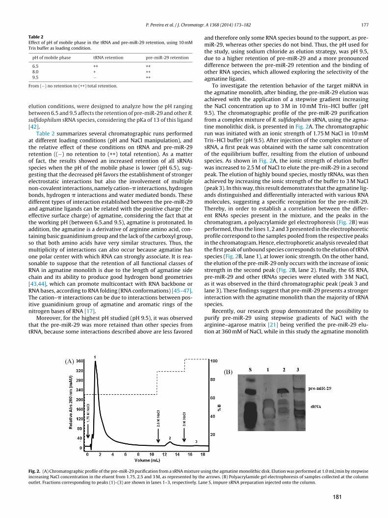
P. Pereira et al. / J. Chromatogr. A 1368 (2014) 173–182 177
Table 2Effect of pH of mobile phase in the tRNA and pre-miR-29 retention, using 10 mMTris buffer as loading condition.
pH of mobile phase tRNA retention pre-miR-29 retention
6.5 ++ ++8.0 + ++9.5 − ++
From (−) no retention to (++) total retention.
elution conditions, were designed to analyze how the pH rangingbetween 6.5 and 9.5 affects the retention of pre-miR-29 and other R.sulfidophilum sRNA species, considering the pKa of 13 of this ligand[42].
Table 2 summarizes several chromatographic runs performedat different loading conditions (pH and NaCl manipulation), andthe relative effect of these conditions on tRNA and pre-miR-29retention ((−) no retention to (++) total retention). As a matterof fact, the results showed an increased retention of all sRNAsspecies when the pH of the mobile phase is lower (pH 6.5), sug-gesting that the decreased pH favors the establishment of strongerelectrostatic interactions but also the involvement of multiplenon-covalent interactions, namely cation–� interactions, hydrogenbonds, hydrogen � interactions and water mediated bonds. Thesedifferent types of interaction established between the pre-miR-29and agmatine ligands can be related with the positive charge (theeffective surface charge) of agmatine, considering the fact that atthe working pH (between 6.5.and 9.5), agmatine is protonated. Inaddition, the agmatine is a derivative of arginine amino acid, con-taining basic guanidinium group and the lack of the carboxyl group,so that both amino acids have very similar structures. Thus, themultiplicity of interactions can also occur because agmatine hasone polar center with which RNA can strongly associate. It is rea-sonable to suppose that the retention of all functional classes ofRNA in agmatine monolith is due to the length of agmatine sidechain and its ability to produce good hydrogen bond geometries[43,44], which can promote multicontact with RNA backbone orRNA bases, according to RNA folding (RNA conformations) [45–47].The cation–� interactions can be due to interactions between pos-itive guanidinium group of agmatine and aromatic rings of thenitrogen bases of RNA [17].
Moreover, for the highest pH studied (pH 9.5), it was observedthat the pre-miR-29 was more retained than other species fromtRNA, because some interactions described above are less favored
and therefore only some RNA species bound to the support, as pre-miR-29, whereas other species do not bind. Thus, the pH used forthe study, using sodium chloride as elution strategy, was pH 9.5,due to a higher retention of pre-miR-29 and a more pronounceddifference between the pre-miR-29 retention and the binding ofother RNA species, which allowed exploring the selectivity of theagmatine ligand.
To investigate the retention behavior of the target miRNA inthe agmatine monolith, after binding, the pre-miR-29 elution wasachieved with the application of a stepwise gradient increasingthe NaCl concentration up to 3 M in 10 mM Tris–HCl buffer (pH9.5). The chromatographic profile of the pre-miR-29 purificationfrom a complex mixture of R. sulfidophilum sRNA, using the agma-tine monolithic disk, is presented in Fig. 2A. The chromatographicrun was initiated with an ionic strength of 1.75 M NaCl in 10 mMTris–HCl buffer (pH 9.5). After injection of the complex mixture ofsRNA, a first peak was obtained with the same salt concentrationof the equilibrium buffer, resulting from the elution of unboundspecies. As shown in Fig. 2A, the ionic strength of elution bufferwas increased to 2.5 M of NaCl to elute the pre-miR-29 in a secondpeak. The elution of highly bound species, mostly tRNAs, was thenachieved by increasing the ionic strength of the buffer to 3 M NaCl(peak 3). In this way, this result demonstrates that the agmatine lig-ands distinguished and differentially interacted with various RNAmolecules, suggesting a specific recognition for the pre-miR-29.Thereby, in order to establish a correlation between the differ-ent RNAs species present in the mixture, and the peaks in thechromatogram, a polyacrylamide gel electrophoresis (Fig. 2B) wasperformed, thus the lines 1, 2 and 3 presented in the electrophoreticprofile correspond to the samples pooled from the respective peaksin the chromatogram. Hence, electrophoretic analysis revealed thatthe first peak of unbound species corresponds to the elution of tRNAspecies (Fig. 2B, lane 1), at lower ionic strength. On the other hand,the elution of the pre-miR-29 only occurs with the increase of ionicstrength in the second peak (Fig. 2B, lane 2). Finally, the 6S RNA,pre-miR-29 and other tRNAs species were eluted with 3 M NaCl,as it was observed in the third chromatographic peak (peak 3 andlane 3). These findings suggest that pre-miR-29 presents a strongerinteraction with the agmatine monolith than the majority of tRNAspecies.
Recently, our research group demonstrated the possibility topurify pre-miR-29 using stepwise gradients of NaCl with thearginine–agarose matrix [21] being verified the pre-miR-29 elu-tion at 360 mM of NaCl, while in this study the agmatine monolith
Fig. 2. (A) Chromatographic profile of the pre-miR-29 purification from a sRNA mixture using the agmatine monolithic disk. Elution was performed at 1.0 mL/min by stepwiseincreasing NaCl concentration in the eluent from 1.75, 2.5 and 3 M, as represented by the arrows. (B) Polyacrylamide gel electrophoresis of samples collected at the columnoutlet. Fractions corresponding to peaks (1)–(3) are shown in lanes 1–3, respectively. Lane S, impure sRNA preparation injected onto the column.
181

178 P. Pereira et al. / J. Chromatogr. A 1368 (2014) 173–182
Fig. 3. (A) Chromatogram showing the purification of pre-miR-29 from sRNA population by using the agmatine monolithic disk. Elution was performed at 1 mL/min bystepwise decreasing (NH4)2SO4 concentration in the eluent from 2.4 to 0 M (NH4)2SO4 in 10 mM Tris buffer (pH 8.0). (B) Polyacrylamide gel electrophoresis analysis ofsamples collected at the column outlet. Fractions corresponding to peaks (1) and (2) are shown in lanes 1 and 2, respectively. Lane S, impure sRNA sample injected onto thecolumn.
allowed the recovery and purification of pre-miR-29 with 2.5 M ofNaCl. In general, this comparative study evidences that the bind-ing mechanism inherent to the biorecognition of pre-miR-29 bythe agmatine amino acid derivative is stronger than the interac-tions established with the arginine amino acid. This phenomenoncan result from the combination of multiple elementary interac-tions such as electrostatic interactions, hydrogen bonds, hydrogen� interactions, water mediated bonds and cation–� interactions,which both ligands may engage through the terminal guanidinegroup. However, in the agmatine monolith a stronger interactionoccurred since the recovery of pre-miR-29 required a higher NaClconcentration. The difference between the ligands is the absence ofthe carboxyl group in agmatine, which can have two positive effectsin the RNA retention. It can prevent repulsion of charged moleculesand enables the establishment of additional hydrophobic inter-actions with the carbon backbone of the ligand. Thus, dependingon the environmental conditions established and the amino acidligands used, some interactions can be more favored than other,becoming more evident under these conditions. In order to provethis hypothesis and mainly exploit hydrophobic interactions, astepwise gradient by decreasing ammonium sulfate ((NH4)2SO4)concentration between 2.4 M and 0 M in 10 mM Tris–HCl buffer(pH 8) was applied. In this case, the agmatine monolith was equili-brated with 2.4 M (NH4)2SO4 in 10 mM Tris–HCl buffer (pH 8). Afterthe binding of the sample to the disk, a first elution step designedto elute the tRNA species with lower affinity to the support wascarried out with the same salt concentration of the equilibriumbuffer, promoting total pre-miR-29 retention (Fig. 3A). The elutionof pre-miR-29 was then achieved with a second step by using 0 M(NH4)2SO4 in 10 mM Tris–HCl buffer (pH 8) (Fig. 3A). In fact, thehigh salt concentration plays a key role on the pre-miR-29 bind-ing to agmatine ligand. Again, a polyacrylamide gel electrophoresiswas used to detect and identify different species eluting in eachchromatographic peak (Fig. 3B). The electrophoretic analysis ofthe fractions eluting from the agmatine monolith (Fig. 3A) provedthat the first peak of unbound material corresponds to the elu-tion of tRNA (Fig. 3B, lane 1), whereas the second peak was mainlyattributed to the elution of pre-miR-29 (Fig. 3B, lane 2). The interac-tions that favor the recognition of pre-miR-29, under hydrophobicelution conditions, can be van der Waals forces and hydrophobicinteractions while the main responsible group for the interactionsestablished between pre-miR-29 and agmatine ligand can be thecarbon chain of the spacer arm.
The functionality of agmatine ligand to biorecognize the pre-miR-29 under hydrophobic- and ionic-based elution conditions
shows the applicability and versatility of this ligand to developseveral pre-miR-29 purification strategies due to the multipleinteractions involved for each condition. Apart from the struc-tural characteristics of the agmatine ligand, also the pre-miR-29structural features seem to be relevant on its distinct interactionbehavior with the ligand. The main explanation for the specificinteractions occurring between the pre-miR-29 and the agmatineamino acid derivative is the single-stranded nature of RNA, whichis normally involved in RNA recognition, due to the high base expo-sure and availability for interactions. Additionally, the negativecharge conferred by the phosphate groups in the RNA backbone isimportant for their interaction with the agmatine monolith, sug-gesting them to have a crucial role in RNA retention. Likewise,pre-miR-29 is a sRNA molecule with the shape of a stem-loopor hairpin consisting of two long irregular double-stranded stemregions, which are interrupted by a largely single-stranded internalloop [21]. This particular structure may also explain the multiplenon-covalent interactions established which are involved in thebiomolecular recognition of pre-miR-29 by the agmatine ligand.
In turn, to obtain an elution strategy more selective and biospe-cific as well as to achieve higher elution efficiency, it was employeda new approach for pre-miR-29 purification using arginine as com-petitive agent. This agent can bind either to the retained pre-miR-29or to the immobilized agmatine ligand depending on their char-acteristics, allowing thus to predict the interactions that can beinvolved once agmatine is derived of arginine. The competitivestudies were performed by adding 250 mM of arginine to theelution buffer in stepwise gradient to exploit specific elution ofpre-miR-29 from the agmatine monolith and therefore to evalu-ate the possibility of reaching higher purification factors. Elutiongradients applied were adapted to ensure that the RNA sample ison ideal conditions to bind to the support and be eluted only dueto the competing agent. Agmatine monolith was first equilibratedwith 1.75 M NaCl in 10 mM Tris–HCl buffer (pH 9.5) to promote thetotal retention of pre-miR-29. After sample application, a first peakof unbound species was obtained and then the ionic strength wasincreased upon arginine addition. In this step, 1.75 M of NaCl sup-plemented with 100 mM arginine in 10 mM Tris–HCl buffer (pH 9.5)was used for the elution of pre-miR-29 in a second peak (Fig. 4A).
These findings are in accordance with previous published resultswhich show that this competition strategy is able to elute boundpre-miR-29 from arginine–agarose column [21]. It is suggestedthat the major mechanism from which pre-miR-29 is eluted fromthe agmatine support is the preferential binding of free arginineto the pre-miR-29, an interaction that we believe to be stronger
182

P. Pereira et al. / J. Chromatogr. A 1368 (2014) 173–182 179
Fig. 4. (A) Separation of pre-miR-29 using agmatine monolithic disk. Elution was performed at 1 mL/min by stepwise increasing of arginine concentration in the eluent to100 mM as represented by the arrows. (B) Polyacrylamide gel electrophoresis analysis of each peak in both experiments is represented in the respective figure. Fractionscorresponding to peaks (1)–(3) are shown in lanes 1–3, respectively. Lane S, sRNA sample injected onto the column.
than the interaction pre-miR-29–agmatine support. In fact, sincethe arginine is positively charged at the pH in study, electrostaticinteractions can possible occur between the pre-miR-29 and thefree arginine. However, another mechanism that can explain pre-miR-29 elution is the interaction of free arginine with the agmatineligand in the immobilized support [48], that could also promote thedisplacement of bound pre-miR-29. The last peak corresponded tothe elution of tRNAs and some residual pre-miR-29 by increasingthe ionic strength to 3 M of NaCl. The results of polyacrylamide gelelectrophoresis (Fig. 4B) showed that the tRNAs were recovered inthe first peak (lane 1), being separated from the pre-miR-29 thatwas attained in the second peak (lane 2), while other tRNAs andpre-miR-29 were obtained in the third peak (lane 3). As judged bypolyacrylamide gel electrophoresis, pre-miR-29 was isolated andsuitably purified in the second chromatographic step with highintegrity. However, even if some arginine remains in the sample,the structure of pre-miR-29 is not changed, as we have previ-ously studied by circular dichroism (data not shown). Actually, theapplication of arginine can also be strongly associated with the pre-served integrity observed in RNA samples since arginine, owing toits multiplicity for interactions, has been largely associated withstabilizing effects on RNA conformation [17,21].
In a general way, the competition study has shown that the freearginine could promote the pre-miR-29 biospecific elution of theagmatine monolith.
Beyond the potential application of agmatine-based affinitychromatography to purify pre-miR-29 described here, this studyalso demonstrates the possibility of using three different strate-gies to achieve the pre-miRNA purification. In fact, the purificationof pre-miR-29 was accomplished either using an increased NaClor decreased (NH4)2SO4 gradients or using a competition strategywith arginine supplemented buffer. In addition, this work provesthe relevance of the establishment of well-defined binding andelution conditions to enhance the pre-miR-29 purification perfor-mance, resulting in an improvement of the final pre-miRNA yields,as this could represent an important impact on therapeutic applica-tions of the purified pre-miR-29. Comparing the elution strategieswith NaCl or arginine to elute bound pre-miR-29 biomolecules, itwas found that the competition strategy favors the selectivity forpre-miR-29 because the arginine concentration needed to promoteelution is lower than the NaCl concentrations. This fact is in accor-dance with what has been previously described by other authors,because as arginine is monovalent at the pH used, it is equivalent toNaCl in terms of valency; however the conductivity is much lower
than for NaCl at identical concentrations [21,49]. This study alsoallowed the purification of pre-miR-29 in 20 min at a flow-rate of1 mL/min.
3.4. Effect of flow rate on pre-miR-29 purification
The effect of flow rate on purification of pre-miR-29 was investi-gated, although no change is expected on the separation selectivityonce some studies proved that the molecules separation is flow-independent due to the physical and chemical constitution ofmonoliths [26,50]. Therefore, to verify the impact of flow rate onpurification of pre-miR-29, the same elution gradient previouslyestablished for the ideal separation was used. Fig. 5A shows theresulting chromatograms for the separation of pre-miR-29 at thedifferent flow rates from 0.5 to 3 mL/min. The purity of pre-miR-29separated with different flow rates was followed by electrophore-sis as shown in Fig. 5B. Evaluating the chromatograms and therespective electrophoresis, it is clearly evident that no changes haveoccurred in the separation efficiency of pre-miR-29 for the differentflow rates under study, but a significant reduction on the chromato-graphic run time was verified when higher flow rates were used(Fig. 5A). It is also possible to observe that the chromatograms over-lap each other even at the highest applied flow rate of 3 mL/min. Inthis case the separation was completed in 9 min, which is in accor-dance with the advantages presented by the monolithic supports,where the chromatographic run was 4 times faster that the timeused in conventional matrix of arginine [21].
3.5. Pre-miR-29 characterization
To ensure the success of the pre-miR-29 purification method-ologies here described for therapeutic applications it is essentialto guarantee the total elimination of impurities such as proteins,assessed by the micro-BCA method and endotoxins evaluatedby LAL assay as well as to isolate the pre-miR-29 from othersRNAs species, which may be assured by polyacrylamide gel elec-trophoresis [5,51]. Moreover, RNA preparations should maintainthe structural integrity, free from enzymatic inhibitors for RT andPCR reactions and free from nucleases [52].
The concentration and quality of the pre-miR-29 preparationsisolated by the agmatine monolith were evaluated by Phoretix1D software (Nonlinear Dynamics, Newcastle, United Kingdom)through the evaluation of the intensity of electrophoretic bands.The quantitative analysis of pre-miR-29 throughout the isolation
183

180 P. Pereira et al. / J. Chromatogr. A 1368 (2014) 173–182
Fig. 5. (A) Effect of flow rate on resolution of pre-miR-29. Experiments were performed in the same salt and gradient conditions applied for the pre-miR-29 purification(presented in Fig. 2) at different flow rates (0.5, 1, 2 and 3 mL/min). (B) The peaks obtained in the chromatographic runs at different flow rates were identified by polyacrylamidegel electrophoresis.
procedure is summarized in Table 3, which presents the puritydegree, purification factor and recovery yield of pre-miR-29 sampleobtained with the agmatine monolith. In a general point of view,the strategy with NaCl enabled a high recovery (97.33%) of the tar-get miRNA but the purity degree only reached 75.21%, whereas thestrategy with (NH4)2SO4 allows the recovery of the partially purifypre-miR-29 (35.51% of purity) when the salt concentration is signif-icantly reduced. On the other hand, the competition studies usingarginine in the elution buffer, demonstrated the possibility of puri-fying pre-miR-29 with higher purity (90.11% of purity and 94.88%of yield) in comparison to the other strategies, even though somepre-miRNA is lost in the final elution step. Thus, if larger amountsof pure pre-miRNA are desired, the strategy that employs NaCl canbe used, while if higher purity levels are necessary, the competitionstrategy with arginine is more appropriate [21].
The protein quantification in the purified pre-miR-29 fractionscollected from the chromatographic peaks was performed usingthe micro-BCA assay (Table 3). The analysis of the protein con-tent in the fractions collected along the pre-miR-29 purificationgradient revealed that proteins tend to elute more in the firstgradient step (data not shown). Therefore, the chromatographicstrategies used in these new pre-miRNA isolation methods (peak2 in Figs. 2A, 3A and 4A) also provide a reduction of protein con-tamination in RNA samples, specifically in the competition strategyusing arginine in the elution buffer, where the protein contami-nation level was negligible (2.774 ± 0.005 ng/�L), as required fortherapeutic applications (Table 3). Endotoxins contamination wasassessed using the ToxinSensorTM Chromogenic Limulus Amoe-bocyte Lysate (LAL) Endotoxin Assay Kit (GenScript, USA, Inc.)according to the manufacturer’s instructions. The endotoxins con-tent in the final pre-miR-29 sample indicates a significant reductionof the endotoxins level throughout the chromatographic step withthe agmatine monolith, with a purification factor of pre-miR-29
Fig. 6. pre-miR-29 identification by reverse-transcription PCR. The agarose-gelelectrophoretic analysis of PCR products shows amplification of pre-miR-29 cDNAfragments. The negative control had no band intensification. Lane M, DNA molec-ular weight marker; lane 1, negative control; lanes 2, 3 and 4, second peaks of thethree strategies under study (sodium chloride, ammonium sulfate and arginine),respectively.
relatively to LPS of 50 times (data not shown). A more accurateidentification of pre-miR-29 molecules purified from the sRNApopulations with the agmatine monolith was performed by PCR.As it can be observed from the electrophoretic analysis of the PCRproducts (Fig. 6), by using specific primers for pre-miR-29 cDNA,the PCR reaction allowed the amplification of pre-miR-29. No fur-ther amplification was seen in the control (lane 1). The negative
Table 3Quantitative analysis of purity and recovery yield of pre-miR-29 isolated by agmatine monolithic disk.
Elution strategy sRNA sample (�g) Pre-miR-29 purity (%) Pre-miR-29 yield (%) Proteins (ng/�L)
NaCl 20 75.21 97.33 3.258 ± 0.001(NH4)2SO4 20 35.51 99.40 5.516 ± 0.006Arginine 20 90.11 94.88 2.774 ± 0.005
The correlation coefficients of protein calibration curves was 0.9998.Data are presented as means with SD (N = 3).
184

P. Pereira et al. / J. Chromatogr. A 1368 (2014) 173–182 181
control was made of PCR reaction solutions without cDNA. There-fore, it was verified the identity of the sRNA isolated from theoriginal population as pre-miR-29.
4. Conclusions
This work combines, for the first time, the structural versatilityprovided by monolithic supports with the specificity and selectivityof agmatine ligand, as a promising strategy for miRNA purification.Moreover, the characterization of the modified monolith revealedthat a maximum binding capacity at 100% breakthrough was8.08 mg/mL for a RNA concentration of 0.25 mg/mL. The obtainedKd value, 2.6 × 10⁻⁷ M, confirms that the agmatine monolith sup-port develops a good affinity interaction with RNA, showing thatagmatine monolith is a good affinity chromatographic support.The implementation of agmatine monolithic chromatography forpre-miR-29 purification was based on the development of spe-cific interactions between the pre-miR-29 and agmatine ligands,allowing the removal of other RNA species, as well as the reductionof proteins and endotoxins contaminants, obtaining highly purepre-miR-29 for therapeutic applications. The type and salt con-centration and pH manipulation in the loading buffer allows theestablishment of different interactions and consequently differentelution strategies. The exploitation of these affinity interactions,resulting from multiple intermolecular forces, namely van derWaals forces, hydrogen bonds, electrostatic and hydrophobic inter-actions, can trigger new insights not only in isolation strategies butalso in many other RNA research fields owing to its implication inmolecular recognition phenomena. Additionally, for the pre-miR-29 purification this monolithic support represents an advantageousalternative to conventional supports due to fast separation andconsequent short contact time, ensuring structural stability of thetarget molecule. In conclusion, our approach revealed an efficienttechnique to obtain pharmaceutical-grade miRNA with high recov-ery yield, purity and good integrity, which may in a near future beused in RNA structural and functional studies as well as in genetherapy.
Acknowledgments
This work was supported by the Portuguese Foundation forScience and Technology (PTDC/EBB-BIO/114320/2009, EXPL/BBB-BIO/1056/2012, COMPETE: FCOMP-01-0124-FEDER-027560 andPest-C/SAU/UI0709/2011). The authors also acknowledge theproject with Reference CENTRO-07-ST24-FEDER-002014, from“Programa Operacional Regional do Centro 2007-2013 QREN”(“Mais Centro” program). Patrícia Pereira and Ângela Sousaacknowledge fellowships (Ph.D. Grant, Ref SFRH/BD/81914/2011and Postdoctoral Grant, Ref SFRH/BPD/79106/2011, respectively).The authors would like to thank Prof. Yo Kikuchi (Toyohashi Uni-versity of Technology) for kindly provide pBHSR1-RM plasmid andto BIA Separations for having kindly provided the agmatine mono-lithic disk and U. Cernigoj for having made the immobilization ofagmatine ligand.
References
[1] D.P. Bartel, MicroRNAs: genomics, biogenesis, mechanism, and function, Cell116 (2004) 281–297.
[2] J.A. Broderick, P.D. Zamore, MicroRNA therapeutics, Gene Ther. 18 (2011)1104–1110.
[3] I. Vomelova, Z. Vanickova, A. Sedo, Methods of RNA purification. All ways(should) lead to Rome, Folia Biol. (Praha) 55 (2009) 243–251.
[4] S.C. Tan, B.C. Yiap, DNA RNA, and protein extraction: the past and the present,J. Biomed. Biotechnol. 2009 (2009) 574398.
[5] F. Re, RNA Methodologies – Laboratory Guide for Isolation and Characterization,Academic Press, York, USA, 2005.
[6] M.A. Dineva, L. MahiLum-Tapay, H. Lee, Sample preparation: a challenge in thedevelopment of point-of-care nucleic acid-based assays for resource-limitedsettings, Analyst 132 (2007) 1193–1199.
[7] L.E. Easton, Y. Shibata, P.J. Lukavsky, Rapid, nondenaturing RNA purificationusing weak anion-exchange fast performance liquid chromatography, RNA 16(2010) 647–653.
[8] N. Nonne, M. Ameyar-Zazoua, M. Souidi, A. Harel-Bellan, Tandem affinity purifi-cation of miRNA target mRNAs (TAP-Tar), Nucleic Acids Res. 38 (2010) e20.
[9] S.M. McCarthy, M. Gilar, J. Gebler, Reversed-phase ion-pair liquid chromatog-raphy analysis and purification of small interfering RNA, Anal. Biochem. 390(2009) 181–188.
[10] J.S. Kieft, R.T. Batey, A general method for rapid and nondenaturing purificationof RNAs, RNA 10 (2004) 988–995.
[11] R. Martins, J.A. Queiroz, F. Sousa, Ribonucleic acid purification, J. Chromatogr.A 1355 (2014) 1–14.
[12] L. Ponchon, F. Dardel, Large scale expression and purification of recombinantRNA in Escherichia coli, Methods 54 (2011) 267–273.
[13] S.C. Walker, F.H. Scott, C. Srisawat, D.R. Engelke, RNA affinity tags for the rapidpurification and investigation of RNAs and RNA–protein complexes, MethodsMol. Biol. 488 (2008) 23–40.
[14] J. Chen, J. Lozach, E.W. Garcia, B. Barnes, S. Luo, I. Mikoulitch, L. Zhou, G. Schroth,J.B. Fan, Highly sensitive and specific microRNA expression profiling usingBeadArray technology, Nucleic Acids Res. 36 (2008) e87.
[15] R. Martins, J.A. Queiroz, F. Sousa, Histidine affinity chromatography-basedmethodology for the simultaneous isolation of Escherichia coli small and ribo-somal RNA, Biomed. Chromatogr. 26 (2011) 781–788.
[16] R. Martins, J.A. Queiroz, F. Sousa, A new affinity approach to isolateEscherichia coli 6S RNA with histidine-chromatography, J. Mol. Recognit. 23(2010) 519–524.
[17] R. Martins, C.J. Maia, J.A. Queiroz, F. Sousa, A new strategy for RNA isolation fromeukaryotic cells using arginine affinity chromatography, J. Sep. Sci. 35 (2012)3217–3226.
[18] F. Sousa, D.M. Prazeres, J.A. Queiroz, Affinity chromatography approaches toovercome the challenges of purifying plasmid DNA, Trends Biotechnol. 26(2008) 518–525.
[19] P. Pereira, A. Sousa, J. Queiroz, A. Figueiras, F. Sousa, New approach for purifi-cation of pre-miR-29 using lysine-affinity chromatography, J. Chromatogr. A1331 (2014) 129–132.
[20] A. Afonso, P. Pereira, J.A. Queiroz, A. Sousa, F. Sousa, Purification of pre-miR-29by a new O-phospho-l-tyrosine affinity chromatographic strategy optimizedusing design of experiments, J. Chromatogr. A 1343 (2014) 119–127.
[21] P. Pereira, A. Sousa, J. Queiroz, I. Correia, A. Figueiras, F. Sousa, Purificationof pre-miR-29 by arginine-affinity chromatography, J. Chromatogr. B: Analyt.Technol. Biomed. Life Sci. 951–952 (2014) 16–23.
[22] A. Strancar, A. Podgornik, M. Barut, R. Necina, Short monolithic columns asstationary phases for biochromatography, Adv. Biochem. Eng. Biotechnol. 76(2002) 49–85.
[23] A. Podgornik, S. Yamamoto, M. Peterka, N.L. Krajnc, Fast separation of largebiomolecules using short monolithic columns, J. Chromatogr. B: Analyt. Tech-nol. Biomed. Life Sci. 927 (2013) 80–89.
[24] A. Sousa, F. Sousa, J.A. Queiroz, Advances in chromatographic supportsfor pharmaceutical-grade plasmid DNA purification, J. Sep. Sci. 35 (2012)3046–3058.
[25] A. Sousa, C.T. Tomaz, F. Sousa, J.A. Queiroz, Successful application of monolithicinnovative technology using a carbonyldiimidazole disk to purify supercoiledplasmid DNA suitable for pharmaceutical applications, J. Chromatogr. A 1218(2011) 8333–8343.
[26] A. Sousa, D. Bicho, C.T. Tomaz, F. Sousa, J.A. Queiroz, Performance of a non-grafted monolithic support for purification of supercoiled plasmid DNA, J.Chromatogr. A 1218 (2011) 1701–1706.
[27] A. Sousa, A.M. Almeida, U. Cernigoj, F. Sousa, J.A. Queiroz, Histamine mono-lith versatility to purify supercoiled plasmid deoxyribonucleic acid fromEscherichia coli lysate, J. Chromatogr. A 1355 (2014) 125–133.
[28] D. El-Agamy, M. Sharawy, E. Ammar, Agmatine attenuates silica-induced pul-monary fibrosis, Hum. Exp. Toxicol. 33 (2014) 650–660.
[29] N. Kitanaka, J. Kitanaka, F.S. Hall, G.R. Uhl, K. Watabe, H. Kubo, H. Takahashi, K.Tanaka, N. Nishiyama, M. Takemura, Agmatine attenuates methamphetamine-induced hyperlocomotion and stereotyped behavior in mice, Behav. Pharmacol.25 (2014) 158–165.
[30] N.R. Kotagale, S. Walke, G.P. Shelkar, D.M. Kokare, M.J. Umekar, B.G. Taksande,Agmatine attenuates nicotine induced conditioned place preference in micethrough modulation of neuropeptide Y system, Behav. Brain Res. 262 (2014)118–124.
[31] H. Suzuki, S. Umekage, T. Tanaka, Y. Kikuchi, Artificial RNA aptamer produc-tion by the marine bacterium Rhodovulum sulfidophilum: improvement of theaptamer yield using a mutated transcriptional promoter, J. Biosci. Bioeng. 112(2011) 458–461.
[32] A. Jungbauer, O. Kaltenbrunner, Fundamental questions in optimizing ion-exchange chromatography of proteins using computer-aided process design,Biotechnol. Bioeng. 52 (1996) 223–236.
[33] A. Soares, J.A. Queiroz, F. Sousa, A. Sousa, Purification of human papillomavirus16 E6/E7 plasmid deoxyribonucleic acid-based vaccine using an arginine mod-ified monolithic support, J. Chromatogr. A 1320 (2013) 72–79.
[34] F. Sousa, T. Matos, D.M. Prazeres, J.A. Queiroz, Specific recognition of super-coiled plasmid DNA in arginine affinity chromatography, Anal. Biochem. 374(2008) 432–434.
185

182 P. Pereira et al. / J. Chromatogr. A 1368 (2014) 173–182
[35] M.V. Latronico, D. Catalucci, G. Condorelli, MicroRNA and cardiac pathologies,Physiol. Genomics 34 (2008) 239–242.
[36] P.T. Nelson, W.X. Wang, B.W. Rajeev, MicroRNAs (miRNAs) in neurodegenera-tive diseases, Brain Pathol. 18 (2008) 130–138.
[37] L. Du, A. Pertsemlidis, Cancer and neurodegenerative disorders: pathogenicconvergence through microRNA regulation, J. Mol. Cell Biol. 3 (2011) 176–180.
[38] M.J. Shin, L. Tan, M.H. Jeong, J.H. Kim, W.S. Choe, Monolith-based immobilizedmetal affinity chromatography increases production efficiency for plasmid DNApurification, J. Chromatogr. A 1218 (2011) 5273–5278.
[39] A. Jungbauer, Chromatographic media for bioseparation, J. Chromatogr. A 1065(2005) 3–12.
[40] R.E. Boto, P. Almeida, J.A. Queiroz, Thiacarbocyanine as ligand in dye-affinitychromatography for protein purification, Biomed. Chromatogr. 22 (2008)278–288.
[41] J. Ruta, C. Ravelet, J. Desire, J.L. Decout, E. Peyrin, Covalently bonded DNAaptamer chiral stationary phase for the chromatographic resolution of adeno-sine, Anal. Bioanal. Chem. 390 (2008) 1051–1057.
[42] D. Grundemann, C. Hahne, R. Berkels, E. Schomig, Agmatine is efficiently trans-ported by non-neuronal monoamine transporters extraneuronal monoaminetransporter (EMT) and organic cation transporter 2 (OCT2), J. Pharmacol. Exp.Ther. 304 (2003) 810–817.
[43] B.J. Calnan, B. Tidor, S. Biancalana, D. Hudson, A.D. Frankel, Arginine-mediatedRNA recognition: the arginine fork, Science 252 (1991) 1167–1171.
[44] T. Janas, J.J. Widmann, R. Knight, M. Yarus, Simple, recurring RNA binding sitesfor l-arginine, RNA 16 (2010) 805–816.
[45] F. Jiang, A. Gorin, W. Hu, A. Majumdar, S. Baskerville, W. Xu, A. Elling-ton, D.J. Patel, Anchoring an extended HTLV-1 Rex peptide within an RNAmajor groove containing junctional base triples, Structure 7 (1999) 1461–1472.
[46] J.D. Puglisi, J.R. Wyatt, I. Tinoco Jr., Solution conformation of an RNA hairpinloop, Biochemistry 29 (1990) 4215–4226.
[47] M. Yarus, J.J. Widmann, R. Knight, RNA–amino acid binding: a stereochemicalera for the genetic code, J. Mol. Evol. 69 (2009) 406–429.
[48] R. Mallik, D.S. Hage, Affinity monolith chromatography, J. Sep. Sci. 29 (2006)1686–1704.
[49] T. Arakawa, D. Ejima, K. Tsumoto, M. Ishibashi, M. Tokunaga, Improved perfor-mance of column chromatography by arginine: dye-affinity chromatography,Protein Exp. Purif. 52 (2007) 410–414.
[50] M.K. Danquah, G.M. Forde, Large-volume methacrylate monolith for plasmidpurification. Process engineering approach to synthesis and application, J. Chro-matogr. A 1188 (2008) 227–233.
[51] L.V. Madabusi, G.J. Latham, B.F. Andruss, RNA extraction for arrays, MethodsEnzymol. 411 (2006) 1–14.
[52] S.A. Bustin, T. Nolan, Pitfalls of quantitative real-time reverse-transcriptionpolymerase chain reaction, J. Biomol. Tech. 15 (2004) 155–166.
186

187
Paper VIII
Characterization of polyplexes involving small RNA
P. Pereira, A. F. Jorge, R. Martins, A. A. C. C. Pais, F. Sousa, A. Figueiras
Journal of Colloid and Interface Science. 2012. 387: 84-94
Short description: This paper presents a systematic study of different polymers, such as
Polyethylenimine, Chitosan and Poly(allylamine), with different molecular weights, charge
densities and backbone structure, to efficiently encapsulate small RNA molecules. The
assessment of the biophysical and structural characteristics (such as size, zeta potential,
morphology, encapsulation efficiency and protection/stability) of the polymer–small RNA
complexes were evaluated and compared, in order to design a successful delivery system for
small RNAs. In addition, a rationale for the experimental observations is provided using Monte
Carlo simulation for systems with polymers of different lengths and charge densities. The
simulations showed that there is an interplay between the size of polymers chains and their
charge density that define the degree of condensation for small RNA.

188

Characterization of polyplexes involving small RNA
Patrícia Pereira a, Andreia F. Jorge b, Rita Martins a, Alberto A.C.C. Pais b, Fani Sousa a, Ana Figueiras a,c,⇑a CICS-UBI – Health Sciences Research Centre, University of Beira Interior, Avenida Infante D. Henrique, 6200-506 Covilhã, Portugalb Department of Chemistry, University of Coimbra, Rua Larga, 3004-535 Coimbra, Portugalc CEF-FFUC – Pharmaceutical Studies Center, Faculty of Pharmacy, University of Coimbra, Pólo das Ciências da Saúde, Azinhaga de Santa Comba, 3000-548 Coimbra, Portugal
a r t i c l e i n f o
Article history:Received 11 May 2012Accepted 31 July 2012Available online 21 August 2012
Keywords:PolyplexesSmall RNAPolyethylenimineChitosanPoly(allylamine)
a b s t r a c t
The purpose of the present study is to provide a tool for an efficient design and synthesis of non-viral vec-tors for small RNA delivery. The effects of properties of the polycation, such as molecular weight, chargedensity and backbone structure, to polyplex structure and physicochemical behavior were systematicallyevaluated. The condensing agents, polyethylenimine (PEI), chitosan (CS) and poly(allylamine) (PAA) wereadded to sRNA molecules at different N/P ratio. The efficiency of encapsulation and protection of sRNA, aswell as polyplex size, zeta potential and morphology were followed and compared. The results show thatPEI/sRNA polyplexes display a small size and positive zeta potential. However, for low molecular weights,this polycation is unable to protect sRNA in the presence of a decompacting agent. With chitosan, sRNA isefficiently compacted at high N/P ratios. The CS/sRNA complexes display small sizes, ca. 200 nm, positivesurface charge and also good stability. Finally, the PAA/sRNA polyplexes were found to be the smallest atlow N/P ratios, displaying a good encapsulation efficiency and high stability. A rationale for the experi-mental observations is provided using Monte Carlo simulation for systems with polycations of differentlength and charge density. The simulations showed that there is an interplay between the size of poly-cation chains and its charge density that define the degree of condensation for sRNA.
� 2012 Elsevier Inc. All rights reserved.
1. Introduction
Gene therapy has gained significant attention over the past twodecades as a promising approach for a future therapeutic strategyin clinical applications. Gene therapy is important not only for thetreatment of diseases caused by genetic defects, but also in thedevelopment of methods for treatment and prevention of a widerange of acquired disorders that include severe combined immu-nodeficiency, cystic fibrosis, rheumatoid arthritis and Parkinson’sdisease, as well as an alternative to the traditional chemotherapyused in treating cancer [1–4].
Over recent years, the importance of RNA in numerous biologi-cal processes has increased substantially. Small RNAs (sRNAs) arebeing increasingly recognized as crucial regulatory molecules inall organisms. The specificity and the potency of sRNAs suggestthat they might become therapeutic agents, especially in interfer-ence RNA strategies [5–7]. Free therapeutic genes can be enzymat-ically degraded by ubiquitous nucleases resulting in a shorthalf-life in the blood. For that reason they show poor cellularuptake when delivered in aqueous solutions, and insufficient tissue
bioavailability, which has stimulated the development of carriersystems for gene delivery [1,8,9]. The gene carriers include viralsystems such as retrovirus and adenovirus or non-viral systems,including cationic polymers, cationic lipids, peptides and dendri-mers [10,11]. The polycations are a promising alternative to com-pact RNA for systemic delivery because of their low cytotoxicity,low immunogenicity, high stability, biocompatibility, and unre-stricted gene materials size. In addition, potential safety benefitsmake these compounds increasingly attractive for gene therapy[4,12–14]. Moreover, non-viral vectors present relatively low costproduction and high flexibility, allowing to design carriers withwell-defined structural and chemical properties [1].
Cationic polymers can efficiently condense RNA molecules intonanometer range complexes, commonly via protonated aminegroups that promote the electrostatic interaction with the nega-tively charged phosphate groups in RNA [15]. They may alsoprovide a positively charged surface in the complex. One of theobstacles for an efficient gene transfection is crossing the negativelycharged cell membrane, and net positive charge of the polyplexespromotes electrostatic interaction with the overall negative chargeof the cell membrane and prevents particle aggregation [14–16]. Inaddition, the size of the polyplexes should be between 50 and200 nm for efficient internalization by endocytic processes, andonce inside the cell, they should dissociate to release the sRNAsfrom the vector system for their respective functions [17–19].
0021-9797/$ - see front matter � 2012 Elsevier Inc. All rights reserved.http://dx.doi.org/10.1016/j.jcis.2012.07.088
⇑ Corresponding author. Address: Centro de Investigação em Ciências da Saúde,Universidade da Beira Interior, Avenida Infante D. Henrique, 6200-506 Covilhã,Portugal. Fax: +351 275 329 099.
E-mail address: [email protected] (A. Figueiras).
Journal of Colloid and Interface Science 387 (2012) 84–94
Contents lists available at SciVerse ScienceDirect
Journal of Colloid and Interface Science
www.elsevier .com/locate / jc is
189

Due to the increased interest in these systems, a large numberof natural and synthetic polymers have been synthesized and char-acterized. Polyethylenimine (PEI), a polycationic polymer hasemerged as one of the most promising candidates for the develop-ment of efficient gene delivery vectors [12]. The complexes PEI-nucleotide show high stability, controllable size, adjustableunpackaging properties in cells, and flexibility for addition of moi-eties that target specific entities on cell membranes and intracellu-lar structures [20]. In particular, PEI has potential as a RNA carrierdue to its superior transfection efficiency which may facilitateendosomal escape after entering the cells as it acts as a ‘‘protonsponge’’ during acidification of the endosome [13,21]. Chitosan(CS) is also considered to be a good candidate to integrate genedelivery systems. CS presents beneficial qualities such as low tox-icity, low immunogenicity, excellent biodegradability, biocompati-bility, as well as a high positive charge that can easily formpolyelectrolyte complexes with negatively charged nucleotidesthrough electrostatic interactions [2,9,22]. Recently, poly(allyla-mine) (PAA), a synthetic cationic polymer having high density ofprimary amino groups, has drawn considerable attention as anon-viral gene delivery system [23]. PAA carries a strong positivecharge, which enables it to bind and package negatively chargedsRNA. It is a polymer extensively used in the formulation of bio-compatible films, nanomaterials and for cell encapsulation.
In the present work, a systematic study of different polymers,PEI, CS and PAA, with different molecular weights and charge den-sities is performed. The aim of this study is to compare the perfor-mance of these different polycations on sRNA condensation, withthe subsequent assessment of the biophysical and structural char-acteristics (such as size, zeta potential, morphology and complexstability) of the polymer–sRNA complexes, in order to design a suc-cessful delivery system for sRNA.
2. Experimental section
2.1. Materials
Five different polycations were used. CS low molecular weight(LMW) (Mw = 50–190 kDa; degree of deacetylation in the 75–85%range), CS medium molecular weight (MMW) (Mw = 190–310 kDa, degree of deacetylation in the 75–85% range), PEI-LMWwith Mw = 1.3 kDa, 50 wt% in H2O and PAA of 17 kDa, 20 wt% inH2O were obtained from Sigma–Aldrich (St. Louis, MO, USA). PEIhigh molecular weight (HMW) of Mw = 10 kDa was purchased fromPolysciences, Inc. (Warrington, PA). Guanidinium salt and all thechemicals used in the lysis buffer were obtained from Sigma (St.Louis, MO, USA). All solution were freshly prepared using 0.05%diethyl pyrocarbonate (DEPC) treated water. DEPC was purchasedfrom Fluka (Sigma). Fetal bovine serum (FBS) was purchased fromBiochrom and heparin (25,000 IU for 5 mL) was obtained fromWinthrop.
2.2. sRNA samples
sRNA samples used in this study were isolated from Escherichiacoli DH5a based on the protocol described by Martins and co-workers (2010), in which an acid guanidinium thiocyanate–phe-nol–chloroform method is used [24]. Briefly, cells were lysed andthe nucleic acid fraction obtained was precipitated with isopropa-nol. The precipitate was recovered by centrifugation at 16,000g for20 min at 4 �C. After centrifuging, the sRNA pellets were air-driedfor 5 min at room temperature and their solubilization was per-formed in 200 lL of 0.05% DEPC-treated water. Finally, the opticaldensity of the samples was determined using a Nanophotometer toassess sRNA quantity and purity.
2.3. Polyplex formation
All the polyplexes were formulated using the method of simplecomplexation between molar concentrations of positive charge,present in the protonated amine groups of polycation (N), andthe negative charge of the phosphate groups of RNA (P) [3,8]. Todetermine specific N/P ratios, the mass of 325 Da correspondingto one phosphate group on sRNA was used. Moreover, over thepH used in this study, sRNA displays an approximately constantanionic charge density, with the pKa of the respective phosphategroup close to 1.5 [25]. The calculation of the positive chargeswas made in accordance with the pKa values and molecular weightof each polycation (Table 1). sRNA stock solution was prepared bysRNA dissolution in sodium acetate buffer (0.1 M sodium acetate/0.1 M acetic acid, pH 4.5), up to a final concentration of 25 lg/mL. The sRNA concentration was determined by UV absorbanceat 260 nm. A solution of 40 lg/mL of RNA will have an absorbanceof 1 [26]. The polycation stock solutions were also prepared in so-dium acetate buffer pH 4.5 in a concentration of 10 mg/mL.Preliminary experiments were performed to identify the concen-tration range where the polyplexes are formed. A fixed volume ofpolycation solution (100 lL) of variable concentration was addedto a sRNA solution (400 lL). The final concentration of sRNA wasequal to 20 lg/mL (60.6 lM) and was kept constant in all themethods used for the characterization of the formed complexes.Particles were obtained by addition of cationic polymer solutionto sRNA solution and immediately vortexed at maximum speedfor 30 s. All the samples were subsequently left for equilibrationat room temperature for 60 min.
2.4. Circular dichroism (CD)
CD was used to monitor the RNA behavior when subjected todifferent pH values. CD spectra were obtained using a 0.2 cm pathlength quartz rectangular cell at a constant temperature of 25 �C, ina Jasco 1850 spectrophotometer. Spectra were recorded from 215to 320 nm at a scan speed of 10 nm/min and a bandwidth of1 mm. Three spectra were accumulated and averaged for eachsample. The final sRNA concentration was 100 lg/mL. All measure-ments were conducted under a constant gas flow, to purge theozon generated by the light source of the instrument. The CD signalwas converted to molar ellipticity, smoothed with a Jasco Fast Fou-rier transform algorithm.
2.5. Gel agarose sRNA assays
A series of polycation/sRNA polyplexes in different ratios wereprepared as described above. The samples (20 lL) were analyzedby horizontal electrophoresis using 0.8% of agarose gels (Hoefer,San Francisco, CA, USA). The electrophoresis was carried out inTris–acetic acid (TAE) buffer in DEPC-treated water (40 mM Trisbase, 20 mM acetic acid and 1 mM EDTA, pH 8.0) and run at 90 Vfor 50 min. The bands corresponding to sRNA and polycation/sRNAcomplexes were visualized under ultraviolet light after staining thegels with ethidium bromide (0.5 lg/mL) or 5 lL of a 10,000� solu-tion of Gelstar. The gels were imaged using a Vilber Lourmat sys-tem (ILC Lda, Lisbon, Portugal).
2.6. Scanning electron microscopy (SEM)
The morphological characteristics of the polyplexes werevisualized with a scanning electron microscopy (SEM) (HitachiS-2700, Tokyo, Japan). Briefly, one drop of the solution containingthe polyplex samples was placed on the surface of cover glassesand stored at 37 �C overnight. Subsequently, the samples were
P. Pereira et al. / Journal of Colloid and Interface Science 387 (2012) 84–94 85
190

then sputter coated with gold using an Emitech K550 SputteringCoater (London, England) for 3 min at 30 mA.
2.7. Particle size and zeta potential measurements
The mean particle diameter of the polyplexes and polydisper-sion index were determined by dynamic light scattering (photoncorrelation spectroscopy, PCS) using an N5 Particle Analyzer (Beck-man Coulter Inc., USA). Particle diameters of the freshly preparedcomplexes were measured at 25 �C, and data were collected at90� scattering angle. The time-averaged autocorrelation functionswere transformed into intensity-weighted distributions of theapparent hydrodynamic diameter using the available MalvernPCS software 6.20. The surface charges (zeta potential) of the poly-plexes were determined by laser Doppler electrophoresis using aZetasizer Nano ZS (Malvern Instruments Ltd., UK), at 25 �C. Theaverage values of size and zeta potential were calculated withthe data obtained from three measurements ± SD.
2.8. Determination of sRNA encapsulation efficiency
The sRNA encapsulation efficiency (EE) was calculated from thedetermined free sRNA concentration in the supernatant recoveredafter particle centrifugation (15,000g, 20 min, 25 �C). The amountof unbound sRNA was determined by UV–Vis analysis (ShimadzuUV–Vis 1700 spectrophotometer) at 260 nm. Supernatant recov-ered from unloaded polycation (without sRNA) was used as ablank. Three repetitions of this procedure were performed for eachsystem. The encapsulation efficiency was calculated as
EE ð%Þ ¼ ½ðTotal sRNA amount
� sRNA supernatant amountÞ=Total sRNA amount�� 100
2.9. Polyplexes: release profile and protection of sRNA
Protection and release profile of the sRNA present in the poly-plexes were also assessed. All the polyplexes were resuspendedin PBS buffer, pH 7.4. The protection experiments were carriedout by incubation of 12.5 lL of polyplexes with 1.5 lL of RNasesolution (10 and 100 lg/mL) for 1 h at 37 �C or with 10% of fetalbovine serum (FBS) for 30 and 60 min at 37 �C. To characterizethe release of the encapsulated sRNA, samples were incubated witha series of heparin solutions (0.01, 0.1, 0.5 and 1 IU/mL of heparin),prepared by diluting aliquots of a heparin stock solution (10 IU/mL). The polyplex solutions (10 lL) were incubated with 10 lL ofeach concentration for 1 h at 37 �C.
2.10. Molecular modeling
An estimation of size and charge density of all polycations usedwas carried out resorting to the Avogadro package [27], using the
UFF force field [28]. The structure of all polycations was estab-lished considering at least ten monomers. The average distance be-tween charged groups, dAB, and also the average size per monomerwere established, considering the structure of each monomer as gi-ven by the manufacturer.
2.11. Monte Carlo simulation
The systems studied in this work were modeled in a coarse-grain approach, where all ions and chain monomers are consideredas charged hard-spheres and the solvent is considered as a contin-uum with a dielectric permittivity of water. We modeled a titrationof a polyanion with 100 negative charged beads, with oppositelycharged polyplexes with variable chain lengths and charge densi-ties. The four different systems are listed in Table 2. The notationcorresponding to the length of the polycation chain and corre-sponding charge density (e.g. Short-HCD) is used. The charge den-sity varies between low (LCD), moderate (MCD) and high (HCD).The charge ratio, N/P, between polyanion and polycation were keptconstant, equal to unity, for all systems considered. It is given by
N=P ¼ zmon;PCNmon;PCNPC
zmon;PANmon;PA
where Zmon is the charge of monomers, Nmon is the number of mono-mers in each chain and N is the number of polycation chains in thesystem. The subscript PC and PA correspond to polycation and pol-yanion, respectively. The linear charge density, dz, of the polycationchains was calculated as,
dz ¼Nmon;PCZmon;PC
ðNmon;PC � 1ÞR0
and taking R0 as the bead-bead separation. A more extensivedescription of this method can be seen in previous work by someof the authors [29]. In the present work, the hard sphere radius ofpolyanion beads was 2.7 Å and of positive beads of polycation 2 Å.A reference distance between connected monomers of 6.5 Å wasimposed for the negative chain, 5.6 Å for the polycations. Theharmonic force constant and angular force constant used was0.4 N m�1 and 1.7 � 10�24 J deg�2, respectively. The polyelectrolytebackbones correspond to semi-flexible polymers, in terms of
Table 1Main characteristics of PEI-LMW, PEI-HMW, CS-LMW, CS-MMW and PAA.
Polymer Molecular weight (MW)a Monomer charge density (e)a Nominal DDb (%) Determined DDc (%) pKaref
PEI-LMW 1300 11+ – – 9.26 [15]PEI-HMW 10,000 3+ – – 8.5 [45]CS-LMW 50,000–190,000 1+ 75–85 83.14 ± 0.39 6.5 [15]CS-MMW 190,000–310,000 1+ 75–85 73.86 ± 0.61 6.5 [15]PAA 17,000 1+ – – 8.5 [46]
a e is the elementar charge.b Provided by the manufacturer.c Determined by 1DUVS. The data represent the mean and standard error of at least three separate measurements (mean ± SD).
Table 2Systems simulated in the present work. In all cases, a negative polyelectrolyte with100 charged beads of unit charge was present.
Systems Number of positivechains
Number of beads/chain
Charge perbead
Short-HCD 5 20 1Intermediate-
LCD8 50 0.25
Intermediate-MCD
4 50 0.5
Long-LCD 2 200 0.25
86 P. Pereira et al. / Journal of Colloid and Interface Science 387 (2012) 84–94
191

intrinsic persistence length. Considering also the electrostaticeffects, the values of the persistence length, in the presence of therespective counterions only, is 28 Å for the polyanion, and ca.22 Å for the polycations, irrespective of the chain length. For eachsystem, Metropolis Monte Carlo simulations were performed inthe canonical ensemble using the Molsim package [30]. The effi-ciency of the simulations was improved by taking concerted movesin the chain particles. These included translation as a whole andslithering [31]. The rate of acceptance of single particle moves forthe polyanion was 33% for a simulation of a compact polyplex (sys-tem Short-HCD), while translation and slithering warranted levelsof acceptance of 4% and 39%, respectively. In general, all remainingsystems yielded higher values for the polyanion. The polycationmoves resulted in significantly higher rates of acceptance: 44%,32% and 38% for the single particle, translation as a whole and slith-ering, respectively, also in system Short-HCD. Again, the remainingsystems displayed higher acceptance ratios for the polycationsbackbone.
3. Results and discussion
3.1. Structural characteristics of the polycations
From the structures of different polycations with optimizedconformation, it is possible to determine the average size andcharge density of the respective monomers (Fig. 1). Also, consider-ing the molecular weight of each monomer and the correspondingaverage chain molecular weight, it is possible to estimate the num-ber of monomers per chain, Nmon,PC. The linear charge density wascalculated similarly to what was described for the Monte Carlosimulation but zmon,PC is the number of charges in each monomer(see Table 1) and R0 is the length per monomer. Table 3 summa-rizes the results obtained from simple molecular mechanics calcu-lations in gas phase. It should be stressed that these are roughestimates, in which the groups are not protonated to avoid thenecessity of a solvent, and aim at facilitate the link between theexperimental and theoretical approaches. Chitosan chains displaythe highest chain length, but the lowest charge density. The
highest charge density is, as expected, obtained for PEI polymers.In this case, and due to the fact that these polycations arebranched, it is more difficult to establish the total chain length.This is especially important in the case of PEI, 10 kDa, in whichthe branching is random. PAA presents an intermediate size andchain length. Further details will be discussed in the next sections.
3.2. Assessment of sRNA stability in different solutions
The conformational stability of the sRNA secondary structurewas assessed at different solutions (H2O–DEPC and acetate buffer,pH 4.5) using CD (Fig. 2). The CD spectra of the sRNA in H2O–DEPCis very similar and show characteristic features of the A-RNA [32].For sRNA in H2O–DEPC, a positive band around 260 nm was ob-served, and a negative signal at 220 nm, with approximately thesame intensity. On the other hand, the sRNA spectrum in acetatebuffer at pH 4.5 depicts a decrease in the value of the molar ellip-ticity of the positive band (260 nm) and maintains the negative sig-nal at 220 nm. These results show that isolated RNA is sensitive tothe buffer used, since the respective CD spectrum is different inacetate buffer and in water. This indicates that the sRNA structureis altered at lower pH.
To check on RNA integrity, further techniques were used. Spec-trophotometry analysis (Nanophotometer) measuring absorbance
Fig. 1. Optimized geometries (UFF) of polycations in gas phase, (a) PAA and (b) CS, (c) PEI, 1.2 kDa and (d) PEI 10 kDa. Each polycation contains not less than 10 monomers.The average distance between the ionizable species (blue) was performed by considering the distance, dAB, and subsequent distances determined following the nearestionizable group. (For interpretation of the references to color in this figure legend, the reader is referred to the web version of this article.)
Table 3Estimates from molecular mechanics of the average length per monomer, averagelength per chain and charge density of each of the polycations used in the presentwork.
Polycations Averaged measurements per chain
Length per monomer(Å)
Length per chain(Å)
Charge density(e/Å)a
PAA 3.4 810 0.29CS-LMW 5.4 3627 0.19CS-MMW 5.4 6347 0.19PEI-LMW 24.9 62.3 0.44PEI-HMW 9.3 – 0.32
a e is the elementar charge.
P. Pereira et al. / Journal of Colloid and Interface Science 387 (2012) 84–94 87
192

at 260 and 280 nm [26] were conducted. The nucleic acid concen-tration was estimated using the absorbance values at 260 nm,while the integrity of each sample was determined by calculatingthe 260/280 ratio. The spectrophotometric data show that RNA inH2O–DEPC displays a high degree of integrity as extracted fromthe absorbance ratio, A260/280 = 2.017 ± 0.046, value which isin close agreement with the reference ratio 2.0 ± 0.1 [26]. Asimilar value is obtained for RNA in acetate buffer,A260/280 = 1.979 ± 0.057. Electrophoresis analysis is an importantcomplementary technique that also enables to assess RNA integrity[26]. For that purpose, the UVIband—1D gel analysis software(Uvitec, Cambridge, UK) is used as an auxiliary of the agaroseelectrophoresis allowing the determination of the intensity ofsRNA bands. Fig. 2B shows the semiquantitative analysis of theintensity of sRNA bands in H2O–DEPC and acetate buffer. The valueof peak volume obtained was 40,505,059, which indicates that theintensity of sRNA in H2O–DEPC band is slightly higher than theintensity of sRNA in acetate buffer (37,224,173). Together, theseresults demonstrate that there is a small decrease of RNA integrity,but it remains stable at pH 4.5 (Fig. 2B).
However, there is no consensus on the precise nature of thechanges that occur in RNA conformation when the pH is lowered,and how pH influences the sRNA degree of ionization. Biologically,hybridization occurs naturally in acidic conditions and promotesthe neutralization of the condensed phosphate molecules [33]. Invitro, this occurs with a pH below 5 and can result in depurination,base pair protonation, adenine, uracil, and cytosine unstacking, andthus degradation of RNA [33].
3.3. UV and agarose gel electrophoresis
In order to determine the optimal complexation conditions, it isnecessary to evaluate the degree of binding between sRNA andpolycation at different N/P ratios. The polycations were mixed withsRNA at several ratios for the formation of sRNA–polycation parti-cles (polyplexes) in acetate buffer at pH 4.5 [14]. The pH 4.5 valuewas chosen because it is located within the range of pH values(4–6) that has been attributed to the endo/lysosomal compartment
[34]. Moreover, at this pH, the amine groups of polycations in studyare protonated and act as a cationic polyelectrolyte that can inter-act with the negatively charged sRNA. In Table 1, it is possible toobserve that the polycations possess pKa values higher than thoseof the RNA phosphate groups. The degree of compaction betweensRNA and all polycations in study are analyzed by gel agarose elec-trophoresis (Fig. 3) and by UV spectrometry (Fig. 4). In preliminarystudies, N/P ratio values in the range of 0.625–50 were studied forall the selected polycations (data not shown). In this range, anintermediate region was found for each polycation, where mostof the sRNA molecules are partially condensed, followed by com-plete condensation of the sRNA at higher concentrations ofpolycation.
Regarding the PEI, it was observed that the bands correspondingto free sRNA in the PEI-LMW/sRNA complex were not observedwhen the polymer was present at the N/P ratio above 3, (Fig. 3A).In accordance with the results obtained from electrophoresis,100% of sRNA loading efficiency was achieved at the N/P ratio 3–4 and, at this point, sRNA is completely complexed with PEI-LMW (Fig. 4). In the case of the PEI-HMW/sRNA complex(Fig. 3B), no suppression of sRNA was identified in those complexeswith N/P ratios of 1.25–2.5, while sRNA migration in complexeswith N/P ratios higher than 2.5 was not observed. These resultswere also observed in the encapsulation studies, in which 97% ofthe sRNA loading efficiency was obtained, for the 3–3.5 N/P ratios(Fig. 4). In comparison, the two PEIs in study display subtle differ-ences in the sRNA condensation. In general, however, PEI-HMWseems to be more efficient in sRNA condensation. It is known thatpolycations with higher charge densities promote the strongestbinding with RNA [10, 12, 32, 33]. However, it should be stressedthat the degree of compaction depends also on the size of the po-sitive chains, and on their number, for a constant charge ratio [35].In Table 3 are listed the main differences between these two poly-cations in terms of size and charge density. It is observed that thecharge density of PEI-LMW is higher than that of PEI-HMW, but thechain is longer and the degree of branching is higher in PEI-HMW.Some previous studies have also pointed out that a branched struc-ture may promote a more efficient interaction with sRNA, when
Fig. 2. CD spectra of E. coli sRNA in the H2O–DEPC and acetate buffer at pH 4.5. The data shown in this CD spectrum are the average from three independent measurements.
88 P. Pereira et al. / Journal of Colloid and Interface Science 387 (2012) 84–94
193

compared to a linear polymer structure, which may be beneficialfor the packaging of sRNA [36,37].
In a second set of experiments, we have used two types of chito-san polymers. The characteristics of the different commercialchitosan (CS-LMW and CS-MMW) used in this work are shown inTable 1. MW and DD are the molecular weight and the deacetyla-tion degree, respectively. The DD was measured by a first deriva-tive UV-spectroscopy (1DUVS) method, using a Shimadzu 1700UV–vis spectrophometer [38]. The DD values represent the per-centage of deacetylated primary amine groups along the molecularchain, which subsequently determines the positive charge densitywhen chitosan is dissolved in acidic conditions (pH � 5.5) [38]. Inthe presence of CS-LMW at an N/P ratio of 15–25, sRNA migratedalong the gel, which indicates the presence of non-complexedsRNA (Fig. 3C). For higher N/P ratios, around 30, the presence ofpolyplexes in the wells (neutralized sRNA) is observed, suggesting
sRNA is fully complexed. In the case of CS-MMW at N/P ratios of10–35, sRNA also migrated into the gel but at a N/P ratio above35 the sRNA is neutralized (Fig. 3D). In Fig. 3D, it is possible to ob-serve that, at the N/P ratio 20–45, some polyplexes move in thedirection of the anode, that is, some polyplexes present an excessof positive charge. It was clear that the efficiency of the polycationcondensation and the charge distribution inside the polyplex isknown to depend on the distance between the charges in the con-densing agent. This reflects the fact that both linear charge densityand the relative number of chains of the condensing agent areimportant factors in the condensation behavior. Overchargingeffects have been previously observed in similar systems, and over-charging is favored in situations where a concentrated solution ofpolycation is added to the polyanion solution [39,40].
Finally in the presence of PAA, sRNA complexation starts at verylow values of the N/P ratio. This condensation becomes more
Fig. 3. Binding efficiency of sRNA/polymer polyplexes at various N/P ratios. (A) sRNA/PEI-LMW (1300 Da), (B) sRNA/PEI-HMW (10 kDa), (C) sRNA/CS-LMW (50–190 kDa), (D)sRNA/CS-MMW (190–310 kDa) and (E) sRNA/PAA (17 kDa). The first lane of the gel corresponds to free sRNA. The numbers in each lane indicate the N/P ratios values. Eachexperiment was performed of three times.
Fig. 4. Encapsulation efficiency of the polyplexes obtained from different polymers. The mean results and standard deviations (vertical lines in figure) of three independentmeasurements are presented (mean ± SD are given, three repetitions each).
P. Pereira et al. / Journal of Colloid and Interface Science 387 (2012) 84–94 89
194

marked at N/P = 0.7 and N/P = 0.8, where most sRNA molecules arecomplexed, as extracted from the intensity decrease of the bandthat corresponds to free sRNA (Fig. 3E). The results obtained forPAA/sRNA encapsulation are in agreement with those from agarosegel electrophoresis, where a 25.6% of sRNA loading efficiency wasachieved at the N/P ratio of 0.7 and the highest N/P ratio was85% for an N/P ratio of 0.8 (Fig. 4). These observations indicate thatthis polycation, which possesses an intermediate size and chargedensity, as compared with the others used in this work, is very effi-cient in sRNA condensation. An illustration from the molecularmechanics calculations (see Fig. 1) suggests that the charges ofthe PAA are more accessible than those in the PEI chains. Also, ahigh degree of charge matching between PAA and sRNA [41] is adefinite possibility. In fact, charges in PAA are separated by ca.3.4 Å (Table 3) yielding a charge density close to that of sRNA.
3.4. Structural properties
The physical properties such as size and surface charge thatinfluence cellular interactions and polyplex biodistribution werealso determined. The results of zeta potential and size of polyplex-es are shown in Table 4. In general, an increase in the charge of thecomplexes, for all systems in study, is observed upon the additionof polymers. The size of sRNA, in the absence of polycation, is325 nm in diameter. The initial size of the sRNA molecules is re-duced when the N/P ratio increases, up to a minimum value, afterwhich the chains tend to expand again. It is also observed that thelowest size of the complexes obtained with CS is slightly higherthan that obtained with the PEI chains. In turn, PAA reaches thelowest size value. Also, PAA complexes at 0.6, 0.7, 0.8 and 0.9 N/Pratios are smaller than 150 nm, which is considered the size limitfor nonspecific endocytosis via clathrin-coated pits [42]. In the caseof PEI and PAA it is seen that the zeta potential increases gradually,from negative to positive values (Table 4). The complexes formedwith sRNA and the two PEIs at N/P ratio of 2.5 displayed small neg-ative zeta potentials. These results are in accordance with the re-sults obtained in the electrophoresis (Fig. 3) and in theencapsulation studies (Fig. 4) at the N/P ratio 2.5, indicating that,at this point, RNA is not fully complexed with the polycation. Theseresults may be due to the fact that most of the positively chargedPEI chains in sRNA/PEI complexes were closely covered with the
strongly anionic RNA. For CS chains, the zeta potential values arepositive also in the region where condensation occurs. These corre-spond to a higher N/P ratio values than for the other polymers. Inthis range, the zeta potential value is almost constant in the case ofCS-LMW, and is only slightly increasing for CS-HMW. As noted, PEIand CS promote sRNA condensation at very different N/P ratios.This can be ascribed to the marked difference of charge density be-tween these two classes (see Table 3). However, comparable sizesand surface charges are obtained in the condensation regions.Again, PAA, that possesses an intermediate size and charge densi-ties, leads to the smaller complexes.
Monte Carlo simulations (Table 2) were performed to explainthe trends observed (see Fig. 5). In the simulation, a charge ratioclose to 1 corresponds to the existence of neutral complexes. Thefocus of the simulation is to assess the influence of charge densityand size of polymer in the polyanion compaction. As such, theshorter chains possess a higher charge density, and in the longerones, the charge density is decreased, so as to mimic the differ-ences between the polycations used in the experimental part. Itis observed that, while the highest density chains promote thehigher degree of compaction, for a constant linear charge density(LCD), the longer chains are the more effective. This means that,in some cases, a decrease in linear charge density may be compen-sated by an increase in size, in what concerns compaction effi-ciency. This is consistent with the experimental observations, inwhich chitosan (long and intermediate-LCD), longer but lessdensely charged, attains a similar degree of compaction of that at-tained by PEI (Short-HCD and Intermediate-MCD). Also, PEI is moredensely charged but shorter than the more efficient PAA. Naturally,these observations should also take into account more detailedstructural aspects, leading to charge matching of charge accessibil-ity, as referred above. The importance of charge density and chargelocalization has been also emphasized in the interaction of oppo-sitely charged microgel-peptide systems [43].
The simulation also shows additional structural differences inthe polyplexes, apart from those observed in the distribution ofthe radii of gyration (Fig. 5). In the presence of the polycation withlonger chain length, the polyanion is neutralized and totally sur-rounded by the oppositely charged chains (Fig. 6). On the otherhand, with shorter and highly charged polycation chains, thepolyanion is condensed but more exposed to the outside. Theseobservations can suggest an explanation for the positive chargeof all CS-sRNA polyplexes studied, which is higher than that foundfor the remaining systems.
SEM was used to determine the morphology of the particles ob-tained with the different polycations. Independently of the polyca-tion used, polyplexes displayed a very well defined spherical anduniform morphology (Fig. 7A–E).
Table 4Average zeta potential and size at various N/P ratios of sRNA and polycation (PEI-LMW, PEI-HMW, CS-LMW, CS-MMW and PAA). The values of zeta potential and sizewere calculated with the data obtained from three independent measurements(mean ± SD, n = 3).
N/P ratios Zeta potential ± SD (mV) Size ± SD (nm)
PEI-LMW PEI-HMW PEI-LMW PEI-HMW
2.5 �11 ± 1 �3 ± 0.4 254 ± 22 371 ± 183 12 ± 1 32 ± 2 198 ± 11 144 ± 23.5 19 ± 1 32 ± 1 144 ± 7 154 ± 24 21 ± 2 30 ± 2 171 ± 1 168 ± 15 24 ± 1 27 ± 2 232 ± 12 265 ± 2
CS-LMW CS-MMW CS-LMW CS-MMW
15 28 ± 1 23 ± 0.2 215 ± 5 298 ± 420 29 ± 0.4 26 ± 0.2 158 ± 5 243 ± 1130 27 ± 1 27 ± 1 157 ± 7 193 ± 235 29 ± 1 28 ± 0.4 182 ± 10 172 ± 440 28 ± 1 29 ± 1 237 ± 20 178 ± 1
PAA
0.5 �14 ± 1 182 ± 60.6 �8 ± 3 116 ± 70.7 1 ± 0.3 98 ± 160.8 17 ± 2 86 ± 110.9 25 ± 0.4 122 ± 5
Fig. 5. Probability distributions for the radius of gyration, P(Rgyr), for thepolyanion. The labels indicate the correspondent system.
90 P. Pereira et al. / Journal of Colloid and Interface Science 387 (2012) 84–94
195

3.5. Stability of sRNA in polyplexes
RNA must be stable to digestion by nuclease so as to have max-imal activity in the cells [9]. As such, to assess if an appropriateprotection of sRNA is promoted by complexation with the polyca-tion, stability studies, for the systems displaying higher encapsula-tion efficiency, were performed using RNase and FBS. The resultsobtained are shown in Figs. 8 and 9, respectively. The UVIband—1D gel analysis software (Uvitec, Cambridge, UK) was again usedas an auxiliary of the agarose electrophoresis allowing to controlthe degradation of sRNA.
In the case of PEI-LMW polyplexes, sRNA degradation was ob-served at a 10 and 100 lg/mL enzyme concentration over a periodof 1 h at 37 �C, for all N/P ratios studied (Fig. 8A, lane 3–8). sRNAdegradation was also observed for PAA (Fig. 8C) polyplexes at allN/P ratios in study while for PEI-HMW polyplexes, it was only ob-served when N/P = 4 (Fig. 8A, lane 9–14). Regarding the protectionpromoted in the nanoparticulated systems, it should be noted thatneither sRNA in the polyplexes produced with CSs nor PEI-HMW
showed signs of degradation when in contact with RNase(Fig. 8B). These results were confirmed by the intensity of thebands in each condition by value of peak volume, since the inten-sity peaks correspond to degraded RNA. PEI-LMW/sRNA complexeswere less stable than PEI-HMW complexes at all charge ratios, with59% maximal dissociation achieved with 100 lg/mL RNase. There-fore, PEI-HMW/sRNA complexes were more stable than the PEI-LMW/sRNA complexes, with 9.5% maximal dissociation achievedwith 100 lg/mL RNase for N/P = 4. PAA/sRNA complexes achieved16% dissociation, and, therefore, were less stable than the PEI-HMW/sRNA complexes. Thus, the structure of sRNA seems to becomplexed more loosely in PEI-LMW polyplexes and, hence, RNasepartially degrades the sRNA in the two enzyme concentrations,whereas PEI-HMW and CSs provide a complete protection(Fig. 8A and B).
Similar results were obtained when the polyplexes were incu-bated with 10% of FBS at 37 �C for 30 and 60 min (Fig. 9A–C). Asshown in Fig. 9A, the degraded sRNA was visualized by the appear-ance of the sRNA bands in the lanes 3–8 of the PEI-LMW/sRNA
Fig. 6. Representative snapshots showing distinct conformations of polycation (green) and polyanion (red) at N/P = 1. (a) System Long-LCD and (b) system Short-HCD. (Forinterpretation of the references to color in this figure legend, the reader is referred to the web version of this article.)
Fig. 7. Polyplexes obtained by simple complexation at pH 4.5 from commercial polymer and sRNA visualized by SEM. (A) sRNA/PEI-LMW (N/P = 3.5), (B) sRNA/PEI-HMW (N/P = 4), (C) sRNA/CS-LMW (N/P = 35), (D) sRNA/CS-MMW (N/P = 45) and (E) sRNA/PAA (N/P = 0.6).
P. Pereira et al. / Journal of Colloid and Interface Science 387 (2012) 84–94 91
196

Fig. 8. Agarose gel electrophoresis of the nanoparticle following incubation with RNase (10 and 100 lg/mL) for 1 h at 37 �C. (A) sRNA/PEI-LMW and sRNA/PEI-HMW, (B)sRNA/CS-LMW and sRNA/CS-MMW and (C) sRNA/PAA. Lane 1 – sRNA + RNase (10 lg/mL) and lane 2 – sRNA + RNase (100 lg/mL). The electrophoresis presents the data ofthree times independents experiments.
Fig. 9. Agarose gel electrophoresis of the nanoparticle protection of sRNA following incubation with FBS for 30 and 60 min. (A) sRNA/PEI-LMW and sRNA/PEI-HMW, (B) sRNA/CS-LMW and sRNA/CS-MMW and (C) sRNA/PAA. Lane 1 – FBS (10%); lane 2 – sRNA + FBS (10%). All experiments were performed of three times.
Fig. 10. Agarose gel electrophoresis of the nanocapsules following incubation with heparin (0.01, 0.1 and 0.5 IU/mL) for 1 h at 37 �C. (A) sRNA/PEI-LMW, (B) sRNA/PEI-HMW,(C) sRNA/CS-LMW, (D) sRNA/CS-MMW, (E) sRNA/PAA. Lane 1 – sRNA + heparin (0.1 IU/mL) and lane 2 – sRNA + heparin (0.5 IU/mL). Each experiment was carried three times.
92 P. Pereira et al. / Journal of Colloid and Interface Science 387 (2012) 84–94
197

complexes. On the other hand, sRNA complexed with the otherpolymers does not show any signs of degradation, even after incu-bating with 10% FBS for 1 h at 37 �C, whereas the free sRNA rapidlydegrades after 30 min of incubation at 37 �C (as shown by the com-plete absence of sRNA bands in the gel in wells 1). It should benoted that the band of the lane 2 results of FBS as shown in thecontrol FBS gel (Fig. 9A–C, lane 2). PEI-LMW/sRNA complexes atall N/P ratios tested, from 3.5 to 5, were very sensitive to FBS with75% maximal dissociation. In general, these results revealed thatPEI-HMW, CS-LMW, CS-MMW and PAA nanoparticles are able toprotect sRNA from nuclease degradation (Fig. 9A – lanes 9–15; Band C), suggesting that they are suitable delivery vehicles forin vitro and in vivo gene delivery applications.
The dissociation properties of the polyplexes were evaluatedthrough heparin/polyanion competition assay. Polyplexes can suf-fer unwanted modifications when interacting with large polyani-ons found outside cells, such as sulfated glycosaminoglycans andproteoglycans during the transfer membrane, since they have pos-itively charged surfaces, which may affect the integrity and themobility of the polyplexes [13,20]. For this reason, it is importantto measure the capability of complexes for sRNA delivery throughthe polyanion competition assay. Heparin is a polysaccharide neg-atively charged bearing sulfonate groups that compete with thenucleic acids, leading to dissociation. For this purpose, polyplexesare incubated with varying amounts of heparin (0.01, 0.1 and0.5 IU/mL) to induce the decomplexation of the sRNA within thecomplexes, prior to separation on agarose gels (Fig. 10A–E). Allthe PEI-LMW/sRNA complexes are unstable in the presence of hep-arin (Fig. 10A). PEI-LMW/sRNA complexes achieved 10.7% maximaldissociation, after incubation with heparin. So, these resultsindicate that PEI-HMW/sRNA complexes are more stable thanPEI-LMW/sRNA complexes at all charge ratios, in the presence ofheparin (Fig. 10A and B). The results obtained are in accordancewith Kwok and Hart [44]. Furthermore, sRNA maintained stablecomplexes with CS-LMW and CS-MMW, even in the presence ofhigh concentrations of heparin (Fig. 10C and D). The PAA/sRNAcomplexes formulated at a N/P ratio of 1.0 and 1.1 dissociated evenwith trace amounts of heparin (Fig. 10E). For PAA/sRNA complexesformulated at N/P ratios of 1.0 and 1.1, 13.7% and 2.9% maximaldissociation was achieved, respectively, after incubation with0.5 IU/mL heparin. Overall, the results concerning the incubationwith heparin for a period of 1 h indicate that PEI-LMW and PAAcomplexes are more strongly affected by heparin than PEI-HMWand CS complexes, and it is also expected that they have a lowerefficiency in transfer of the cell membrane. Except for that withPAA, polyplexes that are more sensitive to RNase degradation arealso more prone to decompaction by heparin. Also, longer polyca-tions seem to promote a higher degree of protection.
The ability to dissociate in the presence of heparin reveals thepotential to dissociate within the cell, releasing the nucleic acid,while providing sufficient stability outside the cells to protectand preserve the size and integrity of the acid nucleic complex.
4. Conclusions
RNA interference technology is emerging as a powerful tool forin vivo research, both to address questions of basic biology and theneeds of drug development formulations. Their successful applica-tion depends on optimizing physicochemical properties such assize, zeta potential, encapsulation efficiency and complex stabilityof the carriers. The PEI-LMW/sRNA complexes showed smallersizes and a higher encapsulation efficiency (100%). However, theywere not stable in the presence of RNase, FBS and heparin. Onthe other hand, the PEI-HMW/sRNA complexes indicate goodencapsulation efficiency, small sizes and stability in the presence
of FBS and heparin. The CS/sRNA particles exhibited positive zetavalues in the relevant range of N/P ratios tested, with size valuesof ca. 200 nm, and good stability in the presence of RNase, heparinand FBS. Additionally, they display high values for the encapsula-tion efficiency. Finally, the PAA/sRNA polyplexes presented thesmaller size, good encapsulation efficiency and high stability inmimetized physiological conditions. We recall that PAA is, how-ever, known to be cytotoxic. Apart from the differences betweenthe structures of the polycations used, it was observed that it ispossible to produce polyplexes with similar structural properties,such as size and potential zeta. Thus, from the polyplexes devel-oped in this work, chitosan complexes seem to be the most prom-ising vehicles for application in RNA therapeutic.
In a more general sense, results have also shown that a longerpolycation with low charge density may induce a similar degreeof condensation of that of a shorter polycation with a high linearcharge density. The accessibility of the positive charge and effectsof charge matching may also favor an efficient condensation. Theseresults may easily be transposed to similar systems, and guide inthe optimization of sRNA particles for sRNA delivery.
Acknowledgments
The authors would like to thank Dr. Vítor Gaspar and Eng. AnaPaula Gomes for acquiring the SEM and Professor Graça Rasteirofor kindly allowing the use of the zeta potential. Patrícia Pereiraand Andreia F. Jorge gratefully acknowledge Fundação para Ciênciae Tecnologia (FCT), Portugal, for financial support Ph.D. Grant, RefSFRH/BD/81914/2011 and Ref SFRH/BD/66748/2009, respectively.A. F. J. and A.A.C.C.P. acknowledge FEDER Funds through the COM-PETE Program ‘‘Programa Operacional Factores de Competitividade(FCOMP-01-0124-FEDER-010831)‘‘ and by National Funds throughFundação para a Ciência e Tecnologia (FCT) under the Project:PTDC/QUI-QUI/101442/2008.
References
[1] C.H. Ahn, S.Y. Chae, Y.H. Bae, S.W. Kim, J. Control Release 80 (2002) 273.[2] T.H. Kim, S.I. Kim, T. Akaike, C.S. Cho, J. Control Release 105 (2005) 354.[3] S. Mao, W. Sun, T. Kissel, Adv. Drug Deliv. Rev. 62 (2010) 12.[4] J.X. Chen, H.Y. Wang, C.Y. Quan, X.D. Xu, X.Z. Zhang, R.X. Zhuo, Org. Biomol.
Chem. 8 (2010) 3142.[5] R.R. Breaker, Nature 432 (2004) 838.[6] C.Y. Chu, T.M. Rana, J. Cell. Physiol. 213 (2007) 412.[7] K.M. Wassarman, A.X. Zhang, G. Storz, Trends Microbiol. 7 (1999) 37.[8] M.S. Huh, S.Y. Lee, S. Park, S. Lee, H. Chung, Y. Choi, Y.K. Oh, J.H. Park, S.Y. Jeong,
K. Choi, K. Kim, I.C. Kwon, J. Control Release 144 (2010) 134.[9] H. Katas, H.O. Alpar, J. Control Release 115 (2006) 216.
[10] M.E. Bonnet, P. Erbacher, A.L. Bolcato-Bellemin, Pharm. Res. 25 (2008) 2972.[11] H. Koo, G.W. Jin, H. Kang, Y. Lee, K. Nam, C. Zhe Bai, J.S. Park, Biomaterials 31
(2010) 988.[12] S. Nimesh, R. Chandra, Eur. J. Pharm. Biopharm. 73 (2009) 43.[13] S.Y. Lee, M.S. Huh, S. Lee, S.J. Lee, H. Chung, J.H. Park, Y.K. Oh, K. Choi, K. Kim,
I.C. Kwon, J. Control Release 141 (2010) 339.[14] V. Dehousse, N. Garbacki, S. Jaspart, D. Castagne, G. Piel, A. Colige, B. Evrard, Int.
J. Biol. Macromol. 46 (2010) 342.[15] S.C. De Smedt, J. Demeester, W.E. Hennink, Pharm. Res. 17 (2000) 113.[16] C. Tros de Ilarduya, Y. Sun, N. Duzgunes, Eur. J. Pharm. Sci. 40 (2010) 159.[17] H. Gao, W. Shi, L.B. Freund, Proc. Natl. Acad. Sci. USA 102 (2005) 9469.[18] H. Aoki, M. Satoh, K. Mitsuzuka, A. Ito, S. Saito, T. Funato, M. Endoh, T.
Takahashi, Y. Arai, FEBS Lett. 567 (2004) 203.[19] D.J. Gary, N. Puri, Y.Y. Won, J. Control Release 121 (2007) 64.[20] M. Glodde, S.R. Sirsi, G.J. Lutz, Biomacromolecules 7 (2006) 347.[21] H. Katas, E. Cevher, H.O. Alpar, Int. J. Pharm. 369 (2009) 144.[22] W.E. Rudzinski, T.M. Aminabhavi, Int. J. Pharm. 399 (2010) 1.[23] A. Pathak, A. Aggarwal, R.K. Kurupati, S. Patnaik, A. Swami, Y. Singh, P. Kumar,
S.P. Vyas, K.C. Gupta, Pharm. Res. 24 (2007) 1427.[24] R. Martins, J.A. Queiroz, F. Sousa, J. Mol. Recognit. 23 (2010) 519.[25] Y.S. Mel’nikova, B. Lindman, Langmuir 16 (2000) 5871.[26] F. Re, RNA Methodologies – Laboratory Guide for Isolation and
Characterization, fourth ed., Academic Press, York, USA, 2005.[27] Avogadro is an open source software under the GNU GPL. It can be
downloaded from <http://avogadro.openmolecules.net/wiki/MainPage>.[28] A.K. Rappe, C.J. Casewit, K.S. Colwell, W.A. Goddard, W.M. Skiff, J. Am. Chem.
Soc. 114 (1992) 10024.
P. Pereira et al. / Journal of Colloid and Interface Science 387 (2012) 84–94 93
198

[29] A.F. Jorge, J.M. Sarraguca, R.S. Dias, A.A. Pais, Phys. Chem. Chem. Phys. 11(2009) 10890.
[30] P. Linse. Moisim, Version 4.0.8, 2007.[31] C. Brender, M. Lax, J. Chem. Phys. 79 (1983) 2423.[32] B. Ranjbar, P. Gill, Chem. Biol. Drug Des. 74 (2009) 101.[33] P. Doty, H. Boedtker, J.R. Fresco, R. Haselkorn, M. Litt, Proc. Natl. Acad. Sci. USA
45 (1959) 482.[34] L.K. Medina-Kauwe, J. Xie, S. Hamm-Alvarez, Gene Ther. 12 (2005) 1734.[35] R.S.P. Dias, J. Chem. Phys. 119 (2003) 8150.[36] P. Kebbekus, D.E. Draper, P. Hagerman, Biochemistry 34 (1995) 4354.[37] P.J. Hagerman, Annu. Rev. Biophys. Biomol. Struct. 26 (1997) 139.[38] S.C. Tan, E. Khor, T.K. Tan, S.M. Wong, Talanta 45 (1998) 713.
[39] A.F. Jorge, R.S. Dias, J.C. Pereira, A.A. Pais, Biomacromolecules 11 (2010) 2399.[40] M.F. Pinto, M.C. Moran, M.G. Miguel, B. Lindman, A.S. Jurado, A.A. Pais,
Biomacromolecules 10 (2009) 1319.[41] M.A. Trejo-Ramos, F. Tristan, J.L. Menchaca, E. Perez, M. Chavez-Paez, J. Chem.
Phys. 126 (2007) 014901.[42] A.C. Grayson, A.M. Doody, D. Putnam, Pharm. Res. 23 (2006) 1868.[43] H. Bysell, P. Hansson, M. Malmsten, J. Phys. Chem. B 114 (2010) 7207.[44] A. Kwok, S.L. Hart, Nanomedicine 7 (2011) 210.[45] S. Choosakoonkriang, B.A. Lobo, G.S. Koe, J.G. Koe, C.R. Middaugh, J. Pharm. Sci.
92 (2003) 1710.[46] G.C. Kini, J. Lai, M.S. Wong, S.L. Biswal, Langmuir 26 (2010) 6650.
94 P. Pereira et al. / Journal of Colloid and Interface Science 387 (2012) 84–94
199

200

201
Paper IX
Recombinant pre-miR-29b for Alzheimer´s disease therapeutics
P. Pereira, J. Tomás, J. A. Queiroz, A. Figueiras, F. Sousa
(Submitted for publication)
Short description: Following the establishment of appropriated strategies for human pre-miR-
29b biosynthesis (paper III), purification (paper VI) and delivery of RNA (paper VIII), here it is
described the application of this recombinant RNA in decreasing the BACE1 expression levels
and, subsequently the Aβ expression, after transfection of N2a695 cells. Primarily, all the
pre-miR-29b loaded polyplexes were formulated using the method of simple complexation
which is based on the electrostatic interactions that occur between polymer and pre-miR-
29b. According to the results obtained in this study, we developed an integrative platform
that allows biosynthesis, purification and transfection of the recombinant pre-miR-29b using
polyplexes to decrease the hBACE1 and endogenous Aβ42 expression levels. At this point, the
main objective was to verify the biological activity of the recombinant pre-miR-29b,
evaluated by the efficiency to knockdown BACE1.

202

203
Recombinant pre-miR-29b for Alzheimer´s disease therapeutics
Patrícia A. Pereira*, Joana F. Tomás*, João A. Queiroz*, Ana R. Figueiras*,#,
Fani Sousa*
*CICS-UBI – Health Sciences Research Centre, University of Beira Interior, Avenida Infante D.
Henrique, Covilhã, 6200-506, Portugal;
#CNC – Center of Neuroscience and Cell Biology, University of Coimbra, Largo Marquês de
Pombal, Coimbra, 3004-517, Portugal
*Corresponding author:
Dr. Fani Sousa
Phone: +351 275 329 074
Fax: +351 275 329 099
E-mail address: [email protected]
Postal address: Health Sciences Research Centre, Universidade da Beira Interior, Avenida
Infante D. Henrique, 6200-506 Covilhã, Portugal

204

205
Abstract
MicroRNAs are arising as the next generation of diagnostic and therapeutic tools for gene
silencing. Studies demonstrated that the miR-29 expression is decreased in Alzheimer's
disease (AD) patients displaying high levels of human β-secretase (hBACE1). Recent advances
toward an effective therapy for AD intend to employ miR-29 to suppress hBACE1 expression
and subsequent Amyloid-β (Aβ) peptide. However, delivery of mature miRNA has
demonstrated modest efficacy in vitro; therefore, the preparation of highly pure and
biologically active pre-miRNA arises as one of the most important challenges in the
development of these therapeutic strategies. Recently, we described a new strategy based
arginine-affinity chromatography to specifically purify the recombinant pre-miR-29b.
Following this strategy, the purified pre-miR-29b was successfully encapsulated into
polyplexes that were further delivered in cytoplasm. It was verified that Chitosan/pre-miR-
29b and Polyethylenimine/pre-miR-29b systems efficiently delivered pre-miR-29b to N2a695
cells, thus reducing the hBACE1 protein expression (around 78% and 86%, respectively) and
Aβ42 levels (approximately 44% and 47%, respectively). Furthermore, pre-miR-29b
downregulates the hBACE1 mRNA expression in 80%. Overall, it was demonstrated that the
recombinant pre-miR-29b using polyplexes allowed to decrease the hBACE1 and Aβ42
expression levels, improving the currently available methodologies of miRNA-based
therapeutics.
Keywords
Alzheimer’s disease; hBACE1; Amyloid-β; Chitosan; Polyethylenimine; Polyplexes;
Recombinant pre-miR-29b;

206

207
Introduction
Over the past decade, several hundred small non-coding regulatory RNA molecules, known as
microRNAs (miRNAs), have been identified as potential biomarkers and therapeutic
products1,2. Briefly, miRNAs are initially transcribed as pri-miRNAs from the regions of the
genome that are subsequently processed by the Drosha into pre-miRNAs stem-loop. Then, pre-
miRNAs are exported to the cytoplasm where they are converted into mature miRNAs after
the removal of their loops by Dicer. Finally, one strand of the miRNA duplex (called mature
miRNA sequence) is loaded onto the RNA-induced silencing complex (RISC) and, posteriorly,
binds to specific sites typically present in the 3’ untranslated region (3’UTR) of messenger
RNA (mRNA), leading to translational inhibition with imperfect base pairing or, less
frequently, to the mRNA degradation with perfect base pairing1,3-6. An important feature of
these RNAs is that an individual miRNA can regulate multiple mRNA targets and,
consequently, can induce multiple effects on the expression of several related genes
families3. Accordingly, it is likely that miRNAs are directly involved in a variety of cellular
processes, namely in cell cycle regulation, proliferation, apoptosis, differentiation, stress
response and metabolism5,9. Thus, the unveiling of the role of several miRNAs present in
cellular processes is critical to develop new diagnosis methods or drugs for understanding and
treating illnesses. To date, there are over 1880 miRNAs that have been discovered in humans,
among which the miR-29 remains as one of the most interesting and intriguing miRNA
families, once the dysregulation of this miRNA has a strong impact on many diseases including
multiple types of cancers10-14 and neurodegenerative15-19 disorders, suggesting that it can be a
promising target in the therapeutic of these diseases.
Recently, several studies have been published concerning the expression of miR-29 in patients
with Alzheimer’s disease (AD)20,25,26, suggesting that this biomolecule can play an important
role in the regulation of this neurodegenerative disease. The generation and subsequent
accumulation of amyloid-β peptides (Aβ) is a histological characteristic that is strongly
implicated in the pathogenesis of AD22-24. One of the mechanisms that contribute to the
accumulation of Aβ is sequential proteolytic cleavages of full-length amyloid precursor
protein (APP) by β-site amyloid precursor protein cleaving enzyme 1 (BACE1), being
considered as a prime drug target for therapeutic inhibition of Aβ production in AD25. Hence,
the regulation of the proteins expression levels involved in the Aβ generation process has
demonstrated to be important in AD. Several research groups showed that the miR-29 is
potentially involved in the regulation of APP and BACE1 expression because in vitro studies
revealed that in sporadic AD patients, displaying abnormally high BACE1 protein levels, the
miR-29 cluster was significantly decreased20, 23, 26. These findings provided support for a causal
relationship between miR-29 and AD, since the miR-29 suppresses the expression levels of
hBACE1 and, consequently, Aβ peptides levels in neuronal cells16. Thus, miR-29 can be used as
a potential therapeutic weapon for pharmacological intervention of AD.

208
Commonly, the miRNAs used for therapeutic purposes have mostly been produced by chemical
synthesis (phosphoamidite chemistry, normally used for the generation of short
oligoribonucleotides), enzymatic synthesis (longer RNAs can be produced by in vitro
transcription) or via a plasmid (for miRNA expression into eukaryotic cell lines)26. Although
these methods can be very efficient in producing miRNA, in general, additional purification
protocols to remove the impurities (failure sequences, impurities of pDNA template,
enzymes, nucleotides, salts or buffer) from the production process have to be employed. The
presence of impurities may lead to non-targeted gene silencing, what is commonly associated
with a decrease in therapeutic effectiveness and still restrict the implementation of these
oligonucleotides onto clinical applications26,27. Furthermore, the purification methods usually
employed are still expensive, difficult to scale up and can cause degradation of the RNA
molecules due to the requirement of toxic solvents and use of denaturing conditions28. Thus,
considering the rapidly growing interest on these novel biopharmaceuticals, as a result of its
potential therapeutic application, novel technologies to improve their preparation are
currently being pursued.
Thus, our research group recently proposed a new process for production and purification
through amino acid-based affinity chromatography, of recombinant pre-miR-29b with
potential therapeutic application29. This strategy could contribute for the establishment of
reliable, simple and more cost-effective processes, easily adopted by biopharmaceutical
industries, while maintaining maximal product quality and biological activity. However, in
order to improve the biological effect in RNAi-based therapies it is also essential the
development of an efficient miRNA delivery system capable to overcome the biological
barriers, protect the integrity of miRNA and trapping miRNA in intracellular space to exert its
function30. During the last decades, several research groups have focused on the production
and characterization of polyplexes formed between sRNA and some cationic polymers such as
Polyethylenimine (PEI)31-35 and Chitosan (CS)36-40. In general, these vehicles can be promising
systems to compact RNA for systemic delivery because they present a mean diameter of 200
nm, high stability, biocompatibility and proved to be useful in protecting the RNA against
fetal bovine serum (FBS) and ribonuclease41. Furthermore, delivery by polyplexes has the
advantage of being more cost-effective. However, the high toxicity of PEI is one of the major
limiting factors especially for in vivo use. Polymers with low molecular weight (< 25KDa)
display low toxicity, but the transfection efficiency is low as well. It is commonly believed
that the most suitable molecular weight of PEI for gene transfer ranges between 5KDa and
25KDa42,43. Thus, the main novelty of the present study is to report the application of the
recombinant pre-miR-29b in cells transfection to decrease the hBACE1 expression levels and,
subsequently the Aβ expression levels. Overall, the implementation of this cutting-edge
approach provides the basis for the improvement of the currently available methodologies of
gene silencing as a putative genetic therapy for AD and their implementation on
biopharmaceutical industry.

209
Results
pre-miR-29b protection by polyplexes formulation and delivery to neuronal cells
The pre-miR-29 loaded polyplexes were formulated using the following conditions: CS/pre-
miR-29b with N/P ratio of 30 and to PEI/pre-miR-29b with N/P ratio of 3.5 (see Methods
section). Relevant parameters such as size, zeta potential and loading capacity were
determined for the different polyplexes. Thus, as shown in Table 1, all of the polyplexes
demonstrated high encapsulation efficiency, small sizes and exhibited a strong positive
charge on their surface.
Table 1 – Characterization of polyplexes. The values of zeta potential and size were
calculated with the data obtained from three independent measurements (mean ± SD, n = 3).
Polyplexes N/P Ratio Size (nm) Zeta potential (mV) Encapsulation
efficiency
Chitosan 30 130.65±8.29 +24.53±2.92 90%
Polyethylenimine 3.5 118.92±5.10 +35.00±4.22 100%
In vitro characterization of the cytotoxic profile of polyplexes
The cellular cytotoxicity effect of all the synthesized formulations (polycations/pre-miR-29b)
was evaluated by MTS and compared with cells treated with ethanol (positive control). As
presented in Fig. 1, at 48 and 72 h after transfection, cellular viability is clearly not affected
by the presence of the CS/pre-miR-29b since the majority of cells remained viable (>96%
viability), suggesting that these carriers are suitable for therapeutic applications. Also, at 48
and 72 h after transfection with PEI/pre-miR-29b, the cells remained viable (>94% viability),
as demonstrated in Fig. 1B.

210
Figure 1 - MTS assays conducted in CS/pre-miR-29b (A) and in PEI/pre-miR-29b (B), which
were cultured in the presence of increasing concentrations of pre-miR-29b for 48 and 72 h.
Untreated cells and cells transfected with an unrelated RNA control were used as negative
controls for cytotoxicity. Ethanol treated cells were used as positive controls to induce
toxicity. Mean percentage values relative to the untreated cells and standard error of the
mean in 3 independent experiments are shown. ANOVA, mean ± SD.
Downregulation of human BACE1 expression induced by polyplex/pre-miR-29b
To ascertain whether recombinant pre-miR-29b could effectively suppress human BACE1
(hBACE1) expression, we used mouse neuroblastoma (N2a) cells stably transfected with cDNAs
encoding human APP695 (N2a695 cells)44. Thus, in order to explore the effect of recombinant
pre-miR-29b administration, N2a695 cells were transfected with CS/pre-miR-29b, PEI/pre-
miR-29b and Lipo/pre-miR-29b using different concentrations of the pre-miR-29b (3.84, 6.32,
8.72 and 9.9 nM).

211
Figure 2 – Recombinant pre-miR-29b effect on BACE1 levels in N2a695 cells at different
concentrations of the pre-miR-29b for 72 h. (A) Representative confocal microscopy images of
N2a695 cells treated with CS/pre-miR-29b, PEI/pre-miR-29b and Lipo/pre-miR-29b stained
against BACE1. Quantification of fluorescence intensity for BACE1 protein expression in cells
transfected with: (B) CS/pre-miR-29b, (C) PEI/pre-miR-29b, and (D) Lipo/pre-miR-29b. All
results are expressed relatively to those in untreated cells and error bars represent standard
deviations derived from three or more independent experiments performed in triplicate.
ANOVA, mean±SD.
Consistent with the Immunocytochemistry results, Western blot analysis revealed that
endogenous hBACE1 protein levels were significantly reduced in the N2a695 cell line
transfected with polyplexes containing the recombinant pre-miR-29b, compared with the
untreated cells and cells transfected with an unrelated RNA control (Figs. 2 and 3), at 48 and

212
72 h. In addition, N2a695 cells were transfected with synthetic miR-29b (positive control) and
scrambled RNA (negative control) (at a final concentration of 9.9 nM) (Fig. 3F). At 24 h after
transfection, no significant changes on the endogenous levels of hBACE1 protein were
detected when compared with untreated cells and cells transfected with the unrelated RNA
control (data not shown). On the other hand, 72 h after transfection, hBACE1 protein
expression was decreased by approximately 78% in cells transfected with CS/pre-miR-29b,
and by 86% in those transfected by PEI/pre-miR-29b complexes (Figs. 2 and 3). In addition, in
N2a695 cells, pre-miR-29b treatment caused concentration-dependent inhibition of hBACE1
expression (Fig. 3). Thus, hBACE1 protein expression was significantly reduced following
transfection with both synthetic miR-29b (around 48% reduction) and recombinant pre-miR-
29b (around 82% reduction) by comparing with the transfection of untreated cells (Fig. 3C).
Figure 3 –Western blot analysis of endogenous hBACE1 and β-actin levels in the cell lysate of
N2a695 cells treated with different concentrations of pre-miR-29b, at 24, 48 and 72 h. (A),
(B) and (C): representative images of Western blot from N2a695 cells after 72 h transfection
with CS/pre-miR-29b, PEI/pre-miR-29b and Synthetic miR-29b and Scrambled RNA,
respectively. (D), (E) and (F): densitometric quantifications of hBACE1 expression from
Western blot images of CS/pre-miR-29b, PEI/pre-miR-29b and synthetic miR-29b and
scrambled RNA, respectively. Error bars represent standard deviations derived from three or
more independent experiments performed in duplicate. ANOVA, mean±SD. (For simplicity
purposes, the blots were cropped and the representative images are shown).
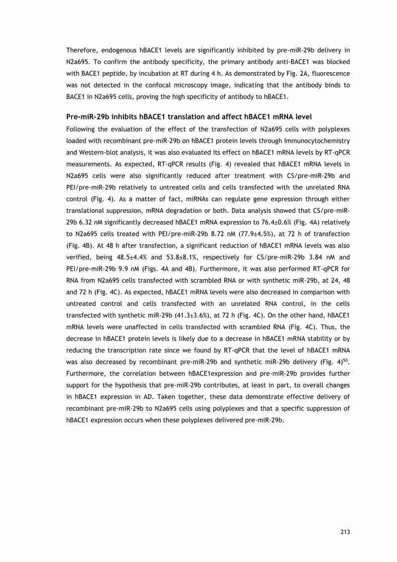
213
Therefore, endogenous hBACE1 levels are significantly inhibited by pre-miR-29b delivery in
N2a695. To confirm the antibody specificity, the primary antibody anti-BACE1 was blocked
with BACE1 peptide, by incubation at RT during 4 h. As demonstrated by Fig. 2A, fluorescence
was not detected in the confocal microscopy image, indicating that the antibody binds to
BACE1 in N2a695 cells, proving the high specificity of antibody to hBACE1.
Pre-miR-29b inhibits hBACE1 translation and affect hBACE1 mRNA level
Following the evaluation of the effect of the transfection of N2a695 cells with polyplexes
loaded with recombinant pre-miR-29b on hBACE1 protein levels through Immunocytochemistry
and Western-blot analysis, it was also evaluated its effect on hBACE1 mRNA levels by RT-qPCR
measurements. As expected, RT-qPCR results (Fig. 4) revealed that hBACE1 mRNA levels in
N2a695 cells were also significantly reduced after treatment with CS/pre-miR-29b and
PEI/pre-miR-29b relatively to untreated cells and cells transfected with the unrelated RNA
control (Fig. 4). As a matter of fact, miRNAs can regulate gene expression through either
translational suppression, mRNA degradation or both. Data analysis showed that CS/pre-miR-
29b 6.32 nM significantly decreased hBACE1 mRNA expression to 76.4±0.6% (Fig. 4A) relatively
to N2a695 cells treated with PEI/pre-miR-29b 8.72 nM (77.9±4.5%), at 72 h of transfection
(Fig. 4B). At 48 h after transfection, a significant reduction of hBACE1 mRNA levels was also
verified, being 48.5±4.4% and 53.8±8.1%, respectively for CS/pre-miR-29b 3.84 nM and
PEI/pre-miR-29b 9.9 nM (Figs. 4A and 4B). Furthermore, it was also performed RT-qPCR for
RNA from N2a695 cells transfected with scrambled RNA or with synthetic miR-29b, at 24, 48
and 72 h (Fig. 4C). As expected, hBACE1 mRNA levels were also decreased in comparison with
untreated control and cells transfected with an unrelated RNA control, in the cells
transfected with synthetic miR-29b (41.3±3.6%), at 72 h (Fig. 4C). On the other hand, hBACE1
mRNA levels were unaffected in cells transfected with scrambled RNA (Fig. 4C). Thus, the
decrease in hBACE1 protein levels is likely due to a decrease in hBACE1 mRNA stability or by
reducing the transcription rate since we found by RT-qPCR that the level of hBACE1 mRNA
was also decreased by recombinant pre-miR-29b and synthetic miR-29b delivery (Fig. 4)50.
Furthermore, the correlation between hBACE1expression and pre-miR-29b provides further
support for the hypothesis that pre-miR-29b contributes, at least in part, to overall changes
in hBACE1 expression in AD. Taken together, these data demonstrate effective delivery of
recombinant pre-miR-29b to N2a695 cells using polyplexes and that a specific suppression of
hBACE1 expression occurs when these polyplexes delivered pre-miR-29b.

214
Figure 4 – Effect of recombinant pre-miR-29b on hBACE1 mRNA levels in N2a695 cells
following 24, 48 and 72 h treatment with: (A) CS/pre-miR-29b and (B) PEI/pre-miR-29b (C)
Synthetic miR-29b and Scrambled RNA. Values in the graphs are mean from triplicates of RT-
qPCR threshold cycles for hBACE1 mRNA normalized to those of mRNA for GAPDH from 3
independent experiments and demonstrate significant differences across treatment
conditions. ANOVA, mean ± SD.
Recombinant pre-miR-29b modulates Aβ42 generation in vitro
Previous studies showed a causal relationship between miR-29b expression and BACE1
activity, and, consequently, Aβ peptides generation16. Interestingly, by Immunocytochemistry
analysis it was found that when the endogenous Aβtotal peptides are secreted, they
accumulate around the nucleus leading to its deformation, appearing like a kidney (Fig. 5A).
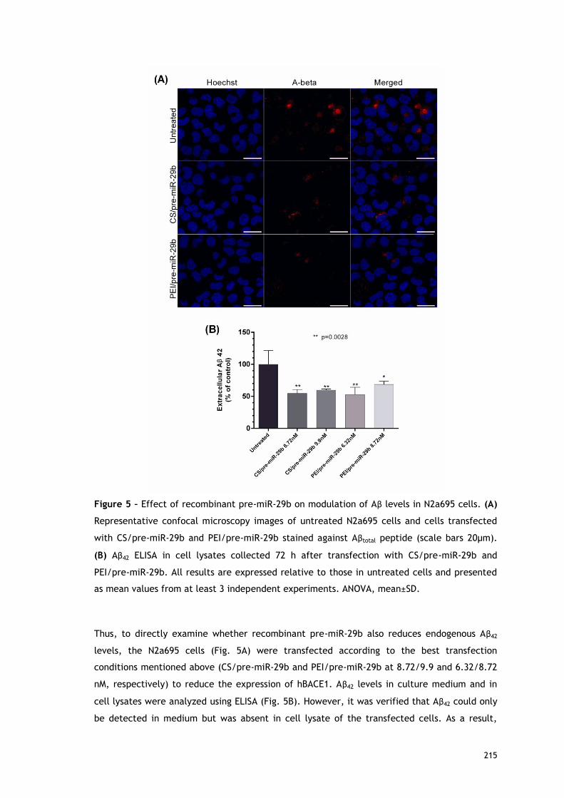
215
Figure 5 – Effect of recombinant pre-miR-29b on modulation of Aβ levels in N2a695 cells. (A)
Representative confocal microscopy images of untreated N2a695 cells and cells transfected
with CS/pre-miR-29b and PEI/pre-miR-29b stained against Aβtotal peptide (scale bars 20µm).
(B) Aβ42 ELISA in cell lysates collected 72 h after transfection with CS/pre-miR-29b and
PEI/pre-miR-29b. All results are expressed relative to those in untreated cells and presented
as mean values from at least 3 independent experiments. ANOVA, mean±SD.
Thus, to directly examine whether recombinant pre-miR-29b also reduces endogenous Aβ42
levels, the N2a695 cells (Fig. 5A) were transfected according to the best transfection
conditions mentioned above (CS/pre-miR-29b and PEI/pre-miR-29b at 8.72/9.9 and 6.32/8.72
nM, respectively) to reduce the expression of hBACE1. Aβ42 levels in culture medium and in
cell lysates were analyzed using ELISA (Fig. 5B). However, it was verified that Aβ42 could only
be detected in medium but was absent in cell lysate of the transfected cells. As a result,

216
endogenous Aβ42 levels were significantly reduced by 45.2±6.0% and 40.07±1.3% with CS/pre-
miR-29b 8.72 and 9.9 nM, respectively. With PEI/pre-miR-29b 6.32 and 8.72 nM, the
endogenous Aβ42 levels were reduced by 47.2±11.4% and 31.31±4.7%, respectively, when
compared to N2a695 untreated cells at 72 h (Fig. 5B). Therefore, endogenous Aβ42 levels were
inhibited by recombinant pre-miR-29b in this neuronal cell line. Once again, these findings
indicate that recombinant pre-miR-29b regulates Aβ42 production through mechanisms
dependent of hBACE1 expression.
Discussion
BACE1 and Aβ are central players in the pathways implicated in AD, it has been established
that reducing BACE1 expression has been suggested as a potential strategy for mitigating the
pathological processes underlying AD25,46. Interestingly, the miR-29a/b-1/c cluster is
significantly decreased in the brains of AD patients, and this decrease is correlated with the
increased level of BACE1 protein16,21. Previously published studies have demonstrated the
importance of using the miR-29 family as a potential major suppressor to silence BACE1
protein expression. However, the success of any therapeutic strategy depends on the
development of economic, effective and efficient methods for miRNAs large-scale production
(recombinant production using prokaryotic hosts) and purification, as an alternative to in
vitro transcription or chemical synthesis29. The global interest is to produce high quantities of
miRNA but also to obtain and preserve its quality, fulfilling the requirements of regulatory
agencies. To accomplish this purpose, we recently described a novel purification methodology
based on arginine-affinity chromatography to selectively purify the pre-miR-29b from
different RNA species with high recovery yield, purity and good integrity, revealing to be an
efficient and reproducible technique to obtain an appropriate RNA quality with potential
applicability for transfection studies29. Arginine was selected because it is a conserved amino
acid of the active center of the PAZ domains of the Argonaute proteins, belonging to the
RISC, suggesting that they have an important role in the specific recognition of the 3’UTR of
the pre-miRNA47.
On the other hand, the development of an efficient miRNA delivery system capable to
overcome the biological barriers, protect the integrity of miRNA from degradation, promote
cellular uptake and, finally, release miRNA in the cytoplasmic compartment is crucial to
achieve an improvement in the biological effect in RNAi-based therapies. Hence, according to
previous results obtained by our research group43, we decided to explore the possibility of
using polycations-based carrier systems to efficiently deliver the recombinant pre-miR-29b to
N2a695 neuronal cell line, to suppress hBACE1 expression. The polyplexes developed have not
a significant cytotoxicity effect of on the selected cell line (Fig. 1). An important in vitro
experimental approach in this study is the use of a stable N2a695 cell line because in these
cells there is a constitutive endogenous hBACE1 expression, enabling greater sensitivity for

217
evaluating the direct or indirect effect of recombinant pre-miR-29b on the cellular levels of
hBACE1 or on the products of its activity (Aβ42 peptides), once in biological systems over-
expressing hBACE1 leads to the formation Aβ peptides20,44,45.
Along with the role that pre-miR-29b plays in the regulation of the BACE1 levels, it was also
evaluated the involvement of this miRNA in the functional regulation of the amyloid pathway.
As a matter of fact, in this study and for the first time, it was demonstrated that the
overexpression of recombinant pre-miR-29b induces a marked decrease in the levels of the
protein hBACE1 and, consequently, a significant decrease in the level of the endogenous Aβ42.
This result suggests that the application of pre-miR-29 lead to a significant decrease in the
levels of two major risk factors commonly associated with the neurodegeneration in AD. We
further verified that a single treatment with CS/pre-miR-29b or PEI/pre-miR-29b dramatically
decreased the amount of hBACE1 expression (suppressed in 78.5±4.5% and 86.1±1.2%,
respectively) and the generation of Aβ42 peptide (44.2±6.0% and 47.2±11.4%, respectively) by
downregulating hBACE1 protein in N2a695 cells (Figs. 2, 3 and 5). These findings reinforce the
fact that the ability of recombinant pre-miR-29b to regulate endogenous hBACE1 protein
expression is likely direct, because it binds to the 3’ UTR of hBACE1 mRNA, complementary to
the miR-29b seed region (Fig. 4). Our results demonstrated that hBACE1 mRNA levels were
also significantly altered following recombinant pre-miR-29b transfection, suggesting a post-
transcriptional mechanism that involves direct degradation/destabilization of hBACE1 mRNA
and is likely that it promotes inhibition of hBACE1 protein translation. With these results it
was verified that hBACE1 is a direct target of pre-miR-29b (Figs. 2, 3 and 4), which was
further validated by the downregulation of hBACE1 gene expression by polycations/pre-miR-
29b in vitro.
Our findings, together with those from other groups, suggest a fundamental role of miR-29 in
AD, and emphasize the potential application of miR-29 in prognosis prediction and AD
therapy. With the successful implementation of this methodology and comparing with
previously published data (~62% to miR-339-5p46; ~35% to miR-29c21; ~50% to miR-29a/b-116),
we obtained the highest decrease ever reported for the hBACE1 expression levels (around 78%
to CS/pre-miR-29b and 86% to PEI/pre-miR-29b) in AD model cells. In addition, it was also
verified a decrease of the endogenous Aβ42 levels (approximately 44% to CS/pre-miR-29b and
47% to PEI/pre-miR-29b) in these cells. Furthermore, the concentrations of recombinant pre-
miR-29b used in this study were much lower than those reported in other works using
synthetic miRNA (25 to 250 nM)16,21, thus a smaller amount of recombinant pre-miR-29b is
required to obtain a higher silencing level of hBACE1. In fact, this result may be due to the
fact that we are using recombinant pre-miRNA, which is more efficiently recognized and
processed within the cell, instead of the mature form of miRNA or the fact that the pre-
miRNA was efficiently purified and delivered to the cell through its encapsulation with
polycations48,49.

218
Our results suggest that recombinant pre-miR-29b can represent a novel biopharmaceutical
product for the therapeutic modulation of hBACE1 levels, once this study have new
implications for hBACE1 biology and offer a new perspective on the treatment of AD.
According to the results obtained in this study, we developed an integrative platform that
allows biosynthesis, purification and transfection of the recombinant pre-miR-29b using
polyplexes to decrease the hBACE1 and endogenous Aβ42 expression levels. In addition, this
approach allowed improving the currently available methodologies of miRNA-based
therapeutics, not only for neurological disorders but also for future therapeutic targets that
may be of potential clinical interest.
Methods
Reagents
CS (Mw = 50–190 kDa) and cell culture reagents were purchased from Sigma–Aldrich. PEI (Mw =
10 kDa) was purchased from Polysciences. (3-(4,5-dimethylthiazol-2-yl)-5-(3-
carboxymethoxyphenyl)-2-(4-sulfophenyl)-2H-tetrazolium) (MTS) was obtained from Promega.
Anti-human BACE1 and BACE1 peptide (Abcam); anti-β-actin and monoclonal anti-β-Amyloid
(Sigma-Aldrich); Hoechst 33342®, AlexaFluor 488® and AlexaFluor 546® (Invitrogen); Goat
anti-rabbit/mouse IgG-HRP (Santa Cruz Biotechnology) were used.
Pre-miR-29b biosynthesis and purification by arginine affinity chromatography
The pre-miR-29b used in the experiments was produced in a bacterial cell culture of
Rhodovulum sulfidophilum DSM 1374 strain (BCCM/LMG, Belgium) modified with the plasmid
pBHSR1-RM containing the sequence of pre-miR-29b, as previously described by Pereira and
collaborators29. Then, the pre-miR-29b isolation was achieved using arginine as a specific
ligand in affinity chromatography29. The endotoxins content was also evaluated by using the
ToxinSensorTM Chromogenic Limulus Amoebocyte Lysate assay kit (GenScript), being verified
a considerably reduction during the chromatographic process, since the sample injected onto
the arginine matrix had 0.164 EU/µg of sRNA and the final pre-miR-29b fraction presented
0.002 EU/µg of pre-miR-29b. Thus, the endotoxins level in the final pre-miR-29b sample
conforms the guidelines of regulatory agencies like Food and Drug Administration (<0.06
EU/mL for the cerebrospinal fluid). In addition, the PCR product corresponding to the purified
pre-miR-29b was sequenced to confirm the identity and orientation of the amplicon (Figure S1
in Supporting Information).
Formulation of polyplexes
Briefly, all the pre-miR-29b loaded polyplexes were formulated using the method of simple
complexation which is based on the electrostatic interactions that occur between molar
concentrations of positive charge, present in the protonated amine groups of each polycation
(N), and the negative charge of the phosphate groups of RNA backbone (P), as described by

219
Pereira and co-workers41. The pre-miR-29b (a final concentration of 2 µg/mL) and polycation
(a concentration of 10 mg/mL) stock solutions were prepared in sodium acetate buffer (0.1 M
sodium acetate/0.1 M acetic acid, pH 4.5). In order to promote encapsulation, cationic
polymer solution (100 µL) was added dropwise to the pre-miR-29b solution (400 µL), under
stirring during 30 s, to particle formation41. The PEI/pre-miR-29b molar ratio is 0.35:1
(mol/mol) and the CS/pre-miR-29b molar ratio is 3:1 (mol/mol). The formulated polyplexes
were incubated at RT for 30 min and then pelleted by centrifugation at 15000 g for 20 min.
The amount of unbound pre-miR-29b was quantified by UV spectrophotometry41. Thus, the
encapsulation efficiency (EE) was determined using the following formula: EE%=[(Total pre-
miRNA amount – pre-miRNA supernatant amount)/Total pre-miRNA amount] x 100. Three
repetitions of this procedure were performed for each system. The hydrodynamic diameter
and zeta potential of the pre-miR-29b-loaded polyplexes were determined by dynamic light
scattering (DLS) using a Zetasizer Nano ZS particle analyzer (Malvern Instruments,
Worcestershire, UK), equipped with a He-Ne laser, at 25ºC. For DLS analysis particle samples
were produced as before mentioned and ressuspended in ultrapure water. Size
characterization was performed in fully automatic mode and with a scattering angle of 173°.
Particle zeta potential measurements were performed in disposable capillary cells and
computed by using Henry’s [F(Ka) 1.5], and Smoluchowsky models. All the data was examined
in Zetasizer software v 7.03. The experiments were performed in triplicate and an average of
30 measurements was acquired individually for each sample. As a positive control,
Lipofectamine 2000 transfection reagent was used (Lipo/pre-miR-29b), according to the
protocol recommended by the manufacturer.
Transfection of N2a695 cells with polyplexes/pre-miR-29b
N2a695 cells at passages 5-27 were cultured in the following medium: 1:1 mixture of
Dulbecco’s modified Eagle’s medium (DMEM) and OptiMEM supplemented with 5% (wt/vol)
heat-inactivated FBS and 1% (wt/vol) penicillin–streptomycin48. N2a695 cells were seeded at
3×10⁴ cells/mL. When a 50 to 60% confluence was achieved, the culture medium was replaced
by serum-free medium. After 12 h, CS/pre-miR-29b, PEI/pre-miR-29b and Lipofectamine/pre-
miR-29b (Lipo/pre-miR-29b) were added to the cells at pre-miR-29b concentration of 3.84 to
9.9 nM and transfection was carried out during 6 h. The culture medium was replaced by
fresh medium supplemented with 1% FBS and 1% antibiotic, to allow the cells to remain
metabolically active, expressing hBACE1 and Aβ. Untreated cells and cells transfected with an
unrelated RNA (5’-UGUGCAAAUCUAUGCAAAACUGA-3′) were used for negative controls (at a
final concentration of 9.9 nM). In addition, cells were also transfected with scrambled miRNA
(5′-UUCUCCGAACGUGUCACGUTT-3′; 3′-TTAAGAGGCUUGCACAGUGCA-5′) and a synthetic miR-
29b in the mature form (5′-UAGCACCAUUUGAAAUCAGUGUU-3´) using CS, as controls (at a
final concentration of 9.9 nM). The cells were harvested 24, 48 and 72 h after transfection.
All transfection experiments were performed in triplicate.

220
MTS assay
The cellular cytotoxicity effect of the different formulations of polyplexes (CS and PEI/pre-
miR-29b) was evaluated using the Cell Titer 96® AQueous Non-Radioactive Cell Proliferation
Assay. Briefly, N2a695 cells were seeded at a density of 2×10⁴ cells per well in a 96-well
plate, 24 h after the cell culture medium was replaced by serum-free culture medium. Cells
were then transfected as described above and the MTS assay was performed at different time
points (48 and 72 h). Subsequently, the medium was exchanged, a mixture of MTS/phenazine
metasulfate (PMS) was added to each well, and cells were incubated during 4 h at 37ºC in a
humidified atmosphere containing 5% CO2. Following incubation, the absorbance
measurements of the soluble brown formazan produced were performed in a microplate
reader (Sanofi, Diagnostics Pauster) at 492 nm. All experiments were repeated at least three
times. Cells incubated with absolute ethanol were used as positive control for cytotoxicity.
Immunocytochemistry and Imaging Analysis
N2a695 cells were seeded on glass coverslips into 12-well plates, to be recovered at 24, 48
and 72 h. After transfection with either Lipofectamine 2000 or polyplexes, the cells were
fixed for 10 min at RT with 4% paraformaldehyde (PFA) buffer solution, followed by
permeabilization with 0.1% Triton X-100 for 5 min. Cell preparations were blocked for 1 h at
RT with 20% FBS in PBS-Tween 20 (PBS-T). The cells were then incubated overnight at 4ºC
with the primary anti-hBACE1 polyclonal antibody (1:100) and anti-Aβ17-24 monoclonal
antibody (1:1000). Then, cells were washed 6 times with PBS-T and incubated for 1 h at RT
with the appropriate fluorescence conjugated secondary antibodies AlexaFluor 488® goat
anti-rabbit or AlexaFluor 546® goat anti-mouse. After washing, the nucleus were
counterstained with Hoechst 33342® (1:1000) for 10 min followed by 3 washing steps with
PBS-T. Glass coverslips were mounted on slides and the preparations were visualized under a
Zeiss LSM 710 laser scanning confocal microscope (Carl Zeiss SMT Inc., USA) equipped with a
plane-apocromat 63×/DIC objective. Images were processed and analyzed using ImageJ
software.
Western blot Analysis
Cells were rinsed in ice-cold PBS and homogenized in cell lysis buffer: 25 mM Tris-HCl buffer,
pH 7.4; 2.5 mM EDTA; 1% Triton X-100; 2.5 mM EGTA; 25 mM phenylmethylsulfonyl fluoride
and complete EDTA Free protease inhibitor cocktail (Roche). Once homogenized, cell extracts
were centrifuged at 11500 rpm for 7 min at 4ºC. The total protein concentration in the
supernatant was determined using Bradford Protein Assay (BioRad) and 50 µg of total protein
from each cell extract were boiled for 10 min in reducing buffer and then fractionated by
electrophoresis on 10% SDS-PAGE. After, proteins were transferred to polyvinylidene
difluoride filter (PVDF) membranes and blocked with TBS-T (200 mM Tris-HCl, pH 7.4; 1.37 M
NaCl; 0.1% Tween 20) supplemented with 5% BSA. Following, membranes were probed with
primary antibodies recognizing the hBACE1 (1:1000 in 5% BSA in TBS-T) and β-actin (1:20000 in

221
TBS-T) at 4ºC overnight. After three washes with TBS-T, membranes were incubated with the
HRP-labeled anti-rabbit/mouse IgG secondary antibody diluted 1:50000, followed by three
washes with TBS-T. Signal detection was performed with ECL substrate (BioRad) according to
manufacturer’s instructions and images were acquired with the ChemiDoc™ XRS system
(BioRad) and analysed with the Image Lab (BioRad).
Expression of BACE1 mRNA in N2a695 cells by RT-qPCR
Total RNA was extracted from the cells using TRIzol reagent (Invitrogen). 1 µg of total RNA
was reverse transcribed using the RevertAid First Strand cDNA Synthesis Kit (Thermo Fisher
Scientific Inc.), according to the manufacturer’s instructions. For quantitative analysis, RT-
qPCR amplification of cDNA was performed using the Maxima® SYBR Green/Fluorescein qPCR
Master Mix (Thermo Fisher Scientific Inc.) in an IQ5 Cycler from BioRad. RT-qPCR efficiencies
were calculated from the given slopes with MyIQ 2.0 software (BioRad). The relative
quantification of the BACE1 expression was based on the comparative threshold cycle (CT)
method in which the amount of the target was determined to be 2-(ΔCT target - ΔCT calibrator),
normalized to levels of glyceraldehyde-3-phosphate dehydrogenase (GAPDH) and relative to
the untreated control cells. The primers used in these experiments were 5’-
AGACGCTCAACATCCTGGTG-3’ (forward) and 5’-CCTGGGTGTAGGGCACATAC-3’ (reverse) for
the amplification of human BACE (hBACE) and 5’- TGACGTGCCGCCTGGAGAAA-3’ (forward),
5’-AGTGTAGCCCAAGATGCCCTTCAG-3’ (reverse) for the amplification of GAPDH. Each sample
was run in triplicate, and threshold cycle (CT) values were averaged from the triplicate. The
final data were averaged from 3 separately conducted experiments.
Sandwich enzyme-linked immunosorbent assay (ELISA)
The endogenous Aβ42 levels were quantified in the culture medium and in total cell lysates
using ELISA (Invitrogen), according to the manufacturer’s instructions. Total cell lysates were
performed as described for Western blot and the culture medium was centrifuged at 13000
rpm for 1 min to remove cell debris. Total cell lysates and culture medium were diluted 1:10
and Aβ42 concentrations were detected with an ELISA (Invitrogen), according to the
manufacturer’s instructions. The results were expressed as referred to dilutions of standard
synthetic control peptides. In addition, absolute Aβ42 values (pg/ml) were measured and
normalized to total protein yield of crude cell lysates.
Statistical analysis
All experiments were repeated at least three times using independent culture preparations.
Data in the figures are expressed as mean ± standard error. Quantitative data were
statistically analyzed by one-way analysis of variance (ANOVA), followed by pair-wise
comparisons using the Fisher’s least significant difference test. * indicate significant
difference versus untreated cells, # indicate significant difference versus cells transfected
with unrelated RNA control and ● indicate significant difference between samples, being

222
considered statistically significant for p<0.05. Statistical analysis was performed by using
GraphPad Prism 6 software.
Acknowledgments
This work was supported by the Portuguese Foundation for Science and Technology (FCT),
through the project EXPL/BBB-BIO/1056/2012 and Pest-OE/SAU/UI0709/2014. Patrícia Pereira
acknowledges the fellowship (Ph.D. Grant, Ref SFRH/BD/81914/2011), from FCT. Joana
Tomás also acknowledges the fellowship in the ambit of the project CENTRO-07-ST24-FEDER-
002015. The authors also acknowledge the program Fundo Europeu de Desenvolvimento
Regional (FEDER) [COMPETE [FCOMP-01-0124-FEDER-027560]]. The authors would like to thank
Professor Yo Kikuchi (Division of Life Science and Biotechnology, Department of Ecological
Engineering, Toyohashi University of Technology) for kindly provided of pBHSR1-RM plasmid,
Prof. Wenjie Luo and Professor Cecília Santos for the kind contribution regarding the cell line
N2a695 and, finally, Professor Manuel Lemos and Eduarda Coutinho for your help in
sequencing.
Author’s contributions
PP carried out all the experimental procedures and wrote the manuscript. PP, JT and FS
conceived and designed the study. JT helped to perform the experimental procedures. All the
authors contributed to the drafting of the manuscript and were responsible for the revision.
Additional information
Competing financial interests: The authors declare no competing financial interests.

223
References
1. Broderick, J. A.; Zamore, P. D. MicroRNA therapeutics. Gene Ther 18, 1104-10 (2011)
2. Bartels, C. L.; Tsongalis, G. J. MicroRNAs: novel biomarkers for human cancer. Clinical
chemistry 55, 623-31 (2009)
3. Selbach, M., et al. Widespread changes in protein synthesis induced by microRNAs.
Nature 455, 58-63 (2008)
4. Ambros, V. The functions of animal microRNAs. Nature 431, 350-5 (2004)
5. He, L.; Hannon, G. J. MicroRNAs: small RNAs with a big role in gene regulation. Nat Rev
Genet 5, 522-31 (2004)
6. Bartel, D. P. MicroRNAs: genomics, biogenesis, mechanism, and function. Cell 116, 281-
97 (2004)
7. Bartel, D. P. MicroRNAs: target recognition and regulatory functions. Cell 136, 215-33
(2009)
8. Chen, F.; Hu, S. J. Effect of microRNA-34a in cell cycle, differentiation, and apoptosis: a
review. Journal of biochemical and molecular toxicology 26, 79-86 (2012)
9. Plaisier, C. L.; Pan, M.; Baliga, N. S. A miRNA-regulatory network explains how
dysregulated miRNAs perturb oncogenic processes across diverse cancers. Genome research
22, 2302-14 (2012)
10. Jiang, H.; Zhang, G.; Wu, J. H.; Jiang, C. P. Diverse roles of miR-29 in cancer (review).
Oncology reports 31, 1509-16 (2014)
11. Wu, Z.; Huang, X.; Zou, Q.; Guo, Y. The inhibitory role of Mir-29 in growth of breast
cancer cells. Journal of experimental & clinical cancer research : CR 32, 98 (2013)
12. Xiong, Y., et al. Effects of microRNA-29 on apoptosis, tumorigenicity, and prognosis of
hepatocellular carcinoma. Hepatology 51, 836-45 (2010)
13. Li, Y., et al. Progressive miRNA expression profiles in cervical carcinogenesis and
identification of HPV-related target genes for miR-29. The Journal of pathology 224, 484-95
(2011)
14. Fabbri, M., et al. MicroRNA-29 family reverts aberrant methylation in lung cancer by
targeting DNA methyltransferases 3A and 3B. Proc Natl Acad Sci U S A 104, 15805-10 (2007)
15. Jiao, J.; Herl, L. D.; Farese, R. V.; Gao, F. B. MicroRNA-29b regulates the expression
level of human progranulin, a secreted glycoprotein implicated in frontotemporal dementia.
PloS one 5, e10551 (2010)
16. Hebert, S. S., et al. Loss of microRNA cluster miR-29a/b-1 in sporadic Alzheimer's disease
correlates with increased BACE1/beta-secretase expression. Proc Natl Acad Sci U S A 105,
6415-20 (2008)
17. Satoh, J. MicroRNAs and their therapeutic potential for human diseases: aberrant
microRNA expression in Alzheimer's disease brains. J Pharmacol Sci 114, 269-75 (2010)

224
18. Kocerha, J.; Kauppinen, S.; Wahlestedt, C. microRNAs in CNS disorders. Neuromolecular
medicine 11, 162-72 (2009)
19. Hebert, S. S., et al. MicroRNA regulation of Alzheimer's Amyloid precursor protein
expression. Neurobiology of disease 33, 422-8 (2009)
20. Bettens, K., et al. APP and BACE1 miRNA genetic variability has no major role in risk for
Alzheimer disease. Hum Mutat 30, 1207-13 (2009)
21. Zong, Y., et al. miR-29c regulates BACE1 protein expression. Brain Res 1395, 108-15
(2011)
22. Dominguez, D. I.; Hartmann, D.; De Strooper, B. BACE1 and presenilin: two unusual
aspartyl proteases involved in Alzheimer's disease. Neuro-degenerative diseases 1, 168-74
(2004)
23. Cole, S. L.; Vassar, R. The Alzheimer's disease beta-secretase enzyme, BACE1. Molecular
neurodegeneration 2, 22 (2007)
24. Schonrock, N.; Matamales, M.; Ittner, L. M.; Gotz, J. MicroRNA networks surrounding APP
and amyloid-beta metabolism-implications for Alzheimer's disease. Experimental Neurology
235, 447-454 (2012)
25. Hunt, C. E.; Turner, A. J. Cell biology, regulation and inhibition of beta-secretase (BACE-
1). FEBS J 276, 1845-59 (2009)
26. Ponchon, L.; Dardel, F. Large scale expression and purification of recombinant RNA in
Escherichia coli. Methods 54, 267-73 (2011)
27. Sherlin, L. D., et al. Chemical and enzymatic synthesis of tRNAs for high-throughput
crystallization. RNA 7, 1671-8 (2001)
28. Martins, R.; Queiroz, J. A.; Sousa, F. Ribonucleic acid purification. J Chromatogr A 1355,
1-14 (2014)
29. Pereira, P., et al. Purification of pre-miR-29 by arginine-affinity chromatography. J
Chromatogr B Analyt Technol Biomed Life Sci 951-952, 16-23 (2014)
30. Pecot, C. V.; Calin, G. A.; Coleman, R. L.; Lopez-Berestein, G.; Sood, A. K. RNA
interference in the clinic: challenges and future directions. Nat Rev Cancer 11, 59-67 (2011)
31. Lee, S. Y., et al. Stability and cellular uptake of polymerized siRNA (poly-
siRNA)/polyethylenimine (PEI) complexes for efficient gene silencing. J Control Release 141,
339-46 (2010)
32. Nimesh, S.; Chandra, R. Polyethylenimine nanoparticles as an efficient in vitro siRNA
delivery system. Eur J Pharm Biopharm 73, 43-9 (2009)
33. Kwok, A.; Hart, S. L. Comparative structural and functional studies of nanoparticle
formulations for DNA and siRNA delivery. Nanomedicine : nanotechnology, biology, and
medicine 7, 210-9 (2011)
34. Grayson, A. C.; Doody, A. M.; Putnam, D. Biophysical and structural characterization of
polyethylenimine-mediated siRNA delivery in vitro. Pharmaceutical research 23, 1868-76
(2006)

225
35. Lungwitz, U.; Breunig, M.; Blunk, T.; Gopferich, A. Polyethylenimine-based non-viral
gene delivery systems. Eur J Pharm Biopharm 60, 247-66 (2005)
36. Sarvaiya, J.; Agrawal, Y. K. Chitosan as a suitable nanocarrier material for anti-
Alzheimer drug delivery. International journal of biological macromolecules 72, 454-65
(2015)
37. Howard, K. A., et al. RNA interference in vitro and in vivo using a novel chitosan/siRNA
nanoparticle system. Molecular therapy: the journal of the American Society of Gene
Therapy 14, 476-84 (2006)
38. Holzerny, P., et al. Biophysical properties of chitosan/siRNA polyplexes: profiling the
polymer/siRNA interactions and bioactivity. J Control Release 157, 297-304 (2012)
39. Rudzinski, W. E.; Aminabhavi, T. M. Chitosan as a carrier for targeted delivery of small
interfering RNA. International journal of pharmaceutics 399, 1-11 (2010)
40. Mao, S.; Sun, W.; Kissel, T. Chitosan-based formulations for delivery of DNA and siRNA.
Advanced drug delivery reviews 62, 12-27 (2010)
41. Pereira, P., et al. Characterization of polyplexes involving small RNA. J Colloid Interface
Sci 387, 84-94 (2012)
42. Godbey, W. T.; Wu, K. K.; Mikos, A. G. Size matters: molecular weight affects the
efficiency of poly(ethylenimine) as a gene delivery vehicle. Journal of biomedical materials
research 45, 268-75 (1999)
43. Godbey, W. T.; Wu, K. K.; Mikos, A. G. Poly(ethylenimine) and its role in gene delivery. J
Control Release 60, 149-60 (1999)
44. Chen, Y., et al. Antidiabetic drug metformin (GlucophageR) increases biogenesis of
Alzheimer's amyloid peptides via up-regulating BACE1 transcription. Proc Natl Acad Sci U S A
106, 3907-12 (2009)
45. Kobayashi, D., et al. BACE1 gene deletion: impact on behavioral function in a model of
Alzheimer's disease. Neurobiology of aging 29, 861-73 (2008)
46. Long, J. M.; Ray, B.; Lahiri, D. K. MicroRNA-339-5p down-regulates protein expression of
beta-site amyloid precursor protein-cleaving enzyme 1 (BACE1) in human primary brain
cultures and is reduced in brain tissue specimens of Alzheimer disease subjects. J Biol Chem
289, 5184-98 (2014)
47. Rivas, F. V., et al. Purified Argonaute2 and an siRNA form recombinant human RISC. Nat
Struct Mol Biol 12, 340-9 (2005)
48. Tsutsumi, A.; Kawamata, T.; Izumi, N.; Seitz, H.; Tomari, Y. Recognition of the pre-
miRNA structure by Drosophila Dicer-1. Nat Struct Mol Biol 18, 1153-8 (2011)
49. Li, M. M.; Wang, W. P.; Wu, W. J.; Huang, M.; Yu, A. M. Rapid production of novel pre-
microRNA agent has-mir-27b in Escherichia coli using recombinant RNA technology for
functional studies in mammmalian cells. Drug Metab Dispos 42, 1791-1794 (2014)

226
Supporting Information
Figure S1 - Sequencing of the pre-miR-29b.

227
Paper X
Brain-targeting study of lactoferrin-stearic acid–
Chitosan/Polyethylenimine pre-miR-29b-delivery system
P. Pereira, J. Tomás, C. Cruz, C. Santos, J. A. Queiroz, A. Figueiras, F. Sousa
(In preparation)
Short description: In view of the promising results previously obtained, it was necessary to
develop a carrier that can cross the BBB and deliver efficient and effectively the recombinant
pre-miR-29b to the brain. Thus, in this paper, the nanodevices of CS and PEI were
functionalized with specific ligands, namely lactoferrin which is recognized by cell surface
receptors of BBB and stearic acid which has emerged as an optimal ligand for targeting the
cell membrane allowing nanoparticle endocytosis, for higher transfection yields. To observe
the cellular uptake of nanodevices, cell live imaging was applied. Ex vivo permeation studies
in brain barrier models were performed in transwell cell culture systems, in order to evaluate
the delivery efficiency of the vector to the brain. Moreover, additional studies were carried
out using in vitro models of the blood-CSF barrier (namely, Z310 cells) and BBB (Rat brain
endothelial cells (RBE4) cells).

228

229
Brain-targeting study of lactoferrin-stearic acid–
Chitosan/Polyethylenimine pre-miR-29b-delivery system
P. Pereira1, J. Tomás1, C. Cruz1, C. Santos1, J.A. Queiroz1, A. Figueiras1,2, F.
Sousa1
1CICS-UBI – Health Sciences Research Centre, University of Beira Interior, Avenida Infante D.
Henrique, Covilhã, 6200-506, Portugal;
2CNC – Center of Neuroscience and Cell Biology, University of Coimbra, Largo Marquês de
Pombal, Coimbra, 3004-517, Portugal
*Corresponding author:
Dr. Fani Sousa
Phone: +351 275 329 074
Fax: +351 275 329 099
E-mail address: [email protected]
Postal address: Health Sciences Research Centre, Universidade da Beira Interior, Avenida
Infante D. Henrique, 6200-506 Covilhã, Portugal

230

231
Abstract
The aim of the present study is to develop nanocarriers for targeted pre-miR-29b delivery
across the Blood-Brain Barrier (BBB), to reach neuronal cells and inhibit the BACE1, with
potential implications in Alzheimer’s disease treatment. A dual-targeting drug delivery system
(Chitosan (CS) or Polyethylenimine (PEI) conjugated with stearic acid (SA) and lactoferrin (Lf))
was synthesized and its BBB penetration was evaluated. Stearic acid and lactoferrin were
successfully exploited as brain-targeting ligands to modify cationic polymers. The
intracellular uptake of the delivery systems by neuronal cells was studied in vitro as well as
the gene silencing efficiency of pre-miR-29b. The CS/PEI-SA-Lf systems showed very strong
fluorescence in the cytoplasm and cell nucleus, and were able to deliver pre-miR-29b to
N2a695 cells in 1 hour after transfection. The experiment of transport across the BBB showed
that CS-SA-Lf delivered 65% of recombinant pre-miR-29b in a period of 4 hours, an higher
transport ratio than the 42% found for PEI-SA-Lf in the same time. The results indicate that
CS-SA-Lf can be a potential pre-miR-29b carrier for AD therapy.
Keywords
Alzheimer’s disease; hBACE1; Chitosan; Polyethylenimine; Blood-Brain-Barrier; Recombinant
pre-miR-29b; Lactoferrin; Stearic Acid

232

233
1. Introduction
Alzheimer’s disease (AD) is the most common and devastating form of dementia in the elderly
and can lead to death within 3 to 9 years after appearance of symptoms [1]. AD is
pathologically characterized by the progressive neuronal loss, mainly due to the accumulation
of extracellular amyloid plaques consisting of aggregates of toxic amyloid-beta (Aβ) peptides
[2-4]. They are generated through sequential proteolytic cleavage of the amyloid precursor
protein (APP) by β-secretase (also known as β-site APP-cleaving enzyme 1 (BACE1)) and γ-
secretase, which play a pivotal role in the dysfunction and death of neurons [5-7]. Several
convincing evidences suggest that the regulation of expression of proteins involved in the
generation and accumulation of Aβ peptides can be extremely important in AD, since Aβ
peptides play the central role in initiating and development of the pathological cascade of AD
[4, 5, 8, 9]. In the last years, several research groups demonstrated that the levels and
activity of BACE1 protein are elevated in sporadic AD brains, suggesting that BACE1
dysregulation is directly implicated in AD pathogenesis. For these reasons, BACE1 has been
recognized as a promising drug target for the therapy of this disease, once BACE1 inhibition
may decrease the formation of all forms of Aβ peptides in AD and, consequently, reduce cell
death [10-12]. On the other hand, significant alterations in the expression of miR-29 family,
in brains of AD patients, have been observed in most of studies [13, 14], being significantly
decreased and displaying abnormally high levels of BACE1 protein. These findings raised the
opportunity to use miR-29 as a possible protective therapeutic strategy for the future of AD
therapy. The continued lack of progress in identifying effective diagnosis and treatment
modalities also contributes to higher health care costs and social burdens that are associated
with this disease [15-17].
One of the reasons limiting the development of therapies for AD is the existence of a physical
barrier, the Blood-Brain Barrier (BBB), which hinders the passage of most of the therapeutic
drugs from the circulating blood to the brain [18]. The BBB prevents the entry of harmful
substances present in the systemic blood circulation into the brain, but allows the access of
necessary nutrients and chemical signaling molecules, making of the brain a place of poor
permeability to various drugs [19-22]. Its function is related to the structural characteristics,
once the BBB is composed by tight intracellular junctions formed between adjacent brain
capillary endothelial cells [19]. On the other hand, the BBB possesses a variety of efflux
transporters, including P-glycoprotein [23], receptors for transferrin [24, 25], lactoferrin [26],
insulin [27], insulin-like growth factors [28], lipoprotein receptor-related protein (LRP) [29,
30], as well as, transmembrane monocarboxylic acid transporters and fatty acid transport
proteins [31], reinforcing BBB function by effectively removing drugs from the brain and
pumping them back into blood.
As a result, it is strictly necessary the development of methods that promote the delivery of
therapeutic drugs to the brain, safely and effectively. During the last decade, several non-

234
viral vectors were developed for brain targeting, including polymers (e.g. polyethyleneimine
(PEI), chitosan (CS), poly(lactic-co-glycolic acid) (PLGA), poly lactic acid (PLA)), polymeric
micelles, liposomes and dendrimers [32-37]. Among the vectors mentioned above, polymeric
delivery systems (polyplexes) have emerged as one of the most attractive carriers to cross the
BBB and increase the uptake of appropriate drugs by the brain. To accomplish this, the
receptors existing on the BBB have been explored as a means to specifically target drugs to
the brain. This can be achieved through the surface modification of delivery systems with a
ligand (such as transferrin (Tf), lactoferrin (Lf), stearic acid (SA)) targeting to the receptor,
which facilitates drug ability to interact with specific molecules expressed on the luminal side
of BBB endothelial cells [38]. In the present study, the recombinant human pre-miR-29b was
chosen as the therapeutic gene to be encapsulated in the polyplexes (CS or PEI) modified with
Lf and SA in the surface, to bind specifically to the Lf receptors on neuronal cells, through a
receptor-mediated pathway.
CS and PEI have gained much attention as delivery systems due to their advantageous
properties such as, ability to encapsulate large amounts of drug, high stability in biological
fluids, targeting ability, rapid cellular uptake, high transfection efficiency, biocompatibility,
biodegradability, low cytotoxicity and immunogenicity, and reduced side effects by targeted
delivery [39-41]. In addition, the cationic charge of CS and PEI, due to the presence of amino
groups, allows the establishment of electrostatic interactions with the negatively charged
RNAs, leading to an effective condensation and protection of the integrity of the RNA. In fact,
the encapsulation of RNAs prevents non-specific interactions and enzymatic degradation,
increasing the drug circulation in blood [39-41].
Lf is a single chain iron-binding glycoprotein that belongs to the Tf family. Its concentration in
the blood plasma is low but is higher in BBB [42, 43]. Recent studies reported that the
transport of Lf across the BBB monolayer model was unidirectional and that there was a
strong affinity of positively charged Lf to Lf receptor, resulting in higher uptake of Lf-
conjugated drug delivery systems to the brain comparing to Tf-modified NPs [26, 44]. For all
these reasons, Lf has been exploited as a promising brain-targeting ligand, facilitating drug
delivery into the brain. A previous study also described the ability of CS-SA for the delivery of
drugs into the brain and to avoid recognition by P-glycoprotein system, yielding an efficient
brain-targeting gene vector [38]. Thus, in the current study, it was explored the potential of
the polyplexes CS and PEI combined with two targeting substances, Lf and SA, to encapsulate
recombinant pre-miR-29b, and act as a brain-targeting delivery system. Various
measurements, including 1H NMR and SDS-PAGE, were used to characterize and confirm the
structure of the modified polyplexes. The properties of the polyplexes CS-SA, CS-SA-Lf, PEI-SA
and PEI-SA-Lf, such as the ability of transport across BBB model, the targeting effects, in
vitro cell uptake and transfection studies were performed on N2a695, RBE4 and Z310 cells.
The efficacy of these systems was finally monitored by examining the induced BACE1
inhibition.

235
2. Materials and Methods
2.1. Materials
Arginine–Sepharose 4B gel was acquired from Amersham Biosciences. All buffers used for the
chromatographic experiments were freshly prepared with 0.05% diethyl pyrocarbonate (DEPC)
treated water and were filtered through a 0.20 µm pore size membrane. Sodium chloride
(NaCl) was purchased from Panreac and tris(hydroxymethyl) aminomethane (Tris) from Sigma-
Aldrich. CS low molecular weight (LMW) (Mw=50–190 kDa; degree of deacetylation in the 75–
85% range), Stearic acid Grade I (98.5%), Lactoferrin human, 2-Iminothiolane hydrochloride
(2-IOT, 98%), 1-ethyl-3-(3-dimethylaminopropyl) carbodiimide (EDC) and all cell culture media
and reagents used in cell culture procedures were purchased from Sigma–Aldrich, unless
otherwise noted. PEI high molecular weight (HMW) (Mw=10 kDa) was acquired from
Polysciences, Inc. (Warrington, PA). Fluorescein isothiocyanate isomer I (FITC), Hoechst
33342® and AlexaFluor 488® were obtained from Invitrogen (Carlsbad, CA, US). Mouse
neuroblastoma (N2a) cells stably transfected with cDNAs encoding human APP695 (N2a695)
were kindly provided by Professor Wenjie Luo (Weill Cornell Medical College). The rat brain
microvascular endothelial cell line (RBE4) was provided as a gift by the laboratory of Dr. M.
Aschner (Department of Pediatrics, Vanderbilt Kennedy Center, Nashville, Tennessee).
2.2. Methods
2.2.1. Synthesis and characterization of PEI and CS polyplexes conjugated with SA and Lf
The conjugation of PEI or CS to SA and Lf (designated as CS-SA-Lf and PEI-SA-Lf) was
performed as previously reported by Xie and co-workers [38], with minor modifications. In
brief, first, SA (2.5 mg) and EDC (25 mg, at a ratio 1:10) were dissolved in 1.0 mL anhydrous
DMSO and stirred at 60ºC for 1 hour, until EDC and SA were well-dissolved and mixed. The
resulting mixture (SA:EDC) was then added slowly to 1% (w/v) of CS-LMW and PEI-HMW in
sodium acetate buffer (0.1 M sodium acetate/0.1 M acetic acid, pH 4.5) and the reaction
solution was kept at room temperature in the dark during 24 hours, with stirring in a water
bath. The amine group on PEI or CS was crosslinked with the carboxylic group of stearic acid
by EDC as linker. Posteriorly, the resulting conjugate, CS/PEI-SA, was dialyzed first against
phosphate buffered saline (PBS, pH 7.4) for 1 day and then against double deionized water for
2 days, using dialysis membrane (SnakeSkinTM Dialysis Tubing, MWCO 3500 Da, 22mm dry
diameter, ThermoScientific). The conjugated CS/PEI-SA was isolated as a “sponge” by
lyophilization, and was further characterized by 1H Nuclear Magnetic Resonance (NMR)
spectroscopy. All proton NMR spectra were recorded in solution of studied compounds in D2O
using a Bruker Avance III 600 MHz equipped with a QCI cryoprobe. For the preparation of
CS/PEI-SA-Lf, Lf (10 mg) was dissolved in 1.0 mL aqueous solution of water and mixed with
0.5 mL aqueous solution of 2-iminothiolane hydrochloride (2-IOT, 0.7 mg) and the reaction
proceed for 1 hour at room temperature, with moderate shaking. The excess of 2-IOT was
removed through a column of Sephadex G 100 column (Hitrap desalting column) using PBS 1x
(pH 7.0) as elution buffer. Fractions were collected according to the chromatogram obtained

236
by analysis of the absorbance (a peak contains about 90% of thiolated Lf) [45]. Subsequently,
CS-SA or PEI-SA were dissolved in 0.5 mL PBS (pH 7.0) and then added to the PBS solution of
Lf, drop by drop, for 20 hours at room temperature, in the dark. To remove unreacted Lf, the
resulting mixtures were purified by dialysis (SnakeSkinTM Dialysis Tubing, MWCO 3500 Da,
22mm dry diameter, ThermoScientific) during 4 days against double deionized water and
freeze dried, for further usage. The SDS-PAGE was used to characterize the carriers CS-SA-Lf
and PEI-SA-Lf.
2.2.2. Formulation and characterization of CS-SA-Lf/pre-miR-29b, PEI-SA-Lf/pre-miR-29b,
CS-SA/pre-miR-29b and PEI-SA/pre-miR-29b complexes
For all experiments, the human pre-miR-29b was obtained from a bacterial cell culture of
Rhodovulum sulfidophilum DSM 1374 strain (BCCM/LMG, Belgium) modified with the plasmid
pBHSR1-RM containing the sequence of pre-miR-29b. The growth was performed in a semi-
defined medium containing 30 µg/mL Kanamycin. After biosynthesis, the recombinant pre-
miR-29b was recovered from a complex mixture of small RNAs, through affinity
chromatography by using a column of arginine, as previously described by Pereira and
collaborators [46]. The quantity and quality of the purified pre-miR-29b was assessed by
measuring its optical density at 260 nm and 280 nm and by polyacrylamide electrophoresis,
respectively. Recombinant pre-miR-29b was covalently labeled with the fluorescent dye,
FITC, when necessary. PEI-SA-Lf/pre-miR-29b, PEI-SA-Lf/pre-miR-29b, CS-Lf/pre-miR-29b and
PEI-Lf/pre-miR-29b systems were formulated using the method of simple complexation
through electrostatic interaction by using different amines to phosphate ratios (N/P ratios,
molar ratio of CS/PEI-nitrogen atoms present in the protonated amine groups to pre-miR-29b-
phosphate groups), as described by Pereira and co-workers [47]. Pre-miR-29b stock solution
was prepared by pre-miRNA dissolution in sodium acetate buffer, up to a final concentration
of 2 µg/mL and was kept constant in all the methods used for the characterization of the
formed complexes [47]. The CS-SA-Lf, PEI-SA-Lf, PEI-Lf and CS-Lf stock solutions were also
prepared in sodium acetate buffer pH 4.5 in a concentration of 10 mg/mL. Briefly, the
complexes were obtained by addition of 100 µL of CS-SA-Lf, PEI-SA-Lf, PEI-Lf or CS-Lf
solution, of variable concentration, dropwise to the pre-miR-29b solution (400 µL). The
complexes were immediately mixed using a vortex mixer at maximum speed for 1 min. After
30 min of stabilization at room temperature, the complexes were recovered by centrifugation
(15000 g, 20 min). The complexation of pre-miR-29b by CS-SA, PEI-SA, CS-SA-Lf and PEI-SA-Lf
was evaluated by agarose gel electrophoresis [47].
2.2.3. Determination of the encapsulation efficiency
The encapsulation efficiency (EE) was calculated by determining free pre-miRNA
concentration in the supernatant recovered after particle centrifugation (15000g, 20 min,
25ºC). The amount of unbound pre-miR-29b was quantified by UV spectrophotometry
(Nanophotometer), at 260 nm. Supernatant recovered from unloaded complexes (without pre-
miRNA) was used as a blank. Three repetitions of this procedure were performed for each

237
system. EE was determined using the following formula: EE % = [(Total pre-miRNA amount –
pre-miRNA supernatant amount)/Total pre-miRNA amount] x 100 [47].
2.2.4. Cell culture
In this study, N2a695, RBE4 and Z310 choroidal epithelial cells were used. N2a695 cells (at
passages 13-30) were cultured in the following medium: 1:1 mixture of Dulbecco’s Modified
Eagle’s Medium (DMEM) and OptiMEM supplemented with 5% (wt/vol) heat-inactivated fetal
bovine serum (FBS) and 1% (wt/vol) penicillin-streptomycin. In these cells there is a
constitutive endogenous human BACE1 expression, enabling greater sensitivity for detecting
pre-miR-29b induced changes in the human BACE1 expression at post transcriptional level.
Z310 cell line (at passages 95-100) was maintained in DMEM high glucose growth medium
containing 10% FBS and 1% penicillin-streptomycin [48]. On the other hand, RBE4 cultures (at
passages 7-30) were grown in the following medium: Minimum Essential Medium (αMEM) and
Ham’s F10 Nutrient Mix (1:1 vol/vol) supplemented with 10% FBS, 1% penicillin-streptomycin,
1% L-glutamine and 0.6% Geneticin (300 μg/m). All cell lines were kept at 37ºC in a
humidified atmosphere containing 5% CO2. Cells were subcultured regularly using trypsin-
EDTA [49].
2.2.5. In vitro transfection and expression
2.2.5.1. In vitro cellular uptake – Cell live imaging
In order to investigate the cellular uptake, pre-miR-29b was labeled with FITC. In brief, an
aliquot of FITC-DMSO solution was added in drops to pre-miR-29b solution (8.72 nM). The
resultant solution was kept under stirring for 3 hours at room temperature. Then, the final
product was incubated with 3 M NaCl and 2.5 volumes of absolute ethanol ice-cold at -20ºC
during 30 min. Excess FITC was removed by centrifugation at 12000g for 30 min at 4ºC. After
centrifuging, the pellet was washed with 75% ethanol twice, followed by a 5 min
centrifugation at 12000 g (4ºC). Finally, the FITC-pre-miR-29b was ressuspended in OptiMEM
and encapsulated with CS-SA-Lf, PEI-SA-Lf, PEI-Lf and CS-Lf. N2a695 cells were seeded at a
density of 2×10⁴ cells/cm2 in μ-Slide 8-well flat bottom imaging plates (Ibidi GmbH,
Germany). In the following day, the medium was replaced by fresh serum-free medium and
cells were stained with Hoechst 33342® nuclear probe for 20 min. Subsequently, the cells
were transfected with the complexes prepared with FITC-labeled pre-miR-29b in serum-free
medium. The cells were transferred to a Zeiss LSM 710 confocal laser scanning microscope
(CLSM; Carl Zeiss SMT Inc., US) equipped with a plane-apocromat 63×/DIC objective and
processed in Zeiss Zen (SP2, 2010) and Imaris software (Bitplane, Switzerland), in order to
evaluate the cellular uptake. The fluorescence images were obtained at 63× amplification.
Cells without the addition of complexes were imaged as control.
2.2.5.2. In vitro transfection studies
Briefly, N2a695 cells were seeded in 12-well plates at a density of 2×10⁴ cells/well in 1.5 mL
of complete medium. When a 50% to 60% confluence was achieved, the media was removed
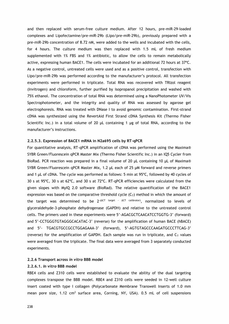
238
and then replaced with serum-free culture medium. After 12 hours, pre-miR-29-loaded
complexes and Lipofectamine/pre-miR-29b (Lipo/pre-miR-29b), previously prepared with a
pre-miR-29b concentration of 8.72 nM, were added to the wells and incubated with the cells,
for 4 hours. The culture medium was then replaced with 1.5 mL of fresh medium
supplemented with 1% FBS and 1% antibiotic, to allow the cells to remain metabolically
active, expressing human BACE1. The cells were incubated for an additional 72 hours at 37ºC.
As a negative control, untreated cells were used and as a positive control, transfection with
Lipo/pre-miR-29b was performed according to the manufacturer’s protocol. All transfection
experiments were performed in triplicate. Total RNA was recovered with TRIzol reagent
(Invitrogen) and chloroform, further purified by isopropanol precipitation and washed with
75% ethanol. The concentration of total RNA was determined using a NanoPhotometer UV/Vis
Spectrophotometer, and the integrity and quality of RNA was assessed by agarose gel
electrophoresis. RNA was treated with DNase I to avoid genomic contamination. First-strand
cDNA was synthesized using the RevertAid First Strand cDNA Synthesis Kit (Thermo Fisher
Scientific Inc.) in a total volume of 20 µL containing 1 µg of total RNA, according to the
manufacturer’s instructions.
2.2.5.3. Expression of BACE1 mRNA in N2a695 cells by RT-qPCR
For quantitative analysis, RT-qPCR amplification of cDNA was performed using the Maxima®
SYBR Green/Fluorescein qPCR Master Mix (Thermo Fisher Scientific Inc.) in an IQ5 Cycler from
BioRad. PCR reaction was prepared in a final volume of 20 μL containing 10 μL of Maxima®
SYBR Green/Fluorescein qPCR Master Mix, 1.2 μL each of 25 µM forward and reverse primers
and 1 μL of cDNA. The cycle was performed as follows: 5 min at 95ºC, followed by 40 cycles of
30 s at 95ºC, 30 s at 62ºC, and 30 s at 72ºC. RT-qPCR efficiencies were calculated from the
given slopes with MyIQ 2.0 software (BioRad). The relative quantification of the BACE1
expression was based on the comparative threshold cycle (CT) method in which the amount of
the target was determined to be 2-(ΔCT target - ΔCT calibrator), normalized to levels of
glyceraldehyde-3-phosphate dehydrogenase (GAPDH) and relative to the untreated control
cells. The primers used in these experiments were 5’-AGACGCTCAACATCCTGGTG-3’ (forward)
and 5’-CCTGGGTGTAGGGCACATAC-3’ (reverse) for the amplification of human BACE (hBACE)
and 5’- TGACGTGCCGCCTGGAGAAA-3’ (forward), 5’-AGTGTAGCCCAAGATGCCCTTCAG-3’
(reverse) for the amplification of GAPDH. Each sample was run in triplicate, and CT values
were averaged from the triplicate. The final data were averaged from 3 separately conducted
experiments.
2.2.6 Transport across in vitro BBB model
2.2.6.1. In vitro BBB model
RBE4 cells and Z310 cells were established to evaluate the ability of the dual targeting
complexes transpose the BBB model. RBE4 and Z310 cells were seeded in 12-well culture
insert coated with type I collagen (Polycarbonate Membrane Transwell Inserts of 1.0 mm
mean pore size, 1.12 cm2 surface area, Corning, NY, USA). 0.5 mL of cell suspensions

239
containing 2.0 x 10⁵ cells was added to the inner (donor) chamber, which was inserted into
the outer (acceptor) chamber containing 1.3 mL of the same culture medium. A cell
monolayer is usually formed 3-5 days after seeding, as judged by three criteria: (1) the cells
formed a confluent monolayer without visible spaces between cells under a light microscope;
(2) the height of the culture medium in the inner chamber had to be at least 2 mm higher
than that in the outer chamber for at least 24 hours; and (3) a constant transendothelial
electrical resistance (TEER) value across the cell layer was obtained [50]. The TEER was used
to measure the formation of tight junctions, indicating the integrity of BBB structure for
studying drug delivery to the brain. The cell monolayer integrity was monitored using an
EVOM Endohmchamber (Word Precision Instruments, Inc. Sarasota, Florida, USA) to measure
the TEER value of the in vitro BBB model. Only cell monolayers with TEER value above 100
Ωcm2 were selected for the transport studies, which indicate that this system could be used
as an in vitro BBB model [50]. All the permeability experiments were performed in serum-free
medium at 37ºC, and the drug carriers, including CS-SA/pre-miR-29b-FITC, CS-SA-Lf/pre-miR-
29b-FITC, PEI-SA/pre-miR-29b-FITC and PEI-SA-Lf/pre-miR-29b-FITC were added into the
donor compartment of the above BBB model with the pre-miR-29b concentration of 8.72 nM.
After 4 hours, the inserts were removed. The concentration of pre-miR-29b transported across
BBB media was determined using a spectrofluorometer (Spectramax Gemini XS, Molecular
Devices LLC, US), with the excitation wavelength of 480 nm and the emission wavelength at
590 nm. The BBB transport ratio of pre-miR-29b was calculated as follows: Transport ratio % =
[(Total Fluorescence in the outer chamber)/ [(Total Fluorescence in the inner chamber +
Total Fluorescence in the outer chamber)] x 100.
2.2.7.2. Immunofluorescence
Endothelial cells from the in vitro BBB model were fixed for 10 min at room temperature with
4% of paraformaldehyde (PFA) buffer solution. Cells were then washed 3 times with PBS for 10
min, followed by permeabilization with PBS containing 0.1% Triton X-100 for 15 min. After
fixation, the nucleus of the endothelial cells was counterstained with Hoechst 33342®
(1:1000) for 10 minutes followed by 3 washing steps with PBS-T. Cells were mounted on slides
and the fluorescence images were acquired using a confocal laser scanning microscope (Zeiss
LSM 710, Carl Zeiss SMT Inc., USA) equipped with a plane-apocromat 63×/DIC objective.
Images were processed and analyzed using ImageJ software. All experiments were repeated
at least three times, and representative images are shown.
2.8. Statistical analysis
All the experiments were repeated at least three times using independent culture
preparations. Data are presented as the mean ± standard error. Statistical comparisons were
performed using a one-way analysis of variance (ANOVA), followed by pair-wise comparisons
using the Fisher’s least significant difference test. A P value <0.05 was considered statistically
significant. Statistical analysis was performed by using GraphPad Prism 6 software.

240
3. Results
3.1. Synthesis and characterization of CS-SA, PEI-SA, CS-SA-Lf, PEI-SA-Lf
complexes
The carriers of CS/PEI-SA were synthesized by chemical reaction between the carboxyl group
of SA and the amino group of CS or PEI in the presence of EDC, a carboxyl activating agent
[38]. 1H NMR experiments were used to characterize and confirm the chemical structure of
the obtained CS/PEI-SA complexes. Also, for comparing the signals, CS and PEI spectra were
also recorded without stearic acid. The 1H-NMR assignment of CS was previously determined
and is as follows: 1H NMR (CS, D2O) δ =5.10 (H1), δ =3.09 (H2), δ =3.43~3.81 (H3, H4, H5, H6), δ
=1.96 (NHCOCH3) ppm. The signal of H1 was effected by the near water peak suppression of
H2O at δ=4.76. Comparing with CS 1H-NMR spectrum, the triplet signals at 0.9 ppm, due to the
terminal CH3 protons of stearic acid and the new signal between 1–1.5 ppm corresponding to
the CH2 chain of acyl chain, showed that stearic acid was linked to CS. Similarly, the 1H NMR
spectrum of the PEI was first assignment and the proton peaks of PEI (–NHCH2CH2–) appeared
at 3.2–2.0 ppm. The molecular structure of PEI-SA was then confirmed by 1H NMR. 1H NMR
analysis indicates that the amino-groups of the PEI were acylated. The 1H-NMR assignment of
PEI-SA was as follows: (ppm): 0.86–0.89 (t, -CH2CH2(CH2)15CH3), 1.25 (br, -
CH2CH2(CH2)15CH3), 1.62 (br, -CH2CH2(CH2)9CH3), 2.18 (br, -CH2CH2(CH2)9CH3), 2.39–3.3 (m, -
CH2CH2NH-, -CH2CH2N-, -CH2CH2NHCO-, -CH2CH2NHCO-). Although, the same chemical shifts
appeared in PEI-SA, however the sharp peaks at 3.0-2.4 ppm could contribute to the chemical
shifts of PEI. Moreover, the ratio of peak area at 3.0–2.4 ppm to the peak area of methyl
group in PEI-SA was bigger than that of PEI. These results indicated that stearic acid was
successfully link to PEI. The NMR studies also demonstrated that very few free amines are
available on the CS-SA complexes comparing with PEI-SA complexes. The functionalization
with Lf was obtained using a common method with 2-iminothiolane hydrochloride as the
sulfhydrylization reagent [45]. After purification, thiolated protein was obtained. The SDS-
PAGE was used to further confirm the surface capping of the terminal amines of the CS/PEI-
SA with Lf. In the SDS-PAGE a new band above 100 KDa was observed, indicating that Lf ligand
was covalently attached to the CS/PEI-SA (Figure 1). NMR experiments are current running to
complement the SDS-PAGE results.

241
Figure 1 - SDS-PAGE of CS-SA, CS-SA-Lf, PEI-SA and PEI-SA-Lf.
3.2. Preparation of CS-SA-pre-miR-29b, PEI-SA-pre-miR-29b, CS-SA-Lf-pre-miR-
29b and PEI-SA-Lf-pre-miR-29b
All pre-miR-29b-loaded complexes were prepared by electrostatic interactions that occur
between the positively charged amine groups of the carriers and the phosphate groups of the
pre-miR-29b [47]. The complexes were mixed with pre-miR-29b for the formation of pre-miR-
29b-loaded complexes in acetate buffer at pH 4.5. In order to determine the degree of
compaction between pre-miR-29b and all carriers in study, a gel retardation assay and UV
spectrometry were performed (Fig. 2). As shown in Fig. 2A, the mobility of pre-miR-29b was
fully retarded by all carriers, indicating that the pre-miR-29b was tightly neutralized within
the carriers. It was also possible to observe that PEI-SA-pre-miR-29b and PEI-SA-Lf-pre-miR-
29b moved in direction of the anode, that is, some polyplexes present an excess of positive
charge, know as overcharging effects. These results are in accordance with what was
observed in the encapsulation studies, in which 100% of encapsulation efficiency was
achieved, indicating that pre-miR-29b was completely complexed with PEI-SA-Lf (Fig. 2B). In
what concerns to the CS-based systems, CS-SA-Lf seems to be more efficient in pre-miR-29b
condensation, once a value of 86% was obtained in comparison with 80% for CS-SA.

242
Figure 2 - Synthesis of pre-miR-29b-loaded complexes. (A) Agarose gel electrophoresis
analysis. Lane 1: naked pre-miR-29b, lane 2-5: pre-miR-29b loaded into the different
complexes, as indicated in the figure. (B) Encapsulation efficiency of different pre-miR-29b-
loaded complexes (Data represent the mean±SD, n = 4).
3.3. In vitro cellular uptake and intracellular trafficking of the pre-miR-29b-
loaded complexes
To evaluate the efficacy of cellular uptake of pre-miR-29b loaded onto the CS-SA-Lf and PEI-
SA-Lf, laser scanning confocal microscopy was employed to quantify the uptake and to
visualize the cellular distribution of pre-miR-29b (see Fig. 3 and 4). To accomplish this
purpose, the pre-miR-29b was labeled with FITC to construct fluorescent complexes for their
localization in N2a695 cells. Figures 3 and 4 present the confocal fluorescence
microphotographs of N2a695 cells treated with CS-SA-Lf/pre-miR-29b-FITC and PEI-SA-Lf/pre-
miR-29b-FITC followed during 2.5 hours, respectively. As demonstrated by serial Z-stacks of
confocal images, pre-miR-29b-loaded complexes were clearly detected within the cells (see
Fig. 3 and 4). After 1 hour of transfection, a homogeneous distribution of CS-SA-Lf/pre-miR-
29b-FITC and PEI-SA-Lf/pre-miR-29b-FITC complexes within the cells was observed. Indeed, a
significant fraction appeared in the cytoplasm near the cell nucleus (see Fig. 3 and 4),
suggesting that the complexes displayed an excellent ability to cross cell membranes by
cellular endocytosis. As shown in Fig. 3 and 4, after 2 hours of incubation, pre-miR-29b-
loaded complexes were also present in the nuclei of cells, being visible the green
fluorescence throughout the entire cytoplasm and nucleus. Following the confirmation that
the pre-miR29b-loaded complexes were efficiently delivered into N2a695 cells, the expression
of pre-miR-29b-FITC was also evaluated by quantitative analysis of cellular accumulation of
FITC.
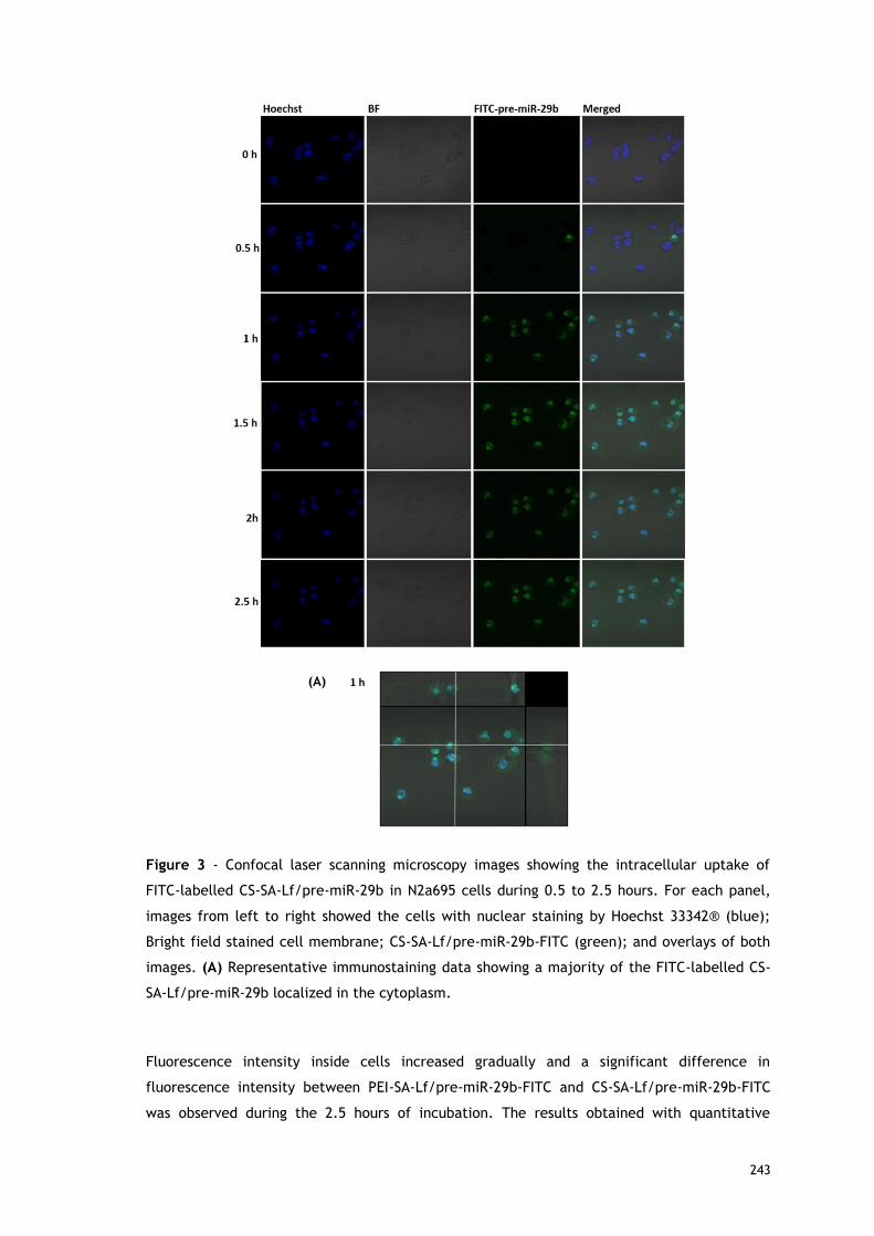
243
Figure 3 - Confocal laser scanning microscopy images showing the intracellular uptake of
FITC-labelled CS-SA-Lf/pre-miR-29b in N2a695 cells during 0.5 to 2.5 hours. For each panel,
images from left to right showed the cells with nuclear staining by Hoechst 33342® (blue);
Bright field stained cell membrane; CS-SA-Lf/pre-miR-29b-FITC (green); and overlays of both
images. (A) Representative immunostaining data showing a majority of the FITC-labelled CS-
SA-Lf/pre-miR-29b localized in the cytoplasm.
Fluorescence intensity inside cells increased gradually and a significant difference in
fluorescence intensity between PEI-SA-Lf/pre-miR-29b-FITC and CS-SA-Lf/pre-miR-29b-FITC
was observed during the 2.5 hours of incubation. The results obtained with quantitative
(A)

244
analysis were extremely similar to those obtained with fluorescence imaging. Interestingly, no
significant differences were found in the mean fluorescence intensity at 1.5 or 2.5 hours,
indicating that a plateau was achieved after 1.5 hours of incubation, in the cells transfected
with CS-SA-Lf (data not shown).
Figure 4 - Confocal laser scanning microscopy images showing the intracellular uptake of
FITC-labelled PEI-SA-Lf/pre-miR-29b in N2a695 cells during 0.5 to 2.5 hours. For each panel,
images from left to right showed the cells with nuclear staining by Hoechst 33342® (blue);
Bright field stained cell membrane; PEI-SA-Lf/pre-miR-29b -FITC (green); and overlays of both
images. (A) Representative immunostaining data showing a majority of the FITC-labelled PEI-
SA-Lf/pre-miR-29b localized in the cytoplasm.
(A)

245
In contrast, the mean fluorescence intensity of the PEI-SA-Lf/pre-miR-29b complexes
increased during the 2.5 hours of incubation, while CS-SA-Lf/pre-miR-29b increased until 1.5
hours of incubation and then kept stable until the end of transfection period (data not
shown). These results indicate that the release of pre-miR-29b from the CS-SA-Lf complexes
was faster than that from the PEI-SA-Lf.
3.4. Evaluation of human BACE1 gene knockdown induced by pre-miR-29b-loaded
complexes
To investigate the effect of recombinant pre-miR-29b on hBACE1 mRNA expression levels, RT-
qPCR measurements were performed. To accomplish this purpose, N2a695 cells were
transfected with CS-SA-Lf/pre-miR-29b, PEI-SA-Lf/pre-miR-29b and Lipo/pre-miR-29b with
8.72 nM of the target miRNA. After 72 hours of transfection, it was verified that the
overexpression of recombinant pre-miR-29b induces a notably decrease in the endogenous
hBACE1 mRNA levels, in comparison with the untreated cells (Fig. 5). In particular, hBACE1
mRNA expression was decreased by approximately 77% in cells transfected with CS-SA-Lf/pre-
miR-29b, and by 69% in those transfected with PEI-SA-Lf/pre-miR-29b complexes, when
compared with untreated cells (Fig. 5). This reduction on mRNA hBACE1 expression was also
significantly higher than the silencing (around 40%) achieved in cells transfected with
Lipo/pre-miR-29b, the positive control (Fig. 5). Thus, in comparison to Lipofectamine 2000,
CS-SA-Lf demonstrated a higher efficacy in delivering pre-miR-29b once it was achieved a 2
fold reduction of BACE1 mRNA levels.
Figure 5 - In vitro gene silencing effect of recombinant pre-miR-29b on hBACE1 mRNA levels
in N2a695 cells following 72 hours treatment with CS-SA-Lf/pre-miR-29b, PEI-SA-Lf/pre-miR-
29b and Lipo/pre-miR-29b. Values in the graph are mean from triplicates of RT-qPCR
threshold cycles for hBACE1 mRNA normalized to those of mRNA for GAPDH from 3
independent experiments and demonstrate significant differences across treatment
conditions. ANOVA, mean±SD.

246
Although the functionalized CS presents a slightly lower encapsulation efficiency (86%)
compared with the unmodified CS (90%), it shows a higher transfection efficiency, and
consequently leads to higher hBACE inhibition, very similar to those obtained for the
unmodified CS (77% to CS-SA-Lf and 76.4% to CS). These results show that the
functionalization did not affect the delivery of recombinant pre-miR-29b to the cells and its
biological activity continues to exceed the synthetic miR-29b. Furthermore, the PEI
functionalized demonstrates a decrease in the inhibition of hBACE1 (69%) compared to the
unmodified PEI (78%) what may be due to their structural characteristics after
functionalization.
3.5. Permeability across the BBB using CS-SA/pre-miR-29b, PEI-SA/pre-miR-29b,
CS-SA-Lf/pre-miR-29b and PEI-SA-Lf/pre-miR-29b
In order to evaluate the ability of all the systems to cross the BBB, in vitro BBB model was
established using RBE4 cells [49], which were transfected with CS-SA/pre-miR-29b-FITC, CS-
SA-Lf/pre-miR-29b-FITC, PEI-SA/pre-miR-29b-FITC and PEI-SA-Lf/pre-miR-29b-FITC. The
fluorescence intensity in the inner and outer chambers of the transwell was determined, in
order to evaluate the targeting effect of Lf and SA in the CS and PEI polymers to cross the
BBB. Fig. 6A shows the transport ability of different complexes to cross BBB model at the
same concentration of pre-miR-29b. After 4 hours of incubation, the transport ratios were of
about 12.28, 41.61, 64.56 and 64.93% for PEI-SA, PEI-SA-Lf, CS-SA and CS-SA-Lf, respectively.
These results indicated that the dual-targeting complexes have enhanced transport ability to
cross the BBB compared to the complexes with only one targeting group. Fig. 6A shows also
the immunostaining microphotographs of CS-SA-Lf and PEI-SA-Lf internalized in RBE4 cells. As
revealed in Fig. 6A, the strong green intensity in cytoplasm of RBE4 cells suggested that
complexes were internalized by endocytosis through the cells. Z310 cells were used as in
vitro blood-cerebrospinal fluid barrier model for evaluating the dual-targeting effects (Fig.
6B).

247
Figure 6 - The transport ratio of pre-miR-29b across the BBB during 4 hours in RBE4 cells (A)
and Z310 cells (B). Data were presented as the mean ± standard deviation. (A) Intracellular
trafficking in RBE4 cells after treating with CS-SA-Lf/pre-miR-29b-FITC and PEI-SA-LF/pre-
miR-29b-FITC. The fluorescence signals were collected by LSCM with three channels: blue
fluorescence from nuclei stained with Hoechst 33258, green fluorescence from FITC labeled
pre-miR-29b and the merged images of three channels.
With the same incubation time and pre-miR-29b concentration, the different formulations
were internalized by Z310 cells in the following order: CS-SA-Lf/pre-miR-29b > PEI-SA-Lf/pre-
miR-29b, with transport ratios of 65.49% and 48.08%, respectively. These results are in
agreement with the BBB transportation result. These findings demonstrated that the dual
targeting complexes (PEI/CS-SA-Lf) could mediate the recognition via Lf receptors and fatty
acid transporter(s)/low-density lipoprotein receptor expressed by RBE4 and Z310 cells,
allowing the transport of exogenous pre-miR-29b into the brain, probably via the pathway of
receptor-mediated transcytosis (RMT). In addition, Lf is a competent protein that intensifies
the interaction between complexes and RBE4 and Z310 cells. On the other hand, these results
can also be explained due to the positive charge of the cationic polymers, which could
promote electrostatic interactions with the negatively charged RBE4 and Z310 cell
membranes, activating the cellular uptake through the pathway of adsorptive-mediated
transcytosis (AMT) [38]. The internalization of CS-SA and CS-SA-Lf by RBE4 cells was higher
than that of PEI-SA and PEI-SA-Lf. Thus, it was found that the chitosan improved the delivery

248
efficacy both in terms of targeting at an earlier time point as well as in the accumulation in
cells. These results can be related to some characteristics of CS that can transiently open
tight junctions, enabling the transport of components via a paracellular pathway through the
epithelial barrier.
Conclusion
In conclusion, the present in vitro study demonstrated that CS-SA-Lf and PEI-SA-Lf could
efficiently cross the BBB and thus, deliver the recombinant pre-miR-29b as a therapeutic
agent to the neuronal cells. The pre-miR-29b induced the inhibition of BACE1 mRNA.
Specifically, the in vitro cellular uptake study showed that lactoferrin and stearic acid
conjugated with chitosan are highly efficient in cell transfection (about 65%) and pre-miRNA
intracellular delivery, and consequently, in the silencing of the BACE1 mRNA in comparison to
PEI-SA-Lf. These results suggest that chitosan conjugated with stearic acid and lactoferrin can
represent a potentially promising and interesting therapeutic strategy for targeting
recombinant pre-miR-29b to neuronal cells, aiming the development of effective strategies to
treat AD.
Acknowledgments
This work was supported by the Portuguese Foundation for Science and Technology (FCT),
through the project EXPL/BBB-BIO/1056/2012 and Pest-OE/SAU/UI0709/2014. Patrícia Pereira
acknowledges the fellowship (Ph.D. Grant, Ref SFRH/BD/81914/2011), from FCT. Joana
Tomás also acknowledges the fellowship in the ambit of the project CENTRO-07-ST24-FEDER-
002015. The authors also acknowledge the program Fundo Europeu de Desenvolvimento
Regional (FEDER) [COMPETE [FCOMP-01-0124-FEDER-027560]]. The authors would like to thank
Professor Yo Kikuchi (Division of Life Science and Biotechnology, Department of Ecological
Engineering, Toyohashi University of Technology) for kindly provided of pBHSR1-RM plasmid
and Professor Wenjie Luo for the kind contribution regarding the cell line N2a695.

249
References
[1] Isik AT (2010) Late onset Alzheimer's disease in older people. Clin Interv Aging 5, 307-311.
[2] Blennow K, de Leon MJ, Zetterberg H (2006) Alzheimer's disease. Lancet 368, 387-403.
[3] Hardy, J, Selkoe, DJ (2002) The amyloid hypothesis of Alzheimer's disease: progress and
problems on the road to therapeutics. Science 297, 353-356.
[4] Mullane K, Williams M (2013) Alzheimer's therapeutics: continued clinical failures question
the validity of the amyloid hypothesis-but what lies beyond? Biochem Pharmacol 85, 289-305.
[5] Glenner GG (1989a) Amyloid beta protein and the basis for Alzheimer's disease. Prog Clin
Biol Res 317, 857-868.
[6] Kim DH, Yeo SH, Park JM, Choi JY, Lee TH, et al. (2014) Genetic markers for diagnosis and
pathogenesis of Alzheimer's disease. Gene 545, 185-193.
[7] Ghiso J, Frangione B (2002) Amyloidosis and Alzheimer's disease. Adv Drug Deliv Rev 54,
1539-1551.
[8] Provost P (2010a) Interpretation and applicability of microRNA data to the context of
Alzheimer's and age-related diseases. Aging 2, 166-169.
[9] Schonrock N, Matamales M, Ittner LM, Gotz J (2012) MicroRNA networks surrounding APP
and amyloid-beta metabolism-implications for Alzheimer's disease. Exp Neurol 235, 447-454.
[10] Vassar R, Kovacs DM, Yan R, Wong PC (2009) The beta-secretase enzyme BACE in health
and Alzheimer's disease: regulation, cell biology, function, and therapeutic potential. J
Neurosci 29, 12787-12794.
[11] Cole SL, Vassar R (2007) The Alzheimer's disease beta-secretase enzyme, BACE1. Mol
Neurodegener 2, 22
[12] Hunt CE, Turner AJ (2009) Cell biology, regulation and inhibition of beta-secretase
(BACE-1). FEBS J 276, 1845-59
[13] Zong Y, Wang H, Dong W, Quan X, Zhu H, et al. (2011) miR-29c regulates BACE1 protein
expression. Brain Res 1395, 108-115.
[14] Hebert SS, Horre K, Nicolai L, Papadopoulou AS, Mandemakers W, et al. (2008) Loss of
microRNA cluster miR-29a/b-1 in sporadic Alzheimer's disease correlates with increased
BACE1/beta-secretase expression. Proc Natl Acad Sci U S A 105, 6415-6420.
[15] Dunkel P, Chai CL, Sperlagh B, Huleatt PB, Matyus P (2012) Clinical utility of
neuroprotective agents in neurodegenerative diseases: current status of drug development for
Alzheimer's, Parkinson's and Huntington's diseases, and amyotrophic lateral sclerosis. Expert
Opin Investig Drugs 21, 1267-1308.
[16] Suehs BT, Davis CD, Alvir J, van Amerongen D, Pharmd NC, et al. (2013) The clinical and
economic burden of newly diagnosed Alzheimer's disease in a medicare advantage population.
Am J Alzheimers Dis other Demen 28, 384-392.

250
[17] Qiu C, Kivipelto M, von Strauss E (2009) Epidemiology of Alzheimer's disease: occurrence,
determinants, and strategies toward intervention. Dialogues Clin Neuros 11, 111-128.
[18] Edwards RH (2001) Drug delivery via the blood-brain barrier. Nat Neurosci 4, 221-2.
[19] Alam MI, Beg S, Samad A, Baboota S, Kohli K, et al. (2010) Strategy for effective brain
drug delivery. Eur J Pharm Sci 40, 385-403.
[20] Egleton RD, Davis TP (1997) Bioavailability and transport of peptides and peptide drugs
into the brain. Peptides 18,1431-9.
[21] Pangalos MN, Schechter LE, Hurko O (2007) Drug development for CNS disorders:
strategies for balancing risk and reducing attrition. Nat Rev Drug Discov 6, 521-32.
[22] Pardridge WM (2005) The blood-brain barrier: bottleneck in brain drug development.
NeuroRx 2, 3-14.
[23] Kusuhara H, Sugiyama Y (2001) Efflux transport systems for drugs at the blood-brain
barrier and blood-cerebrospinal fluid barrier (Part 1). Drug Discov Today 6,150-6.
[24] Descamps L, Dehouck MP, Torpier G, Cecchelli R (1996) Receptor-mediated transcytosis
of transferrin through blood-brain barrier endothelial cells. Am J Physiol 270, H1149-58.
[25] Visser CC, Stevanovic S, Heleen Voorwinden L, Gaillard PJ, Crommelin DJ, et al. (2004)
Validation of the transferrin receptor for drug targeting to brain capillary endothelial cells in
vitro. J Drug Target 12, 145-50.
[26] Fillebeen C, Descamps L, Dehouck MP, Fenart L, Benaissa M, et al. (1999) Receptor-
mediated transcytosis of lactoferrin through the blood-brain barrier. J Biol Chem 274, 7011-7.
[27] Duffy KR, Pardridge WM (1987) Blood-brain barrier transcytosis of insulin in developing
rabbits. Brain Res 420, 32-8.
[28] Reinhardt RR, Bondy CA (1994) Insulin-like growth factors cross the blood-brain barrier.
Endocrinology 135, 1753-61.
[29] Candela P, Gosselet F, Miller F, Buee-Scherrer V, Torpier G, et al. (2008) Physiological
pathway for low-density lipoproteins across the blood-brain barrier: transcytosis through brain
capillary endothelial cells in vitro. Endothelium 15, 254-64.
[30] Dehouck B, Fenart L, Dehouck MP, Pierce A, Torpier G, et al. (1997) A new function for
the LDL receptor: transcytosis of LDL across the blood-brain barrier. J Cell Biol 138, 877-89.
[31] Edmond J (2001) Essential polyunsaturated fatty acids and the barrier to the brain. J Mol
Neurosci 2, 181-193
[32]Kabanov AV, Batrakova EV, Alakhov VY. (2002) Pluronic block copolymers as novel
polymer therapeutics for drug and gene delivery. J Control Release 82, 189-212.
[33] Chen J, Xie J (2012) Progress on RNAi-based molecular medicines. Int J Nanomedicine 7,
3971-80.
[34] Gao K, Huang L (2009) Nonviral Methods for siRNA Delivery. Mol Pharm 6, 651-8.

251
[35] Morille M, Passirani C, Vonarbourg A, Clavreul A, Benoit JP (2008) Progress in developing
cationic vectors for non-viral systemic gene therapy against cancer. Biomaterials 29, 3477-96.
[36] Yin H, Kanasty RL, Eltoukhy AA, Vegas AJ, Dorkin JR, et al. (2014) Non-viral vectors for
gene-based therapy. Nat Rev Genetics 15, 541-55.
[37] Zhang Y, Wang Z, Gemeinhart RA (2013) Progress in microRNA delivery. J Control Release
172, 962-74.
[38] Xie Y-T, Du YZ, Yuan H, Hu F-Q (2012) Brain-targeting study of stearic acid-grafted
chitosan micelle drug-delivery system. Int J Nanomedicine 7, 3235-3244.
[39] Huwyler J, Wu D, Pardridge WM (1996) Brain drug delivery of small molecules using
immunoliposomes. Proc. Natl. Acad. Sci. U.S.A. 93, 14164–14169.
[40] Swami A, Goyal R, Tripathi SK, Singh N, Katiyar N, et al. (2009) Effect of
homobifunctional crosslinkers on nucleic acids delivery ability of PEI nanoparticles. Int J
Pharm 374(1–2), 125–38.
[41] Sarvaiya J, Agrawal YK (2015) Chitosan as a suitable nanocarrier material for anti-
Alzheimer drug delivery. Int J Biol Macromol 72, 454-465.
[42] Ward PP, Paz E, Conneely OM (2005) Multifunctional roles of lactoferrin: a critical
overview. Cell Mol Life Sci 62, 2540–2548.
[43] Huang RQ, KeWL, Qu YH, Zhu JH, Pei YY, et al. (2007) Characterization of lactoferrin
receptor in brain endothelial capillary cells and mouse brain. J Biomed Sci 14, 121–128.
[44] Ji B, Maeda A, Higuchi M, Inoue K, Akita H, et al. (2006) Pharmacokinetics and brain
uptake of lactoferrin in rats. Life Sci 78, 851–855.
[45] Huang R, Ke W, Han L, Liu Y, Shao K, et al. (2009) Brain-targeting mechanisms of
lactoderrin-modified DNA-loaded nanoparticles. J Cerebral Blood Flow & Metabolism 29,
1914-1923.
[46] Pereira P, Sousa A, Queiroz JA, Correia I, Figueiras A, et al. (2014) Purification of pre-
miR-29 by arginine-affinity chromatography. J Chrom B Analyt Technol Biomed Life Sci 16-23,
951-952.
[47] Pereira P, Jorge A, Martins R., Pais ACCC, Sousa F, et al. (2012) Characterization of
polyplexes involving small RNA. J Colloid Interface Sci 387, 84-94.
[48] Shi LZ, Li GJ, Wang S, Zheng W (2008) Use of Z310 cells as an in vitro blood-cerebrospinal
fluid barrier model: Tight junction proteins and transport properties. Toxicology in Vitro 22,
190-199
[49] Balbuena P, Li W, Ehrich M (2011) Assessments of tight junction proteins occludin,
claudin 5 and scaffold proteins ZO1 and ZO2 in endothelial cells of the rat blood-brain barrier:
Cellular responses to neurotoxicants malathion and lead acetate. NeuroToxicology 32, 58-67
[50] Re F, Cambianica I, Zona C, Sesana S, Gregori M, et al. (2011) Functionalization of
liposomes with ApoE-derived peptides at different density affects cellular uptake and drug
transport across a blood-brain barrier model. Nanomed: Nanotech, Biol, Med 7, 551-559

252

253
Chapter 4

254

255
Concluding remarks
Over the recent years, the study and application of miRNAs has been considered useful and
promising for the diagnosis and treatment of several diseases, in particular Alzheimer's
disease. Most miRNAs employed in the development of these therapeutic approaches are
produced either by enzymatic or chemical synthesis methods. Although, the synthesis process
is efficient for large scale production, several limitations have been found regarding the use
of synthetic miRNAs, once the isolation of these formulations are based on methods that
employ chemical solvents and denaturing agents. Consequently, the final product is not
suitable to be biologically applied, because of the health risks associated, and thus strongly
compromise the success of several RNA based-procedures in the clinical field. Aiming to
overcome some of these limitations, this project was centered on the establishment of a new
strategy to biosynthesize and to purify the recombinant pre-miR-29b, also focusing on the
preparation of suitable non-viral systems to efficiently deliver the pre-miR-29b to the cell. In
addition, it was demonstrated the efficiency of the recombinant pre-miR-29b in BACE1
knockdown, using in vitro neuronal cells lines.
The recombinant production of pre-miR-29b was performed using the bacterium R.
sulfidophilum DSM 1374, harboring a plasmid encoding for human pre-mir-29b. Regarding the
growth conditions, and conversely to what was previously described for this bacterium, it was
possible to develop an original approach for the aerobic growth of R. sulfidophilum, which
resulted in a cellular growth improvement (higher growth rates, higher optical densities and a
shorter cultivation time) followed by an enhanced production of intracellular (about 358
μg/L) and extracellular (approximately 182 µg/L) pre-miR-29b. In addition, the results
indicated that the extracellular medium is not highly contaminated with genomic DNA,
proteins or endotoxins, which is an important parameter concerning the integrity, stability
and activity of the pre-miRNA (Paper III). In general, the use of this alternative strategy,
based on the secretion of the pre-miRNA, avoids time-consuming and laborious RNA extraction
methods that frequently induce RNA denaturation and, simultaneously suppresses the use of
organic solvents and extremely toxic chemicals which are biologically hazardous. Despite this
great advantage, in the course of this thesis it was difficult to recover the pre-miRNA from
the extracellular medium, due to the high dilution factor, and the purification experiments
were developed by using the RNAs recovered from the cells.
Considering the previous knowledge that pre-miR-29b can be successfully obtained by a
simple, fast and economic process using the R. sulfidophilum host, it is required the
development and implementation of an adequate and efficient purification method that
allows to overcome the challenges of the already existing chromatographic techniques, for
the purification of miRNAs. In this project, it is suggested that amino acid-based affinity
chromatography can be considered as a promising chromatographic strategy, to specifically

256
and efficiently purify the pre-miR-29b, eliminating the host impurities (small RNAs, proteins
and endotoxins) by exploiting the specific interactions occurring between the L-lysine and L-
arginine ligands and the pre-miRNA. Thus, it was developed an initial study to characterize
the binding of pre-miR-29b onto the amino acids-based agarose supports using Surface
Plasmon Resonance (Paper IV). The understanding of the conditions favoring these
interactions was essential to control, manipulate, determine and establish the selective
binding/elution chromatographic conditions (such as the composition and ionic strength of
elution buffer and temperature), in order to improve the binding specificity of the pre-miR-
29b onto the amino acids-based supports, preserving their structural stability and integrity.
Considering the equilibrium dissociation constant, it was verified that the recombinant pre-
miR-29b binds more strongly to L-arginine (KD between 10-6 and 10-7 M) than to L-lysine (KD
between 10-5 and 10-7 M). This study also disclosed that the structure of the pre-miR-29b is
sensitive to changes in the physical and chemical environmental conditions. In general, the
results obtained provide valuable information for the optimization and implementation of L-
lysine and L-arginine amino acids as chromatographic ligands, in order to improve the
efficiency of pre-miR-29b purification.
Lysine-agarose matrix was the first support to be tested for the purification of pre-miR-29b.
Due the presence of the amine groups in its structure, it is suggested that the main
interactions established between nucleic acids and the L-lysine amino acid are electrostatic.
Thus, initial experiments were performed to choose the best binding/elution conditions for
pre-miR-29b, using sodium chloride gradients. However, under these conditions the RNA
binding was not effective and the selective purification of pre-miRNA was not achieved.
Therefore, the retention behavior of pre-miR-29b was evaluated using an ammonium sulfate
stepwise gradient (Paper V), in order to achieve higher specificity. Despite the effectiveness
of lysine-based methodology in the purification of pre-miR-29b, the requirement for high salt
concentration can be seen as a disadvantage, especially with regard to biotechnological and
clinical applications.
To overcome this limitation and to improve pre-miR-29b purification, the arginine-agarose
matrix was used. The application of arginine as specific ligand allowed the pre-miR-29b
recovery under mild salt conditions, using sodium chloride gradients, showing that this matrix
can be a good alternative to purify pre-miR-29b (Paper VI). The arginine ligand allowed the
purification of the pre-miR-29b using three different elution strategies, namely by using
sodium chloride (90% of purity and 97% of yield using a low salt concentration) and ammonium
sulfate (33% of purity and requires high salt concentrations) gradients, or by establishing a
competitive elution strategy with arginine (98% of purity and 57% of yield). Although
electrostatic interactions are most prevalent in the arginine matrix, it was verified the
presence of other interactions due the binding and elution conditions used. Moreover, the
quality control analysis revealed high integrity of pre-miR-29b preparations as well as high
purity degree and recovery yields, eliminating R. sulfidophilum impurities (proteins and

257
endotoxins) as well as other species of small RNAs, which can origin adverse effects and
inflammatory response to the patients.
Taking into account that the structure of the chromatographic supports is continuously
optimized to afford rapid and efficient separations, it was also tested a monolithic support to
purify the pre-miR-29b, in order to overcome the limitations associated with the conventional
supports in the purification of miRNAs. The application of the agmatine monolithic support for
pre-miR-29b purification revealed a specific recognition of the pre-miR-29b that was totally
isolated from other small RNAs, which represented an advantage to obtain highly pure RNA in
a short time (Paper VII). These results were useful for the implementation of a new affinity
chromatographic strategy using monoliths immobilized with specific ligands, such as
agmatine, to efficiently and selectively purify the pre-miR-29b. In addition, this strategy
allowed an efficient pre-miR-29b separation without flow rate dependence using similar
binding and elution strategies to those used with arginine-agarose. With this study it was
verified that the dynamic binding capacity for RNA molecules is dependent of the feed
concentration and the characterization of the modified monolith revealed that a maximum
binding capacity of 8.08 mg/mL was reached to a RNA concentration of 0.25 mg/mL.
Moreover, the dissociation constant value obtained was 2.6x10⁻⁷ M, revealing a good affinity
interaction between the agmatine ligand and pre-miR-29b. The quality control analysis in the
final pre-miR-29b sample revealed that the removal of host impurities (proteins and small
RNAs) was efficient. In fact, this new monolithic support arises as a powerful instrument on
the miRNA purification to be used in further clinical applications, providing a more rapid and
economical purification platform, resulting in a short contact time with pre-miR-29b
molecules, which preserves the structural stability and integrity of target molecules.
Thus, the results and conclusions obtained in these studies suggest that the mechanism
inherent to the purification of the pre-miR-29b results from the occurrence of multiple non-
covalent interactions, including electrostatic, hydrophobic, hydrogen bonds, van der Waals
forces and cation–π interactions, which combined induce a specific biorecognition of the pre-
miR-29b by arginine, lysine and agmatine amino acids. In general, the application of amino
acids-based affinity chromatography allowed the selective, efficient and specific isolation of
the target pre-miRNA from the complex biological mixture of other sRNAs with high recovery
yield, integrity and good purity, demonstrating thus the potential and versatility of these
ligands in the purification of pre-miR-29b, in a single unit operation. Furthermore, the
purification of the pre-miR-29b using the amino acids-based affinity chromatography showed
several improvements over the currently used purification methods, thus proving to be an
interesting option for the downstream processing of miRNA therapeutic products.
After the purification of the target pre-miRNA, the project was focused on the development
of delivery systems able to encapsulate, protect and promote the delivery of pre-miR-29b to
the target cells. Initially, as described in paper VIII, a systematic study of three polymers,

258
polyethylenimine (PEI), chitosan (CS) and poly(allylamine), with different structural
characteristics (such as molecular weights, charge densities and backbone structure) was
performed, in order to design a successful non-viral delivery system for small RNAs. The
polymers of PEI and CS stood out in the condensation of small RNA as well as in relation to its
physicochemical characteristics, namely size, zeta potential, morphology, encapsulation
efficiency and complex stability.
In the previous papers it was possible to develop efficient methodologies to recover pre-miR-
29b with a high purity degree, in particular with arginine-agarose matrix. Thus, in the paper
IX, is described how the recombinant pre-miR-29b was condensed and some systems were
prepared and characterized to encapsulate and protect the pre-miR-29b activity. All of the
polyplexes demonstrated high loading capacity, small sizes and exhibited a strong positive
charge on their surface. Moreover, the results showed that the levels of human BACE1 protein
expression were significantly decreased, by approximately 80%, in cells transfected with
polyplexes loaded with recombinant pre-miR-29b relatively to untreated cells and cells
treated with scrambled RNA and miR-29 synthetic. Consequently, endogenous Aβ42 levels
were also significantly reduced in about 45%. It is important to highlight that the therapeutic
effect of pre-miR-29b in BACE1 mRNA silencing was also achieved, with a mRNA reduction of
approximately 77% in cells transfected with CS-SA-Lf/pre-miR-29b and 69% in those
transfected by PEI-SA-Lf/pre-miR-29b. Overall, these results showed that the miR-29b can
induce the suppression of the expression levels of Aβ peptides in neuronal cells, showing a
causal relationship between miR-29b expression and BACE1 activity, and, consequently, Aβ
generation. Considering the application of the pre-miR-29b in the brain, the delivery systems
previously characterized were functionalized with ligands, to facilitate their access and
interaction with specific molecules expressed on the surface of BBB endothelial cells. In the
paper X, it was demonstrated that CS functionalized with lactoferrin and stearic acid cross
the BBB quickly and with high transfection efficiency, causing an increase of the pre-miRNAs
concentration in the brain.
According to the results obtained in this doctoral research work and comparing with
previously published data, it was developed a biotechnological platform that allows
biosynthesis, purification and transfection of the recombinant pre-miR-29b using polyplexes.
With the successful implementation of this methodology it was obtained the highest decrease
ever reported for the hBACE1 and endogenous Aβ42 expression levels in AD model cells.
Overall, the development and implementation of these cutting-edge approaches allows
obtaining high quantities of pure pre-miR-29b, in its biologically active form, for further
application in many molecular biology subjects. This approach also provides the basis for the
improvement of the currently available methodologies of microRNA-based therapeutics, not
only for neurological disorders but also for other diseases, and can be translated to additional
therapeutic targets that may be of potential clinical interest in the future.

259
Future trends
Overall, it can be asserted that the main aims of this research work were fulfilled. However
several challenging questions arisen that can be addressed as future perspectives. The
integration of innovative affinity chromatographic strategies in the global bioprocess of pre-
miR-29b biosynthesis and purification will be required for removing the host contaminants
from the extracellular pre-miR-29b. Thus, the development of a selective strategy for
efficiently isolate the extracellular pre-miR-29b will allow recovering this target miRNA
directly from the culture medium without cells lysis and, consequently, without employing
organic solvents and other harmful reagents.
Following the work already performed, more studies can be developed to motivate the
improvement of the already implemented chromatographic techniques or the establishment
of new purification approaches. In this way, in order to improve the specificity, selectivity
and throughput of the bioseparation methodology for purification of the target pre-miRNAs,
other affinity purification strategies can be implemented, using other immobilized amino
acids, its derivatives or peptides or even testing other specific ligands like complementary
nucleotide sequences to pre-miRNAs. These ligands could be immobilized onto different
chromatographic matrices, such as agarose-based and monoliths to purify pre-miRNAs from
the constituents of culture medium, expecting the improvement of the process performance,
namely considering the purification degree and recovery yield.
Likewise, it is extremely necessary to understand the phenomenon of biorecognition, since
the identification of the interactions involved in the isolation of pre-miRNA can be very
important, allowing the control and manipulation of the chromatographic conditions
responsible for the recognition of the pre-miRNA molecules. Accordingly, to characterize the
binding recognition, as well as the thermodynamic parameters underlying the main
interaction mechanism between pre-miRNAs and the different affinity ligands, and to
determine the magnitude of kinetics and/or affinity constants, it could be used Nuclear
Magnetic Resonance (NMR) spectroscopy, Flow Microcalorimetry (FMC) and Surface Plasmon
Resonance (SPR), respectively. Furthermore, in order to fully characterize and compare the
novel supports, dynamic binding capacity studies with target pre-miRNAs can be performed
under different operation conditions (e.g. feed concentration, flow rate, pH and salt
concentration), allowing the establishment of the best ligands and conditions to purify the
target pre-miRNAs.
On the other hand, to improve the delivery of pre-miRNA therapeutics to the cells and the
cellular response, different delivery systems based on polymers, such as polymeric
nanoparticles (e.g. Poly(lactic-co-glycolic acid)) and poloxamer micelles (Pluronic® block
copolymers) can be developed and characterized. Moreover, as these systems are so
260
versatile, they can be functionalized with brain-targeting molecules, either on their surface
or on the polymeric network, to be recognized by cell surface receptors of BBB, and promote
targeting to specific cells and tissues.
With the successful implementation of these methodologies, it could be further evaluated the
therapeutic function of the recombinant pre-miRNA in in vivo studies and test its efficacy in
improving cognition and biochemical hallmarks of disease, using mice models of AD (3xTg and
APPswe/PSEN1ΔE9). To accomplish this objective, several studies are required to establish
the range of doses and administration routes to be employed in the pharmacological studies
(biodistribution/pharmacokinetics and toxicity studies). Finally, it could also be evaluated
and compared the performance of recombinant pre-miRNAs and conventional therapeutic
drugs. These studies should be first performed in vitro, but the development of some in vivo
studies could bring important insights about the efficacy and safety of gene therapy, either
used separately or in combination with conventional drugs, opening a new prospect of cure,
due to the possible synergistic effect.