Embed Size (px)
Citation preview

2130
diagnosis and treatment of GC, the current tre-atments available to patients with advanced GC are very limited because of the distant metasta-sis. Thus, understanding the precise molecular mechanisms underlying the tumorigenesis and progression of gastric cancer is urgently needed and can provide the basis for the development of novel therapeutic strategies.
MicroRNAs (miRNAs) are endogenous non-coding small RNAs, roughly 18-25 nucleo-tides in length that that can down-regulate gene expression by targeting the 3’-UTR region of specific mRNA sequences5-7. Besides, it is clo-sely related to the development and progression of tumor and plays an important role in the proliferation, apoptosis, metastasis of tumor8,9. miRNAs can function as either oncogenes or tumor suppressors according to the roles of their target genes. For instance, Zhao et al10 showed that miR-874 was significantly down-regulated in colorectal cancer tissues, and ove-rexpression of miR-874 suppressed cell growth and induced apoptosis in colorectal cancer cel-ls by targeting STAT3. Zhang et al11 reported that miR-198 inhibited tumorous behaviors of human osteosarcoma through directly targeting ROCK1. Zhao et al12 found that up-regulated expression of miR-494 could inhibit GC cells proliferation, migration, and invasion by tar-geting IGF-1R. These findings suggested that miRNAs potentially serve as therapeutic tar-gets for novel treatment strategies against tu-mors, including GC.
In the current work, we detected the expres-sion of miR-425-5p in GC tissues and cell lines. We further investigated the role of miR-425-5p in the metastasis of GC cells. Fur-thermore, we investigated the exact roles of the miR-425-5p and its underlying molecular mechanisms in GC.
Abstract. – OBJECTIVE: MicroRNAs (miRNAs) have emerged as important gene regulators and are recognized as key players in carcinogenesis. The present study investigated the role of miR-425-5p in the development and progression of gastric cancer (GC).
PATIENTS AND METHODS: The miR-425-5p level in GC tissues and cells was assayed by qRT-PCR. Then, the effects of miR-425-5p ex-pression on the biological behavior of GC cells were investigated. Analysis of target protein ex-pression was determined by Western blotting. Bioinformatic prediction and luciferase assays were employed to identify the predicted miRNA which regulates CYLD.
RESULTS: miR-425-5p was found to be up-reg-ulated in GC tissues and cell lines. Knockdown of miR-425-5p in GC cells attenuated migration and invasion of GC cells, whereas overexpression of miR-425-5p promoted cell migration and invasion. The luciferase assay demonstrated that CYLD was a direct target of miR-425-5p. Furthermore, the miR-425-5p level was inversely correlated with lev-els of CYLD in Western blotting assay.
CONCLUSIONS: Our findings indicate that miR-425-5p may contribute to the progression of GC through a mechanism involving CYLD, sug-gesting that miR-425-5p may have the potential to be a novel important alternative therapeutic target for GC.
Key Words:miR-425-5p, Gastric cancer, CYLD, Invasion, Migration.
Introduction
Gastric cancer (GC) is the second most com-mon cause of cancer-related death and the fourth most common cancer worldwide1-3. In addition, The Chinese Cancer Registry Annual Report reported that GC is the third commonest mali-gnancy and the second leading cause of cancer death in China4. Despite improvements in the
European Review for Medical and Pharmacological Sciences 2017; 21: 2130-2136
Y.-F. YAN, F.-M. GONG, B.-S. WANG, W. ZHENG
General Surgery Department II, Chinese PLA General Hospital, Beijing, China
Yong-feng Yan and Fang-ming Gong contributed equally to this work
Corresponding Author: Wei Zheng, MD; e-mail: [email protected]
MiR-425-5p promotes tumor progression via modulation of CYLD in gastric cancer

MiR-425-5p promotes progression of gastric cancer
2131
Patients and Methods
Cell Culture and Tissue SamplesFifteen paired GC and matched normal non-tu-
mor tissues were obtained from our department. All the tissues were immediately stored in liquid nitrogen until use. Written informed consent was obtained from all the patients. All specimens were handled and made anonymous according to the ethical and legal standards. The Ethical Com-mittee of our Hospital approved the investigation.
An immortal gastric epithelial cell line, GES-1, and four GC cell lines, BGC-823, AGS, HGC-27, and MKN-45, were commercially obtained from American Type Culture Collection (ATCC, Manassas, VA, USA). The above cells were pro-pagated in Dulbecco’s Modified Eagle Medium (DMEM; Invitrogen, Carlsbad, CA, USA) sup-plemented with 10% FCS at 37°C in 5% CO2 cell culture incubator.
Cell TransfectionThe hsa-miR-425-5p mimics, hsa-miR-425-5p
inhibitor, and negative control miRNA (NC) were chemically synthesized by Shanghai GenePhar-ma Co., Ltd. (Songjiang, Shanghai, China). Lipo-fectamine 2000 (Invitrogen, Eugene, OR, USA) was used for transfection according to the manu-facturer’s protocol.
Quantitative Reverse Transcription-poly-merase Chain Reaction
Total RNA extraction and reverse transcrip-tion were performed in strict accordance with the manufacturer’s instructions. Quantitative reverse transcription-polymerase chain reaction (qRT-PCR) was performed using the PrimeScript RT Reagent Kit and SYBR Premix Ex Taq kit (TaKa-Ra Bio, Inc., Otsu, Shiga, Japan). PCR parameters for cycling were as follows: 95°C for 20 seconds, 40 cycles of PCR at 95°C for 3 seconds, and 60°C for 30 seconds. All reactions were done in a 10-mL reaction volume in triplicate. The result com-pared with U6 and GAPDH was calculated by the 2-dCt method. Primers used for Real-time PCR were designed by Primer Express 3.0 and synthe-sized in Invitrogen.
Western BlottingA total of 20 mg of protein was used for We-
stern blotting. After gels electrophoresis, sam-ples were transferred to polyvinylidene fluoride (PVDF) membranes. The membrane was blocked with 5% skimmed milk in TBST and incubated
with the antibody against CYLD (1:1000 dilution; Santa Cruz Biotechnology, Santa Cruz, CA, USA) and glyceraldehyde 3-phosphate dehydrogena-se (GAPDH, 1:5000; Santa Cruz Biotechnology, Santa Cruz, CA, USA) overnight 4°C. Second an-tibodies conjugated with horseradish peroxidase (anti-mouse IgG and anti-rabbit IgG) were used to detect primary antibodies. For HRP detection, an ECL chemiluminescence kit (CWBIO) was used. Protein quantity was detected by β-catenin as a loading control.
Wound Healing AssayThe BGC-823 cells transfected with RNA
were plated in 6-well plates. When the cells grew to full confluence, a line was scratched using a pipette tip. The cells were then washed with serum-free medium and incubated with serum-free DMEM.
Cell Migration and Invasion AssaysWe used a transwell cell migration and Ma-
trigel invasion assay to measure the migration and invasion ability of BGC-823 cell lines. For migration assay, 5×10⁴ transfected cells were placed in the upper chamber of each insert. For invasion assay, 5×10⁴ transfected cells were placed in the upper chamber of each insert co-ated with 150 mg Matrigel. The lower chamber was filled with 500 mL Roswell Park Memorial Institute-1640 (RPMI-1640) medium with 10% FBS to attract cells. After cells had been incu-bated for 36 hours at 37°C and 5% CO2, those left in the upper chamber were removed with a cotton swab. Then, cells invading cells across the membrane were counted under a light mi-croscope.
Luciferase Reporter AssaysBGC-823 cells were seeded into a 24-well pla-
te and cotransfected with miR-425-5p or control and 3′UTR-luciferase plasmids. The cells were lysed at 48 h post-transfection, and the luciferase activity was measured using the Dual-Luciferase Reporter Assay System (Promega, Haiding, Bei-jing, China), and Renilla-luciferase was used for normalization.
Statistical AnalysisExperiments were performed in triplica-
te, and the results were investigated with the SPSS 22 software (IBM, New York, NY, USA). Differences between groups were compared

Y.-F. Yan, F.-M. Gong, B.-S. Wang, W. Zheng
2132
with standard deviation followed by indepen-dent-samples t-test. p-values < 0.05 were defi-ned as significant.
Results
MiR-425-5p was up-regulated in GC tissues and cell lines
Expression levels of miR-425-5p in GC sam-ples and cells lines were analyzed by RT-PCR. As shown in Figure 1A, miR-425-5p was highly expressed in GC tissue than in normal samples. Figure 1B also showed that miR-425-5p levels were significantly increased in GC tissue com-pared with normal gastric cell lines.
Overexpression of miR-425-5p promoted BGC-823 cells invasion and migration
To reveal the biological role of miR-425-5p in migration and invasion, miR-425-5p mimic or miR-NC was transfected into BGC-823 cel-ls. The qRT-PCR analysis confirmed that tran-sfection with the miR-425-5p mimic resulted in significant overexpression of miR-425-5p (Figure 2A). Then, the effects of miR-425-5p on the invasiveness and migration of GC cells were examined by Transwell and scra-tch-wound assays, respectively. As shown in Figure 2B, the movement ability of BGC-823 cells was improved by miR-425-5p. Then, It was found that miR-425-5p over-expression
clearly promoted BGC-823 cells migration and invasion, compared with that observed in control cells (Figure 2C).
Knockdown of miR-425-5p suppressed BGC-823 cells invasion and migration
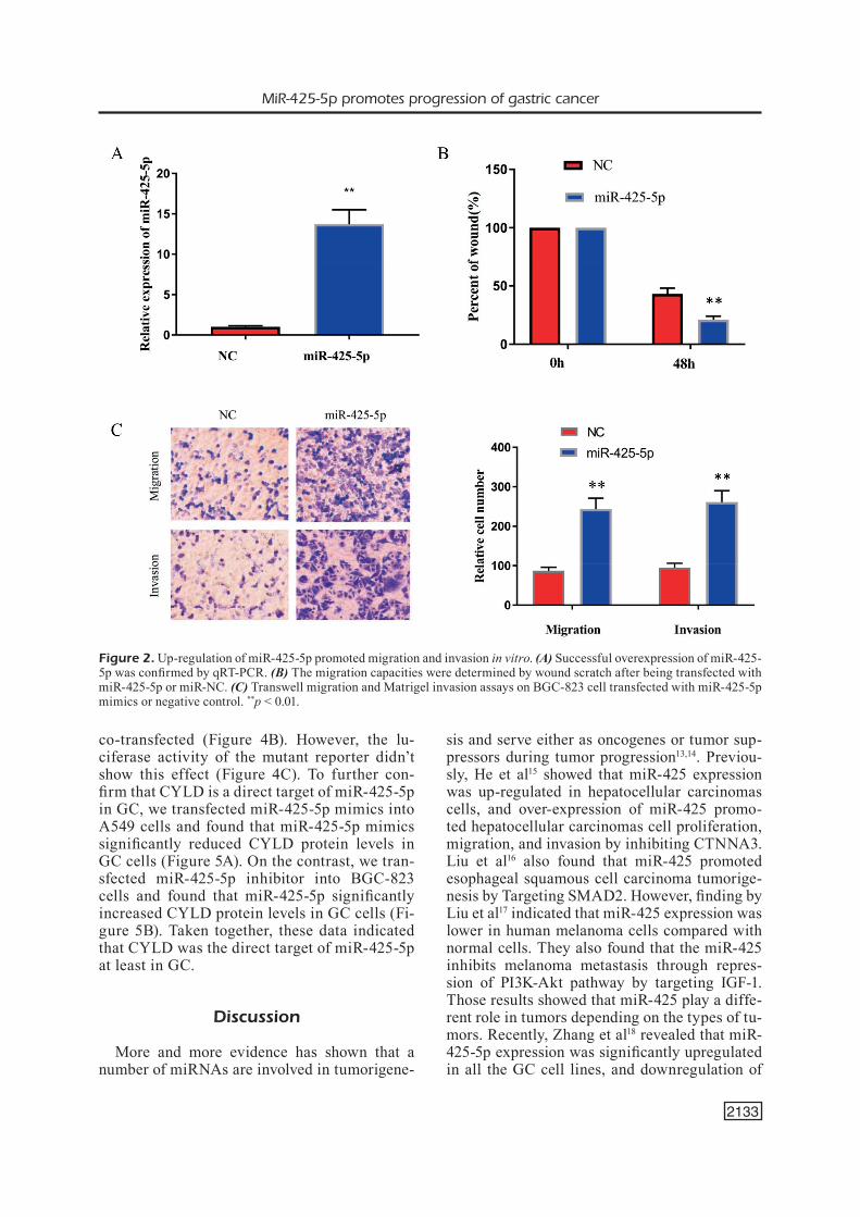
Moreover, we transfected BGC-823 cells with a miR-425-5p inhibitor to downexpress this miR-NA, then analyzed cell migration and invasion. The qRT-PCR analysis confirmed that tran-sfection with the miR-425-5p inhibitor resulted in significant down-regulation of miR-425-5p (Fi-gure 3A). Moreover, the results showed that the miR-425-5p inhibitor significantly suppressed the migration and invasion abilities of BGC-823 cells (Figure 3B-3C).
MiR-425-5p regulated CYLD expression by targeting 3′-UTR of CYLD mRNA
To explore the molecular mechanism of CYLD regulation, three bioinformatic algori-thms (TargetScan, PicTar, and miRanda) were applied to identify potential miRNAs which may involve in the regulation of CYLD. The data suggested a putative binding site for miR-425-5p in the 3’UTR of CYLD mRNA (Figu-re 4A). Thus, reporter vectors, containing lu-ciferase complementary DNA followed by the CYLD 3’UTR, were constructed (Figure 4A). The relative luciferase activity of the repor-ter containing the wild-type 3′ UTR of CYLD was significantly suppressed when 425-5p was
Figure 1. miR-425-5p expression is increased in GC tissues and cell lines. (A) Relative expression of miR-425-5p determined by quantitative PCR in the indicated cell lines. (B) Relative expression of miR-425-5p in a cohort of 15 human GC tissues and adjacent normal tissues. The endogenous U6 RNA was used as the internal control. **p < 0.01 versus normal.
MiR-425-5p promotes progression of gastric cancer
2133
co-transfected (Figure 4B). However, the lu-ciferase activity of the mutant reporter didn’t show this effect (Figure 4C). To further con-firm that CYLD is a direct target of miR-425-5p in GC, we transfected miR-425-5p mimics into A549 cells and found that miR-425-5p mimics significantly reduced CYLD protein levels in GC cells (Figure 5A). On the contrast, we tran-sfected miR-425-5p inhibitor into BGC-823 cells and found that miR-425-5p significantly increased CYLD protein levels in GC cells (Fi-gure 5B). Taken together, these data indicated that CYLD was the direct target of miR-425-5p at least in GC.
Discussion
More and more evidence has shown that a number of miRNAs are involved in tumorigene-
sis and serve either as oncogenes or tumor sup-pressors during tumor progression13,14. Previou-sly, He et al15 showed that miR-425 expression was up-regulated in hepatocellular carcinomas cells, and over-expression of miR-425 promo-ted hepatocellular carcinomas cell proliferation, migration, and invasion by inhibiting CTNNA3. Liu et al16 also found that miR-425 promoted esophageal squamous cell carcinoma tumorige-nesis by Targeting SMAD2. However, finding by Liu et al17 indicated that miR-425 expression was lower in human melanoma cells compared with normal cells. They also found that the miR-425 inhibits melanoma metastasis through repres-sion of PI3K-Akt pathway by targeting IGF-1. Those results showed that miR-425 play a diffe-rent role in tumors depending on the types of tu-mors. Recently, Zhang et al18 revealed that miR-425-5p expression was significantly upregulated in all the GC cell lines, and downregulation of
Figure 2. Up-regulation of miR-425-5p promoted migration and invasion in vitro. (A) Successful overexpression of miR-425-5p was confirmed by qRT-PCR. (B) The migration capacities were determined by wound scratch after being transfected with miR-425-5p or miR-NC. (C) Transwell migration and Matrigel invasion assays on BGC-823 cell transfected with miR-425-5p mimics or negative control. **p < 0.01.
Y.-F. Yan, F.-M. Gong, B.-S. Wang, W. Zheng
2134
miR-425-5p expression inhibited GC cell pro-liferation, invasion, and migration, suggesting that miR-425-5p function as a tumor promoter in GC. In the present work, we further explore the underlying mechanisms of miR-425-5p-induced GC cell progression.
In the present study, we confirmed that the expression of miR-425-5p was significantly hi-gher in GC tissues than in normal gastric tis-sues. Next, More importantly, miR-425-5p ove-rexpression promoted in vitro cell migration and invasion. On the contrary, downregulation of miR-425-5p suppressed in vitro cell migra-tion and invasion. These data confirmed that miR-425-5p exhibits tumor-promoting activity in GC.
CYLD has been reported to be down-regu-lated in several types of cancers including co-lorectal cancer, breast cancer, hepatocellular carcinoma and gastric cancer, suggesting that CYLD exhibits broad tumor suppressor fun-ctions19-22. Using the algorithms TargetScan, and miRanda website tools, we identified CYLD as the potential target of miR-425-5p. Furthermo-re, we performed Luciferase reporter assays and the results showed that miR-425-5p may directly target CYLD-3’UTR with the seed sequence of 47-98 sites. The result of western blot also con-firmed that over-expression of miR-425-5p could suppress the expression level of CYLD. All the above suggested that CYLD was a potential fun-ctional target of miR-425-5p.
Figure 3. Down-regulation of miR-425-5p suppressed migration and invasion in vitro. (A) Successful knockdown of miR-425-5p was confirmed by qRT-PCR. (B) The migration capacities were determined by wound scratch after being transfected with miR-425-5p inhibitor or miR-NC. (C) Transwell migration and Matrigel invasion assays on BGC-823 cell transfected with miR-425-5p inhibitor or negative control. **p < 0.01.

MiR-425-5p promotes progression of gastric cancer
2135
Conclusions
Taken together, our in vitro experimental re-sults demonstrated that miR-425-5p may target
CYLD to promote the invasion and metastasis of GC. miR-425-5p may serve as a novel therapeutic target for GC.
Figure 4. MiR-425-5p directly targets CYLD. (A) Predicted miR-425-5p target sequences in the 3’-UTRs of CYLD. (B,C) Relative luciferase activity was analyzed upon co-transfection with wild-type (WT) or mutant-type (mt) reporter plasmids and miR-425-5p, miR-425-5p inhibitor or miR-Ctrl in BGC-823 cells. **p < 0.01.
Figure 5. The effects of miR-425-5p on the expression of CYLD protein in GC cell line. (A) Levels of CYLD in BGC-823 cells. Cells were transfected with miR-425-3p mimetic or NC-miR for 24 h. Levels of CYLD were determined by Western blot. (B) Levels of CYLD in BGC-823 cells. Cells were transfected with miR-425-3p inhibitor or NC-miR for 24 h. Levels of CYLD were determined by Western blot. **p < 0.01.

Y.-F. Yan, F.-M. Gong, B.-S. Wang, W. Zheng
2136
Conflict of interestThe authors declare no conflicts of interest.
References
1) Jemal a, Bray F, Center mm, Ferlay J, Ward e, For-man d. Global cancer statistics. CA Cancer J Clin 2011; 61: 69-90.
2) Berretta S, Berretta m, FioriCa F, di FranCia r, ma-giStri P, Bertola g, FiSiChella r, Canzonieri V, di Be-nedetto F, tarantino g. Multimodal approach of advanced gastric cancer: based therapeutic al-gorithm. Eur Rev Med Pharmacol Sci 2016; 20: 4018-4031.
3) Ferro a, Peleteiro B2, malVezzi m3, BoSetti C3, Ber-tuCCio P3, leVi F4, negri e3, la VeCChia C5, lunet n6. Worldwide trends in gastric cancer mortality (1980-2011), with predictions to 2015, and inci-dence by subtype. Eur J Cancer 2014; 50: 1330-1344.
4) Chen W, zheng r, zeng h, zhang S, he J. Annual report on status of cancer in China, 2011. Chin J Cancer Res 2015; 27: 2-12.
5) lai eC. Micro RNAs are complementary to 3’ UTR sequence motifs that mediate negative post-tran-scriptional regulation. Nat Genet 2002; 30: 363-364.
6) Calin ga, CroCe Cm. MicroRNA signatures in hu-man cancers. Nat Rev Cancer 2006; 6: 857-866.
7) Meister G. miRNAs get an early start on transla-tional silencing. Cell 2007; 131: 25-28.
8) zhang y, Wen X, hu Xl, Cheng lz, yu Jy, Wei zB. Downregulation of miR-145-5p correlates with poor prognosis in gastric cancer. Eur Rev Med Pharmacol Sci 2016; 20: 3026-3030.
9) ren C, Chen h, han C, Fu d, zhou l, Jin g, Wang F, Wang d, Chen y, ma l, zheng X, han d. miR-486-5p expression pattern in esophageal squamous cell carcinoma, gastric cancer and its prognostic value. Oncotarget 2016; 7: 15840-15853.
10) zhao B, dong aS. MiR-874 inhibits cell growth and induces apoptosis by targeting STAT3 in human colorectal cancer cells. Eur Rev Med Pharmacol Sci 2016; 20: 269-277.
11) zhang S, zhao y, Wang l. MicroRNA-198 inhibi-ted tumorous behaviors of human osteosarco-ma through directly targeting ROCK1. Biochem Biophys Res Commun 2016; 472: 557-565.
12) zhao XQ, liang tJ, Fu JW. miR-494 inhibits inva-sion and proliferation of gastric cancer by targe-ting IGF-1R. Eur Rev Med Pharmacol Sci 2016; 20: 3818-3824.
13) iorio mV, FerraCin m, liu Cg, VeroneSe a, SPizzo r, SaBBioni S, magri e, Pedriali m, FaBBri m, CamPiglio m, ménard S, Palazzo JP, roSenBerg a, muSiani P, Volinia S, nenCi i, Calin ga, Querzoli P, negrini m, CroCe Cm. MicroRNA gene expression deregula-tion in human breast cancer. Cancer Res 2005; 65: 7065-7070.
14) garzon r, Calin ga, CroCe Cm: MicroRNAs in can-cer. Annu Rev Med 2009; 60: 167-179
15) he B, li t, guan l, liu Fe, Chen Xm, zhao J, lin S, liu zz, zhang hQ. CTNNA3 is a tumor suppressor in hepatocellular carcinomas and is inhibited by miR-425. Oncotarget 2016; 7: 8078-8089
16) liu l, zhao z, zhou W, Fan X, zhan Q, Song y. Enhanced Expression of miR-425 Promotes Esophageal Squamous Cell Carcinoma Tumori-genesis by Targeting SMAD2. J Genet Genomics 2015; 42: 601-611.
17) liu P, hu y, ma l, du m, Xia l, hu z. miR-425 inhi-bits melanoma metastasis through repression of PI3K-Akt pathway by targeting IGF-1. Biomed Pharmacother 2015; 75: 51-57.
18) zhang z, li y, Fan l, zhao Q, tan B, li z, zang a. mi-croRNA-425-5p is upregulated in human gastric can-cer and contributes to invasion and metastasis in vitro and in vivo. Exp Ther Med 2015; 9: 1617-1622.
19) hellerBrand C, BumeS e, Bataille F, dietmaier W, maSSoumi r, BoSSerhoFF aK. Reduced expression of CYLD in human colon and hepatocellular carci-nomas. Carcinogenesis 2007; 28: 21-27.
20) hutti Je, Shen rr, aBBott dW, zhou ay, SPrott Km, aSara Jm, hahn WC, Cantley lC. Phosphorylation of the tumor suppressor CYLD by the breast can-cer oncogene IKKepsilon promotes cell transfor-mation. Mol Cell 2009; 34: 461-472.
21) Welte S, urBaniK t, elSSner C, Kautz n, Koehler BC, WaldBurger n, BermeJo Jl, Pinna F, WeiSS Kh, SChemmer P, Jaeger d, longeriCh t, Breuhahn K, SChulze-BergKamen h. Nuclear expression of the deubiquitinase CYLD is associated with impro-ved survival in human hepatocellular carcinoma. PLoS One 2014; 9: e110591.
22) zhu m, zhou X, du y, huang z, zhu J, Xu J, Cheng g, Shu y, liu P, zhu W, Wang t. miR-20a induces cispla-tin resistance of a human gastric cancer cell line via targeting CYLD. Mol Med Rep 2016; 14: 1742-1750.

![Research Paper Exosomal miR-17-5p promotes angiogenesis …important physiological responses during development and disease [23, 24]. Among this cluster, miR-17-5p is particularly](https://img.dokumen.tips/doc/110x75/5f09f1827e708231d4294077/research-paper-exosomal-mir-17-5p-promotes-angiogenesis-important-physiological.jpg)