Embed Size (px)
Citation preview

JOURNAL OF NEUROINFLAMMATION
Sharma et al. Journal of Neuroinflammation (2015) 12:30 DOI 10.1186/s12974-015-0249-0
RESEARCH Open Access
miR-146a suppresses cellular immune responseduring Japanese encephalitis virus JaOArS982strain infection in human microglial cellsNikhil Sharma1, Ruhi Verma1, Kanhaiya Lal Kumawat3, Anirban Basu3 and Sunit K Singh1,2*
Abstract
Background: Japanese encephalitis virus (JEV) is the causative agent of Japanese encephalitis which is moreprevalent in South and Southeast Asia. JEV is a neurotropic virus which infiltrates into the brain through vascularendothelial cells. JEV infects neurons and microglial cells which causes neuronal damage and inflammation.However, JEV also evades the cellular immune response to survive in host cells. Viruses are known to modulate theexpression of microRNAs, which in turn modulate cellular immune response by targeting expression of antiviralgenes. The aim of this study is to understand the anti-inflammatory role of miR-146a during JEV infection, whichfacilitates immune evasion.
Methods: Human brain microglial cells (CHME3) were infected by JEV: JaOArS982 and P20778 strain, and expressionof miR-146a were analyzed. Overexpression and knockdown studies of miR-146a were done to see the effect onNF-κB pathway and antiviral Jak-STAT pathway. Regulatory role of miR-146a on expression of interferon-stimulatedgenes was determined by real-time PCR and luciferase assays.
Results: JEV infection elevated the expression of miR-146a in JaOArS982 strain which caused downregulation of TRAF6,IRAK1, IRAK2, and STAT1 genes. Exogenous overexpression of miR-146a led to suppression of NF-κB activation andabrogation of Jak-STAT pathway upon JEV infection which led to downregulation of interferon-stimulated genes (IFIT-1and IFIT-2) and facilitated viral replication. JEV infection initially upregulated cytokine production and activated STAT1activity but STAT1 levels reduced at later time point, which led to the downregulation of interferon-stimulated genes.
Conclusion: Upregulation of miR-146a by JEV JaOArS982 strain leads to suppression of NF-κB activity and disruption ofantiviral Jak-STAT signaling which helps the virus to evade the cellular immune response. This effect of JEV infection onmiR-146a expression was found to be strain specific.
Keywords: JE, miR-146a, Neuropathogenesis, STAT1, NF-κB activity, ISG, Viral immune evasion
IntroductionJapanese encephalitis virus (JEV) is a mosquito-borneneurotropic virus which belongs to the family flaviviri-dae. JEV is mostly prevalent in South and SoutheastAsia. JEV leads to death of 25% of infected patientswhereas 50% of survivors suffer from neuronal damage,
* Correspondence: [email protected] of Neurovirology and Inflammation Biology, CSIR-Centre forCellular and Molecular Biology (CCMB), Uppal Road, 500007 Hyderabad, AP,India2Current Affiliation: Laboratory of Human Molecular Virology andImmunology, Molecular Biology Unit, Faculty of Medicine, Institute ofMedical Sciences (IMS), Banaras Hindu University (BHU), 221005 Varanasi,IndiaFull list of author information is available at the end of the article
© 2015 Sharma et al.; licensee BioMed CentraCommons Attribution License (http://creativecreproduction in any medium, provided the orDedication waiver (http://creativecommons.orunless otherwise stated.
loss of memory, and cognitive dysfunction [1]. JEVcauses encephalitis and death in patients. A comparativestudy between the different parts of the brain depictedthat JEV has more affinity to the midbrain and thalamus[2]. JEV maintains a zoonotic life cycle where pigs aremajor reservoir hosts and mosquitoes act as vectors [3].JEV infects macrophages and PBMCs [4] and infectedmacrophages help the virus to cross blood brain barrier[5]. However JEV also persists latently in T-lymphocytes[6] and PBMCs [7]. JEV has been also reported to sup-press dendritic cell maturation and causes expansion ofTreg cells [8]. Hence, JEV evades the cellular innateresponse which facilitates its survival in the host. JEV
l. This is an Open Access article distributed under the terms of the Creativeommons.org/licenses/by/4.0), which permits unrestricted use, distribution, andiginal work is properly credited. The Creative Commons Public Domaing/publicdomain/zero/1.0/) applies to the data made available in this article,
Table 1 Sequence of RNA oligos used
Name of oligos Sequences
miR-146a mimic UGAGAACUGAAUUCCAUGGGUU
Scramble GGAUGUAUGCUGCUGCUAAUAA
Sharma et al. Journal of Neuroinflammation (2015) 12:30 Page 2 of 16
infects microglial cells and causes microglial activationwhich leads to neuronal damage due to inflammation[9]. Microglial cells are resident macrophages in thebrain which harbor JEV. A study on mouse microglialcells demonstrated persistence of JEV in microglial cellsand suggested that these cells may serve as reservoirs forvirus [10].MicroRNAs are the small regulatory RNAs which are 19
to 24 nucleotides in length and are reported to regulate theexpression of around 60% of human genes [11]. Micro-RNAs (miRNAs) lead to regulation of gene expression bybinding to complementary sites present in 3′UTR of targetgene via their seed region [12,13]. Viruses modulate the ex-pression of cellular microRNAs [14] which may help thevirus to augment its replication [15] or in evasion of cellu-lar immune response [16]. Recently, the expression of miR-29b [17] and miR-155 [18] has been reported to be modu-lated upon JEV infection. Viruses like DENV, CHIKV andVSV have been reported to overexpress miR-146a whichhelps the virus to shut down inflammatory responses of cell[19,20,21]. miR-146a is a well-known anti-inflammatorymicroRNA which targets TNF receptor-associated factor 6(TRAF6) and IL-1 receptor associated kinase-1 (IRAK1)and IRAK 2 genes [20]. These genes encode for variousadaptor proteins involved in NF-κB activation. miR-146ahas been also reported to target STAT1 gene which acts asa transcription factor for expression of interferon-stimulated genes [22]. miR-146a is induced by NF-κB acti-vation, where induction of miR-146a targets genes involvedin NF-κB activation and forms a regulatory negative feed-back loop in monocytes [23]. miR-146a was found to beoverexpressed in Tregs and its deficiency led to disruptionof immunological tolerance in mice [24]. miR-146a overex-pression leads to suppression of cellular inflammatory re-sponse and decrease in cytokine secretion [25,26].In our study, we found that JEV-induced miR-146a up-
regulation led to the downregulation of TRAF6 and IRAK1and IRAK2 genes and suppression of NF-κB activationalong with decreased expression of pro-inflammatory cyto-kines. The initial activation of NF-κB by virus resulted inincreased expression of miR-146a, which targeted theadapter molecules involved in NF-κB activation through anegative feedback loop. JEV-mediated miR-146a upregula-tion downregulated the STAT1 expression and abrogatedthe Jak-STAT pathway, which led to the decreased expres-sion of interferon-stimulated genes (ISGs). This observa-tion suggested the exploitation of cellular miR-146a by JEVto suppress cellular inflammatory responses in order tocreate favorable cellular environment for their survival.
Materials and methodsCell cultureHuman microglial cell line CHME3 was obtained as agift from Dr. Anirban Basu (National Brain Research
Centre, Manesar, Haryana). CHME3 cells were grown inComplete Dulbecco Modified Eagle Medium (DMEM)(#12100-046, Gibco, Rockville, MD, USA) with 10%heat-inactivated fetal bovine serum (16000–044; GibcoBRL) and 100 U penicillin and 100 μg/ml streptomycin(#10378016; Gibco-BRL). Porcine stable kidney cells (PScells) for JEV Plaque Assay and C6/36 cells for JEVpropagation were also cultured in Complete DulbeccoModified Eagle Medium.
JEV propagation and infectionJEV strains (JaOArS982 and P20778 Vellore strain) weregiven as a gift by Dr. Anirban Basu, NBRC which wasfurther propagated in mosquito cell line C6/36 (Aedesalbopictus). The 2 × 10⁵ cells were seeded in 75-cm2
flask and infected with JEV at MOI 0.1 in incompleteDMEM (without FBS and antibiotic) medium. The in-complete DMEM media was replaced by completeDMEM media 3 h post infection, and cells were incubatedfor 8 days in humidified 5% CO2 incubator at 28°C. Thesupernatant was collected, and the virus was precipitatedusing PEG virus precipitation kit (#ab102538; Abcam,Cambridge, MA, USA). Virus titer was determined byusing plaque assay. For plaque assay, 2 × 10⁵ PS cells wereseeded in six-well plates and different dilutions of virus(10−3 to 10−9) were used for infection. Three hours postinfection, cells were washed with PBS and agarose overlaymedium (2X incomplete DMEM, 5% FBS, 2% low meltingagarose, and 1% penicillin-streptomycin) was added oncells and kept at 37°C incubator for 72 h. Later, the cellswere fixed by 10% formaldehyde and the overlay was re-moved. The cells were stained with crystal violet stain, andplaques were counted to determine the virus titer. For in-fection experiments, 5 × 10⁵ CHME3 cells were seeded in25 cm2 flask and infected by JEV at MOI 5 in incompleteDMEM. Three hours post infection, the media was re-placed by complete DMEM and cells were harvested at24 h post infection.
miR-146a overexpressionCHME3 cells were seeded in six-well dishes, and 100 pmolof miR-146a seed sequence mimic (Bioserve, Hyderabad,India) was transfected by using Lipofectamine 2000(#11668-019; Invitrogen, Carlsbad, CA, USA) according tomanufacturer’s protocol. Scrambled seed sequence ofmiR-146a and mock Lipofectamine treatment were usedas control. The sequence of miR-146a oligo and scramblehas been mentioned in Table 1. The overexpression of

Table 2 List of primers used for real-time PCR
Genes Primer sequences
IL-6 Forward: 5′ ACTCACCTCTTCAGAACGAATTG 3′
Reverse: 5′CCATCTTTGGAAGGTTCAGGTTG 3′
TNF-α Forward: 5′ CCTCTCTAATCAGCCCTCTG 3′
Reverse: 5′GAGGACCTGGGAGTAGATGAG 3′
JEV NS3 Forward: 5′ AGAGCGGGGAAAAAGGTCAT 3′
Reverse: 5′ TTTCACGCTCTTTCTACAGT 3′
GAPDH Forward: 5′ ATGGGGGAAGGTGAAGGTCG 3′
Reverse: 5′ GGGGTCATTGATGGCAACAATA 3′
IFIT-1 Forward: 5′ AGAAGCAGGCAATCACAGAAAA 3′
Reverse: 5′ CTGAAACCGACCATAGTGGAAAT 3′
IFIT-2 Forward: 5′ CACATGGGCCGACTCTCAG 3′
Reverse: 5′ CCACACTTTAACCGTGTCCAC 3′
Sharma et al. Journal of Neuroinflammation (2015) 12:30 Page 3 of 16
miR-146a was confirmed by real-time PCR using TaqManprobe. The cells were harvested after 48 h post transfec-tion for RNA isolation and Western blotting.
Anti-miR-146a (miR inhibitor) overexpressionCHME3 cells were transfected with 100 pmol of anti-miR-146a (#AM 10722, Ambion) along with 100 pmolCy3-labeled scrambled anti-miR (#AM17011; Ambion)as negative control by using Lipofectamine transfectionreagent. Cy3-labeled control enables to determine trans-fection efficiency into cells. Knockdown of miR-146awas confirmed by real-time PCR using Taqman probe.Cells were harvested 48 h post transfection.
RNA isolation and real-time PCRQiagen miRNeasy kit (#217004; Qiagen, Venlo, Netherlands)was used for miRNA isolation from harvested cells. Com-plementary DNA (cDNA) synthesis was done by usingmultiscribe TaqMan reverse transcriptase (#4366596; Ap-plied Biosystems, Waltham, MA, USA) with miR-146aspecific primers. Real-time analysis of miR-146a level wasdone by real-time PCR (ABI VII A7 RT-PCR) by usingmiR-146a-specific TaqMan probe and universal PCR mas-ter mix (#4324018; Applied Biosystems). The expressionof miR-146a was normalized by endogenous controlRNU24 expression.For estimation of IL-6, IFIT-1, and IFIT-2 transcript
levels and viral RNA, total RNA was extracted by usingRNeasy mini kit (Qiagen, Cat No. 74106) according tomanufacturer’s protocol. Quantification of RNA wasdone, and cDNA was prepared by using superscript II(Invitrogen, Cat No. 11904-018) according to manufac-turer’s protocol. List of primers used in this study isgiven in Table 2.
Western blottingThe harvested cell pellet was lysed in RIPA buffer(150 mM NaCl, 50 mM Tris-HCl, pH 7.5, 1% NP-40,0.5% sodium deoxycholate, 0.1% SDS) with 1 μM PMSFand 1X proteCEASE-50 (#427P; G-Biosciences, St. Louis,MO, USA). The lysate was sonicated, and protein wasquantified by bicinchoninic acid (BCA) assay. Equalamount of protein was loaded into each well, resolved on12% SDS-PAGE gel, and transferred on PVDF membrane.Five percent skimmed milk in 1X TBS-Tween 20 wasused for blocking the membrane. The membranes werethen incubated in primary antibody (1:1,000) overnightfollowed by three washes with TBST each for 15 min.Later, the membrane was incubated in HRP-conjugatedsecondary antibody for 1 h and then washed thrice with1X TBST (15 min each) and developed by using super-signal developing reagent as HRP substrate. Primary anti-bodies against TRAF6 (#8028; Cell Signaling Technology,Danver, MA, USA), anti-IRAK1 (#4504; Cell Signaling
Technology), anti-IRAK2 (#4367; Cell Signaling Tech-nology), anti-phospho-NFKB p65 (#3037; Cell SignalingTechnology), anti-NFKB p65 (#4764; Cell SignalingTechnology), anti-STAT1 (#9172 Cell Signaling Tech-nology), anti-phospho-STAT1 (#8826 Cell SignalingTechnology), anti-JEV NS1 (#ab41651; Abcam) andanti-β-tubulin (#ab6046; Abcam) antibodies were dilutedin 5% BSA in TBST buffer. Goat-raised HRP-conjugatedanti-rabbit secondary antibody (ab6721-1; Abcam) wasused in 1:50,000 dilution.
Luciferase assaysCHME3 cells were seeded in six-well dishes and weretransfected with NF-κB luciferase reporter plasmid(1 μg) along with β-galactosidase (700 ng) vector byusing Lipofectamine 2000. For miR-146a overexpressionand anti-miR experiments, 100 pmol of scrambled miRand miR-146a and 100 pmol of Cy3-labeled scrambledanti-miR and anti-miR-146a were co-transfected alongwith plasmids, and luciferase activity was measured 48 hpost transfection. For infection, cells were counted 24 hpost transfection and infected with JEV (MOI 5). Cellswere incubated for 24 h after infection and were lysed tomeasure luciferase activity. ISRE and IFN-β luciferaseassay was also done in similar manner. Luciferase activ-ity was measured 24 h post infection. For measuring theluminescence activity, cells were lysed in 1X lysis bufferprovided by Luciferase Assay kit (#E4030; Promega,Madison, WI, USA) and luminescence was measured byadding luciferase assay reagent as per manufacturer’sprotocol. Luciferase activity was measured in Perkin Elmermultiplate reader (Enspire 2300 Multimode plate reader).The luciferase activity was normalized by β-galactosidaseactivity. β-galactosidase activity was measured by usingβ-Galactosidase kit (#E2000; Promega, Madison, WI, USA)as per manufacturer’s protocol.
Sharma et al. Journal of Neuroinflammation (2015) 12:30 Page 4 of 16
ELISA of TNF-αCHME3 cells were seeded into six-well plate, and 100pmol of miR-146a was transfected by using Lipofecta-mine 2000. Scramble sequence was used as control.Twenty four hours post transfection, JEV infection wasgiven, the supernatant was collected after 24 h, andELISA was performed to determine secreted TNF-αlevel by using human TNF-α ELISA kit (#-KHC3011,Invitrogen) according to manufacturer’s protocol. Foranti-miR experiments, 100 pmol of anti-miR-146a wastransfected along with Cy3-labeled negative control priorto JEV infection.
Statistical analysisAll experiments were done in triplicates, and compari-son was made between all data sets by a one-tailed, un-paired Student’s t-test or one-way ANOVA. Data wasconsidered significant when P < 0.05. * denotes P < 0.05,** denotes P < 0.005, and *** denotes P < 0.001.
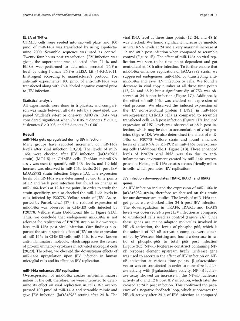
ResultmiR-146a gets upregulated during JEV infectionMany groups have reported increment of miR-146alevels after viral infection [19,20]. The levels of miR-146a were checked after JEV infection (JaOArS982strain) (MOI 5) in CHME3 cells. TaqMan microRNAassay was used to quantify miR-146a levels, and 1.9-foldincrease was observed in miR-146a levels, 24 h post JEVJaOArS982 strain infection (Figure 1A). The expressionlevels of miR-146a were determined at two time pointsof 12 and 24 h post infection but found no change inmiR-146a levels at 12-h time point. In order to study thestrain specificity, we also checked the miR-146a levels incells infected by P20778, Vellore strain of JEV. As re-ported by Pareek et al. [27], the reduced expression ofmiR-146a was observed in CHME3 cells infected byP20778, Vellore strain (Additional file 1: Figure S1A).Thus, we conclude that endogenous miR-146a is notrelevant for replication of P20778 strain as it downregu-lates miR-146a post viral infection. Our findings sup-ported the strain-specific effect of JEV on the expressionof miR-146a in CHME3 cells. miR-146a is a well-knownanti-inflammatory molecule, which suppresses the releaseof pro-inflammatory cytokines in activated microglial cells[28,29]. Therefore, we checked the downstream effects ofmiR-146a upregulation upon JEV infection in humanmicroglial cells and its effect on JEV replication.
miR-146a enhances JEV replicationOverexpression of miR-146a creates anti-inflammatorymilieu in the cell; therefore, we were interested to deter-mine its effect on viral replication in cells. We overex-pressed 100 pmol of miR-146a and scramble mimic andgave JEV infection (JaOArS982 strain) after 24 h. The
viral RNA level at three time points (12, 24, and 48 h)was checked. We found significant increase by ninefoldin viral RNA levels at 24 and a very marginal increase at12 and 48 h post infection when compared to scramblecontrol (Figure 1B). The effect of miR-146a on viral rep-lication was seen to be time point dependent and gotneutralized at 48 h after infection. To further ensure thatmiR-146a enhances replication of JaOArS982 strain, wesuppressed endogenous miR-146a by transfecting anti-miR-146a and gave JEV infection to cells. We found adecrease in viral copy number at all three time points(12, 24, and 48 h) but a significant dip of 75% was ob-served at 24 h post infection (Figure 1C). Additionally,the effect of miR-146a was checked on expression ofviral proteins. We observed the induced expression ofthe JEV non-structural protein 1 (NS1) in miR-146aoverexpressing CHME3 cells as compared to scrambletransfected cells 24 h post infection (Figure 1D). Inducedexpression of NS1 levels was observed at 48 h post in-fection, which may be due to accumulation of viral pro-teins (Figure 1D). We also determined the effect of miR-146a on P20778 Vellore strain and found enhancedlevels of viral RNA by RT-PCR in miR-146a overexpress-ing cells (Additional file 1: Figure S1B). These enhancedlevels of P20778 viral RNA was also due to anti-inflammatory environment created by miR-146a overex-pression. Hence, miR-146a creates a virus-friendly milieuin cells, which promotes JEV replication.
JEV infection downregulates TRAF6, IRAK1, and IRAK2genesAs JEV infection induced the expression of miR-146a inJaOArS982 strain, therefore we focused on this strainfor our downstream studies. The levels of miR-146a tar-get genes were checked after 24 h post JEV infection.The downregulation in TRAF6, IRAK1, and IRAK2levels was observed 24 h post JEV infection as comparedto uninfected cells used as control (Figure 2A). Sincethese genes are major adaptor molecules involved inNF-κB activation, the levels of phospho-p65, which isthe subunit of NF-κB activator complex, were deter-mined by Western blotting and found a decrease in ra-tio of phospho-p65 to total p65 post infection(Figure 2C). NF-κB luciferase construct containing NF-κB response element upstream firefly luciferase genewas used to ascertain the effect of JEV infection on NF-κB activation at various time points. β-galactosidasevector was co-transfected in order to normalize lucifer-ase activity with β-galactosidase activity. NF-κB lucifer-ase assay showed an increase in the NF-κB luciferaseactivity at 6 and 12 h post JEV infection, which later de-creased at 24 h post infection. This confirmed the pres-ence of a negative feedback loop, which suppresses theNF-κB activity after 24 h of JEV infection as compared
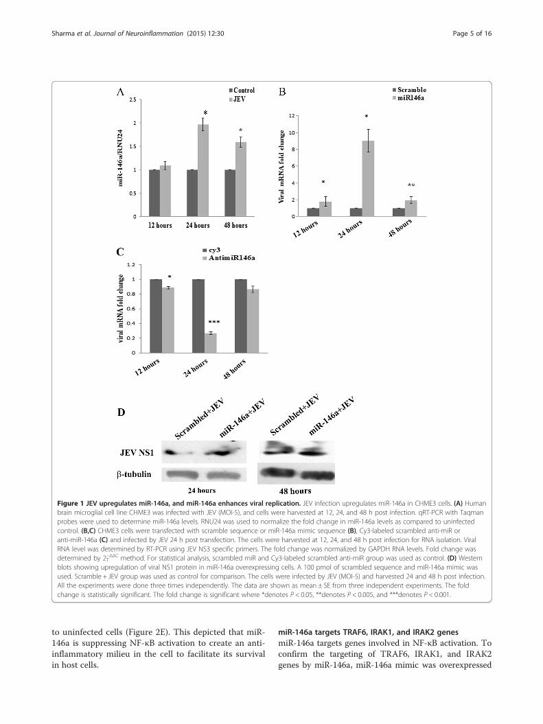
Figure 1 JEV upregulates miR-146a, and miR-146a enhances viral replication. JEV infection upregulates miR-146a in CHME3 cells. (A) Humanbrain microglial cell line CHME3 was infected with JEV (MOI-5), and cells were harvested at 12, 24, and 48 h post infection. qRT-PCR with Taqmanprobes were used to determine miR-146a levels. RNU24 was used to normalize the fold change in miR-146a levels as compared to uninfectedcontrol. (B,C) CHME3 cells were transfected with scramble sequence or miR-146a mimic sequence (B), Cy3-labeled scrambled anti-miR oranti-miR-146a (C) and infected by JEV 24 h post transfection. The cells were harvested at 12, 24, and 48 h post infection for RNA isolation. ViralRNA level was determined by RT-PCR using JEV NS3 specific primers. The fold change was normalized by GAPDH RNA levels. Fold change wasdetermined by 2−ΔΔCT method. For statistical analysis, scrambled miR and Cy3-labeled scrambled anti-miR group was used as control. (D) Westernblots showing upregulation of viral NS1 protein in miR-146a overexpressing cells. A 100 pmol of scrambled sequence and miR-146a mimic wasused. Scramble + JEV group was used as control for comparison. The cells were infected by JEV (MOI-5) and harvested 24 and 48 h post infection.All the experiments were done three times independently. The data are shown as mean ± SE from three independent experiments. The foldchange is statistically significant. The fold change is significant where *denotes P < 0.05, **denotes P < 0.005, and ***denotes P < 0.001.
Sharma et al. Journal of Neuroinflammation (2015) 12:30 Page 5 of 16
to uninfected cells (Figure 2E). This depicted that miR-146a is suppressing NF-κB activation to create an anti-inflammatory milieu in the cell to facilitate its survivalin host cells.
miR-146a targets TRAF6, IRAK1, and IRAK2 genesmiR-146a targets genes involved in NF-κB activation. Toconfirm the targeting of TRAF6, IRAK1, and IRAK2genes by miR-146a, miR-146a mimic was overexpressed
Figure 2 (See legend on next page.)
Sharma et al. Journal of Neuroinflammation (2015) 12:30 Page 6 of 16

(See figure on previous page.)Figure 2 JEV downregulates TRAF6, IRAK1, IRAK2, and NF-κB activation. CHME3 cells were infected by JEV (MOI-5) and harvested 24 h postinfection. (A) Western blots showing downregulation of TRAF6, IRAK1, and IRAK2 genes. Uninfected cells were used as control. (B) Graph barsrepresenting densitometry plot depicting downregulation of TRAF6, IRAK1, and IRAK2 genes post JEV infection. The image density of blots wasnormalized by β-tubulin by using ImageJ software. (C) Western blot showing downregulation of phospho-p65 upon JEV infection. (D) Densitometryplot showing decreased phosphorylation of p65 subunit of NF-κB. The ratio of phospho/total p-65 decreases upon JEV infection. (E) Graph bar showingincreased NF-κB luciferase activity at 6 and 12 h after JEV infection which later decreased 24 h post JEV infection. One-way ANOVA was used todetermine statistical significance. P values were considered significant where *denotes P < 0.05, **denotes P < 0.005, and ***denotes P < 0.001 from 6 hsample, #P from 12 h sample. NF-κB luciferase vector was co-transfected with β-galactosidase vector. β-galactosidase activity was used to normalizeluciferase activity. All experiments were repeated thrice and are represented as mean ± SE.
Sharma et al. Journal of Neuroinflammation (2015) 12:30 Page 7 of 16
in CHME3 cells. CHME3 cells were transfected by using100 pmol of miR-146a and incubated for 48 h posttransfection. Additionally, CHME3 cells were also trans-fected with scrambled miR-146a and used as a control.Significant upregulation of miR-146a was confirmed byTaqMan qPCR microRNA assay. TaqMan qPCR did notshow any upregulation of miR-146a in CHME3 cellstransfected with scrambled miR-146a (Figure 3C). TheWestern blot analysis has shown downregulation ofTRAF6, IRAK1, and IRAK2 genes in the CHME3 cellsoverexpressing miR-146a (Figure 3A).
Anti-miR-146a rescues TRAF6, IRAK1, and IRAK2 genesduring JEV infectionTo further validate the role of miR-146a during JEV infec-tion, we silenced miR-146a by using anti-miR-146a. Cy3-labeled scramble anti-miR was used as negative control.We confirmed the suppression of miR-146a by qPCR(Figure 4C). By Western blot analysis, we found that anti-miR-146a was able to rescue the expression of TRAF6,IRAK1, and IRAK2 genes from downregulation after JEVinfection (Figure 4A). Increased expression of miR-146aled to the downregulation of TRAF6, IRAK1, and IRAK 2genes, and anti-miR-146a was able to neutralize the effectof JEV on TRAF6, IRAK1, and IRAK2 genes.
miR-146a suppresses NF-κB activation and cytokineproductionTo unveil the downstream effects of miR-146a overex-pression which promoted JEV replication, the effect ofmiR-146a overexpression on NF-κB activation was ana-lyzed. The NF-κB luciferase activity was found to bediminished by 40% in the presence of miR-146a, com-pared to scramble control (Figure 4D). JEV infection re-sulted into the downregulation of the luciferase activityby 50% in presence of miR-146a. To further delineatethe suppressive effect of miR-146a, miR-146a activitywas silenced by anti-miR-146a, which led to the in-creased luciferase activity. Elevated luciferase activitywas observed in the presence of JEV (24 h post-infection) after anti-miR-146a transfection (Figure 4E).JEV infection also elevated NF-κB activity in the pres-ence of anti-miR-146a, compared to untransfected JEV-infected cells (Figure 4E).
The effect of miR-146a overexpression was checked oncytokine production after JEV infection. JEV is known totrigger cytokine secretion in microglial cells. To see theeffect of exogenous expression of miR-146a on JEV-induced cytokine production, the transcript levels ofIL-6 were checked. We found reduced messenger RNA(mRNA) levels of IL-6 in miR-146a overexpressing cells,compared to scrambled control (Additional file 2: FigureS2A). The levels of TNF-α was also checked by ELISAof supernatants of JEV-infected miR-146a overexpressingCHME3 cells, and we found reduced TNF-α secretion(Additional file 2: Figure S2C). We also analyzed theIFN-β promoter activity in miR-146a overexpressingCHME3 cells after JEV infection and found 50% de-crease in IFN-β promoter activity in the presence ofmiR-146a (Additional file 2: Figure S2E).
miR-146a targets STAT1 genemiR-146a overexpression resulted in decreased IFN-β pro-moter activity, so the effect of miR-146a upregulation oninterferon signaling was also investigated. The expressionof STAT1 protein in miR-146a overexpressing cells wasdetermined. Signal transducer and activator of transcrip-tion (STAT1) is a well-known downstream molecule inIFN signaling which binds to Janus kinases (Jak) and trig-gers expression of interferon-stimulated gene and is aknown to target of miR-146a. The targeting of STAT1gene by miR-146a was confirmed in our cells, and down-regulation in STAT1 levels was observed upon miR-146aoverexpression (Figure 5C). Anti-miR-146a rescued STAT1from downregulation (Figure 5D).Type I interferon activates STAT1 by its phosphoryl-
ation and dimerization. JEV has been reported to in-crease the STAT1 phosphorylation [30]. We observedupregulation in STAT1 phosphorylation (ser727) 12 hpost JEV infection (Figure 5A). However, the level oftotal STAT1 was not changed in 12 h samples. Later,downregulation in total and phospho-STAT1 levelswas noticed at 24 h post JEV infection (Figure 5A).We assume that downregulation in STAT1 levelswould be due to miR-146a upregulation, which targetsexpression of STAT1. To confirm our hypothesis,miR-146a was silenced by using anti-miR-146a andfound increased STAT1 levels in the presence of JEV

Figure 3 miR-146a targets TRAF6, IRAK1, and IRAK2. CHME3cells were transfected with 100 pmol miR-146a mimic sequence.Scrambled seed sequence was used as control along with mocktreatment as control. The cells were harvested 48 h post transfection.(A) Western blots showing downregulation of TRAF6, IRAK1, andIRAK2 genes upon miR-146a overexpression. (B) Densitometry plotshowing downregulation of TRAF6, IRAK1, and IRAK2 genes uponmiR-146a overexpression. The overexpressed samples were comparedto scramble control for statistical analysis. (C) RT-PCR analysis of miR-146alevels in miR-146a overexpressed cells. The cells were harvested 48 hpost transfection. miR-146a levels were determined by RT-PCRby using Taqman probe. RNU24 was used for normalization. Allexperiments were repeated thrice and are represented as mean ± SE.The fold change is significant where *denotes P < 0.05, **denotesP < 0.005, and ***denotes P < 0.001.
Sharma et al. Journal of Neuroinflammation (2015) 12:30 Page 8 of 16
(Figure 5D). So we speculate that miR-146a downregu-lates STAT1 levels which could hinder interferon sig-naling pathway.
miR-146a suppresses JEV-induced ISRE promoter activityActivated STAT1 dimerizes and binds to other factors toenter the nucleus and activates the expression ofinterferon-stimulated genes after binding to interferon-stimulated response elements (ISRE) in the promoter ofinterferon-inducible genes. Since miR-146a reduced theSTAT1 activation, the effect of miR-146a overexpressionwas further studied on ISRE activity by ISRE luciferaseassay. miR-146a overexpressing cells displayed reducedluciferase activity, which depicted suppression of ISREpromoter activity upon JEV infection in miR-146a overex-pressing cells (Figure 6A). However, we did not observeany significant decrease in ISRE activity in scramble trans-fected cells infected by JEV (Figure 6A). ISRE activity wasincreased upon JEV infection when miR-146a was silencedby using anti-miR-146a prior to JEV infection, comparedto Cy3-labeled, scrambled transfected cells infected byJEV (Figure 6B).To study the time-dependent effect of JEV infection on
ISRE activity, the luciferase activity was checked at 12 and24 h post JEV infection. ISRE activity got increased atearly time point of 12 h of infection; but later, it decreasedat 24 h post JEV infection (Figure 6C). JEV infection sup-pressed the ISRE activity after 24 h of JEV infection, whichweakens the cellular immune response against the virus.
Downregulation of interferon-stimulated genes bymiR-146aSTAT1 acts as a transcription factor which increases theexpression of many interferon-stimulated genes. Abroga-tion of STAT1 gene leads to reduced ISRE activity, whichin turn affects the expression interferon-stimulated genes(ISGs). As we observed, miR-146a downregulated STAT1levels; therefore, the levels of ISGs were determinedafter miR-146a overexpression. Interferon-induced pro-tein with tetratricopeptide repeat (IFIT) proteins aremajor interferon-induced proteins. We checked themRNA levels of two IFIT genes IFIT-1 and IFIT-2 aftermiR-146a overexpression. About 40% reduction in IFIT-1mRNA levels (Figure 7A) and 30% reduction in IFIT-2levels (Figure 7C) were noticed in cells, transfected miR-146a followed by JEV infection, compared to scramblecontrol mimic. Anti-miR-146a reversed the effect ofmiR-146a by increasing the levels of IFIT-1 and IFIT-2(Figure 7B,D). The miR-146a downregulates ISGs anddisrupts Jak-STAT signaling.
JEV modulates pro-inflammatory cytokines and expressionof IFIT-1 and IFIT-2Viruses elicit the secretion of pro-inflammatory cytokinesin cells. This strategy is used by the host to restrict viralreplication and survival. JEV triggers the innate immuneresponse of the cells and elicits the secretion of pro-inflammatory cytokines like IL-6 and TNF-α. Significant
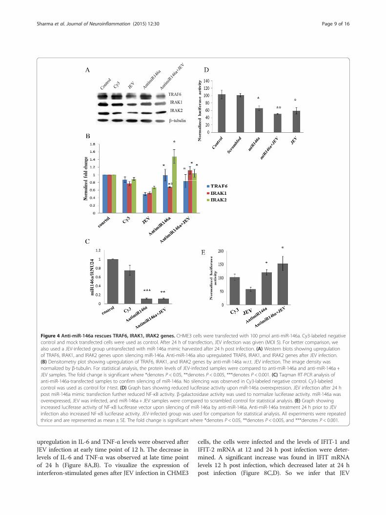
Figure 4 Anti-miR-146a rescues TRAF6, IRAK1, IRAK2 genes. CHME3 cells were transfected with 100 pmol anti-miR-146a. Cy3-labeled negativecontrol and mock transfected cells were used as control. After 24 h of transfection, JEV infection was given (MOI 5). For better comparison, wealso used a JEV-infected group untransfected with miR-146a mimic harvested after 24 h post infection. (A) Western blots showing upregulationof TRAF6, IRAK1, and IRAK2 genes upon silencing miR-146a. Anti-miR-146a also upregulated TRAF6, IRAK1, and IRAK2 genes after JEV infection.(B) Densitometry plot showing upregulation of TRAF6, IRAK1, and IRAK2 genes by anti-miR-146a w.r.t. JEV infection. The image density wasnormalized by β-tubulin. For statistical analysis, the protein levels of JEV-infected samples were compared to anti-miR-146a and anti-miR-146a +JEV samples. The fold change is significant where *denotes P < 0.05, **denotes P < 0.005, ***denotes P < 0.001. (C) Taqman RT-PCR analysis ofanti-miR-146a-transfected samples to confirm silencing of miR-146a. No silencing was observed in Cy3-labeled negative control. Cy3-labeledcontrol was used as control for t-test. (D) Graph bars showing reduced luciferase activity upon miR-146a overexpression. JEV infection after 24 hpost miR-146a mimic transfection further reduced NF-κB activity. β-galactosidase activity was used to normalize luciferase activity. miR-146a wasoverexpressed, JEV was infected, and miR-146a + JEV samples were compared to scrambled control for statistical analysis. (E) Graph showingincreased luciferase activity of NF-κB luciferase vector upon silencing of miR-146a by anti-miR-146a. Anti-miR-146a treatment 24 h prior to JEVinfection also increased NF-κB luciferase activity. JEV-infected group was used for comparison for statistical analysis. All experiments were repeatedthrice and are represented as mean ± SE. The fold change is significant where *denotes P < 0.05, **denotes P < 0.005, and ***denotes P < 0.001.
Sharma et al. Journal of Neuroinflammation (2015) 12:30 Page 9 of 16
upregulation in IL-6 and TNF-α levels were observed afterJEV infection at early time point of 12 h. The decrease inlevels of IL-6 and TNF-α was observed at late time pointof 24 h (Figure 8A,B). To visualize the expression ofinterferon-stimulated genes after JEV infection in CHME3
cells, the cells were infected and the levels of IFIT-1 andIFIT-2 mRNA at 12 and 24 h post infection were deter-mined. A significant increase was found in IFIT mRNAlevels 12 h post infection, which decreased later at 24 hpost infection (Figure 8C,D). So we infer that JEV

Figure 5 miR-146a upregulation by JEV targets STAT1 phosphorylation. (A) CHME3 cells were infected with JEV and pelleted at 12 and24 h post infection. Western blots representing phospho-STAT1 and STAT1 levels at 12 and 24 h post infection. Both STAT1 and phospho-STAT1got downregulated 24 h post infection. (B) Densitometry plot showing increase in phosphorylation of STAT1 at 12 h post infection. Later, noincrease in phosphorylation was observed 24 h after infection due to downregulation in levels of STAT1. Both phospho-STAT1 and STAT1image density was normalized by β-tubulin. For statistical analysis, the total and phospho-STAT1 levels of 12 and 24 h JEV-infected sampleswere compared to control uninfected samples at 12 and 24 h. (C) Western blot showing downregulation of STAT1 upon miR-146a mimicoverexpression. (D) Western blot showing that silencing of miR-146a by anti-miR-146a upregulates STAT1 upon JEV infection. (E) Densitometryplot showing upregulation of STAT1 upon silencing of miR-146a. JEV infection also upregulates STAT1 when anti-miR-146a is transfected 24 hprior to JEV infection. The image density was normalized by β-tubulin. For statistical analysis, the anti-miR-146a and anti-miR-146a + JEV groupswere compared to JEV-infected group. All experiments were repeated thrice and are represented as mean ± SE. The fold change is significantwhere *denotes P < 0.05, **denotes P < 0.005, and ***denotes P < 0.001.
Sharma et al. Journal of Neuroinflammation (2015) 12:30 Page 10 of 16

Figure 6 miR-146a suppresses ISRE promoter activity. CHME3 cells were co-transfected with 1 μg ISRE luciferase vector and 700 ng β-galactosidase vector to measure ISRE luciferase activity. (A) Graph bars showing reduced ISRE luciferase activity upon JEV infection in miR-146aoverexpressing cells. miR-146a mimic was transfected along with vectors, and scrambled sequence was used as control for statistical analysis.Scrambled + JEV group did not show any significant decrease in ISRE activity upon JEV infection. JEV infection was given 24 h post transfection,and luciferase activity was measured after 24 h. (B) Graph bars showing increased ISRE luciferase activity upon JEV infection in anti-miR-146atransfected cells. Cy3-labeled scrambled anti-miR was used as control. Anti-miR-146a increased ISRE activity upon JEV infection as compared toscrambled Cy3 + JEV group. (C) Graph bars showing ISRE luciferase activity upon JEV infection at two time points - 12 and 24 h post infection.The ISRE activity decreases at later time point after JEV infection. All experiments were repeated thrice and are represented as mean ± SE. The foldchange is significant where *denotes P < 0.05, **denotes P < 0.005, and ***denotes P < 0.001.
Sharma et al. Journal of Neuroinflammation (2015) 12:30 Page 11 of 16
downregulates IFIT levels and expression of pro-inflammatory cytokines at late hours of infection to sup-press the cellular innate immune response to alleviate itssurvival in the cell.
DiscussionJEV infection leads to neuroinflammation in JEV-infectedpatients, which leads to high morbidity and mortality.However, the survivors still show neurological and
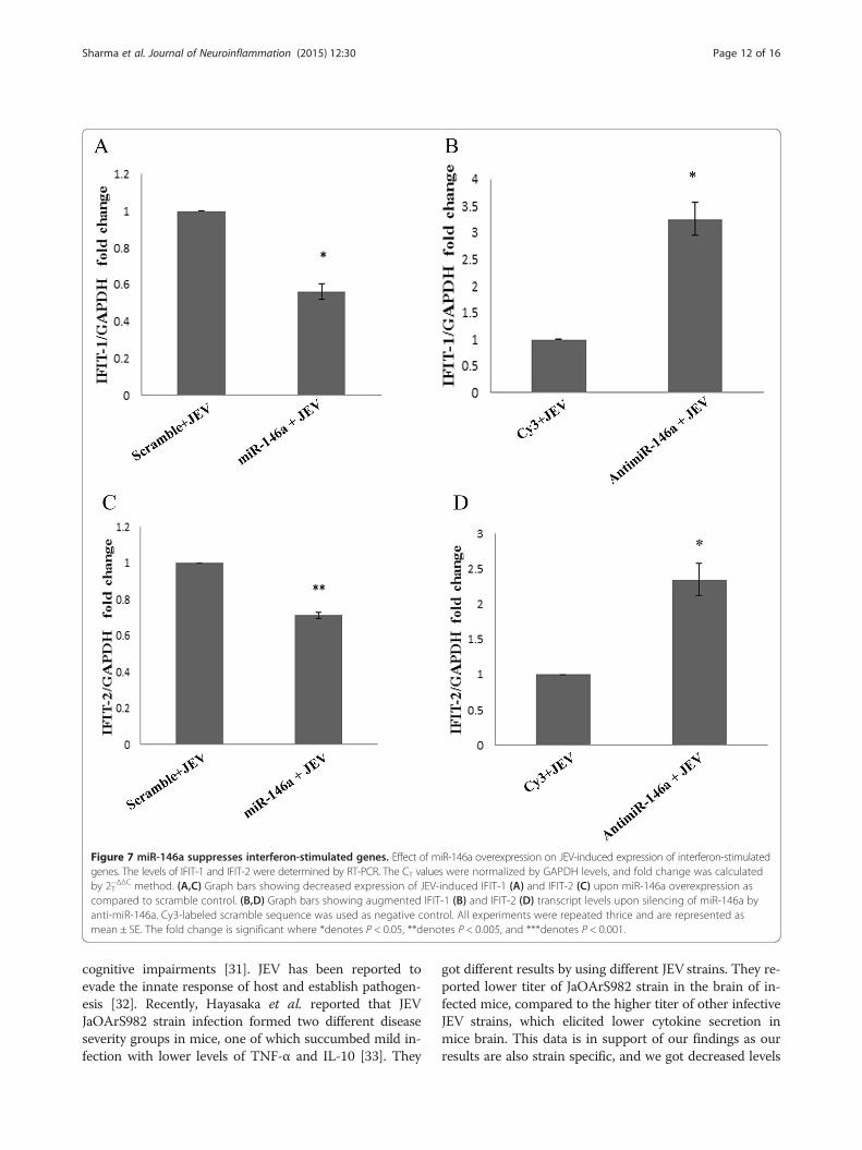
Figure 7 miR-146a suppresses interferon-stimulated genes. Effect of miR-146a overexpression on JEV-induced expression of interferon-stimulatedgenes. The levels of IFIT-1 and IFIT-2 were determined by RT-PCR. The CT values were normalized by GAPDH levels, and fold change was calculatedby 2−ΔΔCT method. (A,C) Graph bars showing decreased expression of JEV-induced IFIT-1 (A) and IFIT-2 (C) upon miR-146a overexpression ascompared to scramble control. (B,D) Graph bars showing augmented IFIT-1 (B) and IFIT-2 (D) transcript levels upon silencing of miR-146a byanti-miR-146a. Cy3-labeled scramble sequence was used as negative control. All experiments were repeated thrice and are represented asmean ± SE. The fold change is significant where *denotes P < 0.05, **denotes P < 0.005, and ***denotes P < 0.001.
Sharma et al. Journal of Neuroinflammation (2015) 12:30 Page 12 of 16
cognitive impairments [31]. JEV has been reported toevade the innate response of host and establish pathogen-esis [32]. Recently, Hayasaka et al. reported that JEVJaOArS982 strain infection formed two different diseaseseverity groups in mice, one of which succumbed mild in-fection with lower levels of TNF-α and IL-10 [33]. They
got different results by using different JEV strains. They re-ported lower titer of JaOArS982 strain in the brain of in-fected mice, compared to the higher titer of other infectiveJEV strains, which elicited lower cytokine secretion inmice brain. This data is in support of our findings as ourresults are also strain specific, and we got decreased levels
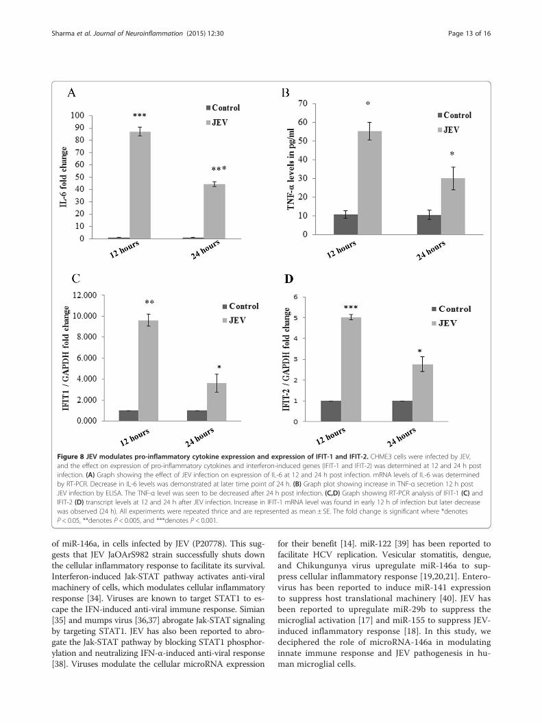
Figure 8 JEV modulates pro-inflammatory cytokine expression and expression of IFIT-1 and IFIT-2. CHME3 cells were infected by JEV,and the effect on expression of pro-inflammatory cytokines and interferon-induced genes (IFIT-1 and IFIT-2) was determined at 12 and 24 h postinfection. (A) Graph showing the effect of JEV infection on expression of IL-6 at 12 and 24 h post infection. mRNA levels of IL-6 was determinedby RT-PCR. Decrease in IL-6 levels was demonstrated at later time point of 24 h. (B) Graph plot showing increase in TNF-α secretion 12 h postJEV infection by ELISA. The TNF-α level was seen to be decreased after 24 h post infection. (C,D) Graph showing RT-PCR analysis of IFIT-1 (C) andIFIT-2 (D) transcript levels at 12 and 24 h after JEV infection. Increase in IFIT-1 mRNA level was found in early 12 h of infection but later decreasewas observed (24 h). All experiments were repeated thrice and are represented as mean ± SE. The fold change is significant where *denotesP < 0.05, **denotes P < 0.005, and ***denotes P < 0.001.
Sharma et al. Journal of Neuroinflammation (2015) 12:30 Page 13 of 16
of miR-146a, in cells infected by JEV (P20778). This sug-gests that JEV JaOArS982 strain successfully shuts downthe cellular inflammatory response to facilitate its survival.Interferon-induced Jak-STAT pathway activates anti-viralmachinery of cells, which modulates cellular inflammatoryresponse [34]. Viruses are known to target STAT1 to es-cape the IFN-induced anti-viral immune response. Simian[35] and mumps virus [36,37] abrogate Jak-STAT signalingby targeting STAT1. JEV has also been reported to abro-gate the Jak-STAT pathway by blocking STAT1 phosphor-ylation and neutralizing IFN-α-induced anti-viral response[38]. Viruses modulate the cellular microRNA expression
for their benefit [14]. miR-122 [39] has been reported tofacilitate HCV replication. Vesicular stomatitis, dengue,and Chikungunya virus upregulate miR-146a to sup-press cellular inflammatory response [19,20,21]. Entero-virus has been reported to induce miR-141 expressionto suppress host translational machinery [40]. JEV hasbeen reported to upregulate miR-29b to suppress themicroglial activation [17] and miR-155 to suppress JEV-induced inflammatory response [18]. In this study, wedeciphered the role of microRNA-146a in modulatinginnate immune response and JEV pathogenesis in hu-man microglial cells.
Sharma et al. Journal of Neuroinflammation (2015) 12:30 Page 14 of 16
We report the JEV-mediated increased expression ofmiR-146a in CHME3 cells. Our results are contradictoryto findings of Pareek et al. who found downregulatedmiR-146a levels upon JEV infection [27]. This discrep-ancy may be due to different strains of virus used byPareek et al. To confirm this strain-specific effect of JEV,we determined miR-146a levels in P20778 strain used byPareek et al. and found similar results to their findings.This suggested that increased expression of miR-146a isstrain specific. JEV infection downregulated the adaptormolecules TRAF6, IRAK1, and IRAK2 involved in NF-κB activation, which are targeted by miR-146a. To de-scribe the specific role of miR-146a in targeting of theseadaptor molecules, miR-146a was silenced and we foundthat anti-miR-146a rescued TRAF6, IRAK1, and IRAK2from downregulation upon JEV infection. To further un-veil the downstream effects on NF-κB activity, NF-κBpromoter luciferase assay and Western blot analysiswere performed. We found reduced luciferase activity at24 h post JEV infection and reduced phosphorylation ofNF-κB p65 subunit upon JEV infection. To confirm thepresence of negative feedback loop, we checked the NF-κB luciferase activity at early time points (6 and 12 h)and found initial upregulation in luciferase activityfollowed by a decrease in luciferase activity at laterhours. Initial NF-κB activation by JEV preludes the miR-146a overexpression, which further led to the downregu-lation of adaptor proteins involved in NF-κB activationand constitute a negative regulatory loop [41]. Hence,we conclude that JEV-mediated upregulation of miR-146a takes place to downregulate NF-κB activation.miR-146a overexpression suppressed NF-κB activation
as demonstrated by luciferase assay, and inhibition ofmiR-146a enhanced NF-κB activation upon JEV infec-tion. Recently, Jin Ho Paik et al. also reported similarresults upon miR-146a overexpression [42]. Our findingsdemonstrated that miR-146a creates an anti-inflammatoryenvironment in cells. NF-κB subunits act as transcriptionfactor for expression of pro-inflammatory cytokines. miR-146a overexpression also suppressed JEV-induced cytokineproduction (IL-6, TNF-α, IFN-β). The replication of JEV isincreased upon miR-146a overexpression due to an anti-inflammatory environment created by overexpression ofmiR-146a. However, this enhanced replication of viralRNA was observed only at 24 h post infection and this ef-fect reduced at later time points. To rule out the possibil-ity that this reduced effect on viral replication could bedue to degradation of overexpressed miR-146a at laterhours, we checked the levels of miR-146a at later hours byRT-PCR and found that the level of miR-146a wasretained in CHME3 cells (data not shown). We also foundelevated levels of viral NS1 protein in miR-146a overex-pressing cells. This may be due to greater accumulation ofviral proteins in miR-146a overexpressing cells. Recently,
Bing-Ching Ho et al. also reported that miR-146a sup-ports the survival of Enterovirus in mice and silencing ofmiR-146a improved the survival of Enterovirus-infectedmouse due to restoration of interferon production [43].This suggested that the cellular anti-viral machinery gotcompromised in miR-146a overexpressing cells and sup-ported viral replication. However, a recent study by Kunduet al. reported increased levels of SOCS1 and SOCS3 dur-ing early time points of infection which promoted JEVreplication at early time points, but the viral titer got de-creased due to the decrease in levels of SOCS1 andSOCS3 during later time points, which led to the activatedcellular immune response [30]. We also found a decreasein viral mRNA levels at later time points. This decreasein viral titer in later time points may help the virus topersist latently in cells. Expression of a truncated form ofviral NS1 protein and production of low virus titer in per-sistently JEV-infected murine neuroblastoma cells as anaftermath of virus-cell interaction [44] also depict that re-striction of viral replication can assist viral persistence.Decrease in viral titer at later time points may be due tothe activation of cellular immune machinery which nulli-fied the effect of miR-146a overexpression.miR-146a targets STAT1 gene which leads to abrogation
of Jak-STAT pathway [45]. We also found similar results,where miR-146a overexpression reduced the STAT1 levelsand inhibition of miR-146a upregulated the STAT1 levels.We also analyzed the effect of miR-146a overexpressionon Jak-STAT pathway. Suppression of ISRE promoteractivity and downregulated expression of interferon-stimulated genes (ISGs) were found in miR-146a overex-pressing cells upon JEV infection. Interferon-inducedprotein with tetratricopeptide repeat (IFIT) proteins arewell-known interferon-induced anti-viral proteins, havinganti-proliferative effects [46]. IFIT-1 has been reported torestrict JEV replication [47]. IFIT-2 has been also reportedto restrict VSV replication [48]. miR-146a downregulatedthe JEV induced expression of IFIT-1 and IFIT-2. Down-regulation of IFIT levels perturb the cellular anti-viral ma-chinery, which helps in JEV replication in host cells. Tanget al. has reported downregulation of other ISGs (OAS1,Mx1, LY6E) upon miR-146a overexpression [22]. Thesefindings indicated that miR-146a abrogated the Jak-STATpathway and downregulated the expression of ISGs, whichled to the suppression of innate immune response againstthe virus and augmented the viral replication in miR-146aoverexpressing cells.We also analyzed the effect of JEV infection on STAT1
activation at different time points. JEV induced theSTAT1 activation at early time points but downregulatedthe STAT1 levels at later time points due to targeting ofthe STAT1 by miR-146a. Inhibition of miR-146a led tothe increased STAT1 levels upon JEV infection. TheISRE promoter activity and expression of IFIT-1 and
Sharma et al. Journal of Neuroinflammation (2015) 12:30 Page 15 of 16
IFIT-2 also displayed similar trends. We observed initialupregulation of ISRE promoter activity and levels of IFIT1and IFIT2 upon JEV infection, which supported the activa-tion of cellular innate immune machinery against thevirus. At 24-h time point, we found decreased levels ofIFIT1 and IFIT2 and reduced ISRE promoter activitywhich indicated that the virus has successfully suppressedthe cellular inflammatory response. We observed the de-creased expression of IL-6 and TNF-α at 24 h as com-pared to 12 h post JEV infection. These findings suggestthat JEV-mediated miR-146a upregulation led to the sup-pression of NF-κB activation and STAT1 degradation,caused the downregulation of ISGs at later time point(24 h). Downregulation of ISGs has been reported to facili-tate persistence of virus in cells [49]. This is a strategy em-braced by JEV to suppress the cellular inflammatoryresponse in human microglial cells and evade innate im-mune response. However; this effect is time dependentand other strategies adopted by the cell to combat viralreplication could play an important role. The host celltriggers various anti-viral signaling pathways to curtailviral replication. Further studies are required to find outother anti-viral strategies adopted by JEV to evade cellularimmune response. This study demonstrated strain-specificeffects of JEV, as different JEV strains may lead to the vary-ing downstream effects in host cellular immune responses.Understanding the regulatory role of cellular microRNAsduring JEV infection in microglial cells would be helpfulin understanding the molecular mechanism of JEVneuropathogenesis.
Additional files
Additional file 1: Figure S1. JEV P20778 strain downregulates miR-146aand effect of miR-146a on P20778 replication. CHME3 cells were infected byJEV P20778 Vellore strain (MOI-5), and cells were harvested after 24 and 48 hfor RNA isolation and qPCR. (A) Graph showing downregulation in miR-146alevels 24 and 48 h post infection obtained by RT-PCR using miR-146a-specific Taqman probes. Uninfected control groups of the sametime points were used for comparison. RNU24 levels were used fornormalization. Fold change was determined by 2−ΔΔCT method. (B) RT-PCRgraph showing upregulated viral RNA levels in miR-146a overexpressingcells. CHME3 cells were transfected with 100 pmol miR-146a mimic, andP20778 JEV infection was given after 24 h. The cells were harvested 24and 48 h post infection. Scrambled + JEV group of the same time pointswas used as control for comparison. Viral RNA level was determined byRT-PCR using JEV NS3 specific primers. The fold change was normalizedby GAPDH RNA levels. All experiments were repeated thrice and arerepresented as mean ± SE. The fold change is significant where *denotesP < 0.05, **denotes P < 0.005, and ***denotes P < 0.001.
Additional file 2: Figure S2. miR-146a suppresses cytokine expression.Graph bars showing the effect of overexpression of miR-146a on variouscytokines upon JEV infection. miR-146a suppressed the expression ofJEV-triggered cytokines (IL-6, TNF-α, IFN-β). (A,B) RT-PCR analysis of IL-6levels in JEV-infected miR-146a overexpressing cells. (A) miR-146asuppressed the IL-6 expression as compared to scramble control.Anti-miR-146a transfection increased the expression of IL-6 upon JEVinfection as compared to Cy3-labeled negative control. (B-D) Graph barsshowing ELISA of supernatants to check TNF-α secretion in miR-146a
overexpressing cells (C) and in anti-miR-146a transfected cells (D) uponJEV infection. (E-F) Graph depicting the effect of miR-146a on IFN-βpromoter activity by IFN-β promoter luciferase assay. miR-146a suppressedthe JEV-triggered IFN-β promoter activity (E) whereas anti-miR-146aenhanced the IFN-β promoter activity upon JEV infection (F). Theinfected samples were compared to uninfected control for statisticalanalysis. All experiments were repeated thrice and are represented asmean ± SE. The fold change is significant where *denotes P < 0.05,**denotes P < 0.005, and ***denotes P < 0.001.
Competing interestsThe authors declare that they have no competing interests.
Authors’ contributionsNS is currently working as a CSIR-Senior Research Fellow and pursuing hisPh.D program. NS designed and carried out most of the experiments andwrote the manuscript. RV performed some initial experiments to establishthe proof of the concept. KLK propagated JEV in mice. AB provided theJEV and microglial cells. SKS guided the team during the planning of theexperimental design, analyzed the data, and wrote the paper. All authorsread and approved the final manuscript.
AcknowledgementsThe authors are thankful to Prof. Adolfo Gracia-Sastre, Department ofMedicine and Microbiology, Mount Sinai School of Medicine, New York, NY,USA, for providing NF-κB and IFN-β luciferase vector as a kind gift. Authorsare thankful to Ms. Ritu Mishra for her suggestions during experiments andmanuscript preparation. Authors are thankful to the director, Centre forCellular and Molecular Biology (CCMB), Hyderabad, for his support. NikhilSharma is a recipient of CSIR-Senior Research Fellowship. Authors highlyacknowledge the financial support from the DST grant number (INT/Korea/P-08/2011) of the Department of Science and Technology, and grant onRNAi Science and Technology of the Department of Biotechnology, Govt.of India; New Delhi respectively.
Author details1Laboratory of Neurovirology and Inflammation Biology, CSIR-Centre forCellular and Molecular Biology (CCMB), Uppal Road, 500007 Hyderabad, AP,India. 2Current Affiliation: Laboratory of Human Molecular Virology andImmunology, Molecular Biology Unit, Faculty of Medicine, Institute ofMedical Sciences (IMS), Banaras Hindu University (BHU), 221005 Varanasi,India. 3National Brain Research Centre, Haryana-122050 Manesar, Haryana, India.
Received: 18 August 2014 Accepted: 15 January 2015
References1. Unni SK, Ruzek D, Chhatbar C, Mishra R, Johri MK, Singh SK. Japanese
encephalitis virus: from genome to infectome. Microbes Infect. 2011;13:312–21.2. Srivastava R, Kalita J, Khan MY, Gore MM, Bondre VP, Misra UK. Temporal
changes of Japanese encephalitis virus in different brain regions of rat.Indian J Med Res. 2013;138:219–23.
3. van den Hurk AF, Ritchie SA, Mackenzie JS. Ecology and geographicalexpansion of Japanese encephalitis virus. Annu Rev Entomol. 2009;54:17–35.
4. Sapkal GN, Wairagkar NS, Ayachit VM, Bondre VP, Gore MM. Detection andisolation of Japanese encephalitis virus from blood clots collected duringthe acute phase of infection. Am J Trop Med Hyg. 2007;77:1139–45.
5. Thongtan T, Thepparit C, Smith DR. The involvement of microglial cells inJapanese encephalitis infections. Clin Dev Immunol. 2012;2012:890586.
6. Mathur A, Kulshreshtha R, Chaturvedi UC. Evidence for latency of Japaneseencephalitis virus in T lymphocytes. J Gen Virol. 1989;70(Pt 2):461–5.
7. Sharma S, Mathur A, Prakash V, Kulshreshtha R, Kumar R, Chaturvedi UC.Japanese encephalitis virus latency in peripheral blood lymphocytes andrecurrence of infection in children. Clin Exp Immunol. 1991;85:85–9.
8. Cao S, Li Y, Ye J, Yang X, Chen L, Liu X, et al. Japanese encephalitis viruswild strain infection suppresses dendritic cells maturation and function, andcauses the expansion of regulatory T cells. Virol J. 2011;8:39.
9. Ghoshal A, Das S, Ghosh S, Mishra MK, Sharma V, Koli P, et al.Proinflammatory mediators released by activated microglia inducesneuronal death in Japanese encephalitis. Glia. 2007;55:483–96.
Sharma et al. Journal of Neuroinflammation (2015) 12:30 Page 16 of 16
10. Thongtan T, Cheepsunthorn P, Chaiworakul V, Rattanarungsan C, Wikan N,Smith DR. Highly permissive infection of microglial cells by Japaneseencephalitis virus: a possible role as a viral reservoir. Microbes Infect.2010;12:37–45.
11. Bartel DP. MicroRNAs: target recognition and regulatory functions. Cell.2009;136:215–33.
12. Ambros V. The functions of animal microRNAs. Nature. 2004;431:350–5.13. Singh SK, Pal Bhadra M, Girschick HJ, Bhadra U. MicroRNAs – micro in size
but macro in function. FEBS J. 2008;275:4929–44.14. Gottwein E, Cullen BR. Viral and cellular microRNAs as determinants of viral
pathogenesis and immunity. Cell Host Microbe. 2008;3:375–87.15. Skalsky RL, Cullen BR. Viruses, microRNAs, and host interactions. Annu Rev
Microbiol. 2010;64:123–41.16. Cullen BR. MicroRNAs as mediators of viral evasion of the immune system.
Nat Immunol. 2013;14:205–10.17. Thounaojam MC, Kaushik DK, Kundu K, Basu A. MicroRNA-29b modulates
Japanese encephalitis virus-induced microglia activation by targeting tumornecrosis factor alpha-induced protein 3. J Neurochem. 2014;129:143–54.
18. Thounaojam MC, Kundu K, Kaushik DK, Swaroop S, Mahadevan A, ShankarSK, et al. MicroRNA 155 regulates Japanese encephalitis virus-inducedinflammatory response by targeting Src homology 2-containing inositolphosphatase 1. J Virol. 2014;88:4798–810.
19. Wu S, He L, Li Y, Wang T, Feng L, Jiang L, et al. miR-146a facilitatesreplication of dengue virus by dampening interferon induction by targetingTRAF6. J Infect. 2013;67:329–41.
20. Hou J, Wang P, Lin L, Liu X, Ma F, An H, et al. MicroRNA-146a feedbackinhibits RIG-I-dependent type I IFN production in macrophages by targetingTRAF6, IRAK1, and IRAK2. J Immunol. 2009;183:2150–8.
21. Selvamani SP, Mishra R, Singh SK. Chikungunya virus exploits miR-146a toregulate NF-kappaB pathway in human synovial fibroblasts. PLoS One.2014;9:e103624.
22. Tang Y, Luo X, Cui H, Ni X, Yuan M, Guo Y, et al. MicroRNA-146A contributesto abnormal activation of the type I interferon pathway in human lupus bytargeting the key signaling proteins. Arthritis Rheum. 2009;60:1065–75.
23. Taganov KD, Boldin MP, Chang KJ, Baltimore D. NF-kappaB-dependentinduction of microRNA miR-146, an inhibitor targeted to signaling proteinsof innate immune responses. Proc Natl Acad Sci U S A. 2006;103:12481–6.
24. Lu LF, Boldin MP, Chaudhry A, Lin LL, Taganov KD, Hanada T, et al. Functionof miR-146a in controlling Treg cell-mediated regulation of Th1 responses.Cell. 2010;142:914–29.
25. Zeng Z, Gong H, Li Y, Jie K, Ding C, Shao Q, et al. Upregulation of miR-146acontributes to the suppression of inflammatory responses in LPS-inducedacute lung injury. Exp Lung Res. 2013;39:275–82.
26. Iyer A, Zurolo E, Prabowo A, Fluiter K, Spliet WG, van Rijen PC, et al.MicroRNA-146a: a key regulator of astrocyte-mediated inflammatoryresponse. PLoS One. 2012;7:e44789.
27. Pareek S, Roy S, Kumari B, Jain P, Banerjee A, Vrati S. MiR-155 induction inmicroglial cells suppresses Japanese encephalitis virus replication andnegatively modulates innate immune responses. J Neuroinflammation.2014;11:97.
28. Saba R, Gushue S, Huzarewich RL, Manguiat K, Medina S, Robertson C, et al.MicroRNA 146a (miR-146a) is over-expressed during prion disease andmodulates the innate immune response and the microglial activation state.PLoS One. 2012;7:e30832.
29. Rom S, Rom I, Passiatore G, Pacifici M, Radhakrishnan S, Del Valle L, et al.CCL8/MCP-2 is a target for mir-146a in HIV-1-infected human microglialcells. FASEB J. 2010;24:2292–300.
30. Kundu K, Dutta K, Nazmi A, Basu A. Japanese encephalitis virus infectionmodulates the expression of suppressors of cytokine signaling (SOCS) inmacrophages: implications for the hosts’ innate immune response. CellImmunol. 2013;285:100–10.
31. Manocha GD, Mishra R, Sharma N, Kumawat KL, Basu A, Singh SK.Regulatory role of TRIM21 in the type-I interferon pathway in Japaneseencephalitis virus-infected human microglial cells. J Neuroinflammation.2014;11:24.
32. Aleyas AG, George JA, Han YW, Rahman MM, Kim SJ, Han SB, et al.Functional modulation of dendritic cells and macrophages by Japaneseencephalitis virus through MyD88 adaptor molecule-dependent and-independent pathways. J Immunol. 2009;183:2462–74.
33. Hayasaka D, Shirai K, Aoki K, Nagata N, Simantini DS, Kitaura K, et al.TNF-alpha acts as an immunoregulator in the mouse brain by reducing the
incidence of severe disease following Japanese encephalitis virus infection.PLoS One. 2013;8:e71643.
34. Jenkins BJ. Transcriptional regulation of pattern recognition receptors byJak/STAT signaling, and the implications for disease pathogenesis. JInterferon Cytokine Res. 2014;10:750–8.
35. Didcock L, Young DF, Goodbourn S, Randall RE. The V protein of simianvirus 5 inhibits interferon signalling by targeting STAT1 for proteasome-mediated degradation. J Virol. 1999;73:9928–33.
36. Kubota T, Yokosawa N, Yokota S, Fujii N, Tashiro M, Kato A. Mumps virus Vprotein antagonizes interferon without the complete degradation of STAT1.J Virol. 2005;79:4451–9.
37. Kubota T, Yokosawa N, Yokota S, Fujii N. C terminal CYS-RICH region ofmumps virus structural V protein correlates with block of interferon alphaand gamma signal transduction pathway through decrease of STAT 1-alpha.Biochem Biophys Res Commun. 2001;283:255–9.
38. Lin RJ, Liao CL, Lin E, Lin YL. Blocking of the alpha interferon-inducedJak-STAT signaling pathway by Japanese encephalitis virus infection. J Virol.2004;78:9285–94.
39. Kambara H, Fukuhara T, Shiokawa M, Ono C, Ohara Y, Kamitani W, et al.Establishment of a novel permissive cell line for the propagation of hepatitis Cvirus by expression of microRNA miR122. J Virol. 2012;86:1382–93.
40. Ho BC, Yu SL, Chen JJ, Chang SY, Yan BS, Hong QS, et al. Enterovirus-inducedmiR-141 contributes to shutoff of host protein translation by targeting thetranslation initiation factor eIF4E. Cell Host Microbe. 2011;9:58–69.
41. Ma X, Becker Buscaglia LE, Barker JR, Li Y. MicroRNAs in NF-kappaB signaling.J Mol Cell Biol. 2011;3:159–66.
42. Paik JH, Jang JY, Jeon YK, Kim WY, Kim TM, Heo DS, et al. MicroRNA-146adownregulates NF-κB activity via targeting TRAF6 and functions as a tumorsuppressor having strong prognostic implications in NK/T cell lymphoma.Clin Cancer Res. 2011;17:4761–71.
43. Ho BC, Yu IS, Lu LF, Rudensky A, Chen HY, Tsai CW, et al. Inhibition ofmiR-146a prevents enterovirus-induced death by restoring the productionof type I interferon. Nat Commun. 2014;5:3344.
44. Chen LK, Liao CL, Lin CG, Lai SC, Liu CI, Ma SH, et al. Persistence of Japaneseencephalitis virus is associated with abnormal expression of thenonstructural protein NS1 in host cells. Virology. 1996;217:220–9.
45. Wang S, Zhang X, Ju Y, Zhao B, Yan X, Hu J, et al. MicroRNA-146a feedbacksuppresses T cell immune function by targeting STAT1 in patients withchronic hepatitis B. J Immunol. 2013;191:293–301.
46. Diamond MS, Farzan M. The broad-spectrum antiviral functions of IFIT andIFITM proteins. Nat Rev Immunol. 2013;13:46–57.
47. Kimura T, Katoh H, Kayama H, Saiga H, Okuyama M, Okamoto T, et al. Ifit1inhibits Japanese encephalitis virus replication through binding to 5′capped 2′-O unmethylated RNA. J Virol. 2013;87:9997–10003.
48. Fensterl V, Wetzel JL, Ramachandran S, Ogino T, Stohlman SA, BergmannCC, et al. Interferon-induced Ifit2/ISG54 protects mice from lethal VSVneuropathogenesis. PLoS Pathog. 2012;8:e1002712.
49. Ooi EL, Chan ST, Cho NE, Wilkins C, Woodward J, Li M, et al. Novel antiviralhost factor, TNK1, regulates IFN signaling through serine phosphorylationof STAT1. Proc Natl Acad Sci U S A. 2014;111:1909–14.
Submit your next manuscript to BioMed Centraland take full advantage of:
• Convenient online submission
• Thorough peer review
• No space constraints or color figure charges
• Immediate publication on acceptance
• Inclusion in PubMed, CAS, Scopus and Google Scholar
• Research which is freely available for redistribution
Submit your manuscript at www.biomedcentral.com/submit
![Original Article Upregulating miR-146a by physcion ... · Upregulating miR-146a by physcion reverses multidrug ... [20]. However, the role of physcion on hemato-logical malignancies](https://img.dokumen.tips/doc/110x75/5bc7678409d3f267298b9f31/original-article-upregulating-mir-146a-by-physcion-upregulating-mir-146a.jpg)