Embed Size (px)
Citation preview
JOURNAL OF VIROLOGY, Aug. 1990, p. 3967-39730022-538X/90/083967-07$02.00/0Copyright C) 1990, American Society for Microbiology
Cloning of Minute Virus of Mice cDNAs and Preliminary Analysisof Individual Viral Proteins Expressed in Murine Cells
KAREN E. CLEMENS, D. ROSELYN CERUTIS,t LISA R. BURGER, CLAIRE Q. YANG, AND DAVID J. PINTEL*Department of Molecular Microbiology and Immunology, School of Medicine, University of Missouri-Columbia,
Columbia, Missouri 65212
Received 28 March 1990/Accepted 8 May 1990
cDNAs corresponding to RNA from the autonomous parvovirus minute virus of mice were cloned intoconstitutive and inducible expression vectors. These clones generate viral NS2, VP1, and VP2 proteinsindividually. Initial examination of these clones by transient expression analysis and analysis of stablytransformed murine cell lines inducibly expressing these constructs indicated that they will be useful tools forcharacterizing the function of individual minute virus of mice gene products.
The single-stranded 5-kilobase (kb) genome of the auton-omous parvovirus minute virus of mice (MVM) is organizedinto two overlapping transcription units (Fig. 1A) (1, 2, 20).Transcripts Ri (4.8 kb) and R2 (3.3 kb), which are generatedfrom a promoter at map unit (m.u.) 4 (2, 4), encode the viralnonstructural proteins NS1 (84 kilodaltons) and NS2 (25kilodaltons), respectively, from open reading frames (ORFs)in the left half of the viral genome (8, 9). The R3 (3.0-kb)transcripts, initiated from a promoter at m.u. 38, encode theoverlapping viral capsid proteins VP1 (83 kilodaltons) andVP2 (64 kilodaltons) from the ORF in the right half of theMVM genome (2, 9, 14, 20). All MVM mRNAs are comple-mentary to the genomic DNA strand and are polyadenylatedat the far-right-hand end of the genome (6, 20). Threesplicing patterns are used to excise a small intron at m.u. 44to 46 common to all three transcript classes, resulting in ninedifferent spliced RNA species (2, 13, 18). The R2 transcriptsare additionally spliced betwen m.u. 10 and 39 (1, 2, 9, 13,18, 20).
Analysis of MVM RNA, utilizing cDNA clones and junc-tion-specific oligonucleotides, has indicated that, for Rl, R2,and R3, the most frequently used splicing pattern of thesmall 44- to 46-m.u. intron joins nucleotides (nt) 2280 to2377, a splicing pattern linking nt 2317 to 2399 is used lessfrequently, and a splicing pattern joining nt 2280 to 2399 hasbeen observed as a rare species (13, 18). A fourth pattern,uniting nt 2317 with 2377, has not been observed and may betoo small (59 nt) for efficient splicing (18). The large intron inR2 is excised, using a single donor (nt 514) and acceptor (nt1990) site (13).NS1 is encoded in ORF 3, which terminates at nt 2276,
upstream from the first small intron donor site at nt 2280 (9).Therefore, synthesis of NS1 is unaffected by differentialsplicing of the small intron. NS2 shares 84 amino-terminalresidues with NS1 in ORF 3 and switches to ORF 2 as thelarge R2 intron is spliced (9). Alternative splicing of the smallintron can theoretically yield three NS2 isoforms differing inthe final 6 to 15 amino acids at the carboxyl terminus (1, 2,13, 18).VP1 is encoded by the subset of R3 transcripts that excise
the small intron at the nt 2317 donor site (1, 9). When thissplicing pattern is used, the AUG at nt 2286 is utilized, and
* Corresponding author.t Present address: Department of Pharmacology, University of
Nebraska Medical Center, Omaha, NE 68105.
a 10-amino-acid ORF is spliced in frame to ORF 1, whichencodes the remainder of VP1. VP2 is encoded by the subsetof R3 transcripts that excise the small intron at the nt 2280donor site. This removes the VP1 AUG at 2286, and adownstream AUG at nt 2794 initiates VP2 in ORF 1 (2, 14).The relative ratio in which the capsid proteins appear duringinfection is proportional to the frequency that the differentsplice sites are used (7).Because of the overlapping organization of the parvovirus
genome, it has been difficult to assign specific functionsexclusively to individual gene products. While some head-way has been gained in various parvovirus systems by usingstandard mutagenesis approaches (12, 22, 26, 27, 29), wehave proceeded to isolate MVM cDNAs in order to assayindividual MVM proteins in both transient analysis andwithin stably transformed cell lines. These cell lines mightadditionally be used to complement viral mutants. In thisreport, we describe the construction of cDNA copies of thetwo small intron junctions at m.u. 44 to 46 that are usedpredominantly, the large intron junction of R2, and theircloning into various constitutive and inducible expressionvectors. A preliminary characterization of murine cell linesexpressing individual MVM gene products will be presented.
Construction of cDNAs. A 15-,ug sample of total RNAtaken 24 h postinfection (6) from synchronized (10) MVM(p)-infected human NB324K fibroblasts was hybridized to 7.5pmol of an oligonucleotide (oligo B, nt 2661 to 2685) com-plementary to MVM mRNA downstream from the smallintron (Fig. 1A) and extended by using 14 U of avianmyeloblastosis virus reverse transcriptase (Promega Biotec,Madison, Wis.) for 3 h at 42°C (11). The complementarystrand was synthesized by denaturing and annealing theRNA-cDNA hybrid to a second oligonucleotide (oligo A, nt327 to 346) complementary to the cDNA strand upstreamfrom the large intron donor site (Fig. 1A). The primer wasextended by using the thermostable Taq (Thermus aquati-cus) DNA polymerase (Stratagene, La Jolla, Calif.) (25). Thedouble-stranded cDNA was further amplified for 30 consec-utive cycles of DNA synthesis in the presence of botholigonucleotide primers (1 ,uM) and Taq polymerase (2 U).Only cDNAs generated from the Rl and R2 transcripts wereamplified, since the cDNA-complementary oligonucleotide(oligo A) failed to hybridize to R3-generated cDNAs (Fig.1A) which terminate at nt 2003 (2, 4, 20).Amplified cDNAs were digested with EcoRV and HindIll,
which cut at MVM nt 385 and 2652, respectively, internal to
3967
Vol. 64, No. 8
on Septem
ber 24, 2018 by guesthttp://jvi.asm
.org/D
ownloaded from

3968 NOTES
-AAo
U).'3(-)7
RIR2
NUCLEOTIDES1000 2000 3000 4000 5000
IC20 30 40 5b ~~~60 7o 80 90 1~0MAP UNITS
36 SE. RV 62 id INI
327-36digo A
2661-266aUg. B
I1664 2376Hae IN Hae IN
BATG
MMTV-Gnomic I N E '225ATO
MMTV-NS2 1 I
Ms5 ( 19w)ATO
MMTV-NS2rf-yjl(2230233,(2280/2377) '
PAI1fi
ATG
MMTV-NS2 I WM (231799)(2317/2399) z22(614/1990)
ATOr228f6
MMTV-VPlNP22072
MMTV-VP2 I , '(2280/2377) 2072
P3 ATO ATr ," 2794
34S3/4212
346314212
346314212
6060
--
6060
6060ATG2794
560Ir*10*td"'77
2794ATG
r*IV171VOMMMTV-VP1 I s a
(2317/2399) 72 2 ffi
,=W,
2794ATG
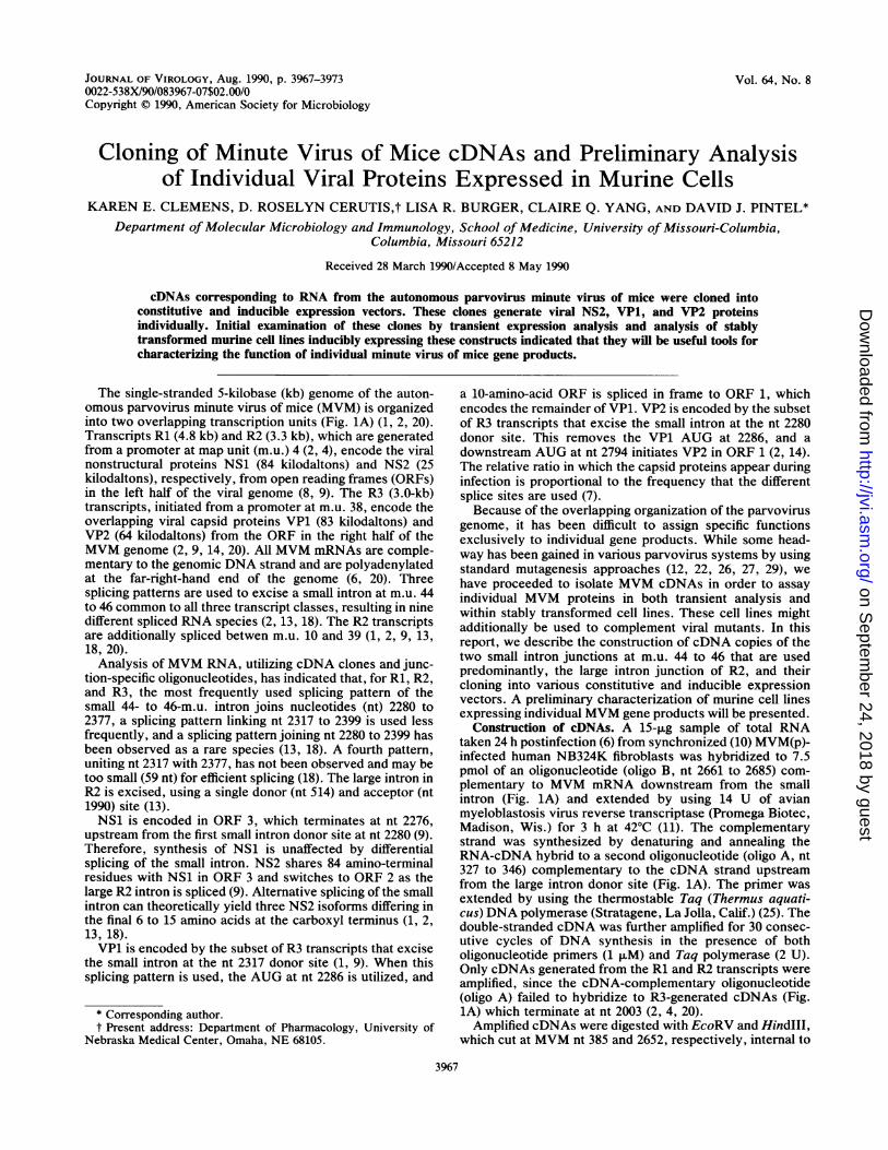
FIG. 1. (A) Map of MVM indicating the three major transcripts (Rl, R2, and R3) in relation to the ORFs deduced from sequence analysis(2). ORFs within the viral complementary strand are depicted as open areas, and termination codons are indicated by vertical lines. Oligo Aand oligo B represent oligonucleotides used in MVM cDNA synthesis and amplification as described in text. EcoRV (nt 385) and HindlIl (nt2652) restriction sites were used to generate termini for cloning cDNA sequences into the infectious clone of MVM in pML (16). Thedesignated MVM HaeIII fragment (nt 1854 to 2378) was cloned into the pGEM vector (Promega Biotec, Madison, Wis.) and used to generatethe antisense MVM RNA probe used for the RNase protection assays (see Fig. 4). (B) MMTV-MVM constructs used in the generation ofMVM protein-producing cell lines. The open box designates the MMTV long terminal repeat, and the solid line represents MVM sequences.pML vector sequences have been omitted for clarity. MMTV sequences include 976 nt of the long terminal repeat containing thedexamethasone-inducible promoter/enhancer (17). Transcription initates 111 nt upstream of the MMTV-MVM fusion. MVM sequences beginat the MVM nt 225 (2) downstream from the MVM P4 promoter for constructs producing the nonstructural protein(s) and at nt 2072downstream from the MVM P38 promoter for constructs exclusively producing the structural proteins. Transcription initiation sites arerepresented as arrows. A 759-nt deletion between two BglII sites is designated 3453/4212. Nucleotides in parentheses represent intron splicejunctions.
6060
6060
wwe..Sf f)I
J. VIROL.
on Septem
ber 24, 2018 by guesthttp://jvi.asm
.org/D
ownloaded from

NOTES 3969
the oligonucleotide primers but flanking the large and smallintron. This fragment was substituted for the analogousEcoRV-HindIII fragment of infectious MVM cloned intopML (16). cDNA-containing clones generated from RNAslacking the small intron and those lacking both the large andsmall intron were differentiated by restriction mapping andwere further characterized by sequence analysis of bothstrands across the entire insert (data not shown). In allclones examined, the large intron splice junction joins nt 514to 1990, confirming the results of previous studies (13, 18).Two classes of small intron splice junctions were obtained,as expected from previous studies, those joining nt 2280 to2377 and those joining nt 2317 to 2399. cDNA clonesrepresenting the rare splicing pattern which joins nt 2280 to2399 were not obtained.
Cloning of cDNAs in constitutive and inducible expressionvectors and production of expressing cell lines. ClonedcDNAs which lacked the large (514/1990) intron (whichremoves the P38 TATA box at approximately nt 1978) andretained the small intron or, alternatively, had the smallintron excised by either the major or minor splicing pattern,were used to construct clones which produce either all threeNS2 isoforms or, uniquely, the major or minor NS2 isoformfrom either the simian virus 40 early promoter (28) or mousemammary tumor virus (MMTV) promoter (17) (Fig. 1B). Inthese constructs, MVM sequences begin at nucleotide 225,downstream from the MVM P4 promoter. Constructs whichproduce both VP1 and VP2 or VP1 or VP2 individually wereobtained by cloning the right-hand end of the MVM genomebeginning at nucleotide 2072 downstream from the P38promoter (Fig. 1B). Constructs that retained the small intronproduced both VP1 and VP2. Those containing cDNArepresenting the major splicing pattern (2280/2377) are pre-dicted to produce VP2, and those containing cDNA repre-senting the minor splicing pattern (2317/2399) are predictedto produce VP1.When analyzed by immunoprecipitation at 48 h posttrans-
fection as described below, the simian virus 40-driven cDNAclones transfected into COS-1 cells produced the anticipatedMVM gene products as identified by comigration with au-thentic MVM proteins from a wild-type infection (data notshown). Immunofluorescence analysis of simian virus 40-driven NS2 cDNAs transfected into COS-1 cells indicatedthat without other viral gene products present, NS2 localizesas it does after transfection of wild-type MVM (data notshown).
After verification by transient analysis, cDNA clones wereconstructed that were driven by the dexamethasone-induc-ible promoter from MMTV (obtained from J. Majors, Wash-ington University, St. Louis, Mo.) (Fig. 1B) for use in thegeneration of stable cell lines expressing individual MVMgene products, as had been done previously for the relatedparvovirus Hi (24). Clones in which the MMTV promoterdrives genomic MVM beginning at nucleotide 225 were alsoconstructed. These were expected to produce NS1 and allthree isoforms of NS2 under the control of the MMTVpromoter and VP1 and VP2 under the control of the MVMP38 promoter. Although the unaltered version of the MMTVpromoter has significant activity when uninduced, it wasused in an attempt to overcome potential toxic effects ofthese MVM gene products.The MMTV-MVM constructs were cotransfected at a 30:1
ratio into A9 mouse fibroblasts along with pko-neo (28), inwhich the neomycin resistance gene is driven by the SV40early promoter, using the CaPO4 technique (19). Neomycinresistant cell clones were isolated in the presence of 1 mg of
Geneticin (G-418 Sulfate; GIBCO, Gaithersburg, Md.) perml and tested for production of MVM-specific gene productsin the presence and absence of dexamethasone by immuno-precipitation as described below.
Characterization of individual MVM gene products ex-pressed in murine cell lines. Approximately 5 x 10⁵ cells fromindividually isolated cell clones were labeled for 2 h by using0.1 mCi of [35S]methionine (Tran "S-label; ICN Radiochem-icals, Irvine, Calif.) per ml 15 to 20 h after the addition ofdexamethasone (ranging between 0.05 and 0.6 ,uM in dif-ferent experiments). A9 cells were infected with MVM at amultiplicity of 10 and labeled similarly at 15 to 20 h postin-fection as a control. MVM-specific proteins were immuno-precipitated as previously described (9) by using antisera(kindly provided by S. Cotmore and P. Tattersall) specificfor either NS1, NS2, or the amino-terminal ORF common toNS1 and NS2, or antiserum generated in response to purifiedempty virions. Multiple clonal cell lines obtained by cotrans-fection with the MVM-expressing constructs diagrammed inFig. 1B were screened by immunofluorescence as previouslydescribed (14). These lines displayed a great variability in thepercentage of expressing cells and in the magnitude of theirinducibility. Cell lines generating high levels of MVM geneproducts upon induction were chosen for further study.
Initially, MVM gene products from cell lines transfectedwith MMTV-Genomic (Fig. 1B) were analyzed. These linesproduced NS1, NS2, VP1, and VP2 that comigrated with thecorresponding proteins from a wild-type MVM infection andwere inducible by dexamethasone (Fig. 2A through C). Theenhancement of the capsid gene products in these lines (Fig.2C) was probably due to elevated levels of NS1, a knowntransactivator of the P38 promoter (21, 23, 27).
Likewise, cells transfected by the capsid protein-pro-ducing constructs MMTV-VP1/VP2, MMTV-VP1 (2317/2399), and MMTV-VP2 (2280/2377) (Fig. 1B) inducibly ex-pressed proteins of the expected size (Fig. 2C). It isinteresting that cDNA clones predicted to express VP1 didnot express detectable VP2 (despite the retention of theAUG at nt 2794 which is normally used for initiation of VP2)and that already spliced MVM transcripts were apparentlytransported to the cytoplasm and translated efficiently. Inaddition, as seen previously (14), cell lines expressing theunspliced R3 transcription unit produced VP1 and VP2 in thesame ratio as that seen in viral infection (Fig. 2C), and theygenerated empty capsids as assayed by hemagglutination ofguinea pig erythrocytes (data not shown). Immunofluores-cence analysis of these cell lines (data not shown) alsoindicated that when expressed individually, VP1 and VP2localize as they do when expressed together.
Cell lines transfected by the NS2-producing cDNA cloneswere examined next. Proteins immunoprecipitated from celllines expressing all three NS2 isoforms (MMTV-NS2) andthose producing either the major [MMTV-NS2 (2280/2377)]or minor [MMTV-NS2 (2317/2399)] isoform comigrated withthe multiple NS2 isoforms normally seen during wild-typeinfection (Fig. 2B), which were verified by Western blot(immunoblot) analysis (data not shown). In most cases,significantly more NS2 was produced in the presence ofdexamethasone. Because the cell lines expressing individualcDNA versions of NS2 generated multiple bands similar tothose observed during MVM infection, we chose to investi-gate the state of NS2 phosphorylation as a possible expla-nation for the presence of multiple bands.When NS2 immunocomplexes were treated with 1 U of
potato acid phosphatase (Boehringer Mannheim Biochemi-cals, Indianapolis, Ind.) in MES [0.1 M 2-(N-morpholino)
VOL. 64, 1990
on Septem
ber 24, 2018 by guesthttp://jvi.asm
.org/D
ownloaded from

3970 NOTES
Ag
k.:. ::__.__._______...............
B M -.14F,-.,.:-
AN# SERA
_N"ISI
_.; _;
IIu I11 "
FIG. 2. Characterization of MVM protein-producing cell lines by immunoprecipitation in the absence (-) or presence (+) of 0.6 ,uMdexamethasone (DEX). Cell lysates from 5 x 10⁵ cells were immunoprecipitated as previously described (9). Control lanes include uninfected(UN) or MVM-infected (INF) pko-neo-transformed or normal A9 mouse fibroblasts (multiplicity of infection, 10) taken 15 to 20 hpostinfection. Immunoprecipitated proteins were separated on a 10% (for VP1 and VP2) or 12.5% (for NS2) sodium dodecyl sulfate-polyacrylamide gel. (A) Immunoprecipitations of lysates from cell lines transfected with MMTV-Genomic with either preimmune (PI) orimmune (I) serum directed against a region of ORF 3 unique to NS1. The band marked with an asterisk is viral VP2 which has beennonspecifically precipitated in this experiment. (B) Immunoprecipitations of lysates from cell lines transfected with MMTV-Genomic,MMTV-NS2, MMTV-NS2 (2280/2377), or MMTV-NS2 (2317/2399) with either preimmune (PI) or immune (I) serum directed against thecarboxyl-terminal ORF common to all three NS2 isoforms (9). (C) Immunoprecipitations of lysates from cell lines transfected withMMTV-Genomic, MMTV-VP1/VP2, MMTV-VP1(2317/2399), or MMTV-VP2(2280/2377) with either preimmune (PI) or immune (I) serumdirected against purified empty capsids.
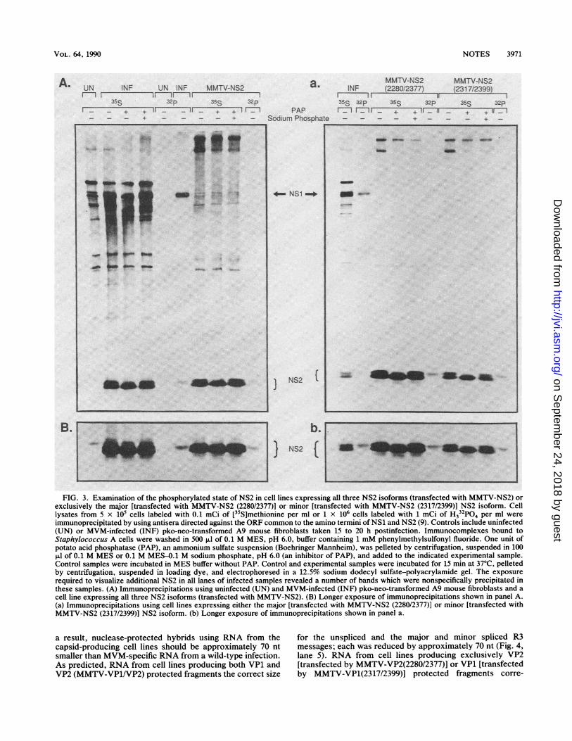
ethanesulfonic acid], pH 6.0, for 15 min at 37°C (3), theamount of the slower-migrating NS2 species was reduced(Fig. 3). When 0.1 M sodium phosphate was included as aphosphatase inhibitor in the reaction mix, the abundance ofthis species was similar to that found in untreated reactions.In addition, this species was specifically labeled when thecells were incubated for 2 h in the presence of 1 mCi ofH332P04 (ICN Radiochemicals, Irvine, Calif.) per ml, con-firming that NS2 exists in a phosphorylated state (Fig. 3).When cell lines producing either all three NS2 isoforms orexclusively the major or minor isoform were examined, theresults were the same, confirming that both the NS2 majorand minor isoforms are phosphorylated (Fig. 3). The assign-ment of individual isoforms of NS2 and their variablyphosphorylated states to particular bands on a gel willrequire further examination of these cDNA clones.
Cell lines that express either all MVM gene products(transfected with MMTV-Genomic), NS2, or capsid proteinswere stable uninduced for at least 3 weeks in culture,retaining full inducibility (data not shown). Upon continuedinduction however, cell lines that express either all MVMgene products (transfected by MMTV-Genomic), NS2 alone[transfected by either MMTV-NS2 or MMTV-NS2 (2280/2377)], or VP1 [transfected by MMTV-VP1 (2317/2399)]demonstrated decreased viability (cell lines expressing theminor NS2 isoform or VP2 alone have not yet been tested).All lines have reduced plating efficiencies and lose MVMexpression during prolonged culture (K. E. Clemens and D.J. Pintel, unpublished data). The toxicity of parvovirusnonstructural proteins has been noted previously (5, 15, 24).A quantitative characterization of the toxic effect of theseMVM proteins is in progress.
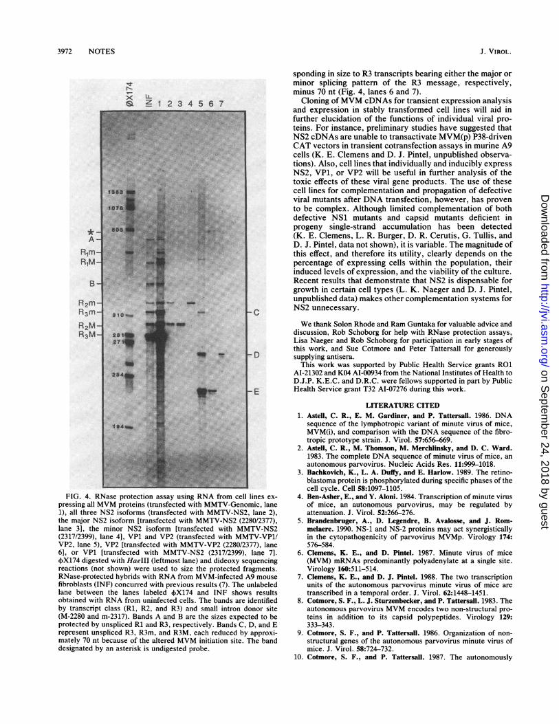
MVM-specific mRNAs produced by the murine cell lines.MVM-specific mRNAs produced by the MMTV-MVMcDNA-generated cell lines were investigated by using RNaseprotection analysis of hybrids formed between total RNAisolated from dexamethasone-induced cells and an SP6-generated antisense MVM RNA probe spanning nt 1854 to2378 (Fig. 1A) as previously described (7). The 5' terminus ofthe probe resides within the R2 large intron, and the 3'terminus is within the small intron common to all threetranscripts. This allows discrimination between the Rl, R2,and R3 transcript classes as well as those subspecies usingalternate small splice donor sites. Briefly, 10 ,ug of total RNAwas hybridized to a 40-fold molar excess of probe at 50°C for16 to 20 h, followed by digestion with 4.5 ,ug of RNase A and0.2 p.g of RNase Ti per ml at 37°C for 45 min. Thenuclease-protected hybrids were separated on an 8% poly-acrylamide-urea gel (Fig. 4).
Cell lines expressing the MMTV-Genomic construct pro-duced transcripts identical in size and similar in abundanceto those found in a wild-type MVM infection (Fig. 4, lanesINF and 1). Cell lines producing all three NS2 isoforms(transfected with MMTV-NS2) produced spliced R2 tran-scripts with both the major and minor small intron splicingpattern (Fig. 4, lane 2). As predicted, cell lines producingeither the major [transfected by MMTV-NS2 (2280/2377)] orthe minor [transfected by MMTV-NS2 (2317/2399)] NS2isoform produced R2 transcripts bearing the major and theminor small intron-splicing pattern, respectively (Fig. 4,lanes 3 and 4).
In constructs used to make the MVM capsid-producingcell lines, MVM sequences begin at nt 2072, downstreamfrom the native MVM R3 transcript start site at nt 2003. As
I!
.VP2
.. V I.,7'4 vP2-,: - :2 ---- .722!9.-' Z260. 125 7:
.
.11 -2: P:
J. VIROL.
A
on Septem
ber 24, 2018 by guesthttp://jvi.asm
.org/D
ownloaded from
VOL. 64, 1990
UN INF UN INF MMTV-NS2
35S 32p 35S 32p
a.
} i PAP- Sodium PhosDhate
- NS1 -
)
NS2 1
b.NS2 (
NOTES 3971
MMTV-NS2INF (2280/2377)
MMTV-NS2(231 7/2399)
I iI -II1135S 32p 35S 32p 35S 32prr_l F_ 11 if_ If - + + if T
~~- + - - - + -
FIG. 3. Examination of the phosphorylated state of NS2 in cell lines expressing all three NS2 isoforms (transfected with MMTV-NS2) orexclusively the major [transfected with MMTV-NS2 (2280/2377)] or minor [transfected with MMTV-NS2 (2317/2399)] NS2 isoform. Celllysates from 5 x 10⁵ cells labeled with 0.1 mCi of [35S]methionine per ml or 1 x 10⁶ cells labeled with 1 mCi of H332P04 per ml wereimmunoprecipitated by using antisera directed against the ORF common to the amino termini of NS1 and NS2 (9). Controls include uninfected(UN) or MVM-infected (INF) pko-neo-transformed A9 mouse fibroblasts taken 15 to 20 h postinfection. Immunocomplexes bound toStaphylococcus A cells were washed in 500 ,ul of 0.1 M MES, pH 6.0, buffer containing 1 mM phenylmethylsulfonyl fluoride. One unit ofpotato acid phosphatase (PAP), an ammonium sulfate suspension (Boehringer Mannheim), was pelleted by centrifugation, suspended in 100,ul of 0.1 M MES or 0.1 M MES-O.1 M sodium phosphate, pH 6.0 (an inhibitor of PAP), and added to the indicated experimental sample.Control samples were incubated in MES buffer without PAP. Control and experimental samples were incubated for 15 min at 37°C, pelletedby centrifugation, suspended in loading dye, and electrophoresed in a 12.5% sodium dodecyl sulfate-polyacrylamide gel. The exposurerequired to visualize additional NS2 in all lanes of infected samples revealed a number of bands which were nonspecifically precipitated inthese samples. (A) Immunoprecipitations using uninfected (UN) and MVM-infected (INF) pko-neo-transformed A9 mouse fibroblasts and acell line expressing all three NS2 isoforms (transfected with MMTV-NS2). (B) Longer exposure of immunoprecipitations shown in panel A.(a) Immunoprecipitations using cell lines expressing either the major [transfected with MMTV-NS2 (2280/2377)] or minor [transfected withMMTV-NS2 (2317/2399)] NS2 isoform. (b) Longer exposure of immunoprecipitations shown in panel a.
a result, nuclease-protected hybrids using RNA from thecapsid-producing cell lines should be approximately 70 ntsmaller than MVM-specific RNA from a wild-type infection.As predicted, RNA from cell lines producing both VP1 andVP2 (MMTV-VP1/VP2) protected fragments the correct size
for the unspliced and the major and minor spliced R3messages; each was reduced by approximately 70 nt (Fig. 4,lane 5). RNA from cell lines producing exclusively VP2[transfected by MMTV-VP2(2280/2377)] or VP1 [transfectedby MMTV-VP1(2317/2399)] protected fragments corre-
- }+ _i----I--------Tl rI
_ _ + - -_ - +
A.
B.
'a.''ml~~~~~~~~~~~~~~~~~~~~~~~~~~~~~~~~~~~~~~~~~~~~~~~~~~~~~~.:...
,. 9.
Se-*
_ e
m. 4 SP So_" Am_m
a-6IS ,",-- As efl__. Amokme
on Septem
ber 24, 2018 by guesthttp://jvi.asm
.org/D
ownloaded from
3972 NOTES
L-Z 1 2 3 4 5 6 7
sponding in size to R3 transcripts bearing either the major orminor splicing pattern of the R3 message, respectively,minus 70 nt (Fig. 4, lanes 6 and 7).
Cloning ofMVM cDNAs for transient expression analysisand expression in stably transformed cell lines will aid infurther elucidation of the functions of individual viral pro-teins. For instance, preliminary studies have suggested thatNS2 cDNAs are unable to transactivate MVM(p) P38-drivenCAT vectors in transient cotransfection assays in murine A9cells (K. E. Clemens and D. J. Pintel, unpublished observa-tions). Also, cell lines that individually and inducibly expressNS2, VP1, or VP2 will be useful in further analysis of thetoxic effects of these viral gene products. The use of thesecell lines for complementation and propagation of defectiveviral mutants after DNA transfection, however, has provento be complex. Although limited complementation of bothdefective NS1 mutants and capsid mutants deficient inprogeny single-strand accumulation has been detected(K. E. Clemens, L. R. Burger, D. R. Cerutis, G. Tullis, andD. J. Pintel, data not shown), it is variable. The magnitude ofthis effect, and therefore its utility, clearly depends on thepercentage of expressing cells within the population, theirinduced levels of expression, and the viability of the culture.Recent results that demonstrate that NS2 is dispensable forgrowth in certain cell types (L. K. Naeger and D. J. Pintel,unpublished data) makes other complementation systems forNS2 unnecessary.-C
_F -FIG. 4. RNase protection assay using RNA from cell lines ex-
pressing all MVM proteins (transfected with MMTV-Genomic, lane1), all three NS2 isoforms (transfected with MMTV-NS2, lane 2),the major NS2 isoform [transfected with MMTV-NS2 (2280/2377),lane 3], the minor NS2 isoform [transfected with MMTV-NS2(2317/2399), lane 4], VP1 and VP2 (transfected with MMTV-VP1/VP2, lane 5), VP2 [transfected with MMTV-VP2 (2280/2377), lane6], or VP1 [transfected with MMTV-NS2 (2317/2399), lane 7].,X174 digested with HaeIII (leftmost lane) and dideoxy sequencingreactions (not shown) were used to size the protected fragments.RNase-protected hybrids with RNA from MVM-infected A9 mousefibroblasts (INF) concurred with previous results (7). The unlabeledlane between the lanes labeled 4X174 and INF shows resultsobtained with RNA from uninfected cells. The bands are identifiedby transcript class (Rl, R2, and R3) and small intron donor site(M-2280 and m-2317). Bands A and B are the sizes expected to beprotected by unspliced Rl and R3, respectively. Bands C, D, and Erepresent unspliced R3, R3m, and R3M, each reduced by approxi-mately 70 nt because of the altered MVM initiation site. The banddesignated by an asterisk is undigested probe.
We thank Solon Rhode and Ram Guntaka for valuable advice anddiscussion, Rob Schoborg for help with RNase protection assays,Lisa Naeger and Rob Schoborg for participation in early stages ofthis work, and Sue Cotmore and Peter Tattersall for generouslysupplying antisera.This work was supported by Public Health Service grants RO1
AI-21302 and K04 AI-00934 from the National Institutes of Health toD.J.P. K.E.C. and D.R.C. were fellows supported in part by PublicHealth Service grant T32 AI-07276 during this work.
LITERATURE CITED1. Astell, C. R., E. M. Gardiner, and P. Tattersall. 1986. DNA
sequence of the lymphotropic variant of minute virus of mice,MVM(i), and comparison with the DNA sequence of the fibro-tropic prototype strain. J. Virol. 57:656-669.
2. Astell, C. R., M. Thomson, M. Merchlinsky, and D. C. Ward.1983. The complete DNA sequence of minute virus of mice, anautonomous parvovirus. Nucleic Acids Res. 11:999-1018.
3. Bachkovich, K., L. A. Duffy, and E. Harlow. 1989. The retino-blastoma protein is phosphorylated during specific phases of thecell cycle. Cell 58:1097-1105.
4. Ben-Asher, E., and Y. Aloni. 1984. Transcription of minute virusof mice, an autonomous parvovirus, may be regulated byattenuation. J. Virol. 52:266-276.
5. Brandenbruger, A., D. Legendre, B. Avalosse, and J. Rom-melaere. 1990. NS-1 and NS-2 proteins may act synergisticallyin the cytopathogenicity of parvovirus MVMp. Virology 174:576-584.
6. Clemens, K. E., and D. Pintel. 1987. Minute virus of mice(MVM) mRNAs predominantly polyadenylate at a single site.Virology 160:511-514.
7. Clemens, K. E., and D. J. Pintel. 1988. The two transcriptionunits of the autonomous parvovirus minute virus of mice aretranscribed in a temporal order. J. Virol. 62:1448-1451.
8. Cotmore, S. F., L. J. Sturzenbecker, and P. Tattersall. 1983. Theautonomous parvovirus MVM encodes two non-structural pro-teins in addition to its capsid polypeptides. Virology 129:333-343.
9. Cotmore, S. F., and P. Tattersall. 1986. Organization of non-structural genes of the autonomous parvovirus minute virus ofmice. J. Virol. 58:724-732.
10. Cotmore, S. F., and P. Tattersall. 1987. The autonomously
*A
RlmR1M
B
R2m-R3m_R2M-R3M-
J. VIROL.
on Septem
ber 24, 2018 by guesthttp://jvi.asm
.org/D
ownloaded from

NOTES 3973
replicating parvoviruses of vertebrates. Adv. Virus Res. 33:91-174.
11. Fouser, L. A., and J. D. Friesen. 1986. Mutations in a yeastintron demonstrate the importance of specific conserved nucle-otides for the two stages of nuclear mRNA splicing. Cell45:81-93.
12. Hermonat, P. L., M. A. Labow, R. Wright, K. I. Bems, and N.Muzyczka. 1984. Genetics of adeno-associated virus: isolationand preliminary characterization of mutants of adeno-associatedtype 2 mutants. J. Virol. 51:329-339.
13. Jongeneel, C. V., R. Sahli, G. K. McMaster, and B. Hirt. 1986.A precise map of splice junctions in the mRNAs of the minutevirus of mice, an autonomous parvovirus. J. Virol. 59:564-573.
14. Labieniec-Pintel, L., and D. Pintel. 1986. The minute virus ofmice P39 transcription unit can encode both capsid proteins. J.Virol. 57:1163-1167.
15. Labow, M. A., L. H. Graf, Jr., and K. I. Berns. 1987. Adeno-associated virus gene expression inhibits cellular transformationby heterologous genes. Mol. Cell. Biol. 7:1320-1325.
16. Lusky, M., and M. Botchan. 1981. Inhibition of SV40 replicationin simian cells by specific pBR322 DNA sequences. Nature(London) 293:79-81.
17. Majors, J., and H. E. Varmus. 1983. A small region of the mousemammary tumor virus long terminal repeat confers glutocorti-coid hormone regulation on a linked heterologous gene. Proc.Natl. Acad. Sci. USA 80:5866-5870.
18. Morgan, W. R., and D. C. Ward. 1986. Three splicing patternsare used to excise the small intron common to all minute virusof mice RNAs. J. Virol. 60:1170-1174.
19. Parker, B. A., and G. R. Stark. 1979. Regulation of simian virus40 transcription: sensitive analysis of the RNA species presentearly in infections by virus or viral DNA. J. Virol. 31:360-369.
20. Pintel, D., D. Dadachanji, C. R. Astell, and D. C. Ward. 1983.The genome of minute virus of mice, an autonomous parvovi-
rus, encodes two overlapping transcription units. Nucleic AcidsRes. 11:1019-1038.
21. Rhode, S. L., III. 1973. Replication process of the parvovirusH-1. I. Kinetics in a parasynchronous cell system. J. Virol.11:856-861.
22. Rhode, S. L., III. 1976. Replication process of the parvovirusH-1. V. Isolation and characterization of temperature sensitiveH-1 mutants defective in progeny DNA synthesis. J. Virol.171:659-667.
23. Rhode, S. L., III. 1985. trans-Activation of parvovirus P38promoter by the 76K noncapsid protein. J. Virol. 55:886-889.
24. Rhode, S. L., III. 1987. Construction of a genetic switch forinducible trans-activation of gene expression in eucaryotic cells.J. Virol. 61:1448-1456.
25. Saiki, R. K., D. H. Gelfand, S. Stoffel, S. J. Scharf, R. Higuchi,G. T. Horn, K. B. Mullis, and H. A. Erlich. 1988. Primer-directed enzymatic amplification of DNA with a thermostableDNA polymerase. Science 239:487-491.
26. Tratschin, J. D., I. L. Miller, and B. J. Carter. 1984. Geneticanalysis of adeno-associated virus: properties of deletion mu-tants constructed in vitro and evidence for an adeno-associatedvirus replication function. J. Virol. 51:611-619.
27. Tullis, G. E., L. Labieniec-Pintel, K. E. Clemens, and D. Pintel.1988. Generation and characterization of a temperature-sensi-tive mutation in the NS-1 gene of the autonomous parvovirusminute virus of mice. J. Virol. 62:2736-2744.
28. Van Doren, K., D. Hanahan, and Y. Gluzman. 1984. Infection ofeucaryotic cells by helper-independent recombinant adenovi-rus: early region 1 is not obligatory for integration of viral DNA.J. Virol. 50:606-614.
29. Wicker, R., and M. Gunther. 1988. Isolation and characteriza-tion of thermo sensitive mutants from Kilham Rat Virus, arodent parvovirus. J. Gen. Virol. 67:163-175.
VOL. 64, 1990
on Septem
ber 24, 2018 by guesthttp://jvi.asm
.org/D
ownloaded from