Embed Size (px)
Citation preview

Minimally InvasivePORT ACCESS
Mitral Valve Surgery

INTRODUCTION
The majority of isolated mitral valve procedures canbe performed with minimally invasive approaches thatreduce patient trauma, blood loss and hospital length ofstay, speed recovery and produce a superior cosmetic result.1-4
The most commonly employed minimally invasiveapproach for mitral valve surgery is a right mini-thoracotomy. This approach relies upon peripheralcannulation for cardiopulmonary bypass, specializedtechniques for myocardial protection, and use oflong-shafted instruments. The right mini-thoracotomyprovides excellent visualization of the mitral valveand facilitates tricuspid valve surgery or surgicalablation of atrial fibrillation when indicated.
The objective of this monograph is to illustrate the rightmini-thoracotomy approach for mitral valve surgery withparticular emphasis on techniques for cardiopulmonarybypass, myocardial protection, and valve exposure. Weillustrate a classic PORT ACCESS approach that employsthe EndoClamp catheter for aortic occlusion and a modifiedapproach that relies upon a transthoracic clamp for aorticocclusion.
Edwards Lifesciences would like to acknowledge A. Marc Gillinov, M.D. and Tomislav Mihaljevic,M.D., Department of Thoracic and Cardiovascular Surgery, Cleveland Clinic for their contributionsto the development of this monograph, Minimally Invasive PORT ACCESS Mitral Valve Surgery.
A. Marc Gillinov, MD and Tomislav Mihaljevic, MD are paid consultants to Edwards Lifesciences.
The surgical techniques presented herein are the techniques used by A. Marc Gillinov, MD andTomislav Mihaljevic, MD. Edwards Lifesciences does not endorse any particular surgical technique.

1
INTRODUCTION
MINIMALLY INVASIVEPORT ACCESS
SURGERY

ANESTHESIA PREPARATION INCLUDINGRETROGRADE CARDIOPLEGIA CATHETERAND PULMONARY ARTERY VENT
Before surgical incision, transesophageal echocardiography is used to confirm
mitral valve dysfunction and to ensure that the aortic valve is competent;
aortic regurgitation that is more than mild is a relative contraindication to a
right mini-thoracotomy approach to the mitral valve. Bilateral upper extremity
arterial monitoring lines are placed if the EndoClamp catheter is to be used
for aortic occlusion. A right internal jugular venous line is placed for possible
conversion to a venous cannula if additional venous drainage is necessary.
Using echocardiographic guidance and if required fluoroscopy assistance,
the anesthesia team places a coronary sinus catheter for delivery of retrograde
cardioplegia and a pulmonary artery vent. With experience, these catheters
can be positioned in only a few minutes.
MINIMALLY INVASIVEPORT ACCESSSURGERY
2
MINIMALLY INVASIVEPORT ACCESS
SURGERY
3
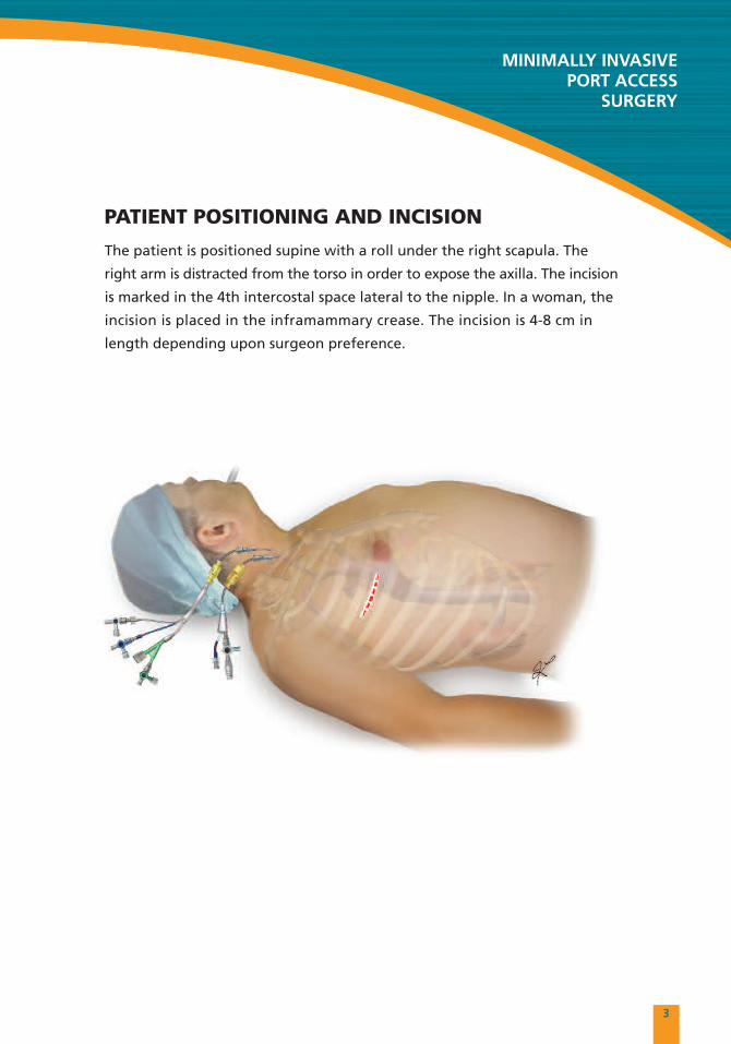
PATIENT POSITIONING AND INCISION
The patient is positioned supine with a roll under the right scapula. The
right arm is distracted from the torso in order to expose the axilla. The incision
is marked in the 4th intercostal space lateral to the nipple. In a woman, the
incision is placed in the inframammary crease. The incision is 4-8 cm in
length depending upon surgeon preference.

4
MINIMALLY INVASIVEPORT ACCESSSURGERY
SOFT TISSUE RETRACTOR
The chest is entered in the 4th intercostal space and a soft tissue retractor
is placed; the arms of the soft tissue retractor are affixed to the skin under
tension to form an “X.” A fine catheter is placed in the chest to insufflate
CO2 at a rate of 6 liters/minute.

CHEST WALL RETRACTOR
The chest wall retractor is placed over the soft tissue retractor. In most
instances, the cross bar is placed toward the patient’s left; however, it may
also be placed with the cross bar to the patient’s right. The retractor articulates
to optimize exposure. A stitch placed through the central tendon of the
diaphragm and taken through the chest wall caudal to the incision improves
exposure if the diaphragm obstructs visualization of the pericardium.
5
MINIMALLY INVASIVEPORT ACCESS
SURGERY
6
MINIMALLY INVASIVEPORT ACCESSSURGERY
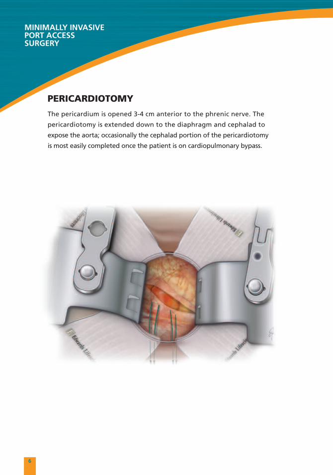
PERICARDIOTOMY
The pericardium is opened 3-4 cm anterior to the phrenic nerve. The
pericardiotomy is extended down to the diaphragm and cephalad to
expose the aorta; occasionally the cephalad portion of the pericardiotomy
is most easily completed once the patient is on cardiopulmonary bypass.
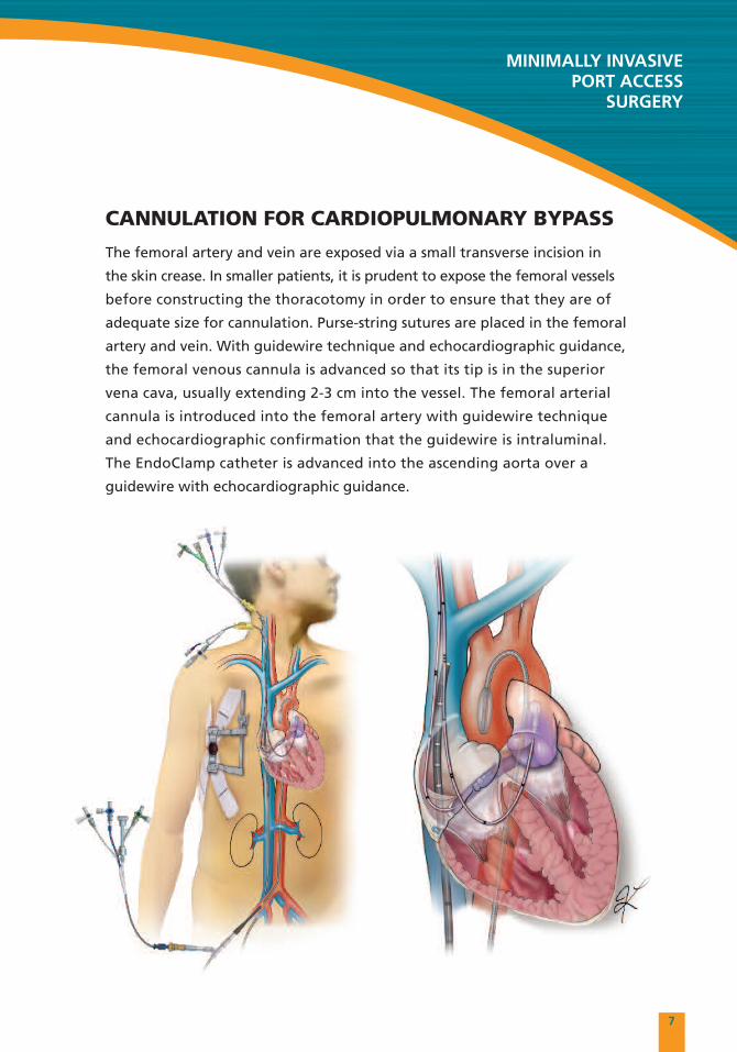
CANNULATION FOR CARDIOPULMONARY BYPASS
The femoral artery and vein are exposed via a small transverse incision in
the skin crease. In smaller patients, it is prudent to expose the femoral vessels
before constructing the thoracotomy in order to ensure that they are of
adequate size for cannulation. Purse-string sutures are placed in the femoral
artery and vein. With guidewire technique and echocardiographic guidance,
the femoral venous cannula is advanced so that its tip is in the superior
vena cava, usually extending 2-3 cm into the vessel. The femoral arterial
cannula is introduced into the femoral artery with guidewire technique
and echocardiographic confirmation that the guidewire is intraluminal.
The EndoClamp catheter is advanced into the ascending aorta over a
guidewire with echocardiographic guidance.
7
MINIMALLY INVASIVEPORT ACCESS
SURGERY

8
MINIMALLY INVASIVEPORT ACCESSSURGERY
AORTIC OCCLUSION AND CARDIOPLEGIA DELIVERY
Cardiopulmonary bypass is initiated and adequate drainage is confirmed.
Vacuum-assisted venous drainage is routinely employed. If drainage is not
adequate, a right internal jugular venous catheter placed by anesthesia is
changed over a wire to a superior vena cava cannula for cardiopulmonary
bypass. The perfusionist achieves the systemic blood pressure that will
be maintained during the case, and the EndoClamp catheter is inflated.
EndoClamp catheter inflation is accomplished in a step-wise fashion, with
constant monitoring of balloon position on echocardiogram, aortic root
pressure, and upper extremity pressures. Initially, 5 cc of saline is added to
the balloon. To prevent balloon migration towards the aortic valve, remove
excess slack from the EndoClamp catheter by gently retracting it at the groin
incision. If the balloon still migrates toward the aortic valve, it is manually
retracted with echocardiographic visualization in order to maintain position in
the ascending aorta. Saline is gradually added in 5 cc increments, maintaining
balloon position. When the aortic root pressure
falls to zero, the root is occluded and the balloon is
locked into place by the rotating hemostasis valve.
Antegrade cardioplegia delivery is started. Balloon
pressure is generally 300-350 mmHg at this point.
Antegrade cardioplegia flow is confirmed by using
color flow Doppler echocardiography to examine
the aortic root. Upper extremity pressures, aortic
root pressure and balloon pressure are monitored
throughout the operation. Myocardial protection
is achieved with a combination of antegrade
cardioplegia, retrograde cardioplegia, and mild
systemic hypothermia (28-32˚ C).
9
MINIMALLY INVASIVEPORT ACCESS
SURGERY
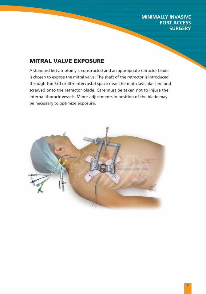
MITRAL VALVE EXPOSURE
A standard left atriotomy is constructed and an appropriate retractor blade
is chosen to expose the mitral valve. The shaft of the retractor is introduced
through the 3rd or 4th intercostal space near the mid-clavicular line and
screwed onto the retractor blade. Care must be taken not to injure the
internal thoracic vessels. Minor adjustments in position of the blade may
be necessary to optimize exposure.

10
MINIMALLY INVASIVEPORT ACCESSSURGERY
MITRAL VALVE EXPOSURE
In most cases, mitral valve exposure is excellent. Early placement of
annuloplasty sutures enhances exposure by distracting the valve toward
the surgeon.

11
MINIMALLYINVASIVE MODIFIED
PORT ACCESSSURGERY

12
MINIMALLY INVASIVEMODIFIED PORT ACCESSSURGERY
ANESTHESIA PREPARATION AND INCISION
Before surgical incision, transesophageal echocardiography is used to confirm
mitral valve dysfunction and to ensure that the aortic valve is competent;
aortic regurgitation that is more than mild is a relative contraindication to this
approach. A right internal jugular venous line is placed for possible conversion
to a venous cannula if additional venous drainage is necessary. Using
echocardiographic guidance and if required fluoroscopy assistance, the
anesthesia team places a coronary sinus catheter for delivery of retrograde
cardioplegia and a pulmonary artery vent. With experience, these catheters
can be positioned in only a few minutes. The patient is positioned supine
with a roll under the right scapula. The right arm is distracted from the torso
in order to expose the axilla. The incision is marked in the 4th intercostal space
lateral to the nipple. In a woman, the incision is placed in the inframammary
crease. The incision is 4-8 cm depending upon surgeon preference.

13
MINIMALLY INVASIVEMODIFIED PORT ACCESS
SURGERY
SOFT TISSUE RETRACTOR
The chest is entered in the 4th intercostal space and a soft tissue retractor
is placed; the arms of the soft tissue retractor are affixed to the skin under
tension to form an “X.” A fine catheter is placed in the chest to insufflate
CO2 at a rate of 6 liters/minute.

14
MINIMALLY INVASIVEMODIFIED PORT ACCESSSURGERY
CHEST WALL RETRACTOR
The chest wall retractor is placed over the soft tissue retractor. In most
instances, the cross bar is placed toward the patient’s left; however, it may
also be placed with the cross bar to the patient’s right. The retractor articulates
to optimize exposure. A stitch placed through the central tendon of the
diaphragm and taken through the chest wall caudal to the incision improves
exposure if the diaphragm obstructs visualization of the pericardium.

15
MINIMALLY INVASIVEMODIFIED PORT ACCESS
SURGERY
PERICARDIOTOMY
The pericardium is opened 3-4 cm anterior to the phrenic nerve. The
pericardiotomy is extended down to the diaphragm and cephalad to expose
the aorta; occasionally the cephalad portion of the pericardiotomy is most
easily completed once the patient is on cardiopulmonary bypass. Two stay
sutures are placed in the pericardium anterior to the phrenic nerve and
brought out through the chest wall laterally, exposing the right atrium
and right pulmonary veins.

16
MINIMALLY INVASIVEMODIFIED PORT ACCESSSURGERY
CANNULATION AND MYOCARDIAL PROTECTION
The femoral artery and vein are exposed by a small transverse incision in
the skin crease. The femoral venous cannula is advanced with guidewire
technique and echocardiographic guidance into the superior vena cava, usually
extending 2-3 cm into the vessel. The femoral arterial cannula is advanced
into the femoral artery. Cardiopulmonary bypass is established and drainage
is assessed; if drainage is inadequate, the surgeon may reposition the cannula
to achieve better drainage. Occasionally, a second venous cannula is advanced
percutaneously into the superior vena cava by rewiring a right internal jugular
venous line. Vacuum-assisted venous drainage is routinely employed. An
antegrade cardioplegia catheter is placed in the proximal ascending aorta.

17
MINIMALLY INVASIVEMODIFIED PORT ACCESS
SURGERY
TRANSTHORACIC AORTIC CROSS-CLAMP
A transthoracic aortic cross-clamp is introduced through the chest wall in
the 3rd intercostal space as posteriorly as possible; the clamp is positioned
in the transverse sinus for aortic occlusion. The clamp is oriented so that
its concave aspect is directed cephalad. Alternatively, a cross-clamp with
a flexible shaft may be introduced through the primary incision.

18
MINIMALLY INVASIVEMODIFIED PORT ACCESSSURGERY
MITRAL VALVE EXPOSURE
A standard left atriotomy is constructed and an appropriately-sized
retractor blade is chosen to expose the mitral valve. The shaft of the retractor
is introduced through the 3rd or 4th intercostal space near the mid-clavicular
line and screwed onto the retractor blade. Care must be taken not to injure
the internal thoracic vessels. Minor adjustments in position of the blade may
be necessary to optimize exposure. In most cases, mitral valve exposure is
excellent. Early placement of annuloplasty sutures enhances exposure by
distracting the valve toward the surgeon.

19
NOTES

20
NOTES


References1. Grossi E, Galloway AC, Ribicove GH, Zakow PK, Derivaux CC, Baumann FG, Schwesinger DW, Colvin SB. Impact of minimallyinvasive valvular heart surgery – a case control study. Ann Thorac Surg. 2001;71:807-810
2. Casselman FP, Slycke SV, Wellens F, De Geest R, Degrieck I, VanPraet F, Vermueulen Y, Vanermen H. Mitral Valve Surgery CanNow Routinely Be Performed Endoscopically. Circulation. 2003: 108 Suppl 1:II48-54
3. Glower DD, Siegel LC, Frischmeyer KJ, Galloway AC, Ribakove GH, Robinson NB, Ryan WH, Colvin SB. Predictors of outcomein a multicenter port-access valve registry. Ann Thorac Surgy. 2000;70:1054-9
4. Wheatley GH, Prince SL, Herbert MA, Ryan WH. Port-access aortic valve surgery: a technique in evolution. Heart Surgery Forum.2004: 7(6):E628-31
This presentation demonstrates use of Edwards Lifesciences devices by trained surgeons and is not intended to be used as a surgicaltraining guide. Physicians should be adequately trained and have familiarity with PORTACCESS surgical procedures. Refer to full labelingprovided with the Edwards Lifesciences products and consult medical literature relative to techniques, Indications for Use, Contraindicationsfor Use, Warnings and Precautions, Complications, and Hazards prior to performance of any cardiac surgical procedure. Many variables,including patient anatomy, pathology, and surgical techniques, may influence procedural approaches and outcomes. Patient and procedureselection are responsibilities of the medical professional.
A. Marc Gillinov, MD and Tomislav Mihaljevic, MD are paid consultants to Edwards Lifesciences.
The surgical techniques presented herein are the techniques used by A. Marc Gillinov, MD and Tomislav Mihaljevic, MD.Edwards Lifesciences does not endorse any particular surgical technique.
Rx only. See instructions for use for full prescribing information.
Edwards Lifesciences devices placed on the European market meeting the essential requirements referred to in Article 3 of the MedicalDevice Directive 93/42/EEC bear the CE marking of conformity.
Edwards is a trademark of Edwards Lifesciences Corporation. Edwards Lifesciences and the stylized E logo are trademarks of EdwardsLifesciences Corporation and are registered in the United States Patent and Trademark Office. PORT ACCESS is a trademark ofEdwards Lifesciences AG. EndoClamp is a trademark of Edwards Lifesciences AG and is registered in the United States Patent andTrademark Office.
© 2009 Edwards Lifesciences, LLC. All rights reserved. AR04221
Edwards Lifesciences LLC · One Edwards Way · Irvine, CA 92614 USA · 949.250.2500 · 800.424.3278 · www.edwards.comEdwards Lifesciences (Canada) Inc. · Mississauga, Ontario · Canada L5C 4R3 · 905.566.4220 · 800.268.3993
Edwards Lifesciences Europe · Ch. du Glapin 6 · 1162 Saint-Prex · Switzerland · 41.21.823.4300Edwards Lifesciences Japan · 2-8 Rokubancho · Chiyoda-ku, Tokyo 102-0085 · Japan · 81.3.5213.5700