Embed Size (px)
Citation preview
7
Minimally Invasive Aortic Valve Surgery - New Solutions to Old Problems
Juan Bustamante, Sergio Cánovas and Ángel L Fernández Hospital Universitario La Princesa, Madrid
Hospital General Universitario de Valencia & Hospital Clinico Universitario de Santiago de Compostela
Spain
1. Introduction
Aortic stenosis is the most frequent valvulopathy in the western world and basically affects people over 60 years old. It is currently the most common cause of valve replacement in Europe and North America (Schmitto et al., 2011) and its incidence increases with age. (Lung et al., 2003) 25% of people over 65 suffer sclerosis of the aortic valve which can be detected in image tests such as echocardiography and 3% of those over 75 develop aortic stenosis (Lindroos et al., 1993; Stewart et al., 1997). Approximately 16% of patients with aortic valve sclerosis develop stenosis within the space of 7 years. (Cosmi et al., 2002) This rate of progression is variable and has been related with factors common to those for arterial sclerosis. The average reduction in the valve area has been stated as being around 0.1 cm2 per year. (Otto et al., 1997) and the annual increase in gradient as about 10 mmHg. Rheumatic etiology is not common in the more developed countries and its origin is usually degenerative. In approximately half of the cases there usually exists a bicuspid aortic valve basis. Bicuspid aortic valve valvulopathy affects 2% of the population (Kurtz & Otto, 2010) and constitutes the most frequent congenital anomaly. Aortic valve stenosis is more prevalent in men than women (Chambers et al., 2009). This disease normally remains asymptomatic for a long time and represents a low sudden death risk of less than 1%. When stenosis becomes symptomatic, however, the prognosis is much worse. The symptoms usually appear at the end of the disease with three of them being typical and each one of them determining a state which is both evolutionary and prognostic: dyspnea (due to heart faillure), angina and syncope. In aortic stenosis there co-exist three physiological phenomena: cardiac ischemia, elevated pressure in the left ventricle and diminished cardiac output. Diminished tolerance to exercise is generally the first symptom of aortic stenosis and could be the result of the three previously mentioned phenomena. A careful medical history investigation is necessary in order to obtain this information as the earliest clinical signs may be insidious and may not be accompanied by dyspnea as such, and may be consciously or unconsciously disguised by the patient in the progressive restriction of their day-to-day activity as the disease develops.
www.intechopen.com
Aortic Stenosis – Etiology, Pathophysiology and Treatment
92
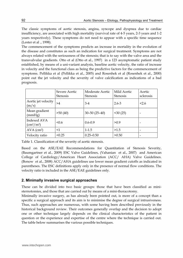
The classic symptoms of aortic stenosis, angina, syncope and dyspnea due to cardiac insufficiency, are associated with high mortality (survival rate of 4-5 years, 2-3 years and 1-2 years respectively). These symptoms do not need to appear with a specific time sequence (Lester et al ., 1998). The commencement of the symptoms predicts an increase in mortality in the evolution of the disease and constitutes as such an indication for surgical treatment. Symptoms are not always related with the seriousness of the stenosis; that is to say with the valve area and the transvalvular gradients. Otto et al (Otto et al., 1997) in a 123 asymptomatic patient study established, by means of a uni-variant analysis, baseline aortic velocity, the ratio of increase in velocity and the functional class as being the predictive factors for the commencement of symptoms. Pellikka et al (Pellikka et al., 2005) and Rosenhek et al (Rosenhek et al., 2000) point out the jet velocity and the severity of valve calcification as indications of a bad prognosis.
Severe Aortic Stenosis
Moderate Aortic Stenosis
Mild Aortic Stenosis
Aortic sclerosis
Aortic jet velocity (m/s)
>4 3-4 2.6-3 <2.6
Mean gradient (mmHg)
>50 (40) 30–50 (25–40) <30 (25) -
Indexed AVA (cm2/m2)
<0.6 0.6-0.9 >0.9 -
AVA (cm2) <1 1-1.5 >1.5 -
Velocity ratio <0.25 0.25–0.50 >0.50 -
Table 1. Classification of the severity of aortic stenosis.
Based on the ASE/EAE Recommendations for Quantitation of Stenosis Severity, (Baumgartner et al., 2009) ESC Valve Guidelines, (Vahanian et al., 2007) and American College of Cardiology/American Heart Association (ACC/ AHA) Valve Guidelines. (Bonow et al., 2008) ACC/AHA guidelines use lower mean gradient cutoffs as indicated in parentheses. The ESC definitions apply only in the presence of normal flow conditions. The velocity ratio is included in the ASE/EAE guidelines only.
2. Minimally invasive surgical approaches
These can be divided into two basic groups: those that have been classified as mini-sternotomies, and those that are carried out by means of a mini-thoracotomy. Minimally invasive surgery, as has already been pointed out, is more of a concept than a specific a surgical approach and its aim is to minimise the degree of surgical intrusiveness. Thus, such approaches are numerous, with some having been described previously in the historical background review. Their outcomes generally overlap and the decision to adopt one or other technique largely depends on the clinical characteristics of the patient in question or the experience and expertise of the centre where the technique is carried out. The table below summarises the various possible techniques.
www.intechopen.com

Minimally Invasive Aortic Valve Surgery - New Solutions to Old Problems
93
Partial sternotomy Para-sternal incision (Cohn et al., 1997; Navia & Cosgrove 1996) Trans-sternal incision (Cohn et al., 1997) Upper sternotomy (Byrne et al., 2000) T mini-sternotomy (Stamou et al., 2003) V-shaped incision (Corbi et al., 2003) Inverted L incision (Stamou et al., 2003) Reversed L incision (Detter et al., 2002) J incision (Cohn et al., 1997; Doll et al., 2002) Reversed C incision Inverted T incision (Farhat et al., 2003) Thoracotomy Right anterior thoracotomy 2º or 3º inter-costal space (Burfeind et al., 2002) Right anterior thoracotomy 4º or 5º inter-costal space (Sharony et al., 2003) Video-assisted vision Port access (Galloway et al., 1999) Video-direct vision AESOP 3000 (Computer Motion, Goleta, CA) ( Falk et al., 1998) Da Vinci System (Intuitive Surgical, Sunnyvale, CA) (Carpentier et al., 1998) Zeus (Computer Motion, Goleta, CA) (Cohn et al., 1997)
Table 2. Minimally Invasive Approaches.
At present, a partial upper sternotomy is the most frequently used incision for a minimally invasive approach to the aortic valve. (Ehrlich et al., 2000). In the case of a thoracotomy approach, this is usually carried out via a parasternal incision over the second and third inter-costal space, which is of 6 to 10 centimetres long, and then proceeds with the re-sectioning of the costal cartilages, finally arriving at the pericardial aperture (Cohn et al., 1997). Other approaches include a partial upper sternotomy with central cannulation, transverse sternotomy, limited sternotomy with a J-incision, right-sided partial sternotomy, reversed L sternotomy, and a limited right thoracotomy for aortic valve replacement (AVR). (Cosgrove et al., 1998; Izzat et al., 1998; Konertz et al., 1996; Svensson et al., 1996; Svensson et al., 1997; Tam et al., 1998; Walther et al., 1999). The most frequently used techniques can be seen in the following figures.
3. Background to minimally invasive surgery
Surgical treatment of valvulopathies was one of the most important medical advances of the twentieth century. This advance began in 1923 with the closed mitral commissurotomy of Cutler and has continued until the present day with the implanting of more complex hemodynamic prostheses with approaches different from the conventional ones. Aortic valve replacement has permitted thousands of lives to be saved since it was first successfully carried out by Harken and Starr in 1960 (Harken et al., 1961; Starr & Edwards, 1961). Cardiac surgery for the treatment of valvulopathies has also advanced considerably through the improvement of various aspects such as circulatory support, myocardial protection measures and, of course, the development of new surgical approaches that have come to be called minimally invasive, which tend to be less surgically intrusive and which will be dealt with later in this text.
www.intechopen.com

Aortic Stenosis – Etiology, Pathophysiology and Treatment
94
Fig. 1. Right anterior thoracotomy 2º
Fig. 2. Reversed C incision 3º inter-costal space
www.intechopen.com

Minimally Invasive Aortic Valve Surgery - New Solutions to Old Problems
95
Fig. 3. T mini-sternotomy
Fig. 4. Reversed L incision
www.intechopen.com
Aortic Stenosis – Etiology, Pathophysiology and Treatment
96
Initially it was in other fields of surgery such as obstetrics, thoracic surgery, urology or general surgery where minimally invasive approaches were developed but they have finally been incorporated into the area of cardiac surgery with their late incorporation largely being due to the very characteristics of this speciality (Cohn et al., 1997). The traditional approach to the treatment of aortic valvulopathy was based on sternotomy with arterial cannulation in the ascendant aorta and venous cannulation in the right atrium. As time passed and surgical techniques developed and improved the objective of reducing surgical aggression by means of using minimally invasive approaches arose. Normal sternotomy has the advantage of a direct view of all the cardiac structures and likewise access to them when compared with other approaches. It has recently been postulated that the use of partial sternotomy in its various forms or mini-thoracotomy, by virtue of being a less intrusive method, could minimise the risk of infection in the wound, loss of blood and postoperative pain without putting surgical results at risk. (Cuenca et al., 1998). Minimally invasive surgery in the realm of cardiac pathologies began with the use of slight thoracotomies without extra-corporal circulation in order to carry out myocardial re-vascularisation, without circulatory support, fundamentally on the front face of the heart (Benetti et al., 1991; Buffolo et al., 1996; Arom et al., 1996, Subramanian et al., 1995; Califiore et al., 1997). At the same time the system of Port-Access from Heartport, Inc., Redwood City, CA, (Stevens et al., 1996) was being developed for the carrying out of myocardial re-vascularisation with cardiac arrest and endovascular clamping. Eventually the development of minimally invasive techniques was incorporated into the treatment of valvulopathies. The development of minimally invasive approaches for the treatment of mitral and aortic pathologies has occurred simultaneously, but here the focus is on aortic approaches. In contrast with minimally invasive myocardial re-vascularisation surgery, which requires special resources and exhaustive training, aortic valvulopathy treatment, originally described by Dr. Cosgrove of the Cleveland Clinic, does not require either special equipment or extensive training. Thus it was that minimally invasive surgery began to be incorporated into the field of cardiac surgery in the 1990s. Some of the first reports of this approach were made by Cosgrove, Minale and Hirose. In the following years the application of the technique and the means for carrying it out had improved considerably, as will later be seen, guaranteeing good results that were comparable with those obtained through a traditional surgical approach. In 1996 Cosgrove and Sabik described a technique with which it was possible to effect aortic valve replacement by means of a mini-thoracotomy through a right parasternal incision 10 cm long. In the technique that they described they performed the peripheral cannulation by means of the femoral artery and vein. The incision ran from the lower edge of the second rib cartilage to the upper edge of the fifth rib cartilage. Then the main pectoral muscle was dissected and the third and fourth rib cartilages were resected. The internal mammary artery was bound just below the second and above the fifth rib cartilage. The inter-costal muscles and the pleura were opened at the edge of the sternum and the pericardium was incised allowing the ascendant aorta to be exposed (Cosgrove & Sabik, 1996). About the same time, Hirose described left side thoractomy for the treatment of aortic valvulopathy (Hirose et al., 1994). Since then there has been a growing interest in minimally invasive approaches due to the results observed. In 1997 Minale et al reported on a series of 27 patients where a right
www.intechopen.com
Minimally Invasive Aortic Valve Surgery - New Solutions to Old Problems
97
parasternal technique with an 8 cm long incision was used. This group accessed the thoracic cavity through the third inter-costal space. The internal mammary artery was tied and sectioned. These authors carried out cannulation on the ascendant aorta and the atrial appendage in order to avoid dissecting the femoral vessels and the morbidity that goes with this (Minale et al., 1997). In 1998 Izzat described another process for the treatment of aortic valvulopathy by means of upper T mini-sternotomy. In the first series that he published he included 9 patients. This technique was carried out by making a 6 cm vertical incision from two finger widths below the sternal fork to the area adjacent to the fourth rib. They performed an upper mini-sternotomy and it was extended laterally and caudally to the level of the third inter-costal space. One of the potential benefits of this procedure was to avoid tying the mammary vessels as well as not having to dissect femoral vessels. It had the advantage that, in case of any complications related with the technique, it could be extended and converted into a complete sternotomy in a short space of time (Izzat et al., 1998). Other similar approaches with the same aim were also described at that time, such as that of Konertz et al, which proposed a technique by means of a sternotomy in J in order to reduce intrusion (Konertz et al., 1996). Rodriguez et al described a series of 25 patients upon whom the technique of an inverted ‘L’ mini-sternotomy through an upper sternal incision for cardiac operations was used. An 8 cm vertical incision was made and then extended laterally to the right to the level of the third or fourth inter-costal space (Rodríguez et al., 1996). While surgeons have been developing new focal points for surgical techniques, the medical industry has also been capable of putting specifically designed stents, instruments, devices and equipment at their disposal for this therapeutic technique, allowing it to be simplified and improving its results as well as broadcasting them and making them more accessible.
4. Characteristics of minimally invasive surger
4.1 Definition The Society of Thoracic Surgeons National Database defines minimally invasive surgery as “any procedure that has not been performed with a full sternotomy and cardiopulmonary bypass support. All other procedures, on or off pump with a small incision or off pump with a full sternotomy are considered minimally invasive.” (STS National Database, 2003). This definition can lead to contradictions depending on the type of approach and the different forms of cannulation used to carry out the surgery. For example, should a mitral valve replacement via a mini-thoracotomy with peripheral femoral and jugular cannulation be considered a minimally invasive procedure? Faced with this problem Chitwood suggests that the concept of minimally invasive surgery is not limited to a specific approach, but is a philosophy in surgical treatment which aims to reduce the degree of surgical aggression (Chitwood & Gulielmos, 2003). In the case of coronary pathology the focus of minimally invasive surgery is on surgical approaches and the extraction of transplants using endoscopic systems, but it also includes circulatory support. The intention is to avoid extra-corporal circulation which is one of the most serious surgical aggressions due to the inflammatory response it provokes. However, in the case of valve surgery, extra-corporal circulation is necessary in order to be able to carry out the surgical procedure, so a less invasive approach is not possible. Attempts to reduce aggression have therefore been centred on the surgical approaches.
www.intechopen.com
Aortic Stenosis – Etiology, Pathophysiology and Treatment
98
There are many options in terms of ways to carry out the procedure using a minimally invasive approach. We must consider, for example, the location of the cannulations (central or peripheral), the type of aortal clamping (external or endovascular) and the form of administering the cardioplegia (antegrade, retrograde or direct via the coronary ostium). All these variants make it difficult to compare results between the different published studies, due to the heterogeneity, not only of the procedures, but also of the sample groups analysed.
4.2 Anaesthesia - Cannulation
Preoperative preparation in patients about to undergo a minimally invasive procedure is similar to that for conventional surgery, with some small differences such as: 1) selective pulmonary intubation in some of the approaches, this tends to be restricted to approaches via mini-thoracotomy for treating mytral tricuspid and congenital pathologies, and some processes involving tumours. 2) The necessity to monitor the pressure curve in both radial arteries if an endoclamp® is used. 3) In the case of approach via mini-thoracotomy the patient should be placed in the supine position with a slight elevation of the right hemithorax by placing the roll at the level of the right shoulder in order to facilitate exposure. The routine use of transesophageal echocardiography is necessary not only to assess the results of the intervention and the purging of air from the cardiac cavities but also to monitor the correct positioning of the cannulas. It is also necessary to install external defibrillator paddles as, given the size of the incision, it may not be possible to insert even paediatric paddles. It is also necessary to consider the possible limits to their effectiveness due to the poor contact with the epicardial surface (Woo et al., 2007).
4.3 Cannulation
Cannulation will depend on the type of procedure undertaken, the approach used and the experience of the team. In the beginning peripheral cannulation was used, as described by Cosgrove. That approach required the dissection of the femoral vessels. Many groups opt for direct cannulation in mininvasive procedures both with partial sternotomy and with mini-thoracotomy to avoid morbidity and reduce surgical aggression. On the other hand, as surgical teams have gained experience it has been possible to apply central cannulation without significant reduction to visibility in the operating field, and without significant increase in technical difficulty.
Direct
The cannulation of the ascendant aorta, close to the aortic arch, is to facilitate exposure selecting a zone free of plaques.
Peripheral
Through an incision in the femoral region the femoral vessels are dissected, isolating the vessels and proceeding to direct cannulation. Another option is percutaneous cannulation (Seldinger technique). It must be borne in mind that in the case of carrying out the procedure by Heart Port and endovascular clamping using endoclamp, larger gauge cannulas, for example 23 F, may be necessary. In these cases it is important to carefully assess the vascular tree using a transesophageal echocardiogram, to rule out the existence of arteriosclerosis or aortic aneurysm. Some teams recommend a prior arteriograph or angio-CT scan. It is important to consider the patient's body surface area, which will be in accordance with the diameter of the vascular tree.
www.intechopen.com
Minimally Invasive Aortic Valve Surgery - New Solutions to Old Problems
99
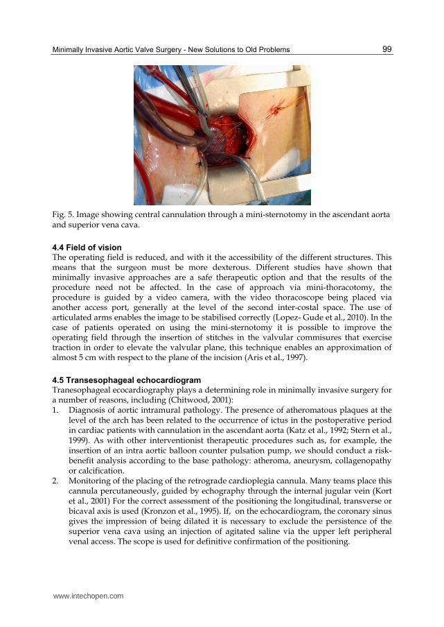
Fig. 5. Image showing central cannulation through a mini-sternotomy in the ascendant aorta and superior vena cava.
4.4 Field of vision The operating field is reduced, and with it the accessibility of the different structures. This means that the surgeon must be more dexterous. Different studies have shown that minimally invasive approaches are a safe therapeutic option and that the results of the procedure need not be affected. In the case of approach via mini-thoracotomy, the procedure is guided by a video camera, with the video thoracoscope being placed via another access port, generally at the level of the second inter-costal space. The use of articulated arms enables the image to be stabilised correctly (Lopez- Gude et al., 2010). In the case of patients operated on using the mini-sternotomy it is possible to improve the operating field through the insertion of stitches in the valvular commisures that exercise traction in order to elevate the valvular plane, this technique enables an approximation of almost 5 cm with respect to the plane of the incision (Aris et al., 1997).
4.5 Transesophageal echocardiogram Tranesophageal ecocardiography plays a determining role in minimally invasive surgery for a number of reasons, including (Chitwood, 2001): 1. Diagnosis of aortic intramural pathology. The presence of atheromatous plaques at the
level of the arch has been related to the occurrence of ictus in the postoperative period in cardiac patients with cannulation in the ascendant aorta (Katz et al., 1992; Stern et al., 1999). As with other interventionist therapeutic procedures such as, for example, the insertion of an intra aortic balloon counter pulsation pump, we should conduct a risk-benefit analysis according to the base pathology: atheroma, aneurysm, collagenopathy or calcification.
2. Monitoring of the placing of the retrograde cardioplegia cannula. Many teams place this cannula percutaneously, guided by echography through the internal jugular vein (Kort et al., 2001) For the correct assessment of the positioning the longitudinal, transverse or bicaval axis is used (Kronzon et al., 1995). If, on the echocardiogram, the coronary sinus gives the impression of being dilated it is necessary to exclude the persistence of the superior vena cava using an injection of agitated saline via the upper left peripheral venal access. The scope is used for definitive confirmation of the positioning.
www.intechopen.com
Aortic Stenosis – Etiology, Pathophysiology and Treatment
100
3. Whether or not the cannula is placed percutaneously it is vital to asses it using echocardiography because we will not have other information, for example that obtained by palpation. Monitoring pressure has not been demonstrated to be sufficient to ensure correct positioning.
4. Positioning of the femoral venous cannula and its progression towards the right atrium. 5. Assessment of air in cavities before the patient is weaned off cardiopulmonary by-pass
to avoid gas embolism and the dysrhythmias resulting from the entry of air in the right heart.
6. Analysis of the segmental contractility and assessment of cardiac function. 7. In patients where the mini-sternotomy approach is used, on occasion it is observed that
when weaning from cardiopulmonary by-pass the right ventricle is under-filled, combined with an increase in central venal pressure. This phenomenon is due to a compression of the front face of the right ventricle by an undivided lower sternum on the anteriorly retracted heart. This can be assessed using echocardiography.
8. The result of the valve surgery in the case of both valve repair and replacement. 9. Location of the level of sternal division, the assessment of the location of the aortic ring
provides orientation as to whether we should extend the sternotomy into the third or fourth inter-costal space with a view to improving access (Sardari et al., 1997).
4.6 Aortic cross clamp 4.6.1 External clamping Aortic clamping in order to be able to carry out the surgical procedure can be done using external pincers (the conventional or classic method) both in the mini-sternotomy or mini-thoractotomy, or using endoluminal clamping with the aid of an inflatable balloon, the endoclamp®. In the case of treatment for aortic pathology this latter method is not used but we will take this opportunity to review some of the concepts. In aortic valvulopathy treatment using minimally invasive surgery, aortic clamping in the case of the mini-sternotomy would be done directly with a conventional clamp. In the case of patients treated via the mini-thoracotomy with Port Access, the Cosgrove flexible aortic cross-clamp or the Glauber tend to be used, via an incision at the level of the third inter-costal space.
4.6.2 Endoclamp®
The endoclamp is introduced via the femoral arterial cannula. It presents a series of advantages and disadvantages as against external clamping. The endoclamp® is positioned at the level of the sinotubular junction and enables the administration of antegrade cardioplegia. The endoclamp® is also equipped with a vent that enables root venting of the air retained in the cardiac cavities. To facilitate placing the endoclamp® and to avoid its possible migration during positioning 0.25 mg/kg of adenosine are administered. The position and correct inflation of the clamp are monitored using echocardiography or radiology. In the case of patients where endovascular clamping is used, which is fundamentally those patients with mitral tricuspid valvulopathies, it is important to be aware that the correct positioning of the clamp is necessary in order to avoid its migration and the possible occlusion of the brachiocephalic trunks which may cause neurological damage as it compromises circulation. That is why it is so important to monitor both radial arteries as well as having a trans-cranial Doppler to assess correct cerebral perfusion.
www.intechopen.com
Minimally Invasive Aortic Valve Surgery - New Solutions to Old Problems
101
The principal complication that can be present with the use of the endoclamp® is aortic dissection which, although it has reduced in frequency as teams have gained experience (1.3% initially as against 0.2% currently), nonetheless has a very high morbimortality (Galloway et al., 1999).
5. Profile of candidates for minimally invasive surgery: indications and contraindications
In principle any patient is a candidate for minimally invasive approaches because of the possible benefits. The different published studies discuss patient profiles in which the application of the technique may be more beneficial.
5.1.1 Elderly
The elderly are a group which has been singled out for the special benefits that minimally invasive surgery can have, given the rapid recovery of patients and the improved clinical evolution. Sharony et al, in a retrospective case-control study of elderly patients (average age 75.3 years) observed a reduction in hospitalisation. Overall, elderly patients are considered to be a high risk group for surgery as expressed in the different risk scales such as the EUROSCORE (Nashef et al., 2002). It is in this type of patient that minimally invasive surgery is thought to be potentially most beneficial. (Sharony et al., 2003).
5.1.2 Re-intervention in coronary patients
It is a widely accepted opinion that reoperations are associated with an increase in morbidity and mortality not only derived from patient-specific clinical factors (i.e. old age and increased comorbidities) but also from the complexity of the surgical technique due to the absence of pericardial closure after initial heart surgery and the development of adhesions. Reoperation status remains an independent predictor of operative death after adjusting for confounding factors. The aorta, right ventricle, and bypass grafts may adhere to the underside of the sternum and can be easily injured during reoperation. In-hospital mortality for patients undergoing cardiac reoperation is higher than that of patients undergoing primary surgical interventions (Yap et al., 2009; Yau et al.., 2000) Previous surgical revascularization is a predictive factor for early mortality (Van Eck et al., 2002). Many authors have proposed reduced approaches with a view to minimising this risk (Byrne et al., 2000; Byrne et al., 1999; Svensson et al., 2001; Tabata et al., 2008). Gaeta et al., in a study of 16 patients with LIMA to LAD analyse the mini-sternotomy as an alternative to the complete sternotomy with a view to reducing the incidence of complications associated with the dissection. Byrne JG et al analyse the results of 39 patients who, following prior revascularization required reintervention due to the development of aortic valvulopathy. In the series of patients analysed they observed a significant reduction in blood loss and in the need for hemoderivatives. It is possible that minimally invasive approaches avoid the potential damage caused by dissecting the structures of the mediastinum as the focus is on the ascendant aorta and partially on the right atrium (Byrne et al., 2000). Myocardial protection was carried out via antegrade or retrograde cardioplegia observing that it was not necessary to dissect and exclude the coronary transplants for correct myocardial protection, as body temperature was reduced (20 degrees in the case of patients with LIMA to LAD and 25 degrees in all cases) and cold cardioplegia was administered, the heart received optimal protection (Byrne et al., 1999).
www.intechopen.com
Aortic Stenosis – Etiology, Pathophysiology and Treatment
102
5.1.3 Contraindications
There are no clearly defined contraindications however it is not recommended to use minimally invasive surgery in cases where the technical difficulty or risk would be increased without justification (von Segesser et al., 1999; Lopez- Gude et al., 2010; Ehrlich et al., 2000): 1. Patients with significant esophageal stricture of any etiology, which means
echocardiography monitoring cannot be used. 2. Patients with pectum excavatum 3. Patients with morbid obesity 4. Patients in which it is intended to use the endoclamp® and who present aortic
regurgitation of a grade greater than I and diameter of the sinotubular junction greater than 35 mm
5. Patients in whom assistance via peripheral cannulation is proposed and who have a history of peripheral arteriosclerosis.
6. In patients where there is documented existence of aortic pathologies such as aneurysms, atheromatous plaques, intramural hematomas or certain collagenopathies the use of the endoclamp may be inadvisable
7. Cardiac ischemia 8. Short or long ascendant aorta 9. Calcifications in the ascendant aorta or porcelain aorta 10. Small aortic ring to which it is planned to apply techniques for expanding the ring
6. Advantages of minimally invasive approaches in the treatment of aortic stenosis
The principal advantages of minimally invasive surgery are associated with the results it obtains, that is to say, to the clinical benefits derived from the technique as well as other benefits fundamentally related to the field of management: the reduction of time spent in intensive care units and overall hospitalisation or possible cosmetic effects. The first published study in which minimally invasive surgical procedures were carried out saw a reduction in the average hospitalisation time, the need for hemoderivatives and a faster functional recovery. Nevertheless they also observed an increase in surgery time (extra-corporal circulation and aortic clamping) (Frazier et al., 1998). There are significant discrepancies between the results of some of the studies listed. Some can be explained by the characteristics of the sample studied, as they deal with samples that are not clinically comparable, or the techniques applied are not superimposable. Others can be explained as due to failings in the design of the study. The interest that minimally invasive surgery has awoken is based on the idea that these less intrusive approaches mean less surgical aggression and therefore less postoperative pain, less hospitalisation time, faster functional recovery and greater cosmetic benefits as the size of scars is reduced (Rao et al., 1993; Wang et al., 2003; Klokocovnik, 1998; Szwerc et al., 1999; Aris et al., 1999; Bonacchi et al., 2002; Caffarelli & Robbins., 2004). Results have changed since the early days of the application of minimally invasive techniques leading, as we will see, to the construction of a safe and effective surgical option for the treatment of aortic valvulopathy. Below we analyse some of the aspects in which minimally invasive surgery offers certain advantages in comparison with conventional treatment.
www.intechopen.com
Minimally Invasive Aortic Valve Surgery - New Solutions to Old Problems
103
6.1 Blood loss
One of the potential advantages attributed to minimally invasive surgery is the reduction of post-operative bleeding, and therefore the need for hemoderivatives. Many studies support this, including Cosgrove et al, Tam et al, Bonachi et al and Macchler et al. One of the limitations of these studies was that the analysis of the effects of minimally invasive approaches on blood loss were carried out without adjusting for pre-operative risk factors or without conducting a multivariant analysis in order to clarify this issue. (Cosgrove et al., 1998; Tam & Almeida, 1998; Bonacchi et al., 2002; Machler et al., 1999). There is a generalised consensus about the possible reduction of bleeding rates, however there is also discordant data on the subject. Stamou et al. conducted a study analysing a sample of 511 patients in which 455 (89%) were treated using sternotomy and 56 (11%) underwent mini-AVR. The aim of the study was to compare perioperative clinical outcomes, transfusion requirements, and early mortality in patients who had mini-AVR versus conventional AVR by using a risk stratification model to adjust for potential differences between the two groups. Nevertheless after adjusting for risk factors the conclusion was reached that there were no differences in the rate of bleeding observed between the two groups (odds ratio [OR] of transfusion postoperatively =0.94, 95% confidence interval [CI] =0.42 to 2.12], p =0.88; OR of transfusion intraoperatively =0.79, 95% CI =0.45 to 1.40, p =0.42). This data contrasts with that of Dogan et al, who found, in a randomised study, that there was a reduction in the amount of surgical drainage in the group of patients treated using minimally invasive approaches. It seems that the evidence of the different studies, even with the methodological limitations to the design and the variability of the groups studied, suggests a reduction in surgical drainage and therefore in the need for hemoderivatives in the immediate postoperative period.
6.2 Pain
The reduction of pain felt by the patient and the demand for analgesics in the immediate postoperative period assessed according to different subjective scales is another aspect that has been analysed at great length, and in principle there is consensus as to the benefits of minimally invasive techniques. There is a certain amount of variation in the ways in which this assessment has been made, however, despite these variations, the results agree. Studies by Candaele et al or Bonacchi et al are among those that record this benefit in comparison with conventional approaches (Candaele et al., 2003; Bonacchi et al., 2002; Liu et al.., 1999). The upper partial sternotomy offers the comfort factor of sternotomy over thoracotomy and prevents complications of other distentions at the costovertebral joint or brachial plexus traction at the thoracic inlet. This causes a reduction in the pain felt by the patient. (Von Segesser et al., 1999).
6.3 Pulmonary function
As the integrity of the thorax is maintained and pain, which is one of the factors that most affects movement in the thorax, is reduced, it is logical to expect that the parameters of pulmonary function will be less affected by minimally invasive approaches. Various authors have analysed this aspect and the results suggest that there is a smaller fall in respiratory parameters using spirometry following intervention with minimally invasive approaches (Moustafa et al., 2007).
www.intechopen.com
Aortic Stenosis – Etiology, Pathophysiology and Treatment
104
Nevertheless, as with the other results obtained pertaining to minimally invasive techniques there are certain discrepancies in this regard (Calderon et al., 2009). Aris et al, in a randomised study of 40 patients did not find any benefits in the respiratory parameters, patients showed a reduction of 26% and 33% in FVC and of 22% and 35% in FEV1 in minimally invasive groups as against sternotomy ones. (Aris et al., 1999) In this analysis it is possible differences were not observed between the groups due to the size of the sample studied.
6.4 Duration of hospital stay and functional recovery
One of the objectives of minimally invasive approaches is to reduce surgical aggression and thus favour functional recovery. The benefit of these approaches in terms of the impact on the duration of hospitalization is quite uniform, and the majority of authors observe benefits in the reduction of the average hospital stay both in time spent in intensive care and total time in hospital. In this vein we find the results of Bonacchi et al, Brinkman et al, Grossi et al, Moustafa M A et al or Liu J et al (Bonacchi et al., 2002; Brinkman et al., 2010; Grossi et al., 2001; Moustafa et al., 2007; Liu et al., 1999). Patients operated on using minimally invasive surgery present, in general terms, less time on mechanical ventilation than patients operated on in the conventional way. It is important to remember that the most significant risk factor in the development of respiratory infection complications is the duration of intubation. It is possible that through minimally invasive surgery there may be a reduction in the incidence of certain infectious complications such as the pneumonia associated with mechanical ventilation. As with other aspects analysed, more studies are required with larger sample sizes in order to confirm what seems, a priori, to be the case. Once again, Aris' study does not show differences in the duration of the hospitalisation (6.3 ± 2.3 days as against 6.3 ± 2.4; p=0.8) (Aris et al., 1999) and again, this could be due to the size of the sample analysed. Other studies, such as Ehrlich, report a reduction in time spent in the ICU but do not observe differences related to the surgical approach in the total time spent in hospital (Ehrlich et al., 2000). Comparative studies have demonstrated that there are no differences in early mortality between minimally invasive approaches and a complete sternotomy (Liu et al., 1999; Machler et al., 1999).
6.5 Reduction of infections, cosmetic effects
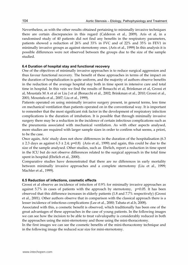
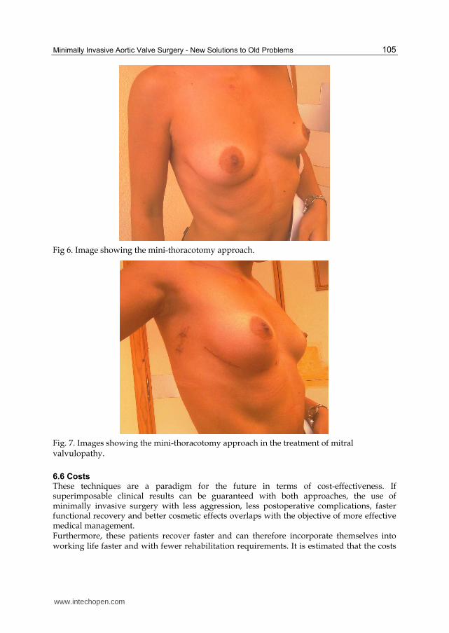
Grossi et al observe an incidence of infection of 0.9% for minimally invasive approaches as against 5.7% in cases of patients with the approach by sternotomy, p=0.05. It has been observed that this difference increases in elderly patients (1.8 and 7.7% respectively) (Grossi et al., 2001). Other authors observe that in comparison with the classical approach there is a lesser incidence of infectious complications (Lee et al., 2000; Tabata et al.b, 2008). Associated with this, a cosmetic benefit is observed, which traditionally has been one of the great advantages of these approaches in the case of young patients. In the following images we can see how the incision to be able to treat valvulopathy is considerably reduced in both the approaches using the mini-sternotomy and those using the mini-thoracotomy. In the first images we can see the cosmetic benefits of the mini-thoracotomy technique and in the following image the reduced scar size for mini-sternotomy.
www.intechopen.com
Minimally Invasive Aortic Valve Surgery - New Solutions to Old Problems
105
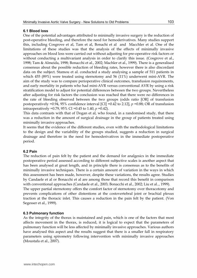
Fig 6. Image showing the mini-thoracotomy approach.
Fig. 7. Images showing the mini-thoracotomy approach in the treatment of mitral valvulopathy.
6.6 Costs These techniques are a paradigm for the future in terms of cost-effectiveness. If superimposable clinical results can be guaranteed with both approaches, the use of minimally invasive surgery with less aggression, less postoperative complications, faster functional recovery and better cosmetic effects overlaps with the objective of more effective medical management. Furthermore, these patients recover faster and can therefore incorporate themselves into working life faster and with fewer rehabilitation requirements. It is estimated that the costs
www.intechopen.com
Aortic Stenosis – Etiology, Pathophysiology and Treatment
106
Fig. 8. Image showing the mini-sternotomy appropach
of post-hospital care surpass tens of millions of dollars in the United States and these costs are rising. If operations can be carried out more efficiently in terms of management of hospital resources and the need for post-hospital care (rehabilitation) can be reduced, this could be important for health management. (Cohn et al., 1997) Many studies have analysed the impact that treatments using minimally invasive approaches can have on health costs. It is clear that if these approaches reduce hospitalization and the incidence of certain complications requiring expensive treatments, the average cost of the procedures will be reduced. Cohn LH indicates that it is possible to observe a reduction of up to 20% of the cost per diagnostic team if minimally invasive approaches are applied. Nevertheless, studying the cost effectiveness of these options is extremely complex as it is affected by a large number of factors many of which are difficult to quantify in economic terms in normal clinical practice, such as the hours the operating theatre is in use or the time taken by the surgeon to carry out the surgical procedure. Studies more in this vein would be necessary, rather than studies of clinical benefits, which at the end of the day are far more important for the patient and the doctor. Nevertheless, in terms of better use of resources the question of costs should not be forgotten. Table 3 shows the results observed in various different studies over the potential benefits of minimally invasive surgery. MI, minimally invasive; C, convencional; CPB cardiopulmonary bypass time; ICU intensive care unit
7. Disadvantages of minimally invasive approaches in the treatment of aortic stenosis
A number of factors have been highlighted as potential disadvantages of minimally invasive surgery as compared with the conventional approach, particularly noteworthy are:
www.intechopen.com
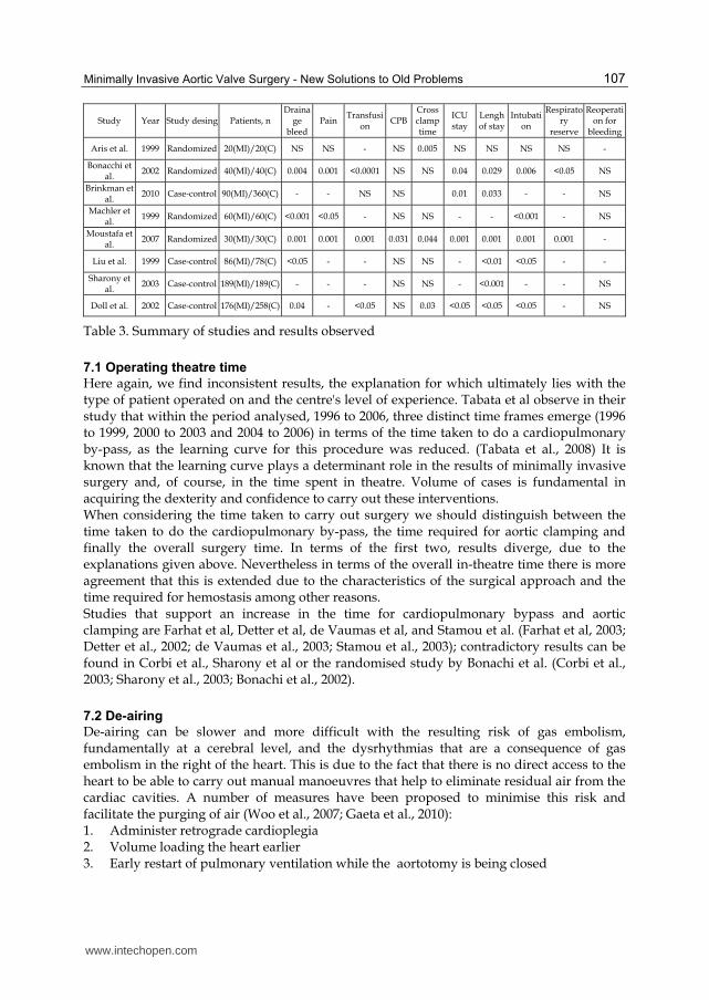
Minimally Invasive Aortic Valve Surgery - New Solutions to Old Problems
107
Study Year Study desing Patients, n Draina
ge bleed
Pain Transfusi
on CPB
Cross clamp time
ICU stay
Lengh of stay
Intubation
Respiratory
reserve
Reoperation for
bleeding
Aris et al. 1999 Randomized 20(MI)/20(C) NS NS - NS 0.005 NS NS NS NS -
Bonacchi et al.
2002 Randomized 40(MI)/40(C) 0.004 0.001 <0.0001 NS NS 0.04 0.029 0.006 <0.05 NS
Brinkman et al.
2010 Case-control 90(MI)/360(C) - - NS NS 0.01 0.033 - - NS
Machler et al.
1999 Randomized 60(MI)/60(C) <0.001 <0.05 - NS NS - - <0.001 - NS
Moustafa et al.
2007 Randomized 30(MI)/30(C) 0.001 0.001 0.001 0.031 0.044 0.001 0.001 0.001 0.001 -
Liu et al. 1999 Case-control 86(MI)/78(C) <0.05 - - NS NS - <0.01 <0.05 - -
Sharony et al.
2003 Case-control 189(MI)/189(C) - - - NS NS - <0.001 - - NS
Doll et al. 2002 Case-control 176(MI)/258(C) 0.04 - <0.05 NS 0.03 <0.05 <0.05 <0.05 - NS
Table 3. Summary of studies and results observed
7.1 Operating theatre time
Here again, we find inconsistent results, the explanation for which ultimately lies with the type of patient operated on and the centre's level of experience. Tabata et al observe in their study that within the period analysed, 1996 to 2006, three distinct time frames emerge (1996 to 1999, 2000 to 2003 and 2004 to 2006) in terms of the time taken to do a cardiopulmonary by-pass, as the learning curve for this procedure was reduced. (Tabata et al., 2008) It is known that the learning curve plays a determinant role in the results of minimally invasive surgery and, of course, in the time spent in theatre. Volume of cases is fundamental in acquiring the dexterity and confidence to carry out these interventions. When considering the time taken to carry out surgery we should distinguish between the time taken to do the cardiopulmonary by-pass, the time required for aortic clamping and finally the overall surgery time. In terms of the first two, results diverge, due to the explanations given above. Nevertheless in terms of the overall in-theatre time there is more agreement that this is extended due to the characteristics of the surgical approach and the time required for hemostasis among other reasons. Studies that support an increase in the time for cardiopulmonary bypass and aortic clamping are Farhat et al, Detter et al, de Vaumas et al, and Stamou et al. (Farhat et al, 2003; Detter et al., 2002; de Vaumas et al., 2003; Stamou et al., 2003); contradictory results can be found in Corbi et al., Sharony et al or the randomised study by Bonachi et al. (Corbi et al., 2003; Sharony et al., 2003; Bonachi et al., 2002).
7.2 De-airing
De-airing can be slower and more difficult with the resulting risk of gas embolism, fundamentally at a cerebral level, and the dysrhythmias that are a consequence of gas embolism in the right of the heart. This is due to the fact that there is no direct access to the heart to be able to carry out manual manoeuvres that help to eliminate residual air from the cardiac cavities. A number of measures have been proposed to minimise this risk and facilitate the purging of air (Woo et al., 2007; Gaeta et al., 2010): 1. Administer retrograde cardioplegia 2. Volume loading the heart earlier 3. Early restart of pulmonary ventilation while the aortotomy is being closed
www.intechopen.com
Aortic Stenosis – Etiology, Pathophysiology and Treatment
108
4. Terminating venting earlier 5. Rotating the operating table upward and on the Leith side 6. The use of CO2 is useful in the treatment of aortic valvulopathy, with it being effective
as almost the only measure applied to a flow of 2l/ min If one is meticulous in the application of these techniques, the problems associated with purging the air are significantly reduced and there is no reason to expect serious disturbances from it.
7.3 Technical difficulties
Another aspect that has been assessed is the increase in technical complexity which could put the effectiveness of the surgical procedure at risk. However, in this respect, many authors have indicated that aortic valve surgery can be carried out with an increase in technical complexity that surgeons can accommodate to without undue efforts in the learning curve, and that results for both approaches are similar (Liu et al., 1999; Machler et al., 1999). Sharony et al indicate that the results for both approaches are similar in the actuarial survival curve at three years. Below we bring together the results of some of the studies that have been carried out in the treatment of aortic valvulopathy with the results observed in each of the aspects that have been considered here.
8. Conclusions
Minimally invasive surgery, both in the treatment of cardiac ischemia and in valvulopathies, has been the subject of considerable developments and improvements in recent years thanks to the perfecting of surgical techniques and the support that the industry has provided for carrying out those techniques. Minimally invasive surgery is a safe and effective therapeutic option in the treatment of aortic valvulopathy that can be compared with conventional treatment, and which, in some centres, represents a not unimportant percentage (majority). It currently has similar results, with a reduction in bleeding rates, pain perceived by the patient, infection of surgical wounds, duration of hospitalisation, functional recovery and significant cosmetic effects. The necessary skills to be able to conduct the procedure are not very demanding and are easily reproduced. Limitations due to the surgical field exposed, procedural challenges and potential pitfalls are easily overcome with the right knowledge and strategies.
9. References
Arís, A., Padró, J.M., & Cámara, M.L. (1997) Minimally invasive aortic valve replacement. Rev Esp Cardiol. 1997 Nov; 50(11):778-81.
Aris, A., Camara, M.L., & Montiel, J. (1999) Ministernotomy versus median sternotomy for aortic valve replacement: a prospective randomized study. Ann Thorac Surg 1999; 67:1583–7.
Arom, K.V., Emery, R.W., & Nicoloff, D.M. (1996) Mini-sternotomy for coronary artery bypass grafting. Ann Thorac Surg 1996; 61: 1.271- 1.272.
Baumgartner, H., Hung, J., & Bermejo, J. (2009) American Society of Echocardiography, European Society of Echocardiography. Echocardiographic assessment of valve
www.intechopen.com

Minimally Invasive Aortic Valve Surgery - New Solutions to Old Problems
109
stenosis: EAE/ASE recommendations for clinical practice. Eur J Echocardiogr 2009; 10: 1–23.
Benetti, F.J., Naselli, G., & Wood, M. (1991) Direct myocardial revascularization without extracorporeal circulation. Experience in 700 patients. Chest 1991; 100: 312-316.
Bonacchi, M., Prifti, E., & Giunti, G. (2002) Does ministernotomy improve postoperative outcome in aortic valve operations? A prospective randomized study. Ann Thorac
Surg 2002; 73:460–5. Bonow, RO, Carabello, BA, & Chatterjee, K. (2008) American College of
Cardiology/American Heart Association Task Force on Practise Guidelines. 2008 focused update incorporated into the ACC/AHA 2006 guidelines for the management of patients with valvular heart disease: a report of the American College of Cardiology/American Heart Association Task Force on Practise Guidelines (Writing Committee to revise the 1998 guidelines for the management of patients with valvular heart disease). Endorsed by the Society of Cardiovascular Anesthesiologists, Society for Cardiovascular Angiography and Interventions, and Society of Thoracic Surgeons. J Am Coll Cardiol 2008; 52:e1–e142.
Brinkman, W.T., Hoffman, W., & Dewey, T.M. (2010) Aortic valve replacement surgery: comparison of outcomes in matched sternotomy and PORT ACCESS groups. Ann
Thorac Surg. 2010 Jul; 90(1):131-5. Buffolo, E., Silva de Andrade, J.C., & Rodrigues, J.N. (1996) Coronary atery bypass grafting
without cardiopulmonary bypass. Ann Thorac Surg 1996; 61: 63-66. Burfeind, W.R., Glower, D.D., & Davis, R.D. (2002) Mitral surgery after prior cardiac
operation: port-access versus sternotomy or thoracotomy. Ann Thorac Surg. 2002 Oct; 74(4):S1323-5.
Byrne, J.G., Aranki, S.F., & Couper, G.S. (1999) Reoperative aortic valve replacement: partial upper hemisternotomy versus conventional full sternotomy. J Thorac Cardiovasc
Surg 1999; 118: 991–7. Byrne, J.G., Karavas, A.N., & Adams, D.H. (2000) Partial upper re sternotomy for aortic
valve replacement or re-replacement after previous cardiac surgery. Eur J
Cardiothorac Surg 2000; 18: 282–6. Caffarelli, A.D. & Robbins, R.C. (2004) Will minimally invasive valve replacement ever
really be important? Curr Opin Cardiol 2004; 19:123–7. Calderon, J., Richebe, P., & Guibaud, J.P. (2009) Prospective randomized study of early
pulmonary evaluation of patients scheduled for aortic valve surgery performed by ministernotomy or total median sternotomy. J Cardiothorac Vasc Anesth. 2009 Dec; 23(6):795-801.Califiore, A.M., Teodori, G., & Di Giammarco, G. (1997) Minimally invasive coronary artery bypass grafting on a beating heart. Ann Thorac Surg 1997 63:S72-S75.
Candaele, S., Herijgers, P., & Demeyere, R. (2003) Chest pain after partial upper versus complete sternotomy for aortic valve surgery. Acta Cardiol 2003, 58:17–21.
Carpentier, A., Loulmet, D., & Aupècle, B. (1998) Computer assisted open heart surgery. First case operated on with success C R Acad Sci III. 1998 May; 321(5):437-42.
Chambers, J.B. (2009) Aortic stenosis. Eur J Echocardiogr. 2009 Jan; 10(1):i11-9.
www.intechopen.com
Aortic Stenosis – Etiology, Pathophysiology and Treatment
110
Chitwood, W.R. Jr. (2001) Minimally invasive aortic valve surgery: What is "port access"? Am Heart J. 2001 Sep; 142(3):391-2.
Chitwood, W.R. Jr. & Gulielmos V. (2003) What is minimally invasive cardiac surgery? CTSnet Web site. 2003. Available at: http://www.ctsnet.org/doc/7525.
Cohn, L.H., Adams, D.H., & Couper, G.S. (1997) Minimally invasive cardiac valve surgery improves patient satisfaction while reducing costs of cardiac valve replacement and repair. Ann Surg. 1997 Oct; 226(4):421-6; discussion 427-8.
Connolly, H.M., Oh J.K., & Orszulak, T.A. (1997) Aortic valve replacement for aortic stenosis with severe left ventricular dysfunction: prognostic indicators. Circulation 1997; 95:2395– 400.
Corbi, P., Rahmati, M., & Donal, E. (2003) Prospective comparison of minimally invasive and standard techniques for aortic valve replacement: initial experience in the first hundred patients. J Card Surg 2003, 18:133–139
Cosgrove, D.M. 3rd. & Sabik, J.F. (1996) Minimally invasive approach for aortic valve operations. Ann Thorac Surg. 1996 Aug; 62(2):596-7.
Cosgrove, D.M., Sabik, J.F., & Navia, J.L. (1998) Minimally invasive valve operations. Ann
Thorac Surg 1998; 65:1535–8. Cosmi, J.E., Tunick, P.A., & Rosenzweig, B.P. (2002) The risk of development of aortic
stenosis in patients with ‘benign’ aortic valve thickening. Arch Intern Med 2002; 162:2345-7.
Cuenca, J., Rodríguez-Delgadillo, M.A., & Valle, J.V. (1998) Is the femoral cannulation for minimally invasive aortic valve replacement necessary? Eur J Cardiothorac Surg. 1998 Oct; 14 Suppl 1:S111-4.
Detter, C., Deuse, T., & Boehm, D.H. (2002) Midterm results and quality of life after minimally invasive vs. conventional aortic valve replacement. Thorac Cardiovasc
Surg 2002, 50:337–341. Doll, N., Borger, M.A., & Hain, J. (2002). Minimal access aortic valve replacement: effects on
morbidity and resource utilization. Ann Thorac Surg. 2002 Oct; 74(4):S1318-22. Ehrlich, W., Skwara, W., & Klövekorn, W. (2000) Do patients want minimally invasive aortic
valve replacement? Eur J Cardiothorac Surg. 2000 Jun; 17(6):714-7. Falk, V., Walther, T., & Autschbach, R. (1998) Robot-assisted minimally invasive solo mitral
valve operation. J Thorac Cardiovasc Surg. 1998 Feb; 115(2):470-1. Farhat, F., Lu, Z., & Lefevre, M. (2003) Prospective comparison between total sternotomy
and ministernotomy for aortic valve replacement. J Card Surg 2003, 18:396–401 Frazier, B.L., Derrick, M.J., & Purewal, S.S. (1998) Minimally invasive aortic valve
replacement. Eur J Cardiothorac Surg. 1998 Oct; 14 Suppl 1:S122-5. Gaeta, R., Lentini, S., & Raffa, G. (2010) Aortic valve replacement by ministernotomy in redo
patients with previous left internal mammary artery patent grafts. Ann Thorac
Cardiovasc Surg. 2010 Jun; 16(3):181-6. Galloway, A.C., Shemin, R.J., & Glower, D.D. (1999) First report of the Port Access
International Registry. Ann Thorac Surg. 1999 Jan; 67(1):51-6; discussion 57-8. Grossi, E.A., Galloway, A.C., & Ribakove, G.H. Impact of minimally invasive valvular heart
surgery: a case-control study. Ann Thorac Surg. 2001 Mar; 71(3):807-10.
www.intechopen.com
Minimally Invasive Aortic Valve Surgery - New Solutions to Old Problems
111
Harken, D.E., Soroff, H.S., & Taylor, W.H. Aortic valve replacement, in Merendino KA (ed): Prosthetic Valves for Cardiac Surgery. Springfield, IL, Thomas, 1961, pp 508-526
Hirose, H., Umeda, S., & Mori, Y. (1994) Another approach for aortic valve replacement through left thoracotomy. Ann Thorac Surg. 1994 Sep; 58(3):884-6.
Izzat, M.B., Yim, A.P., & El-Zufari, M.H. (1998) Upper T ministernotomy for aortic valve operations. Chest 1998; 114:291–4.
Katz, E.S., Tunick, P.A., & Rusinek, H. (1992) Protruding aortic arch atheromas predict stroke in elderly patients undergoing cardiopulmonary bypass: experience with intraoperative transesophageal echocardiography. J Am Coll Cardiol 1992; 20:70-7.
Klokocovnik, T. (1998) Aortic and mitral replacement through a minimally invasive approach. Tex Heart Inst J 1998; 25:166–9.
Konertz, W., Waldenberger, F., & Schmutzler, M. (1996) Minimal access valve surgery through superior partial sternotomy: a preliminary study. J Heart Valve Dis. 1996 Nov; 5(6):638-40.
Kort, S., Applebaum, R.M., & Grossi, E.A. (2001) Minimally invasive aortic valve replacement: echocardiographic and clinical results. Am Heart J. 2001 Sep; 142(3):476-81.
Kouchoukos, N.T., Davila-Roman, V.G., & Spray, T.L. (1994) Replacement of the aortic root with a pulmonary autograft in children and young adults with aortic-valve disease. N Engl J Med 1994; 330:1– 6.
Kronzon, I., Tunick, P.A., & Jortner, R. (1995) Echocardiographic evaluation of the coronary sinus. J Am Soc Echocardiogr 1995; 8:518-26.
Kurtz, C.E. & Otto, C.M. (2010) Aortic stenosis: clinical aspects of diagnosis and management, with 10illustrative case reports from a 25-year experience. Medicine (Baltimore). 2010 Nov; 89(6):349-79.
Kvidal, P., Bergstrom, R., & Horte, L.G. (2000) Observed and relative survival after aortic valve replacement. J Am Coll Cardiol 2000; 35: 747–56.
Lee, J.W., Lee, S.K., & Choo, S.J. (2000) Routine minimally invasive aortic valve procedures. Cardiovasc Surg. 2000 Oct; 8(6):484-90.
Lester, S.J., Heilbron, B., & Gin, K. (1998) The natural history and rate of progression of aortic stenosis. Chest. 1998 Apr; 113(4):1109-14.
Lindroos, M., Kupari, M., & Heikkala, J. (1993) Prevalence of aortic valve abnormalities in the elderly: an echocardiographic study of a random population sample. J Am Coll
Cardiol 1993; 21:1220-5. Liu, J., Sidiropoulos, A., & Konertz, W. (1999) Minimally invasive aortic valve replacement
(AVR) compared to standard AVR. Eur J Cardiothorac Surg. 1999 Nov; 16 Suppl 2:S80-3.
Lopez- Gude, M.J., Garcia, D., & Forteza, A. (2010) Heart PortTM access technique for valvular surgery. Cir. Cardiov. 2010; 17(4):345-50.
Lund, O. (1990) Preoperative risk evaluation and stratification of long-term survival after valve replacement for aortic stenosis: reasons for earlier operative intervention. Circulation 1990; 82:124 –39.
www.intechopen.com
Aortic Stenosis – Etiology, Pathophysiology and Treatment
112
Lung, B., Baron, G., & Butchart, E.G. (2003) A prospective survey of patients with valvular heart disease in Europe: the Euro Heart Survey on valvular heart disease. Eur Heart
J 2003; 24:1231–1243. Machler, H.E., Bergmann, P., & Anelli-Monti, M. (1999) Minimally invasive versus
conventional aortic valve operations: a prospective study in 120 patients. Ann
Thorac Surg 1999, 67:1001–1005 Minale, C., Reifschneider, H.J., & Schmitz, E. (1997) Single access for minimally invasive
aortic valve replacement. Ann Thorac Surg. 1997 Jul; 64(1):120-3. Moustafa, M.A., Abdelsamad, A.A., & Zakaria, G. (2007) Minimal vs median sternotomy for
aortic valve replacement. Asian Cardiovasc Thorac Ann. 2007 Dec; 15(6):472-5. Murphy, E.S., Lawson, R.M., & Starr, A. (1981) Severe aortic stenosis in patients 60 years of
age or older: left ventricular function and 10-year survival after valve replacement. Circulation 1981; 64:II184–8.
Nashef, S.A., Roques, F., & Hammill, B.G. (2002) EurpSCORE Project Group: Validation of European System for Cardiac Operative Risk Evaluation (EuroSCORE) in North American cardiac surgery. Eur J Cardiothorac Surg 2002, 22:101–105
National Adult Cardiac Surgical Database Report 1999–2000. The United Kingdom Cardiac Surgical Register. Accessed 10 May 2006.
http://www. scts.org/file/NACSDreport2000ukcsr.pdf Navia, J.L. & Cosgrove, D.M. Minimally invasive mitral valve operations. Ann Thorac Surg.
1996 Nov; 62(5):1542-4. Otto, C.M., Burwash, I.G., & Legget, M.E. (1997) Prospective study of asymptomatic
valvular aortic stenosis. Clinical, echocardiographic, and exercise predictors of outcome. Circulation 1997; 95:2262-70.
Pellikka, P.A., Sarano, M.E., & Nishimura, R.A. (2005) Outcome of 622 adults with asymptomatic, hemodynamically significant aortic stenosis during prolonged follow-up. Circulation. 2005 111:3290–3295.
Rao, P.N. & Kumar, A.S. (1993) Aortic valve replacement through right thoracotomy. Tex
Heart Inst J 1993; 20:307–8. Rodríguez, J.E., López, M.J., & Carrascal, Y. (1996) Aortic valve replacement via
ministernotomy. Rev Esp Cardiol. 1996 Dec; 49(12):928-30. Rosenhek, R., Binder, T., & Porenta, G. (2000) Predictors of outcome in severe, asymptomatic
aortic stenosis. N Engl J Med. 2000; 343:611–617. Sardari, F.F., Schlunt, M.L., & Applegate, R.I. (1997) The use of transesophageal
echocardiography to guide sternal division for cardiac operations via mini-sternotomy. J Cardiac Surg 1997; 12:67±70.
Schmitto, J.D., Mohr, F.W., & Cohn, L.H. (2011) Minimally invasive aortic valve replacement: how does this perform in high-risk patients? Curr Opin Cardiol. 2011 Mar; 26(2):118-22.
Sharony, R., Grossi, E.A., & Saunders, P.C. (2003) Minimally invasive aortic valve surgery in the elderly: a case-control study. Circulation 2003, 108:43II–47II
Smith, N., McAnulty, J.H., & Rahimtoola, S.H. (1978) Severe aortic stenosis with impaired left ventricular function and clinical heart failure: results of valve replacement. Circulation 1978; 58:255– 64.
www.intechopen.com

Minimally Invasive Aortic Valve Surgery - New Solutions to Old Problems
113
Stamou, S.C., Kapetanakis, E.I., & Lowery, R. Allogeneic blood transfusion requirements after minimally invasive versus conventional aortic valve replacement: a risk-adjusted analysis. Ann Thorac Surg 2003, 76:1101–1106
Starr, A. & Edwards, M.L. (1961) Mitral replacement: Clinical experience with a ball valve prosthesis. Ann Surg 1961 154:726-740.
Stern, A., Tunick, P.A., & Culliford, A.T. Protruding aortic arch atheromas: risk of stroke during heart surgery with and without aortic arch endarterectomy. Am Heart J 1999; 138:746-52.
Stevens, J.H., Burdon, T.A., & Siege1, L.C. Port-access coronary artery bypass with cardioplegic arrest: Acute and chronic studies. Ann Thorac Surg 62:435-441,1996
Stewart, B.F., Siscovick, D., & Lind, B.K. Clinical factors associated with calcific aortic valve disease: Cardiovascular Health Study. J Am Coll Cardiol 1997; 29:630-4.
STS National Database: Spring 2003 Executive Summary. Durham, NC: Duke Clinical Research Institute; 2003.
STS national database: STS U.S. cardiac surgery database: 1997 Aortic valve replacement patients: preoperative risk variables. Chicago, Society of Thoracic Surgeons; 2000. Accessed 10 May 2006. http:// www.ctsnet.org/doc/3031
Subramanian, V.A., Sani, G., & Benetti, F.J. (1995) Minimally invasive coronary bypass surgery: a multi-center report of preliminary clinical experience. Circulation 1995; 92 (Supl 1): 645.
Svensson, L.G. (1997) Minimal-access “J” or “j” sternotomy for valvular, aortic, and coronary operations or reoperations. Ann Thorac Surg 1997; 64:1501–3.
Svensson, L.G. & D’Agostino, R.S. (1998) Minimal-access aortic and valvular operations, including the “J/j” incision. Ann Thorac Surg 1998; 66:431–5.
Svensson, L.G., Nadolny, E.M., & Kimmel, W.A. (2001) Minimal access aortic surgery including re-operations. Eur J Cardiothorac Surg 2001; 19: 30–3.
Szwerc, M.F., Benckart, D.H., & Wiechmann, R.J. (1999) Partial versus full sternotomy for aortic valve replacement. Ann Thorac Surg 1999; 68:2209–13.
Tabata, M., Khalpey, Z., & Shekar, P.S. (2008a) Reoperative minimal access aortic valve surgery: minimal mediastinal dissection and minimal injury risk. J Thorac
Cardiovasc Surg. 2008 Dec; 136(6):1564-8. Epub 2008 Oct 23. Tabata, M., Umakanthan, R., & Cohn, L.H. (2008b) Early and late outcomes of 1000
minimally invasive aortic valve operations. Eur J Cardiothorac Surg. 2008 Apr; 33(4):537-41. Epub 2008 Feb 5.
Tam, R.K. & Almeida, A.A. (1998) Minimally invasive aortic valve replacement via partial sternotomy. Ann Thorac Surg 1998; 65:275–6.
Vahanian, A., Baumgartner, H., & Bax, J. (2007) Task Force on the Management of Valvular Hearth Disease of the European Society of Cardiology; . ESC Committee for Practice Guidelines. Eur Heart J 2007; 28:230–268.
Van Eck, F.M., Noyez, L., & Verheugt, F.W. (2002) Preoperative prediction of early mortality in redocoronary artery surgery. Eur J Cardiothorac Surg. 2002; 21:1031-6.
de Vaumas, C., Philip, I., & Daccache, G. (2003) Comparison of minithoracotomy and conventional sternotomy approaches for valve surgery. J Cardiothorac Vasc Anesth 2003, 17:325–328.
www.intechopen.com

Aortic Stenosis – Etiology, Pathophysiology and Treatment
114
von Segesser, L.K., Westaby, S., & Pomar, J. Less invasive aortic valve surgery: rationale and technique. Eur J Cardiothorac Surg. 1999 Jun; 15(6):781-5.
Walther, T., Falk, V., & Metz, S. (1996) Pain and quality of life after minimally invasive versus conventional cardiac surgery. Ann Thorac Surg 1999; 67:1643–7.
Wang, W.L., Cai, K.C., & Zeng, W.S. (2003) Experience in using three different minimally invasive approaches in cardiac operations. Med Sci Monit 2003; 9:109–13.
Woo, Y.J., Seeburger, J., & Mohr, F.W. (2007) Minimally invasive valve surgery. Semin Thorac
Cardiovasc Surg. 2007 Winter; 19(4):289-98. Yap, C.H., Sposato, L., & Akowuah, E. (2009) Contemporary results show repeat coronary
artery bypass grafting remains a risk factor for operative mortality. Ann Thorac
Surg. 2009; 87:1386-91. Yau, T.M., Borger, M.A., & Weisel, R.D. (2000) The changing pattern of reoperative coronary
surgery: trends in 1230 consecutive reoperations. J Thorac Cardiovasc Surg. 2000; 120:156-63.
www.intechopen.com
Aortic Stenosis - Etiology, Pathophysiology and TreatmentEdited by Dr. Masanori Hirota
ISBN 978-953-307-660-7Hard cover, 254 pagesPublisher InTechPublished online 10, October, 2011Published in print edition October, 2011
InTech EuropeUniversity Campus STeP Ri Slavka Krautzeka 83/A 51000 Rijeka, Croatia Phone: +385 (51) 770 447 Fax: +385 (51) 686 166www.intechopen.com
InTech ChinaUnit 405, Office Block, Hotel Equatorial Shanghai No.65, Yan An Road (West), Shanghai, 200040, China
Phone: +86-21-62489820 Fax: +86-21-62489821
Currently, aortic stenosis (AS) is the most prevalent valvular disease in developed countries. Pathological andmolecular mechanisms of AS have been investigated in many aspects. And new therapeutic devices such astranscatheter aortic valve implantation have been developed as a less invasive treatment for high-risk patients.Due to advanced prevalent age of AS, further discovery and technology are required to treat elderly patientsfor longer life expectancy. This book is an effort to present an up-to-date account of existing knowledge,involving recent development in this field. Various opinion leaders described details of established knowledgeor newly recognized advances associated with diagnosis, treatment and mechanism. Thus, this book willenable close intercommunication to another field and collaboration technology for new devices. We hope that itwill be an important source, not only for clinicians, but also for general practitioners, contributing todevelopment of better therapeutic adjuncts in the future.
How to referenceIn order to correctly reference this scholarly work, feel free to copy and paste the following:
Juan Bustamante, Sergio Ca novas and Angel L Ferna ndez (2011). Minimally Invasive Aortic Valve Surgery -New Solutions to Old Problems, Aortic Stenosis - Etiology, Pathophysiology and Treatment, Dr. MasanoriHirota (Ed.), ISBN: 978-953-307-660-7, InTech, Available from: http://www.intechopen.com/books/aortic-stenosis-etiology-pathophysiology-and-treatment/minimally-invasive-aortic-valve-surgery-new-solutions-to-old-problems

© 2011 The Author(s). Licensee IntechOpen. This is an open access articledistributed under the terms of the Creative Commons Attribution 3.0License, which permits unrestricted use, distribution, and reproduction inany medium, provided the original work is properly cited.