Embed Size (px)
Citation preview

TECHNICAL NOTE
Julio J. Mulero,1 Ph.D.; Chien Wei Chang,1 Ph.D.; Robert E. Lagac�,1 B.A.; Dennis Y. Wang,1 Ph.D.;Jennifer L. Bas,2� M.F.S.; Timothy P. McMahon,2� Ph.D.; and Lori K. Hennessy,1 Ph.D.
Development and Validation of the AmpF‘STR�
MiniFilerTM PCR Amplification Kit: A MiniSTRMultiplex for the Analysis of Degraded and ⁄orPCR Inhibited DNA*
ABSTRACT: DNA typing of degraded DNA samples can be a challenging task when using the current commercially available multiplex shorttandem repeat (STR) analysis kits. However, the ability to type degraded DNA specimens improves by redesigning current STR marker ampliconssuch that smaller sized polymerase chain reaction (PCR) products are generated. In an effort to increase the amount of information derived fromthese types of DNA samples, the AmpF‘STR� MiniFilerTM PCR Amplification Kit has been developed. The kit contains reagents for the amplifica-tion of eight miniSTRs which are the largest sized loci in the AmpF‘STR� Identifiler� PCR Amplification Kit (D7S820, D13S317, D16S539,D21S11, D2S1338, D18S51, CSF1PO, and FGA). Five of these STR loci (D16S539, D21S11, D2S1338, D18S51, and FGA) also are some of thelargest loci in the AmpF‘STR� SGM Plus� kit. This informative nine-locus multiplex, which includes the gender-identification locus Amelogenin,has been validated according to the FBI ⁄ National Standards and SWGDAM guidelines. Our results demonstrate significant performance improve-ments in models of DNA degradation, PCR inhibition, and nonprobative samples when compared to the AmpF‘STR� Identifiler� and SGM Plus�
kits. These data support that the MiniFilerTM kit will increase the likelihood of obtaining additional STR information from forensic samples in situa-tions in which standard STR chemistries fail to produce complete profiles.
KEYWORDS: forensic science, DNA typing, mini short tandem repeat, D7S820, D13S317, D16S539, D21S11, D2S1338, D18S51,CSF1PO, FGA, humic acid, hematin
Extracting sufficient high quality DNA for conventional shorttandem repeat (STR) typing from a sample of poor quality is achallenging task and at times not possible. Often, polymerase chainreaction (PCR) amplification with commercial STR multiplexesresults in partial or no information due to extensive DNA fragmen-tation. Many laboratories often do not attempt further analysis ofthese limiting samples and some laboratories resort to single nucle-otide polymorphism (SNP) analysis (1) or mitochondrial DNAsequencing of the hypervariable regions (2,3) so as to obtain someinformation. These approaches, while helpful, are inherently lessdiscriminating than STR multiplex systems and increase analysistime and cost. Another approach increases the number of amplifica-tion cycles to the recommended PCR protocol for commercial STRmultiplexes (4,5). However, low level DNA analysis is highly sus-ceptible to stochastic effects and can result in allele drop-outs,allele drop-ins, imbalance of heterozygote peak height and area,
inconsistent peak size of stutter products, and increased risk of lab-oratory-based contamination (6). Recently, several laboratories (7–14) have demonstrated improvements in genotyping degraded DNAsamples by repositioning primers in as close as possible to the STRrepeat region. These primer changes result in smaller PCR productstermed ‘‘miniSTRs’’ that increase the potential number of templatemolecules available for the PCR.
The AmpF‘STR� MiniFilerTM PCR Amplification Kit wasdesigned to amplify as miniSTRs eight of the largest sized loci in theAmpF‘STR� Identifiler� PCR Amplification Kit (D7S820,D13S317, D16S539, D21S11, D2S1338, D18S51, CSF1PO, FGA).Five of these loci (D16S539, D21S11, D2S1338, D18S51, and FGA)also are five of the largest loci in the AmpF‘STR� SGM Plus� kit.Together with the gender-identification locus Amelogenin, this nine-locus multiplex enables simultaneous amplification of the loci thatoften fail detection during the amplification of compromised DNAsamples (12). Seven of these loci (D7S820, D13S317, D16S539,D21S11, D18S51, CSF1PO, FGA) are part of the core loci requiredby the Combined DNA Index System (CODIS) to maintain compati-bility with convicted offender database profiles generated with con-ventional commercial STR multiplexes. Furthermore, a populationstudy involving 1308 samples demonstrated that the MiniFilerTM andIdentifiler� kits share a very high degree of concordance (15).
This article describes the developmental validation performedaccording to guidelines issued by the Director of the FBI (16), andthe revised guidelines issued by SWGDAM (17). Performancecomparisons between the MiniFilerTM, Identifiler�, and SGM Plus�
kits using models of DNA degradation, PCR inhibition, and non-probative samples are described.
1Applied Biosystems, 850 Lincoln Centre Dr., Foster City, CA 94404.2Armed Forces DNA Identification Lab, 1413 Research Blvd, Rockville,
MD 20850.*Oral presentation at the 17th International Symposium on Human Identi-
fication, Nashville, TN 2006. Some of the data are also presented in theMiniFilerTM PCR Amplification Kit User Guide ‘‘Experiments and Results’’section.
�Present address: Las Vegas Metropolitan Police Department, Las Vegas,NV 89101.
�Present address: Applied Biosystems, 850 Lincoln Centre Dr, FosterCity, CA 94404.
Received 27 June 2007; and in revised form 28 Oct. 2007; accepted 18Nov. 2007.
J Forensic Sci, July 2008, Vol. 53, No. 4doi: 10.1111/j.1556-4029.2008.00760.x
Available online at: www.blackwell-synergy.com
838 � 2008 American Academy of Forensic Sciences

Materials and Methods
DNA Samples
Anonymous DNA samples were purchased from Seracare LifeSciences (Oceanside, CA), Raji DNA (Biochain Institute, Hayward,CA) and 9947A was purchased from Marligen Biosciences
(Ijamsville, MD). The 9948 DNA sample was purchased from Cori-ell Cell Repositories (Camden, NJ) and the AmpF‘STR� ControlDNA 007 was obtained from Applied Biosystems (Foster City, CA).The population study encompassed DNA samples from Seracare LifeSciences as well as samples analyzed in collaboration with theNational Institute of Standards and Technology (NIST) (15). Thequantity of the DNA samples was determined prior to amplificationusing the Quantifiler� Human DNA Quantification Kit (AppliedBiosystems) on the ABI PRISM� 7000 Sequence Detection System(Applied Biosystems) according to the manufacturer’s specifications.
Primer Set Optimization
Each of the STR loci amplified by the MiniFilerTM kit has beencharacterized previously by other groups and can be found in com-mercial STR multiplexes such as the Identifiler� and SGM Plus�
Kits (18,19). The MiniFilerTM kit primers were designed and opti-mized to obtain small amplified products (less than currently gener-ated using other commercially available Applied Biosystems kits)with robust signal intensity and balanced peak heights from humansamples. Initially, singleplex reactions were tested using ourdesigned primer sets to ensure locus-specific amplification. Thenmultiplex PCR studies were carried out through primer concentra-tion adjustment and empirical performance testing in an effort togenerate sensitive, balanced, and specific signals for the eight STRloci and the sex determining marker Amelogenin in a single PCRreaction. The MiniFilerTM kit STR loci span a range between 71and 250 bp. To accommodate the co-amplification of eight loci
FIG. 1—Representative electropherogram showing the profile of 500 pg of control DNA 007 amplified with the MiniFilerTM kit. The four panels correspondto (from top to bottom) 6-FAMTM, VIC�, NEDTM, and PET� dye-labeled peaks. The genotype is shown with the allele number displayed underneath eachpeak.
FIG. 2—Effect of PCR cycle number on intracolor peak height average.Three genomic DNA samples (control DNA 007, 9947A, and 9948) wereamplified in triplicates using as an input 500 pg of DNA. The samples wereamplified on the Applied Biosystems GeneAmp� PCR System 9700 (operat-ing in 9600 emulation mode).
MULERO ET AL. • MINIFILERTM KIT VALIDATION 839

within this narrow size range, we employed a five-dye fragmentanalysis system developed at Applied Biosystems and currently uti-lized in the AmpF‘STR� SEfilerTM (20), Identifiler� (21), andYfiler� kits (22). The five dyes (6-FAMTM, NEDTM, PET�, VIC�,and LIZ�) expand the fluorescent detection range to 660 nmthereby enabling more loci to be multiplexed in a single PCR. Theelectrophoretic spacing between different loci was optimized byintroducing monomeric non-nucleotide linkers between the DNAoligonucleotide sequence and the fluorescent tag during the primersynthesis process (23,24) for most loci. The resulting PCR producthas a slower mobility, which correlates with the number of linkersused. This approach has been successfully applied to the Yfiler andIdentifiler� kits. The corresponding allelic ladders also contain non-nucleotide linkers. Three 500 pg samples (9947A, 9948, and the
control DNA 007) were amplified in triplicate at the standard pri-mer concentration and at 10% intervals up to €30% levels to evalu-ate primer performance (data not shown).
PCR Components
The buffer components and concentrations are proprietary andinclude a DNA polymerase, salts, dNTPs, carrier protein, and0.05% sodium azide. Each of these components was tested indi-vidually at a series of titrations around the standard conditionused in the AmpF‘STR� MiniFilerTM Master Mix. The compo-nents were tested in increments of €10% up to €30% from thestandard concentration to test for reliability and robustness of theamplification components. Each concentration was tested withthree samples (9947A, 9948, and control DNA 007) and ampli-fied in triplicate using 500 pg of template DNA (data notshown).
Thermal Cycling Parameters
Thermal cycling parameters were evaluated to establish the opti-mal performance window of amplification for the MiniFilerTM kit.Cycling parameters around the standard set of conditions weretested. For each study, 500 pg of DNA from three samples(9947A, 9948, and control DNA 007) were prepared in triplicateand stored at 4�C while awaiting amplification in the same thermalcycler for each of the parameters tested.
The following thermal cycler parameters were examined (therecommended parameters are shown in bold):
FIG. 3—Representative electropherograms from an annealing temperature study of the control DNA 007 DNA. The DNA was amplified with the MiniFilerTM
kit at the indicated temperatures. Peak heights were measured in relative fluorescent units (RFUs).
TABLE 1—Stutter average and range for MiniFilerTM kit loci.
Locus Stutter Average Stutter Range (%) SD
D13S317 6.3 1.9–13.7 1.7D7S820 4.9 1.8–10.7 1.5D2S1338 8.8 3.5–17.7 1.9D21S11 8.1 2.2–15.7 1.5D16S539 6.8 1.2–14.3 2.0D18S51 7.9 2.1–17.3 2.3CSF1PO 6.2 1.4–13.9 1.7FGA 7.4 1–14.5 1.6
All loci have tetranucleotide repeats yielding )4 bp stutters. Stutters weredetermined for those samples with peak heights between 400 and 5000RFUs. The threshold minimum stutter peak height was 20 RFUs.
840 JOURNAL OF FORENSIC SCIENCES
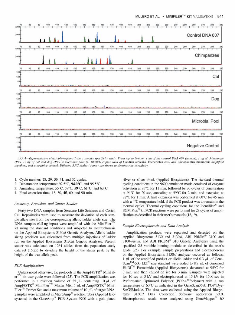
1. Cycle number: 28, 29, 30, 31, and 32 cycles.2. Denaturation temperature: 92.5�C, 94.0�C, and 95.5�C.3. Annealing temperature: 55�C, 57�C, 59�C, 61�C, and 63�C.4. Final extension time: 15, 30, 45, 60, and 90 min.
Accuracy, Precision, and Stutter Studies
Forty-two DNA samples from Seracare Life Sciences and CoriellCell Repositories were used to measure the deviation of each sam-ple allele size from the corresponding allelic ladder allele size. TheDNA samples (0.5 ng input) were amplified with the MiniFilerTM
kit using the standard conditions and subjected to electrophoresison the Applied Biosystems 3130xl Genetic Analyzer. Allelic laddersizing precision was calculated from multiple injections of ladderrun on the Applied Biosystems 3130xl Genetic Analyzer. Percentstutter was calculated on 1264 alleles from the population studydata set (15,25) by dividing the height of the stutter peak by theheight of the true allele peak.
PCR Amplification
Unless noted otherwise, the protocols in the AmpF‘STR� MiniFil-erTM kit user guide were followed (25). The PCR amplification wasperformed in a reaction volume of 25 lL containing 10 lL ofAmpF‘STR� MiniFilerTM Master Mix, 5 lL of AmpF‘STR� Mini-FilerTM Primer Set, and a maximum volume of 10 lL of target DNA.Samples were amplified in MicroAmp� reaction tubes (Applied Bio-systems) in the GeneAmp� PCR System 9700 with a gold-plated
silver or silver block (Applied Biosystems). The standard thermalcycling conditions in the 9600 emulation mode consisted of enzymeactivation at 95�C for 11 min, followed by 30 cycles of denaturationat 94�C for 20 sec, annealing at 59�C for 2 min, and extension at72�C for 1 min. A final extension was performed at 60�C for 45 minwith a 4�C temperature hold, if the PCR product was to remain in thethermal cycler. Thermal cycling conditions for the Identifiler� andSGM Plus� kit PCR reactions were performed for 28 cycles of ampli-fication as described in their user’s manuals (18,19).
Sample Electrophoresis and Data Analysis
Amplification products were separated and detected on theApplied Biosystems 3130 and 3130xl, ABI PRISM� 3100 and3100–Avant, and ABI PRISM� 310 Genetic Analyzers using thespecified G5 variable binning module as described in the user’sguide (25). For example, sample preparations and electrophoresison the Applied Biosystems 3130xl analyzer occurred as follows:1 lL of the amplified product or allelic ladder and 0.3 lL of Gene-ScanTM-500 LIZ� size standard were added to 8.7 lL of deionizedHi-DiTM Formamide (Applied Biosystems), denatured at 95�C for3 min, and then chilled on ice for 3 min. Samples were injectedfor 10 sec at 3 kV and electrophoresed at 15 kV for 1500 sec inPerformance Optimized Polymer (POP-4TMpolymer) with a runtemperature of 60�C as indicated in the GeneScan36vb_POP4Dye-SetG5Module. The data were collected using the Applied Biosys-tems 3130xl Data Collection Software application v3.0.Electrophoresis results were analyzed using GeneMapper� ID
FIG. 4—Representative electropherograms from a species specificity study. From top to bottom: 1 ng of the control DNA 007 (human), 1 ng of chimpanzeeDNA, 10 ng of cat and dog DNA, a microbial pool (c. 100,000 copies each of Candida albicans, Escherichia coli, and Lactobacillus rhamnosus amplifiedtogether), and a negative control. Different RFU scales (y-axis) are shown to demonstrate specificity.
MULERO ET AL. • MINIFILERTM KIT VALIDATION 841

Software v3.2. Allele peaks were interpreted when greater than orequal to 50 relative fluorescence units (RFUs).
Species Specificity
DNA samples from primates (1 ng each from gorilla, chimpanzee,orangutan, and macaque), nonprimates (10 ng each from mouse,dog, pig, cat, horse, chicken, and cow), and micro-organisms(c. 100,000 copies each from Candida albicans, Staphylococcusaureus, Escherichia coli, Neisseria gonorrhoeae, Bacillus subtilis,and Lactobacillus rhamnosus) were subjected to PCR amplificationusing the MiniFilerTM kit in triplicates. The microbial DNAs werepooled together prior to amplification.
Sensitivity Study
Two DNA samples (9948 and control DNA 007) were seriallydiluted to amounts of 1 ng, 500, 250, 125, 62, and 31 pg andamplified in replicates of four with the MiniFilerTM kit.
Models of DNA Degradation and PCR Inhibition
Performance of the kit with inhibited and degraded samples wasexamined using laboratory models. Degraded DNA was producedfirst by sonicating Raji DNA followed by a 20-min incubation withincreasing amounts (0–6 U) of DNase I (Ambion Inc., Austin, TX).The resulting DNA was examined by agarose gel analysis to assessthe level of DNA degradation at each dose. An inhibition studywas performed with hematin (Sigma, St. Louis, MO) diluted to
1 mM in 0.1 N NaOH and added to the PCR to obtain final con-centrations ranging from 0 to 80 lM. Another inhibition study alsowas performed by diluting humic acid (Sigma) to 250 ng ⁄lL inTE buffer (10 mM Tris pH 8.0, 0.1 mM EDTA) and adding it tothe PCR to obtain final concentrations ranging from 0 to50 ng ⁄lL.
Nonprobative Samples
DNA from the formaldehyde-fixed paraffin-embedded (FFPE)tissue specimen was extracted from two 2-lm sections from tissueblock using the RecoverAllTM Total Nucleic Acid Isolation Kit(Ambion). This procedure included an initial deparaffinization withxylene, followed by an ethanol rinse and thorough drying. Theresulting deparaffinized sample was digested with proteinase K andincubated for 48 h. The DNA solution was applied to a glass-fiberfilter, washed, treated with RNase, and eluted into a microcentri-fuge tube using reagents provided by the manufacturer.
The bone sample from a skeletal remain was obtained and ana-lyzed by the Armed Forces DNA Identification Laboratory. Thebone was processed and the DNA extracted according to theirestablished procedures.
Mixture Studies
Mixtures of two DNA samples were examined at various ratios(1:1, 1:3, 1:7, 1:10, and 1:15) while holding the total amount ofinput DNA constant at 1 ng. For example, a 1:1 mixture contains0.5 ng of each individual. Mixture ratios of 1:3, 1:7, 1:10, and 1:15
FIG. 5—Effect of varying inputs of template DNA on peak height. The results depicted are representative of the amplification of the control DNA 007 atthe indicated amounts. The data were analyzed with a peak amplitude threshold of 50 RFUs.
842 JOURNAL OF FORENSIC SCIENCES

FIG. 6—Representative electropherograms from a model of DNA degradation. Panels A, C, and E are 1 ng of untreated Raji DNA and Panels B, D, and Fcorrespond to 2 ng samples that were sonicated and treated with 5 U of DNase I for 20 min (Materials and Methods). Panel A and B correspond to DNAamplified with the Identifiler� kit. Panels C and D correspond to DNA amplified with the SGM Plus� kit. Panels E and F correspond to DNA amplified withthe MiniFilerTM kit.
MULERO ET AL. • MINIFILERTM KIT VALIDATION 843

contain 0.25, 0.125, 0.091, and 0.0625 ng of the minor component,respectively.
Statistical Analysis
Peak height ratios were calculated by dividing the lower peakheight by the higher peak height within a heterozygote at a locus.Intracolor peak balance was calculated by dividing the lowest peakheight value by the highest peak height value within a color(homozygote peak heights are divided by two and heterozygotepeak heights are averaged for each marker).
The probability of identity (PI) for each locus was calculated bysumming the square of the genotype frequencies (26). The com-bined PI was determined by multiplying the individual PIs for eachlocus tested. The population data set and relevant statistics weredocumented in the Identifiler� kit user’s manual (18).
Results
Thermal Cycling Parameters
A representative MiniFilerTM kit STR profile using as template500 pg of the control DNA 007 is shown in Fig. 1. The optimalthermal cycling parameters were determined to be in the middle ofa window that balances specificity and sensitivity. PCR cycle num-bers did not have a significant effect on peak height balance in therange studied (28–32 cycles) (Fig. 2). Each increase in cycle num-ber generally led to an approximately corresponding twofoldincrease in overall peak height. The average peak height at 28cycles for a heterozyogus peak was 346 € 94 RFUs and the aver-age peak height at the standard cycle number of 30 was1420 € 470 RFUs. Several peaks were offscale at 32 cycles (aver-age heterozygous peak height 5080 € 1329 RFUs), while no off-scale peaks were detected at lower cycle numbers. The optimalannealing temperature was based on a balance among specificamplification of DNA, sensitivity, and reproducible intracolor peakbalance. No locus dropout was observed with annealing tempera-tures between 55�C and 61�C. However, a significant decrease inpeak heights in the D7S820 locus and Amelogenin products as wellas occasional allele dropouts in D16S539 were noted at 63�C(Fig. 3). These experiments indicated that a 2�C window aroundthe set point of 59�C yields specific PCR products with the desiredsensitivity for the DNA samples tested. At the standard annealing
temperature (59�C), intracolor peak balance greater than 40% wasreproducibly observed for 500 pg of input DNA (data not shown).
Although, the primers used in the MiniFilerTM kit were designedto promote nonspecific terminal nucleotide addition by the DNApolymerase (27), a 60�C, 45-min final extension step was added tothe protocol. The final extension step ensures the completion of +Aaddition to the 3¢ end of all double-stranded PCR products. This isespecially important in samples containing PCR inhibitors. It ispossible that some types of PCR inhibition not encountered in thisstudy may require an even longer final extension step.
Accuracy, Precision, and Stutter Studies
Determining sizing accuracy and precision includes measurementerror and assessing performance for accurate and reliable genotyp-ing. Forty-two DNA samples were used to measure the deviationof each sample allele size from the corresponding allelic ladderallele size. All sample alleles tested were within €0.5 bp of a corre-sponding allele in the allelic ladder (25). Allelic ladder sizing preci-sion was calculated from multiple injections of ladder run. Thestandard deviation (SD) of the mean was calculated and shown tobe within 0.15 bp or less (25).
Stutter products are a result of strand slippage (28) during PCRamplification. The most common stutter is one unit in length smal-ler than the true allele resulting in a product that could be, forexample, four bases smaller for the tetranucleotide repeat markersin the MiniFilerTM kit. Percent stutter was calculated on 1264alleles from the population study data set (15,25) (Table 1). All locishowed the trend of increasing stutter percentages with increasingallele size. On average, the stutter product formation forMiniFilerTM is slightly higher than for the Identifiler� kit (18). Thiscould be due to the higher number of amplification cycles for theMiniFilerTM kit (30 vs. 28 cycles) increasing the chance of slippageevents. Alternatively, the MiniFilerTM kit has a higher concentrationof MgCl2 in the amplification reaction potentially contributing tomore slippage events by the polymerase (29,30).
Species Specificity
A variety of animal and microbial species were tested to assessthe human specificity of the assay. Of the primate DNA samples, notsurprisingly chimpanzee (Fig. 4) and to a lesser extent gorilla (datanot shown) yielded partial profiles. Chimpanzee STRs homologous
TABLE 2—Amplification efficiency of Raji DNA incubated with increasing doses of DNaseI for 20 min.
DNaseI MiniFilerTM Kit (n = 3) Identifiler� Kit (n = 3) SGM Plus� Kit (n = 4)
0 1.0 1.0 (1.0) 1.0 (1.0)4 U 1.0 0.36 € 0.19 (0.65 € 0.08) 0.43 € 0.17 (0.71 € 0.09)5 U 1.0 0.26 € 0.04 (0.52 € 0.05) 0.20 € 0.0 (0.44 € 0.08)6 U 0.98 € 0.04 0 (0.024 € 0.021) 0.05 € 0.06 (0.025 € 0.029)
A full profile in MiniFilerTM consists of 14 amplification products (14 ⁄ 14 = 1.0). Identifiler� and SGM Plus� kits were scored for the loci shared with theMiniFilerTM kit and for their total number of amplification products as shown in brackets (28 and 20 amplification products respectively for Identifiler� andSGM Plus� kits). The values are expressed as mean € SD.
TABLE 3—Combined probability of Identity (PI) values for the Identifiler� and MiniFilerTM kit loci.
African American U.S. Caucasian U.S. Hispanic Native American
Identifiler� kit loci (without MiniFilerTM kit loci)* 2.01 · 10)08 6.10 · 10)08 7.32 · 10)08 1.75 · 10)07
MiniFilerTM kit loci 6.52 · 10)11 8.21 · 10)11 1.05 · 10)10 2.08 · 10)10
Identifiler� kit loci 1.31 · 10)18 5.01 · 10)18 7.65 · 10)18 3.62 · 10)17
*The Identifiler� kit loci included in the PI calculation are D8S1179, D3S1358, TH01, vWA, D5S818, and TPOX.
844 JOURNAL OF FORENSIC SCIENCES

FIG. 7—Representative electropherograms from a model of hematin inhibition. Panels A, C, and E are 1 ng of untreated control DNA 007, and Panels B, D,and F correspond to 1 ng samples that were amplified in the presence of 80 lM hematin. Panels A and B correspond to DNA amplified with the Identifiler� kit.Panels C and D correspond to DNA amplified with the SGM Plus� kit. Panels E and F correspond to DNA amplified with the MiniFilerTM kit.
MULERO ET AL. • MINIFILERTM KIT VALIDATION 845

to human STRs have been previously characterized (31). The ampli-fication of genomic DNA from nonprimate species and pooled DNAfrom a number of microorganisms did not yield reproducible PCRproducts in triplicate amplifications. A representative electrophero-gram containing selected species is shown in Fig. 4.
Sensitivity
Sensitivity studies were performed using serial dilutions of twoDNA samples including the control DNA 007 contained within thekit (Fig. 5). The optimal quantity of template DNA for the MiniFil-erTM kit PCR ranged from 0.5 to 0.75 ng. Full profiles wereobtained with amounts as low as 0.125 ng. Low input DNAamounts (<100 pg) yielded more variable results occasionallyresulting in partial profiles. Depending on the sensitivity of theinstrument, some minor dye artifacts may appear in the electrophe-rogram (FAM70, FAM117, FAM127, VIC80, VIC117, andNED166) (25). These artifacts can be distinguished from actualallele peaks by using appropriate negative controls and shouldtherefore not compromise the accurate typing of samples. PCR arti-facts may occur when the amount of input DNA exceeds the rec-ommended amount (500–750 pg). These artifacts werecharacterized as secondary stutter products in D13S317 andD21S11 and their mobility varies with that of the main amplifica-tion product (25). Electrophoretic artifacts may arise if the ambienttemperature falls below 20�C. Under these conditions, D16S539and D18S51 may yield broad and occasionally split peak profiles(data not shown).
Model of DNA Degradation
Environmental insults on forensic samples may result in DNAdegradation or damage at random locations. As with any multi-locus system, the possibility exists that not every locus will amplifyif the DNA sample has been severely degraded with thelargest sized loci being the most susceptible. The ability of theMiniFilerTM kit primers to amplify degraded DNA was investigatedand compared to the Identifiler� and SGM Plus� kits (Fig. 6,Table 2). High molecular weight genomic DNA sonicated andincubated with DNase I for several time periods was tested (Materi-als and Methods). Two nanograms of degraded DNA (and 1 ngnondegraded DNA) was amplified. As expected, with increasingDNase I digestion, there was a reduction in PCR product yield atall loci. At each of the DNase I doses shown in Table 2, the Mini-FilerTM kit outperformed both the SGM Plus� and Identifiler� kits.The information content in Identifiler� and SGM Plus� comes pri-marily from the smaller loci.
The probability of Identity values were calculated for the Identi-filer� kit loci with and without the MiniFilerTM kit loci so as toestimate the discrimination power of a partial STR profile missingthe largest loci in Identifiler� when compared to a full profile with
the MiniFilerTM kit. Table 3 demonstrates that a full profile withthe MiniFilerTM kit is more discriminating than a partial STR pro-file consisting of the Identifiler� loci without the MiniFilerTM loci.
Models of PCR Inhibition
DNA samples from crime scenes may contain inhibitors that canaffect amplification of DNA samples. For example, heme com-pounds have been identified as PCR inhibitors in DNA samplesextracted from bloodstains (32). The effect of hematin on theamplification efficiency of the MiniFilerTM kit was examined byvarying concentrations of hematin (0–80 lM) in the PCR. Overallinhibition of the Identifiler� and SGM Plus� PCR reactions wasobserved as the concentration of hematin increased to 60 lM(Fig. 7, Table 4). Complete PCR inhibition for Identifiler� andSGM Plus� kits was seen at 80 lM. Complete profiles were repro-ducibly obtained with the MiniFilerTM kit at the testedconcentrations.
Forensic evidence collected from soil may result in the co-extraction of other soil components, such as humic acids (HA)which negatively interfere with DNA analytical processes (33). Theinfluence of increasing amounts of HA (0–50 ng ⁄lL) on the ampli-fication of DNA with the MiniFilerTM, Identifiler�, and SGMPlus� kits was examined. Complete inhibition of the Identifiler�
and SGM Plus� kits was observed as the concentration of HAreached 30 ng ⁄lL (Fig. 8, Table 5). Full profiles were reproduciblyachieved by the MiniFilerTM kit even at 50 ng ⁄lL. We observed a50% peak height reduction in the MiniFilerTM kit loci D7S820,D16S539, and Amelogenin loci at the highest concentration tested(50 ng ⁄lL HA).
This overcoming of inhibition is not directly related to reductionin amplicon size. The small sized loci in the Identifiler� and SGMPlus� kits failed to amplify in the presence of high concentrationsof inhibitor where the MinifilerTM loci were successfully amplified.Yet, these loci are similar in size. The introduction of the proprie-tary PCR buffer overcomes some of the inhibitory effects and thuscontributes to the overall robustness of the MinifilerTM kit.
Nonprobative Sample Analysis
The ability to isolate and identify DNA from archived tissue sam-ples provides a powerful tool in retrospective identity studies of clini-cal tissue samples. The standard preservation technique of FFPEtissues results in extensive cross-linking between the nucleo-histonematrices. During long-term storage, chemical modification can causenucleic acid fragmentation causing a potential problem in obtainingDNA typing results. Although not typeable with conventional STRtyping kits, the MiniFilerTM kit enabled successful analysis of DNAextracted from several samples including a breast tumor tissue sam-ple dating back to 1997 and extracted 9 years later (2006) (Fig. 9). Abone sample from a skeletal remain dating back to World War II
TABLE 4—Amplification efficiency of control DNA 007 in the presence of increasing amounts of hematin.
Hematin (lM) MiniFilerTM Kit (n = 3) Identifiler� Kit (n = 3) SGM Plus� Kit (n = 5)
0 1.0 1.0 (1.0) 1.0 (1.0)20 1.0 1.0 (1.0) 1.0 (1.0)40 1.0 0.84 € 0.27 (0.91 € 0.16) 1.0 (1.0)60 1.0 0.08 € 0.07 (0.16 € 0.16) 0.13 € 0.03 (0.12 € 0.07)80 1.0 0 (0) 0 (0)
A full MiniFilerTM profile consists of 17 amplification products (17 ⁄ 17 = 1.0). Identifiler� and SGM Plus� kits were scored for the loci shared with theMiniFiler kit and for their total number of amplification products as shown in brackets (29 and 22 amplification products respectively for Identifiler� andSGM Plus� kits). The values are expressed as mean € SD.
846 JOURNAL OF FORENSIC SCIENCES
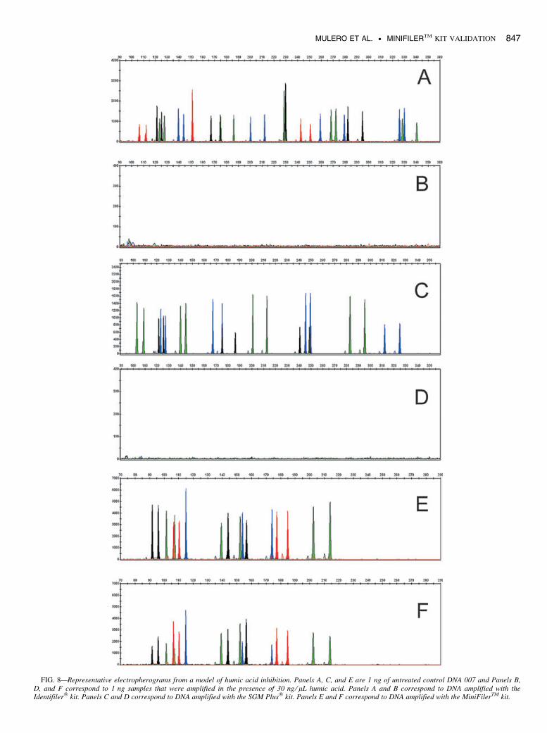
FIG. 8—Representative electropherograms from a model of humic acid inhibition. Panels A, C, and E are 1 ng of untreated control DNA 007 and Panels B,D, and F correspond to 1 ng samples that were amplified in the presence of 30 ng ⁄ lL humic acid. Panels A and B correspond to DNA amplified with theIdentifiler� kit. Panels C and D correspond to DNA amplified with the SGM Plus� kit. Panels E and F correspond to DNA amplified with the MiniFilerTM kit.
MULERO ET AL. • MINIFILERTM KIT VALIDATION 847

was amplified with the Identifiler� kit and resulted in no genetic pro-file. In contrast, analysis of the sample with the MiniFilerTM kityielded typeable results at all loci (Fig. 10).
Mixture Studies
Evidence samples that contain body fluids and ⁄or tissues origi-nating from more than one individual are commonly encounteredin forensic casework. Table 6 depicts the genotype of the minorcomponent of two individuals mixed at several mixture ratios. Mostof the alleles at each locus for the two individuals are different andthus do not overlap; however, some alleles of the minor contribu-tors do reside at stutter positions of alleles from the major contribu-tor. The minor component at 1:1, 1:3, and 1:7 mixture ratios wasreadily typeable. At a 1:10 ratio, all three profiles yielded completeprofiles. However at 1:15 ratios, alleles at the D7S820 and D18S51loci for one of the profiles were not detected because they werebelow the stutter filter threshold. In summary, ratios greater than1:10 (where the minor component is c. 91 pg) may result in partialprofiles for the minor component.
Population and Concordance Studies
Concordance data for four population groups (U.S. Caucasian,U.S. Hispanic, Asians, and African Americans) have been pub-lished (15) and documented in the MiniFilerTM kit user guide (25).In summary, 1308 samples were evaluated with both the MiniFil-erTM and Identifiler� STR kits: 449 African American, 445 Cauca-sian, 207 Hispanic, and 207 Asian individuals. Full concordancebetween the Identifiler� and MiniFilerTM kits was observed in99.7% of the STR allele calls compared (15). Twenty-five of the27 discordant results were due to allele dropout. Two discordantresults in the Identifiler� kit amplicon outside of the MinifilerTM
kit primer binding region were due to a base insertion in one sam-ple and a five base deletion in the other. The population set wasalso analyzed for peak height ratios as shown on Table 7. Themean peak height ratios indicate that the two alleles of a heterozy-gous individual are generally very well balanced. However, occa-sional low peak heights ratios were observed as outlying datapoints as shown in the column labeled as ‘‘Minimum’’ on Table 7.
Discussion
The MiniFilerTM PCR amplification kit simultaneously amplifieseight miniSTR loci and the sex determining marker amelogenin,making it a highly discriminating STR detection system for humanidentification. The combination of five-dye chemistry (6-FAMTM,NEDTM, PET�, VIC�, and LIZ� dyes) and the inclusion of non-nucleotide linkers made it technologically possible to simulta-neously amplify all loci in a single PCR. Because the primers forthe MiniFilerTM loci were redesigned from the conventional STRkits to yield smaller amplicons, these loci may be typeable with the
MiniFilerTM kit when previously not possible. Additionally, asshown in Table 3, the MiniFilerTM kit is more discriminating thana partial profile with the Identifiler� kit (minus the MiniFilerTM
loci). Thus, a forensic analyst has alternatives in deciding whichmultiplex system to use, especially when confronted with a com-promised DNA sample that yields sufficient DNA only for a singlePCR. If there is sufficient DNA for multiple PCRs but the DNA issufficiently degraded, then the combination of the results that maybe derived from a partial Identifiler� or SGM Plus� kit profile anda MiniFilerTM kit profile may notably increase the discriminationpower of the STR profile.
The validation of the MiniFilerTM kit encompassed the verifica-tion of the best reaction conditions and reagent concentrations forthe amplification of pristine as well as compromised DNA. Theperformance criteria included overall peak heights, peak heightratios, intracolor peak balance, and lack of cross-reactive peaks inthe presence of nonhuman DNA. The thermal cycling parametersof the MiniFilerTM kit differ from other autosomal AmpF‘STR�
kits in three ways: (1) the number of amplification cycles (30cycles), (2) the denaturation time (20 sec), and (3) annealing time(2 min). These changes enhanced sensitivity for detection of smallamounts of DNA in the presence of inhibitors of the PCR. Thesensitivity studies demonstrated that an input amount of 500 pg ofDNA does not produce off-scale peaks while yielding enough sig-nal strength for reproducible detection of 125 pg or less of templateDNA (Fig. 5). The DNA mixture study (Table 6) demonstrates thatthe MiniFilerTM kit is capable of producing robust profiles for theminor contributor even at ratios of 1:10.
In summary, the results from the models of PCR degradation andinhibition and nonprobative samples demonstrate that theMiniFilerTM kit can be extremely useful for amplifying DNA underconditions where other commercial autosomal STR kits yield partialor no profiles. As such, this kit is a useful and robust complement toconventional STR kits and will be especially applicable to challeng-ing situations involving the identification of human remains.
Finally, Applied Biosystems performed the developmental vali-dation studies described in this paper. It is recommended that eachlaboratory conduct its own internal validation according to FBI ⁄National Standards and SWGDAM guidelines (16,17).
Acknowledgments
The authors would like to thank our collaborators John But-ler, Carolyn Hill, and Margaret Kline from NIST for performingpopulation studies. We would like to gratefully acknowledgeWalther Parson from the Institute of Legal Medicine in Inns-bruck, Niels Morling and Helle Smidt Mogensen from the Insti-tute of Forensic Medicine in Copenhagen, and Arthur Eisenbergand Xavier Aranda from the University of North Texas HealthScience Center in Ft. Worth for testing prototypes. The authorsalso wish to thank Michael Malicdem for technical assistance.We also extend our gratitude to Lisa Calandro and Bruce
TABLE 5—Amplification efficiency of control DNA 007 in the presence of increasing amounts of humic acid.
Humic acid (ng ⁄ lL) MiniFilerTM Kit (n = 3) Identifiler� Kit (n = 3) SGM Plus� Kit (n = 5)
0 1.0 1.0 (1.0) 1.0 (1.0)10 1.0 0.96 € 0.08 (0.98 € 0.05) 1.0 (1.0)30 1.0 0 (0) 0 (0)50 1.0 0 (0) 0 (0)
A full MiniFilerTM profile consists of 17 amplification products (17 ⁄ 17 = 1.0). Identifiler� and SGM Plus� kits were scored for the loci shared with the Mini-FilerTM kit and for their total number of amplification products as shown in brackets (29 and 22 amplification products respectively for Identifiler� and SGMPlus� kits). The values are expressed as mean € SD.
848 JOURNAL OF FORENSIC SCIENCES

FIG. 9—Results from the amplification of 1 ng of degraded DNA isolated from a formalin fixed paraffin-embedded breast tumor tissue sample from 1997.(A) Identifiler� kit (modified to 30 cycles of amplification) and (B) MiniFilerTM kit electropherograms. The circles in panel A identify the loci missing fromthe Identifiler� kit but amplified by the MiniFilerTM kit.
MULERO ET AL. • MINIFILERTM KIT VALIDATION 849

FIG. 10—Results from the amplification of 1 ng of degraded DNA extracted from a skeletal remain. (A) Identifiler� kit and (B) MiniFilerTM kit electropher-ograms. The circles in panel A identify the loci missing from the Identifiler� kit but amplified by the MiniFilerTM kit.
850 JOURNAL OF FORENSIC SCIENCES

Budowle for valuable comments on this work, and the membersof the Human Identification R&D, Marketing and Manufactur-ing groups (Applied Biosystems) for their assistance.
References
1. Dixon LA, Dobbins AE, Pulker HK, Butler JM, Vallone PM, CobleMD, et al. Analysis of artificially degraded DNA using STRs andSNPs–results of a collaborative European (EDNAP) exercise. ForensicSci Int 2006;164(1):33–44.
2. Bender K, Schneider PM, Rittner C. Application of mtDNA sequenceanalysis in forensic casework for the identification of human remains.Forensic Sci Int 2000;113(1–3):103–7.
3. Just RS, Irwin JA, O’Callaghan JE, Saunier JL, Coble MD, Vallone PM,et al. Toward increased utility of mtDNA in forensic identifications.Forensic Sci Int 2004;146:(Suppl):147–9.
4. Whitaker JP, Cotton EA, Gill P. A comparison of the characteristics ofprofiles produced with the AMPFlSTR SGM Plus� multiplex system forboth standard and low copy number (LCN) STR DNA analysis. ForensicSci Int 2000;129(1):25–34.
5. Kloosterman AD, Kersbergen P. Efficacy and limits of genotyping lowcopy number (LCN) DNA samples by multiplex PCR of STR loci. JSoc Biol 2003;197(4):351–9.
6. Gill P, Whitaker J, Flaxman C, Brown N, Buckleton J. An investigationof the rigor of interpretation rules for STRs derived from less than100 pg of DNA. Forensic Sci Int 2000;112(1):17–40.
7. Wiegand P, Kleiber M. Less is more—length reduction of STR ampli-cons using redesigned primers. Int J Legal Med 2001;114(4–5):285–7.
8. Butler JM, Shen Y, McCord BR. The development of reduced size STRamplicons as tools for analysis of degraded DNA. J Forensic Sci2003;48(5):1054–64.
9. Drabek J, Chung DT, Butler JM, McCord BR. Concordance studybetween Miniplex assays and a commercial STR typing kit. J ForensicSci 2004;49(4):859–60.
10. Chung DT, Drabek J, Opel KL, Butler JM, McCord BR. A study on theeffects of degradation and template concentration on the amplificationefficiency of the STR Miniplex primer sets. J Forensic Sci2004;49(4):733–40.
11. Coble MD, Butler JM. Characterization of new miniSTR loci to aidanalysis of degraded DNA. J Forensic Sci 2005;50(1):43–53.
12. Grubwieser P, Muhlmann R, Berger B, Niederstatter H, Pavlic M, Par-son W. A new ‘‘miniSTR-multiplex’’ displaying reduced ampliconlengths for the analysis of degraded DNA. Int J Legal Med2006;120(2):115–20.
13. Opel KL, Chung DT, Drabek J, Tatarek NE, Jantz LM, McCord BR.The application of miniplex primer sets in the analysis of degradedDNA from human skeletal remains. J Forensic Sci 2006;51(2):351–6.
14. Tsukada K, Takayanagi K, Asamura H, Ota M, Fukushima H. Multiplexshort tandem repeat typing in degraded samples using newly designedprimers for the TH01, TPOX, CSF1PO, and vWA loci. Legal Med2002;4:239–45.
15. Hill CR, Kline MC, Mulero JJ, Lagace RE, Chang CW, Hennessy LK,et al. Concordance study between the AmpFlSTR MiniFilerTM PCRamplification kit and conventional STR typing kits. J Forensic Sci2007;52(4):870–3.
16. Quality assurance standards for forensic DNA testing laboratories.Forensic Sci Commun 2000;2(3). Available at http://www.fbi.gov/hq/lab/fsc/backissu/july2000/codis2a.htm. Accessed on April 28, 2008.
17. Revised Validation Guidelines-Scientific Working Group on DNA Anal-ysis Methods (SWGDAM). Forensic Sci Commun 2004;6(3). Availableat http://www.fbi.gov/hq/lab/fsc/backissu/july2004/standards/2004_03_standards02.htm. Accessed on April 28, 2008.
18. Applied Biosystems. AmpF‘STR� IdentifilerTM PCR amplification kituser’s manual. Foster City, CA: Applied Biosystems, 2001.
19. Applied Biosystems. AmpF‘STR� SGM PlusTM PCR amplification kituser’s manual. Foster City, CA: Applied Biosystems, 2001.
20. Coticone SR, Oldroyd N, Philips H, Foxall P. Development of theAmpF‘STR SEfiler PCR amplification kit: a new multiplex containingthe highly discriminating ACTBP2 (SE33) locus. Int J Legal Med2004;118:224–34.
21. Collins PJ, Hennessy LK, Leibelt CS, Roby RK, Reeder DJ, Foxall PA.Developmental validation of a single-tube amplification of the 13 CO-DIS STR loci, D2S1338, D19S433, and amelogenin: the AmpF‘STRIdentifiler� PCR Amplification Kit. J Forensic Sci 2004;49:1265–77.
22. Mulero JJ, Chang CW, Calandro LM, Green RL, Li Y, Johnson CL,et al. Development and validation of the AmpF‘STR Yfiler PCR ampli-fication kit: a male specific, single amplification 17 Y-STR multiplexsystem. J Forensic Sci 2006;51(1):64–75.
23. Grossman PD, Bloch W, Brinson E, Chang CC, Eggerding FA, Fung S,et al. High-density multiplex detection of nucleic acid sequences: oligo-nucleotide ligation assay and sequence-coded separation. Nucleic AcidsRes 1994;22:4527–34.
24. Baron H, Fung S, Aydin A, Bahring S, Luft FC, Schuster H. Oligo-nucleotide ligation assay (OLA) for the diagnosis of familial hyper-cholesterolemia. Nat Biotechnol 1996;14:1279–82.
25. Applied Biosystems. AmpF‘STR� MiniFilerTM PCR amplification kituser guide. Foster City, CA: Applied Biosystems, 2006.
TABLE 6—DNA mixture study.
Mixture Ratio Genotype D13S317 D7S820 D2S1338 D21S11* D16S539 D18S51 CSF1PO* FGA
0:1 Major GT 11 7,12 20,23 28,31 9,10 12,15 11,12 24,261:0 Minor GT 12,14 8,9 20,21 28,30 12,13 17,19 10 21,221:1 Minor GT 12,14 8,9 21 30 12,13 17,19 10 21,221:1 Minor GT 12,14 8,9 21 30 12,13 17,19 10 21,221:3 Minor GT 12,14 8,9 21 30 12,13 17,19 10 21,221:3 Minor GT 12,14 8,9 21 30 12,13 17,19 10 21,221:7 Minor GT 12,14 8,9 21 30 12,13 17,19 10 21,221:7 Minor GT 12,14 8,9 21 30 12,13 17,19 10 21,221:10 Minor GT 12,14 8,9 21 30 12,13 17,19 10 21,221:10 Minor GT 12,14 8,9 21 30 12,13 17,19 10 21,221:15 Minor GT 12,14 8,9 21 30 12,13 17,19 10 21,221:15 Minor GT 12,14 9 21 30 12,13 19 10 21,22
Major GT, major component genotype; Minor GT, minor component genotype.Minor component allele calls at nonoverlapping STR loci from duplicate mixture amplifications. Detected genotype of minor component using a peak
amplitude threshold of 50 RFU.*Stutter peak present, which may mask allele.
TABLE 7—Peak height ratio for eight loci obtained from genotypedpopulation samples (input DNA = 500 pg).
AlleleNumber of
Observations (n) Mean Median SD Minimum Maximum
CSF1PO 781 87.9 89.3 29.8 57.9 100D2S1338 911 87.0 88.8 33.8 52.3 100D7S820 820 88.1 89.7 29.4 58.4 100D13S317 733 88.4 90.4 35.0 50.5 100D16S539 804 87.5 89.1 38.1 46.1 100D18S51 906 87.9 89.2 31.7 55.1 100D21S11 856 88.2 90.0 37.4 47.1 100FGA 904 88.0 89.4 32.6 53.9 100
Peak height ratios were determined only for those heterozygous sampleswith peak heights >200 RFUs.
MULERO ET AL. • MINIFILERTM KIT VALIDATION 851

26. Sensabaugh GF. Biochemical markers of individuality. In: Saferstein R,editor. Forensic science handbook. New York: Prentice-Hall, Inc, 338–415.
27. Brownstein MJ, Carpten JD, Smith JR. Modulation of non-templatednucleotide addition by Taq DNA polymerase: primer modifications thatfacilitate genotyping. BioTechniques 1996;20:1004–6.
28. Walsh PS, Fildes NJ, Reynolds R. Sequence analysis and characteriza-tion of stutter products at the tetranucleotide repeat locus vWA. NucleicAcids Res 1996;24:2807–12.
29. Mulero JJ, Chang CW, Hennessy LK. Characterization of the N + 3stutter product in the trinucleotide repeat locus DYS392. J Forensic Sci2006;51(5):1069–73.
30. Viguera E, Canceill D, Ehrlich SD. In vitro replication slippage byDNA polymerases from thermophilic organisms. J Mol Biol2001;312(2):323–33.
31. Lazaruk K, Wallin J, Holt C, Nguyen T, Walsh PS. Sequence variationin humans and other primates at six short tandem repeat loci used inforensic identity testing. Forensic Sci Int 2001;119(1):1–10.
32. Akane A, Matsubara K, Nakamura H, Takahashi S, Kimura K. Identifi-cation of the heme compound copurified with deoxyribonucleic acid(DNA) from bloodstains, a major inhibitor of polymerase chain reaction(PCR) amplification. J Forensic Sci 1994;39:362–72.
33. Sutlovic D, Definis Gojanovic M, Andelinovic S, Gugic D, Primorac D.Taq polymerase reverses inhibition of quantitative real time polymerasechain reaction by humic acid. Croat Med J 2005;46(4):556–62.
Additional information and reprint requests:Julio J. Mulero, Ph.D.Applied Biosystems850 Lincoln Centre Dr. MS 404-3Foster City, CA 94404E-mail: [email protected]
852 JOURNAL OF FORENSIC SCIENCES