Embed Size (px)
Citation preview

ORAL & Implantology - Anno V - N. 2-3/201258
MIH: EPIDEMIOLOGIC CLINIC STUDYIN PAEDIATRIC PATIENTR. CONDÒ, C. PERUGIA*, P. MATURO*, R. DOCIMO*
Department of Clinical Science and Traslational Medicine University of Rome “Tor Vergata”*Department of Experimental Medicine and Surgeon
SUMMARYThe Molar Incisor Hypomineralization (MIH) is a qualitative and quantitative defect of the enamel structure of thefirst permanent molars, which may vary from 1 to 4 with involvement of maxillary and jaw permanent incisors.Aim. Aim of this study is that to evaluate, among 1500 paediatric patients chosen at random aged between 0 and 14 years,afferent by the Paediatric Dentistry of the Azienda Ospedialiera Policlinico Tor Vergata of Rome from 1996 to 2011, theincidents and the prevalence of the MIH distribution, and furthermore to ascertain the possible relationship with the datadescribed in the literature. Results and discussion. From the sample of 1500 paediatric patients, the number of those affections from MIH has turnedout to be pairs to 110 (7.3%) aged between 4 and 15 years, and an average age equal to 9.7. The incidence of the hy-poplastic defects is greater in the elements of the permanents series in which the functional class mainly interested is thatof the first molars, with a percentage of 39.8%. Regarding the elements of the deciduous series affections from hypoplasia,they turn out to be in all in number of 20 represented in 80% of the cases from the seconds molars while in the remain-ing 20% of the cases the items involved are the central incisors. About the percentage of elements involved in the MIH:the molars, involved with a frequency of 56%, turn out to be more hit regarding incisors (44%). As reported in the litera-ture, it can be asserted that the MIH can hit in equal measure both the male sex that feminine one. Conclusions. MIH represents a condition quite frequent in the paediatric population. In managing this anomaly takes anessential role in the early diagnosis and in the differential one. The study done underlined the importance of a correct ap-plication of the therapeutic protocol which, starting from a careful diagnosis and articulating themselves in the executionof preventive treatments and in severe cases restorative and prosthetic, has the aim to certify the functionality and theaesthetic of the dental elements affected by MIH.
Key words: MIH, paediatric patient, epidemiology.
Introduction
The Molar Incisor Hypomineralization (MIH) is aqualitative and quantitative defect of the dentalenamel, to idiopathic aetiology characterized bythe progressive and simultaneous hypomineraliza-tion and/or hypoplasia of the enamel structure of thefirst permanent molars, which may vary from 1 to4 with involvement of maxillary and jaw permanentincisors.In the past many Authors described clinically sucha peculiar and specific pathological condition mak-ing reference to it, any time with different terms andeven contradictory.It was only in the 2000 that the European Academy
of Paediatric Dentistry recognizes the necessity toidentify it in an univocal and unequivocal way withthe terms of Molar Incisors Hypomineralization(MIH) not to confuse with the Molar Hypominer-alization (MH) that does not affect at all the inci-sors.Therefore, the lack of appropriate definition madeit, for many years, that the related literature oftenconfused and however insufficient to the extentthat even today it is difficult to establishing if someof the studies made actually refer to said pathology.Aim of this study is that to evaluate, among 1500paediatric patients chosen at random aged between0 and 14 years, afferent by the Paediatric Dentistryof the Azienda Ospedaliera Policlinico Tor Vergataof Rome from 1996 to 2011, the incidents and the
rese
arc
h a
rtic
le

research article
ORAL & Implantology - Anno V - N. 2-3/2012 59
prevalence of the MIH distribution, and further-more to ascertain the possible relationship with thedata described in the literature.
Aetiology
Although the international literature agrees in rec-ognizing that, the aetiology of the MIH might bedue to some idiopathic factors of systemic origin, inthe 2008 the European Society of Paediatric Den-tistry, assumed that it could be tied up also withsome genetic component and, since the suscepti-bility to such a pathology may vary a lot from oneindividual to another, even if casual factors may bethe same, it indicates that, in reality the MIH recog-nises an aetiology of a multifactor type.Lygidakis, among 2000 individuals aged between 3and 15 years, demonstrated that, in a percentage of14.5%, the MIH is most correlated to any system-atic pathology, in a percentage of 19.2% it is linkedwith prenatal problems and with a birth delay, in the44.3% it is due to perinatal problems and in the21.8% it is due to neonatal problems, coming to theconclusion, in a second time, that in the 12.2% it isnot associated with any important medical historywhile, in the remaining 87.8% may be correlatedwith numerous systematic problems incurred infrom the birth to the first childhood like: asthma,pneumonia, infection of the upper airway, otitismedia, tonsillitis and tonsillectomies, taking up antibiotic molecules, some presence of dioxin in the mother’s milk and some childhood spotted fever (1-7).According to a systematic revision of 2007, WilliamV. has declared that, even if a specific aetiologic forthe MIH is not yet known, it is sure instead that thechildren characterized by a bad general health sta-tus during the first three years of life, the ones bornbefore the term or exposed to contaminated areas(phytoestrogens, mercury, phthalates, bipocidi, pes-ticides) are more subject to be affected by MIH (8).In general, in any case, the surveys made towardsthe analysis of all possible factors aetiologic as oftoday resulted to be retrospective studies, that sendback to an individual memory, often incompleteand inexact. This to support the fact that, in reality,
the perspective studies, conducted at the birth andup to the eruption of the first permanent molars arenecessary and contribute to clarify the question.Therefore at today, the literature agrees and con-cludes that it is not yet possible to define a specificaetiology for the MIH (4).
Epidemiology
The epidemiologic data, present in the literature ofthe last fifteen years, are only limited and turnedmostly to studies of prevalence. The epidemiolog-ical studies on the frequency of hypoplasia of thedental enamel in the contemporary populationsdemonstrate the liaison between the socio-eco-nomical conditions and the prevalence of this de-fect. In the developed countries the incidence of thiscondition for an average of 10%, while in the de-veloping countries the same incidence is higherthan the 50% (9).In the 1987, Koch noted that, in the permanent el-ements the prevalence of hypomineralizationand/or idiopatic hypoplasia of the dental enamelvaried between the 3.6 and the 21.5 according to theyear of birth (10-12).The studies undertaken between the 1987 and the2001 are of difficult interpretation because they donot have as objective the study of prevalence,recorded as secondary result only. They certify thatit varies within an ample interval, included betweenthe 2.8 and 2.5%, up to assume a more reliable andconstant value, around the 15%, immediately aftersuch time term (7, 12-24).In reality, the more recent epidemiological studiesseem to suggest on the contrary that the MIH preva-lence is more and more increasing (15).According to a study dated 2008 on 3518 Greekchildren, aged between 5.5 and 12 years, it resultsthat: the 10.2% of them (58.6 female and 41.4 male)present MIH, while the total of the tooth affected re-sulted equal to 1286, of which 776 molars and 510incisors with an average of involved elements, perchild, equal to 5.7 (3.4 molars and 2.2 incisors). Inthe 28.4% of the children affected by MIH are in-terested only molars while in the 71.6% both themolars and the incisors. The association of the af-

ORAL & Implantology - Anno V - N. 2-3/2012
rese
arc
h a
rtic
le
60
fected tooth more frequently noticed has resulted tobe: 4 molars/2 incisors (23.5%), 4 molars/4 incisors(16.8%), only 4 molars (15.1%), only 2 molars(9.7%) with a major involvement of the maxillaryelements in respected to the lower jaw. Such studydemonstrates finally the such pathologic conditionin progressive because the interested dental ele-ments are hit by a post-eruptive repartition of theenamel in relation to the age, and determine that thegravity of the MIH increases in a way proportionalto the chronologic age while the more light defectsappear prevalently charged to the permanent inci-sors (2, 14).The major part of the studies undertaken betweenthe 2001 and the 2007 demonstrate that it does notexist any difference of distribution among the twosexes (7, 14, 17-21).In any case, recently some Authors report a majorevidence charged to female sex, but with no demon-stration if it is really a significant data (3, 23).The numerous studies undertaken to establish theaverage value, per child, of tooth affected by MIHdemonstrate that it varies from 5 to 5.7 elements perindividual; they highlight furthermore that the dam-ages involving the first permanent maxillary molarsare the more serious as well as the more frequent (2,7, 14, 16, 17, 19-23).On the other hand, four clinic studies demonstrate,on the contrary, that the permanent molars affectedby MIH belong prevalently to the jaw dental arch(7, 14, 20, 25).
Diagnosis: clinical signs and symptoms
In the 2003, Weerheijm has established that theMIH clinic evaluation must be executed only ondental elements well washed and wet, underliningthat the age optimal for a diagnostic survey of suchpathological condition corresponds to the 8 yearsold, time in which we may suppose that both thefirst molars and the permanent incisors are alreadythere (15).From the diagnostic point of view it is importantalso to consider that the MIH hits, in a prevailingmanner but not exclusively, the permanent molars
and incisors, because as Weerheijm himself demon-strated that also the second deciduous molars, thesecond permanent molars and the cusps of the per-manent canines may occasionally show some de-fects of the dental enamel (26).Notwithstanding that, very different from the his-tological point of view, clinically the damages hy-pomineralization and that ones hypoplastic of theMIH are considered referred to the same patho-logic environment (15). Even if, the literature de-fines as “hypomineralization” the qualitative defectof deposit of matrix and minerals and as “hy-poplastic” the quantitative defect of formation of thematrix. From the clinical point of view, both ofthem are classified as defects inadequate of thedental amelogenesis and they are characterized bysome areas of enamel opacity and of colour whitish,yellow, yellowish, dark, up to the black. The dam-ages tend to further worse and often the molarsmay suffer of the so called post-eruptive breakdown, either to say a structural collapse, frequentlydiagnosed as a caries damage, due to the presenceof a light and porous structure, that it is also re-sponsible of forming of atypical cavities and par-ticular distortion of the crown morphology (27).The incisors affected, rarely show the divisionspost eruptive because, in reality, we think that saidphenomenon is due to back of occlusive charge onthe opacity, event that in the contrary involvesprevalently the first permanent molars. It is impor-tant underline that the division of the enamel, beingan event post-eruptive, may complete a clinic situ-ation of hypomineralization or hypoplasia in thesecases it is imposed a differential diagnosis withthose enamel defects, appeared during the processof amelogenesis and therefore before the eruptionof the element in the arch (19, 26, 28).Some Authors agree in considering the dental el-ements affected by MIH hyper sensitive to thestimulus (26, 29). Both of the clinic level and ra-diographic they are characterized by the presenceof an inflammation of the pulp due actually to theincreased dental sensibility (29). The first per-manent molars affected by MIH may turn intocaries pathologies very quick and the presence ofa caries damages may hide the true diagnosis ofMIH (26, 30).Therefore, the appropriate diagnosis of MIH needs a

research article
ORAL & Implantology - Anno V - N. 2-3/2012 61
selective registration of the clinic evidences relatedto the single dental elements (4 first permanent mo-lars and 8 permanent incisors) in order to evaluate:• the presence or absence of limited opacities;• the presence of enamel division post eruptive;• the presence of atypical repairs;• verification of past extractions of first permanent
molars;• check the lack of a molar eruption or of a per-
manent incisor (Fig. 1) (26).
MIH methodology of clinicclassification
The classifications to which the scientific literaturesend in order to evaluate the clinic entity of thedamages caused by the MIH and therefore to es-tablish the gravity of such pathological condition,are essentially two:• Classification of dental amelogenesis (31);
• Classification of gravity of defects caused by thedental amelogenesis (5, 13).
The defects caused by deficits happened during theamelogenesis process have been classified in threetypes:1) hypoplastic, it happens during the formative
phase of the matrix deposition. The enamel,characterized by a thickness of the tissue verylight, may have an aspect dot-shaped, smooth orrough;
2) hypocalcified, that it is produced at the time ofthe calcification, during which the matrix min-eralizes. It is characterized by an enamel easilypierceable with probes;
3) hypomaturational, that happens during the ma-turity during which we have an increase of crys-tallites; the enamel may be pigmented or whiteand it is characterized by a thin stratum enam-elled soft and crumbly; such a defect, often islinked to taurodontism (31).
Last, in order to evaluate the defects of the amelo-
Figure 1Clinical case 1.
ORAL & Implantology - Anno V - N. 2-3/2012
rese
arc
h a
rtic
le genesis taking into consideration their peculiarity,extention and position in the teeth crown, it is pos-sible to correlate the gravity of the MIH to that ofthe areas hit by such damages and therefore identifyit in:• severe (loss of enamel correlated to a dental im-
pairment);• moderate (limited to the only loss of enamel);• slight change of the colour (into white, yellow or
brown).
The areas with a minor MIH appear only with acolour change, while the moderate with the loss ofenamel. In the cases of serious MIH the loss ofstructure interests also the dentin (5, 13).
Materials and methods
The purpose of this study is that of evaluating asample of 1500 paediatric patients taken at ran-dom, aged between 0 and 14 years, all of them af-ferent by the Paediatric Dentistry of the AziendaOspedaliera Policlinico Tor Vergata of Rome from1996 to 2011, the incidence, prevalence and dis-tribution of MIH, and also to establish the even-tual relationship with the data referred in litera-ture.Of any patient have been recorded the data relatedto sex and age and it has been analyzed with atten-tion the dental formula, in order to register the pres-ence and the gravity of eventual defects of qualityand/or quantity inherent to the dental enamel.Of any patient affected by hypomineralization
and/or hypoplasia have been recorded number andfunctional class of any interested dental elements,specifying if they were deciduous or permanent.Subsequently, it has been evaluated the near or postmedical history, in order to underline the presenceof eventual pathologies linked or in any case cor-related to MIH.It has been made a statistical analysis, based onnumerical values and percentages. Have beencalculated the averages arithmetic of the variousdistribution, the averages of the various inci-dences, linking them to two sex and patient age,and it has been evaluated the correlation witheventual pathologies linked with the presence ofMIH. The data so obtained have been included intables and graphics, namely hystograms and aero-grammes, later on described and commented indetails.
Results
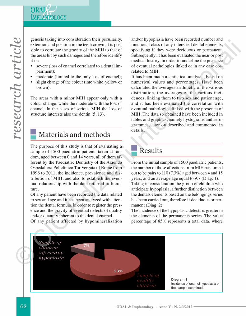
From the initial sample of 1500 paediatric patients,the number of those affections from MIH has turnedout to be pairs to 110 (7.3%) aged between 4 and 15years, and an average age equal to 9.7 (Diag. 1).Taking in consideration the group of children whoanticipate hypoplasia, a further distinction betweenthe dentals elements based on the belongings serieshas been carried out, therefore if deciduous or per-manent (Diag. 2).The incidence of the hypoplasic defects is greater inthe elements of the permanents series. The valuepercentage of 85% represents a total data, where
Diagram 1Incidence of enamel hypoplasia onthe sample examined.
62
research article
ORAL & Implantology - Anno V - N. 2-3/2012
still a distinction between the various dental func-tional classes is not carried out.Regarding the elements of the deciduous series af-fections from hypoplasia, they turn out to be in allin number of 20 represented in 80% of the casesfrom the seconds molars (tot.16) while in the re-maining 20% of the cases the items involved are thecentral incisors (tot. 4).The obtained data concerning dental elements af-fected by MIH were divided in two tables, the firstregarding the deciduous set of the teeth (Tab. 1) andthe second one inheres that permanent one (Tab. 2);in both tables were distinct functional classes andsexes.In a decreasing order of incidence, it has turned outthat the dental elements that mainly anticipate de-fects of the dental enamel are represented by thesecond deciduous molars (tot. 11); successively
followed, therefore less frequently, from a conditionof generalized hypoplasia of the incisors; at lastfrom the involvement of first deciduous molars(Tab. 1)Overall distribution of hypoplasia has turned outto have a slightly frequency greater in the male(Diag. 3).Regarding the permanent dentition, Table 2 showsthat there are not cases of second molars affected byhypoplastic defects. The hypoplastic instead envis-ages with a greater incidence of first molars fol-lowed, in a decreasing order, by conditions of gen-eralized hypoplasia, hypoplasia of the elements in-cisors and at last by cases of MIH, condition gen-eralized from the simultaneous presence of defectsit is at the level of the molars that the incisors. Thedistribution of these data between males and fe-males is evident in Diagram 4.
Diagram 2Incidence of enamel hypoplasia indeciduous and permanent teeth.
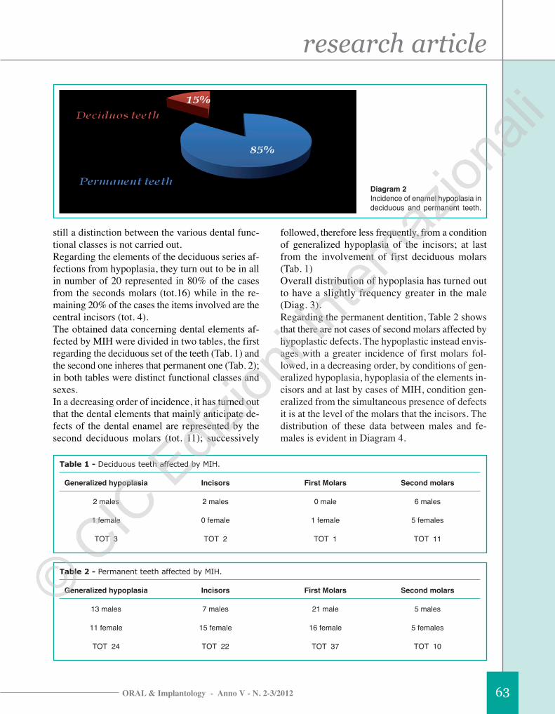
Table 1 - Deciduous teeth affected by MIH.
Generalized hypoplasia Incisors First Molars Second molars
2 males 2 males 0 male 6 males
1 female 0 female 1 female 5 females
TOT 3 TOT 2 TOT 1 TOT 11
Table 2 - Permanent teeth affected by MIH.
Generalized hypoplasia Incisors First Molars Second molars
13 males 7 males 21 male 5 males
11 female 15 female 16 female 5 females
TOT 24 TOT 22 TOT 37 TOT 10
63

ORAL & Implantology - Anno V - N. 2-3/3012
rese
arc
h a
rtic
le
64
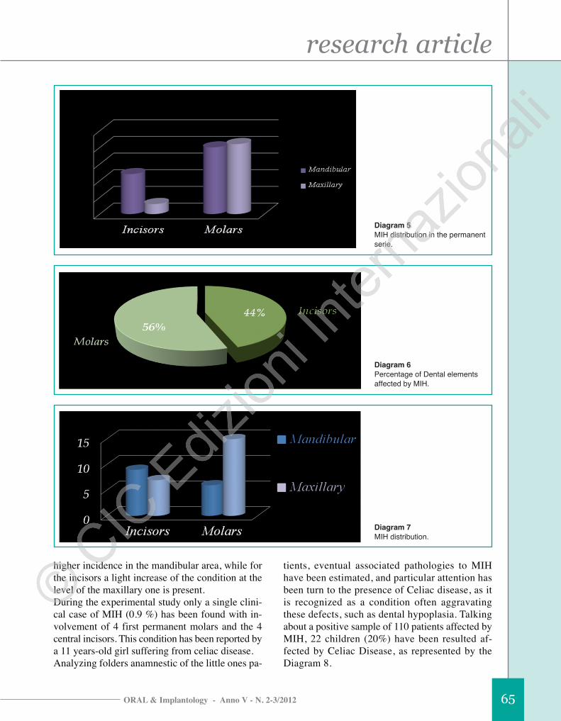
Later, it was determined the distribution of dentalelements permanent suffering from hypoplasia dis-tinguishing them between maxillary and mandibu-lar (Diag. 5). The study then has place the attentionon the MIH condition, considering the presence ofhypoplasia not more isolated to single elements, butat the present level of molars and incisors. Out of110 children with hypoplastic defects, those inwhich was detected MIH, were found to be 10,with an average age of 10.5. The distribution turnsout to be absolutely fair between the two sexes, as5 males and 5 females are interested. Entering morespecifically, the distribution of such hypoplasic
condition anticipates the following distributionwhich showed in Diagram 6.From this analysis it turns out that on 10 children af-fections from MIH, molars, interested in number of21, are more involved than incisors, which are in-terested in number of 16.The dentals elements mainly hit from MIH are themandibular molars, involved in number of 14 (Diag.7). This value is performed by the maxillary inci-sors, in number of 8, and subsequently by mandibu-lar incisors, in number of 6, and finally by maxillarymolars, in number of 5. Overall, it is possible to saythat for the functional class of molars is found an
Diagram 3Hypoplasia of deciduos teeth.
Diagram 4Hypoplasia of permanent teeth.
research article
ORAL & Implantology - Anno V - N. 2-3/2012 65
higher incidence in the mandibular area, while forthe incisors a light increase of the condition at thelevel of the maxillary one is present.During the experimental study only a single clini-cal case of MIH (0.9 %) has been found with in-volvement of 4 first permanent molars and the 4central incisors. This condition has been reported bya 11 years-old girl suffering from celiac disease.Analyzing folders anamnestic of the little ones pa-
tients, eventual associated pathologies to MIHhave been estimated, and particular attention hasbeen turn to the presence of Celiac disease, as itis recognized as a condition often aggravatingthese defects, such as dental hypoplasia. Talkingabout a positive sample of 110 patients affected byMIH, 22 children (20%) have been resulted af-fected by Celiac Disease, as represented by theDiagram 8.
Diagram 5MIH distribution in the permanentserie.
Diagram 6Percentage of Dental elementsaffected by MIH.
Diagram 7MIH distribution.

ORAL & Implantology - Anno V - N. 2-3/2012
rese
arc
h a
rtic
le
66
Discussion
Pointed out that, from the data collected duringthe study, the incidence of hypoplasic condition,in a sample of 1500 paediatric patients, it is equalto 7% (Diag. 1), it can be asserted that this valueturns out to be slightly lower than the data in theliterature, which provides a range of incidencebetween the 4 and the 20%, with an average of12% (10, 11). Looking at Diagram 2 it is clearthat the incidence of hypoplasia turns out to besignificantly greater in the elements of the per-manent series with a frequency of 85% with theaverage of the sample pairs to 9.7 years. This datais perfectly on line with studies in literaturewhich reported a higher frequency of hypoplas-tic conditions in the permanent series comparedto the deciduous one (11).On the total sample of 110 children, those whohave hypoplasic defects at the level of the decidu-ous teeth have turned out to be in number of 17, in-cluding 10 males and 7 females, with an averageage of 5 years. Although a light prevalence of thehypoplasia is stressed in the males, this is notenough to assume a real correlation between hy-poplasia and sex. The functional class of deciduousmore interested, with a frequency of 64.7%, is thatof the second molars. The central incisors insteadare involved in the 11.8% of the cases (Tab. 1). Assaid by Weerheijm and other Authors, the MIH is acondition detail that regards, for definition, the el-ements of the permanent series (12). To support of
such consideration, in our study were not encoun-tered cases of MIH in deciduous teeth (6, 32, 33).The permanent teeth, as pointed out, is by definitionmore affected by this anomaly dental development.As a matter of fact are involved 93 children on110, of which 46 males and 47 females, with amedium age of 8 years. Even this value has beenconfirmed by the literature, since many Authorsclaim that there is a correlation between the inci-dence of disease and sex, as evidenced by this study(7,14, 17, 19, 20).Some Authors however argue that the frequency ofthe MIH, in permanent teeth, is light greater in thefemale, aspect also found in this study (3, 23, 34).The functional class of the permanent series mainlyinterested is that of the first molars, with a per-centage of 39.8%. This can be explained by the factthat, in the eruption sequence, the first molars arethe first elements to erupt in arch, at the age of 6 ap-proximately.On line with what is reported in the literature, thestudy showed that in the permanent teeth, as well asdeciduous, the incisors are elements that are in-volved to a lesser extent, with a frequency of 23.6%.Diagram 3 and 4 represent graphically what is re-ported in Table 1 and Table 2, therefore the distri-bution of different dental elements hypoplasia, re-spectively, in deciduous and permanent.The Cartesian diagram shows on the x-axis dentalfunctional classes, while on the y-axis numeric val-ues representing the elements involved.Permanent dental elements affected by hypoplasiawere then divided according to functional class
Diagram 8MIH and Celiac Disease.
research article
ORAL & Implantology - Anno V - N. 2-3/2012 67
(Diag. 4) and then, on the basis of the distinction be-tween maxillary and mandibular, to assess the pos-sible prevalence (Diag. 5). By processing the data,you can highlight that, as regards the maxillary in-cisors, those elements are involved in number of 20(90.9%), while the maxillary envolved in number of2 (9.1%). The molars instead present a balanced dis-tribution of hypoplastic overall between the twoarches: these are indeed results involved 39 maxil-lary and 38 mandibular molars. Diag. 5, 6 and 7 regard the MIH condition, inwhich it is possible to have a contemporary in-volvement of permanent elements, molars and in-cisors; such anomaly is found in 10 children (Tab.2), therefore with a frequency of the 9.1% (Diag.5). Making a first distinction on the sex, being in-terested 5 males and 5 females, it is not possibleto indicate a prevalence or a greater predispositionof one of the two sexes regarding the other, so asalready it is evidenced for the conditions of hy-poplasia previously discussed. Also in this casethe data turn out to be agree with the literature(14, 17-21, 35). As reported in the literature, it can be asserted thatthe MIH can hit in equal measure both the male sexthat feminine one. Diagram 6 shows the percentageof elements involved in the MIH: the molars, in-volved with a frequency of 56%, turn out to bemore hit regarding incisors (44%).
Conclusions
The clinical study has considered the conditionmade of MIH, which manifest with as a quality andquantity teeth enamel, to idiopathic aetiology char-acterized by progressive and simultaneous hy-pomineralization or hypoplasia of the structure ofthe enamel of the first permanent molars in variablenumber from 1 to 4 with frequent involvement ofmaxillary and mandibular permanent incisors (11,12). Areas with light MIH are only manifested withdental discromation while, those moderated with aloss of enamel. In case of severe MIH, tissue losscan also affect the dentin (5, 13).The exposure to certain environmental pollutingagents, a general state of ill health in the first three
years of life, as well as a genetic predisposition, rep-resent the main etiologic factors of MIH.From the analysis of literature and from the resultsemerged from this clinical study, it has been high-lighted that MIH represents a condition quite fre-quent in the paediatric population. Specifically, theliterature reports a medium frequency equal to 12%.In the study being evaluated in a random sample ofchildren aged between 3 and 14 years old, it hasbeen turned out to be equal to 9%.In managing this anomaly, take an essential role,the early diagnosis and the differential one. Thefirst one is fundamental not only for the planningof the treatment of the lesions, but also to provideparents a correct information about the state ofhealth of the dental elements concerned, in orderto arise promptly to support a more favourableprognosis. The differential diagnosis is importantto the aim not to confuse potential hypoplasticdefects with other anomalies of enamel structureof the enamel, such as post-traumatic discoloura-tion or tetracycline, fluorosis and processes ofcaries.The therapeutic protocol articulates with preventionat different levels, including the sealing of the fur-rows of the permanent molars hypoplastic or hy-pomineralized and treatments of systematic andtopical fluoride prophylaxis; it is important there-fore, the clinical monitoring, with a continuous fol-low-up every 3 months.The first permanent molars affected by MIH, inview of the mineralization’s defects, can found veryquickly a sharing post-eruptive of the enamel and acarious pathology, which can hide a hypoplasic le-sions. The progress of the caries can also be aggra-vated by the fact that children suffering from thisparticular pathology neglect their oral hygiene, dueto increased sensitivity, with further progression ofthe carious phatologies (30).The study done underlined the importance of a cor-rect application of the therapeutic protocol which,starting from a careful diagnosis and articulatingthemselves in the execution of preventive treatmentsand in severe cases restorative and prosthetic, has theaim to certify the functionality and the aesthetic ofthe dental elements affected by MIH.This study represents a preliminary investigation onMIH: the values obtained are infact to be considered
ORAL & Implantology - Anno V - N. 2-3/2012
rese
arc
h a
rtic
le
68
baseline data, which can be expanded and sup-ported by successive sample.
References
1. Lygidakis NA, Chaliasou A, Siounas G. Evaluation ofcomposite restaurations in hypomineralised permanentmolars: a four-year clinical trial. Eur J Paediatr Dent2003; 3: 143-148.
2. Lygidakis NA, Dimou G, Marinou D. Molar-incisor-hypomineralisation (MIH). A retrospective clinicalstudy in Greek children. II. Possible medical aetiolog-ical factors. Eur Arch Paediatr Dent. 2008aDec;9(4):207-17.
3. Lygidakis NA, Dimou G, Briseniou E. Molar-incisor-hypomineralisation (MIH). Retrospective clinical studyin Greek children. I. Prevalence and defect character-istics. Eur Arch Paediatr Dent. 2008b Dec;9(4):200-6.
4. Whatling R, Fearne JM. MIH: a study of aetiologicalfactors in a group of UK children. Int J Paediatr Dent2008; 18: 155-162.
5. Alaluusua S, Lukinmaa P-J, Vartiainen T et al. Poly-chlorinated dibenzo-p-dioxins and dibenzofurans viamother’s milk may cause developmental defects in thechild’s teeth. Environ toxicol pharmacol 1996a; 1:193-197.
6. Alaluusua S, Lukinmaa P-L, Torppa J, Tuomisto J,Vartiainen T. Developing teeth as biomarker of diodiexposure. Lancet 1999; 353:206.
7. Jalevik B, Norén JG, Klingberg G, Barregard I. Etio-logic factors influencing the prevalence of demar-cated opacities in permanent first molars in a group ofSwedish children. Eur J Oral Sci 2001; 109: 230-234.
8. William V, Burrow MF, Messer LB. Microshear bondstrength of resin composite to teeth affected by molarhypoplasia using two adhesive system. Paediatr Dent2007; 28: 233-41.
9. Willmott NS, Bryan RA, Duggal MS. Molar-incisor-hypomineralisation: a literature review. Eur Arch Pae-diatr Dent. 2008 Dec; 9(4):172-9.
10. Koch G, Hallonsten AL, Ludvigsson N, Hansson BO,Holst A, Ullbro C. Epidemiologic study of idiopathicenamel hypomineralization in permanent teeth ofSwedish children. Community Dent Oral Epidemiol.1987 Oct;15(5):279-85.
11. Weerheijm KL, Jalevik B, Alaluusua S. Molar-Incisor-Hypomineralisation. Caries Res 2001a; 35: 390-391.
12. Weerheijm KL, Mejàre I. MIH A questionnaire inven-tory of its occurrence in member countries of the Eu-ropean Academy of Paediatric Dentistry (EAPD). Int JPaediatr Dent 2003; 13: 411-416.
13. Alaluusua S, Lukinmaa P-L, Koskimies M et al. De-velopmental dental defects associated with long breastfeeding. Eur J Oral Sci 1996b; 104: 493-497.
14. Leppaniemi A, Lukinmaa P-L, Alaluusua S. Nonfluo-ride Hypomineralisation in the permanent first molarsand their impact on treatment need. Caries Res 2001;35: 36-40.
15. Weerheijm KL, Duggal M, Mejàre I et al. Judgementcriteria for Molar-Incisor-Hypomineralisation (MIH) inepidemiologic studies: a summary of the Europeanmeeting on Mih held in Athens, 2003. Eur Arch Pae-diatr Dent 2003; 3: 110-113.
16. Dietrich G, Sperling S, Hetzer G. MIH in a group ofchildren and adolescents living in Dresden (Germany).Eur J Paediatr Dent 2003; 3: 133-137.
17. Calderara PC, Gerthoux PM, Mocarelli P et al. Theprevalence of MIH in a group of Italian school children.Eur J Paediatr Dent 2005; 2: 79-83.
18. Fteita D, Ali A, Alaluusua S. MIH in a group of school-aged children in Benghazi, Lybia. Eur Arch PaediatrDent 2006; 7: 92-95.
19. Muratbegovic A, Markovic N, Selinovic MG. MIH inBosnia and Herzegovina: Prevalence, aetiology andclinical consequences in medium caries activity popu-lation. Eur Arch Paediatr Dent 2007; 8: 189-194.
20. Preusser SE, Ferring V, Wleklinski C, Wetzel E-E.Prevalence and severity of MIH in a region of Germany– A brief communication. J Public Healt Dent 2007; 67:148-150.
21. Jasulaityte I, Veerkamp JS, Weerheijm KL. MIH reviewand prevalence data from a study of primary schoolchildren in Kaunas (Lithuania). Eur Arch Paediatr Dent2007; 8: 87-94.
22. Arrow P. Prevalence of developmental enamel defectsof the first permanent molars among school children inWestern Australia. Aust Dent J 2008; 53: 250-259.
23. Cho S-Y, Ki Y, Chu V. MIH in Hong Kong Chinesechildren. Int J Paediatr Dent 2008; 18: 348-352.
24. Crombie FA, Manton DJ, Weerheijm KL, KilpatrickNM. MIH: a survey of members of the Australian andNew Zeland Society of Paediatric Dentistry. Aust DentJ 2008; 53: 160-166.
25. Chawla N, Messer LB, Silva M. Clinical studies in mo-lar hypoplasia. Part I: Distribution and putative asso-ciations. Eur Arch Paediatr Dent 2008; 9: 180-190.
26. Weerheijm KL. MIH Eur J Paediatr Dent 2003; 4: 114-120.
27. Marthu-Muju K, Wright TJ. Diagnosis and treatment ofMolar-Incisor-Hypomineralisation. Compendium 2006;27(11): 604-611.
28. Fayle SA. MIH Restorative management. Eur J Pae-diatr Dent 2003; 4: 121-126.
29. Jalevik B, Klingberg G. Dental treatment, dental fearand behaviour management problems in children withsevere enamel hypoplasia in their permanent first mo-lars. Int J Paediatr Dent 2002; 12: 24-32.

research article
ORAL & Implantology - Anno V - N. 2-3/2012 69
30. Beentjes VE, Weerheijm KL, Groen HJ. Factors in-volved in the aetiology of MIH. Eur J Paediatr Dent2002; 3: 9-13.
31. Witkop CJ Jr. Amelogenesis imperfecta, dentinogene-sis imperfecta and dentin dysplasia revisited: prob-lems in classification. J Oral Pathol. 1988 Nov; 17(9-10):547-53.
32. Jalevik B, Klingberg G, Norén JG, Barregard L. Epi-demiological study of idiopathic enamel hypomineral-isation in permanent first molars. European Academyof Pediatric Dentistry Congress Abstract number 99.Eur J Paediatr Dent 2000; 1:128.
15. Jalevik B, Moller M. Evaluation of spontaneous spaceclosure and development of permanent dentition afterextraction of hypoplasic permanent first molars. Int JPaediatr Dent 2007; 17: 328-335.
34. Lygidakis NA, Dimou G, Marinou D, Gouva G. Aeti-
ology of MIH. A retrospective study. European Acad-emy of Paediatric Dentistry Congress Abstract number069. Eur J Paediatr Dent 2004; 5: 19.
35. Willmott NS, Bryan RA, Duggal MS. Molar-incisor-hypomineralisation: a literature review. Eur Arch Pae-diatr Dent 2008 Dec; 9(4):172-9.
Correspondence to:Roberta CondòDepartment of Clinical Scienceand Traslational MedicineUniversity of Rome “Tor Vergata”Via Montpellier, 100166 Rome, ItalyE-mail: [email protected]