Embed Size (px)
Citation preview

Tumor and Stem Cell Biology
MIG-7 Controls COX-2/PGE2-Mediated Lung CancerMetastasis
Ming-Yi Ho1, Shu-Mei Liang1,2, Shao-Wen Hung2, and Chi-Ming Liang1,3
AbstractMore effective treatments for metastatic lung cancer remain a pressing clinical need. In this study, we
identified migration inducting gene-7 (MIG-7) protein as critical for COX-2/prostaglandin E2 (PGE2)- andAkt/GSK-3b-dependent tumor invasion/metastasis. COX-2/PGE2 activated EP4 to enhance Akt and GSK-3bphosphorylation and b-catenin/T-cell factor/lymphoid enhancer factor signaling leading to MIG-7 upregula-tion. RNAi-mediated attenuation of MIG-7 blocked COX-2/PGE2- and Akt/GSK-3b-mediated migration/invasion effects. Furthermore, MIG-7 protein inhibited protein phosphatase 2A to sustain Akt/GSK-3bphosphorylation and cancer-cell migration/invasion. Cancer cells overexpressing MIG-7 exhibited increasedexpression of ZEB-1 and Twist in parallel with epithelial–mesenchymal transition, metastasis and cancerlethality. MIG-7 protein level positively correlated with advanced stages of human lung cancers. MIG-7 thusoffers a theranostic target for cancer metastases arising from aberrant activation of the cellular COX-2/PGE2and Akt/GSK-3b signaling pathways. Cancer Res; 73(1); 439–49. �2012 AACR.
IntroductionLung cancer is the leading cause of cancer death worldwide.
The major cause of treatment failure and mortality is cancermetastasis (1–3). The cyclooxygenases (COX), key enzymes inthe biosynthesis of prostaglandins, are potential mediators ofmetastasis. Overexpression of COX-2, an inducible form ofCOX, is frequently found in early and advanced lung cancertissues and is associated with poor prognosis (4–7). Elevatedlevels of tumor COX-2 and its metabolite prostaglandin E2(PGE2) contribute to a decrease in E-cadherin, induction oftumor angiogenesis, augmentation of cancer motility andinvasiveness, resistance to apoptosis, and suppression of anti-tumor immunity (5, 8, 9).The binding of PGE2 to 1 or more of its 4 G protein-coupled
receptors, designated EP1, EP2, EP3, and EP4, has been shown tostimulate phosphatidylinositol 3-kinase/protein kinaseB (PI3K/Akt) and extracellular signal-regulated kinase 1/2 (ERK1/2)signaling and is implicated in cancer-cell growth and progres-sion (8–11). However, both the COX-2/PGE2 and PI3K/Aktsignaling cascades exist in normal cells with pleiotropic func-tions and inhibition of these 2 important signaling pathwayscausesavarietyofundesirable sideeffects (12, 13).Onenotorious
example of the effects of the long-termuse of COX-2 inhibitors isrofecoxib (Vioxx) that resulted in renal and cardiovascularsystem impairment and was withdrawn from the market in2004 (14, 15). Discovery of agents that target the downstreameffector(s) of COX-2/PGE2 and PI3K/Akt signaling cascades isdesirable to aid thedevelopmentof therapeutics that havebetterselectivity and specificity and no severe adverse effects.
Another potential mediator of metastasis is migrationinducting gene-7 (MIG-7) protein (16). Increase in MIG-7mRNA is found in embryonic cytotrophoblast cells duringplacenta development as well as in more than 80% of tumorcells, but not found in 25 different normal tissues or in bloodfrom normal subjects (16–18). Transduction of MIG-7 incarcinoma cells results in invasion by carcinoma cells in 3-dimensional culture in vitro (17). Stably expressing MIG-7-specific shRNA, on the other hand, reduces phosphorylationof Akt and ERK1/2 and attenuates membrane-type 1 matrixmetalloproteinase (MT1-MMP) activity in endometrial carci-noma cell lines (19). Whether or not this holds true for lungcancer has not been examined and functional associationbetween MIG-7 and COX-2/PGE2 signaling in promoting lungcancer invasion/metastasis remains elusive.
In this study, we found thatMIG-7 was functionally associatedwithCOX-2/PGE2-induced lungcancermetastasis throughphos-phorylation of Akt and glycogen sythase kinase-3 b (GSK-3b),activation of b-catenin/TCF-4/LEF-1 signaling and decrease inthe activity of protein phosphatase 2A (PP2A). The critical roleof MIG-7 protein was further substantiated by examining itslevel in the human lung cancers as well as the effects of overex-pressingMIG-7 on lung cancer cells in a xenograft mousemodel.
Materials and MethodsMaterials
A549 cell line [American Type Culture Collection (ATCC:CCL-185)], H1299 (ATCC: CRL-5803), CL1-0, and CL1-5 (20)
Authors' Affiliations: 1Genomics Research Center, 2Agricultural Biotech-nology Research Center, and 3Institute of Biological Chemistry, AcademiaSinica, Taipei, Taiwan, ROC
Note: Supplementary data for this article are available at Cancer ResearchOnline (http://cancerres.aacrjournals.org/).
CorrespondingAuthors:Chi-MingLiang, AcademiaSinica, Rm727, ABRC,No. 128, Sec. 2, Academia Road, Taipei 11529, Taiwan. Phone: 8862-2787-2501; Fax: 8862-2651-8049; E-mail: [email protected]; andShu-Mei Liang, Academia Sinica, Rm 626, ABRC, No. 128, Sec. 2,Academia Road, Taipei 11529, Taiwan. Phone: 8862-2652-2870;E-mail: [email protected]
doi: 10.1158/0008-5472.CAN-12-2220
�2012 American Association for Cancer Research.
CancerResearch
www.aacrjournals.org 439
on June 25, 2018. © 2013 American Association for Cancer Research. cancerres.aacrjournals.org Downloaded from
Published OnlineFirst November 13, 2012; DOI: 10.1158/0008-5472.CAN-12-2220

were maintained in Dulbecco's Modified Eagle's Media orRPMI medium (GibcoBRL Life Technologies) supplementedwith 10% FBS (GibcoBRL Life Technologies) and 1% penicillin-streptomycin-neomycin (GibcoBRL Life Technologies). Ahuman lung fibroblast cell line (WI-38) was obtained fromAbcam. A549 and H1299 cells were certified by the FoodIndustry Research and Development Institute (Hsinchu, Tai-wan) in 2012.MIG-7 siRNA and scrambled control siRNA werepurchased from Dharmacon (Thermo Fisher Scientific). Thesequences of the MIG7-1 siRNA and MIG7-2 siRNA are: GUC-GAAGAAAUGAAACUUUUU and CUUAAAUCACAGGAAAU-CUUU. MIG-7 siRNA transfection was undertaken by usingDharmacon Accell SMARTpool siRNA reagent (Thermo FisherScientific) according to the protocol recommended by Dhar-macon. The MIG7 shRNA-encoding sequences cloned inpGPU6/Neo siRNA Expression vectors were obtained fromGenDiscovery Biotechnology. The MIG7-1 or MIG7-2 shRNAencoding sequence was created by using the 2 complementaryoligonucleotides indicated below, each containing the 21nucleotides target sequence of MIG7 (313–333 or 1523–1543), followed by a short spacer TTCAAGAGA: shMIG7-1sense, 50-CACCGCAAGTACAGGGCAGAATTTCTTCAAGAGA-GAAATTCTGCCCTGTACTTGCTTTTTTG-30; shMIG7-1 anti-sense, 50-GATCCAAAAAAGCAAGTACAGGGCAGAATTTC-TCTCTTGAAGAAATTCTGCCCTGTACTTGC-30; shMIG7-2sense, 50-CACCGCCATCTGTGAGATTACAAATTTCAAGAGA-ATTTGATATCTCACAGATGGCTTTTTTG-30; and shMIG7-2antisense, 50-GATCCAAAAAAGCCATCTGTGAGATATCAAA-TTCTCTTGAAATTTGATATCTCACAGATGGC-30. Mice, anti-bodies, and other reagents are described on the SupplementaryMaterials and Methods.
Knockdown and overexpression of proteinsFull-length human COX-2 cDNA (NM 000963.2) and human
MIG-7 (DQ080207.2) cDNA derived from A549 cells wereamplified by using specific primers (Supplementary TableS1; Sigma-Proligo) and subcloned into pcDNA6/BioEase-DESTby Gateway cloning technology (Invitrogen) to generate pCOX-2 plasmids (pCOX2) and pMIG7 plasmids. The insertsequences in the plasmids were confirmed by automated DNAsequencing. The plasmids or siRNAs were transfected into celllines by PolyJet In Vitro DNA Transfection Reagent or Gene-Mute siRNA and DNA Transfection Reagent (SignaGen labo-ratories). The expression of target proteins in the transfectedcells was determined 48 hours after transfection, unlessspecified otherwise.
Immunoblotting, immunoprecipitation, and gelatinzymographic analysis
Gelatinolytic activities of MMP-2 and MMP-9, immunopre-cipitation, and immunoblotting were conducted as describedpreviously (21, 22).
Migration, invasion, and anchorage-independentgrowth assays
In vitromigration, invasion, and anchorage-independentgrowth assays were conducted as described previously(23).
Luciferase reporter assayTo examine transcriptional regulation of MIG7 promoters,
genomic DNA was isolated from the A549 cell line and approx-imately 3 kb of theMIG7 promoter was amplified by PCR usingthe specific primers listed in Supplementary Table S1 (Sigma-Proligo) and subcloned into the pGL3-basic vector (Promega)to generate the MIG7 promoter/firefly luciferase reporterconstruct, designated pGL3-MIG7. To analyze T-cell factor/lymphoid enhancer factor (TCF/LEF)-dependent promoteractivity, we purchased TOPflash (wild-type TCF binding sitesreporter plasmid) and FOPflash (nonresponsive mutated TCFbinding sites reporter plasmid; Millipore). pRL-TK plasmid(Promega) containingRenilla luciferasewas used as an internalcontrol. At 24 to 36 hours after transfection, cell lysates wereassayed using the Dual-Luciferase Reporter Assay System(Promega) and luciferase activities were measured with aluminometer (Wallac Vector3; PerkinElmer).
PP2A activity assayCellular PP2A activity was assayed using a PP2A immuno-
precipitation phosphatase assay kit (Upstate) according to themanufacturer's instructions.
Preparation of pCMV-GFP/luciferase-lentivirus andestablishment of stable cell lines
A549GL cells were produced by infecting A549 cells withcytomegalovirus promoter (pCMV)-GFP/luciferase-lentivirusas described previously (22). A549GL cells were then transfectedwith pcDNA6/BioEase-DEST (empty vector; EV) or pMIG7plasmids and then selected with blasticidin (100 mg/mL) toproduce A549EV-GL and A549MIG7-GL stable cell lines, respec-tively. Some 549MIG7-GL cells were transfected with MIG7-1short hairpin RNA (shRNA) and then selected with G418(400 mg/mL) to produce A549MIG7shMIG7-GL stable cell lines.CL1-0EV-GL, CL1-0MIG7-GL, and CL1-0MIG7shMIG7-GL stable celllines were prepared according to the same procedure.In addition, CL1-5GL cells were transfected with controlshRNA, MIG7-1 shRNA, and MIG7-2 shRNA separately andselected with G418 (400 mg/mL) to produce CL1-5shCont-GL,CL1-5shMIG7-1-GL, and CL1-5shMIG7-2-GL stable cell lines. Theexpression level of MIG-7 in each stable cell lines was analyzedby immunoblot analysis (Supplementary Fig. S5E).
Experimental xenograft murine metastasis modelMale severe combined immunodeficient mice (n ¼ 9/group)
were implanted with 50 mL RPMI medium (vehicle) or 1 � 106/50 mL lung tumor cells on day 0 by lateral tail vein injection.Metastatic progression was monitored weekly and quantifiedusing a noninvasive bioluminescence In Vivo Imaging System(IVIS) (Xenogen) as described previously (22). Thirty-five daysafter injection, 3 mice were killed for necropsy and the other6 mice were kept for survival studies. The percentage of animalsurvival in each group was routinely recorded.
Immunohistochemistry, histopathology examination,and assessment of MIG-7 protein level
Human lung carcinoma tissuemicroarray (TMA) slideswerepurchased from US Biomax (BC41114) and verified by a
Ho et al.
Cancer Res; 73(1) January 1, 2013 Cancer Research440
on June 25, 2018. © 2013 American Association for Cancer Research. cancerres.aacrjournals.org Downloaded from
Published OnlineFirst November 13, 2012; DOI: 10.1158/0008-5472.CAN-12-2220

qualified, experienced pathologist (Dr. Wei-Hwa Lee). Immu-nohistochemistry and histopathology analyses were con-ducted as described previously (22, 24). The images werescanned into a digital format by Scanscope XT system (AperioTechnologies) and analyzed using Aperio ImageScope 9.1software (Aperio Technologies). The percentage of positivecells from 5 fields of each tissue sample was scored as follows: 0(0%–5%), 1 (5%–20%), 2 (20%–40%) and 3 (>40%) of tumortissue stained. Signal intensity from 5 fields of each tissuesample was scored by their staining color as follows: 0 (noimmunostaining), 1 (light brown), 2 (medium brown), and 3(dark brown). Two scores for each tissue sample were com-bined to evaluate the relative intensity and expression per-centage of MIG-7 in each tissue sample.
StatisticsAll statistical comparisons were made with 2-tailed tests.
The survival timewas assessed usingKaplan–Meier curves and
tested for significance by the log-rank test. Statistical evalua-tion was conducted using GraphPad Prism version 5.0 forMicrosoftWindows (GraphPad Software). Differences betweengroups were considered statistically significant at �, P < 0.05;��, P < 0.01, or ��� , P < 0.0001.
ResultsMIG-7 protein plays an important role in COX-2-inducedmigration and invasion of lung cancer cells
MIG-7 protein expressed in A549, H1299, CL1-0, and CL1-5human lung cancer cell lines (Supplementary Fig. S1A). Toexamine the potential relationship between COX-2, MIG-7protein, and cell invasiveness, we treated these lung cancercells with COX-2-specific inhibitor NS398 or COX-2 siRNA(siCOX2) and found that it inhibited MIG-7 protein (by 2- to4.5-fold) and decreased epithelial–mesenchymal transition(EMT) as indicated by a 2- to 4.5-fold increase in the level ofepithelial cellmarker E-cadherin and a 3- to 4.5-fold decrease in
Figure 1. MIG-7protein is functionally associatedwithCOX-2-mediatedupregulationof EMT,MMP-2activity, andmigration/invasionof lungcancer cells.Cellswere examined by immunoblotting for protein expression, zymography for MMP activity and transwell assay for cell migration/invasion as described inMaterials andMethods. Blots are representative of 3 independent experiments. Data representmeans�SDof 3 independent experiments; �,P <0.05by t test.AandB, lungcancer cells (2�105/mL)were treatedwithCOX-2 antagonistNS398 for 24hours or control scrambledsiRNA (siCont) andCOX-2 siRNA(siCOX2)for 48 hours as indicated. C and D, knockdown of MIG-7 suppressed COX-2-mediated effects but not COX-2 or PGE2 level. Parental lung cancer cells (A549,H1299, and CL1-0; C) were transfected with EV, pCOX2, control scrambled siRNAs (-siMIG7-1), or MIG7-1 siRNAs (þsiMIG7-1) for 48 hours as indicated.
COX-2/PGE2-Driven Cancer Metastasis Requires MIG-7
www.aacrjournals.org Cancer Res; 73(1) January 1, 2013 441
on June 25, 2018. © 2013 American Association for Cancer Research. cancerres.aacrjournals.org Downloaded from
Published OnlineFirst November 13, 2012; DOI: 10.1158/0008-5472.CAN-12-2220
the level of mesenchymal cell marker vimentin (Fig. 1A and B).siCOX2 also decreased PGE2, MMP-2 activity, and migration/invasion by 2- to 4.5-fold in lung cancer cells (SupplementaryFig. S1B–S1D). In contrast, overexpressing COX-2 by transfec-tion with pCOX2 increased COX-2 together with MIG-7protein, PGE2, EMT, MMP-2 activity, and migration/invasionby 2- to 3.5-fold in lung cancer cells (Fig. 1C and D). MIG-7knockdown (þsiMIG7-1 orþsiMIG7-2) attenuated the effect ofCOX-2 overexpression onMIG-7, EMT, MMP-2, and the migra-tion/invasion of the cells by 30% to 60%, but did not affectexpression level of COX-2 and PGE2 (Fig. 1C and D, Supple-mentary Fig. S1E and S1F).
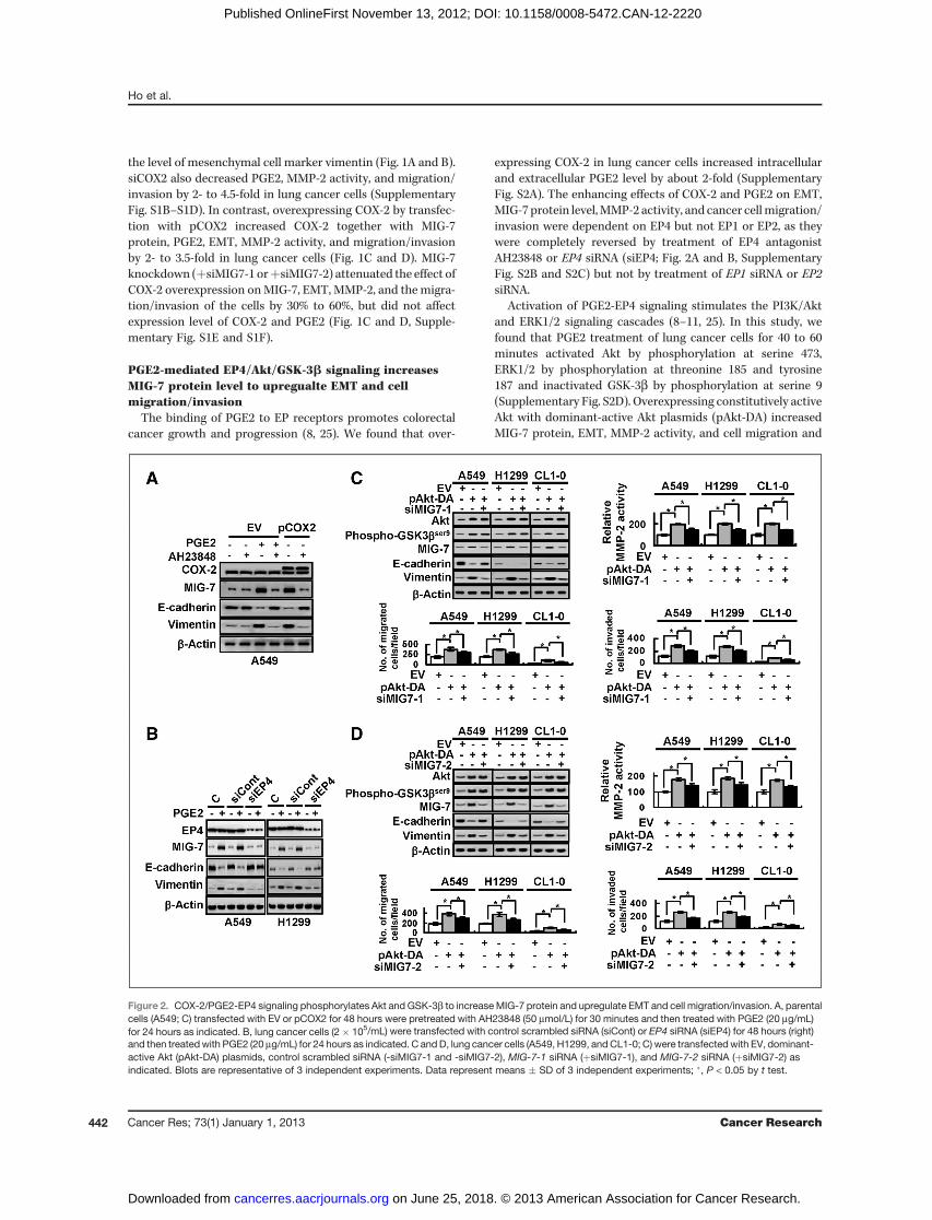
PGE2-mediated EP4/Akt/GSK-3b signaling increasesMIG-7 protein level to upregualte EMT and cellmigration/invasion
The binding of PGE2 to EP receptors promotes colorectalcancer growth and progression (8, 25). We found that over-
expressing COX-2 in lung cancer cells increased intracellularand extracellular PGE2 level by about 2-fold (SupplementaryFig. S2A). The enhancing effects of COX-2 and PGE2 on EMT,MIG-7 protein level,MMP-2 activity, and cancer cellmigration/invasion were dependent on EP4 but not EP1 or EP2, as theywere completely reversed by treatment of EP4 antagonistAH23848 or EP4 siRNA (siEP4; Fig. 2A and B, SupplementaryFig. S2B and S2C) but not by treatment of EP1 siRNA or EP2siRNA.
Activation of PGE2-EP4 signaling stimulates the PI3K/Aktand ERK1/2 signaling cascades (8–11, 25). In this study, wefound that PGE2 treatment of lung cancer cells for 40 to 60minutes activated Akt by phosphorylation at serine 473,ERK1/2 by phosphorylation at threonine 185 and tyrosine187 and inactivated GSK-3b by phosphorylation at serine 9(Supplementary Fig. S2D). Overexpressing constitutively activeAkt with dominant-active Akt plasmids (pAkt-DA) increasedMIG-7 protein, EMT, MMP-2 activity, and cell migration and
Figure 2. COX-2/PGE2-EP4 signaling phosphorylates Akt and GSK-3b to increaseMIG-7 protein and upregulate EMT and cell migration/invasion. A, parentalcells (A549; C) transfected with EV or pCOX2 for 48 hours were pretreated with AH23848 (50 mmol/L) for 30 minutes and then treated with PGE2 (20 mg/mL)for 24 hours as indicated. B, lung cancer cells (2� 105/mL) were transfected with control scrambled siRNA (siCont) or EP4 siRNA (siEP4) for 48 hours (right)and then treatedwith PGE2 (20 mg/mL) for 24 hours as indicated. C andD, lung cancer cells (A549, H1299, andCL1-0; C) were transfectedwith EV, dominant-active Akt (pAkt-DA) plasmids, control scrambled siRNA (-siMIG7-1 and -siMIG7-2), MIG-7-1 siRNA (þsiMIG7-1), and MIG-7-2 siRNA (þsiMIG7-2) asindicated. Blots are representative of 3 independent experiments. Data represent means � SD of 3 independent experiments; �, P < 0.05 by t test.
Ho et al.
Cancer Res; 73(1) January 1, 2013 Cancer Research442
on June 25, 2018. © 2013 American Association for Cancer Research. cancerres.aacrjournals.org Downloaded from
Published OnlineFirst November 13, 2012; DOI: 10.1158/0008-5472.CAN-12-2220

invasion by 2- to 3-fold (Fig. 2C and D). Knockdown of MIG-7attenuated effects of Akt overexpression on EMT, MMP-2activity, and cell invasion (by 30%–60%) but not phosphory-lation of GSK-3b (Fig. 2C and D), indicating that MIG-7 is thedownstream target of Akt/GSK-3b signaling.
Activation of Akt/GSK-3b signaling enhances b-catenin-TCF-4/LEF-1 signaling cascade to elevate MIG-7 proteinlevelIn some cancer cells, inactivation of GSK-3b by phosphory-
lationat serine9hasbeen shown to increase the accumulationofnuclear b-catenin, which associates with TCF/LEF to turn onWnt signaling (26, 27) and transactivate transcription of targetgenes (28). To observe whether phosphorylated GSK-3bSer9
causes activation of Wnt signaling in lung cancer cells, we useda luciferase reporter system in which the promoter modulecontained TCF binding sites (TOPflash) or nonresponsive,mutated binding sites (FOPflash). COX-2 and PGE2 increasedthe EP4-dependent luciferase activity of TOPflash/FOPflash upto 45-fold that was accompanied with 11-fold increase inMIG-7transcription in the lung cancer cells (Supplementary Fig. S3A).
We further transfected A549 cells with dominant active (non-phosphorylated S9A-GSK-3b mutant) and dominant negative(kinase-deficient K85A-GSK-3bmutant) plasmids (29) to exam-ine whether they can regulate Wnt signaling and MIG-7 level.Our results showed that constitutive overexpression of activeGSK-3b by transfection of S9A-GSK-3b plasmids (pGSK3b-DA)downregulated phosphorylation of GSK-3b and attenuatedaccumulation of b-catenin, TOPflash reporter activity, andMIG-7 expression by 60% to 75% under PGE2 stimulation (Fig.3A). Transfection of K85A-GSK-3b plasmids (pGSK3b-DN), onthe other hand, resulted in 2- to 4-fold increase in levels ofb-catenin andMIG-7 proteins as well as around 40-fold increasein TOPflash reporter activity in lung cancer cells (Fig. 3A). Inaddition, increase of b-catenin by transfection of active form ofb-catenin (pb-catenin-S33A) enhanced TOPflash reporter activ-ity by 45-fold and MIG-7 expression more than 2-fold in A549cells (Fig. 3B). These results showed activation of Wnt signalingand induction of MIG-7 expression by phosphorylated GSK-3bSer9 in lung cancer cells.
Transfection with TCF-4 plasmids (pTCF4; 0.5–2 mg), LEF-1plasmids (pLEF1; 0.5–2 mg), or both pTCF4 and pLEF1
Figure 3. PGE2-mediatedtransactivation of MIG7 geneinvolves a b-catenin-TCF-4/LEF-1pathway. A and B, parental cells(A549; C) cotransfected with reporterplasmids TOPflash/FOPflash andpRL-TK and plasmids of emptyvector (-pGSK3b-DA, -pGSK3b-DN,-pb-catenin-S33A), dominant-activeGSK3b (þpGSK3b-DA), dominant-inactive GSK3b (þpGSK3b-DN), anddominant-active b-catenin (þpb-catenin-S33A) were treated with orwithout PGE2 (20mg/mL) for 24hoursas indicated. C, parental cells (A549;C) were cotransfected with reporterplasmids pGL3-MIG7 and pRL-TKand plasmids of EV, TCF-4 (pTCF4),LEF-1 (pLEF1), or a combination ofTCF-4 and LEF-1 plasmids (pTCF4þpLEF1) for 24 hours as indicated.D, parental cells (A549; C)cotransfected with reporter plasmidspGL3-MIG7 and pRL-TK and controlscrambled siRNA (siCont) or TCF-4siRNA (siTCF4) or LEF-1 siRNA(siLEF1) or combinationofTCF-4 andLEF-1 siRNAs (siTCF4 þ siLEF1)were treated with or without PGE2(20 mg/mL) for 24 hours as indicated.Blots are representative of 3independent experiments. Datarepresent means � SD of 3independent experiments. �, P < 0.05and ��, P < 0.01 by t test.
COX-2/PGE2-Driven Cancer Metastasis Requires MIG-7
www.aacrjournals.org Cancer Res; 73(1) January 1, 2013 443
on June 25, 2018. © 2013 American Association for Cancer Research. cancerres.aacrjournals.org Downloaded from
Published OnlineFirst November 13, 2012; DOI: 10.1158/0008-5472.CAN-12-2220
plasmids enhanced MIG-7 transcription and expression up to4-fold in a dose-dependent manner (Fig. 3C). PGE2 increasedtranscription and expression of MIG-7 protein as well asb-catenin level (Fig. 3D and Supplementary Fig. S3B) up to3-fold. The PGE-2-mediated increase of MIG-7 protein wasattenuated partially (47%) by transfection with either TCF-4siRNA (siTCF4) or LEF-1 siRNA (siLEF1) and abolishedby combination of siTCF4 and siLEF1 (Supplementary Fig.S3B). These results suggest that PGE2 activates Akt/GSK-3bsignaling to increase MIG-7 protein via enhancement of theb-catenin/LEF/TCF signaling cascade.
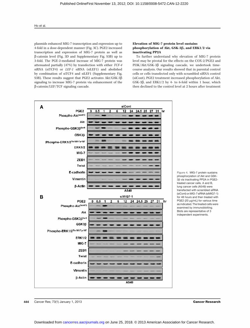
Elevation of MIG-7 protein level sustainsphosphorylation of Akt, GSK-3b, and ERK1/2 viainactivating PP2A
To further understand why elevation of MIG-7 proteinlevel may be pivotal for the effects on the COX-2/PGE2 andPI3K/Akt/GSK-3b signaling cascade, we undertook time-course analysis. Our results showed that in parental controlcells or cells transfected only with scrambled siRNA control(siCont), PGE2 treatment increased phosphorylation of Akt,GSK-3b, and ERK1/2 by 4- to 6-fold within 1 hour, whichthen declined to the control level at 2 hours after treatment
Figure 4. MIG-7 protein sustainsphosphorylation of Akt and GSK-3b via inactivating PP2A in PGE2-treated cancer cells. A and B,lung cancer cells (A549) weretransfected with scrambled siRNA(siCont) orMIG-7 siRNA (siMIG7-1)for 48 hours and then treated withPGE2 (20 mg/mL) for various timeas indicated. The treated cells wereexamined by immunoblotting.Blots are representative of 3independent experiments.
Ho et al.
Cancer Res; 73(1) January 1, 2013 Cancer Research444
on June 25, 2018. © 2013 American Association for Cancer Research. cancerres.aacrjournals.org Downloaded from
Published OnlineFirst November 13, 2012; DOI: 10.1158/0008-5472.CAN-12-2220

(Fig. 4A and Supplementary Fig. S4A). MIG-7 protein levelwas not elevated until 12 hours after PGE2 treatment andreached a 4-fold plateau at 24 to 31 hours. After elevation ofMIG-7 protein level, phosphorylation of Akt, GSK-3b, andERK1/2 was increased again and sustained at significantlyhigh levels (Fig. 4A and Supplementary Fig. S4A). In com-parison, in MIG-7 knockdown cells, even though phosphor-ylation of Akt, GSK-3b, and ERK1/2 increased in 1 hour anddeclined to control level at 2 hours after PGE2 treatment inthe same manner as control cells or siCont-transfected cells(Fig. 4B and Supplementary Fig. S4B), increase in MIG-7protein level after 12 hours or later was attenuated and sowas phosphorylation of Akt, GSK-3b, and ERK1/2 (Fig. 4Band Supplementary Fig. S4B).Phosphorylation of Akt, GSK-3b, and ERK1/2 is negatively
regulated by phosphatases, notably PP2A, which accounts forthe majority of serine-threonine phosphatase activity ineukaryotic cells (30). We thus examined whether MIG-7 inac-tivates PP2A to sustain phosphorylation of Akt, GSK-3b, andERK1/2. By using methods to determine the interaction ofPP2A with Akt and assay the PP2A phosphatase activity (31),we found that in PGE2-treated cancer cells, the amount of Aktassociated with PP2A regulatory unit B55-a and the phospha-tase activity of PP2Aweremaintained at the same levels beforethe elevation of MIG-7 protein level (Supplementary Fig. S5Aand S5B). Twelve hours after PGE2 treatment, the 2.5-foldincrease in MIG-7 protein level was accompanied with a 55%decrease in PP2A activity and a 45% decline in the amount ofAkt associated with B55-a (Supplementary Fig. S5A and S5B).Knockdown ofMIG-7 in PGE2-treated cancer cells reversed thedecline in the amount of Akt associated with B55-a andrestored the activity of PP2A (Supplementary Fig. S5C andS5D).
Overexpression of MIG-7 increases anchorage-independent growth and invasive ability of lung cancercellsKnockdownofMIG-7 in lung cancer cells attenuatedCOX-2/
PGE2 and Akt-mediated EMT (Figs. 1C, 2C, 2D, and 4B, andSupplementary Fig. S1E and S4B), a central tumorigenic pro-cess related to cellularmigration/invasion andmetastasis (32).Our results showed that MIG-7 overexpression upregulatedactivities of migration and invasion and increased growthability of lung cancer cells (A549, H1299, and CL1-0) in softagar by 2-fold (Fig. 5A). KnockdownofMIG-7withMIG-7 siRNA(siMIG7-1 or siMIG7-2) in highly invasive CL1-5 human lungcancer cells decreased cell migration and invasion as well asclone-forming ability in soft agar by 40% to 55% (Fig. 5B).Similar effects of MIG-7 were confirmed in A549EV-GL,A549MIG7-GL, and A549MIG7shMIG7-GL as well as CL1-0EV-GL,CL1-0MIG7-GL cells, and CL1-0MIG7shMIG7-GL lung cancer cellsstably expressing EV, MIG-7 protein, MIG7 shRNA, greenfluorescent protein, and luciferase (GL; Fig. 5C and Supple-mentary Fig. S5E).
MIG-7 protein promotes lung cancer metastasis in vivoTo investigate whether MIG-7 is sufficient to induce lung
cancer metastasis in vivo, we injected medium (vehicle),
A549EV-GL, A549MIG7-GL, or A549MIG7shMIG7-GL cells, respective-ly, into the tail vein of a murine metastasis model. Our resultsshowed that A549MIG7-GL-bearing mice developed more num-ber of tumor nodules (a 2.5-fold increase) in the lung comparedwith A549EV-GL- and A549MIG7shMIG7-GL-bearing mice on day 35postinoculation (Fig. 6A and 6B). Therewasmore Ki67-positiveproliferating cells (a 2-fold increase) and larger tumor area inmice inoculated with A549MIG7-GL cells than in those inocu-lated with A549EV-GLand A549MIG7shMIG7-GL cells (Fig. 6C).Analysis of lung tissue lysates showed that mice implantedwith A549MIG7-GL cells had higher expression levels of MIG-7,ZEB1, Twist, and vimentin and lower expression level of E-cadherin (Fig. 6D) as compared with those implanted withA549EV-GL and A549MIG7shMIG7-GL. The survival time ofA549MIG7-GL-bearing mice was significantly shortened thanthat of A549EV-GL- and A549MIG7shMIG7-GL-bearing mice (Fig.6E; median survival time of 39.5 days versus 52 and 51.5days, P < 0.001). Similar trends were found in comparingCL1-0MIG7-GL- and CL1-0MIG7shMIG7-GL-bearing mice (Supple-mentary Fig. S6A–S6D).Moreover, all the CL1-0MIG7-GL-bearingmice perished before day 50 postinoculation; whereas allthe mice inoculated with vehicle and CL1-0EV-GL as well as 5of 6 mice inoculated with CL1-0MIG7shMIG7-GL survived even onday 70 postinoculation (Supplementary Fig. S6E). Stably silenc-ing MIG-7 protein in CL1-5 cells (CL1-5shMIG7-1 and CL1-5shMIG7-2) by transfection with MIG7-1 or MIG7-2 shRNA alsoshowed that downregulation of MIG-7 reduced in vivo lungmetastasis of CL1-5 cells and prolonged survival time of tumor-bearing mice (Supplementary Fig. S7A–S7E).
MIG-7 protein level positively correlates with advancedstages of cancers in human lung tumor tissues
Analysis of MIG-7 protein levels in tumor and normal lungtissues of a human lung carcinoma TMA showed that themedian MIG-7 protein level was higher in lung cancer (T) thannormal (N) lung tissues (P < 0.0001); in locally advanced (T3þT4) versus noninvasive or minimally advanced (T1 þ T2) lungcancers (P < 0.0001); in cancers with lymph node involvement(N1 þ N2 status) versus cancers without lymph node involve-ment (N0 status; P< 0.0001); and in stage II/III/IV versus stage Ilung cancers (P < 0.0001; Fig. 7).
DiscussionAgonist of EP1 (ONO-DI-004) but not EP2, EP3 or EP4 was
reported to increase invasion of hepatocellular carcinoma(HCC) (33). As PGE2 receptors transactivate epidermalgrowth factor receptor (EGFR) and mesenchymal-epithelialtransition (MET), it has been proposed that there is a cross-talk between the COX-2/PGE2/EP1 and EGFR/c-Met signal-ing pathways that coordinately regulate human HCC cellinvasion (33). In this study, we found, however, that theeffects of COX-2 and PGE2 on EMT, MIG-7 protein level,MMP-2, and cancer cell migration/invasion were reversed byinhibition of EP4 (Fig. 2A, 2B, Supplementary Fig. S2B andS2C) but not EP1 or EP2. It remains to be elucidated whetherthese differences in EP4 and EP1 are due to different celltypes and whether COX-2/PGE2/EP4 has any cross-talk withEGFR/c-Met signaling in lung cancer cells.
COX-2/PGE2-Driven Cancer Metastasis Requires MIG-7
www.aacrjournals.org Cancer Res; 73(1) January 1, 2013 445
on June 25, 2018. © 2013 American Association for Cancer Research. cancerres.aacrjournals.org Downloaded from
Published OnlineFirst November 13, 2012; DOI: 10.1158/0008-5472.CAN-12-2220

Although Twist has been shown to activate the Akt signalingpathway (34) and play an essential role in tumormetastasis (35),our time-course analysis of PGE2 effects showed that elevationof MIG-7 was associated with increase of EMT and phosphor-ylation of Akt, GSK-3b, and ERK, which occurred several hoursbefore Twist induction (Fig. 4A and Supplementary Fig. S4). Asvariation in MIG-7 protein level did not affect COX-2 and PGE2
level (Fig. 1C and Supplementary S1E) and elevation in MIG-7protein level by 2.5-fold was accompanied by a 55% decrease inthe activity of PP2A (Supplementary Fig. S5A and S5B), anegative regulator of Akt phosphorylation, it is most likely thatMIG-7 mediates sustainment of Akt, GSK-3b, and ERK1/2phosphorylationprimarily, if not exclusively, via this inactivatingeffect on PP2A. To what extent Twist and other phosphatases
Figure 5. MIG-7 increases 3-dimensional growth and invasive ability of lung cancer cells. A and B, lung cancer cells (A549, H1299, and CL1-0; C) weretransfected with EV, MIG-7 plasmid (pMIG7), or control scrambled siRNA (-siCont), MIG-7-1 siRNA(siMIG7-1), and MIG-7-2 siRNA (siMIG7-2) asindicated. The transduced cells were examined by immunoblotting, anchorage-independent growth,migration, and invasion assays as described inMaterialsand Methods. C, the relative invasive ability of different stable cell lines were examined by invasion assay. Blots are representative of 3 independentexperiments. Data represent means � SD of 3 independent experiments. �, P < 0.05 by t test.
Ho et al.
Cancer Res; 73(1) January 1, 2013 Cancer Research446
on June 25, 2018. © 2013 American Association for Cancer Research. cancerres.aacrjournals.org Downloaded from
Published OnlineFirst November 13, 2012; DOI: 10.1158/0008-5472.CAN-12-2220

contribute to the PGE2-mediated phosphorylation of Akt, GSK-3b, and ERK1/2 remains to be elucidated.MIG-7 activates MT1-MMP to regulate EMT and has been
reported to be required for metastasis (19, 36–38). Our resultsshow thatMIG-7 protein facilitates EMTandmetastasis of lungcancer cells resulting in the death of animals bearing thesecancer cells (Fig. 5 and 6). As MT1-MMP regulates MMP-2activation (39), it is likely that the increase of up to 2-foldMMP-2 activity by COX-2/PGE2 (Fig. 1D and Supplementary Fig. S1F)in lung cancer cells is at least in part through effect ofMIG-7 onMT1-MMP.In this study, we found that siMIG7 reduced phosphor-
ylation of serine 473 in Akt and Thr 185/Tyr 187 in ERK1/2in lung cancer cells (Fig. 4). This result is consistent
with the report of MIG-7-specific shRNA effects in endo-metrial carcinoma cell lines (19). Our finding of a positivecorrelation between MIG-7 protein level and malignantphenotypes and advanced stages of lung cancer (Fig. 7) isalso consistent with that found in breast cancer tissue array(19).
Overexpressing and/or knockdown of MIG-7 in A549,CL1-0 and CL1-5 cells show that MIG-7 promotes lungcancer metastasis in vivo (Fig. 6, Supplementary Figs. S6and S7). In addition, we have recently found that EMT andmigration/invasion of other kinds of human cancers such ascervical HeLa cancer cells were promoted by overexpressingCOX-2 and these effects of COX-2 were attenuated by 35%to 55% under MIG-7 knockdown. MIG-7 protein might thus
Figure 6. MIG-7 enhances lungcancermetastasis in an experimentalmurine metastasis model.A549EV-GL, A549MIG7-GL,and A549MIG7shMIG7-GL cells weregenerated as described in Materialsand Methods and were injected intotail vein of mice (1 � 106/50 mL/mouse), respectively. Five weeksafter cell implantation,bioluminescent images of wholebody as well as H&E,immunohistochemical staining andcell lysates of lung were taken. A andB, the whole bodies of mice weredetected by bioluminescent imagingand the lungs ofmicewere dissectedfrom the surrounding tissue forweight and tumor nodulemeasurement. Data representmeans�SDof at least 3miceof eachgroup; �, P < 0.05; ��, P < 0.01 by ttest. C and D, the murine lungsections were analyzed after H&E(at �40 magnification) andimmunohistochemistry staining(at �40 magnification; insets �400magnification) and cell lysates wereextracted and analyzed byimmunoblotting. Blots of 3 tissuesamples of each group arerepresentative of 3 independentexperiments. E, percentage ofsurvival in mice inoculated withA549MIG7-GL cells was comparedwith that in mice inoculated withA549EV-GL or A549MIG7shMIG7-GL;P < 0.001, n ¼ 6 by t test.
COX-2/PGE2-Driven Cancer Metastasis Requires MIG-7
www.aacrjournals.org Cancer Res; 73(1) January 1, 2013 447
on June 25, 2018. © 2013 American Association for Cancer Research. cancerres.aacrjournals.org Downloaded from
Published OnlineFirst November 13, 2012; DOI: 10.1158/0008-5472.CAN-12-2220

play an important role in invasion/metastasis of a variety ofcancers.
As MIG-7 is rarely found in normal cells (Fig. 7 and Sup-plementary Fig. S1A; refs. 16–19) and change inMIG-7 level didnot affect COX-2 and PGE2 level (Fig. 1C and SupplementaryFig. S1E), whereas, knockdown of MIG-7 attenuated the effectsof COX-2/PGE2 and Akt signaling on EMT, MMP2, and cancercell migration/invasion by 30% to 60% (Fig. 1C and D, 2C and Dand Supplementary Fig. S1E and S1F), inhibiting metastaticcancer cells with MIG-7 inhibitors, siMIG7, or MIG-7 shRNAmight selectively block effects of COX-2/PGE2 andAkt/GSK-3bsignaling thus inhibiting or reducing cancer cell invasion andmetastasis without causing severe adverse effects on normalcells.
In summary, this report represents the first demonstrationof a functional link between COX-2/PGE2, Akt/GSK-3b, b-cate-nin/LEF/TCF, MIG-7, and PP2A. These findings shed light onthe mechanism of action of MIG-7 protein and suggest thatMIG-7 may be an important therapeutic target for COX-2/PGE2- and Akt/GSK-3b-driven cancer metastasis
Disclosure of Potential Conflicts of InterestNo potential conflicts of interest were disclosed.
Authors' ContributionsConception and design: M.-Y. Ho, S.-M. Liang, C.-M. LiangDevelopment of methodology: M.-Y. Ho, S.-W. Hung, C.-M. LiangAcquisition of data (provided animals, acquired and managed patients,provided facilities, etc.): M.-Y. Ho, S.-M. Liang, S.-W. Hung, C.-M. LiangAnalysis and interpretation of data (e.g., statistical analysis, biostatistics,computational analysis): M.-Y. Ho, S.-M. LiangWriting, review, and/or revision of the manuscript: M.-Y. Ho, S.-M. Liang,C.-M. LiangAdministrative, technical, or material support (i.e., reporting or orga-nizing data, constructing databases): M.-Y. Ho, C.-M. LiangStudy supervision: S.-M. Liang, C.-M. Liang
AcknowledgmentsThe authors thank Dr. Wei-Hwa Lee for verifying the pathologic and clinical
grade and tumor-node-metastasis classification of human lung carcinoma TMAand Dr. Michael Hsiao for offering the pCMV-GFP/luciferase-lentivirus. Theauthors also thank Ms. Tzu-Wen Tai for her assistance with flow cytometryanalysis, and Ms. Miranda Loney for English editorial assistance.
Grant SupportThis work was supported by Academia Sinica (S.-M. Liang and C.-M. Liang).The costs of publication of this article were defrayed in part by the payment of
page charges. This article must therefore be hereby marked advertisement inaccordance with 18 U.S.C. Section 1734 solely to indicate this fact.
Received June 8, 2012; revised November 7, 2012; accepted November 9, 2012;published OnlineFirst November 13, 2012.
Figure 7. MIG-7 protein levelpositively correlateswith advancedstages of cancers in human lungtumor tissues. Lung tissue sectionsfrom a human lung carcinomamicroarray were analyzed byimmunostaining with anti-MIG-7antibodies. The percentage ofpositive cells and protein level ofMIG-7 in each tissue sample werescored as described in Materialsand Methods. More MIG-7 proteinwas found in cancers (T; n ¼ 89)than normal tissues (N; n ¼ 10); inadvanced stage T3 þ T4 cancers(n ¼ 28) versus noninvasive orminimally advanced T1 þ T2cancers (n ¼ 61); in cancers withlymph node involvement (N1þN2;n¼ 44) versus those without lymphnode involvement (N0; n¼ 45); andin stage IIþIIIþIV (n ¼ 51) versusstage I lung cancer (n ¼ 38). Thetissues and tumors were examinedat �200 magnification for MIG-7immunostaining. Data areexpressed as medians relativeto each group of tissues;���, P < 0.0001 by t test.
Ho et al.
Cancer Res; 73(1) January 1, 2013 Cancer Research448
on June 25, 2018. © 2013 American Association for Cancer Research. cancerres.aacrjournals.org Downloaded from
Published OnlineFirst November 13, 2012; DOI: 10.1158/0008-5472.CAN-12-2220

References1. Fidler IJ. The pathogenesis of cancer metastasis: the 'seed and soil'
hypothesis revisited. Nat Rev Cancer 2003;3:453–8.2. Liotta LA, Stetler-Stevenson WG. Tumor invasion and metastasis: an
imbalance of positive and negative regulation. Cancer Res 1991;51:5054s–9s.
3. Sun S, Schiller JH, Spinola M, Minna JD. New molecularly targetedtherapies for lung cancer. J Clin Invest 2007;117:2740–50.
4. Brown JR, DuBois RN. Cyclooxygenase as a target in lung cancer. ClinCancer Res 2004;10:4266s–9s.
5. Castelao JE, Bart RD III, DiPerna CA, Sievers EM, Bremner RM. Lungcancer and cyclooxygenase-2. Ann Thorac Surg 2003;76:1327–35.
6. Hida T, Yatabe Y, Achiwa H, Muramatsu H, Kozaki K, Nakamura S,et al. Increased expression of cyclooxygenase 2 occurs frequently inhuman lung cancers, specifically in adenocarcinomas. Cancer Res1998;58:3761–4.
7. Wolff H, Saukkonen K, Anttila S, Karjalainen A, Vainio H, Ristimaki A.Expression of cyclooxygenase-2 in human lung carcinoma. CancerRes 1998;58:4997–5001.
8. Chell S, Kaidi A, Williams AC, Paraskeva C. Mediators of PGE2synthesis and signalling downstream of COX-2 represent potentialtargets for the prevention/treatment of colorectal cancer. BiochimBiophys Acta 2006;1766:104–19.
9. Wang D, Dubois RN. Eicosanoids and cancer. Nat Rev Cancer 2010;10:181–93.
10. Fresno Vara JA, Casado E, de Castro J, Cejas P, Belda-Iniesta C,Gonzalez-Baron M. PI3K/Akt signalling pathway and cancer. CancerTreat Rev 2004;30:193–204.
11. Hanada M, Feng J, Hemmings BA. Structure, regulation and functionof PKB/AKT–a major therapeutic target. Biochim Biophys Acta2004;1697:3–16.
12. Mukherjee D, Nissen SE, Topol EJ. Risk of cardiovascular eventsassociated with selective COX-2 inhibitors. JAMA 2001;286:954–9.
13. Yun J. Allosteric AKT inhibitors as a targeted cancer therapy. CancerBiol Ther 2010;9:504–6.
14. Vanchieri C. Vioxx withdrawal alarms cancer prevention researchers.J Natl Cancer Inst 2004;96:1734–5.
15. Zhang J, Ding EL, Song Y. Adverse effects of cyclooxygenase 2inhibitors on renal and arrhythmia events: meta-analysis of random-ized trials. JAMA 2006;296:1619–32.
16. Crouch S, Spidel CS, Lindsey JS. HGF and ligation of alphavbeta5integrin induce a novel, cancer cell-specific gene expression requiredfor cell scattering. Exp Cell Res 2004;292:274–87.
17. Petty AP, Garman KL, Winn VD, Spidel CM, Lindsey JS. Overexpres-sion of carcinoma and embryonic cytotrophoblast cell-specific Mig-7induces invasion and vessel-like structure formation. Am J Pathol2007;170:1763–80.
18. PhillipsTM, Lindsey JS.Carcinomacell-specificMig-7: a newpotentialmarker for circulating and migrating cancer cells. Oncol Rep 2005;13:37–44.
19. Petty AP, Wright SE, Rewers-Felkins KA, Yenderrozos MA, Vorder-strasse BA, Lindsey JS. Targeting migration inducting gene-7 inhibitscarcinoma cell invasion, early primary tumor growth, and stimulatesmonocyte oncolytic activity. Mol Cancer Ther 2009;8:2412–23.
20. ChuYW,YangPC,YangSC,ShyuYC,HendrixMJ,WuR,etal. Selectionof invasive and metastatic subpopulations from a human lung adeno-carcinoma cell line. Am J Respir Cell Mol Biol 1997;17:353–60.
21. Chen TA, Wang JL, Hung SW, Chu CL, Cheng YC, Liang SM. Recom-binant VP1, an Akt inhibitor, suppresses progression of hepatocellular
carcinomaby inducing apoptosis andmodulation ofCCL2 production.PLoS One 2011;6:e23317.
22. Peng JM, Chen YH, Hung SW, Chiu CF, Ho MY, Lee YJ, et al.Recombinant viral protein promotes apoptosis and suppresses inva-sionof ovarian adenocarcinomacells by targeting alpha5beta1 integrinto down-regulate Akt and MMP-2. Br J Pharmacol 2012;165:479–93.
23. Ho MY, Leu SJ, Sun GH, Tao MH, Tang SJ, Sun KH. IL-27 directlyrestrains lung tumorigenicity by suppressing cyclooxygenase-2-medi-ated activities. J Immunol 2009;183:6217–26.
24. Chiu CF, Ho MY, Peng JM, Hung SW, Lee WH, Liang CM, et al. Rafactivation by Ras and promotion of cellular metastasis require phos-phorylation of prohibitin in the raft domain of the plasma membrane.Oncogene 2012 Mar 12. [Epub ahead of print].
25. Sheng H, Shao J, Washington MK, DuBois RN. Prostaglandin E2increases growth and motility of colorectal carcinoma cells. J BiolChem 2001;276:18075–81.
26. Buchanan FG, DuBois RN. Connecting COX-2 and Wnt in cancer.Cancer Cell 2006;9:6–8.
27. DarAA,Belkhiri A, El-RifaiW. Theaurora kinaseA regulatesGSK-3betain gastric cancer cells. Oncogene 2009;28:866–75.
28. Shao J, JungC, Liu C, Sheng H. Prostaglandin E2 Stimulates the beta-catenin/T cell factor-dependent transcription in colon cancer. J BiolChem 2005;280:26565–72.
29. Ma C, Wang J, Gao Y, Gao TW, Chen G, Bower KA, et al. The role ofglycogen synthase kinase 3 beta in the transformation of epidermalcells. Cancer Res 2007;67:7756–64.
30. Westermarck J, Hahn WC. Multiple pathways regulated by the tumorsuppressor PP2A in transformation. TrendsMolMed 2008;14:152–60.
31. Kuo YC, Huang KY, Yang CH, Yang YS, Lee WY, Chiang CW.Regulation of phosphorylation of Thr-308 of Akt, cell proliferation, andsurvival by the B55alpha regulatory subunit targeting of the proteinphosphatase 2A holoenzyme to Akt. J Biol Chem 2008;283:1882–92.
32. Thiery JP, Sleeman JP. Complex networks orchestrate epithelial-mesenchymal transitions. Nat Rev Mol Cell Biol 2006;7:131–42.
33. Han C, Michalopoulos GK, Wu T. Prostaglandin E2 receptor EP1transactivates EGFR/MET receptor tyrosine kinases and enhancesinvasiveness in human hepatocellular carcinoma cells. J Cell Physiol2006;207:261–70.
34. Li J, ZhouBP.Activationof beta-catenin andAktpathwaysbyTwist arecritical for the maintenance of EMT associated cancer stem cell-likecharacters. BMC Cancer 2011;11:49.
35. Yang J, Mani SA, Donaher JL, Ramaswamy S, Itzykson RA, Come C,et al. Twist, a master regulator of morphogenesis, plays an essentialrole in tumor metastasis. Cell 2004;117:927–39.
36. KoshikawaN,Minegishi T, Sharabi A, Quaranta V, Seiki M.Membrane-type matrix metalloproteinase-1 (MT1-MMP) is a processing enzymefor human laminin gamma 2 chain. J Biol Chem 2005;280:88–93.
37. KoshikawaN, Giannelli G, Cirulli V, Miyazaki K, Quaranta V. Role of cellsurface metalloprotease MT1-MMP in epithelial cell migration overlaminin-5. J Cell Biol 2000;148:615–24.
38. Hotary KB, Allen ED, Brooks PC, Datta NS, Long MW, Weiss SJ.Membrane type I matrix metalloproteinase usurps tumor growth con-trol imposed by the three-dimensional extracellular matrix. Cell 2003;114:33–45.
39. Lehti K, Lohi J, Valtanen H, Keski-Oja J. Proteolytic processing ofmembrane-type-1 matrix metalloproteinase is associated with gelati-nase A activation at the cell surface. Biochem J 1998;334 (Pt 2):345–53.
COX-2/PGE2-Driven Cancer Metastasis Requires MIG-7
www.aacrjournals.org Cancer Res; 73(1) January 1, 2013 449
on June 25, 2018. © 2013 American Association for Cancer Research. cancerres.aacrjournals.org Downloaded from
Published OnlineFirst November 13, 2012; DOI: 10.1158/0008-5472.CAN-12-2220
2013;73:439-449. Published OnlineFirst November 13, 2012.Cancer Res Ming-Yi Ho, Shu-Mei Liang, Shao-Wen Hung, et al. MIG-7 Controls COX-2/PGE2-Mediated Lung Cancer Metastasis
Updated version
10.1158/0008-5472.CAN-12-2220doi:
Access the most recent version of this article at:
Material
Supplementary
http://cancerres.aacrjournals.org/content/suppl/2012/11/13/0008-5472.CAN-12-2220.DC1
Access the most recent supplemental material at:
Cited articles
http://cancerres.aacrjournals.org/content/73/1/439.full#ref-list-1
This article cites 38 articles, 13 of which you can access for free at:
Citing articles
http://cancerres.aacrjournals.org/content/73/1/439.full#related-urls
This article has been cited by 3 HighWire-hosted articles. Access the articles at:
E-mail alerts related to this article or journal.Sign up to receive free email-alerts
Subscriptions
Reprints and
To order reprints of this article or to subscribe to the journal, contact the AACR Publications Department at
Permissions
Rightslink site. Click on "Request Permissions" which will take you to the Copyright Clearance Center's (CCC)
.http://cancerres.aacrjournals.org/content/73/1/439To request permission to re-use all or part of this article, use this link
on June 25, 2018. © 2013 American Association for Cancer Research. cancerres.aacrjournals.org Downloaded from
Published OnlineFirst November 13, 2012; DOI: 10.1158/0008-5472.CAN-12-2220