Embed Size (px)
Citation preview

Middle and Inner Ear Trauma
Steven T. Wright, M.D.
Faculty Advisor: Shawn D. Newlands, M.D., Ph.D.
The University of Texas Medical Branch
Department of Otolaryngology
Grand Rounds Presentation
October 23, 2002

Anatomy

Anatomy

Anatomy

Anatomy

Etiology
TM is much more traumatized than the
Inner Ear
1.4-8.6 per 100,000
Men> Women
Children are curious

Classification of TM Perforations
Quadrant
Size
% vs. mm
Marginal vs. Central

Traumatic TM Perforations
Compression Injuries
Barotrauma
Penetrating Injuries
Thermal Injuries
Lightning/Electrical Injuries

Middle Ear Trauma
Is usually associated with TM or inner ear
trauma unless Iatrogenic
Ossicular discontinuity
Facial Nerve Injury
Chorda tympani Nerve Injury
Barotrauma to Stapes footplate

Inner Ear Trauma
Blunt Trauma
Penetrating Trauma
Barotrauma

Blunt Trauma
Temporal Bone Fractures
Longitudinal vs. Transverse
Oblique

Longitudinal fractures
80% of Temporal
Bone Fractures
Lateral Forces along
the petrosquamous
suture line
15-20% Facial Nerve
involvement
EAC laceration

Transverse fractures
20% of Temporal
Bone Fractures
Forces in the Antero-
Posterior direction
50% Facial Nerve
Involvement
EAC intact

Penetrating Trauma
Increase in violence and firearms
Associated with more dismal outcome
More likely to involve intracranial lesions

Barotrauma
Rapid pressure fluctuations with the inner
ear
Air travel or SCUBA diving
“the bends”

Evaluation and Management
ATLS
H & P
Thorough head & neck examination

Physical Examination
Basilar Skull Fractures
Periorbital Ecchymosis (Raccoon’s Eyes)
Mastoid Ecchymosis (Battle’s Sign)
Hemotympanum

Physical Examination
Tuning Fork exam
Pneumatic Otoscopy

Imaging
HRCT
MRI
Angiography/ MRA

Symptoms
Hearing Loss
Dizziness
CSF Otorrhea and Rhinorrhea
Facial Nerve Injuries

Hearing Loss
Formal Audiometry vs. Tuning Fork
71% of patients with Temporal Bone
Trauma have hearing loss
TM Perforations
CHL > 40db suspicious for ossicular
discontinuity

Hearing Loss
Longitudinal Fractures
Conductive or mixed hearing loss
80% of CHL resolve spontaneously
Transverse Fractures
Sensorineural hearing loss
Less likely to improve

Dizziness
Otic capsule fracture, labyrinthine
concussion, Perilymphatic Fistula

Dizziness
Perilymphatic Fistulas
Fluctuating dizziness and/or hearing loss
Tulio’s Phenomenon
Management
40% spontaneously close
Surgical management

Dizziness
BPPV
Acute, latent, and fatigable vertigo
Can occur any time following injury
Dix Hallpike
Epley Maneuver
CSF Otorrhea and Rhinorrhea
Temporal bone Fractures are the most
common cause of CSF Otorrhea
Beta-2-transferrin
HRCT

CSF Otorrhea and Rhinorrhea
Management
Conservative therapy
Antibiotics
Surgery

CSF Otorrhea and Rhinorrhea
Surgical Management
Surgical approach
Status of hearing
Meningocele/encephalocele
Fistula location
Transmastoid
Middle Cranial Fossa
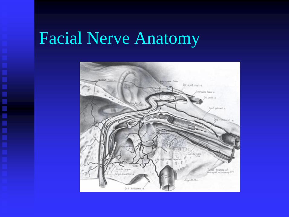
Facial Nerve Anatomy

Facial Nerve Injuries
Evaluation
Previous status
Time
Onset and progression
Complete vs. Incomplete

House Brackman grading system
I Normal Normal facial function
II Mild Slight synkinesis/weakness
III Moderate Complete eye closure, noticeable
synkinesis, slight forehead
movement
IV Moderately
Severe
Incomplete eye closure, symmetry
at rest, no forehead movement
V Severe Assymetry at rest, barely noticeable
motion
VI Total No movement

Electrophysiologic Testing
NET
MST
ENoG

Nerve Excitability Test
Maximal Stimulation Test
>3.5mA difference suggests a poor
prognosis for return of facial function.

Electroneuronography
Most accurate, qualitative measurement
Reduction of >90% amplitude correlates
with a poor prognosis for spontaneous
recovery

Electromyography
Limited use until 10-14 days
Polyphasic potentials= Good

Facial Nerve Injuries
Decision to treat is primarily based on
whether there is complete vs. incomplete
paralysis

Treatment
Conservative treatment candidates
Surgical candidates

Conservative Treatment
Candidates
Chang and Cass
Normal Facial Function regardless of
progression
Incomplete paralysis and no progression
to complete paralysis
Less than 95% degeneration by ENoG

Surgical Candidates
Critical Prognostic factors
Immediate vs. Delayed
Complete vs. Incomplete paralysis
ENoG criteria

Algorithm for Facial Nerve Injury
Surgical Approach
Suspect location of neural injury
Presence or absence of hearing

Surgical Approach
Lateral to the geniculate ganglion
transmastoid

Surgical Approach
Medial to the Geniculate Ganglion
No useful hearing
Transmastoid-translabyrinthine
Intact hearing
Transmastoid-trans-epitympanic
Middle Cranial Fossa

Surgical findings
Nerve repair
Direct anastomosis
Nerve graft
Decompression

Iatrogenic Facial Nerve Injuries
Mastoidectomy (55%)
Tympanoplasty (14%)
Bony Exostoses (14%)
79% were not identified at the time of
surgery
Management of Iatrogenic Facial
Nerve Injuries
<50% decompression
75% had HB of 3 or better!
>50% nerve repair
No patients had better than a HB 3

Case Report
32 yr old fisherman was wading
Minding his own business
Hit in head by a flying fish
Immediate profound vertigo, hearing loss
CT scan revealed longitudinal Temp bone
fracture


![Inner Ear Anatomy[1]](https://img.dokumen.tips/doc/110x75/5528566b4979591c048b47a6/inner-ear-anatomy1.jpg)


