Embed Size (px)
Citation preview

MIDAG@UNCMIDAG@UNC
Medical Image Synthesis Medical Image Synthesis via Monte Carlo Simulationvia Monte Carlo SimulationMedical Image Synthesis Medical Image Synthesis
via Monte Carlo Simulationvia Monte Carlo SimulationAn Application of Statistics in GeometryAn Application of Statistics in Geometry
&&Building a Geometric Model with CorrespondenceBuilding a Geometric Model with Correspondence
James Z. Chen, Stephen M. Pizer, James Z. Chen, Stephen M. Pizer,
Edward L. Chaney, Sarang Joshi, Joshua StoughEdward L. Chaney, Sarang Joshi, Joshua Stough
Presented by: Joshua StoughPresented by: Joshua StoughMedical Image Display & Analysis Group, UNCMedical Image Display & Analysis Group, UNC
midag.cs.unc.edumidag.cs.unc.edu
An Application of Statistics in GeometryAn Application of Statistics in Geometry&&
Building a Geometric Model with CorrespondenceBuilding a Geometric Model with Correspondence
James Z. Chen, Stephen M. Pizer, James Z. Chen, Stephen M. Pizer,
Edward L. Chaney, Sarang Joshi, Joshua StoughEdward L. Chaney, Sarang Joshi, Joshua Stough
Presented by: Joshua StoughPresented by: Joshua StoughMedical Image Display & Analysis Group, UNCMedical Image Display & Analysis Group, UNC
midag.cs.unc.edumidag.cs.unc.edu

MIDAG@UNCMIDAG@UNC
Population Simulation Requires Statistical Population Simulation Requires Statistical Profiling of ShapeProfiling of Shape
Population Simulation Requires Statistical Population Simulation Requires Statistical Profiling of ShapeProfiling of Shape
Goal: Develop a methodology for generating realistic synthetic medical images AND the attendant “ground truth” segmentations for objects of interest.
Why: Segmentation method evaluation.
How: Build and sample probability distribution of shape.
Goal: Develop a methodology for generating realistic synthetic medical images AND the attendant “ground truth” segmentations for objects of interest.
Why: Segmentation method evaluation.
How: Build and sample probability distribution of shape.

MIDAG@UNCMIDAG@UNC
Basic IdeaBasic IdeaBasic IdeaBasic Idea
New images via New images via deformation of template deformation of template geometry and image.geometry and image.
CharacteristicsCharacteristics Legal images represent Legal images represent
statistical variation of statistical variation of shape over a training set.shape over a training set.
Image quality as in a Image quality as in a clinical setting.clinical setting.
Ht

MIDAG@UNCMIDAG@UNC
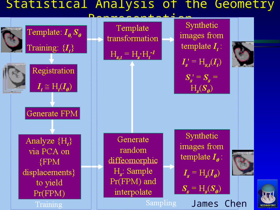
The ProcessThe ProcessThe ProcessThe Process
James Chen

MIDAG@UNCMIDAG@UNC
RegistrationRegistrationRegistrationRegistration
Registration – Composition of Two TransformationsRegistration – Composition of Two Transformations Linear – MIRIT, Frederik MaesLinear – MIRIT, Frederik Maes
Affine transformation, 12 dofAffine transformation, 12 dof Non-linear–Deformation Diffeomorphism, JoshiNon-linear–Deformation Diffeomorphism, Joshi
For all For all IItt , , IItt H Htt((II00) and ) and SStt H Htt((SS00))

MIDAG@UNCMIDAG@UNC
Consequence of an Erroneous HConsequence of an Erroneous HttConsequence of an Erroneous HConsequence of an Erroneous Htt
James Chen

MIDAG@UNCMIDAG@UNC
Generating the Statistics of HGenerating the Statistics of HttGenerating the Statistics of HGenerating the Statistics of Htt
James Chen

MIDAG@UNCMIDAG@UNC
Fiducial Point ModelFiducial Point ModelFiducial Point ModelFiducial Point Model
HHtt isis locally correlated locally correlated
Fiducial point choice via Fiducial point choice via greedy iterative algorithmgreedy iterative algorithm
HHtt'' determined by Joshi determined by Joshi
Landmark Deformation Landmark Deformation Diffeomorphism Diffeomorphism
The Idea: Decrease The Idea: Decrease
MIDAG@UNCMIDAG@UNC
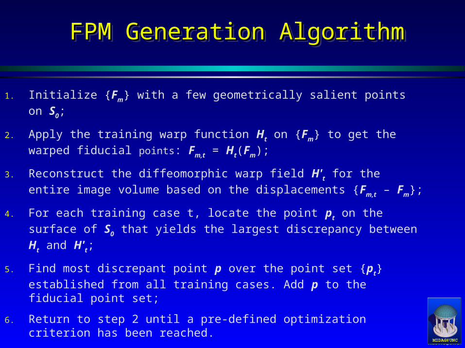
FPM Generation AlgorithmFPM Generation AlgorithmFPM Generation AlgorithmFPM Generation Algorithm
1. Initialize {Fm} with a few geometrically salient points on S0;
2. Apply the training warp function Ht on {Fm} to get the warped
fiducial points: Fm,t = Ht(Fm);
3. Reconstruct the diffeomorphic warp field H't for the entire image
volume based on the displacements {Fm,t – Fm};
4. For each training case t, locate the point pt on the surface of S0
that yields the largest discrepancy between Ht and H't;
5. Find most discrepant point p over the point set {pt} established
from all training cases. Add p to the fiducial point set;
6. Return to step 2 until a pre-defined optimization criterion has been reached.

MIDAG@UNCMIDAG@UNC
A locally accurate warp via FPM landmarksA locally accurate warp via FPM landmarksA locally accurate warp via FPM landmarksA locally accurate warp via FPM landmarks
Volume overlapVolume overlap optimizationoptimization criterion trackscriterion tracks mean warpmean warp discrepancydiscrepancy
Under 100 fiducialUnder 100 fiducial points, ofpoints, of thousands onthousands on surfacesurface
Volume overlapVolume overlap optimizationoptimization criterion trackscriterion tracks mean warpmean warp discrepancydiscrepancy
Under 100 fiducialUnder 100 fiducial points, ofpoints, of thousands onthousands on surfacesurface
ATLASATLAS WARPWARP TRAININGTRAINING

MIDAG@UNCMIDAG@UNC
Human Kidney ExampleHuman Kidney ExampleHuman Kidney ExampleHuman Kidney Example
36 clinical CT 36 clinical CT images in the images in the training settraining set
Monotonic Monotonic OptimizationOptimization
88 fiducial points 88 fiducial points sufficiently mimick sufficiently mimick inter-human rater inter-human rater results (94% volume results (94% volume overlap)overlap)
36 clinical CT 36 clinical CT images in the images in the training settraining set
Monotonic Monotonic OptimizationOptimization
88 fiducial points 88 fiducial points sufficiently mimick sufficiently mimick inter-human rater inter-human rater results (94% volume results (94% volume overlap)overlap)
0.50
0.60
0.70
0.80
0.90
1.00
1.10
1.20
1.30
32 42 52 62 72 82 92
Fiducial Points
Dis
tanc
es (i
n vo
xel u
nits
)
91.0
91.5
92.0
92.5
93.0
93.5
94.0
94.5
95.0
Volu
me
Ove
rlap
(%)
<Closest Surface Distance>
<Ht-H't Surface Distance>
<Volume Overlap>

MIDAG@UNCMIDAG@UNC
Fiducial Point Model Is an Object Representation Fiducial Point Model Is an Object Representation with Positional Correspondencewith Positional Correspondence
Fiducial Point Model Is an Object Representation Fiducial Point Model Is an Object Representation with Positional Correspondencewith Positional Correspondence
Positional Positional correspondence is via correspondence is via the Hthe H'' interpolated from interpolated from the displacements at the the displacements at the fiducial pointsfiducial points
The correspondence The correspondence makes this makes this representation suitable representation suitable for statistical analysisfor statistical analysis
MIDAG@UNCMIDAG@UNC
Statistical Analysis of the Geometry RepresentationStatistical Analysis of the Geometry RepresentationStatistical Analysis of the Geometry RepresentationStatistical Analysis of the Geometry Representation
James Chen

MIDAG@UNCMIDAG@UNC
Principal Components Analysis of the FPM Principal Components Analysis of the FPM DisplacementsDisplacements
Principal Components Analysis of the FPM Principal Components Analysis of the FPM DisplacementsDisplacements
Points in 3M-d spacePoints in 3M-d space Analyze deviation from meanAnalyze deviation from mean
Example: Example: first seven modes of FPM cover 88% of the total variation.
Points in 3M-d spacePoints in 3M-d space Analyze deviation from meanAnalyze deviation from mean
Example: Example: first seven modes of FPM cover 88% of the total variation.
0
10
20
30
40
50
60
70
80
90
100
1 2 3 4 5 6 7
Modes
Varia
tion
Cove
rage
(%)
Component Coverage
Accumulative Coverage

MIDAG@UNCMIDAG@UNC
Modes of Variation – Human KidneyModes of Variation – Human KidneyModes of Variation – Human KidneyModes of Variation – Human Kidney
-2-2 +2+2-1-1 +1+1
II
IIII
IIIIII
ATLASATLAS
MEANMEAN

MIDAG@UNCMIDAG@UNC
Generating Samples of Image Intensity PatternsGenerating Samples of Image Intensity PatternsGenerating Samples of Image Intensity PatternsGenerating Samples of Image Intensity Patterns
James Chen

MIDAG@UNCMIDAG@UNC
ResultsResultsResultsResults
James Chen

MIDAG@UNCMIDAG@UNC
ResultsResultsResultsResults

MIDAG@UNCMIDAG@UNC
MiscellaneousMiscellaneousMiscellaneousMiscellaneous
National Cancer Institute Grant P01 CA47982National Cancer Institute Grant P01 CA47982 National Cancer Institute Grant P01 CA47982National Cancer Institute Grant P01 CA47982ReferencesGerig, G., M. Jomier, M. Chakos (2001). “Valmet: A new validation tool for assessing and improving 3D object
segmentation.” Proc. MICCAI 2001, Springer LNCS 2208: 516-523.
Cootes, T. F., A. Hill, C.J. Taylor, J. Haslam (1994). “The Use of Active Shape Models for Locating Structures in Medical Images.” Image and Vision Computing 12(6): 355-366.
Rueckert, D., A.F. Frangi, and J.A. Schnabel (2001). “Automatic Construction of 3D Statistical Deformation Models Using Non-rigid Registration.” MICCAI 2001, Springer LNCS 2208: 77-84.
Christensen, G. E., S.C. Joshi and M.I. Miller (1997). “Volumetric Transformation of Brain Anatomy.” IEEE Transactions on Medical Imaging 16: 864-877.
Joshi, S., M.I. Miller (2000). “Landmark Matching Via Large Deformation Diffeomorphisms.” IEEETransactions on Image Processing.
Maes, F., A. Collignon, D. Vandermeulen, G. Marchal, P. Suetens (1997). “Multi-Modality Image Registration by Maximization of Mutual Information.” IEEE-TMI 16: 187-198.
Pizer, S.M., J.Z. Chen, T. Fletcher, Y. Fridman, D.S. Fritsch, G. Gash, J. Glotzer, S. Joshi, A. Thall, G. Tracton, P. Yushkevich, and E. Chaney (2001). “Deformable M-Reps for 3D Medical Image Segmentation.” IJCV, submitted.


![[PPT]PowerPoint Presentation - MIDAG - Medical Image …midag.cs.unc.edu/.../Tutorial/NeuroAnatomyReview.ppt · Web viewAnatomy for Neuroimaging J. Keith Smith, M.D., Ph.D. Neuroradiology](https://img.dokumen.tips/doc/110x75/5b29e20c7f8b9a9c1a8b5937/pptpowerpoint-presentation-midag-medical-image-midagcsuncedututorial.jpg)