Embed Size (px)
Citation preview
Microvascular Free Flaps Used in Head and Neck
Reconstruction.
Jeffrey Buyten, MD
Faculty Advisor: Shawn Newlands, MD, PhD, MBA
Faculty Advisor: Francis B. Quinn, MD
The University of Texas Medical Branch Department of Otolaryngology
Grand Rounds Presentation
October 19, 2005
Outline
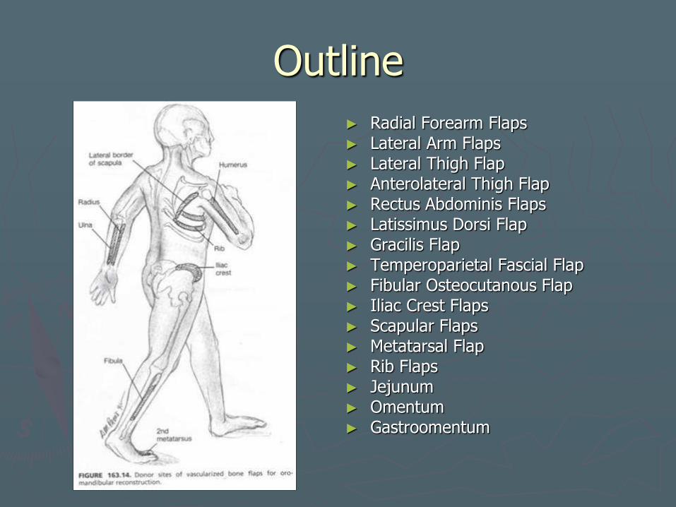
► Radial Forearm Flaps ► Lateral Arm Flaps ► Lateral Thigh Flap ► Anterolateral Thigh Flap ► Rectus Abdominis Flaps ► Latissimus Dorsi Flap ► Gracilis Flap ► Temperoparietal Fascial Flap ► Fibular Osteocutanous Flap ► Iliac Crest Flaps ► Scapular Flaps ► Metatarsal Flap ► Rib Flaps ► Jejunum ► Omentum ► Gastroomentum
Radial Forearm Flap ► 1981 (China), 1985 (pharyngeal recon) ► Oral cavity, base of tongue, pharynx, soft palate,
cutaneous defects, base of skull, small volume bone and soft tissue defects of face
► Thin, pliable skin Reconstitution of contours, sulci, vestibules Tongue mobility
► Fasciocutaneous flaps are highly tolerant of radiation therapy
► Composite flap with bone, tendon, brachioradialis muscle and vascularized nerve. Sensory recovery reported in patients even
when a neural anastomosis is not performed. ► Fasciocutaneous flaps >
musculocutaneous flaps ► Incomplete and unpredictable
► Skin from entire forearm ► 2 team approach
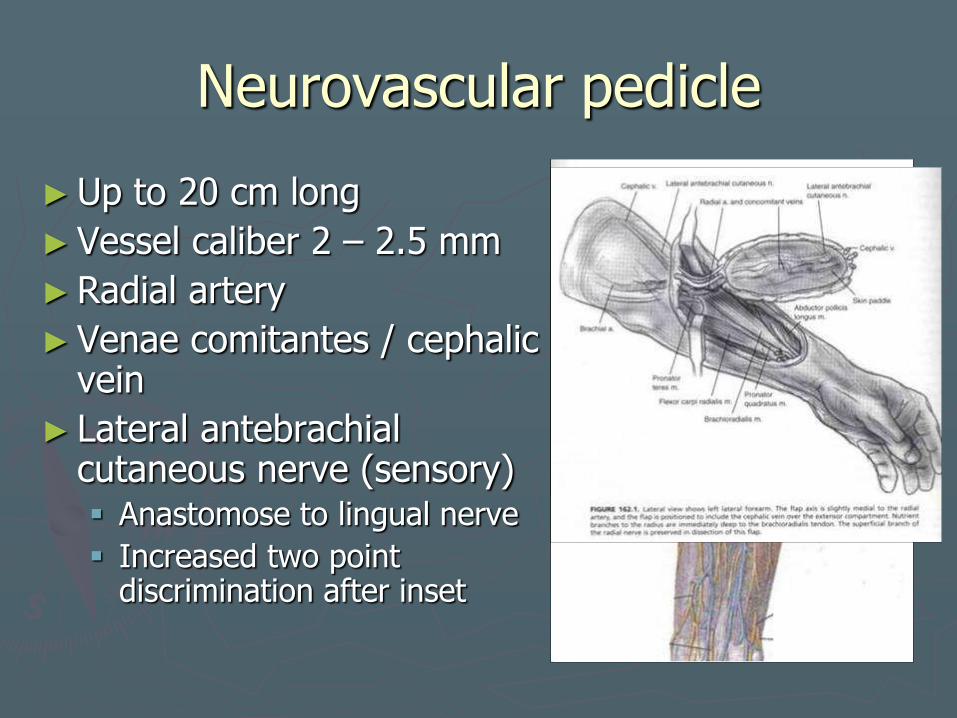
Neurovascular pedicle
►Up to 20 cm long
►Vessel caliber 2 – 2.5 mm
►Radial artery
►Venae comitantes / cephalic vein
► Lateral antebrachial cutaneous nerve (sensory) Anastomose to lingual nerve
Increased two point discrimination after inset
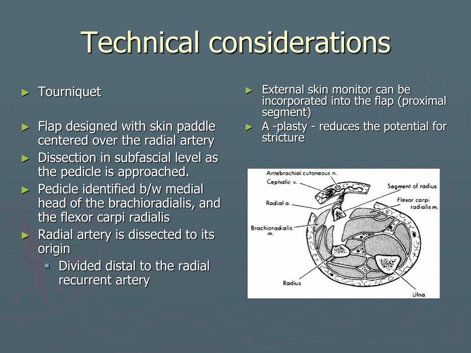
Technical considerations
► Tourniquet
► Flap designed with skin paddle centered over the radial artery
► Dissection in subfascial level as the pedicle is approached.
► Pedicle identified b/w medial head of the brachioradialis, and the flexor carpi radialis
► Radial artery is dissected to its origin
Divided distal to the radial recurrent artery
► External skin monitor can be incorporated into the flap (proximal segment)
► A -plasty - reduces the potential for stricture
Technical considerations
► Osteocutaneous flap
Monocortical
Cuff of flexor pollicis longus
10 – 12 cm of radius
Up to 40% circumference
Limited by amount of available bone and risk for pathologic fracture.
► Pollicis longus tendon
Suspending flap laterally in palatal and total lower lip recon
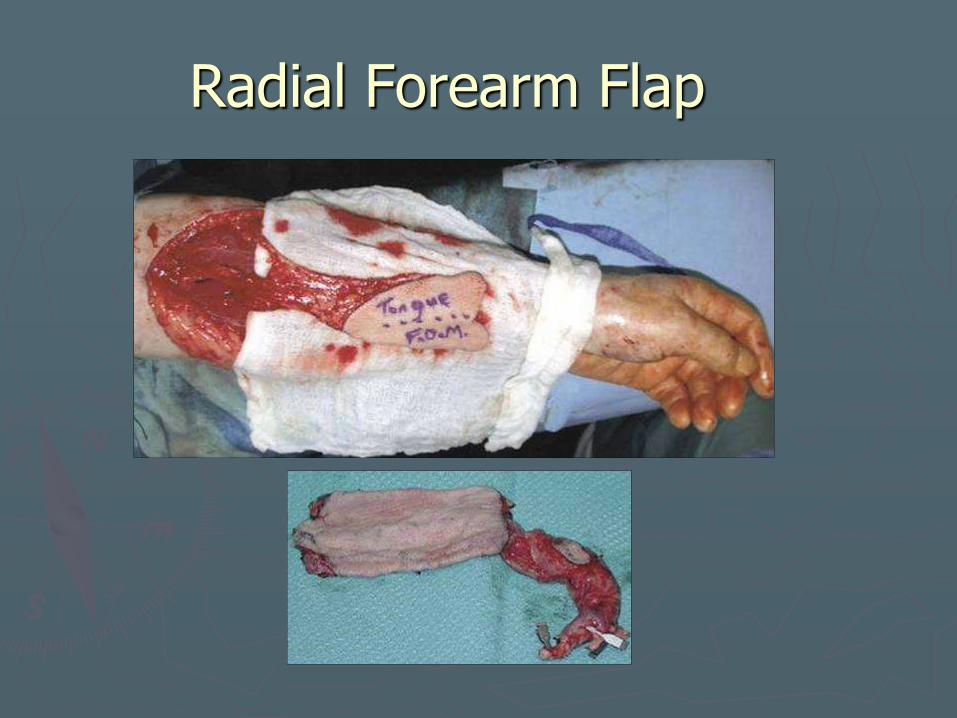
Radial Forearm Flap
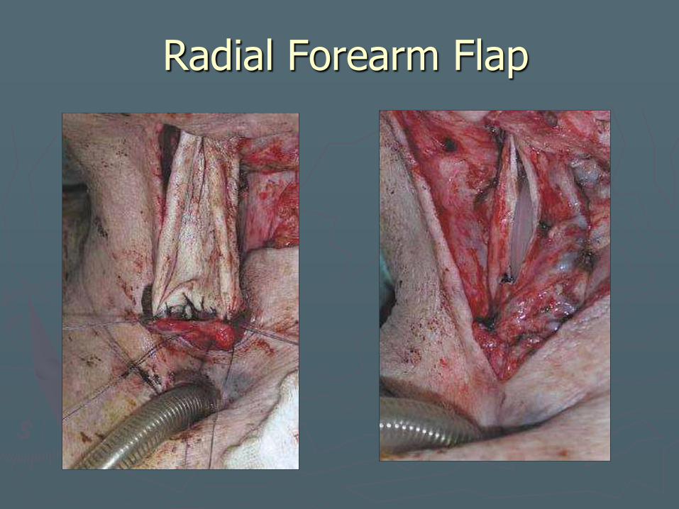
Radial Forearm Flap

Radial Forearm Flap
Radial Forearm Flap
► Morbidity
Hand ischemia
Fistula rates - 42% to 67% in early series
► Subsequent series - 15% and 38%.
► Creation of a controlled fistula or use of a salivary bypass stent can protect the suture line from salivary soilage and decrease the potential for fistulization.
Stricture formation - 9% to 50%.
Radial nerve injury
Variable anesthesia over dorsum of hand.
Radial Forearm Flap
► Preoperative considerations
Allen test
► Tests viability of palmar arch system
No IVs / blood draws in donor arm.
Skin graft (must preserve paratenon layer)
Osteocutaneous flaps
► Radius fracture
►Weakened supination, wrist flexion, grip strength and pinch strength.
Should not be used defect extends below the thoracic inlet
► Postoperative management
Forearm and wrist immobilization w/volar splint
7-10 days
Oral intake can generally begin within 7 to 10 days ► 2 weeks is best if the patient
has been previously irradiated.
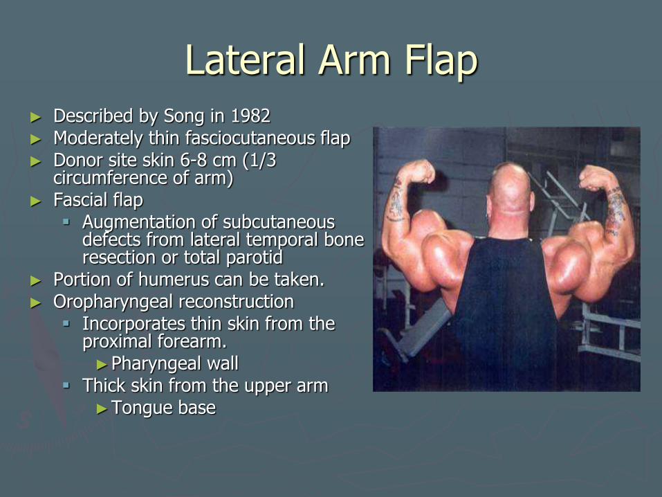
Lateral Arm Flap ► Described by Song in 1982 ► Moderately thin fasciocutaneous flap ► Donor site skin 6-8 cm (1/3
circumference of arm) ► Fascial flap
Augmentation of subcutaneous defects from lateral temporal bone resection or total parotid
► Portion of humerus can be taken. ► Oropharyngeal reconstruction
Incorporates thin skin from the proximal forearm. ►Pharyngeal wall
Thick skin from the upper arm ►Tongue base
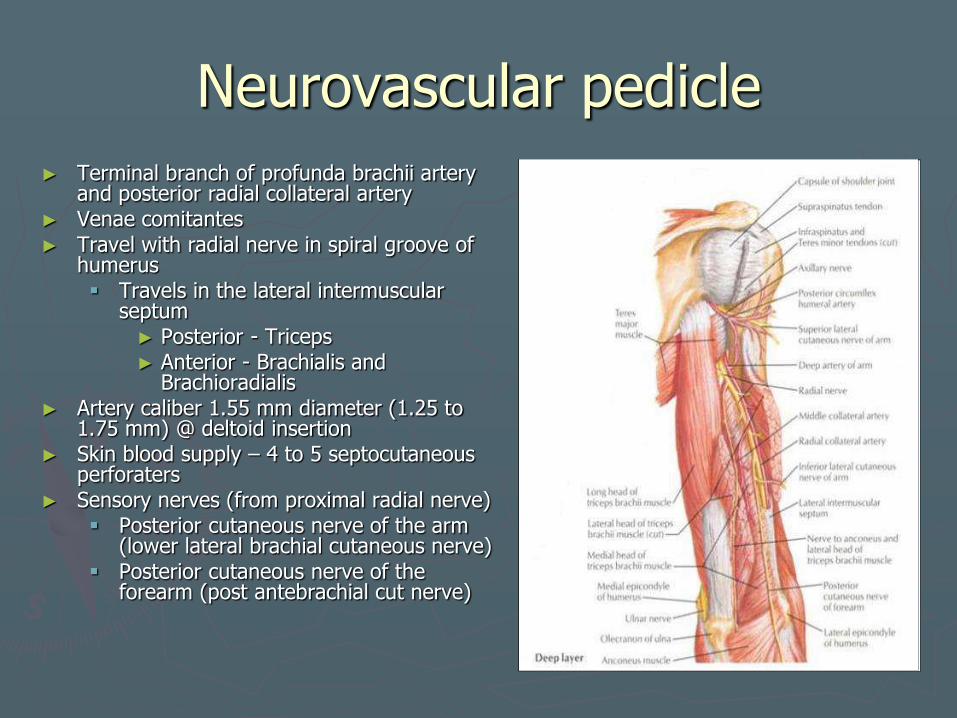
Neurovascular pedicle
► Terminal branch of profunda brachii artery and posterior radial collateral artery
► Venae comitantes ► Travel with radial nerve in spiral groove of
humerus Travels in the lateral intermuscular
septum ► Posterior - Triceps ► Anterior - Brachialis and
Brachioradialis ► Artery caliber 1.55 mm diameter (1.25 to
1.75 mm) @ deltoid insertion ► Skin blood supply – 4 to 5 septocutaneous
perforaters ► Sensory nerves (from proximal radial nerve)
Posterior cutaneous nerve of the arm (lower lateral brachial cutaneous nerve)
Posterior cutaneous nerve of the forearm (post antebrachial cut nerve)
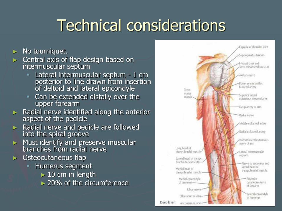
Technical considerations
► No tourniquet. ► Central axis of flap design based on
intermuscular septum Lateral intermuscular septum - 1 cm
posterior to line drawn from insertion of deltoid and lateral epicondyle
Can be extended distally over the upper forearm
► Radial nerve identified along the anterior aspect of the pedicle
► Radial nerve and pedicle are followed into the spiral groove
► Must identify and preserve muscular branches from radial nerve
► Osteocutaneous flap Humerus segment
► 10 cm in length ► 20% of the circumference
Lateral Arm Flap
►Morbidity
Radial nerve damage
►Palsy 2/2 constrictive dressings or tight wound closure.
Primary closure if less than 1/3 of arm
►Use STSG if closure under too much tension.

Lateral Arm Flap
►Preoperative Considerations
Easy scar camouflage
Male patients may have less hair in this region when compared to forearm
►Consider for intraoral reconstruction
Flap becomes thinner more distally

Lateral Thigh Flap
►Described by Baek in 1983
► Large surface area
► Expendable tissue
► Flap size up to 25 x 14 cm
► Fasciocutaneous flap – thin to moderately thick
► Intraoral and pharyngeal reconstruction
►Reinnervated via lateral femoral cutaneous nerve

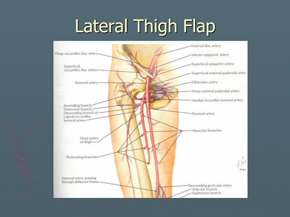
Neurovascular pedicle
► Third perforator of profunda femoris
► Travels w/in intermuscular septum
► Pedicle 8 – 12 cm
► Vessel caliber 2 – 4 mm
► Lateral femoral cutaneous nerve of the thigh
Anterosuperior entry into flap
Does not travel with vascular pedicle
► Terminal cutaneous branch of second or fourth perforators are the dominant arterial supply (rare)
4th perforator usually included in dissection to account for variations
When 2nd perforator dominant – pedicle length limited by muscular branch vessels to preserve femoral blood supply.
Lateral Thigh Flap
Lateral Thigh Flap
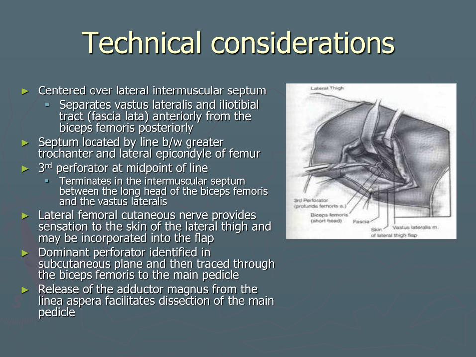
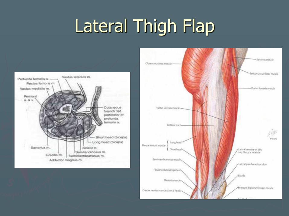
Technical considerations
► Centered over lateral intermuscular septum Separates vastus lateralis and iliotibial
tract (fascia lata) anteriorly from the biceps femoris posteriorly
► Septum located by line b/w greater trochanter and lateral epicondyle of femur
► 3rd perforator at midpoint of line Terminates in the intermuscular septum
between the long head of the biceps femoris and the vastus lateralis
► Lateral femoral cutaneous nerve provides sensation to the skin of the lateral thigh and may be incorporated into the flap
► Dominant perforator identified in subcutaneous plane and then traced through the biceps femoris to the main pedicle
► Release of the adductor magnus from the linea aspera facilitates dissection of the main pedicle
Lateral Thigh Flap
►Morbidity
Atherosclerosis of profunda femoris and its branches
Avoid in pts with h/o PVD
Sciatic nerve injury
Lateral Thigh Flap
► Preoperative Considerations
Assess for PVD (palpate peripheral pulses)
Not advised for use in obese individuals or in those with previous surgery or trauma to the thigh
► Postoperative management
Primary closure of donor site
Early walking

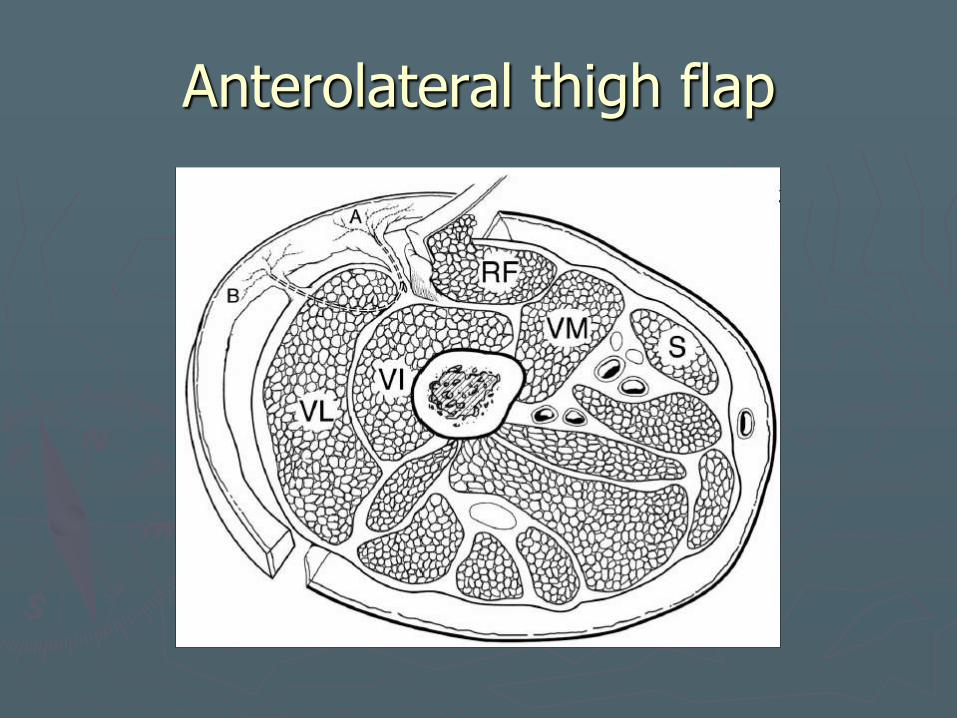
Anterolateral thigh flap
► First reported by Song et al ► Subcutaneous, fasciocutaneous, myocutaneous, adipofascial ► Laryngopharynx, oral cavity, oropharynx, external skin and
maxilla ► Flap may be thinned or suprafascial flaps taken for thinner
flaps ► Popular in Asia ► Less popular in Europe and America
Difficult perforator dissection (bountiful subcutaneous tissue) Variation in vascular anatomy
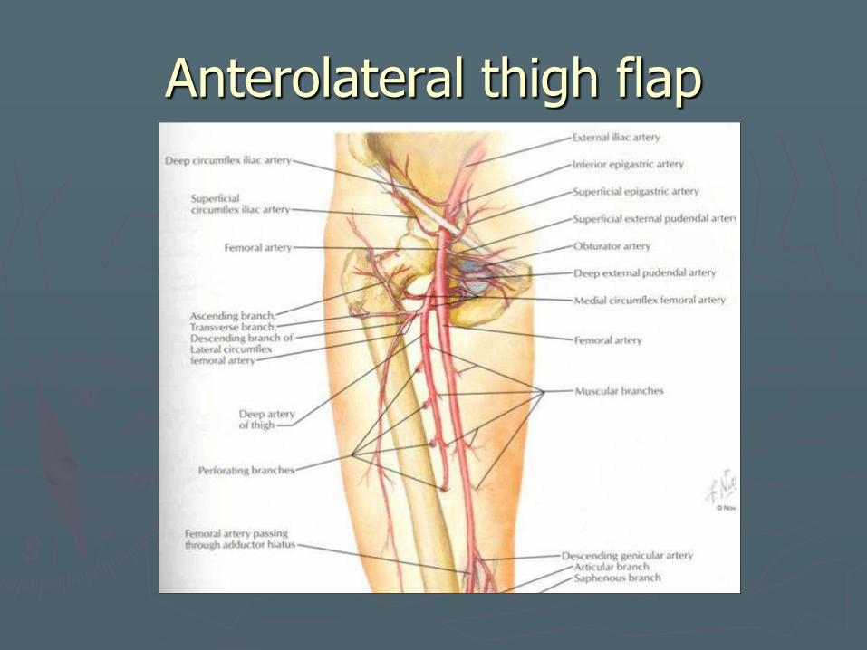
Neurovascular pedicle
► Descending branch of lateral circumflex femoral artery Septocutaneous
►Traverse the fascia lata Musculocutaneous
perforators ►Traverse the vastus
lateralis muscle and the deep fascia
► Venae comitantes ► Descending branch travels
inferiorly in intramuscular space b/w rectus femoris and vastus lateralis
► Caliber – 2.1 mm artery, 2.6 mm vein
► Vascular pedicle up to 16 cm
► Lateral femoral cutaneous nerve – sensory nerve
Branch of lumbar plexus
Enters thigh deep to lateral aspect of inguinal ligament near ASIS
Runs with deep circumflex iliac artery and vein
Runs anterior, posterior or through sartorius, continuing through fascia lata
Neurovascular pedicle
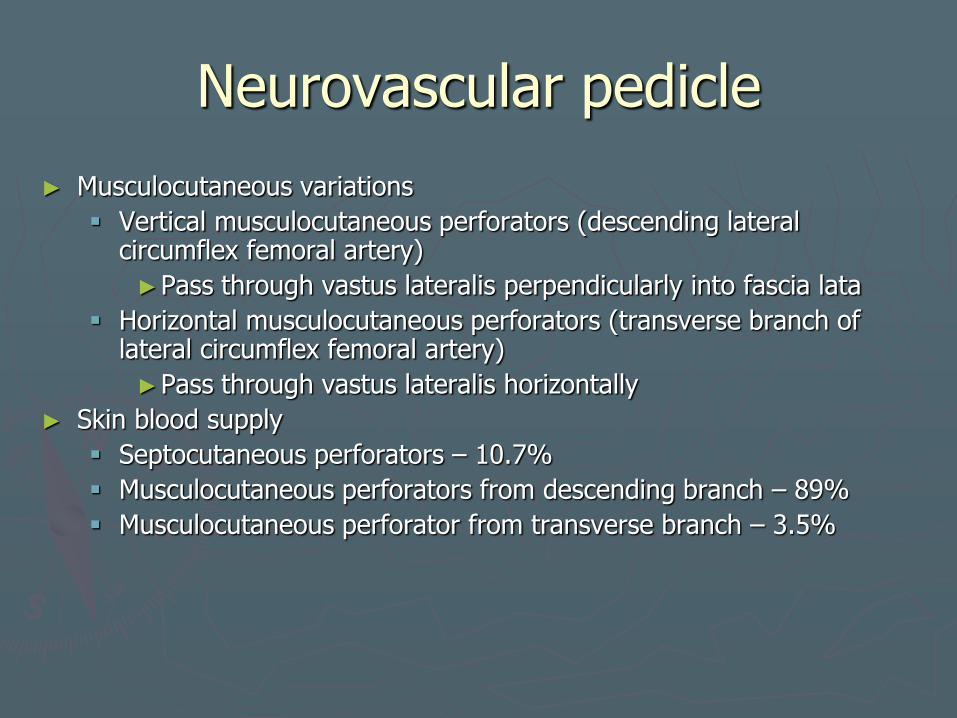
► Musculocutaneous variations
Vertical musculocutaneous perforators (descending lateral circumflex femoral artery)
►Pass through vastus lateralis perpendicularly into fascia lata
Horizontal musculocutaneous perforators (transverse branch of lateral circumflex femoral artery)
►Pass through vastus lateralis horizontally
► Skin blood supply
Septocutaneous perforators – 10.7%
Musculocutaneous perforators from descending branch – 89%
Musculocutaneous perforator from transverse branch – 3.5%
Anterolateral thigh flap
Anterolateral thigh flap
Technical considerations
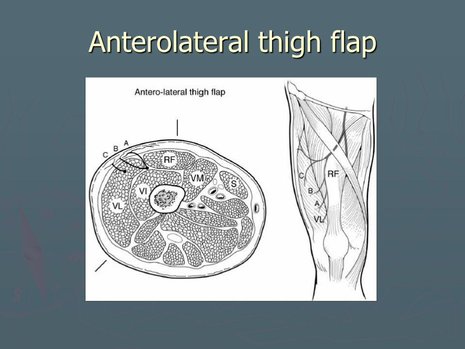
► Draw line from ASIS to lateral patellar border ► Cutaneous perforator exit point from intermuscular
septum or from vastus lateralis 2 cm lateral to and 2 cm inferior to midpoint of
line from ASIS and lateral border of patella ► Use Doppler to mark perforators ► Dissect (medial to lateral) to intermuscular septum
b/w rectus femoris and vastus lateralis. ► Retract rectus femoris medially exposing perforators
Leave muscle cuff around myocutaneous perforators
► Fasciocutaneous flap, suprafascial flap, cutaneous flap (up 5 mm thickness), adipofascial flap
► May include lateral cutaneous nerve of thigh ► Max size – horizontal line from greater trochanter
down to a parallel line 3 cm above patella 25 x 18 cm 20 x 26 cm
► Close donor site primarily if less than 8 cm wide
Anterolateral thigh flap
Anterolateral thigh flap
►Morbidity
Possible STSG
Depends on extent of injury to vastus lateralis
Thinned flaps with more complications in intraoral defects
Anterolateral thigh flap
►Preoperative Considerations
Reduced donor site morbidity compared to RFF
Can be as thin as RFF
Contraindicated in pts with prior upper thigh surgery, vascular procedures, big eaters…
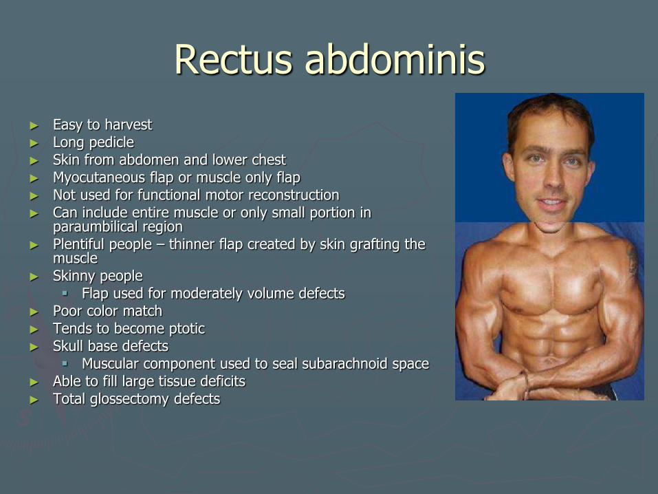
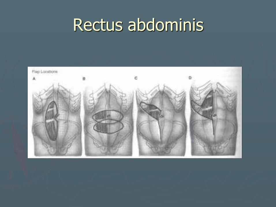
Rectus abdominis
► Easy to harvest ► Long pedicle ► Skin from abdomen and lower chest ► Myocutaneous flap or muscle only flap ► Not used for functional motor reconstruction ► Can include entire muscle or only small portion in
paraumbilical region ► Plentiful people – thinner flap created by skin grafting the
muscle ► Skinny people
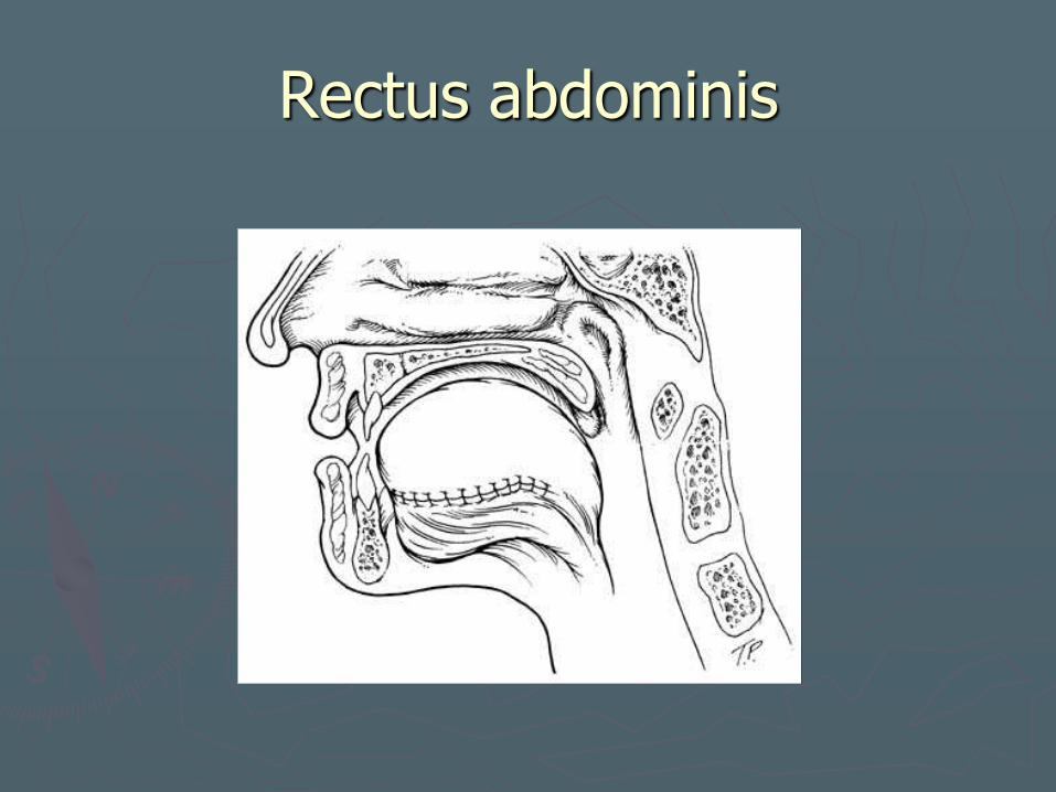
Flap used for moderately volume defects ► Poor color match ► Tends to become ptotic ► Skull base defects
Muscular component used to seal subarachnoid space ► Able to fill large tissue deficits ► Total glossectomy defects

Neurovascular pedicle
► Two dominant pedicles Deep superior epigastric artery/vein Deep inferior epigastric artery and vein
► Based on inferior epigastrics when used for h/n recon because of larger pedicle size
► Inferior epigastric diameter – 3 to 4 mm
► Reinnervated with any of the lower six intercostal nerves.
► Pedicle may travel along lateral aspect of muscle before taking intramuscular route

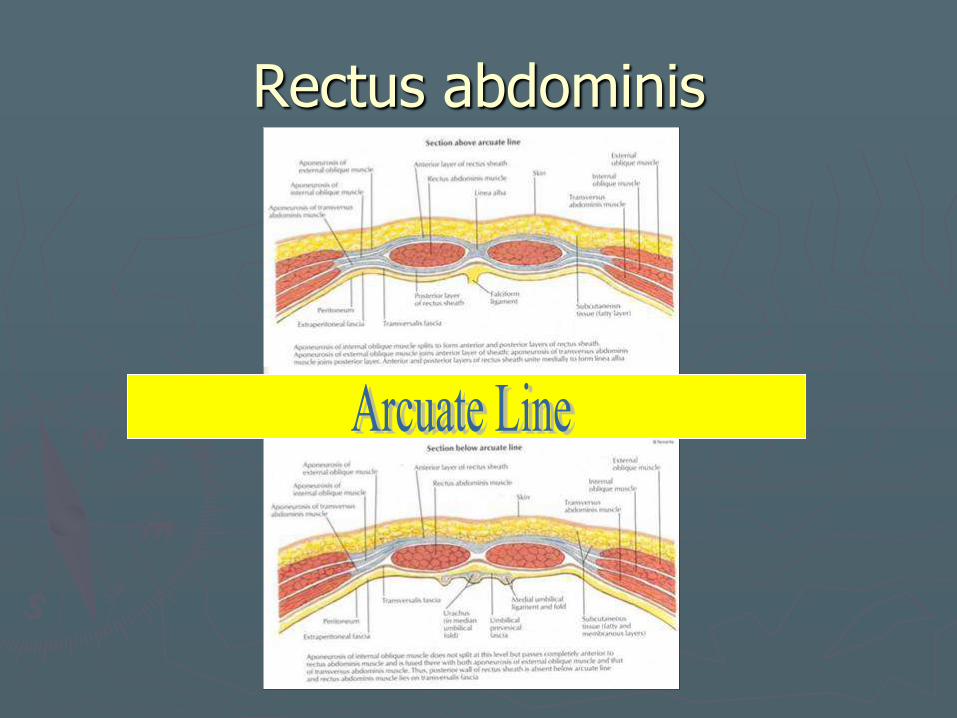
Technical considerations
► Cutaneous blood supply Harvest anterior rectus sheath in paraumbilical
region (dominant perforators located here) Skin paddle designed with epicenter above the
umbilicus ► Primary closure ► Hernia prevention depends on restoring abdominal
wall. ► Arcuate line (level of ASIS)
Superior – posterior sheath with transversalis fascia, internal oblique and transversus abdominis ► Closure of posterior sheath prevents
herniation Inferior – only transversalis fascia posterior to
muscle ► Must close anterior sheath to prevent
herniation
Technical considerations
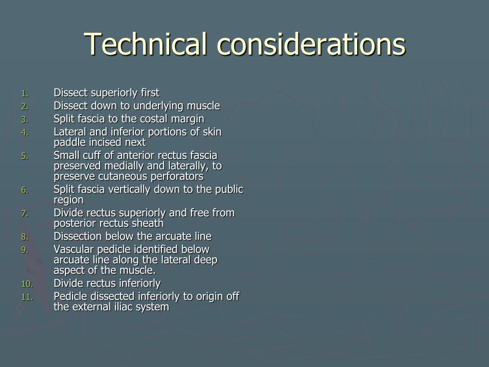
1. Dissect superiorly first 2. Dissect down to underlying muscle 3. Split fascia to the costal margin 4. Lateral and inferior portions of skin
paddle incised next 5. Small cuff of anterior rectus fascia
preserved medially and laterally, to preserve cutaneous perforators
6. Split fascia vertically down to the public region
7. Divide rectus superiorly and free from posterior rectus sheath
8. Dissection below the arcuate line 9. Vascular pedicle identified below
arcuate line along the lateral deep aspect of the muscle.
10. Divide rectus inferiorly 11. Pedicle dissected inferiorly to origin off
the external iliac system
Rectus abdominis
Rectus abdominis
Rectus abdominis
Rectus abdominis
►Morbidity
Abdominal weakness
Hernia
Rectus abdominis
► Preoperative Considerations Prior abdominal surgery
Prior inguinal herniorrhapy may compromise pedicle dissection 2/2 scarring
Hernia
Diastasis recti
► Postoperative management
Ileus
Avoid abdominal strain for 6 weeks.
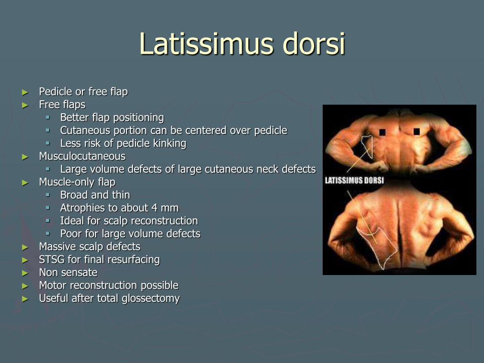
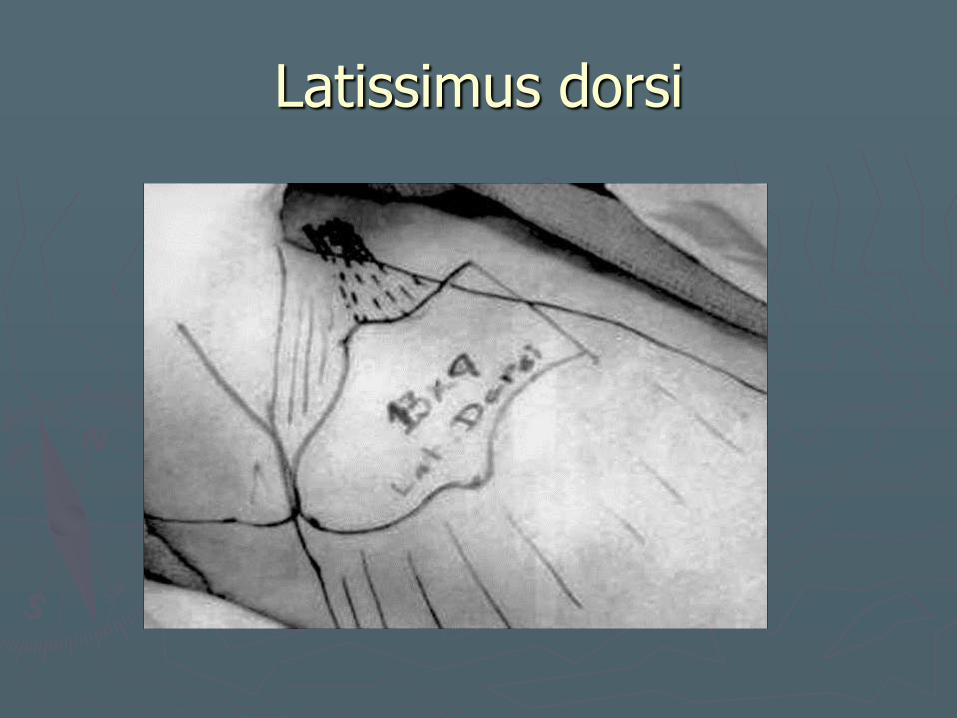
Latissimus dorsi
► Pedicle or free flap ► Free flaps
Better flap positioning Cutaneous portion can be centered over pedicle Less risk of pedicle kinking
► Musculocutaneous Large volume defects of large cutaneous neck defects
► Muscle-only flap Broad and thin Atrophies to about 4 mm Ideal for scalp reconstruction Poor for large volume defects
► Massive scalp defects ► STSG for final resurfacing ► Non sensate ► Motor reconstruction possible ► Useful after total glossectomy
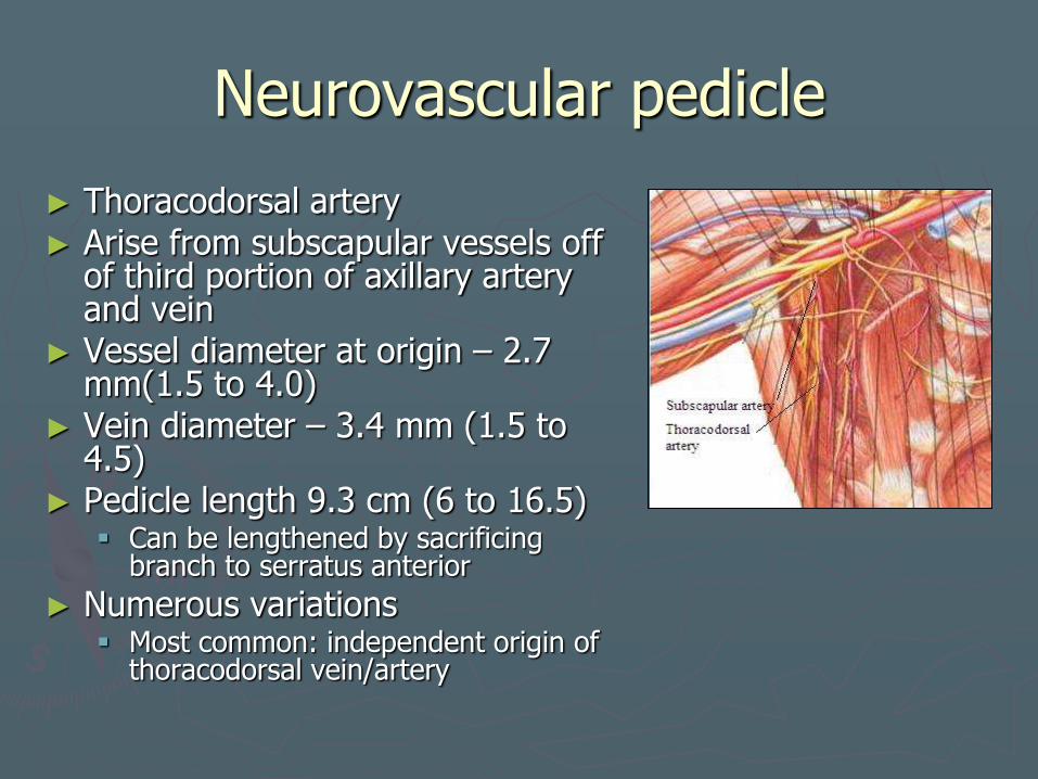
Neurovascular pedicle
► Thoracodorsal artery ► Arise from subscapular vessels off
of third portion of axillary artery and vein
► Vessel diameter at origin – 2.7 mm(1.5 to 4.0)
► Vein diameter – 3.4 mm (1.5 to 4.5)
► Pedicle length 9.3 cm (6 to 16.5) Can be lengthened by sacrificing
branch to serratus anterior
► Numerous variations Most common: independent origin of
thoracodorsal vein/artery

Technical considerations
► Lateral decubitis position If at 15 degrees, flap may be
harvested simultaneously with primary lesion resection
Anterior muscle border along line b/w midpoint of axilla and point midway b/w ASIS and PSIS
► Vessels enter undersurface of muscle 8 to 10 cm below midpoint of axilla
► Serratus vessels ligated during harvest
► Can design two paddle flap based on medial and lateral branches of thoracodorsal vessels
► Total glossectomy insetting. Muscle inset as a sling on
undersurface of mandible Sutured to pterygoid, masseter, or
superior constrictor... Thoracodorsal nerve anastomosed
to a hypoglossal nerve ► Gives reconstructed tongue the
ability to elevate superiorly toward the palate
Latissimus dorsi
Latissimus dorsi
►Morbidity
Marginal flap necrosis
Pedicled flaps pass b/w pec major and minor
►Changes in arm position may occlude pedicle
►Should immobilize arm in flexed position
Latissimus dorsi
► Preoperative Considerations
Relative contraindications - prior axillary LN dissection
Preop angiography advocated to assess vessel patency
► Postoperative management
Suction drains
High incidence of seroma

Gracilis flap
►1976
►Thin muscle flap
►Dynamic facial reanimation
►Muscle revasularized and reinnervated
►Long vascular pedicle
►Easy dissection
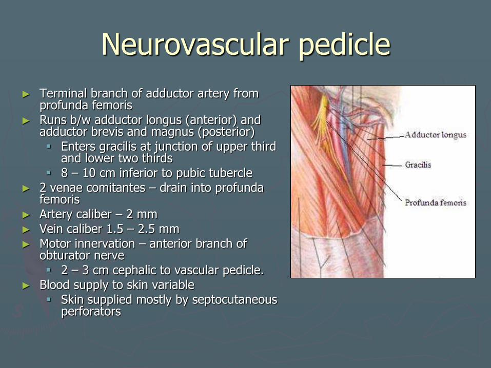
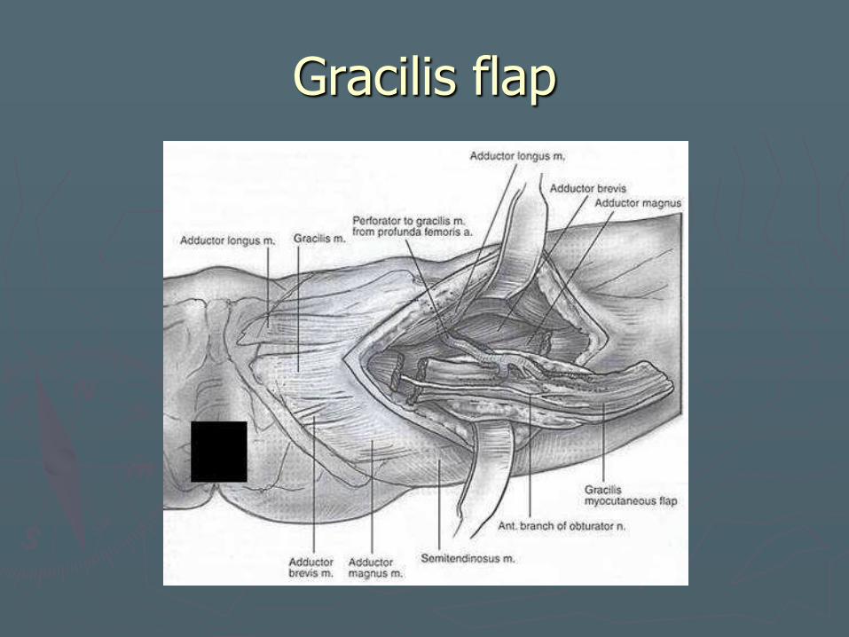
Neurovascular pedicle
► Terminal branch of adductor artery from profunda femoris
► Runs b/w adductor longus (anterior) and adductor brevis and magnus (posterior) Enters gracilis at junction of upper third
and lower two thirds 8 – 10 cm inferior to pubic tubercle
► 2 venae comitantes – drain into profunda femoris
► Artery caliber – 2 mm ► Vein caliber 1.5 – 2.5 mm ► Motor innervation – anterior branch of
obturator nerve 2 – 3 cm cephalic to vascular pedicle.
► Blood supply to skin variable Skin supplied mostly by septocutaneous
perforators

Technical considerations
► Muscle can be split into at least two functional muscular units
► Single neuromuscular unit can be transferred to decrease bulk
► Orient skin paddle longitudinally
Must be centered over dominant musculocutaneous perforator
► For synchronous mimetic movement when proximal facial nerve not available.
2 stage procedure with cross face sural nerve graft
Tinel sign used to monitor axonal growth across the face – 9-12 months
After adequate axonal regrowth – muscle transferred
Gracilis flap
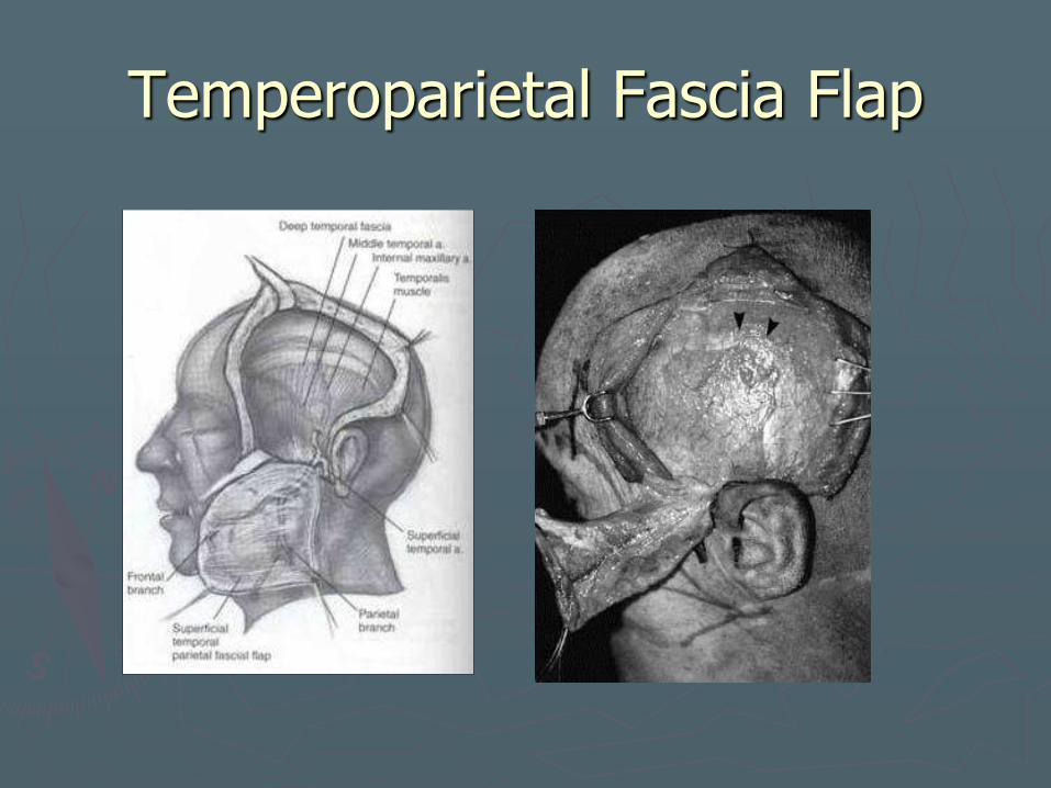
Temperoparietal Fascia Flap
► More commonly transferred as a pedicled flap but can be used as a free flap when arc of rotation is inadequate
► Ultra thin – 2 to 4 mm thick ► Highly vascular, pliable and durable ► Fascial, fasciocutaneous ► Up to 17 x 14 cm with extensive
scalp undermining ► Oral cavity, hemilaryngectomy
defects, middle and upper regions of face w/split calvarial bone graft
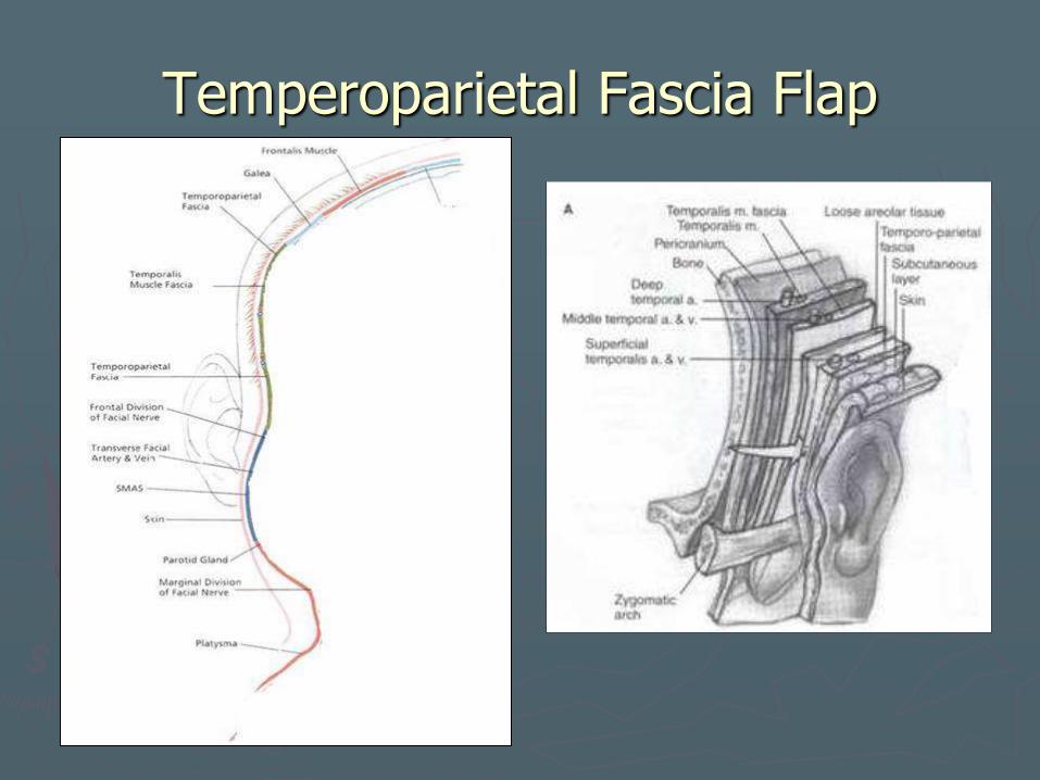
Neurovascular pedicle
► 5 layers – scalp ► Temperoparietal fascia (TPF) deep
to skin and subcutaneous tissue. ► Superficial to temporalis muscular
fascia ► Above superior temporal line it’s
continuous with galea aponeurotica ► Base centered over helix
► Superficial temporal artery and vein – travel in TPF layer 3 cm superior to root of helix Vessels branch into frontal and
temporal divisions Most commonly based on parietal
branch Ligation of frontal artery 3 – 4 cm
distal to branching point to avoid frontal nerve injury
Venous pedicle may course with arteries or 2 to 3 cm posteriorly
► Middle temporal artery – proximal superficial temporal artery at zygomatic arch (supplies temporalis muscular fascia)
► Including middle temporal artery enables a two-layered fascial flap on a single pedicle.
Temperoparietal Fascia Flap
Technical considerations
►Vertical incision over root of helix to superior temporal line
►V-shaped extension at superior limit of incision
►Scalp elevation ant and post
►Dissect deep to flap
►Loose areolar tissue deep to flap
Temperoparietal Fascia Flap
Temperoparietal Fascia Flap
►Morbidity
Frontal branch weakness (travels in TPF)
Secondary alopecia – damage to hair follicles due to superficial dissection
Temperoparietal Fascia Flap
►Preoperative Considerations
Relative contraindications - prior XRT, neck surgery, bicoronal incision or external carotid embolization.
Doppler assessment of pedicle
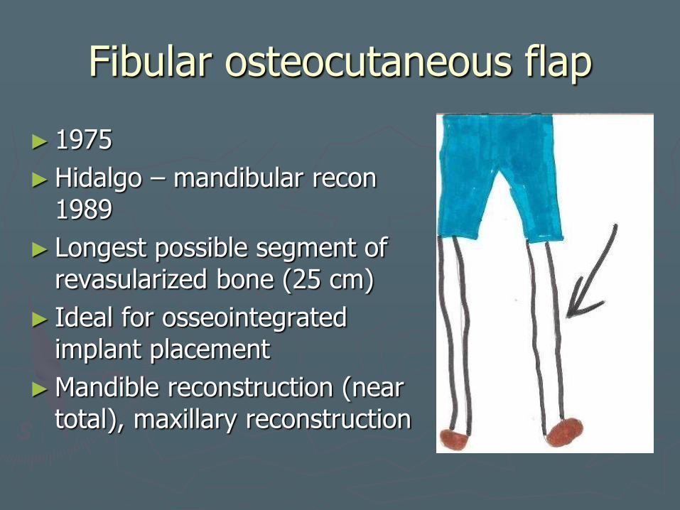
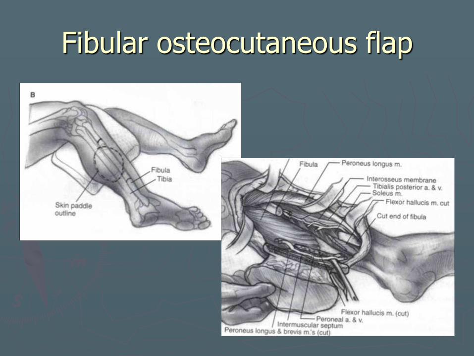
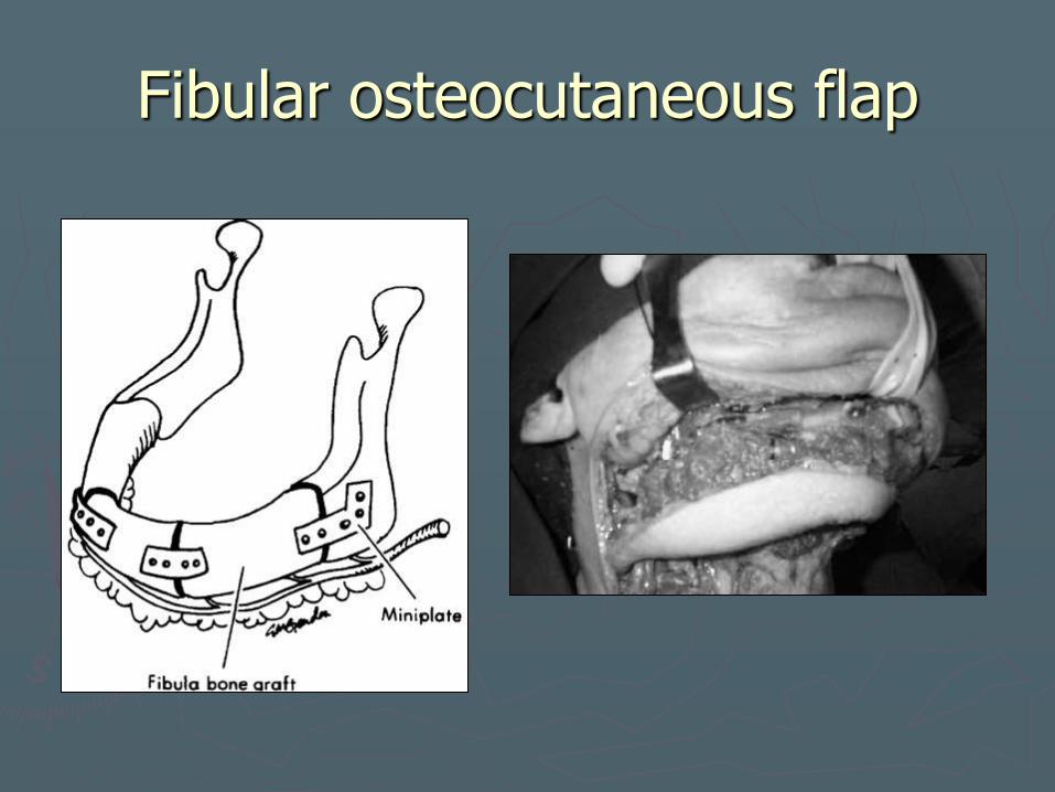
Fibular osteocutaneous flap
► 1975
►Hidalgo – mandibular recon 1989
► Longest possible segment of revasularized bone (25 cm)
► Ideal for osseointegrated implant placement
►Mandible reconstruction (near total), maxillary reconstruction
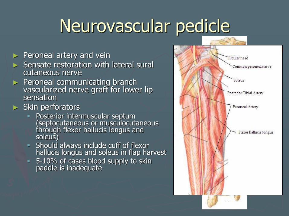
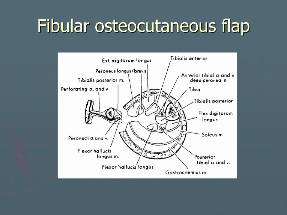
Neurovascular pedicle
► Peroneal artery and vein ► Sensate restoration with lateral sural
cutaneous nerve ► Peroneal communicating branch
vascularized nerve graft for lower lip sensation
► Skin perforators Posterior intermuscular septum
(septocutaneous or musculocutaneous through flexor hallucis longus and soleus)
Should always include cuff of flexor hallucis longus and soleus in flap harvest
5-10% of cases blood supply to skin paddle is inadequate
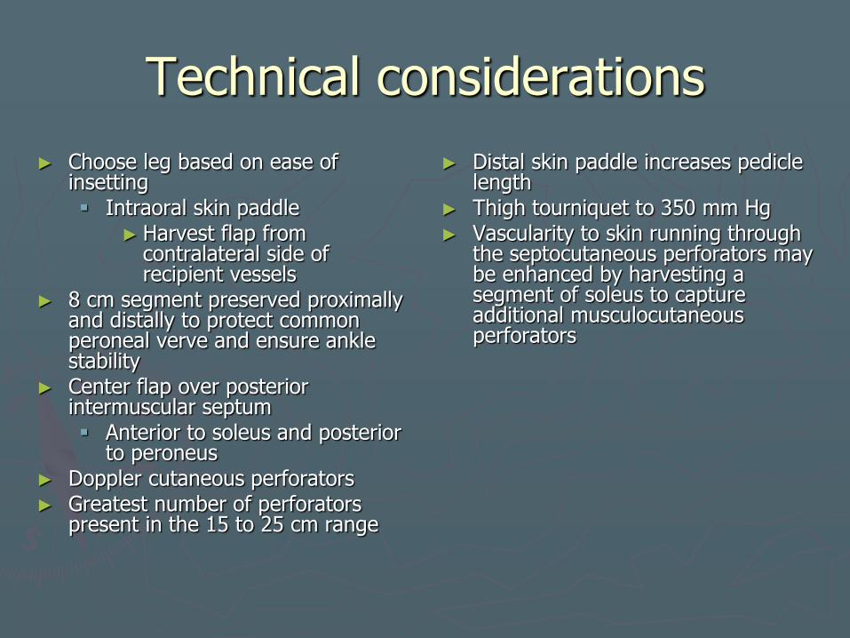
Technical considerations
► Choose leg based on ease of insetting Intraoral skin paddle
► Harvest flap from contralateral side of recipient vessels
► 8 cm segment preserved proximally and distally to protect common peroneal verve and ensure ankle stability
► Center flap over posterior intermuscular septum Anterior to soleus and posterior
to peroneus ► Doppler cutaneous perforators ► Greatest number of perforators
present in the 15 to 25 cm range
► Distal skin paddle increases pedicle length
► Thigh tourniquet to 350 mm Hg ► Vascularity to skin running through
the septocutaneous perforators may be enhanced by harvesting a segment of soleus to capture additional musculocutaneous perforators
Fibular osteocutaneous flap
Fibular osteocutaneous flap
Fibular osteocutaneous flap
Fibular osteocutaneous flap
►Morbidity Donor site complications
►Edema
►Weakness in dorsiflexion of great toe
Skin loss in 5 – 10% of flaps ►reliability of the skin is questionable, and both the
surgeon and the patient should be prepared for the possible need for a second soft tissue flap, either free or pedicled, when reconstructing composite defects with a fibular osteocutaneous flap
May need STSG over donor site closure
Fibular osteocutaneous flap
► Preoperative Considerations
Angiography
MRA
h/o distal lower extremity fracture
Look for varicose veins, edema
► Postoperative management
Distal pulses monitored
Posterior splint for 10 days

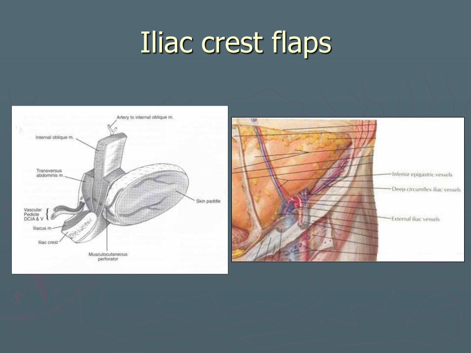
Iliac crest flaps
► Osteocutaneous, osteomusculocutaneous ► Segmental mandibular defects ► Up to 16 cm bone ► Oromandibular reconstruction ► No motor or sensate reconstruction ► Only vascularized bone used extensively with
simultaneous or delayed endosteal dental implant placement
► Skin paddle was not ideal for relining the oral cavity Too thick for accurate restoration of the
3D anatomy ► Inclusion of internal oblique flap
Denervated muscle undergoes atrophy that leaves a thin, fixed, soft tissue coverage over the bone.
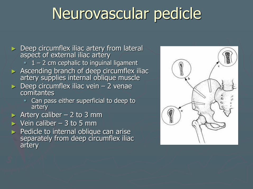
Neurovascular pedicle
► Deep circumflex iliac artery from lateral aspect of external iliac artery 1 – 2 cm cephalic to inguinal ligament
► Ascending branch of deep circumflex iliac artery supplies internal oblique muscle
► Deep circumflex iliac vein – 2 venae comitantes Can pass either superficial to deep to
artery
► Artery caliber – 2 to 3 mm ► Vein caliber – 3 to 5 mm ► Pedicle to internal oblique can arise
separately from deep circumflex iliac artery
Iliac crest flaps
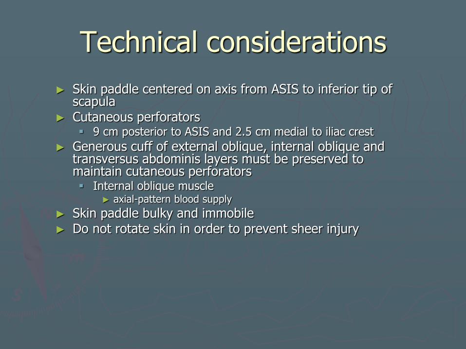
Technical considerations
► Skin paddle centered on axis from ASIS to inferior tip of scapula
► Cutaneous perforators 9 cm posterior to ASIS and 2.5 cm medial to iliac crest
► Generous cuff of external oblique, internal oblique and transversus abdominis layers must be preserved to maintain cutaneous perforators Internal oblique muscle
► axial-pattern blood supply
► Skin paddle bulky and immobile ► Do not rotate skin in order to prevent sheer injury
Iliac crest flaps

Iliac crest flaps
Iliac crest flaps
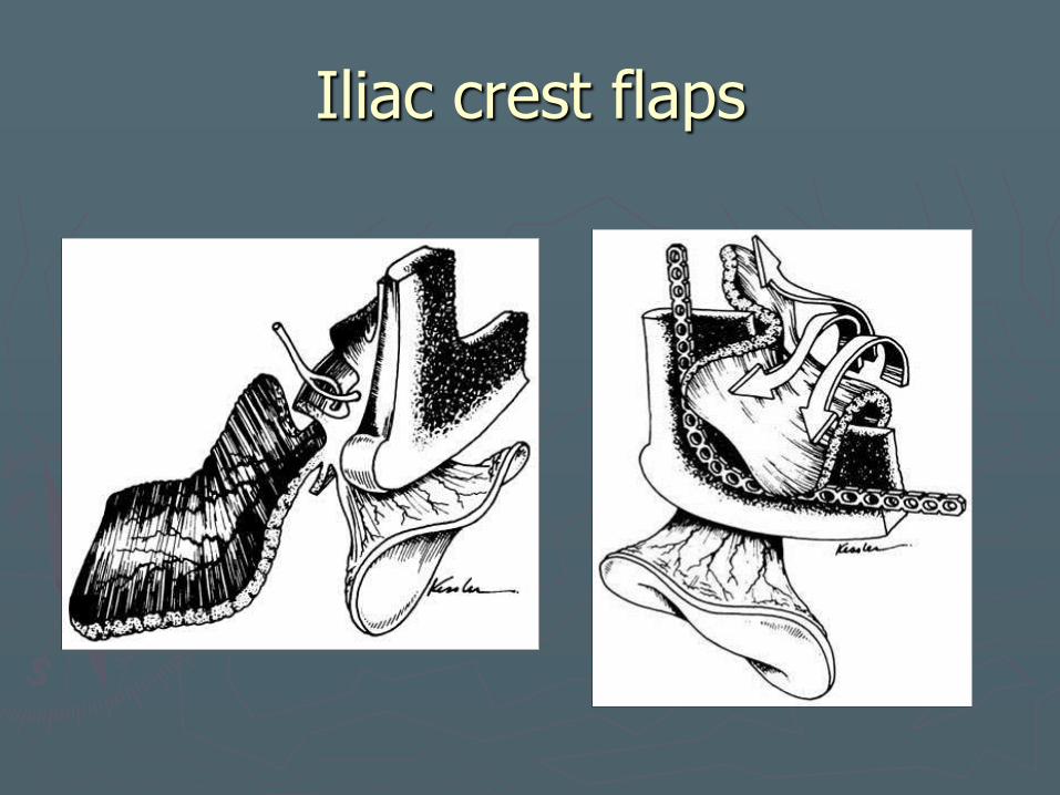
►Morbidity
Hernia
►Need to approximate cut edge of iliacus muscle to transversus abdominis
►Can be reinforced by drilling holes into cut edge of iliac bone
►Approximate external obliques and aponeurosis to tensor fascia lata and gluteus muscles
►Keep inferior oblique inferior and anterior to ASIS
Skin loss from perforator sheer injury
poor color match

Iliac crest flaps
►Preoperative Considerations
h/o hernias, prior iliac bypass graft
Severe PVD,
Preop angio
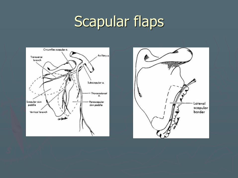
Scapular flaps
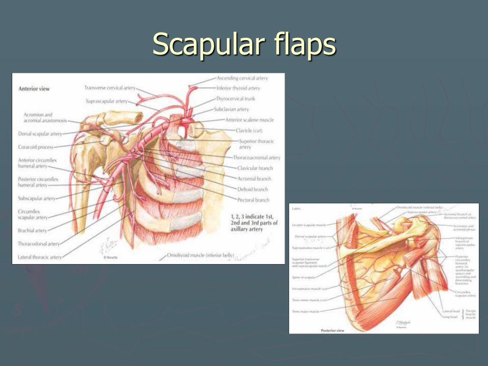
► Fasciocutaneous, osteofasciocutaneous, cutaneous flap, parascapular cutaneous flap, latissimus dorsi myocutaneous flap, and serratus anterior flap
► Thin, hairless skin ► Two cutaneous flaps may be harvested
Horizontally oriented flap – transverse cutaneous branch
Vertically oriented flap parascapular flap – descending cutaneous branch
► Long pedicle length ► Large surface area ► Complex composite midfacial or
oromandibular defects ► Up to 10 cm bone ► Osseointegrated implants possible ► Single team approach
Neurovascular pedicle
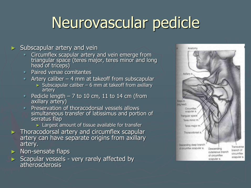
► Subscapular artery and vein Circumflex scapular artery and vein emerge from
triangular space (teres major, teres minor and long head of triceps)
Paired venae comitantes Artery caliber – 4 mm at takeoff from subscapular
► Subscapular caliber – 6 mm at takeoff from axillary artery
Pedicle length – 7 to 10 cm, 11 to 14 cm (from axillary artery)
Preservation of thoracodorsal vessels allows simultaneous transfer of latissimus and portion of serratus flap ► Largest amount of tissue available for transfer
► Thoracodorsal artery and circumflex scapular artery can have separate origins from axillary artery.
► Non-sensate flaps ► Scapular vessels - very rarely affected by
atherosclerosis
Scapular flaps
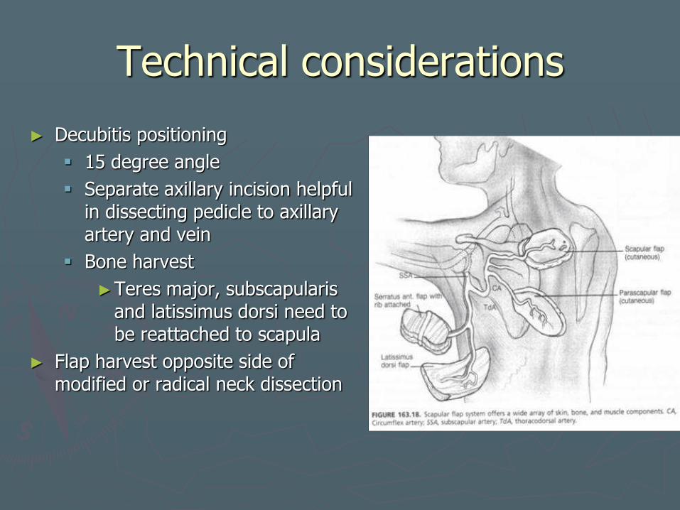
Technical considerations
► Decubitis positioning
15 degree angle
Separate axillary incision helpful in dissecting pedicle to axillary artery and vein
Bone harvest
►Teres major, subscapularis and latissimus dorsi need to be reattached to scapula
► Flap harvest opposite side of modified or radical neck dissection
Scapular flaps
Scapular flaps
►Morbidity
Brachial plexus injury 2/2 lateral decubitis positioning
►Use axillary roll
Stay 1 cm inferior to glenoid fossa
Detach teres major and minor to harvest bone
►Can cause shoulder weakness and limit range of motion.
Scapular flaps
► Preoperative Considerations
Prior axillary node dissection – contraindication
► Postoperative management
Immobilize for 3 to 4 days
Early ambulation
5 days for bone harvest
PT

Rib flap
► First vascularized bone to be used in mandibular reconstruction. (osteocutaneous)
► Blood supply to the rib Internal mammary artery Posteriorly or posterolaterally
on the posterior intercostal vessels
Transferred with the pectoralis major, serratus anterior, or latissimus dorsi muscle
► Poor bone stock except for condylar reconstruction
► Not commonly used
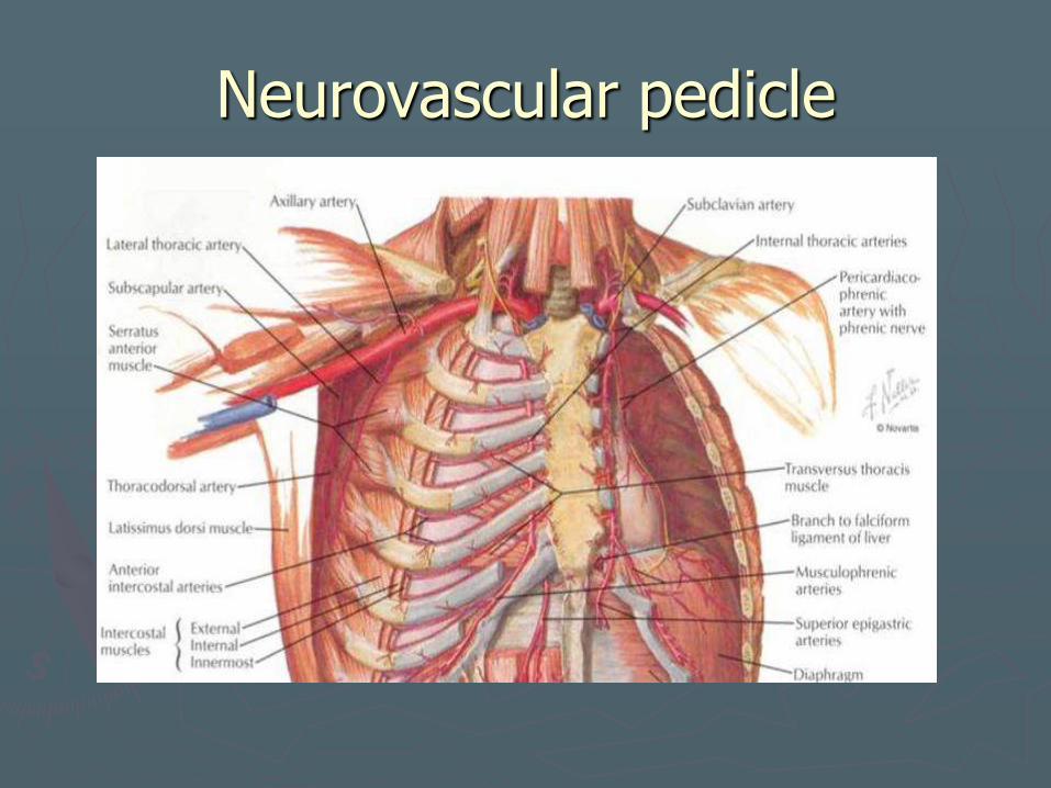
Neurovascular pedicle
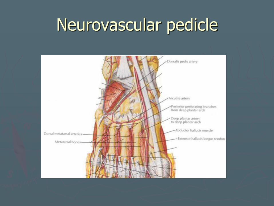
Metatarsus flap
►Osteocutaneous flap based on the first dorsal metatarsal artery
►Thin sensate skin with the second metatarsal.
►Limited bone volume
►Not commonly used
Neurovascular pedicle

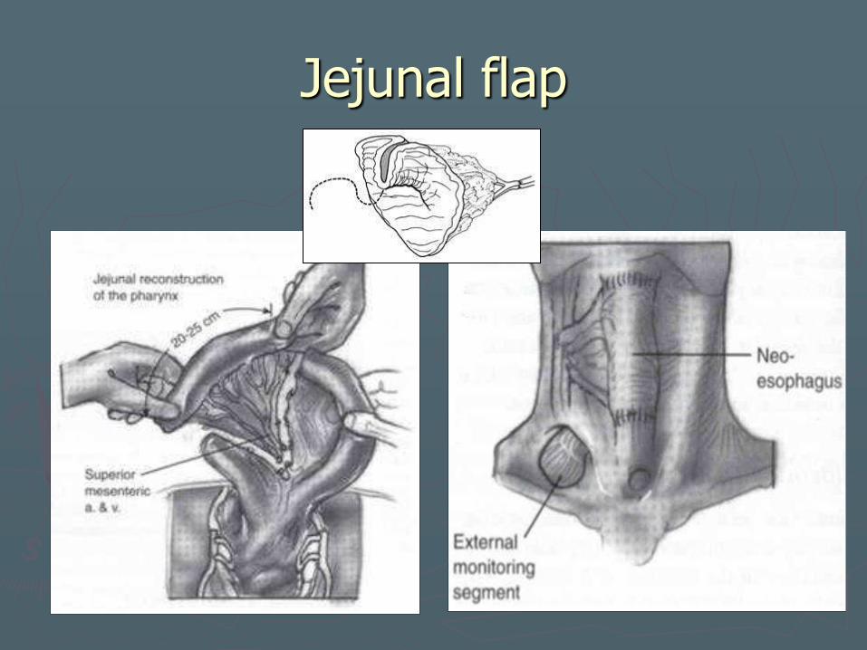
Jejunal flap ► 1959 ► Circumferential pharyngoesophageal
defects ► Patch graft ► Diameter of jejunum – good match to
cervical esophagus ► Ideal mucosal surface ► Two team approach ► Advantages
Better superior positioning
► Disadvantage Inferior positioning limited by thoracic
inlet 3 anastomoses
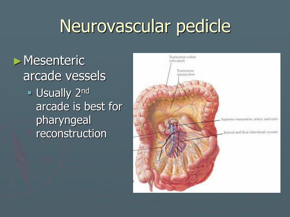
Neurovascular pedicle
►Mesenteric arcade vessels
Usually 2nd arcade is best for pharyngeal reconstruction
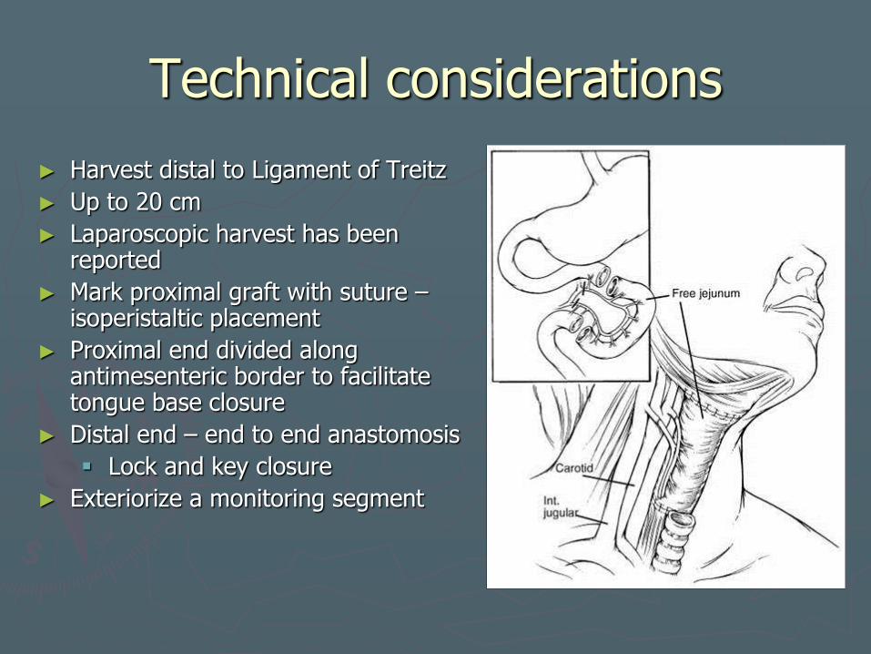
Technical considerations
► Harvest distal to Ligament of Treitz
► Up to 20 cm
► Laparoscopic harvest has been reported
► Mark proximal graft with suture – isoperistaltic placement
► Proximal end divided along antimesenteric border to facilitate tongue base closure
► Distal end – end to end anastomosis
Lock and key closure
► Exteriorize a monitoring segment
Jejunal flap
Jejunal flap
►Morbidity Most susceptible to primary ischemia
Fistula formation – 18%
11% rate of anastomotic stricture ►Higher rate if cervical anastomosis stapled
Wet voice (TEP)
Functional obstruction 2/2 peristalsis
Dysgeusia
Harvest site complications
Jejunal flap
► Preoperative Considerations Absolute contraindications
► Disease extension into proximal thoracic esophagus
► Ascites ► Crohn’s disease
Relative contraindications ► Chronic intestinal diseases ► h/o abdominal surgery
Consider angio
► Intraperitoneal sepsis
Do not use in laryngeal sparing procedures
► Postoperative management Remove monitoring segment
pod 7. Jejunostomy tube
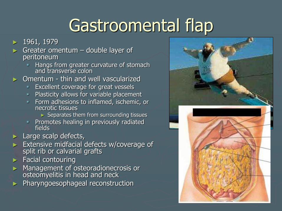
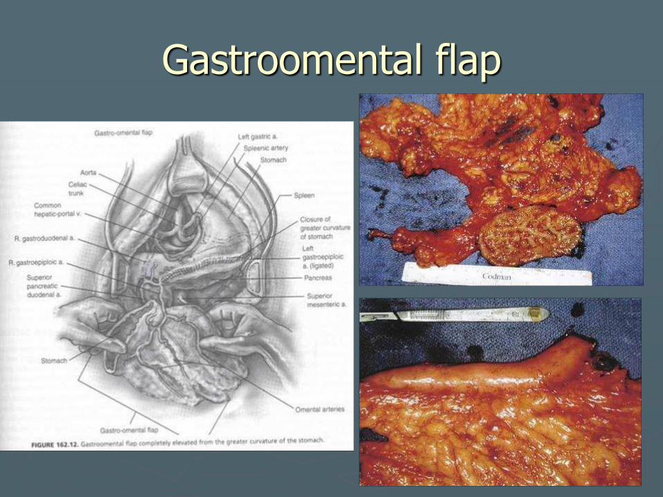
Gastroomental flap ► 1961, 1979 ► Greater omentum – double layer of
peritoneum Hangs from greater curvature of stomach
and transverse colon
► Omentum - thin and well vascularized Excellent coverage for great vessels Plasticity allows for variable placement Form adhesions to inflamed, ischemic, or
necrotic tissues ► Separates them from surrounding tissues
Promotes healing in previously radiated fields
► Large scalp defects, ► Extensive midfacial defects w/coverage of
split rib or calvarial grafts ► Facial contouring ► Management of osteoradionecrosis or
osteomyelitis in head and neck ► Pharyngoesophageal reconstruction
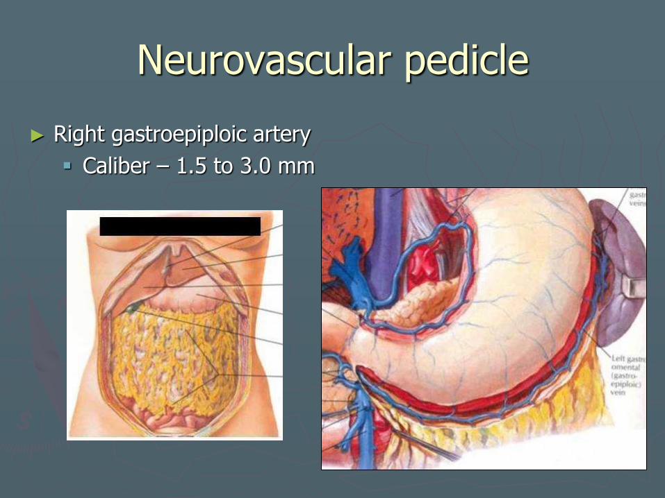
Neurovascular pedicle
► Right gastroepiploic artery
Caliber – 1.5 to 3.0 mm
Gastroomental flap
►Morbidity Intraabdominal complications
►Gastric leak ►Peritonitis ►Intraabdominal abscess ►Volvulus ►Gastric outlet obstruction
If mucosal flap too large or if placed too close to pylorus
Fistula
► Preoperative Considerations h/o GOO h/o PUD
Gastroomental flap

Bibliography
1. Chepeha, DB, Teknos, TN. Microvascular Free Flaps in Head and Neck Reconstruction. In: Head and Neck Surgery—Otolaryngology, 3rd ed., Bailey, BJ Ed. Philadelphia, Lippincott-Raven Publishers, 2001; 2045 – 2065.
2. Urken, ML, Buchbinder, D, Genden, EM. Reconstruction of the Mandible and Maxilla. In Otolaryngology Head and Neck Surgery, 4th Ed. Edited by Cummings CC, St. Louis: Mosby Year Book Inc.; 2004. 1618 – 1635.
3. Chang, KE, Gender, EM, Funk, G. Reconstruction of the Hypopharynx and Esophagus. In Otolaryngology Head and Neck Surgery, 4th Ed. Edited by Cummings CC, St. Louis: Mosby Year Book Inc.; 2004. 1945.
4. Taylor, SM, Haughey, BH. Reconstruction of the Oropharynx. In Otolaryngology Head and Neck Surgery, 4th Ed. Edited by Cummings CC, St. Louis: Mosby Year Book Inc.; 2004. 1758.
5. Lee, KJ. Essentials of Otolaryngology. 891. 6. Lin, DT, Coppit, GL, Burkey, B. Use of the Anterolateral Thigh Flap in Reconstruction
of the Head and Neck. Curr Opin Otolaryngol Head Neck Surg. 12: 300-304. 2004. Lippincott Williams and Wilkins.
7. Genden, E, Haughey, BH. Mandibular Reconstruction by Vascularized Free Flap Tissue Transfer. Am Journ Otolaryngol. 1996; 17 (4): 219 – 227.