Embed Size (px)
Citation preview

Atlas Oral Maxillofacial Surg Clin N Am 13 (2005) 151–171
Microvascular Free Bone Flaps
Remy H. Blanchaert, Jr, MD, DDSa,*,Christopher M. Harris, MD, DMDb
aOral and Maxillofacial Surgery Associates, 1919 N. Webb Road, Wichita, KS 67206, USAbDepartment of Oral and Maxillofacial Surgery,
University of Missouri-Kansas City School of Dentistry, University of
Missouri-Kansas City School of Medicine, 2301 Holmes Street, Kansas City, MO 64108, USA
In the not-so-distant past, composite bone-containing defects of the maxillofacial regionwere reconstructed in a series of staged operations over a long period of time. The developmentand refinement of microvascular free bone flaps has revolutionized the management of suchcases by allowing immediate one-stage reconstruction. The advantages of such an approach areobvious. Restoration of facial form, before the development of the scarring process, providesa more natural facial appearance. Economic advantages include decreased direct costs becausehospital stay is markedly reduced and total surgical time is decreased. Patients benefit byavoiding a series of operations that require time away from work and family. Complicationrates of microvascular free bone flaps paralleldor are better thandthose published for bonegrafting procedures. Perhaps most importantly, with proper planning, microvascular free boneflap reconstruction allows for definitive dental implant-based mandibular reconstruction(Fig. 1A–D). Reconstruction is important because the ability to eat and speak normallycorrelates strongly with patient quality of life after maxillofacial reconstruction, as demon-strated in many clinical studies.
Why do some surgeons within the field of oral and maxillofacial surgery condemn the use offree bone flaps for mandibular reconstruction? Perhaps their experience is limited to cases inwhich appropriate planning and proper attention to detail were not exercised and the outcomewas suboptimal. Microvascular free bone flaps are challenging. This article was designed toaddress the challenges associated with microvascular free bone flaps. The intent is to guidereaders through the process of applying microvascular free bone flaps in their practice byoutlining the lessons learned through 8 years of extensive involvement in maxillofacialreconstruction. General principles are discussed first, followed by a discussion of the variousflaps applicable to maxillofacial reconstruction. Several site-specific case examples also areprovided to emphasize the challenges faced in mandibular reconstruction.
Basic principles
Reconstructive surgeons must understand fully the anticipated defect before preoperativeplanning can begin. It is important that surgeons carefully evaluate the operative site, reviewappropriate imaging, and discuss the planned operation with other surgeons involved in the caseto comprehend the anticipated defect in three dimensions. Microvascular surgeons must selectthe most appropriate donor site, consider the vessel geometry, and devise a plan for shaping,contouring, and insetting the flap in an expedient manner to minimize the ischemia time andfacilitate the microvascular anastomosis.
* Corresponding author.
E-mail address: [email protected] (R.H. Blanchaert, Jr).
1061-3315/05/$ - see front matter � 2005 Elsevier Inc. All rights reserved.
doi:10.1016/j.cxom.2005.05.002 oralmaxsurgeryatlas.theclinics.com

152 BLANCHAERT & HARRIS
In most cases of composite bone-containing defects of the maxillofacial region, fibula flapsand deep circumflex iliac artery (DCIA) flaps are the most suitable choices. In rare instances, thescapula flap and the free radial forearm osteocutaneous flap may prove useful. The fibula flapis generally favored over the DCIA flap because of ease of harvest, minimal donor sitecomplications, and length of the vascular pedicle. The major advantage of the DCIA flap is thatthe donor site is completely hidden.
The donor site should be examined carefully to determine suitability. The vascular supply tothe flap and the remaining tissues must be assured. Prior surgery can compromise the bloodsupply (ie, axillary dissection, scapula flap), and vascular anomalies can put the foot or hand atrisk after flap harvest (fibula, free radial forearm flap). The site selected for flap harvest affectsthe orientation of the vascular pedicle and must be considered in designing the flap to ensureappropriate vessel geometry. In general, the vessels of free flaps do not tolerate tension,redundancy, or sharp angles, which can kink the vein obstruction outflow. In the case ofmandibular reconstruction, the vessels should be oriented in a manner that protects them fromcompression by orienting the flap to place the vascular pedicle along the medial aspect of theinferior border of the neomandible. The flap should be designed so that the vessels extend fromthe flap as near the recipient vessels as possible. For example, if the flap pedicle is to exit the flapat the angle of the right mandible and the skin paddle is to cross over the lateral surface of theneomandible, then the right fibula should be used. Likewise, if the flap is to be similarly orientedbut the pedicle must leave the flap at its anterior aspect (ie, angle and ramus reconstruction),then the left leg should be used.
The recipient site must be prepared appropriately before the flap is harvested. Often, the flapcan be elevated and allowed to reperfuse while the recipient site is prepared. If surgery involvesa patient who underwent irradiation or is a reoperation on a neck that underwent previous neckdissection, however, it may be better to delay flap elevation until after ensuring the presence ofacceptable recipient vessels. In either setting, it is best to restore the continuity of the mandiblewith a reconstruction bar before flap harvest. This approach ensures that the shape of themandible and drape of the facial tissues are acceptable. The flap can be adapted to this shapeat the time of flap inset. In many instances, the reconstruction plate can be adapted beforeresection, removed for resection, and replaced. When there is lateral expansion or full-thickness
Fig. 1. (A–D) This series of images illustrates anterior mandibular reconstruction with a free fibula flap. Note the
character of the healing at the sites of osteotomy of the fibula. Dental implant-based rehabilitation completed the
reconstruction.
153MICROVASCULAR FREE BONE FLAPS
tumor involvement, alternate means of plate adaptation must be considered. Stereolithographicmodels or CAD-CAM polymer models can be created from CT scans, and the reconstructionplate can be adapted to these models after recontouring to a more normal mandibular anatomy(Fig. 2). The need for such a model must be appreciated at the time of initial patient evaluationbefore imaging is ordered, because special imaging protocols may be required. In some instancesit may be helpful to use an external fixation device applied away from the area of the mandibleinvolved in the resection. The device holds the remaining segments of the mandible in place toallow for plate contouring, adaptation, and stabilization (Fig. 3A, B). In other instances, themandibular contour must be established by placing the teeth in occlusion and repositioningsegments that are non–tooth bearing in appropriate position by reference to lateral andposteroanterior cephalometric radiographs. Sometimes a surgeon must adapt the bone plateusing only the opposing dental arch as a guide, based on knowledge of normal jaw anatomy. Insuch cases it is necessary to view the plate adaptation from multiple angles to ensure appropriatepositioning (Fig. 4A, B).
The authors favor the use of locking reconstruction plates for stabilization of microvascularfree bone flaps in mandibular reconstruction. Larger plates were used initially; however, thesenewer plates have advantages in many applications, particularly for segmental resection. Thelarger plates are still used when reconstructing massive resections and for condylar re-construction. The small 2-mm plates are lower in profile but possess more than enough rigidityto stabilize the bone segments until bone healing is complete. Because of the excellent bloodsupply, bony union is achieved rapidly (4–6 weeks). This union depends on excellent adaptationof the bone, however. The osteotomies should be created to ensure intimate contact betweensegments of the flap and between the flap segments and the native mandible. Failure to do somay result in nonunion and subsequent hardware failure (Fig. 5). Standard screws (nonlocking)are used to secure the bone segments to the reconstruction bar. The use of the miniplatetechnique provides adequate stability if applied properly, using paired plates at each osteotomyor flap-native mandible interface. A single miniplate provides insufficient stability (Fig. 6). Theauthors do not use miniplate technique because the use of a previously adapted and appliedreconstruction plate provides such a useful guide for bone flap recontouring.
Revascularization of the flap is completed after recontouring, stabilization and skin paddleinset, and suturing. Manipulation of the flap while it is ischemic simplifies this process. The flapinset is completed before revascularization to ensure that the vessel geometry is appropriate.Microvascular anastomosis is completed with the use of the operating microscope. Themicroscope has significant advantages over the use of loupe magnification. The scope offersexcellent illumination, variable magnification, and equal visualization of the field by theoperator and assistant. The practice of heparinization of the patient at the time of microvascularanastomosis has been shown to be unnecessary and associated with increased local woundcomplications (hematoma). Irrigation of the vessels is accomplished with a solution of 500 Uheparin per 100 mL of saline. An anterior chamber irrigating needle on 5-mL syringes provides
Fig. 2. Preoperative adaptation of a mandibular reconstruction plate to a CAD-CAM model is accomplished after
recontouring the site of tumor expansion to approximate a more normal mandibular anatomy.

154 BLANCHAERT & HARRIS
a suitable irrigation stream. The authors use standard interrupted technique with 9-0 nylon forvessel anastomosis. Disposable approximation clamps are used because they provide reliableclamping forces. End-to-end technique is used most commonly for microvascular anastomosis.The artery is typically approximated first because it is generally deeper than the vein. The facialartery, superior thyroid artery, and external carotid artery are the most commonly usedrecipient arteries. The common facial vein and external jugular vein are the typically usedrecipient veins.
Fibula flap
Background
The fibula flap has gained a well-deserved reputation for reliability, ease of harvest, andsuitability for definitive implant-supported dental rehabilitation. A few oral and maxillofacial
Fig. 3. (A,B) This case series demonstrates the use of a mandibular bridge (temporary external fixator device) to
maintain the position of the proximal mandibular segments after mandibulectomy. The second image illustrates the
appearance after inset of a fibula free flap. Note the position of the vascular pedicle along the medial surface of the
neomandible and the lateral position of the vascular perforators to the skin paddle after inset in the mouth.
Fig. 4. (A,B) This series of images demonstrates the challenges faced in secondary reconstruction. This case involved
multiple operations and bone grafts after shotgun injury. The two images show how viewing the reconstruction from
multiple angles facilitates anatomic mandibular reconstruction.

155MICROVASCULAR FREE BONE FLAPS
surgeons proclaim this flap unsuitable for mandibular reconstruction. The fibula mandibularreconstruction is different in appearance and height in comparison to the native mandible insome cases. The literature that describes success with the flap for this purpose and the seniorauthor’s extensive experience dispute those who downplay the use of this flap. The flap heightand shape of the flap resemble closely the structure of an atrophic edentulous mandible.Throughout the world, the fibula flap has become the most common means of primarymandibular reconstruction. The distance of the donor site from the head and neck field is anadvantage because it allows for simultaneous surgery. Likewise, the lack of significant morbidityof the flap harvest with proper attention to detail is also a significant advantage of the flap.
Early reports of poor reliability of the skin paddle have proved to be related to a poorunderstanding of the flap anatomy, which has led to errors in flap harvest rather than a trueproblem with the flap itself. These errors occurred because the vascular perforators to the skin donot always pass to the skin within the lateral intramuscular septum and can be musculocuta-neous perforators (Fig. 7). The presence of musculocutaneous perforators complicates theharvest and limits the mobility of the skin paddle slightly, but it is managed easily by anexperienced surgeon.
The fibula flap is based on the peroneal artery and vein. These vessels are of good diameterand quality. Preoperative evaluation of the perfusion of the lower extremity should beperformed before planning surgery. The authors perform a clinical assessment and determinethe necessity for additional studies based on the findings of that examination. The examinationshould consider color and character of the skin, hair distribution, temperature, nail thickness,and the character and quality of the pulses within the foot. Identification of any abnormalityshould trigger a vascular imaging study. The authors prefer magnetic resonance angiography(Fig. 8) over arteriography because of the radiation and the dye load required for the
Fig. 5. This image displays hardware failure caused by nonunion of the DCIA flap to the native mandible. The source of
this problem seems to be inadequate flap inset.
Fig. 6. This image demonstrates the inadequate stability provided by a single miniplate. The free fibula flap did not heal
to the native mandible. The cause of this nonunion could be either inadequate stability or poor inset. Miniplates should
be applied in two planes as a paired plate construct.

156 BLANCHAERT & HARRIS
arteriography. Based on studies, it is estimated that 10% to 20% of lower extremities displayabnormal characteristics, which causes the harvest of the peroneal artery to potentiallycompromise the vascularity of the foot. The rationale for not studying all legs is that with anexcellent clinical examination the likelihood of a significant abnormality is low and all threemajor vessels will be seen in the course of flap elevation, which makes it possible to identify anyabnormality before flap harvest.
Fig. 7. This artist sketch is an excellent depiction of the flap harvest, character of the perforators, and proximity to
adjacent structures.
Fig. 8. This is an AP MRA image of the right leg. The detail seen with this imaging technique is remarkable.

157MICROVASCULAR FREE BONE FLAPS
The fibula flap offers flexibility in flap design and placement that provides reconstructivesurgeons with important options in the selection of appropriate recipient vessels. Surgeonsshould select the donor leg and design the flap to simplify inset and maximize vascular pediclelength. The length of the available fibula allows for nearly total mandibular reconstruction. Thecharacter and quality of the bone, along with the excellent segmental vascular supply, allowsurgeons to custom contour the flap to the ideal dimensions of the mandible. Closingosteotomies allow precision inset of the flap to ensure excellent, direct bone contact. Whenproperly recontoured and inset, the flap heals rapidly and restores mandibular continuitydespite the administration of postoperative adjuvant radiation therapy. The bone supports theplacement of dental implants immediately at the time of flap reconstruction, after bone healing,or after resolution of the acute effects of the radiation therapy. Mandibular hardware should beremoved at the time of implant placement to ensure load transfer to the bone element of the flap.
Technique
A patient’s donor leg should be shaved with clippers and the surgical anatomic landmarksoutlined in detail. It is often possible to identify the location of the perforators to the skin witha Doppler probe. Typically they are located near the junction of the middle and distal third ofthe fibula. If they can be identified, they should be marked clearly, as in the clinical exampleprovided. Protection of the articulations of the fibula and peroneal nerve is enhanced byappropriately marking the skin (Fig. 9A–C). Stabilization of the leg can be facilitated with theplacement of a bump at an appropriate location to serve as a foot rest. A sandbag or liter bag ofintravenous fluid secured with tape across the bed is appropriate for this purpose (Fig. 10). Thesite is prepared simultaneously with the head and neck site. The two fields are kept isolatedthroughout the procedure. Surgical skin prepping solution is excellent for site preparation, andan extremity drape is used to isolate the field. A sterile tourniquet is placed above the knee. Thetourniquet is inflated to 100 mm Hg above systolic blood pressure after exsanguination of theleg by elevation for 3 to 5 minutes.
Simultaneous flap harvest and head and neck surgical site preparation can be performedin a two-team manner. The surgical time required for fibula flap elevation is approximately70 minutes. Although the flap can be elevated and modified in situ while pedicled to decreaseischemia time, the authors prefer not to do this. Instead, the authors design the flap backward,beginning from the site of the vascular reanastomosis first to ensure acceptable vessel geometry.The time required to osteotomize, recontour the flap, and complete the inset and reanastomosisis well within the allowable time for warm ischemia. There is little advantage to complicating theprocedure by trying to perform osteotomies with the flap pedicled. The total ischemia time fora fibula flap is approximately 3 hours 20 minutes, including tourniquet time.
The lateral intramuscular septum is easily palpable in all but the heaviest of patients. The skinpaddle is oriented over the perforating vessels and the lateral intramuscular septum (Fig. 11).The anterior skin paddle elevation is completed first with dissection through the skin,subcutaneous tissue, and fascia. Refinement of the location of the skin paddle later can bemade after visualizing the perforators. It is helpful to mark them on the skin paddle to assist increation of the posterior incision to center the perforators within the skin paddle.
The peroneus longus, peroneus brevis, and flexor hallicus longus are reflected from theanterior and medial portion of the fibula to the intramuscular septum (Fig. 12A, B). Thisprocedure is best accomplished with electrocautery while taking care to preserve a small musclecuff. After incision of the intramuscular septum, the deep peroneal nerve and the anterior tibialvessels are easily visualized at this point within the reflected medial tissue elevation (Fig. 13).Gentle medial reflection is necessary to identify the interosseous membrane. The posterior skinpaddle elevation is then accomplished. It is necessary to remain subfascial throughout thisdissection and to remain vigilant for possible musculocutaneous perforators.
Proximal and distal osteotomies are made through the fibula at least 6 cm away from theterminations of the fibula to maintain adequate stability of the knee and ankle joints. The fibulais then rotated and the intraosseous membrane is incised along its length with a scissors toexpose the tibialis posterior muscle. Typically the most distal aspect of the peroneal vessels canbe seen easily at this point. Ligation of the vessels allows increased lateral movement of the flap

158 BLANCHAERT & HARRIS
and eases visualization of the remainder of the dissection. The vascular pedicle is then dissectedfree by dividing the overlying chevron-shaped tibialis posterior muscle. Many large branchesfrom the peroneal vessels to the soleus and gastrocnemius muscles require ligation to facilitateretraction. As the dissection proceeds proximally, the posterior dissection also must becompleted. This procedure is slightly more difficult if the perforators are of the intramusculartype. To facilitate this, it helps to put a finger along the medial aspect of the vascular pedicle anddirectly visualize the perforators while cutting down with electrocautery to join the medialdissection. A small muscle cuff also is usually harvested, which helps to fill the voids presentalong the medial aspect of the mandibular reconstruction.
Throughout this portion of the dissection the posterior tibial vessels are visualized within themedially retracted tissues. Extreme care should be given to avoid undue pressure on the posteriortibial nerve. The operating surgeon must examine frequently the position of the retractors used toensure safe positioning because the retracting surgeon, who is frequently on the opposite side ofthe table, has no direct view. The vascular pedicle should be skeletonized before flap harvest(Fig. 14), which greatly facilitates subsequent microvascular anastomosis. There is routinelya large soleus branch approximately 4 cm from the bifurcation that is often difficult to visualize.Care should be taken to identify and ligate this branch because of the consequences of bleedingobscuring visualization at this critical location. There is often what seems to be a cutaneousperforator along the upper one third of the fibula length. The surgeon must take care not to rely
Fig. 9. (A–C ) This series of images demonstrates the depiction of the relevant anatomy for the fibula harvest.
Maintenance of 6 to 8 cm of the fibula near the joints and the common peroneal nerve is depicted.
159MICROVASCULAR FREE BONE FLAPS
on this vessel unless he or she is certain that it arises from the peroneal vessels. This perforator isoften from a circumflex branch.
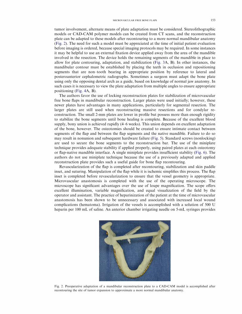
After complete isolation of the flap pedicle, the tourniquet should be released to visualize flapperfusion and ensure hemostasis. Appropriate preparation of the recipient bed should includethorough mobilization of recipient vessels and assurance of excellent flow at the site of plannedanastomosis. The field should be prepared to facilitate the flap modification and inset. Ifpossible, accommodation should be made for a contouring bur and a sagittal saw to be availablefor use simultaneously, which avoids wasting time switching appliances. Division of the flap andtransfer to the head and neck field is accomplished when the site is appropriately prepared. Thedonor site leg may be packed with lap sponges and wrapped in gauze for later management, ora member from the operative team may close the leg over a drain while the flap inset isoccurring. Closure should be accomplished by loose approximation of the muscles, buried
Fig. 10. This image demonstrates the value of a support placed on the operating table to allow hands-free support of the
leg to facilitate fibula harvest.
Fig. 11. This image shows the positioning of the skin paddle of the fibula flap over the lateral intramuscular septum. The
location of the perforators to the skin paddle have been identified and depicted on the leg. The flap is oriented directly
over these vessels.

160 BLANCHAERT & HARRIS
closure of the dermis, and stabilization of the skin at the skin paddle donor site. The skin paddledonor site is then grafted with split-thickness skin from the thigh. The graft should be securedwith resorbable suture and a tie-over bolster dressing. The foot and ankle should be stabilized ina lower leg posterior splint that is well padded. If no skin paddle is harvested, the skin may beapproximated directly and the lower leg dressed with light elastic compression.
The osteotomies of the fibula are made precisely to allow maximum bone contact at theinterface of the fibula segments and the fibula-to-native-mandible interface. When the surgery isproperly performed, the bone flap literally snaps into place. Stabilization is limited to one or twoscrews per segment. This portion of the reconstruction is critical to the outcome. Failure toachieve proper bone inset can result in nonunion and subsequent hardware failure, which arecommon reasons why the senior author sees other surgeons’ patients for revisions. The skinpaddle inset often requires customization of the skin paddle by excision of skin and dermiswhere excess exists and maintenance of the underlying dermal plexus, fat, and fascia to ensureadequate perfusion. The flap should be sutured into position without tension using horizontalmattress sutures. A general principle that should be followed is that more tissue is better thannot enough; some degree of redundancy is shared across wounds to err on the side of excesstissue maintenance.
Postoperative mobilization can begin immediately if there is no skin graft or at the time ofremoval of the bolster dressing (5 days) if a skin graft is used. Donor site elevation is required
Fig. 12. (A) The anterior incision and posterior dissection of the skin paddle of the fibula flap. The vascular perforator is
seen directly opposite the retractor. (B) The appearance of the donor site during the anterior-medial dissection.
Fig. 13. The contents of the anterior compartment are visualized in this image. The interosseous septum is seen anterior
to the fibula. Beneath the septum the peroneal vessels are identified.

161MICROVASCULAR FREE BONE FLAPS
throughout the first few weeks to prevent lymphedema in the leg. Return to normal activitiescan be ensured as soon as 4 to 6 weeks after surgery.
Deep circumflex iliac artery flap
Background
The DCIA flap was developed from early experience with the groin flap. The proper vascularpedicle for the osteomusculocutaneous flap was determined to be the DCIA and vein. Thesevessels arise from the external iliac artery and vein just above the inguinal ligament. They haveaverage diameters of 2 to 3 mm. The ascending branch of the DCIA provides the blood supplyto the internal oblique muscle, which has become the favored soft tissue for harvest with theflap. Harvesting a well-vascularized muscle and allowing secondary epithelialization withinthe oral cavity provides a character and quality of tissue that are well suited to reconstruction ofthe dentoalveolar apparatus. The muscle undergoes a process of atrophy and mucosalizationthat results in a fixed mucosal lining that adheres densely to the iliac bone over which it has beendraped. This process provides an ideal site for dental implant-based oral rehabilitation.
The flap initially was described to include the overlying skin and fat and the internal obliquemuscle. The skin is supplied by muscular perforators and is well vascularized. The only problemwith the skin element of the flap is that it is often too thick and immobile. The flap is commonlyused with only the internal oblique muscle.
The donor site provides some interesting challenges in the course of its reconstruction.Potential exists for hernia formation through the bone harvest site and the weakened abdominalwall. To avoid hernia, the iliac harvest defect is typically spanned with either titanium mesh orpolypropylene mesh, and the abdominal wall likewise is supported with polypropylene mesh. Itis not uncommon to spend twice as much time on the closure of the defect site as in theharvesting of the flap. Proper patient selection is important. The authors prefer not to performthe DCIA flap in active men or laborers. Instead, the DCIA flap has been used typically in thinwomen and retired thin men. One surprising challenge encountered with the use of the DCIAflap in mandibular reconstruction has been too much bone height, which interferes with theinterarch space required for implant-based dental rehabilitation. The DCIA flap is the clearsecond choice for mandibular reconstruction after the fibula.
A significant advantage of the flap is that the distance from the head and neck field allowssimultaneous two-team surgery, which helps considerably to offset the challenges imposed bythe difficulty of the flap harvest and the donor site reconstruction.
Fig. 14. Flap elevation and dissection of the proximal vascular pedicle.

162 BLANCHAERT & HARRIS
Technique
The patient is positioned supine with the table flat. It may be helpful to place a small bolsterunder the hip opposite the harvest site to facilitate visualization of the vascular pedicle.Assistance is mandatory and is best provided by a retractor holder opposite the surgeon and anassistant on the same side as the surgeon. The site should be shaved, prepared, draped, andcovered with an occlusive barrier to isolate the field. The surgical anatomic landmarks should bemarked clearly (Fig. 15A). If a skin paddle is to be used, it should be oriented directly over theiliac crest, should be elliptical shaped, and should be of sufficient width to ensure capture of themusculocutaneous perforators. In the more typical flap harvest without skin paddle, the accessincision is placed in a natural skin crease within the groin that extends laterally superior to theiliac crest. The skin is incised and the external oblique muscle is identified and incisedapproximately 2 cm from the iliac crest (Fig. 15B). The external oblique muscle is then retractedand the internal oblique muscle is dissected along its entire length to the linea semilunaris
Fig. 15. (A–F ) This series of images illustrates the harvest of a DCIA flap. The text supports these drawings well.

163MICROVASCULAR FREE BONE FLAPS
medially and superior to the inferior margin of the rib cage (Fig. 15C). The required amount ofinternal oblique muscle is then incised and the muscle is elevated from the transversalis muscle.The ascending branch of the DCIA is traced along the deep surface of the internal obliquemuscle to the major DCIA pedicle.
Upon identification of the major pedicle, it is wise to extend the dissection medially to theexternal iliac vessels. There is substantial variability in the arrangement of the deep circumflexvessels and the lateral femoral cutaneous nerve (Fig. 15D). The DCIV can provide surgeonswith some challenges because it may exist as two distinct vessels. The authors’ experience is thatthese vessels always come together before draining into the external iliac vein. It is possible,however, that they can remain separate, as described in the literature. If the veins remainseparate, it is possible to evaluate flow and reanastomose only the vein with the greatest flow.
After isolation of the flap pedicle, it is appropriate to begin the osteotomies required toharvest the bone element of the flap. The transversalis muscle must be transected approximately2 cm from the iliac crest or preferably with direct visualization of the vascular pedicle to avoidits injury (Fig. 16A). The preperitoneal fat must be retracted medially and the iliacus musclemust be transected to expose the iliac bone. The muscles are released laterally from the iliac crestto the level of the planned osteotomy. Osteotomies are then completed to free the flap, which
Fig. 16. (A,B) The flap harvest and flap inset. Note the position of the vascular pedicle along the medial surface of the
neomandible.
164 BLANCHAERT & HARRIS
remains pedicled to the DCIA and DCIV at the external iliac vessels. The authors prefer tomaintain approximately 2 to 3 cm of iliac bone intact at the anterior superior iliac spine, whichfacilitates reconstruction of the continuity of the iliac crest to avoid hernia formation. Thevascular pedicle is divided and the flap transferred to the recipient site after proper preparationof the recipient site (Fig. 15E).
Inset of the bone flap is completed in such a way as to ensure protection of the vascularpedicle on the medial aspect of the flap with the internal oblique muscle on the medial-inferioraspect of the flap (ie, with the iliac crest positioned inferiorly and the cut surface superiorly) (seeFig. 15F; Fig. 16B). Care must be taken in design of the flap because the somewhat limitedpedicle length must be allowed for. The maintenance of the ASIS assists in that regard byeffectively lengthening the vascular pedicle. Curvature of the bone element of the flap requirescreating opening osteotomies of the lateral cortex with greenstick fracture of the inner cortex.The gap is packed with particulate cancellous bone (this is demonstrated in Fig. 15F). Themuscle is then draped around the neomandible. The muscle fascia is sutured to the mucosaledges within the oral cavity to provide an effective seal. Rapid granulation of the exposedmuscle is common. The color and character of the exposed muscle provide an excellent meansof monitoring.
The donor site is reconstructed by stabilizing a double thickness of polypropylene mesh tospan the bone defect, which is secured via drill holes in the ilium. The transversalis and externaloblique muscles are then sutured to the aponeurosis and supported with another layer of mesh.The skin is approximated in layers (Scarpa’s fascia, dermis, skin) over a closed suction drain. Anappropriate bowel regimen is recommended for use in the preoperative and postoperative timeframe to avoid constipation and straining at stool. Likewise, lifting weights of more than15 pounds, strenuous exercise, and labor are delayed until at least 4 weeks after surgery to avoidthe development of hernia.
Radial forearm osteocutaneous flap
Background
The free radial forearm fasciocutaneous flap has excellent use in head and neck surgeryprimarily because of the thin, pliable character of the tissue and the reliability of its vasculature.A major advantage of the flap is that the distant location of the flap donor site allowssimultaneous flap harvest and cancer resection or recipient site preparation. The use of this flapas a bone-containing flap is, however, somewhat limited because of the minimal volume of bonethat can be harvested. The primary uses of the flap are to restore facial form and providesupport of adjacent structures, as in reconstruction of the premaxilla to provide nasal and lipsupport. The senior author believes that the bone stock is insufficient for use in mandibularreconstruction because it does not support endosseous dental implant rehabilitation. Dentalrehabilitation should be a primary goal of all mandibular reconstructions. Quality-of-life studiesdemonstrate that for patients, the most important aspects that affect their quality of life areeating normally and speaking clearly. Without dental rehabilitation, the achievement of thesegoals would be impossible; the free radial forearm osteocutaneous flap remains a rarely used flapin the surgical armamentarium.
The free radial forearm flap receives its vascular supply from the radial artery. The flap’svenous drainage is from either the venae comitantes or the cephalic vein. The authors prefer thecephalic vein because its size more closely approximates that of many potential recipient veins inthe neck and facilitates end-to-end anastomosis. These vessels average 2 mm in diameter. Beforeflap harvest, a surgeon must ensure adequate communication between the superficial and deeppalmar arches, which is most easily done using the relatively simple Allen’s test. The absence ofan appropriate communication has been described in 12% of specimens investigated. Althoughthe nondominant hand is generally selected, a poor Allen’s test result has governed the selectionof flap donor site in several of the senior author’s cases. A positive Allen’s test resultdemonstrates the absence of communication between the palmar arches in the patient beingevaluated (Fig. 17). Note that the portion of the hand that appears well perfused with the radial

165MICROVASCULAR FREE BONE FLAPS
artery occluded is limited to that area primarily supplied by the ulnar artery. In this instance,selection of an alternate donor site is required.
The limitations of the bone stock available in the radial forearm fasciocutaneous flap alreadyhave been outlined. The amount of bone that can be harvested is limited to 40% of the diameterof the radius between the insertion of the pronator teres proximally and the brachioradialisdistally (Fig. 18). To avoid the creation of internal stress, the osteotomy should be completedwithout sharp internal line angles. It is best to plate prophylactically across the defect at theradius harvest site because the rate of fracture is approximately 25%. The deformity that resultsfrom fracture of the radius is unacceptable. The blood supply to the radius is through segmentalperiosteal branches within the anterolateral intramuscular septum and the flexor pollicis longusmuscle. The nature of this blood supply allows for osteotomies to be performed, providedappropriate care is given to avoiding excessive periosteal elevation.
Technique
An appropriate donor site is selected based on the character of the tissue and the appearance ofthe vasculature. An Allen’s test must confirm adequate ulnar artery perfusion of the thumbwith radial artery occlusion. If it is difficult to determine perfusion based on color of the skin (ie,
Fig. 17. An Allen’s test that demonstrates the lack of communication between the superficial and deep palmar arches.
Note the pale character of the thumb and first finger.
Fig. 18. A cross-section of the radius harvest site and the appropriate shape of the radius osteotomy.

166 BLANCHAERT & HARRIS
dark-skinned subject), a pulse oximeter probe can be attached to the thumb and the test repeated.The appropriate donor site should be selected well before surgery and the cephalic vein should beprotected from venipuncture. On the day of surgery, the authors prefer to mark the radial arteryand cephalic vein with a surgical marker in the preoperative holding area. This preparationfacilitates design of the flap once the final dimensions of the defect site have been determined.
The donor site arm is prepared simultaneously with the resection/recipient site. The arm isprepared to the axilla and draped with an extremity drape. A sterile tourniquet is applied to theupper arm after the flap has been outlined on the ventral surface of the forearm overlying theradial artery. Inflation of the tourniquet is accomplished after exsanguination of the arm with anelastic bandage. The tourniquet should be inflated to 250 mm Hg or approximately 100 mm Hgabove systolic blood pressure. Dissection and flap elevation are aided by stabilizing the hand/wrist with a lap sponge across the palm secured by nonpenetrating towel clamps.
The flap elevation is performed from the distal. The radial artery, its venae comitantes, andthe cephalic vein are ligated. A judgment call as to the fate of the dorsal radial nerve must bemade based on the degree of its exposure and the possibility of painful altered sensations uponits postoperative stimulation. This nerve may be covered only by the skin graft and may beprone to constant stimulation. It is perhaps best to sacrifice this nerve from the distal of the flapharvest site to beneath the forearm musculature to avoid postoperative dysesthesia. The fascialcompartment that contains the vascular supply is dissected free from the underlying musclesfrom the lateral aspect of the skin paddle. Proximal incision is made in the groove between theflexor carpi radialis and brachioradialis muscle or along or near the cephalic vein. The dissectionis then carried down to the level of the brachioradialis muscle. The brachioradialis muscle can bereflected laterally to expose the proximal vascular pedicle. The medial skin paddle elevation andproximal dissection are then completed. The flexor carpi radialis can be retracted medially toexpose fully the vascular pedicle and the radius. The appropriate osteotomy site is thenidentified and isolated. The osteotomy is made carefully to avoid creation of areas of isolatedstress and avoid removing more than 40% of the diameter of the radius. The flap then remainspedicled on the proximal vascular pedicle. The proximal vascular pedicle is dissected free fromits venae comitantes at the site at which vascular anastamosis is likely to occur.
The authors prefer to allow reperfusion of the flap at this time to ensure adequate perfusionand hemostasis. The flap may be repositioned and wrapped in lap sponges and gauze until thepreparation of the recipient site is ensured.
The head and neck field always must remain isolated from the donor site. The morbidity ofa local infection in the wrist is significant. Extreme limitation of mobility is the likely outcome.The vascular pedicle of the flap is transected and the flap transferred to the recipient site. Theflap inset is completed and the bone stabilized with miniplates or a reconstruction plate beforevascular reanastomosis. The surgical team may be split, and a team member or members may beleft behind during the flap inset to complete the closure of the proximal portion of the donor siteover a drain, stabilization of the skin edges of the flap harvest site to the underlying fascia, andcoverage of the defect with a split-thickness skin graft. The arm should be splinted in theposition of maximum function (wrist dorsiflexed 45 �; fingers neutral) with a simple volar splint.
Scapula flap
Background
The scapula free flap is based on the circumflex scapular artery and vein. These vessels areof large caliber, with common diameters of 4 mm (range 2–6 mm). This flap has perhaps themost versatility and adaptability of any of the bone-containing microvascular free flaps incommon usage. The flap is generally reserved for specific indications in head and neckreconstruction because of the challenges imposed by the need to reposition the patient to allowflap harvest and the donor site morbidity related to the flap harvest. Such indications includethrough-and-through composite defects of the mandible and complex three-dimensionaldefects of the maxilla. The major advantage of this flap is the availability of two separatelymobile cutaneous segments.

167MICROVASCULAR FREE BONE FLAPS
Approximately 8 cm of bone can be harvested along the lateral border of the scapula. Thebone is well perfused through numerous branches. Care must be taken to identify the origin ofthe blood supply to the scapular tip via the angular artery if it is to be included. The angularartery commonly arises from the thoracodorsal artery. Proximal dissection of the vascularpedicle and transection of the flap pedicle at the subscapular artery and vein results in the abilityto transfer simultaneously the latissimus dorsi muscle and the serratus anterior muscle with theflap. Although this is a common point of discussion, the authors know personally of no surgeonwho actually has performed such a flap.
The scapula bone is of acceptable quality and quantity for midface reconstruction in mostcases but may fall short of ideal for mandibular reconstruction because of its limitations in sizeand volume. The bone is commonly insufficient in width to accept endosseous implants inwomen and often is insufficient in men, which limits the flap’s acceptance.
Technique
The patient must be positioned in the lateral decubitus position. For use in head and neckreconstruction, the primary operative site must be packed and covered before repositioning andrepreparation of the operative field. Lap sponges should be placed in the operative site, anda large transparent adhesive drape should be used to cover the site of the resection and the neckdissection. The surgeon also should realize that closure of the flap harvest site and repositioningare required again after flap harvest and before flap inset and revascularization. This processrequires operative team coordination and planning to ensure a smooth transition with minimalloss of time to limit flap ischemia. To avoid brachial plexus injury, careful support andmovement of the arm at the site of the flap harvest and support and protection of thecontralateral brachial plexus with an axillary roll are required.
The scapular outline should be marked, and the orientation of the cutaneous branches of thecircumflex scapular artery should be drawn to facilitate the orientation of the skin paddles. Thetransverse cutaneous branch runs horizontally across the scapula midway between the scapularspine and the scapular tip. The descending cutaneous branch is located vertically along a parallelto the lateral border of the scapula. The origination of these two cutaneous branches from thecircumflex scapular artery is identified in the triangular space formed by the teres major, teresminor, and the long head of the triceps brachii muscle (Fig. 19). This site typically can bepalpated or identified with a simple Doppler probe. The cutaneous elements should be dissected
Fig. 19. An excellent depiction of the scapula flap design and harvest.

168 BLANCHAERT & HARRIS
first to allow identification of the vascular pedicle. The inferior margin of the cutaneous flap isincised through skin and subcutaneous tissue down to the level of the fascia overlying therhomboid and infraspinatus muscles. When the teres major is identified in the course of thedissection, the superior margin of this muscle is followed to the triangular space. The superiormargin of the skin paddle is then incised and elevated in a similar manner. Handling of the flapis facilitated by harvesting a small cuff of the fascia overlying the infraspinatus musclesurrounding the cutaneous branches near the vascular pedicle. After elevation of the skinpaddles, the bone exposure and osteotomies can be undertaken.
The blood supply to the lateral border of the scapula arises from multiple periosteal branchesof the circumflex scapular artery and vein. To preserve these branches, the teres major muscle
Fig. 20. (A–F) The use of a fibula flap in the reconstruction of osteoradionecrosis of the left mandible. The patient was
treated with combined modality chemotherapy and radiation for cancer of the base of the tongue. (A) Bone destruction
on the left posterior body and vertical ramus. (B) The character of the tissues and mandible within the radiated field.
Note the change in character of the mandible in the mid-body region. (C) The extracorporeal adaptation of the healthy
native condyle to the reconstruction plate. (D) The fibula before osteotomies and flap inset. (E) The completed inset of
the flap before vascular anastamosis. (F) Panorex demonstrates the character of the flap in the early postoperative time
frame.

169MICROVASCULAR FREE BONE FLAPS
must be transected and a muscle cuff must be included along the lateral surface of the scapula.The latissimus dorsi muscle can be preserved by dissecting it free from the lateral border of theteres major muscle. Once this muscle transection is completed, excellent visualization of thethoracodorsal, angular, and circumflex scapular artery is created. The thoracodorsal artery isligated and transected (unless the latissimus dorsi muscle is to be included in the flap), as is theangular artery. The proximal dissection of the vascular pedicle is best completed at this timewhile the structures are stable. The circumflex scapular artery and vein should be isolated tofacilitate the later anastomosis of these vessels to acceptable recipient vessels in the neck afterflap transfer.
The osteotomies of the lateral border of the scapular are accomplished after dividing theinfraspinatus muscled in a longitudinal direction to create a 3-cm muscle cuff along the lateralborder. With these muscles retracted, the bone is best cut with an oscillating saw. The tip of thescapula is seldom harvested because of its separate blood supply. The available bone length isapproximately 8 cm. After completion of the osteotomies, the bone flap is elevated and thesubscapularis muscle is transected with a similar 3-cm muscle cuff to complete the mobilizationof the flap. The flap can be harvested with the desired length of vascular pedicle and set asidewhile the donor site is closed.
The teres major muscle must be repaired via direct suture or sutured to the remaining scapulathrough drill holes. Either method significantly reduces the function of the muscle. The functionsof the teres major muscle include internal rotation, extension, and adduction of the arm. Thelimitation of these functions remains the primary drawback to the use of this flap. The overlyingskin is then advanced and reapproximated over a drain in layers. The operative site is dressedand the patient repositioned for flap inset and vascular reanastomosis.
Clinical examples
Case #1: Lateral mandibular resection for osteoradionecrosis
The patient is a 45-year-old white man who 4 years ago underwent right radical neckdissection and combined modality chemotherapy-radiation therapy for tongue base squamouscell carcinoma. He had undergone extraction of partially impacted third molars 3 weeks beforeinitiation of radiation therapy. The left third molar site never healed, and osteoradionecrosisdeveloped. He received approximately 45 hyperbaric oxygen therapy treatments and numerouscourses of antibiotic therapy without resolution before being referred for definitive intervention.Extensive bone destruction was seen on panoramic radiograph. The extent of the diseasedmandibular bone can be seen in a preoperative MRI study. The posterior body, angle, andvertical ramus of the mandible clearly are affected. This case illustrates the use of the native
Fig. 21. (A) The use of a CAD-CAM model to adapt the reconstruction bar in a case of a hemimandibulectomy,
including the articulation of the mandible. (B) A panoramic radiograph shows the early postoperative appearance of the
flap.

170 BLANCHAERT & HARRIS
condyle and its articular surface as a graft. The bone plate was placed before resection andremoved. The condyle was reattached and the plate replaced (Fig. 20A–F).
Case #2: Vertical ramus and condylar reconstruction: recurrent ameloblastoma
A 25-year-old patient presented with recurrent ameloblastoma that involved the posteriorbody, angle, vertical ramus, and condyle. This case example demonstrates the use of amicrovascular free fibula flap in condylar reconstruction. A computer-generated model was usedto prebend the mandibular reconstruction plate. Suspension of the condylar reconstruction isrequired at the articular fossa. Arch bars and elastic traction help suspend the neocondylethroughout its early healing stages (Fig. 21A, B).
Fig. 22. (A–G) The management of an anterior floor of mouth squamous cell carcinoma that infiltrates the mandible.
The reconstruction plate was adapted directly to the mandible and removed before resection. Flap inset and
reanastomosis are depicted, as is proper vessel geometry (E). The skin paddle provides excellent mobility of the tongue,
and the fibula is more than adequate to support dental implant-based rehabilitation.

171MICROVASCULAR FREE BONE FLAPS
Case #3: Floor-of-mouth squamous cell carcinoma
A 64-year-old woman was referred with a large floor-of-mouth squamous cell carcinomainfiltrating themandible.This case illustrates adaptationof the2-mm lockingplate before resection,flap inset, appropriate vessel geometry, and the mobility of the skin paddle. Definitive dentalrehabilitationwas accomplishedwith endosseous implants.The implants areplacedas soonas bonehealing is completed or after resolution of the acute effects of radiation therapy (Fig. 22A–G).
Summary
Microvascular free bone flaps are a modern means of restoring bone-containing compositedefects of the maxillofacial region. The techniques are simple and reliable. The results arereproducible and offer significant advantages over staged mandibular reconstruction. Inparticular, these techniques decrease costs and provide a means of rapid definitive reconstruc-tion. Patients avoid multiple surgical procedures with immediate reconstruction that allowsthem to return to productive lives in society. Proper selection of an appropriate donor site andappropriate preoperative planning facilitate application of these techniques in an expedientmanner. Microvascular free bone flap reconstruction should be considered for all patients withcomposite bone-containing defects of the maxillofacial region.
Further readings
Brown JS. Deep circumflex iliac artery free flap with internal oblique muscle as a new method of immediate
reconstruction of maxillectomy defect. Head Neck 1996;18:412–21.
Brown MR, McCulloch TM, Funk GF, et al. Resource utilization and patient morbidity in head and neck
reconstruction. Laryngoscope 1997;107:1028–31.
Frodel JL, Funk GF, Capper DT, et al. Osseointegrated implants: a comparative study of bone thickness in four
vascularized bone flaps. Plast Reconstr Surg 1993;92:449–55.
Hidalgo DA, Rekow A. A review of 60 consecutive fibula free flap mandibular reconstruction. Plast Reconstr Surg 1995;
96:585–96; discussion 597–602.
Kroll SS, Schusterman MA, Reece GP. Costs and complications in mandibular reconstruction. Ann Plast Surg 1992;29:
341–7.
Sullivan MJ, Baker SR, Crompton R, et al. Free scapula osteocutaneous flap for mandibular reconstruction. Arch
Otolaryngol Head Neck Surg 1989;115:1334–40.
Urken ML, Weinberg H, Vickery C, et al. The internal oblique-iliac crest free flap in composite defects of the oral cavity
involving bone, skin and mucosa. Laryngoscope 1991;101:257–70.
Vaughan ED. The radial forearm free flap in orofacial reconstruction: personal experience in 120 consecutive cases.
J Craniomaxillofac Surg 1990;18:2–7.


![Bone Transport versus Acute Shortening for the Management ...676639/UQ676639_OA.pdf · combined approaches with both microvascular tissue transfers and bone grafts [3] . Masquelet](https://img.dokumen.tips/doc/110x75/5fa3f7aa1a49a1064954e5b0/bone-transport-versus-acute-shortening-for-the-management-676639uq676639oapdf.jpg)