Embed Size (px)
Citation preview

MicroscopyMicroscopy
Light and Electron Light and Electron MicroscopyMicroscopy

The First Light The First Light MicroscopesMicroscopes
Around 1590 Zaccharias and Around 1590 Zaccharias and Hans Janssen experimented with Hans Janssen experimented with lenses in a tube, leading to the lenses in a tube, leading to the forerunner of the microscope and forerunner of the microscope and the telescopethe telescope
In the late 1600’s, Anton van In the late 1600’s, Anton van Leeuwenhoek was the first to see Leeuwenhoek was the first to see bacteria, yeast, and many other bacteria, yeast, and many other microbes using a microscopemicrobes using a microscope

How Light Microscopes How Light Microscopes WorkWork First, the objective lens gathers light First, the objective lens gathers light
from the specimen and magnifies the from the specimen and magnifies the imageimage Most microscopes have several objective Most microscopes have several objective
lenses that can be rotated into position lenses that can be rotated into position to provide different levels of to provide different levels of magnification (4X, 10X, 40X)magnification (4X, 10X, 40X)
The ocular lens in the eyepiece The ocular lens in the eyepiece magnifies and transmits the image to magnifies and transmits the image to your eyeyour eye The magnification of the ocular lens is The magnification of the ocular lens is
10X10X To find the total magnification of the To find the total magnification of the
microscope you are using, multiply the microscope you are using, multiply the magnification of the objective lens by magnification of the objective lens by the magnification of the ocular lens. the magnification of the ocular lens. For example: 40X (objective lense) x 10X For example: 40X (objective lense) x 10X
(ocular lense) = 400X magnification(ocular lense) = 400X magnification
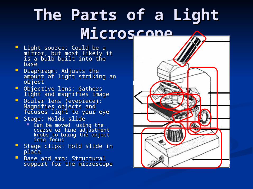
The Parts of a Light The Parts of a Light MicroscopeMicroscope
Light source: Could be a Light source: Could be a mirror, but most likely it is a mirror, but most likely it is a bulb built into the basebulb built into the base
Diaphragm: Adjusts the Diaphragm: Adjusts the amount of light striking an amount of light striking an objectobject
Objective lens: Gathers light Objective lens: Gathers light and magnifies imageand magnifies image
Ocular lens (eyepiece): Ocular lens (eyepiece): Magnifies objects and focuses Magnifies objects and focuses light to your eyelight to your eye
Stage: Holds slideStage: Holds slide Can be moved using the Can be moved using the
coarse or fine adjustment coarse or fine adjustment knobs to bring the object into knobs to bring the object into focus focus
Stage clips: Hold slide in Stage clips: Hold slide in placeplace
Base and arm: Structural Base and arm: Structural support for the microscopesupport for the microscope

Can you name the parts?Can you name the parts?Start on the left side and work from the top down. Start on the left side and work from the top down.
Then go to the right side and work from the top down.Then go to the right side and work from the top down.
Nice Job !
Objective Lenses
Stage
Diaphragm
Light Source
Base
Fine adjustment
Course adjustment
Stage clip
Arm
Ocular lens (eyepiece)

Images Produced by Light Images Produced by Light MicroscopesMicroscopes
Amoeba Streptococcus bacteria Anthrax bacteria
Human cheek cells Plant cells Yeast cells
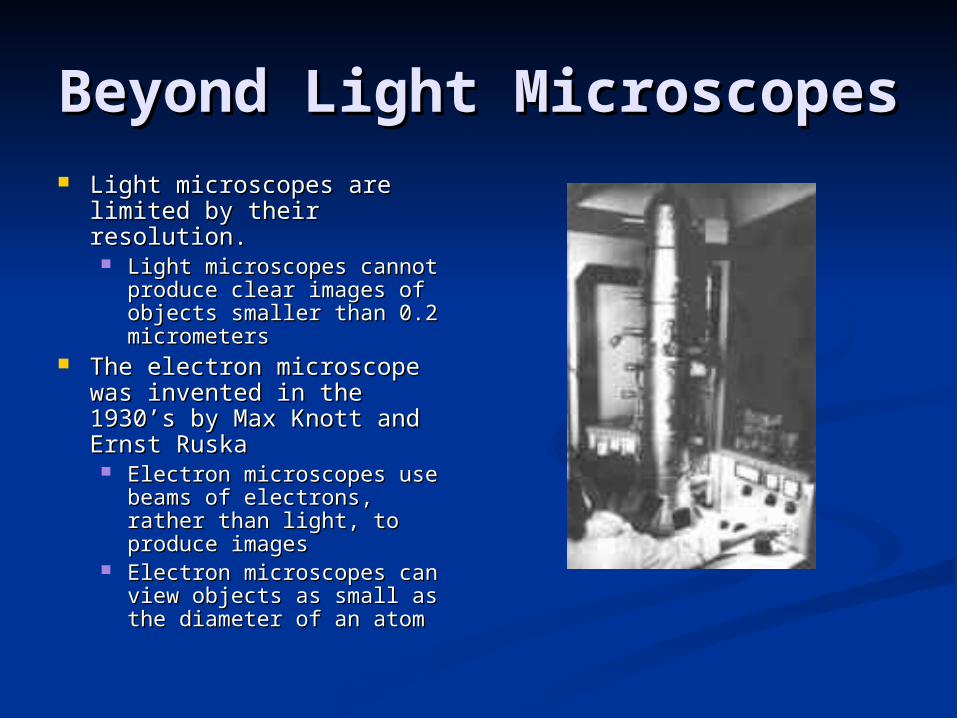
Beyond Light Beyond Light MicroscopesMicroscopes
Light microscopes are Light microscopes are limited by their resolution.limited by their resolution. Light microscopes cannot Light microscopes cannot
produce clear images of produce clear images of objects smaller than 0.2 objects smaller than 0.2 micrometersmicrometers
The electron microscope The electron microscope was invented in the 1930’s was invented in the 1930’s by Max Knott and Ernst by Max Knott and Ernst RuskaRuska Electron microscopes use Electron microscopes use
beams of electrons, rather beams of electrons, rather than light, to produce than light, to produce imagesimages
Electron microscopes can Electron microscopes can view objects as small as view objects as small as the diameter of an atomthe diameter of an atom


Types of Electron Types of Electron MicroscopesMicroscopes
Transmission electron microscopes Transmission electron microscopes (TEMs) pass a beam of electron (TEMs) pass a beam of electron through a thin specimenthrough a thin specimen
Scanning electron microscopes Scanning electron microscopes (SEMs) scan a beam of electrons (SEMs) scan a beam of electrons over the surface of a specimenover the surface of a specimen
Specimens from electron microscopy Specimens from electron microscopy must be preserved and dehydrated, must be preserved and dehydrated, so living cells cannot be viewedso living cells cannot be viewed

Images Produced by Images Produced by Electron MicroscopesElectron Microscopes
Cyanobacteria (TEM) Lactobacillus
(SEM)Campylobacter
(SEM) Deinococcus(SEM)
House ant Avian influenza virus Human eyelash Yeast

Using Microscopes to Using Microscopes to Visualize the Three Shapes Visualize the Three Shapes
of Bacteriaof Bacteria Cocci (round)Cocci (round) Bacilli (rod)Bacilli (rod) Spirilla (spiral)Spirilla (spiral)
Light microscope:Light microscope:Three shapes of bacteria
taken with an SEM
BacilliCocciSpirilla

ReferencesReferences
http://education.denniskunkel.com/http://education.denniskunkel.com/catalog/product_info.php?catalog/product_info.php?products_id=1123products_id=1123
http://micro.magnet.fsu.edu/http://micro.magnet.fsu.edu/ http://inventors.about.com/library/http://inventors.about.com/library/
inventors/blroberthooke.htminventors/blroberthooke.htm