Embed Size (px)
Citation preview

International Journal of
Molecular Sciences
Review
MicroRNA Biomarkers in IBD—DifferentialDiagnosis and Prediction of Colitis-Associated Cancer
Jaslin P. James 1,* , Lene Buhl Riis 1, Mikkel Malham 2,3 , Estrid Høgdall 1, Ebbe Langholz 4,5
and Boye S Nielsen 6
1 Department of Pathology, Herlev University Hospital, 2730 Herlev, Denmark;[email protected] (L.B.R.); [email protected] (E.H.)
2 The Pediatric Department, Copenhagen University Hospital, 2650 Hvidovre, Denmark;[email protected]
3 The Pediatric Department, Holbæk Sygehus, 4300 Holbæk, Denmark4 Gastroenheden D, Herlev University Hospital, 2730 Herlev, Denmark; [email protected] Institute for Clinical Medicine, University of Copenhagen, 2200 Copenhagen, Denmark6 Bioneer A/S, Hørsholm, Kogle Allé 2, 2970 Hørsholm, Denmark; [email protected]* Correspondence: [email protected]
Received: 24 September 2020; Accepted: 21 October 2020; Published: 24 October 2020�����������������
Abstract: Inflammatory bowel disease (IBD) includes Crohn’s disease (CD) and ulcerative colitis(UC). These are chronic autoimmune diseases of unknown etiology affecting the gastrointestinaltract. The IBD population includes a heterogeneous group of patients with varying disease coursesrequiring personalized treatment protocols. The complexity of the disease often delays the diagnosisand the initiation of appropriate treatments. In a subset of patients, IBD leads to colitis-associatedcancer (CAC). MicroRNAs are single-stranded regulatory noncoding RNAs of 18 to 22 nucleotideswith putative roles in the pathogenesis of IBD and colorectal cancer. They have been explored asbiomarkers and therapeutic targets. Both tissue-derived and circulating microRNAs have emerged aspromising biomarkers in the differential diagnosis and in the prognosis of disease severity of IBD aswell as predictive biomarkers in drug resistance. In addition, knowledge of the cellular localizationof differentially expressed microRNAs is a prerequisite for deciphering the biological role of theseimportant epigenetic regulators and the cellular localization may even contribute to an alternativerepertoire of biomarkers. In this review, we discuss findings based on RT-qPCR, microarray profiling,next generation sequencing and in situ hybridization of microRNA biomarkers identified in thecirculation and in tissue biopsies.
Keywords: biomarkers; circulating miRNA; colitis-associated cancer (CAC); Crohn’s disease (CD);inflammatory bowel disease (IBD); microRNA (miRNA); ulcerative colitis (UC)
1. Introduction
Inflammatory bowel disease (IBD) refers to Crohn’s disease (CD) and ulcerative colitis (UC). In UC,inflammation generally includes the rectum and extends towards the coecum and remains confinedto the colon. In contrast, in CD, inflammation can involve any part of the gastrointestinal tract (GI)from the oral cavity to the anus. Both CD and UC are associated with multiple pathogenic factors suchas environmental changes, the array of susceptibility gene variants, qualitatively and quantitativelyabnormal gut microbiota and broadly dysregulated immune response [1]. Although CD and UChave some common pathological and clinical characteristics, they have several different attributesthat imply that they are two distinct disease subtypes. In CD, fissuring ulceration and sub-mucosalfibrosis can be observed along with granulomatous inflammation. In UC, the inflammatory process
Int. J. Mol. Sci. 2020, 21, 7893; doi:10.3390/ijms21217893 www.mdpi.com/journal/ijms

Int. J. Mol. Sci. 2020, 21, 7893 2 of 19
always involves the rectum [2] and general histological findings include crypt distortion, infiltration oflymphocytes and granulocytes and chronic inflammation, usually confined to the lamina propria [3].The diagnosis of IBD is usually established by a collective assessment of clinical presentation andendoscopic, histopathological, radiographic and laboratory findings. A definitive diagnosis of IBDcannot be made without detailed endoscopic and histologic assessment [4]. However, a subset of IBDcases cannot be classified as either CD or UC but are categorized as IBD unclassified (IBDU). Molecularbiomarkers may support differential diagnosis of IBDU cases into CD or UC, or even be helpful indetermining if IBDU represents a unique IBD diagnostic entity.
IBD starts developing at a younger age, including in infants [5], and is often characterized bya considerable diagnostic and therapeutic challenge because of the disease’s clinical features andassociated complications. The prevalence of IBD in the Western world is projected to be up to 0.5%of the overall population [6]. In Denmark, where one of the highest annual incidence rates of IBD inEurope is seen, the incidence has been increasing over the last three decades [7]. In 2013, the incidencewas 9.1 per 100,000 persons and 18.6 per 100,000 persons for CD and UC, respectively [8]. Since theturn of the 21st century, IBD has become a global disease with accelerating incidence rates alsoin developing countries whose societies have adopted a western diet and lifestyle. Although theincidence rate has become steady in western countries, the burden remains high, as prevalence exceeds0.3%. The chronical inflammatory condition in the affected colon of IBD patients has been linked todevelopment of neoplastic lesions in the colon. Several studies have shown a higher incidence ofcolorectal cancer (CRC) in IBD patients [9–11]. No biomarkers exist for the identification of IBD patientsat risk of developing colitis-associated cancer (CAC), strongly advocating for more translationalresearch in this field.
In this review, we give an overview of microRNAs (miRNAs) as candidate biomarkers in the IBDdiagnostic assessment. Changes in miRNA levels are associated with disease development and can bemeasured both within the diseased tissue and in the circulation by a variety of molecular methods.MiRNAs have been found to be well conserved in archived tissue specimens, enabling retrospectiveanalyses of clinical sample cohorts.
2. MicroRNA—An Introduction
MiRNAs play a central role in the regulation of several immune-mediated disorders includingIBD [12–14]. MiRNAs are a group of small noncoding RNAs, approximately 18–22 nucleotides [15]which are found conserved across species. Their discovery was first described first in 1993 inCaenorhabditis elegans [16]. MiRNAs are transcribed as primary transcripts by RNA polymerase,processed into a precursor miRNA by the RNase III enzyme, Drosha, and exported from the nucleus tothe cytoplasm. The precursor miRNA is cleaved by the RNase III enzyme, Dicer, into its mature form,which becomes stably incorporated into an RNA induced silencing complex (RISC). The miRNA guidesthe binding of the RNA-induced silencing complex to complementary sequences in the 3′-untranslatedregions (UTR) of target mRNA molecules, resulting in either mRNA degradation or translationalinhibition [17]. During stages of miRNA biogenesis, several factors can influence the development ofthe mature miRNA. These include regulation of transcription, cleavage of the stem loop structuresby Drosha and Dicer enzymes, and editing as well as regulation of miRNA turnover. Each of thesemechanisms acts as part of a signaling network that modulates gene expression in response to cellularor environmental changes.
MiRNA expression has been shown to be of importance in a wide variety of human diseases suchas cancer, autoimmune, cardiovascular, and neurodegenerative diseases [14,18–24]. The miRNAs notonly circulate in the human peripheral blood in a stable form, they are also present in other body fluidssuch as urine, saliva, milk, cerebrospinal fluid, and feces [25–28]. The miRNAs are engaged in diseaseorigin and development, and some are pathology-specific [29], thus, changes in miRNA expressionprofiles have been addressed for applications in early detection as well as prognostics, diagnosticclassification and drug response prediction.

Int. J. Mol. Sci. 2020, 21, 7893 3 of 19
3. MiRNAs in IBD
In IBD, miRNAs have been found to be involved in pathogenesis and have been identified as bothdiagnostic biomarkers and therapeutic targets [30]. Most of the recent research in the IBD field hasmeasured levels of circulating miRNAs in body fluids such as blood or feces, and in homogenized tissuebiopsies using techniques like microarray profiling, RT-qPCR, and NGS [27,31–34]. Studies have alsoperformed tissue miRNA expression analysis using in situ hybridization (ISH) methods [35–37]. ISHmethods for expression analyses of miRNAs can determine the cellular origin of miRNA expressionand can offer insight into the biology of the disease mechanisms involved. Local expression levelsof miRNAs can greatly vary from those of circulating miRNAs, e.g., due to contribution of miRNAsfrom circulating cells. Esquela-Kerscher and Slack [38] proposed that tumor cells release miRNAs intothe neighboring microenvironment and enter circulation during angiogenesis. Some studies suggestthat this likely occurs through exosomal release from cells [39,40]. Changes in the levels of circulatingmiRNA may occur due to other inflammatory reactions or the host immune response rather than onlydue to the intrinsic changes within the lesion [41]. Thus, as discussed further below, it is not surprisingthat miRNAs analyzed in tissue biopsies poorly correlate with those found in the circulation [42].
There is an increasing interest in exploring epigenetic mechanisms in common diseases,with notable progress in characterizing the contribution of miRNAs [43]. In their 2008 study, Wu et al.found that miRNAs regulate colonic epithelial cell-derived chemokine expression and were the first torelate miRNAs to IBD [44]. The field of miRNA research has grown rapidly after their discovery inhuman disease biology including in IBD [43]. We have listed a series of IBD-related miRNA studiesfrom recent years in Table 1, with a focus on sample type and quantitative method. MiR-21, miR-155,and miR-31 have repeatedly been identified and seem to be the most studied miRNAs related toIBD [15,19,35,45–48]. MiR-21 is possibly the most intriguing miRNA involved in IBD, with associationsbetween miR-21 and IBD being replicated in several studies, as well as functional relevance reported inmouse models of IBD [19,23,24,30,35,49]. Each miRNA can potentially target hundreds of mRNAsresulting in mRNA destabilization and/or inhibition of translation, however, restricted to a specificcellular context, the number of relevant targetable transcripts is probably quite low.
MiRNAs regulate important cellular functions such as cell differentiation and proliferation andsignal transduction and apoptosis and exhibit highly specific regulated patterns of gene expression [15].In autoimmune diseases, miRNAs can act through interference with inflammatory signaling pathways,such as the nuclear transcription factor kappa B (NF-κB) pathway, IL23/IL23R pathway, and IL-6/STAT3pathway [50–54]. Studying the RhoB pathway of cell adhesion in UC mucosa and cultured coloncancer cells, Yang et al. [36] examined the role of miR-21 in regulation of intestinal epithelial barrierfunction and found that miR-21 induced the degradation of RhoB mRNA, reduction in RhoB protein,causing loss of tight junctions in intestinal epithelial cells. Tian et al. showed miR-31 to be highlyexpressed in tissues from IBD patients, and miR-31 reduced the inflammatory response in the DextranSodium Sulphate (DSS)-induced colitis mouse model (see below), by preventing the expression ofinflammatory cytokine receptors such as IL7R and IL17RA and signaling proteins such as GP130 [55].Another study based on the DSS model showed that miR-155 directly binds to SHIP-1 mRNA andcauses a significant decrease in SHIP-1 levels, which regulate cell membrane trafficking, and therebycontribute to the pathogenesis of colitis [56]. Taken together, these examples indicate the complexity ofhow miRNAs may act through signaling pathways in the biological settings of IBD.
Studies of circulating miRNAs have shown that miRNAs are potential candidates as biomarkersfor diagnosing IBD and various other diseases [57–61]. The high stability of miRNAs in the body fluidsand the ability to obtain rapid and accurate quantitative estimates are some merits of using circulatingmiRNAs as biomarkers in IBD [28]. MiRNAs are not only interesting tools for diagnosis, but also forpotential future therapeutic applications by miRNA mimics or miRNA antagonists [62,63].

Int. J. Mol. Sci. 2020, 21, 7893 4 of 19
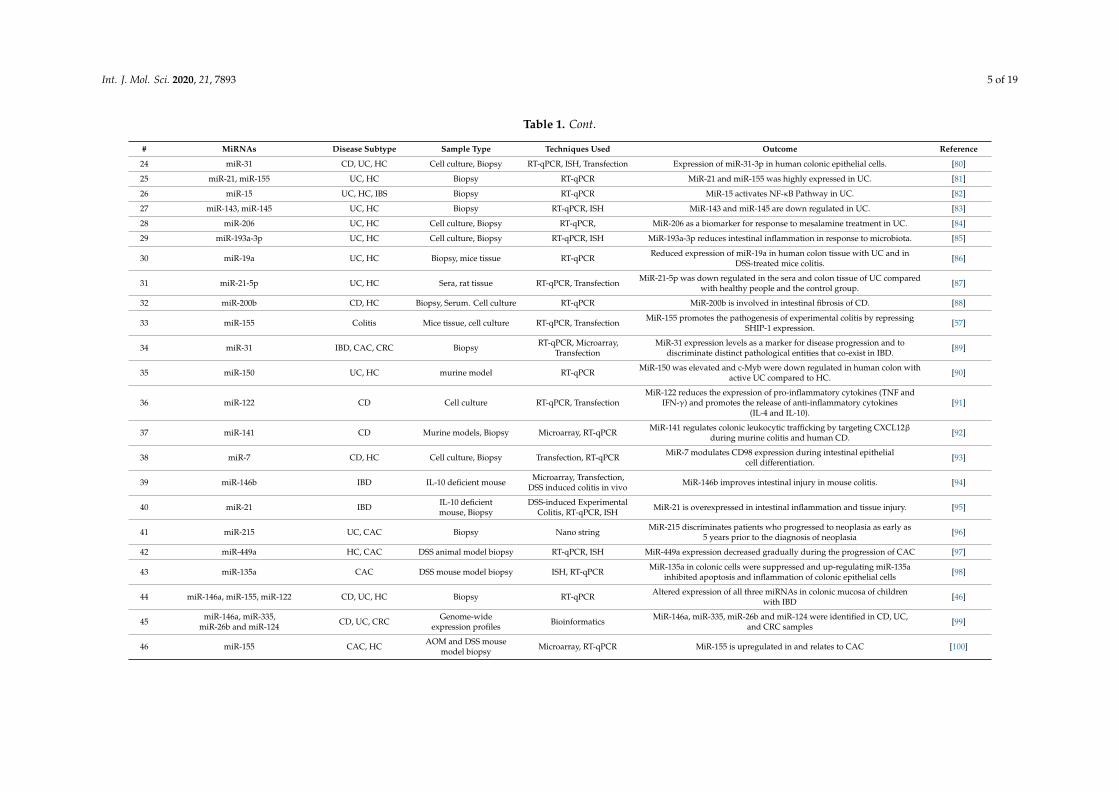
Table 1. A summary of studies on microRNA research in inflammatory bowel disease (IBD). CD: Crohn’s disease, UC: Ulcerative colitis, HC: Healthy controls,RT-qPCR: Quantitative real time polymerase chain reaction, Biopsy: colon tissue biopsy, ISH: In situ hybridization, QISH: Quantitative in-situ hybridization, PBMC:Peripheral blood mononuclear cells, DSS: Dextran sodium sulphate, AOM: Azoxymethane, TNF: Tumor necrosis factor alpha.
# MiRNAs Disease Subtype Sample Type Techniques Used Outcome Reference
1
miR-16, miR-29a, miR-199a-5p,miR-363-3p, miR-340, miR-532-3p,
miRplus-1271, miR-140-3p, miR-127-3p,miR-196b, miR-877, miR-150
CD, UD, HC Serum, Biopsy RT-qPCR, Microarray Mixed outcomes [42]
2 miR-223-3p, miR-31-5p CD, HC Biopsy Nano string Mir-223-3p expression showed age and sex effects and miR-31-5p expressionwas driven by location [45]
3 miR-29b CD Fibroblasts RT-qPCR MCL-1 is modulated in CD fibrosis by miR-29b via IL-6 and IL-8 [64]
4 miR-141, miR-200a, miR-200b, miR-200c UC, CD Biopsy RT-qPCRAll investigated miRNAs were significantly down regulated in CD, and 3 of
them were downregulated in UC in comparison to the normal or the leastaffected mucosa.
[65]
5 miR-141 UC, HC Biopsy Microarray, RT-qPCR MiR-141 plays a role in the bowel inflammation of individuals with active UCvia down regulation of CXCL5 expression. [66]
6 miR-124 UC, HC Biopsy RT-qPCRMiR-124 regulates the expression of STAT3. Reduced levels of miR-124 incolon tissues of children with active UC appear to increase expression and
activity of STAT3.[67]
7 miR-19b CD, HC Biopsy, Cell culture RT-qPCR, ISH MiR-19b suppresses the inflammation and prevents the pathogenesis of CD. [68]
8 miR-590-5p CD, HC Human and mice tissues RT-qPCR Decreased miR-590-5p levels in CD. [69]
9 miR-122 CD, HC Biopsy RT-qPCR, Sequencing Significant increase of miR-122 expression in cells treated with 5′-AZA. [70]
10 miR-10a CD, UC, HC Biopsy RT-qPCR Dendritic cell activation and Th1/Th17 cell immune responses were inhibitedvia miR-10a in IBD. [71]
11 miR-192 CD, UC, HC Biopsy RT-qPCR, Microarray, ISH MiR-192 with decreased expression in active UC. [44]
12 miR-15a CD, UC, HC Biopsy, Cell cultures RT-qPCR MiR-15a negatively regulates epithelial junctions through Cdc42 inCaco-2 cells [72]
13 miR-146a, miR-155 CD Biopsy RT-qPCR MiR-146a and -155 shows increased duodenal expression in pediatric CD. [73]
14 miR-146b-5p CD, UC, HC Serum RT-qPCR Higher expression of serum miR-146b-5p in patients with CD and UC thanin HC. [74]
15 miR-425 CD, UC, HC Biopsy, PBMC RT-qPCR Increased expression of miR-425 in IBD. [75]
16 miR-301a IBD PBMC, Biopsy RT-qPCR MiR-301a promotes intestinal mucosal inflammation via induction of IL-17aand TNF in IBD. [76]
17 miR-125b, miR-155,miR-223 and miR-138 UC Biopsy RT-qPCR, Microarray Differential expression of miR-223, miR-125b, miR-138, and miR-155 in the
inflamed mucosa compared to non-inflamed mucosa and controls. [48]
18 miR-16, miR-21, miR-155, and miR-223 CD, UC, HC Serum, Feces RT-qPCR Differential expression of miR-16, miR-155, miR-21, and miR-223 in IBD. [47]
19 miR-21 UC, HC Biopsy RT-qPCR, ISH Over expression of miR-21 in UC. [36]
20 miR-133a IBD Mice Tissue RT-qPCR MiR-133a-UCP2 pathway participates in IBD by altering downstreaminflammation, oxidative stress, and markers of energy metabolism. [77]
21 miR-20b, miR-98, miR-125b-1, let-7e CD, UC, HC Biopsy RT-qPCR, Microarray MiR-20b, miR-98, miR-125b-1, and let-7e are deregulated in patients with UC. [78]
22 miR-155 CD, HC PBMC RT-qPCR, Transfection MiR-155 regulates IL-10-producing CD24 CD27+ B Cells. [79]
23 miR-21, miR-126 CD, UC, HC Biopsy RT-qPCR, qISH Endothelial expression of miR-126 are increased in UC. MiR-21 is expressed insubsets of monocytes/macrophages and T cells. [35]
Int. J. Mol. Sci. 2020, 21, 7893 5 of 19
Table 1. Cont.
# MiRNAs Disease Subtype Sample Type Techniques Used Outcome Reference
24 miR-31 CD, UC, HC Cell culture, Biopsy RT-qPCR, ISH, Transfection Expression of miR-31-3p in human colonic epithelial cells. [80]
25 miR-21, miR-155 UC, HC Biopsy RT-qPCR MiR-21 and miR-155 was highly expressed in UC. [81]
26 miR-15 UC, HC, IBS Biopsy RT-qPCR MiR-15 activates NF-κB Pathway in UC. [82]
27 miR-143, miR-145 UC, HC Biopsy RT-qPCR, ISH MiR-143 and miR-145 are down regulated in UC. [83]
28 miR-206 UC, HC Cell culture, Biopsy RT-qPCR, MiR-206 as a biomarker for response to mesalamine treatment in UC. [84]
29 miR-193a-3p UC, HC Cell culture, Biopsy RT-qPCR, ISH MiR-193a-3p reduces intestinal inflammation in response to microbiota. [85]
30 miR-19a UC, HC Biopsy, mice tissue RT-qPCR Reduced expression of miR-19a in human colon tissue with UC and inDSS-treated mice colitis. [86]
31 miR-21-5p UC, HC Sera, rat tissue RT-qPCR, Transfection MiR-21-5p was down regulated in the sera and colon tissue of UC comparedwith healthy people and the control group. [87]
32 miR-200b CD, HC Biopsy, Serum. Cell culture RT-qPCR MiR-200b is involved in intestinal fibrosis of CD. [88]
33 miR-155 Colitis Mice tissue, cell culture RT-qPCR, Transfection MiR-155 promotes the pathogenesis of experimental colitis by repressingSHIP-1 expression. [57]
34 miR-31 IBD, CAC, CRC Biopsy RT-qPCR, Microarray,Transfection
MiR-31 expression levels as a marker for disease progression and todiscriminate distinct pathological entities that co-exist in IBD. [89]
35 miR-150 UC, HC murine model RT-qPCR MiR-150 was elevated and c-Myb were down regulated in human colon withactive UC compared to HC. [90]
36 miR-122 CD Cell culture RT-qPCR, TransfectionMiR-122 reduces the expression of pro-inflammatory cytokines (TNF and
IFN-γ) and promotes the release of anti-inflammatory cytokines(IL-4 and IL-10).
[91]
37 miR-141 CD Murine models, Biopsy Microarray, RT-qPCR MiR-141 regulates colonic leukocytic trafficking by targeting CXCL12βduring murine colitis and human CD. [92]
38 miR-7 CD, HC Cell culture, Biopsy Transfection, RT-qPCR MiR-7 modulates CD98 expression during intestinal epithelialcell differentiation. [93]
39 miR-146b IBD IL-10 deficient mouse Microarray, Transfection,DSS induced colitis in vivo MiR-146b improves intestinal injury in mouse colitis. [94]
40 miR-21 IBD IL-10 deficientmouse, Biopsy
DSS-induced ExperimentalColitis, RT-qPCR, ISH MiR-21 is overexpressed in intestinal inflammation and tissue injury. [95]
41 miR-215 UC, CAC Biopsy Nano string MiR-215 discriminates patients who progressed to neoplasia as early as5 years prior to the diagnosis of neoplasia [96]
42 miR-449a HC, CAC DSS animal model biopsy RT-qPCR, ISH MiR-449a expression decreased gradually during the progression of CAC [97]
43 miR-135a CAC DSS mouse model biopsy ISH, RT-qPCR MiR-135a in colonic cells were suppressed and up-regulating miR-135ainhibited apoptosis and inflammation of colonic epithelial cells [98]
44 miR-146a, miR-155, miR-122 CD, UC, HC Biopsy RT-qPCR Altered expression of all three miRNAs in colonic mucosa of childrenwith IBD [46]
45 miR-146a, miR-335,miR-26b and miR-124 CD, UC, CRC Genome-wide
expression profiles Bioinformatics MiR-146a, miR-335, miR-26b and miR-124 were identified in CD, UC,and CRC samples [99]
46 miR-155 CAC, HC AOM and DSS mousemodel biopsy Microarray, RT-qPCR MiR-155 is upregulated in and relates to CAC [100]

Int. J. Mol. Sci. 2020, 21, 7893 6 of 19
To study the pathogenesis and intricacy of IBD, the advancement of a variety of animal modelshas provided important information. The most extensively used mouse model of colitis utilizes DSS,a so-called chemical colitogen with anticoagulant properties, to stimulate epithelial damage. The DSScolitis model is simple and easy to administer. Acute and persistent colitis is achieved by alteringthe concentration of DSS and the frequency of administration [101]. A genetically engineered in vivomodel that has been widely used to examine IBD etiology is the interleukin-10 (IL-10)-deficient mousemodel [102]. IL-10 is an anti-inflammatory cytokine. Mutated IL-10 signaling systems shows earlyand aggressive expansion of systemic inflammation in IBD. IL-10 knockout mice develop spontaneouscolitis and CAC [103]. Nata et al. [94] performed miRNA microarray profiling on IL-10-deficient miceand identified that several miRNAs were upregulated, including miR-146b that, through further studies,was found to contribute to increased intestinal inflammation by upregulating NF-κB. Shi et al. [95]showed that knockout of miR-21 in mice improved the survival rate in DSS-induced fatal colitis viaprotecting against inflammation and tissue injury. Hence, it was suggested that impaired expressionof miR-21 in gut may block the onset or progression of IBD. Other animal models used in IBDresearch include genetically engineered mice, congenic mouse strains, chemically induced models,and cell-transfer models [104]. Most of the studies investigating miRNA expression in IBD have usedhigh-throughput methods such as a microarray combined with RT-qPCR as a validation method forprioritized miRNAs.
4. MiRNA Biomarkers for IBD Diagnosis
The diagnostic assessment of IBD can be challenging; particularly, discriminating CD from UCcan be a diagnostic encounter in cases where the inflammatory lesions are limited to the colon. It isestimated that 10–15% of IBD cases are categorized as IBDU [105]. Although many IBDU patientsare eventually reclassified as either CD or UC, approximately 75% of the IBDU cases maintain thediagnosis of IBDU, suggesting that most of the IBDU patients have a distinct diagnostic entity of atrue overlap phenotype between large bowel CD and typical UC [106,107]. Recently, a study thatexamined colon biopsies from patients with IBD suggested miR-19a, miR-21, miR-31, miR-146a andmiR-375 as a biomarker profile for discriminating CD and UC [108]. A Study by Peck et al. used anext-generation sequencing–based approach and found that a combination of miR-31-5p, miR-215,miR-223-3p, miR-196b-5p and miR-203 could stratify patients with CD according to disease behaviorindependent of the effect of inflammation [109]. The lack of reproducibility in miRNA profilinganalyses of IBD samples in independent studies could be due to the technology applied, as well asthe variation in control groups, disease activity and data normalization. It was recently reportedthat miR-21 is a potential diagnostic marker for discriminating CD from UC, as both RT-qPCR andquantitative ISH (qISH) identified significantly higher levels in UC compared with CD [35]. The authorssuggested that miR-21 is not just an unspecific marker of inflammation, but that miR-21 is specificto the immunopathological process of UC. miR-21 ISH analyses reveal complex expression patterns,where the miR-21 staining is identified mainly in cells of the inflamed lamina propria as well as insubsets of epithelial cells of partly damaged crypt structures (example in Figure 1).

Int. J. Mol. Sci. 2020, 21, 7893 7 of 19
Int. J. Mol. Sci. 2020, 21, x FOR PEER REVIEW 8 of 20
Figure 1. MiR-21 in situ hybridization in ulcerative colitis. The example shows the inflamed colon mucosa with transversally cut crypts and the lamina propria (indicated by LP). The miR-21 ISH signal is represented by the blue stain and is seen in inflammatory cells located in the lamina propria in and some of the epithelial cells (arrows) in some collapsed crypts. Nuclear Fast Red was used in counterstaining.
5. MiRNAs and CAC
Chronic inflammation is linked to the development of a variety of cancers such as CRC, pancreatic, breast, and skin cancer [110,111] and is a key hallmark of cancer [112]. Local chronic inflammation in the colon, typically caused by an unbalance in the regulation of the immune response, may damage the epithelial barrier, which induces self-sustained inflammation linked to continued microbial influx or increased levels of pro-inflammatory cytokines like tumor necrosis factor alpha (TNF) and IL-1β [110,113,114]. Increased levels of oxidants in inflamed tissue cause cell death, or more deliberately, mutations in epithelial cells that, in turn, can initiate neoplastic growth. The persisting inflammation develops into and probably shapes the tumor microenvironment that is inherent to most solid tumors with their additional presence of blood vessels and fibroblastic cells.
Patients with extensive IBD or diagnosed with IBD in childhood [115], have a shorter life expectancy that may be related to the higher risk of CRC [116–118]. CAC represents a type of CRC in which the IBD paved the way for the cancer, probably through mutations in K-RAS and the adenomatous polyposis coli (APC) gene [119]. The risk of CAC may be further increased in untreated IBD patients [120]. Mucosal mapping studies indicate that the chronically inflamed colonic mucosa of patients with IBD undergoes a “field change” in cancer-associated molecular alterations before there is histologic evidence of epithelial dysplasia [121,122], which is one the initial morphological changes in the stepwise progression to CRC [123–125].
MiRNAs are believed to take part in the inflammation in IBD and to be implicated in the process from inflammation to CRC [126]. Despite the fact that CD and UC can affect the entire colon, Ranjha et al. [127] found that CRC in UC patients developed primarily in the rectosigmoid areas of the colon, whereas other parts, such as the ascending colon, showed less frequent development of CRC. Analyzing tissue from rectosigmoid and ascending colon, the authors found differences in the miRNA expression patterns, and suggested that the local miRNA profile could contribute to the development of CRC.
MiRNAs likely play both oncogenic and tumor-suppressive roles in the carcinogenesis and progression of CRC by regulating the expression of numerous cancer-related genes. The role of the inflammatory burden has also been studied in animal models and indicates that both the initiation and the progression of colonic neoplasia can be aggravated or accelerated by the inflammatory conditions [30,126,128–130]. The DSS-induced colitis model has been used to study the role of multiple miRNAs in IBD and CAC, including miR-21, miR-155, and miR-301a, which will be addressed briefly in the following.
Figure 1. MiR-21 in situ hybridization in ulcerative colitis. The example shows the inflamed colonmucosa with transversally cut crypts and the lamina propria (indicated by LP). The miR-21 ISH signal isrepresented by the blue stain and is seen in inflammatory cells located in the lamina propria in and someof the epithelial cells (arrows) in some collapsed crypts. Nuclear Fast Red was used in counterstaining.
5. MiRNAs and CAC
Chronic inflammation is linked to the development of a variety of cancers such as CRC, pancreatic,breast, and skin cancer [110,111] and is a key hallmark of cancer [112]. Local chronic inflammation inthe colon, typically caused by an unbalance in the regulation of the immune response, may damagethe epithelial barrier, which induces self-sustained inflammation linked to continued microbialinflux or increased levels of pro-inflammatory cytokines like tumor necrosis factor alpha (TNF) andIL-1β [110,113,114]. Increased levels of oxidants in inflamed tissue cause cell death, or more deliberately,mutations in epithelial cells that, in turn, can initiate neoplastic growth. The persisting inflammationdevelops into and probably shapes the tumor microenvironment that is inherent to most solid tumorswith their additional presence of blood vessels and fibroblastic cells.
Patients with extensive IBD or diagnosed with IBD in childhood [115], have a shorter lifeexpectancy that may be related to the higher risk of CRC [116–118]. CAC represents a type of CRCin which the IBD paved the way for the cancer, probably through mutations in K-RAS and theadenomatous polyposis coli (APC) gene [119]. The risk of CAC may be further increased in untreatedIBD patients [120]. Mucosal mapping studies indicate that the chronically inflamed colonic mucosa ofpatients with IBD undergoes a “field change” in cancer-associated molecular alterations before there ishistologic evidence of epithelial dysplasia [121,122], which is one the initial morphological changes inthe stepwise progression to CRC [123–125].
MiRNAs are believed to take part in the inflammation in IBD and to be implicated in theprocess from inflammation to CRC [126]. Despite the fact that CD and UC can affect the entire colon,Ranjha et al. [127] found that CRC in UC patients developed primarily in the rectosigmoid areas of thecolon, whereas other parts, such as the ascending colon, showed less frequent development of CRC.Analyzing tissue from rectosigmoid and ascending colon, the authors found differences in the miRNAexpression patterns, and suggested that the local miRNA profile could contribute to the developmentof CRC.
MiRNAs likely play both oncogenic and tumor-suppressive roles in the carcinogenesis andprogression of CRC by regulating the expression of numerous cancer-related genes. The role of theinflammatory burden has also been studied in animal models and indicates that both the initiationand the progression of colonic neoplasia can be aggravated or accelerated by the inflammatoryconditions [30,126,128–130]. The DSS-induced colitis model has been used to study the role of multiplemiRNAs in IBD and CAC, including miR-21, miR-155, and miR-301a, which will be addressed brieflyin the following.

Int. J. Mol. Sci. 2020, 21, 7893 8 of 19
MiR-21 is one of the most prevalent miRNAs in CRC and other cancer types [131,132],and the increased expression levels in CRC are associated with poor prognosis [24,133]. MiR-21acts on tumor-suppressor genes, like PTEN and PDCD4 [134–136], and is thus categorized as anoncomiR [137]. Since miR-21 is upregulated in IBD [35,44,47], and miR-21 reduction in the DSSmodel lowers inflammation in DSS-induced colitis [95], it is tempting to speculate that miR-21is a key facilitator of CAC. In support of this hypothesis, a study of human IBD, found thatthe tumor-suppressor-programmed cell death 4 (PDCD4) was downregulated, while miR-21 wasupregulated [134]. Suppression of PDCD4 and NF-κB activation was found along with reducedlevels of pro-inflammatory TNF [134,135]. In addition, epithelial miR-21 upregulation in UC wasreported to increase intestinal permeability, which is believed to be a key pathophysiological step inthe development of IBD [138].
MiR-155 is upregulated in both UC and CD patients compared to healthy controls [48,139–141]and is upregulated in both tissue and blood from CRC patients, and is furthermore an indicator ofpoor prognosis [142,143]. MiR-155 promotes intestinal inflammation in UC and CD, probably via avariety of inflammation-related pathways [46,56,73,79,81,139]. In a recent study by Liu et al. [144,145],it was shown that miR-155 mediates intestinal barrier dysfunction in DSS-induced mice colitis throughtargeting the HIF-1α/TFF-3 axis. Paraskevi et al. [140] found that miR-155 is the most highly expressedUC-associated miRNA in blood samples, however, in the study by Schönauen [47], the authors did notfind increased miR-155 levels in the blood from IBD patients, suggesting that more studies are neededto determine whether miR-155 is a putative blood-related biomarker.
MiR-301a is upregulated in both blood and tissue from IBD and CRC patients [76,141,146].He et al. [76,129] found increased levels of miR-301a in peripheral blood monocytes and in the mucosafrom IBD patients and in mice after administration of DSS. Using the DSS-induced IBD model inmice with an inactivated miR-301a, miR-301a was found to reduce the inflammation through thesuppression of BTG anti-proliferation factor 1 (BTG1) and to reduce the development of CAC [129].Thus, miR-301a should be investigated in future studies to establish possible use as a clinically relevantdiagnostic biomarker in IBD and for prediction of CAC.
6. MiRNAs as Predictive Biomarkers and in IBD Treatment
The goal of the treatment of IBD patients is to obtain remission and mucosal healing, and therebylower surgery rates. The classical therapies include corticosteroids, thiopurines, and amino salicylates(5-ASA), which have been in use for decades. 5-ASA has minor side effects [147,148] and it is veryeffective for treating mild to moderate UC patients, but not recommended for treatment of patientswith CD [149]. The last-line medical treatment in IBD is administration of biologics targeting keyelements in the inflammatory process. Anti-TNF therapies include TNF inhibitors that antagonizethe pro-inflammatory cytokine TNF [150]. The use of anti-TNF therapy has improved long-termoutcomes for IBD patients [149,151]. Even though TNF inhibitors have improved the overall conditionsfor a large group of IBD patients, approximately 30% of patients fail to respond to TNF inhibitors(primary non-responders), and up to 50% of the patients who initially benefited from treatment withTNF inhibitors lose the response over time (secondary non-responders) [152,153]. Thus, identifyingpredictors of responders/non-responders and choosing a treatment strategy according to biomarkerprofiles could improve overall IBD disease management. Interestingly, Morilla et al. [154], found thatnine miRNAs, together with five clinical factors correlated with response to treatment of IBD patients,and that neural-network-developed algorithms based on certain miRNA levels identified respondersto the anti-TNF antibody therapy, infliximab, vs. non-responders.
Currently used therapies in IBD also include Ustekinumab, Vedolizumab and Tofacitinib.Ustekinumab is a monoclonal antibody against IL-12 and IL-23, which is used in patients withmoderate to severe CD who are resistant to anti-TNF treatment [155]. Considering the efficacy ofustekinumab, it is possible to extrapolate the efficacy of miR-29 mimicry as a mechanism to reduceIL-23 levels [12]. With respect to potential secondary target effects, miR-29c has been described

Int. J. Mol. Sci. 2020, 21, 7893 9 of 19
as a tumor-suppressor in liver cancer [156]. Vedolizumab binds specifically to α4β7-integrin onT-helper lymphocytes. Blocking the α4β7-integrin results in anti-inflammatory activity that is causedby the inhibition of leukocyte adhesion to endothelial cells, which consequently reduces leukocyterecruitment to affected tissue [157]. Previous studies have suggested a similar effect of miRNAs inthe posttranscriptional regulation of leukocyte trafficking [158]. Harris et al. [158] described howendogenous miR-126 inhibits leukocyte adherence through the regulation of an intercellular adhesionmolecule expressed by endothelial cells (VCAM-1). Tofacitinib is a janus kinase (JAK) inhibitor,approved for treating moderate-to-severely active UC patients who have deteriorated disease anddid not improve after conventional or antibody-based therapies [159,160]. Pathak et al. [51] identifiedSOCS1, a potent molecular switch that tunes the JAK pathway that is also a direct target of miR-155.
In general, miRNA-based therapies comprise two fundamental strategies: miRNA antagonismand mimicry [161–164]. Physiologic miRNA over-expression resulting in pathologically reduced targetgene expression can be hindered by using miRNA antagonists, while reduced miRNA expressionresulting in enhanced target function can be restored by utilizing miRNA mimics [12]. A study byLu et al. [56] reported that a so-called antagomir towards miR-155 alleviated DSS-induced intestinalinflammation in mice, and the authors propose that anti-miR-155 could be a promising candidate fora novel IBD therapy. Jin et al. conducted a study on miR-133a and its target UCP2 (mitochondrialuncoupling protein 2) using the DSS-induced IBD mouse model [77]. miR-133a levels were found tobe decreased upon DSS treatment, and by introducing a miR-133a mimic, the DSS-induced IBD wasalleviated, suggesting that miRNA mimics could also function as therapy in IBD [77].
7. Circulating miRNAs vs. Tissue miRNAs
It is of importance to determine whether miRNA dysregulation in the circulation reflects similarchanges in the lesion. The detection and quantification of circulating miRNAs and the interpretationof their impending role as novel non-invasive biomarkers could be very beneficial in the diagnosisand treatment of IBD. As mentioned above, miRNAs can be detected in distinct body fluids such assaliva, plasma or urine [32]. Current diagnostic and predictive findings in IBD on miRNA expressionprofiling have mainly focused on the assessment of miRNAs in blood. Even though blood samplescan be relatively easily obtained from IBD patients, miRNA measurement in blood samples, as withother biological samples, comes with some inherent obstacles, such as sample procurement, storage,measurement platform and normalization of the acquired data. Circulating miRNAs may derivefrom both the diseased tissue and by leakage from the normal vascular network and circulating cells.Obtaining tissue samples, on the other hand, requires an invasive procedure, where small biopsiesfrom the affected part of the bowel are obtained during endoscopy. The tissue samples can be eitherfrozen or fixed in formalin and paraffin-embedded (FFPE) for histological examination. MiRNAs canbe isolated from both fresh-frozen and FFPE tissue samples. Normalization of miRNA data fromboth blood and tissue samples is an important step for data interpretation in the comparison betweenpatients, and between different study cohorts. MiRNA expression levels measured in tissue sampleswill have been derived from cells in the normal tissue and from activated cells in the lesion. To be ableto find the same miRNAs in the tissue as in the circulation would require substantial expression inthe lesion and/or for the background level in the circulation to be low. Thus, it may not be surprisingthat the study by Iborra et al. [42] of tissue biopsies and peripheral blood showed that none of theserum miRNAs corresponded with tissue miRNAs in the CD and UC patients. Feces samples representanother liquid biopsy that is relevant in relation to IBD and may be better linked to expression levels inthe diseased mucosa than to the levels in the blood circulation. Schönauen et al. [47] analyzed bothserum and fecal miRNAs in IBD and found increased levels of miR-16, miR-21, and miR-223 in bothsera and feces from the IBD patients compared to controls. In addition, the authors found that fecallevels, but not sera levels, of miR-16 and miR-223 correlated with clinical parameters, like C-reactiveprotein and calprotectin. Thus, fecal samples seem to be a promising alternative to blood for miRNAprofiling in IBD.

Int. J. Mol. Sci. 2020, 21, 7893 10 of 19
As seen in Table 1, most miRNA studies have used high-throughput methods such as RT-qPCRand microarray for miRNA analysis in IBD. It is important to note that these techniques requirehomogenization of the tissue to isolate the miRNAs. Homogenization of the tissue will degrade thespatial arrangement and, hence, will give an overview of the miRNA expression at the tissue level.ISH using Locked Nucleic Acid (LNA) probes is a method that allows detection of miRNAs in tissuesections [165]. Detection of miRNAs at the cellular level determines the cellular origin of expression andcan provide evidence on expression levels in different cell populations and tissue compartments [35].More knowledge about the cellular localization of miRNAs in the framework of IBD is needed asthis will provide a vital link between the growing amounts of miRNA biomarkers discovered inIBD and functional studies identifying various miRNA target genes. Thorlacius-Ussing et al. usedquantitative ISH on IBD tissue samples and showed that miR-126 levels are increased in UC andexpressed in endothelial cells and miR-21 is expressed in subsets of monocytes/macrophages and Tcells [35]. As also suggested from Figure 1, ISH data provide information of contextual expression inthe tissue, as exemplified by focal upregulation in certain tissue compartments. Simple histologicalanalysis from ISH analysis if IBD tissue can often determine if a miRNA is expressed in the epithelialor stromal (lamina propria) compartment. Nielsen and Holmstrøm presented a method to combinemiRNA ISH using LNA-containing probes with immunohistochemical detection of cell-specific proteinmarkers in order to better characterize the miRNA’s cellular characteristics [166]. This approach couldalso be used to monitor parallel downregulation of the specific downstream target protein. MiRNAISH is a powerful tool when also combined with parallel characterization of the cell population inquestion and of mRNAs using combined staining methods [23]. Thus, for better understanding ofthe role of miRNAs in IBD and CAC, miRNA ISH analyses will be a helpful tool both for validatingexpression and for deciphering the related inflammatory molecular context.
8. Concluding Remarks
MiRNAs in IBD research started with the extensive pioneering work by Wu et al. in 2008 [44],who found altered expression of several miRNAs in tissue from IBD patients. Since then, there havebeen tremendous advancements in the field both regarding mechanistic studies and studies evaluatingthe use of miRNAs as diagnostic and predictive biomarkers in IBD. The miRNAs are involved in theregulation of the NF-κB and the IL-6 pathways, regulating the inflammatory activity. The inflammationis fueled by cytokines like TNF, which is currently a key therapeutic target. Thus, the dysregulatedmiRNAs may be considered also as therapeutic targets in IBD. Tracking the immune status in IBDbased on miRNA signatures determined from liquid or tissue biopsies, may be powerful for designingindividualized therapies that could be, e.g., combinations of conventional drugs and biologically activedrugs, like anti-TNF. In this review, we discussed the possibility of using miRNA expression profilesto understand the link between inflammation in IBD and CAC, where animal models of IBD haveprovided new information on the role of miRNAs both as biomarkers and as possible therapeutictargets. Future studies may apply new sequencing techniques and histology-based multiplexinganalyses in well-annotated independent patient cohorts to address the possible value of miRNAs asdiagnostic and predictive biomarkers.
Author Contributions: Conceptualization, J.P.J., B.S.N., L.B.R. and E.H.; resources, J.P.J., B.S.N., L.B.R., E.H., M.M.and E.L.; data curation, J.P.J., B.S.N., L.B.R., E.H., M.M. and E.L.; writing—original draft preparation, J.P.J. andB.S.N.; writing—review and editing, J.P.J., B.S.N., L.B.R., E.H., M.M. and E.L.; supervision, B.S.N., L.B.R., E.H.,M.M. and E.L. All authors have read and agreed to the published version of the manuscript.
Funding: This research received no external funding.
Conflicts of Interest: The authors declare no conflict of interest.

Int. J. Mol. Sci. 2020, 21, 7893 11 of 19
References
1. De Souza, H.S.P.; Fiocchi, C. Immunopathogenesis of IBD: Current state of the art. Nat. Rev.Gastroenterol. Hepatol. 2016, 13, 13–27. [CrossRef] [PubMed]
2. Bouma, G.; Strober, W. The immunological and genetic basis of inflammatory bowel disease.Nat. Rev. Immunol. 2003, 3, 521–533. [CrossRef] [PubMed]
3. Hendrickson, B.A.; Gokhale, R.; Cho, J.H. Clinical aspects and pathophysiology of inflammatory boweldisease. Clin. Microbiol. Rev. 2002, 15, 79–94. [CrossRef] [PubMed]
4. Chen, M.; Shen, B. Overview of Diagnosis and Medical Treatment of Inflammatory Bowel Diseases; Academic Press:New York, NY, USA, 2018; ISBN 9780128113882.
5. Kappelman, M.D.; Grand, R.J. Does inflammatory bowel disease develop in infants? Inflamm. Bowel Dis.2008, 14 (Suppl. 2), S6–S8. [CrossRef] [PubMed]
6. Molodecky, N.A.; Soon, I.S.; Rabi, D.M.; Ghali, W.A.; Ferris, M.; Chernoff, G.; Benchimol, E.I.; Panaccione, R.;Ghosh, S.; Barkema, H.W.; et al. Increasing incidence and prevalence of the inflammatory bowel diseaseswith time, based on systematic review. Gastroenterology 2012, 142, 46–54.e42. [CrossRef]
7. Burisch, J.; Jess, T.; Martinato, M.; Lakatos, P.L. The burden of inflammatory bowel disease in Europe.J. Crohn’s Colitis 2013, 7, 322–337. [CrossRef]
8. Lophaven, S.N.; Lynge, E.; Burisch, J. The incidence of inflammatory bowel disease in Denmark 1980–2013:A nationwide cohort study. Aliment. Pharmacol. Ther. 2017, 45, 961–972. [CrossRef]
9. Hendriksen, C.; Kreiner, S.; Binder, V. Long term prognosis in ulcerative colitis—Based on results from aregional patient group from the county of Copenhagen. Gut 1985, 26, 158–163. [CrossRef]
10. Loftus, E.V., Jr. Clinical epidemiology of inflammatory bowel disease: Incidence, prevalence,and environmental influences. Gastroenterology 2004, 126, 1504–1517. [CrossRef]
11. Ng, S.C.; Shi, H.Y.; Hamidi, N.; Underwood, F.E.; Tang, W.; Benchimol, E.I.; Panaccione, R.; Ghosh, S.;Wu, J.C.Y.; Chan, F.K.L.; et al. Worldwide incidence and prevalence of inflammatory bowel disease in the21st century: A systematic review of population-based studies. Lancet 2017, 390, 2769–2778. [CrossRef]
12. Chapman, C.G.; Pekow, J. The emerging role of miRNAs in inflammatory bowel disease: A review.Therap. Adv. Gastroenterol. 2015, 8, 4–22. [CrossRef] [PubMed]
13. Schaefer, J.S. MicroRNAs: How many in inflammatory bowel disease? Curr. Opin. Gastroenterol. 2016, 32,258–266. [CrossRef] [PubMed]
14. Wang, C.; Chen, J. microRNAs as therapeutic targets in intestinal diseases. ExRNA 2019, 1, 1–12. [CrossRef]15. Ambros, V. microRNAs: Tiny Regulators with Great Potential. Cell 2001, 107, 823–826. [CrossRef]16. Lee, R.C.; Feinbaum, R.L.; Ambros, V. The C. elegans Heterochronic Gene lin-4 Encodes Small RNAs with
Antisense Complementarity to &II-14 Rosalind. Cell 1993, 75, 843–854. [CrossRef]17. Bartel, D.P. MicroRNAs: Genomics, Biogenesis, Mechanism, and Function Review. Cell 2004, 116, 281–297.
[CrossRef]18. Calin, G.A.; Croce, C.M. MicroRNA signatures in human cancers. Nat. Rev. Cancer 2006, 6, 857–866.
[CrossRef]19. Zarjou, A.; Yang, S.; Abraham, E.; Agarwal, A.; Liu, G. Identification of a microRNA signature in renal
fibrosis: Role of miR-21. AJP Ren. Physiol. 2011, 301, F793–F801. [CrossRef]20. Agarwal, S.; Hanna, J.; Sherman, M.E.; Figueroa, J.; Rimm, D.L. Quantitative assessment of miR34a as an
independent prognostic marker in breast cancer. Br. J. Cancer 2015, 112, 61–68. [CrossRef]21. Xuan, Y.; Yang, H.; Zhao, L.; Bond Lau, W.; Bonnie, L.; Ning, R.; Yuehong, H.; Tao, Y.; Xia, Z.; Shengtao, Z.; et al.
MicroRNAs in colorectal cancer: Small molecules with big functions. Cancer Lett. 2015, 360, 89–105. [CrossRef]22. Nielsen, B.S.; Balslev, E.; Poulsen, T.; Nielsen, D.; Møller, T.; Mortensen, C.E.; Holmstrøm, K.; Høgdall, E.
miR-21 expression in cancer cells may not predict resistance to adjuvant trastuzumab in primary breastcancer. Front. Oncol. 2014, 4, 207. [CrossRef] [PubMed]
23. Møller, T.; James, J.P.; Holmstrøm, K.; Sørensen, F.B.; Lindebjerg, J.; Nielsen, B.S. Co-Detection of miR-21and TNF-α mRNA in Budding Cancer Cells in Colorectal Cancer. Int. J. Mol. Sci. 2019, 20, 1907. [CrossRef][PubMed]
24. Kjaer-Frifeldt, S.; Hansen, T.F.; Nielsen, B.S.; Joergensen, S.; Lindebjerg, J.; Soerensen, F.B.;dePont Christensen, R.; Jakobsen, A.; Group, D.C.C. The prognostic importance of miR-21 in stage IIcolon cancer: A population-based study. Br. J. Cancer 2012, 107, 1169–1174. [CrossRef]

Int. J. Mol. Sci. 2020, 21, 7893 12 of 19
25. Weber, J.A.; Baxter, D.H.; Zhang, S.; Huang, D.Y.; Huang, K.H.; Lee, M.J.; Galas, D.J.; Wang, K. The microRNAspectrum in 12 body fluids. Clin. Chem. 2010, 56, 1733–1741. [CrossRef]
26. Galimberti, D.; Villa, C.; Fenoglio, C.; Serpente, M.; Ghezzi, L.; Cioffi, S.M.G.; Arighi, A.; Fumagalli, G.;Scarpini, E. Circulating miRNAs as potential biomarkers in alzheimer’s disease. J. Alzheimer’s Dis. 2014, 42,1261–1267. [CrossRef]
27. Correia, C.N.; Nalpas, N.C.; McLoughlin, K.E.; Browne, J.A.; Gordon, S.V.; MacHugh, D.E.; Shaughnessy, R.G.Circulating microRNAs as potential biomarkers of infectious disease. Front. Immunol. 2017, 8, 1. [CrossRef][PubMed]
28. Alamdari-palangi, V.; Vahedi, F.; Shabaninejad, Z.; Dokeneheifard, S.; Movehedpour, A.; Taheri-Anganeh, M.;Savardashtaki, A. microRNA in inflammatory bowel disease at a glance. Eur. J. Gastroenterol. Hepatol. 2020,1–9. [CrossRef]
29. Landgraf, P.; Rusu, M.; Sheridan, R.; Sewer, A.; Iovino, N.; Aravin, A.; Pfeffer, S.; Rice, A.; Kamphorst, A.O.;Landthaler, M.; et al. A Mammalian microRNA Expression Atlas Based on Small RNA Library Sequencing.Cell 2007, 129, 1401–1414. [CrossRef]
30. Feng, Y.; Zhang, Y.; Zhou, D.; Chen, G.; Li, N. MicroRNAs, intestinal inflammatory and tumor. Bioorg. Med.Chem. Lett. 2019, 29, 2051–2058. [CrossRef]
31. Chen, X.; Ba, Y.; Ma, L.; Cai, X.; Yin, Y.; Wang, K.; Guo, J.; Zhang, Y.; Chen, J.; Guo, X.; et al. Characterizationof microRNAs in serum: A novel class of biomarkers for diagnosis of cancer and other diseases. Cell Res.2008, 18, 997–1006. [CrossRef]
32. Mitchell, P.S.; Parkin, R.K.; Kroh, E.M.; Fritz, B.R.; Wyman, S.K.; Pogosova-Agadjanyan, E.L.; Peterson, A.;Noteboom, J.; O’Briant, K.C.; Allen, A.; et al. Circulating microRNAs as stable blood-based markers forcancer detection. Proc. Natl. Acad. Sci. USA 2008, 105, 10513–10518. [CrossRef] [PubMed]
33. Tölle, A.; Jung, M.; Rabenhorst, S.; Kilic, E.; Jung, K.; Weikert, S. Identification of microRNAs in bloodand urine as tumour markers for the detection of urinary bladder cancer. Oncol. Rep. 2013, 30, 1949–1956.[CrossRef] [PubMed]
34. Ben-Shachar, S.; Yanai, H.; Horev, H.S.; Elad, H.; Baram, L.; Issakov, O.; Tulchinsky, H.; Pasmanik-Chor, M.;Shomron, N.; Dotan, I. MicroRNAs expression in the ileal pouch of patients with ulcerative colitis is robustlyup-regulated and correlates with disease phenotypes. PLoS ONE 2016, 11, e159956. [CrossRef]
35. Thorlacius-Ussing, G.; Schnack Nielsen, B.; Andersen, V.; Holmstrøm, K.; Pedersen, A.E. Expression andLocalization of miR-21 and miR-126 in Mucosal Tissue from Patients with Inflammatory Bowel Disease.Inflamm. Bowel Dis. 2017, 23, 739–752. [CrossRef]
36. Yang, Y.; Ma, Y.; Shi, C.; Chen, H.; Zhang, H.; Chen, N.; Zhang, P.; Wang, F.; Yang, J.; Yang, J.; et al.Overexpression of miR-21 in patients with ulcerative colitis impairs intestinal epithelial barrier functionthrough targeting the Rho GTPase RhoB. Biochem. Biophys. Res. Commun. 2013, 434, 746–752. [CrossRef]
37. Nagy, Z.B.; Barták, B.K.; Kalmár, A.; Galamb, O.; Wichmann, B.; Dank, M.; Igaz, P.; Tulassay, Z.; Molnár, B.Comparison of Circulating miRNAs Expression Alterations in Matched Tissue and Plasma Samples DuringColorectal Cancer Progression. Pathol. Oncol. Res. 2019, 25, 97–105. [CrossRef]
38. Esquela-Kerscher, A.; Slack, F.J. Oncomirs—MicroRNAs with a role in cancer. Nat. Rev. Cancer 2006, 6,259–269. [CrossRef]
39. Ghosh, A.K.; Secreto, C.R.; Knox, T.R.; Ding, W.; Mukhopadhyay, D.; Kay, N.E. Circulating microvesicles inB-cell chronic lymphocytic leukemia can stimulate marrow stromal cells: Implications for disease progression.Blood 2010, 115, 1755–1764. [CrossRef]
40. Lima, L.G.; Chammas, R.; Monteiro, R.Q.; Moreira, M.E.C.; Barcinski, M.A. Tumor-derived microvesiclesmodulate the establishment of metastatic melanoma in a phosphatidylserine-dependent manner. Cancer Lett.2009, 283, 168–175. [CrossRef]
41. Waters, P.S.; McDermott, A.M.; Wall, D.; Heneghan, H.M.; Miller, N.; Newell, J.; Kerin, M.J.; Dwyer, R.M.Relationship between Circulating and Tissue microRNAs in a Murine Model of Breast Cancer. PLoS ONE2012, 7, e50459. [CrossRef] [PubMed]
42. Iborra, M.; Bernuzzi, F.; Correale, C.; Vetrano, S.; Fiorino, G.; Beltrán, B.; Marabita, F.; Locati, M.; Spinelli, A.;Nos, P.; et al. Identification of serum and tissue micro-RNA expression profiles in different stages ofinflammatory bowel disease. Clin. Exp. Immunol. 2013, 173, 250–258. [CrossRef]
43. Kalla, R.; Ventham, N.T.; Kennedy, N.A.; Quintana, J.F.; Nimmo, E.R.; Buck, A.H.; Satsangi, J. MicroRNAs:New players in IBD. Gut 2015, 64, 504–517. [CrossRef] [PubMed]

Int. J. Mol. Sci. 2020, 21, 7893 13 of 19
44. Wu, F.; Zikusoka, M.; Trindade, A.; Dassopoulos, T.; Harris, M.L.; Bayless, T.M.; Brant, S.R.; Chakravarti, S.;Kwon, J.H. MicroRNAs Are Differentially Expressed in Ulcerative Colitis and Alter Expression of MacrophageInflammatory Peptide-2α. Gastroenterology 2008, 135, 1624–1635.e24. [CrossRef] [PubMed]
45. Mohammadi, A.; Kelly, O.B.; Smith, M.I.; Kabakchiev, B.; Silverberg, M.S. Differential miRNA expression inileal and colonic tissues reveals an altered immunoregulatory molecular profile in individuals with Crohn’sdisease versus healthy subjects. J. Crohn’s Colitis 2019, 13, 1459–1469. [CrossRef]
46. Béres, N.J.; Szabó, D.; Kocsis, D.; Szucs, D.; Kiss, Z.; Müller, K.E.; Lendvai, G.; Kiss, A.; Arató, A.; Sziksz, E.; et al.Role of Altered Expression of MIR-146a, MIR-155, and MIR-122 in Pediatric Patients with InflammatoryBowel Disease. Inflamm. Bowel Dis. 2016. [CrossRef]
47. Schönauen, K.; Le, N.; Von Arnim, U.; Schulz, C.; Malfertheiner, P.; Link, A. Circulating and Fecal microRNAsas Biomarkers for Inflammatory Bowel Diseases. Inflamm. Bowel Dis. 2018, 24, 1547–1557. [CrossRef][PubMed]
48. Valmiki, S.; Ahuja, V.; Paul, J. MicroRNA exhibit altered expression in the inflamed colonic mucosa ofulcerative colitis patients. World J. Gastroenterol. 2017, 23, 5324. [CrossRef]
49. Feng, Y.; Tsao, C. Emerging role of microRNA-21 in cancer (Review). Biomed. Rep. 2016, 5, 395–402. [CrossRef]50. Chen, W.-X.; Ren, L.-H.; Shi, R.-H. Implication of miRNAs for inflammatory bowel disease treatment:
Systematic review. World J. Gastrointest. Pathophysiol. 2014, 5, 63. [CrossRef]51. Pathak, S.; Grillo, A.R.; Scarpa, M.; Brun, P.; D’Incà, R.; Nai, L.; Banerjee, A.; Cavallo, D.; Barzon, L.;
Palù, G.; et al. MiR-155 modulates the inflammatory phenotype of intestinal myofibroblasts by targetingSOCS1 in ulcerative colitis. Exp. Mol. Med. 2015, 47, e164. [CrossRef]
52. Kim, H.-Y.; Kwon, H.Y.; Ha Thi, H.T.; Lee, H.J.; Kim, G.Il.; Hahm, K.-B.; Hong, S. MicroRNA-132 andmicroRNA-223 control positive feedback circuit by regulating FOXO3a in inflammatory bowel disease.J. Gastroenterol. Hepatol. 2016, 31, 1727–1735. [CrossRef] [PubMed]
53. Pierdomenico, M.; Cesi, V.; Cucchiara, S.; Vitali, R.; Prete, E.; Costanzo, M.; Aloi, M.; Oliva, S.; Stronati, L.NOD2 Is Regulated By Mir-320 in Physiological Conditions but this Control Is Altered in Inflamed Tissues ofPatients with Inflammatory Bowel Disease. Inflamm. Bowel Dis. 2016, 22, 315–326. [CrossRef]
54. Moein, S.; Vaghari-Tabari, M.; Qujeq, D.; Majidinia, M.; Nabavi, S.M.; Yousefi, B. MiRNAs and inflammatorybowel disease: An interesting new story. J. Cell. Physiol. 2018. [CrossRef]
55. Tian, Y.; Xu, J.; Li, Y.; Zhao, R.; Du, S.; Lv, C.; Wu, W.; Liu, R.; Sheng, X.; Song, Y.; et al. MicroRNA-31Reduces Inflammatory Signaling and Promotes Regeneration in Colon Epithelium, and Delivery of Mimicsin Microspheres Reduces Colitis in Mice. Gastroenterology 2019, 156, 2281–2296.e6. [CrossRef]
56. Lu, Z.J.; Wu, J.J.; Jiang, W.L.; Xiao, J.H.; Tao, K.Z.; Ma, L.; Zheng, P.; Wan, R.; Wang, X.P. MicroRNA-155promotes the pathogenesis of experimental colitis by repressing SHIP-1 expression. World J. Gastroenterol.2017, 23, 976–985. [CrossRef]
57. Oliveira, D.N.P.; Carlsen, A.L.; Heegaard, N.H.H.; Prahm, K.P.; Christensen, I.J.; Høgdall, C.K.; Høgdall, E.V.Diagnostic plasma miRNA-profiles for ovarian cancer in patients with pelvic mass. PLoS ONE 2019,14, e225249. [CrossRef]
58. Thakral, S.; Ghoshal, K. miR-122 is a unique molecule with great potential in diagnosis, prognosis of liverdisease, and therapy both as miRNA mimic and antimir. Curr. Gene Ther. 2015, 15, 142–150. [CrossRef]
59. Ma, J.; Lin, Y.; Zhan, M.; Mann, D.L.; Stass, S.A.; Jiang, F. Differential miRNA expressions in peripheral bloodmononuclear cells for diagnosis of lung cancer. HHS Public Interes. 2016, 95, 1197–1206. [CrossRef] [PubMed]
60. Coskun, M.; Bjerrum, J.T.; Seidelin, J.B.; Nielsen, O.H. MicroRNAs in inflammatory boweldisease—Pathogenesis, diagnostics and therapeutics. World J. Gastroenterol. 2012, 18, 4629–4634. [CrossRef]
61. El-Khoury, V.; Pierson, S.; Kaoma, T.; Bernardin, F.; Berchem, G. Assessing cellular and circulating miRNArecovery: The impact of the RNA isolation method and the quantity of input material. Sci. Rep. 2015,6, 19529. [CrossRef] [PubMed]
62. Lu, Y.; Cao, D.L.; Zhao, L.X.; Han, Y.; Zhang, Y.L. MicroRNA-146a-5p attenuates visceral hypersensitivitythrough targeting chemokine CCL8 in the spinal cord in a mouse model of colitis. Brain Res. Bull. 2018, 139,235–242. [CrossRef] [PubMed]
63. Li, M.; Zhang, S.; Qiu, Y.; He, Y.; Chen, B.; Mao, R.; Cui, Y.; Zeng, Z.; Chen, M. Upregulation of miR-665promotes apoptosis and colitis in inflammatory bowel disease by repressing the endoplasmic reticulumstress components XBP1 and ORMDL3. Cell Death Dis. 2017, 8. [CrossRef] [PubMed]

Int. J. Mol. Sci. 2020, 21, 7893 14 of 19
64. Nijhuis, A.; Curciarello, R.; Mehta, S.; Feakins, R.; Bishop, C.L.; Lindsay, J.O.; Silver, A. MCL-1 is modulatedin Crohn’s disease fibrosis by miR-29b via IL-6 and IL-8. Cell Tissue Res. 2017, 368, 325–335. [CrossRef]
65. Zidar, N.; Boštjancic, E.; Jerala, M.; Kojc, N.; Drobne, D.; Štabuc, B.; Glavac, D. Down-regulation of microRNAsof the miR-200 family and up-regulation of Snail and Slug in inflammatory bowel diseases—Hallmark ofepithelial−mesenchymal transition. J. Cell. Mol. Med. 2016, 20, 1813–1820. [CrossRef] [PubMed]
66. Cai, M.; Chen, S.; Hu, W. MicroRNA-141 Is Involved in Ulcerative Colitis Pathogenesis via Aiming at CXCL5.J. Interferon Cytokine Res. 2017, 37, 415–420. [CrossRef]
67. Koukos, G.; Polytarchou, C.; Kaplan, J.L.; Morley-Fletcher, A.; Gras-Miralles, B.; Kokkotou, E.; Baril-Dore, M.;Pothoulakis, C.; Winter, H.S.; Iliopoulos, D. MicroRNA-124 regulates STAT3 expression and is down-regulatedin colon tissues of pediatric patients with ulcerative colitis. Gastroenterology 2013, 145, 842. [CrossRef]
68. Cheng, X.; Zhang, X.; Su, J.; Zhang, Y.; Zhou, W.; Zhou, J.; Wang, C.; Liang, H.; Chen, X.; Shi, R.; et al.MiR-19b downregulates intestinal SOCS3 to reduce intestinal inflammation in Crohn’s disease. Sci. Rep.2015, 5, 10397. [CrossRef]
69. Yu, M.; Luo, Y.; Cong, Z.; Mu, Y.; Qiu, Y.; Zhong, M. MicroRNA-590-5p Inhibits Intestinal Inflammation byTargeting YAP. J. Crohn’s Colitis 2018, 12, 993–1004. [CrossRef]
70. Bai, J.; Yu, J.; Wang, J.; Xue, B.; He, N.; Tian, Y.; Yang, L.; Wang, Y.; Wang, Y.; Tang, Q. DNA Methylationof miR-122 Aggravates Oxidative Stress in Colitis Targeting SELENBP1 Partially by p65NF-κB Signaling.Oxidative Med. Cell. Longev. 2019, 2019, 5294105. [CrossRef]
71. Wu, W.; He, C.; Liu, C.; Cao, A.T.; Xue, X.; Evans-Marin, H.L.; Sun, M.; Fang, L.; Yao, S.; Pinchuk, I.V.; et al.miR-10a inhibits dendritic cell activation and Th1/Th17 cell immune responses in IBD. Gut 2015, 64, 1755–1764.[CrossRef]
72. Tang, W.-J.; Peng, K.-Y.; Tang, Z.-F.; Wang, Y.-H.; Xue, A.-J.; Huang, Y. MicroRNA-15a—Cell division cycle42 signaling pathway in pathogenesis of pediatric inflammatory bowel disease. World J. Gastroenterol. 2018,24, 5234–5245. [CrossRef] [PubMed]
73. Szcs, D.; Béres, N.J.; Rokonay, R.; Boros, K.; Borka, K.; Kiss, Z.; Arató, A.; Szabó, A.J.; Vannay, Á.; Sziksz, E.; et al.Increased duodenal expression of miR-146a and -155 in pediatric Crohn’s disease. World J. Gastroenterol.2016, 22, 6027–6035. [CrossRef] [PubMed]
74. Chen, P.; Li, Y.; Li, L.; Yu, Q.; Chao, K.; Zhou, G.; Qiu, Y.; Feng, R.; Huang, S.; He, Y.; et al. CirculatingmicroRNA146b-5p is superior to C-reactive protein as a novel biomarker for monitoring inflammatory boweldisease. Aliment. Pharmacol. Ther. 2019, 49, 733–743. [CrossRef] [PubMed]
75. Yang, X.; He, Q.; Guo, Z.; Xiong, F.; Li, Y.; Pan, Y.; Gao, C.; Li, L.; He, C. MicroRNA-425 facilitates pathogenicTh17 cell differentiation by targeting forkhead box O1 (Foxo1) and is associated with inflammatory boweldisease. Biochem. Biophys. Res. Commun. 2018, 496, 352–358. [CrossRef]
76. He, C.; Shi, Y.; Wu, R.; Sun, M.; Fang, L.; Wu, W.; Liu, C.; Tang, M.; Li, Z.; Wang, P.; et al. MIR-301a promotesintestinal mucosal inflammation through induction of IL-17A and TNF-α in IBD. Gut 2016, 65, 1938–1950.[CrossRef] [PubMed]
77. Jin, X.; Chen, D.; Zheng, R.H.; Zhang, H.; Chen, Y.P.; Zun, X. MiRNA-133a-UCP2 pathway regulatesinflammatory bowel disease progress by influencing inflammation, oxidative stress and energy metabolism.World J. Gastroenterol. 2017, 23, 76–86. [CrossRef]
78. Coskun, M. miR-20b, miR-98, miR-125b-1*, and let-7e* as new potential diagnostic biomarkers in ulcerativecolitis. World J. Gastroenterol. 2013, 19, 4289. [CrossRef]
79. Zheng, Y.; Ge, W.; Ma, Y.; Xie, G.; Wang, W.; Han, L.; Bian, B.; Li, L.; Shen, L. miR-155 regulates IL-10-producingCD24hiCD27+ B cells and impairs their function in patients with Crohn’s disease. Front. Immunol. 2017, 8.[CrossRef]
80. Fang, K.; Law, I.K.M.; Padua, D.; Sideri, A.; Huang, V.; Kevil, C.G.; Iliopoulos, D.; Pothoulakis, C.MicroRNA-31-3p Is Involved in Substance P (SP)-Associated Inflammation in Human Colonic EpithelialCells and Experimental Colitis. Am. J. Pathol. 2018, 188, 586–599. [CrossRef]
81. Takagi, T.; Naito, Y.; Mizushima, K.; Hirata, I.; Yagi, N.; Tomatsuri, N.; Ando, T.; Oyamada, Y.; Isozaki, Y.;Hongo, H.; et al. Increased expression of microRNA in the inflamed colonic mucosa of patients with activeulcerative colitis. J. Gastroenterol. Hepatol. (Aust.) 2010, 25, S129–S133. [CrossRef]
82. Zhang, H.; Li, W. MicroRNA-15 Activates NF-κB pathway via down regulating expression of adenosine A2receptor in ulcerative colitis. Cell. Physiol. Biochem. 2018, 51, 1932–1944. [CrossRef] [PubMed]

Int. J. Mol. Sci. 2020, 21, 7893 15 of 19
83. Pekow, J.R.; Dougherty, U.; Mustafi, R.; Zhu, H.; Kocherginsky, M.; Rubin, D.T.; Hanauer, S.B.; Hart, J.;Chang, E.B.; Fichera, A.; et al. MiR-143 and miR-145 are downregulated in ulcerative colitis: Putativeregulators of inflammation and protooncogenes. Inflamm. Bowel Dis. 2012, 18, 94–100. [CrossRef] [PubMed]
84. Minacapelli, C.D.; Bajpai, M.; Geng, X.; Van Gurp, J.; Poplin, E.; Amenta, P.S.; Brant, S.R.; Das, K.M. miR-206as a Biomarker for Response to Mesalamine Treatment in Ulcerative Colitis. Inflamm. Bowel Dis. 2019, 25.[CrossRef] [PubMed]
85. Dai, X.; Chen, X.; Chen, Q.; Shi, L.; Liang, H.; Zhou, Z.; Liu, Q.; Pang, W.; Hou, D.; Wang, C.; et al.MicroRNA-193a-3p reduces intestinal inflammation in response to microbiota via down-regulation of colonicPepT1. J. Biol. Chem. 2015, 290, 16099–16115. [CrossRef]
86. Chen, B.; She, S.; Li, D.; Liu, Z.; Yang, X.; Zeng, Z.; Liu, F. Role of miR-19a targeting TNF-α in mediatingulcerative colitis. Scand. J. Gastroenterol. 2013, 48, 815–824. [CrossRef]
87. Lu, X.; Yu, Y.; Tan, S. The role of the miR-21-5p-mediated inflammatory pathway in ulcerative colitis.Exp. Ther. Med. 2019, 19, 981–989. [CrossRef]
88. Chen, Y.; Ge, W.; Xu, L.; Qu, C.; Zhu, M.; Zhang, W.; Xiao, Y. miR-200b is involved in intestinal fibrosis ofCrohn’s disease. Int. J. Mol. Med. 2012, 29, 601–606. [CrossRef]
89. Olaru, A.V.; Selaru, F.M.; Mori, Y.; Vazquez, C.; David, S.; Paun, B.; Cheng, Y.; Jin, Z.; Yang, J.; Agarwal, R.; et al.Dynamic changes in the expression of MicroRNA-31 during inflammatory bowel disease-associated neoplastictransformation. Inflamm. Bowel Dis. 2011, 17, 221–231. [CrossRef]
90. Bian, Z.; Li, L.; Cui, J.; Zhang, H.; Liu, Y.; Zhang, C.-Y.; Zen, K. Role of miR-150-targeting c-Myb in colonicepithelial disruption during dextran sulphate sodium-induced murine experimental colitis and humanulcerative colitis. J. Pathol. 2011, 225, 544–553. [CrossRef]
91. Chen, Y.; Wang, C.; Liu, Y.; Tang, L.; Zheng, M.; Xu, C.; Song, J.; Meng, X. MiR-122 targets NOD2 to decreaseintestinal epithelial cell injury in Crohn’s disease. Biochem. Biophys. Res. Commun. 2013, 438, 133–139.[CrossRef]
92. Huang, Z.; Shi, T.; Zhou, Q.; Shi, S.; Zhao, R.; Shi, H.; Dong, L.; Zhang, C.; Zeng, K.; Chen, J.; et al. MIR-141regulates colonic leukocytic trafficking by targeting CXCL12β during murine colitis and human crohn’sdisease. Gut 2014, 63, 1247–1257. [CrossRef] [PubMed]
93. Nguyen, H.T.T.; Dalmasso, G.; Yan, Y.; Laroui, H.; Dahan, S.; Mayer, L.; Sitaraman, S.V.; Merlin, D.MicroRNA-7 modulates CD98 expression during intestinal epithelial cell differentiation. J. Biol. Chem. 2010,285, 1479–1489. [CrossRef] [PubMed]
94. Nata, T.; Fujiya, M.; Ueno, N.; Moriichi, K.; Konishi, H.; Tanabe, H.; Ohtake, T.; Ikuta, K.; Kohgo, Y.MicroRNA-146b improves intestinal injury in mouse colitis by activating nuclear factor-κB and improvingepithelial barrier function. J. Gene Med. 2013, 15, 249–260. [CrossRef] [PubMed]
95. Shi, C.; Liang, Y.; Yang, J.; Xia, Y.; Chen, H.; Han, H.; Yang, Y.; Wu, W.; Gao, R.; Qin, H. MicroRNA-21Knockout Improve the Survival Rate in DSS Induced Fatal Colitis through Protecting against Inflammationand Tissue Injury. PLoS ONE 2013, 8, e66814. [CrossRef]
96. Pekow, J.; Meckel, K.; Dougherty, U.; Haider, H.I.; Deng, Z.; Hart, J.; Rubin, D.T.; Bissonnette, M. Increasedmucosal expression of miR-215 precedes the development of neoplasia in patients with long-standingulcerative colitis. Oncotarget 2018, 9, 20709–20720. [CrossRef]
97. Feng, Y.; Dong, Y.W.; Song, Y.N.; Xiao, J.H.; Guo, X.Y.; Jiang, W.L.; Lu, L.G. MicroRNA-449a is a potentialpredictor of colitis-associated colorectal cancer progression. Oncol. Rep. 2018, 40, 1684–1694. [CrossRef]
98. Lou, C.; Li, Y. Functional role of microRNA-135a in colitis. J. Inflamm. 2018, 15, 7. [CrossRef]99. Bai, J.; Li, Y.; Shao, T.; Zhao, Z.; Wang, Y.; Wu, A.; Chen, H.; Li, S.; Jiang, C.; Xu, J.; et al. Integrating analysis
reveals microRNA-mediated pathway crosstalk among Crohn’s disease, ulcerative colitis and colorectalcancer. Mol. Biosyst. 2014, 10, 2317–2328. [CrossRef]
100. Li, W.; Han, W.; Zhao, X.; Wang, H. [Changes of expression of miR-155 in colitis-associated coloniccarcinogenesis]. Zhonghua Zhong Liu Za Zhi 2014, 36, 257–262.
101. Eichele, D.D.; Kharbanda, K.K. Dextran sodium sulfate colitis murine model: An indispensable tool foradvancing our understanding of inflammatory bowel diseases pathogenesis. World J. Gastroenterol. 2017, 23,6016–6029. [CrossRef]
102. Keubler, L.M.; Buettner, M.; Häger, C.; Bleich, A. A multihit model: Colitis lessons from theinterleukin-10-deficient mouse. Inflamm. Bowel Dis. 2015, 21, 1967–1975. [CrossRef] [PubMed]

Int. J. Mol. Sci. 2020, 21, 7893 16 of 19
103. Mizoguchi, E.; Kanneganti, M.; Mino-Kenudson, M. Animal models of colitis-associated carcinogenesis.J. Biomed. Biotechnol. 2011, 2011. [CrossRef]
104. Mizoguchi, A.; Mizoguchi, E. Animal models of IBD: Linkage to human disease. Curr. Opin. Pharmacol.2010, 10, 578–587. [CrossRef] [PubMed]
105. Guindi, M.; Riddell, R.H. Indeterminate colitis. J. Clin. Pathol. 2004, 57, 1233–1244. [CrossRef]106. Tontini, G.E.; Vecchi, M.; Pastorelli, L.; Neurath, M.F.; Neumann, H. Differential diagnosis in inflammatory
bowel disease colitis: State of the art and future perspectives. World J. Gastroenterol. 2015, 21, 21–46.[CrossRef]
107. Birimberg-Schwartz, L.; Zucker, D.M.; Akriv, A.; Cucchiara, S.; Cameron, F.L.; Wilson, D.C.; Łazowska, I.;Yianni, L.; Paul, S.P.; Romano, C.; et al. Development and validation of diagnostic criteria for IBD subtypesincluding IBdunclassified in children: A multicentre study from the pediatric IBD porto group of ESPGHAN.J. Crohn’s Colitis 2017, 1078–1084. [CrossRef]
108. Schaefer, J.S.; Attumi, T.; Opekun, A.R.; Abraham, B.; Hou, J.; Shelby, H.; Graham, D.Y.; Streckfus, C.;Klein, J.R. MicroRNA signatures differentiate Crohn’s disease from ulcerative colitis. BMC Immunol. 2015,16, 5. [CrossRef]
109. Peck, B.C.E.; Weiser, M.; Lee, S.E.; Gipson, G.R.; Iyer, V.B.; Sartor, R.B.; Herfarth, H.H.; Long, M.D.; Hansen, J.J.;Isaacs, K.L.; et al. MicroRNAs classify different disease behavior phenotypes of Crohn’s disease and mayhave prognostic utility. Inflamm. Bowel Dis. 2015, 21, 2178–2187. [CrossRef]
110. Kantono, M.; Guo, B. Inflammasomes and Cancer: The Dynamic Role of the Inflammasome in TumorDevelopment. Front. Immunol. 2017, 8. [CrossRef]
111. Khalafalla, F.G.; Khan, M.W. Inflammation and Epithelial-Mesenchymal Transition in Pancreatic DuctalAdenocarcinoma: Fighting Against Multiple Opponents. Cancer Growth Metastasis 2017, 10. [CrossRef]
112. Hanahan, D.; Weinberg, R.A. Hallmarks of cancer: The next generation. Cell 2011, 144, 646–674. [CrossRef][PubMed]
113. Shalapour, S.; Karin, M. Cruel to Be Kind: Epithelial, Microbial, and Immune Cell Interactions inGastrointestinal Cancers. Annu. Rev. Immunol. 2020, 38, 649–671. [CrossRef] [PubMed]
114. Todoric, J.; Karin, M. The Fire within: Cell-Autonomous Mechanisms in Inflammation-Driven Cancer.Cancer Cell 2019, 35, 714–720. [CrossRef] [PubMed]
115. Malham, M.; Jakobsen, C.; Paerregaard, A.; Virta, L.J.; Kolho, K.L.; Wewer, V. The incidence of cancer andmortality in paediatric onset inflammatory bowel disease in Denmark and Finland during a 23-year period:A population-based study. Aliment. Pharmacol. Ther. 2019, 50, 33–39. [CrossRef]
116. Chen, R.; Lai, L.A.; Brentnall, T.A.; Pan, S. Biomarkers for colitis-associated colorectal cancer.World J. Gastroenterol. 2016, 22, 7882–7891. [CrossRef]
117. Duricova, D.; Pedersen, N.; Elkjaer, M.; Gamborg, M.; Munkholm, P.; Jess, T. Overall and cause-specificmortality in Crohn’s disease: A meta-analysis of population-based studies. Inflamm. Bowel Dis. 2010, 16,347–353. [CrossRef]
118. Jess, T.; Gamborg, M.; Matzen, P.; Munkholm, P.; Sorensen, T.I. Increased Risk of Intestinal Cancer in Crohn’sDisease: A Meta-Analysis of Population-Based Cohort Studies. Am. J. Gastroenterol. 2005, 100, 2724–2729.[CrossRef]
119. Foersch, S.; Neurath, M.F. Colitis-associated neoplasia: Molecular basis and clinical translation. Cell. Mol.Life Sci. 2014, 71, 3523–3535. [CrossRef]
120. Romano, M.; De Francesco, F.; Zarantonello, L.; Ruffolo, C.; Ferraro, G.A.; Zanus, G.; Giordano, A.; Bassi, N.;Cillo, U. From inflammation to cancer in inflammatory bowel disease: Molecular perspectives. Anticancer Res.2016, 36, 1447–1460.
121. Galandiuk, S.; Rodriguezjusto, M.; Jeffery, R.; Nicholson, A.M.; Cheng, Y.; Oukrif, D.; Elia, G.; Leedham, S.J.;McDonald, S.A.C.; Wright, N.A.; et al. Field cancerization in the intestinal epithelium of patients withCrohn’s ileocolitis. Gastroenterology 2012, 142, 855. [CrossRef]
122. Risques, R.A.; Lai, L.A.; Himmetoglu, C.; Ebaee, A.; Li, L.; Feng, Z.; Bronner, M.P.; Al-Lahham, B.;Kowdley, K.V.; Lindor, K.D.; et al. Ulcerative colitis-associated colorectal cancer arises in a field of shorttelomeres, senescence, and inflammation. Cancer Res. 2011, 71, 1669–1679. [CrossRef]
123. Karvellas, C.J.; Fedorak, R.N.; Hanson, J.; Wong, C.K.W. Increased risk of colorectal cancer in ulcerativecolitis patients diagnosed after 40 years of age. Can. J. Gastroenterol. 2007, 21, 443–446. [CrossRef] [PubMed]

Int. J. Mol. Sci. 2020, 21, 7893 17 of 19
124. Den Low, E.N.; Mokhtar, N.M.; Wong, Z.; Raja Ali, R.A. Colonic mucosal transcriptomic changes in patientswith long-duration ulcerative colitis revealed colitis-associated cancer pathways. J. Crohn’s Colitis 2019, 13,755–763. [CrossRef] [PubMed]
125. Nowacki, T.M.; Brückner, M.; Eveslage, M.; Tepasse, P.; Pott, F.; Thoennissen, N.H.; Hengst, K.; Ross, M.;Bettenworth, D. The Risk of Colorectal Cancer in Patients with Ulcerative Colitis. Dig. Dis. Sci. 2015, 60,492–501. [CrossRef] [PubMed]
126. Josse, C.; Bours, V. MicroRNAs and inflammation in colorectal cancer. Adv. Exp. Med. Biol. 2016, 937, 53–69.[CrossRef]
127. Ranjha, R.; Aggarwal, S.; Bopanna, S.; Ahuja, V.; Paul, J. Site-specific MicroRNA expression may lead todifferent subtypes in ulcerative colitis. PLoS ONE 2015, 10, e0142869. [CrossRef]
128. Itzkowitz, S.H.; Yio, X. Inflammation and cancer—IV. Colorectal cancer in inflammatory bowel disease:The role of inflammation. Am. J. Physiol. Gastrointest. Liver Physiol. 2004, 287, G7–G17. [CrossRef]
129. He, C.; Yu, T.; Shi, Y.; Ma, C.; Yang, W.; Fang, L.; Sun, M.; Wu, W.; Xiao, F.; Guo, F.; et al. MicroRNA301A Promotes Intestinal Inflammation and Colitis-Associated Cancer Development by Inhibiting BTG1.Gastroenterology 2017, 152, 1434–1448.e15. [CrossRef]
130. Signs, S.A.; Fisher, R.C.; Tran, U.; Chakrabarti, S.; Sarvestani, S.K.; Xiang, S.; Liska, D.; Roche, V.; Lai, W.;Gittleman, H.R.; et al. Stromal miR-20a controls paracrine CXCL8 secretion in colitis and colon cancer.Oncotarget 2018, 9, 13048–13059. [CrossRef]
131. Krichevsky, A.M.; Gabriely, G. miR-21: A small multi-faceted RNA. J. Cell. Mol. Med. 2008, 13, 39–53.[CrossRef]
132. Bautista-Sánchez, D.; Arriaga-Canon, C.; Pedroza-Torres, A.; De La Rosa-Velázquez, I.A.; González-Barrios, R.;Contreras-Espinosa, L.; Montiel-Manríquez, R.; Castro-Hernández, C.; Fragoso-Ontiveros, V.;Álvarez-Gómez, R.M.; et al. The Promising Role of miR-21 as a Cancer Biomarker and Its Importance inRNA-Based Therapeutics. Mol. Ther. Nucleic Acids 2020, 20, 409–420. [CrossRef] [PubMed]
133. Nielsen, B.S.; Jørgensen, S.; Fog, J.U.; Søkilde, R.; Christensen, I.J.; Hansen, U.; Brünner, N.; Baker, A.;Møller, S.; Nielsen, H.J. High levels of microRNA-21 in the stroma of colorectal cancers predict shortdisease-free survival in stage II colon cancer patients. Clin. Exp. Metastasis 2011, 28, 27–38. [CrossRef][PubMed]
134. Ludwig, K.; Fassan, M.; Mescoli, C.; Pizzi, M.; Balistreri, M.; Albertoni, L.; Pucciarelli, S.; Scarpa, M.;Sturniolo, G.C.; Angriman, I.; et al. PDCD4/miR-21 dysregulation in inflammatory bowel disease-associatedcarcinogenesis. Virchows Arch. 2013, 462, 57–63. [CrossRef] [PubMed]
135. Frankel, L.B.; Christoffersen, N.R.; Jacobsen, A.; Lindow, M.; Krogh, A.; Lund, A.H. Programmed cell death 4(PDCD4) is an important functional target of the microRNA miR-21 in breast cancer cells. J. Biol. Chem. 2008,283, 1026–1033. [CrossRef] [PubMed]
136. Chu, E.C.; Tarnawski, A.S. PTEN regulatory functions in tumor suppression and cell biology. Med. Sci. Monit.2004, 10, RA235-41.
137. Cho, W.C.S. OncomiRs: The discovery and progress of microRNAs in cancers. Mol. Cancer 2007, 6, 60.[CrossRef]
138. Ando, Y.; Yang, G.-X.; Kenny, T.P.; Kawata, K.; Zhang, W.; Huang, W.; Leung, P.S.C.; Lian, Z.-X.; Okazaki, K.;Ansari, A.A.; et al. Overexpression of microRNA-21 is associated with elevated pro- inflammatory cytokinesin dominant-negative TGF-β receptor type II mouse. J. Autoimmun. 2013, 41, 111–119. [CrossRef]
139. Hou, J.; Hu, X.; Chen, B.; Chen, X.; Zhao, L.; Chen, Z.; Liu, F.; Liu, Z. miR-155 targets Est-1 and inducesulcerative colitis via the IL-23/17/6-mediated Th17 pathway. Pathol. Res. Pract. 2017, 213, 1289–1295.[CrossRef]
140. Paraskevi, A.; Theodoropoulos, G.; Papaconstantinou, I.; Mantzaris, G.; Nikiteas, N.; Gazouli, M. CirculatingMicroRNA in inflammatory bowel disease. J. Crohn’s Colitis 2012, 6, 900–904. [CrossRef]
141. Kara, M.; Yumrutas, O.; Ozcan, O.; Celik, O.I.; Bozgeyik, E.; Bozgeyik, I.; Tasdemir, S. Differential expressionsof cancer-associated genes and their regulatory miRNAs in colorectal carcinoma. Gene 2015, 567, 81–86.[CrossRef]
142. Shibuya, H.; Iinuma, H.; Shimada, R.; Horiuchi, A.; Watanabe, T. Clinicopathological and prognostic value ofmicroRNA-21 and microRNA-155 in colorectal cancer. Oncology 2011, 79, 313–320. [CrossRef] [PubMed]
143. Lv, Z.-c.; Fan, Y.-s.; Chen, H.-b.; Zhao, D.-w. Investigation of microRNA-155 as a serum diagnostic andprognostic biomarker for colorectal cancer. Tumor Biol. 2014, 36, 1619–1625. [CrossRef] [PubMed]

Int. J. Mol. Sci. 2020, 21, 7893 18 of 19
144. Liu, J.; Chen, Z.; Xiang, J.; Gu, X. MicroRNA-155 acts as a tumor suppressor in colorectal cancer by targetingCTHRC1 in vitro. Oncol. Lett. 2018, 15, 5561–5568. [CrossRef] [PubMed]
145. Liu, Y.; Zhu, F.; Li, H.; Fan, H.; Wu, H.; Dong, Y.; Chu, S.; Tan, C.; Wang, Q.; He, H.; et al. MiR-155contributes to intestinal barrier dysfunction in DSS-induced mice colitis via targeting HIF-1α/TFF-3 axis.Aging (Albany N.Y.) 2020, 12, 14966–14977. [CrossRef]
146. Karimi, N.; Feizi, M.A.H.; Safaralizadeh, R.; Hashemzadeh, S.; Baradaran, B.; Shokouhi, B.; Teimourian, S.Serum overexpression of miR-301a and miR-23a in patients with colorectal cancer. J. Chin. Med. Assoc. 2019,82, 215–220. [CrossRef]
147. Loftus, E.V.; Kane, S.V.; Bjorkman, D. Systematic review: Short-term adverse effects of 5-aminosalicylic acidagents in the treatment of ulcerative colitis. Aliment. Pharmacol. Ther. 2004, 19, 179–189. [CrossRef]
148. Scarpa, M.; Castagliuolo, I.; Castoro, C.; Pozza, A.; Scarpa, M.; Kotsafti, A.; Angriman, I. Inflammatorycolonic carcinogenesis: A review on pathogenesis and immunosurveillance mechanisms in ulcerative colitis.World J. Gastroenterol. 2014, 20, 6774–6785. [CrossRef]
149. Williams, C.; Panaccione, R.; Ghosh, S.; Rioux, K. Optimizing clinical use of mesalazine (5-aminosalicylicacid) in inflammatory bowel disease. Ther. Adv. Gastroenterol. 2011, 4, 237–248. [CrossRef]
150. Olesen, C.M.; Coskun, M.; Peyrin-Biroulet, L.; Nielsen, O.H. Mechanisms behind efficacy of tumor necrosisfactor inhibitors in inflammatory bowel diseases. Pharmacol. Ther. 2016, 159, 110–119. [CrossRef]
151. Espinosa Morales, R.; Díaz Borjón, A.; Barile Fabris, L.A.; Esquivel Valerio, J.A.; Medrano Ramírez, G.;Arce Salinas, C.A.; Barreira Mercado, E.R.; Cardiel Ríos, M.H.; Díaz Jouanen, E.; Flores Murrieta, F.J.; et al.Long-term infliximab maintenance therapy for ulcerative colitis: The ACT-1 and -2 extension studies.Inflamm. Bowel Dis. 2012. [CrossRef]
152. Nielsen, O.H.; Ainsworth, M.A. Tumor necrosis factor inhibitors for inflammatory bowel disease. N. Engl.J. Med. 2013, 369, 754–762. [CrossRef] [PubMed]
153. Ben-Horin, S.; Chowers, Y. Review article: Loss of response to anti-TNF treatments in Crohn’s disease.Aliment. Pharmacol. Ther. 2011, 33, 987–995. [CrossRef] [PubMed]
154. Morilla, I.; Uzzan, M.; Laharie, D.; Cazals-Hatem, D.; Denost, Q.; Daniel, F.; Belleannee, G.; Bouhnik, Y.;Wainrib, G.; Panis, Y.; et al. Colonic MicroRNA Profiles, Identified by a Deep Learning Algorithm, That PredictResponses to Therapy of Patients With Acute Severe Ulcerative Colitis. Clin. Gastroenterol. Hepatol. 2019, 17,905–913. [CrossRef] [PubMed]
155. Sandborn, W.J.; Gasink, C.; Gao, L.L.; Blank, M.A.; Johanns, J.; Guzzo, C.; Sands, B.E.; Hanauer, S.B.; Targan, S.;Rutgeerts, P.; et al. Ustekinumab induction and maintenance therapy in refractory Crohn’s disease. N. Engl.J. Med. 2012, 367, 1519–1528. [CrossRef] [PubMed]
156. Bae, H.J.; Noh, J.H.; Kim, J.K.; Eun, J.W.; Jung, K.H.; Kim, M.G.; Chang, Y.G.; Shen, Q.; Kim, S.-J.;Park, W.S.; et al. MicroRNA-29c functions as a tumor suppressor by direct targeting oncogenic SIRT1 inhepatocellular carcinoma. Oncogene 2014, 33, 2557–2567. [CrossRef] [PubMed]
157. Fedyk, E.R.; Wyant, T.; Yang, L.L.; Csizmadia, V.; Burke, K.; Yang, H.; Kadambi, V.J. Exclusive antagonism oftheα4β7 integrin by vedolizumab confirms the gut-selectivity of this pathway in primates. Inflamm. Bowel Dis.2012, 18, 2107–2119. [CrossRef] [PubMed]
158. Harris, T.A.; Yamakuchi, M.; Ferlito, M.; Mendell, J.T.; Lowenstein, C.J. MicroRNA-126 regulates endothelialexpression of vascular cell adhesion molecule 1. Proc. Natl. Acad. Sci. USA 2008, 105, 1516–1521. [CrossRef]
159. Sandborn, W.J.; Ghosh, S.; Panes, J.; Vranic, I.; Su, C.; Rousell, S.; Niezychowski, W. Tofacitinib, an Oral JanusKinase Inhibitor, in Active Ulcerative Colitis. N. Engl. J. Med. 2012, 367, 616–624. [CrossRef]
160. Magro, F.; Estevinho, M.M. Is tofacitinib a game-changing drug for ulcerative colitis? United Eur.Gastroenterol. J. 2020, 8, 755–763. [CrossRef]
161. Kim, H.; Banerjee, N.; Sirven, M.A.; Minamoto, Y.; Markel, M.E.; Suchodolski, J.S.; Talcott, S.T.;Mertens-Talcott, S.U. Pomegranate polyphenolics reduce inflammation and ulceration in intestinalcolitis—Involvement of the miR-145/p70S6K1/HIF1α axis in vivo and in vitro. J. Nutr. Biochem. 2017,43, 107–115. [CrossRef]
162. Maschmeyer, P.; Petkau, G.; Siracusa, F.; Zimmermann, J.; Zügel, F.; Kühl, A.A.; Lehmann, K.;Schimmelpfennig, S.; Weber, M.; Haftmann, C.; et al. Selective targeting of pro-inflammatory Th1 cells bymicroRNA-148a-specific antagomirs in vivo. J. Autoimmun. 2018, 89, 41–52. [CrossRef] [PubMed]

Int. J. Mol. Sci. 2020, 21, 7893 19 of 19
163. Whiteoak, S.R.; Claridge, A.; Balendran, C.A.; Harris, R.J.; Gwiggner, M.; Bondanese, V.P.; Erlandsson, F.;Hansen, M.B.; Cummings, J.R.F.; Sanchez-Elsner, T. MicroRNA-31 Targets Thymic Stromal Lymphopoietinin Mucosal Infiltrated CD4+ T Cells: A Role in Achieving Mucosal Healing in Ulcerative Colitis?Inflamm. Bowel Dis. 2018, 24, 2377–2385. [CrossRef] [PubMed]
164. Chu, X.Q.; Wang, J.; Chen, G.X.; Zhang, G.Q.; Zhang, D.Y.; Cai, Y.Y. Overexpression of microRNA-495improves the intestinal mucosal barrier function by targeting STAT3 via inhibition of the JAK/STAT3 signalingpathway in a mouse model of ulcerative colitis. Pathol. Res. Pract. 2018, 214, 151–162. [CrossRef]
165. Jørgensen, S.; Baker, A.; Møller, S.; Nielsen, B.S. Robust one-day in situ hybridization protocol for detectionof microRNAs in paraffin samples using LNA probes. Methods 2010, 52, 375–381. [CrossRef]
166. Nielsen, B.S.; Holmstrøm, K. Target Identification and Validation in Drug Discovery; Moll, J., Colombo, R., Eds.;Methods in Molecular Biology; Humana Press: Totowa, NJ, USA, 2013; Volume 986, ISBN 978-1-62703-310-7.
Publisher’s Note: MDPI stays neutral with regard to jurisdictional claims in published maps and institutionalaffiliations.
© 2020 by the authors. Licensee MDPI, Basel, Switzerland. This article is an open accessarticle distributed under the terms and conditions of the Creative Commons Attribution(CC BY) license (http://creativecommons.org/licenses/by/4.0/).


![MicroRNA biomarkers predicting risk, initiation and ... · cancer and inherited genetic risk[3]. Modifiable risk factors include diet, physical activity, obesity, cigarette smoking](https://img.dokumen.tips/doc/110x75/5fa0a9102fb69967ca30ad38/microrna-biomarkers-predicting-risk-initiation-and-cancer-and-inherited-genetic.jpg)

![Research Paper microRNA biomarkers for detection of mild … · 2016. 5. 24. · organ is not necessarily accompanied by an increase in its plasma level [53,54]. In this study we](https://img.dokumen.tips/doc/110x75/603cb234ba382405024c5fd2/research-paper-microrna-biomarkers-for-detection-of-mild-2016-5-24-organ-is.jpg)