Embed Size (px)
Citation preview
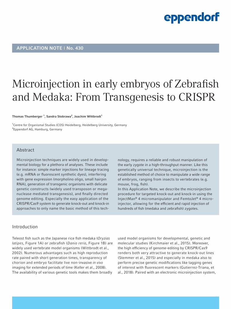
Teleost fish such as the Japanese rice fish medaka (Oryzias latipes, Figure 1A) or zebrafish (Danio rerio, Figure 1B) are widely used vertebrate model organisms (Wittbrodt et al., 2002). Numerous advantages such as high reproduction rate paired with short generation times, transparency of chorion and embryo facilitate live non-invasive in vivo imaging for extended periods of time (Keller et al., 2008). The availability of various genetic tools makes them broadly
used model organisms for developmental, genetic and molecular studies (Kirchmaier et al., 2015). Moreover, the high efficiency of genome-editing by CRISPR/Cas9 renders both very attractive to generate knock-out lines (Stemmer et al., 2015) and especially in medaka also to perform precise genetic modifications like tagging genes of interest with fluorescent markers (Gutierrez-Triana, et al., 2018). Paired with an electronic microinjection system,
Introduction
Microinjection in early embryos of Zebrafish and Medaka: From Transgenesis to CRISPR
Abstract
Microinjection techniques are widely used in develop-mental biology for a plethora of analyses. These include for instance: simple marker injections for lineage tracing (e.g. mRNA or fluorescent synthetic dyes), interfering with gene expression (morpholino oligo, small hairpin RNA), generation of transgenic organisms with delicate genetic constructs (widely used transposon or mega-nuclease mediated transgenesis), and finally directed genome editing. Especially the easy application of the CRISPR/Cas9 system to generate knock-out and knock-in approaches to only name the basic method of this tech-
APPLICATION NOTE I No. 430
Thomas Thumberger ¹, Sandra Stobrawa², Joachim Wittbrodt¹
¹Centre for Organismal Studies (COS) Heidelberg, Heidelberg University, Germany²Eppendorf AG, Hamburg, Germany
nology, requires a reliable and robust manipulation of the early zygote in a high-throughput manner. Like this genetically universal technique, microinjection is the established method of choice to manipulate a wide range of embryos, ranging from insects to vertebrates (e.g. mouse, frog, fish).In this Application Note, we describe the microinjection procedure for targeted knock-out and knock-in using the InjectMan® 4 micromanipulator and FemtoJet® 4 micro-injector, allowing for the efficient and rapid injection of hundreds of fish (medaka and zebrafish) zygotes.

APPLICATION NOTE I No. 430 I Page 2
Materials and Methods
Part 1: Harvesting of fertilized eggs and preparation for microinjection
Material:> Small fish net> Petri dish> Transfer pipette> Fine forceps > 10 µL pipet tip> Agarose mold type injection plate – trench width 1 mm
(medaka), 0.8 mm (zebrafish)
Media and solutions: > Medaka Embryo Rearing Medium (ERM): 0.1 % (w/v)
NaCl, 0.003 % (w/v) KCl, 0.004 % (w/v) CaCl₂ x 2 H₂O, 0.016 % (w/v) MgSO₄ x 7H₂O, optional: 0.0001 % (w/v) methylene blue
> Zebrafish E3 medium: 5 mM NaCl, 0.17 mM KCl, 0.33 mM CaCl₂, 0.33 mM MgSO₄, optional 0.0001 % methylene blue
Mating of medaka and zebrafish are set up as described (Köster et al., 1997; Westerfield, 2000). In brief:
Mating and egg harvesting procedure for medaka: 1. The evening before injections, male medaka fish are
separated from the females.2. Directly before the planned injection, males are put back
to the female tank (preferentially: 1 male, 3 females) and fish are left undisturbed.
high-throughput in vivo screening applications are in reach (Hammouda et al., 2019). This Application Note details our microinjection workflow for CRISPR/Cas9 mediated knock-out and knock-in approaches using the Eppendorf microinjection system. As example for a knock-out approach, we address the loss of the oculocutaneous albinism 2 (oca2, Lischik et al., 2019) gene responsible for melanin pigmentation in zebrafish and medaka. For precise tagging with the green fluorescent protein (gfp) in medaka (knock-in approach; Gutierrez-Triana et al., 2018), we turn to the retinal homeobox transcription factor 2 (rx2) as retina specific gene (Reinhardt et al., 2015).
3. After approx. 15 min, eggs are fertilized and can be collected. For medaka the clutch sticks to the female’s abdomen. It is thus necessary to net the females and to carefully strive off the eggs using a little hook.
4. Medaka zygotes can be collected in a Petri dish with ice cold ERM to slow down development for a prolonged one-cell stage.
5. Bristles and threads that attach to the outside of the chorions need to be removed for the zygotes to be sepa-rated. This is achieved by rolling the clutch with the tip of the finger under modest force inside of the Petri dish. Alternatively, especially with a large number of clutches, pull all batches together, use two forceps and hold onto some threads. Now wind the clutches around the pinched forceps under water. This movement coils up the threads onto the forceps and the zygotes are released individually.
Mating and egg harvesting procedure for zebrafish:1. The night prior to injection, set up the zebrafish in a breed-
ing tank with a divider in place. To increase total egg pro-duction, fish can be set up in a ratio of two females to one male if desired.
2. The following morning, after the room lights turn on, pull the divider from the tank, tilt the top-tank (by approx. 15-20°) to generate shallow water on one side (Sessa et al., 2008) and allow for approx. 20 min of undisturbed mating time.
3. After successful mating collect the fertilized eggs from the bottom of the breeding tank and rinse them in zebrafish medium. Fish can (later) be regrouped for additional mating.
Figure 1: Adult medaka (Oryzias latipes) and zebrafish (Danio rerio).

APPLICATION NOTE I No. 430 I Page 3
For most approaches, it is advisable to inject into the one-cell stage, hence adjust the timing of mating/collection without letting the zygotes pass the single cell stage. For injection, one-cell stage zygotes of either species are trans-ferred into injection plates. These were prepared before by pouring 1.5 % agarose dissolved in water and placing a mold that yields 1 mm (medaka) or 0.8 mm (zebrafish) broad trenches after polymerization of the liquid agarose (Figure 2). Once cooled, the mold is removed from the agarose and the
resulting trenches are filled with (ice-cold) ERM in case of medaka or room temperature zebrafish medium. Place your injection plate under the stereo microscope and transfer the fertilized eggs using a transfer pipet. With fine forceps or a white 10 µL pipet tip, push the zygotes into the grooves and orient with the one cell stage atop. Ensure that the embryos positioned in the trenches of the injection plate have not developed past the four-cell stage.
A B
Figure 2: Sorting of the fish embryos into the injection plate (A) made of agarose with trenches allowing to hold either zebrafish or medaka eggs (B).
Part 2: Preparation of sample, capillary and workstation
1. Injection mixes For the sake of completeness, we describe not only CRISPR component preparations but also the preparation of other injection samples.
Reagents and solutions:> 10x Yamamoto’s isotonic BSS, Ringer’s solution: 7.5 g NaCl,
0.2 g KCl, 0.2 g CaCl₂, 0.02 g NaHCO₃, fill up to final volume of 100 mL with nuclease free water, adjust pH to 7.3
> Nuclease free water > Optional: 1x Ringer’s solution
DNA:For transgenesis, the meganuclease system can be used (Grabher and Wittbrodt, 2007). Plasmid DNA is prepared and purified using a high-purity plasmid preparation kit. DNA concentration and purity can be checked by spectro-photometry. The ratio of A260/A280 should be between 1.8-2.0. For transgenesis experiments, DNA is co-injected with the meganuclease enzyme I-SceI. Due to the low stabil-ity of the meganuclease, aliquots of enzyme solution should be prepared (e.g. 2 μL) upon arrival and stored at -80°C. The microinjection solution should be prepared shortly before injection and kept on ice. Medaka and zebrafish fer-tilized oocytes may be injected using an identical solution: DNA 10-20 ng/μL, I-SceI buffer 0.5x (optional for medaka: Yamamoto buffer 0.5x), I-SceI enzyme 0.3 U/μL, ddH₂O ad

APPLICATION NOTE I No. 430 I Page 4
30 μL. For consistent results it is crucial to inject into the cytoplasm of the embryos and not into the yolk.250-500 pL (approx. 1/6 of the cell volume) of plasmid DNA at a concentration of 10 µg/mL (transgenesis experiments) to 50 µg/mL (mosaic transient expression) are injected into the cytoplasm of early embryos.
RNA:Capped mRNA is synthesized in vitro using a “mMESSAGE mMACHINE®” Kit (ThermoFisher Scientific®, U.S.A.). mRNA is purified through standard purification columns, precipitated and resuspended in RNase-free water. The mRNA is injected in nuclease free water or 1x Ringer’s solu-tion at concentrations of 10 ng/µL up to 1 ng/µL (i.e. from 5 pg to 500 pg mRNA per cell).
Tracer Dyes:Tracer dyes such as FITC-dextran or rhodamine-dextran are injected at a concentration of 1.5 % in 1x Ringer’s solution.
CRISPR/Cas9 knock-out and knock-in design:Find good sgRNA target sites (low predicted off-targets) us-ing a computational sgRNA design tool like CCTop (Stemmer et al., 2015). Anneal the given oligonucleotides and clone them into a suitable expression vector (e.g. plasmid DR274, Addgene®, U.S.A., #42250; Hwang et al., 2013). Release the template for in vitro transcription e.g., with DraI (of DR274 vector) and transcribe the sgRNA with MEGAscript™ T7 Transcription Kit (ThermoFisher Scientific, U.S.A.).
Purification is performed using the RNeasy® Mini Kit (QIAGEN®, Germany). Homology directed repair donors are designed according to the sgRNA that cuts the genomic DNA. The sgRNA target site should be in close vicinity (best +/- 10 nt) to the anticipated point of insertion. About 400 nt homology flanks are used on either side of the insert. If the sgRNA target site fully resides in the sequence used as homology flank, it is required to at least mutate the PAM (protospacer adjacent motif) in the donor sequence for the CRISPR system not to cut the donor template. Assembly of (more complex) donors can easily be achieved with the Golden GATEway cloning setup (Kirchmaier et al., 2013). To improve single-copy HDR-mediated insertion, the donor cassette is PCR amplified using 5’ biotinylated primers and phosphorothioate bonds in the first five nucleotides (further details: Stemmer et al., 2015; Gutierrez-Triana et al., 2018).
To prepare the CRISPR injection mix, use filter-tips and nuclease free water. No special buffer needed.
CRISPR/Cas9 knock-out and knock-in mix:> 150 ng/µL Cas9 mRNA> 15 ng/µL per sgRNA (knock-out attempts may use multi-
plexing)> 10 ng/µL GFP mRNA (optional injection reporter)> (5 ng/µL biotinylated PCR donor – only applies for HDR-
mediated integration)> ad 10 µL nuclease free water
2. Capillary preparation
Consumables: > For zebrafish: Femtotip II, Eppendorf AG, (Germany) or
borosilicate capillary with OD/ID: 1.0 mm/0.58 mm for pulling
> For medaka: Borosilicate capillary with OD/ID: 1.2 mm/0.7 mm for pulling
> MicroloaderTM, Eppendorf AG, (Germany)
Devices: > Microcapillary puller> 0.5-10 µL pipette Eppendorf Research® plus,
Eppendorf AG, (Germany)> Centrifuge, e.g. Centrifuge 5418 R, Eppendorf AG, (Germany)
For zebrafish injection use either the Femtotip II with a long and thin tapered tip end of 0.5 µm opening or a self-pulled microcapillary (e.g. borosilicate capillary with 1.0 mm OD and 0.21 mm wall thickness pulled into two micropipettes
with a fine tip end). The microcapillary for medaka injec-tions needs to have a stronger tip end (e.g. borosilicate capillary with 1.2 mm OD with 0.25 mm wall thickness pulled into two micropipettes with shorter, non-flexible tip ends) allowing to penetrate the chorion. The microcapillar-ies can be pulled in advance. When using self-pulled capillar-ies, the tips are closed. For zebrafish injections, breaking off the tips with forceps is recommended. In contrast, manual opening of the short-pointed tip of medaka injection capil-laries would create a too large opening. Instead use the fully assembled microinjection setup after loading the capillary to scrub the tip of the self-pulled needle across the chorion of a medaka egg. This will break off a tiny tip, ensuring the microcapillary to remain sharp and pointed.Prior to the capillary loading, centrifuge the sample for 10 min at 16.000 x g to avoid any capillary clogging by pre-cipitates. Transfer the supernatant in a new dust-free tube. Load the microcapillary with 2-5 μL of the injection mix as close as possible to the capillary tip using the MicroloaderTM. Avoid air bubbles in the capillary.

APPLICATION NOTE I No. 430 I Page 5
3. Setup of Microinjection workstation
Devices:> Stereo microscope with transmitted light base > InjectMan® 4 micromanipulator, Eppendorf AG, (Germany)> FemtoJet® 4i microinjector, Eppendorf AG, (Germany)> Universal stand for mounting the micromanipulator next
to the stereo microscope, Eppendorf AG, (Germany)> Ferromagnetic plate for fixing the universal stand next to
the stereo microscope
The capillary holder of the FemtoJet 4i is mounted onto the motors of the InjectMan 4 in a 45° angle. The FemtoJet 4i and InjectMan 4 are connected via the interface cable to allow a semi-automatic injection. For the InjectMan 4 the application-specific mask “Developmental biology” is selected to display the optimized function keys for this injection application. The “axial” movement mode is acti-vated. The axial injection movement is controlled by turn-ing the rotary head of the joystick. If the injection pressure
should be triggered via the joystick key, go to menu function “synchronize injection“ and set it to “pressure”. For the FemtoJet 4i microinjector, recommended starting settings are 500-700 hPa for injection pressure (pi) and 80-100 hPa for compensation pressure (pc). The optimal injection time is to be established empirically. The operating mode of the injector is thus set to “manual“ injection. For zebrafish in-jection, a drop of mineral oil on a micrometer can be used to calculate the injection volume. When injected into the oil, an injection droplet with a diameter of 100 µm corresponds to 525 pL injection volume. Ideal injection volumes will fill ap-prox. 10-15 % of the egg volume, thus volumes up to 500 pL are typically injected. After each successful injection the actual injection time (ti) is displayed on the FemtoJet 4i and can be used for semi-automatic injection setup of zebrafish. Due to the stiff medaka chorion, pre-calculation of drop-size is not recommended, as each injection may open the needle a bit more or cell debris may clock the opening. The injected volume is controlled by sight.
Part 3: Injection
1. Insert the sample-loaded microcapillary into the capillary holder of the microinjector and ensure a tight seal. Check that the micromanipulator and the capillary holder are in a proper position to allow for a wide range of movement and adjustment.
2. Lower the microcapillary toward the eggs in your injec-tion plate, holding the dish in place with your opposite hand. Bring the capillary tip into the plane of view of the micro-scope and focus on the thinnest region of the tip. In case of self-pulled capillaries, the tip end must be opened either with help of fine forceps (zebrafish) or by scratching the tip end slightly across the chorion of the medaka egg.
Figure 3: Microinjection workstation for injection into early embryos of zebrafish and medaka. The injection station is assembled by a stereo microscope, the InjectMan 4 and the FemtoJet 4i.

APPLICATION NOTE I No. 430 I Page 6
Break the capillary at a point which leaves it narrow enough to pierce the chorion and yolk but still capable of delivering the sample.
3A. Injection into medaka cells: Just prior the injection each medaka embryo needs to be oriented in the agarose trench with the embryonic cell atop of the yolk with help of fine forceps or small pipet tips. Move the microcapillary down to the embryo using the joystick of the micromanipulator. Pierce the surface of the chorion with the capillary tip and enter the capillary into the embryonic cell in one smooth stroke using the axial movement mode. Trigger the injection pressure by pressing the joystick but-ton. The sample is injected as long as the button is pressed until you see a droplet of approx. 10-15% of the cell (Figure 4A). Avoid injecting air bubbles. After injection retract the capillary out of the egg with same injection angle. Move the petri dish manually to position the next zygote close to the capillary tip in the microscopic focus and repeat the injection.
3B. Injection into zebrafish cells: In contrast to medaka, zebrafish zygotes can be injected into the yolk sack. Move the microcapillary (e.g. Femtotip II) down to the embryo. Pierce the surface of the chorion with the capillary tip and enter the capillary into the yolk in one smooth stroke using the axial movement mode. Trigger the injection pressure by pressing the joystick button until you see a small volume released into the yolk sac (Figure 4B). Avoid injecting air bubbles or stretching the yolk as either can be lethal to the embryo. After injection retract the capillary out of the egg in axial direction. Move the Petri dish to repeat injection of the neighboring embryo. For CRISPR experiments, it is advised to inject as close as pos-sible to the embryonic cell or better directly into the cyto-plasm of the one-cell stage rather than the yolk.In high-throughput routine it is also possible to use the semi-automatic injection mode for zebrafish injection. The InjectMan 4 menu function “Step injection“ is activated, the “Step injection speed” is set to 500-700 µm/sec, the “Step injection distance” is set to 800-1000 µm and the “Synchronize injection” function is set to “Limit”. Switch the FemtoJet 4i to the “automatic” injection mode and set the injection time (ti) that has been established empirically. To trigger the synchronized injection movement and pres-sure just press the joystick key after positioning the egg under the microcapillary.
4. Working down the lines remove eggs which look unfertil-ized or are destroyed during the injection process. Injected embryos are transferred to appropriate rearing media and kept at 26 °C (medaka) or 28°C (zebrafish) until hatching. Keep also some non-injected embryos as controls at same conditions.
5. At the end of the day or early next day, remove dead em-bryos and record the number of injected embryos. Replace the embryo rearing medium in the dish periodically to reduce the risk of infection or add 0.0001 % methylene blue to avoid mold to grow. Note that methylene blue displays autofluo-rescence under UV light and should hence only be used when screening for fluorophores is not required.Figure 4: (A) Microinjection into the cytoplasm of the
medaka one-cell stage. (B) Microinjection into the yolk sac of the one-cell stage embryo of zebrafish.
A
B

APPLICATION NOTE I No. 430 I Page 7
The day after injection, sort the embryos for proper de-velopment and according to expected phenotype or ex-pression; i.e. fluorphores, lineage tracers and constructs containing fluorescent reporters are easily detectable at adequate developmental stages using a fluorescent screening microscope.
Depending on the injection mix, embryos may survive less than the non-injected siblings. However, as injections are easily performed with an electronic microinjection system, a huge number of modified embryos can be generated and selected afterwards. This is particularly relevant for CRISPR-mediated knock-out or knock-in attempts that do not yield an obvious phenotype. In such a case, PCR genotyping is required on a subset of injected embryos (single as well as pooled embryos). Knock-out approaches can be tested by administering the amplified targeted locus in question to the so-called surveyor assay. In brief, if the locus amplicon yields a mixture of different alleles stemming from indel mutations, heteroduplex DNA amplicons are detected and cut. Subsequent gel-electrophoresis reveals a single band for wild-type loci, and three bands (wt, left and right piece of the heteroduplex) after proper cutting (Hammouda et al., 2019). If a targeted knock-in was done using a specific donor
sequence, a pair of primers with one binding the genomic DNA (outside of the homology flank), and the second one targeting the insert sequence can be employed to reveal proper insertion. Note however, extended numbers of cycles during the PCR may yield false-positive amplicons as result of an in vitro fusion PCR event (Won and Dawid, 2017; Gutierrez-Triana et al., 2018).
By the abundance of the targeted genome editing, the general success-rate of the targeting attempt can be deduced and the injected siblings can be raised. With todays’ efficient CRISPR/Cas approaches, loss-of-function screens can al-ready be performed in the injected generation (Hammouda et al., 2019). To illustrate the typical results of microinjections, we used the above protocol to successfully generate a CRISPR/Cas9-mediated knock-out of the eye pigmentation gene oca2 (oculocotaneous albinism 2) in medaka and zebrafish (see Hammouda et al., 2019 for details; Figure 5 A-C) and an in-frame knock-in approach of the sequence of GFP with the endogenous rx2 (retina-specific homeobox gene 2) gene in medaka (see Gutierrez-Triana et al., 2018 for details; Figure 5D).
Expected results
Figure 5: Successful genome editing in the injected generation. Oca2 crispants in medaka (A) and zebrafish (B, C) showing loss of pigmentation (predominant in the retinal pigmented epithelium of the eye, as well as in body pigmen-tation) upon bi-allelic knock-out (oca2-/-) in comparison to pigmented wildtypes (wt). (D) GFP fluorescence in the retina of developing medaka upon HDR-mediated tagging of the rx2 gene.
APPLICATION NOTE I No. 430 I Page 8
One of the strengths of the fish model systems is the ease with which specific gene products can be added to or eliminated from the embryo by microinjection. Using the reliable micromanipulation system like the InjectMan 4 and FemtoJet 4, various manipulations of the fish embryos can be performed in high number and short time. The different properties of the injected agents can be used to receive the desired effect. For instance, injection of mRNA will lead to a uniform distribution throughout the developing organism. Injecting protein or plasmids in contrast will result in a more stochastic (salt-and pepper) distribution.
In combination with the CRISPR/Cas system, the intuitive and convenient operation of the micromanipulation setup described here is a perfect match to perform simple as well as high-throughput knock-out screens on a large number of embryos with little effort and time. More delicate, employing HDR for tagging/inserting donor sequences at designated loci, a larger number of specimens need to be generated to screen for the proper integration event. Especially for zebraf-ish, the option for semi-automated injection steps facilitates efficient and rapid injection of hundreds of fish zygotes.
Discussion
Literature
[1] Wittbrodt J, Shima A, Schartl M. Medaka--a model organism from the far East. Nat Rev Genet. 2002; 3(1): 53-64. Review.
[2] Keller, PJ, Schmidt AD, Wittbrodt J & Stelzer EH. Reconstruction of zebrafish early embryonic development by scanned light sheet microscopy. Science. 2008; 322: 1065-69.
[3] Kirchmaier S, Naruse K, Wittbrodt J, Loosli F. The genomic and genetic toolbox of the teleost medaka (Oryzias latipes). Genetics. 2015; 199(4): 905-18.
[4] Stemmer M, Thumberger T, Del Sol Keyer M, Wittbrodt J, Mateo JL. CCTop: An Intuitive, Flexible and Reliable CRISPR/Cas9 Target Prediction Tool. PLoS One. 2015; 10(4): e0124633.
[5] Gutierrez-Triana JA, Tavhelidse T, Thumberger T, Thomas I, Wittbrodt B, Kellner T, Anlas K, Tsingos E, Wittbrodt J. Efficient single-copy HDR by 5’ modified long dsDNA donors. eLife. 2018; 7: e39468.
Figure 5A-C shows loss of pigmentation in body and retina pigmented epithelium with varying degrees of mosaicism. This is caused by the fact that loss of pigmentation requires a biallelic mutation in oca2 and the NHEJ repair machinery leads to random insertion/deletions at the introduced double strand break which may still yield a functional allele in some cells. In addition, targeted genome editing following Cas9 mRNA injections is delayed as the Cas9 mRNA needs first to be translated. After screening, some embryos should be administered to genotyping (Hammouda et al., 2019) and following positive on-target genome editing, the siblings can be further investigated or raised for line generation. Besides NHEJ based knock-out, the introduction and spe-cific integration of a donor template can be performed by HDR in medaka. To this end, a biotinylated PCR-donor, a
locus specific sgRNA and the Cas9 mRNA are co-injected as detailed above. The donor comprises an insert sequence (here GFP) and flanking homology arms corresponding to the position in the genome, where the insert should be integrat-ed (here the transcriptional start site of the retina specific rx2 gene). For further details on donor template design see Gutierrez-Triana et al. (2018). As this experiment aimed at integration of the GFP sequence in frame with the endoge-nous rx2 gene in medaka (see Figure 5D), it is not advisable to co-inject a fluorescent reporter, as its fluorescence may shield the desired GFP signal. In addition, the fluorescent signal stemming from the tagged protein is under the con-trol of its endogenous promoter and hence may be faint.

APPLICATION NOTE I No. 430 I Page 9
Thermo Fisher Scientific® and mMESSAGE mMACHINE® are registered trademarks of Thermo Fisher Scientific, Inc., USA. Addgene® is a registered trademark of Addgene Inc., USA. QIAGEN® and RNeasy® are registered trademarks of QIAGEN GmbH, Germany. MEGAscript™ is a trademark of ThermoFisher Scientific, Inc., USA. Eppendorf®, the Eppendorf Brand Design, InjectMan®, FemtoJet®, Femtotips® and Eppendorf Research® are registered trademarks of Eppendorf AG, Germany. Microloader™ is a trademark of Eppendorf AG, Germany. All rights reserved, including graphics and images. Copyright © 2020 by Eppendorf AG, Germany.
Your local distributor: www.eppendorf.com/contactEppendorf AG · Barkhausenweg 1 · 22339 Hamburg · [email protected] · www.eppendorf.com
www.eppendorf.com
Ordering informationDescription Order no.
InjectMan® 4, micromanipulator with dynamic movement control, 100 – 240 V/50 – 60 Hz 5192 000 019
FemtoJet® 4x, programmable microinjector with external pressure supply (not included), including foot control 5253 000 017
FemtoJet® 4i, programmable microinjector with integrated pressure supply, including foot control 5252 000 013
Femtotip II, injection capillary (for research use only), sterile, set of 20 5242 957 000
Microloader™, tip for filling Femtotips® and other glass microcapillaries, 0.5 – 20 µL, 100 mm, light gray, 192 pcs.
(2 racks × 96 pcs.)
5242 956 003
Universal stand, for mounting Eppendorf micromanipulators on upright microscopes and stereo microscopes independent
of the microscope tripod used
5192 325 007
[6] Hammouda OT, Böttger F, Wittbrodt J, Thumberger T. Swift Large-scale Examination of Directed Genome Editing. PLoS One. 2019; 14(3): e0213317.
[7] Lischik CQ, Adelmann L, Wittbrodt J. Enhanced in vivo-imaging in medaka by optimized anaesthesia, fluorescent protein selection and removal of pigmentation. PLoS One. 2019; 14(3): e0212956
[8] Reinhardt R, Centanin L, Tavhelidse T, Inoue D, Wittbrodt B, Concordet JP, Martinez-Morales JR, Wittbrodt J. Sox2, Tlx, Gli3, and Her9 converge on Rx2 to define retinal stem cells in vivo. The EMBO Journal. 2015; 34: 1572-88.
[9] Köster R, Stick R, Loosli F, Wittbrodt J. Medaka spalt acts as a target gene of hedgehog signaling. Development. 1997; 124(16): 3147-56.
[10] Westerfield, M. The zebrafish book. A guide for the laboratory use of zebrafish (Danio rerio). 4th ed., Univ. of Oregon Press, Eugene; 2000
[11] Sessa AK, White R, Houvras Y, Burke C, Pugach E, Baker B, Gilbert R, Look AT, and Zon LI. The Effect of a Depth Gradient on the Mating Behavior, Oviposition Site Preference, and Embryo Production in the Zebrafish, Danio rerio. Zebrafish 2008; 5(4): 335-339
[12] Grabher C & Wittbrodt J. Meganuclease and transposon mediated transgenesis in medaka. Genome Biol. 2007; 8 Suppl 1, S10.
[13] Hwang WY, Fu Y, Reyon D, Maeder ML, Tsai SQ, Sander JD, Peterson RT, Yeh JR, and Joung JK. Efficient genome editing in zebrafish using a CRISPR-Cas system. Nat Biotechnol. 2013; 31(3):227-9
[14] Kirchmaier S, Lust K, Wittbrodt J. Golden GATEway cloning--a combinatorial approach to generate fusion and recombination constructs. PLoS One. 2013; 8(10): e76117.
[15] Won M & Dawid IB. PCR artifact in testing for homologous recombination in genomic editing in zebrafish. PLoS One. 2017;12(3): e0172802