Embed Size (px)
Citation preview

Microencapsulation Technology
Kinam ParkDepartments of Pharmaceutics and Biomedical Engineering, Purdue University,West Lafayette, Indiana, U.S.A.
Yoon YeoDepartment of Industrial and Physical Pharmacy, Purdue University,West Lafayette, Indiana, U.S.A.
INTRODUCTION
Microencapsulation technology has been used from1930s in packaging flavors and vitamins. Since the firstcommercial product was introduced for the carbonlesscopying paper,[1] the technology has advanced to anew level. Various microencapsulation techniques areavailable nowadays, and the microencapsulated pro-ducts are widely used in pharmaceutical, biomedical,agricultural, food, consumer products, and cosmeticindustries. Representative applications of microparti-cles in the pharmaceutical and biomedical industriesinclude:
� Taste and odor masking[2]
� Protection of drugs from the environment[3]
� Particle size reduction for enhancing solubility ofthe poorly soluble drugs[4]
� Sustained or controlled drug delivery[5]
� Cell encapsulation[6]
The microparticle system has become an indispens-able part of the controlled drug delivery fields for thepast few decades since it can readily be adapted for vari-ous administration methods. In particular, biodegrad-able polymeric microparticles can provide a number ofadvantages over conventional parenteral formulations:
� Sustained delivery: By encapsulating a drug in apolymer matrix, which limits access of the biologi-cal fluid into the drug until the time of degrada-tion, microparticles maintain the blood level of thedrug within a therapeutic window for a prolongedperiod. Toxic side effects can be minimized, andpatient compliance can be improved by reducingthe frequency of administration.
� Local delivery: Subcutaneously or intramuscu-larly applied microparticles can maintain a thera-peutically effective concentration at the site ofaction for a desirable duration. The local deliverysystem obviates systemic drug administration forlocal therapeutic effects and can reduce the related
systemic side effects. This system has proven ben-eficial for delivery of local anesthetics.[7]
� Pulsatile delivery: While burst and pulsatile releaseis not considered desirable for the sustained deliveryapplication, this release pattern proves to be usefulfor delivery of antibiotics and vaccines. Pulsatilerelease of antibiotics can alleviate evolution of thebacterial resistance. In the vaccine delivery, initialburst followed by delayed release pulses can mimican initial and boost injection, respectively.[8]
With the recent advance of biotechnology and poly-mer chemistry, the use of microparticle systems willcontinue to grow for a variety of applications. Theobjective of this article is to provide a review of thetechnical aspects of the microencapsulation techniquesthat have been widely used in the pharmaceuticalindustry and recent advances of the technology sothat the pharmaceutical scientists can take full advan-tage of the existing assets of this area in developingnew microparticle systems.
TERMINOLOGY
The microencapsulation processes produce smallparticles ranging in size from 1 to 1000mm. There aredifferent names for these particles: microparticle, micro-sphere, microcapsule, and micromatrix. Although theyare often used interchangeably, distinctions can bemadesuch that microcapsules are made of one or multiplecore substances (solid or liquid) that are surroundedby a distinct capsule wall, whereas micromatrices arepolymeric matrices in which the encapsulated sub-stances are homogeneously dispersed. Microparticlesor microspheres are general terminologies that involveboth. Although micromatrices are also called micro-spheres depending on the authors,[5] we will follow theformer definition in this chapter. In consideration ofthe scope of this chapter, current discussion is limitedto the microparticles that utilize natural or syntheticpolymers as an encapsulating material.
Encyclopedia of Pharmaceutical Technology DOI: 10.1081/E-EPT-120028567Copyright # 2007 by Informa Healthcare USA, Inc. All rights reserved. 2315
Enc
yclo
pedi
a of
Pha
rmac
eutic
al T
echn
olog
y D
ownl
oade
d fr
om in
form
ahea
lthca
re.c
om b
y IB
I C
ircu
latio
n -
Ash
ley
Publ
icat
ions
Ltd
on
11/1
1/10
For
pers
onal
use
onl
y.
MICROENCAPSULATION TECHNIQUES
Existing microencapsulation techniques have beenreviewed extensively,[5,9–12] and for this reason, herewe will briefly summarize representative microen-capsulation techniques.
Coacervation
The coacervation method is one of the earliest micro-encapsulation techniques, which has been used forvarious consumer products. This method is based onseparation of a solution of hydrophilic polymer(s)into two phases, which are small droplets of a densepolymer-rich phase and a dilute liquid phase. Coacer-vation can be divided into simple and complexcoacervation depending on the number of polymersthat are involved in the formation of microparticles.
Simple coacervation
This process involves only one polymer (e.g., gelatin, poly-vinyl alcohol, carboxymethyl cellulose), and the phaseseparation can be induced by conditions that result indesolvation (or dehydration) of the polymer phase.These conditions include addition of a water-misciblenon-solvent, such as ethanol, acetone, dioxane, isopro-panol, or propanol,[13] addition of inorganic salts, suchas sodium sulfate,[14] and temperature change.[5]
Complex coacervation
This process involves two hydrophilic polymers ofopposite charges.[15] Neutralization of the overall posi-tive charges on one of the polymers by the negative
charge on the other is used to bring about separationof the polymer-rich phase (Fig. 1). The best-knownexample is the gelatin-gum arabic system pioneered byBungenberg de Jong in the early 1940s.[16] Since electro-static interactions are involved, the pH of the medium isvery important. For example, in the gelatin-gum arabicsystem, pH should be below the isoelectric point of gela-tin so that the gelatin can maintain the positive charge.Once embryonic coacervates form around the dispersedoil or solid phases, these polymer complexes are stabi-lized by cross-linking using glutaraldehyde.
Commercial products
The first commercial microparticle product based on thecomplex coacervationmethodwas carbonless copy paperdeveloped by National Cash Register Corp.[1] The backside of the first page is coated with microcapsules in the3–10mm size range made of a gelatin-gum arabic shellby the coacervation technique. In the center of the cap-sules is the oil containing colorless color-forming agent(e.g., crystal violet lactone). The front side of the secondpage is coated with a developing layer. The pressureimposed on both sheets of paper upon writing inducesbreakage of the microcapsules and makes the colorlesscolor-forming agent released and react with the develop-ing layer to develop color. The microcapsules have alsobeen used in Scratch-N-Sniff� scent strips and Snap-N-Burst� fragrance samplers.
Emulsion Solidification
Microparticles can be produced from emulsion oftwo or more immiscible liquids. For example, a
Fig. 1 Phase diagram for complex
coacervation.
2316 Microencapsulation Technology
Enc
yclo
pedi
a of
Pha
rmac
eutic
al T
echn
olog
y D
ownl
oade
d fr
om in
form
ahea
lthca
re.c
om b
y IB
I C
ircu
latio
n -
Ash
ley
Publ
icat
ions
Ltd
on
11/1
1/10
For
pers
onal
use
onl
y.

solution of hydrophobic drug and polymer in anorganic solvent (oil phase, dispersed phase) is emulsi-fied in an aqueous solution containing an emulsifyingagent (water phase, continuous phase) to produce oil-in-water (o/w) emulsion. The drug containing polymerparticles can be solidified as the solvent is removed.Depending on the solubility of drug in water andencapsulating polymer, the emulsion type can bevaried from water-in-oil-in-water (w/o/w for encap-sulation of water-soluble drug in water-insoluble poly-mer), water-in-oil-in-oil (w/o/o for encapsulation ofwater-soluble drug in water-insoluble polymer), orwater-in-oil (w/o for encapsulation of water-solubledrug in water-soluble polymer) to solid-in-oil-in-water (s/o/w for encapsulation of water-soluble drugparticles in water-insoluble polymer). Depending onthe method of solidifying the discontinuous droplets,the emulsion method can be classified as solvent evapo-ration, solvent extraction, and cross-linking method.
Solvent evaporation
Typically, the polymer is dissolved in a volatile organicsolvent such as methylene chloride. Drugs or diag-nostic agents, either in soluble form or dispersed asfine solid particles, are added to the polymer solution,and then this mixture is emulsified in an aqueoussolution that contains an emulsifying agent such aspoly(vinyl alcohol) (PVA). The resulting emulsion isstirred until most of the organic solvent evaporates,leaving solid microparticles that may be washedwith water and freeze-dried. To facilitate solventevaporation, the emulsion is often heated slightlyabove the boiling point of the solvent. For example,when methylene chloride (boiling point: 39.8�C) isused as an organic solvent, the emulsion is heatedto 40�C.
Solvent extraction
The solvent evaporation method depends on highvapor pressure of the solvent; therefore, this methodrequires volatile solvents such as methylene chloride.Otherwise, the solidification process takes too longthat it results in irregular morphology,[17] high porosityof the microspheres,[18] loss of payload,[19,20] orincreased polydispersity of size distribution.[17] Inthis case, the relatively non-volatile solvents can beremoved by extraction into the continuous phase.[21]
This can be done by using a solvent that has signifi-cant solubility in the continuous phase,[22] increasingthe concentration difference between the dispersedand continuous phase,[19] or adding a third solventinto the continuous phase to facilitate extraction ofthe solvent.[21]
Cross-linking
A number of hydrophilic polymers from natural origin,such as gelatin, albumin, starch, dextran, hyaluronicacid, and chitosan,[23] can be solidified by a chemicalor thermal cross-linking process. A w/o emulsion isprepared by emulsifying the polymer solution in anoil phase (typically vegetable oils or oil–organic solventmixtures) containing an emulsifying agent such asSpan 80. Most proteins are cross-linked using glutaral-dehyde, but its toxicity remains a problem for pharma-ceutical applications. Heating[24] and adding counterpolyions[25] or cross-linking reagents[26] (Fig. 2) arealternative cross-linking methods.
Hot-Melt Microencapsulation
The polymer is first melted and then mixed with soliddrug particles or liquid drugs.[27] This mixture is sus-pended in an immiscible solvent and heated to 5�Cabove the melting point of the polymer under continu-ous stirring. The emulsion is then cooled below themelting point until the droplets solidify.
Ionic Gelation/Polyelectrolyte Complexation
Ionic gelation involves cross-linking of polyelectrolytesin the presence of multivalent counter ions. Forexample, spraying a sodium alginate solution into cal-cium chloride solution produces rigid gel particles.Ionic gelation is often followed by polyelectrolytecomplexation with oppositely charged polyelectrolytes.This complexation forms a membrane of polyelectro-lyte complex on the surface of the gel particles, whichincreases the mechanical strength of the particles. Forcalcium alginate gel particles, polylysine is often usedfor this purpose. Other polymer systems that can beused for ionic gelation (polyelectrolyte complexation)are chitosan/triphosphate, carboxymethylcellulose/aluminum (or chitosan), k-carrageenan/potassium (orchitosan), pectin/calcium, gelan gum/calcium, andpolyphosphazene/calcium (or polylysine). This methodwas developed by Lim and Sun[6] for cell encapsulation;nowadays, it has widely been used for both cell anddrug encapsulation.
Interfacial Polymerization
Monomers can be polymerized at the interface of twoimmiscible substances to form a membrane. Anexample is a nylon membrane resulting from poly-merization of two monomers (typically dichlorideand diamine) at the interface. A non-aqueous phasecontaining surfactant and an aqueous phase con-taining drugs and diamine are mixed to form a w/o
Microencapsulation Technology 2317
Enc
yclo
pedi
a of
Pha
rmac
eutic
al T
echn
olog
y D
ownl
oade
d fr
om in
form
ahea
lthca
re.c
om b
y IB
I C
ircu
latio
n -
Ash
ley
Publ
icat
ions
Ltd
on
11/1
1/10
For
pers
onal
use
onl
y.

emulsion. Then additional non-aqueous phase con-taining acid chloride is added to the emulsion to allowinterfacial polymerization. Polymerization can beterminated by adding excess non-aqueous phase.[28]
Spray Drying
Spray drying is a single-step, closed-system processapplicable to a wide variety of materials. The drug isdissolved or suspended in a suitable (either aqueousor non-aqueous) solvent containing polymer materials.The solution or suspension is atomized into a dryingchamber, and microparticles form as the atomizeddroplets are dried by heated carrier gas. The resultof the spray drying process is heavily dependenton the material properties: The instrument settings,such as inlet temperature, rate of feed flow, spray airflow, and aspirator flow, can together influence theproduct parameters such as particle size, yield, tem-perature load, and content of residual solvents.Optimization of these parameters is usually madethrough trial and error.
Spray Desolvation
Spray desolvation involves spraying a polymer solutiononto a desolvating liquid. For example, microparticlescan be made by spraying a PVA solution onto an acet-one bath. Here, the polymer solvent (water) is extractedinto acetone, and PVA precipitates to form solid micro-particles.[29] In another example, bovine serum albumin(BSA) was encapsulated in poly(lactic-co-glycolic acid)(PLGA) by this method. The micronized drug was sus-pended in a PLGA–acetone solution and atomizedultrasonically into ethanol bath.[30]
A modification of this method is the cryogenicsolvent extraction method.[31] Drugs are dissolved ordispersed in the polymer phase consisting of PLGAand methylene chloride. This drug–polymer mixturesolution is atomized over a bed of frozen ethanol over-laid with liquid nitrogen. The microdroplets freezeupon contacting the liquid nitrogen, then sink ontothe frozen ethanol layer. As temperature increases,the frozen microdroplets begin to sink into the thawingethanol. Methylene chloride of the polymer phase thenthaws and is slowly extracted into ethanol, resulting in
Fig. 2 Microencapsulation based on w/o emulsion and in situ cross-linking. (From Ref.[26].)
2318 Microencapsulation Technology
Enc
yclo
pedi
a of
Pha
rmac
eutic
al T
echn
olog
y D
ownl
oade
d fr
om in
form
ahea
lthca
re.c
om b
y IB
I C
ircu
latio
n -
Ash
ley
Publ
icat
ions
Ltd
on
11/1
1/10
For
pers
onal
use
onl
y.

hardened microparticles. This process was used toproduce a microparticle formulation for humangrowth hormone (hGH).[32,33] Prior to the encapsula-tion process, hGH is formulated with zinc to produceinsoluble Zn-hGH complex. The encapsulation of thiscomplex contributed to stabilize hGH during thefabrication process and within the microspheres afterhydration.[32] An in vivo study of this formulationdemonstrated a lower Cmax and an extended serumlevel for weeks with a biocompatibility comparableto that of the protein solution.[33] The hGH formu-lation using this method (ProLease�) was marketedas an injectable suspension for once- or twice-a-monthadministration (Nutropin Depot�).
Spray Coating
In spray coating, the coating material is sprayed ontosolid drug core particles that are rotated in a coatingchamber. This method is typically used for coatingtables or capsules.
Fluid-bed coating (Air-suspension technique)
There are three commonly used fluid-bed processes:top, tangential, and bottom spray methods. When thegranules are coated by the top-spray granulator sys-tem, granules usually have a porous surface and aninterstitial void space; therefore, the bulk density ofproduced granules is usually lower than that attain-able by other granulation techniques. A rotating-diskmethod (also called a tangential-spray coatingmethod), which combines centrifugal, high-densitymixing, and the efficiency of fluid-bed drying, yields aproduct that has a higher bulk density but still hassome interstitial void space. This method results inparticles that are less friable and more spherical inshape. In the Wurster process (bottom spray), the solidcore particles are fluidized by air pressure and a sol-ution of wall materials is sprayed on to the particlesfrom the bottom of the fluidization chamber parallelto the air stream. Since the spraying nozzle is immersedin the airflow and sprays the coating materials concur-rently into the fluidized particles, the coating solutiondroplets travel only a short distance before contactingthe solid particles. As a result, the film is applied moreevenly and the coated film is more homogeneous. Thecoated particles are lifted on the air stream, which driesthe coating as the particles are carried away from thenozzle. The particles rise on the air stream, then settledown, and then begin another cycle. The cyclescontinue until the desired film thickness is achieved.The Wurster process is particularly well suited for uni-form coating of particles with a polymeric membranein a single operation.
Pan coating
Relatively large particles can be encapsulated by pancoating. Size of solid particles should be greater than600 mm to achieve effective coating using this method.This is a typical method used to apply sugar coatingson candies. This method employs a rotating drum con-taining core materials (such as candies), onto whichwarm sucrose solution is ladled. The rotation distri-butes the syrup evenly as a thin coat on the coresand increases the surface area of the syrup that aidsin evaporation of the water. As the water evaporates,the sugar hardens and coats the cores. For pharmaceu-tical products, perforated pans are used and the coat-ing solution, usually an aqueous solution, is sprayedonto the tumbling cores.
Supercritical Fluid
The supercritical fluid method is a relatively newmethod, which can minimize the use of organic solventsand harsh manufacturing conditions taking advantageof two distinctive properties of supercritical fluids(i.e., high compressibility and liquid-like density). Thismethod can be broadly divided into two parts: rapidexpansion of supercritical solutions (RESS), whichutilizes the supercritical fluid (e.g., carbon dioxide) asa solvent for the polymer,[34] and supercritical anti-solvent crystallization (SAS), using the fluid as anantisolvent that causes polymer precipitation.[35]
Recent reviews of the supercritical technology forparticle production are available in the literature.[36]
Selection of the Microencapsulation Methods
A single microencapsulation method cannot be uni-versally applied for a variety of drugs. In developinga new microparticle system for a given drug, it isimportant to understand the physicochemical proper-ties of the drug and find an encapsulation methodand polymeric materials that best match the properties.Since water is the most widely used solvent system,solubility of the drug in water often serves a good start-ing point of the survey. Physical status of the drug canalso limit the selection. The microencapsulation meth-ods that have widely been used for drugs of differentproperties are summarized in Table 1.
ACHIEVEMENTS AND LIMITATIONS
Natural polymers such as proteins, carbohydrates, fats,and waxes constitute an important group of encapsula-tion materials; however, microparticles for the con-trolled drug delivery purpose have been prepared
Microencapsulation Technology 2319
Enc
yclo
pedi
a of
Pha
rmac
eutic
al T
echn
olog
y D
ownl
oade
d fr
om in
form
ahea
lthca
re.c
om b
y IB
I C
ircu
latio
n -
Ash
ley
Publ
icat
ions
Ltd
on
11/1
1/10
For
pers
onal
use
onl
y.
using almost invariably water-insoluble syntheticpolymers. To date, poly(lactic acid) (PLA) and PLGAhave been the most preferred polymers because theyhave been used in products approved by the U.S. Foodand Drug Administration (U.S. FDA), such as surgicalsutures and depot formulations, and they have arelatively long history of use for biomedical applica-tions. Poly(lactic acid)- and PLGA-based microparticlesystems are commercially available. Lupron Depot�
(leuprolide acetate, TAP Pharmaceuticals Inc.) is thefirst commercial product based on PLA polymers.[47]
Since then, various PLGA- or PLA-based productssuch as Zoladex� Depot (goserelin acetate, Astra-Zeneca), Sandostatin LAR� Depot (octreotide acetate,Norvatis), and TrelstarTM Depot (triptorelin pamoate,Pfizer) were introduced. Nutropin Depot (humangrowth hormone, Genentech Inc.) entered the marketas the first protein-encapsulated PLGA microparticleproduct in 1999.
Despite the extensive use of PLGA polymers inthe microencapsulation arena, it has been foundthrough decades of research that the PLGA micro-particle systems are not universally suited for differentapplications. One of the limitations in the prevalentPLGA systems is that bulk hydrolysis of the polymerinduces acidification of microenvironment of themicroparticles, which can be detrimental to variouspayloads such as proteins[48] and nucleic acids.[49] Inaddition, their drug release kinetics are not readilytunable and, thus, are inappropriate for specificapplications.[50,51]
RECENT ADVANCES IN THEMICROENCAPSULATION TECHNOLOGY
In this section, a few examples of current issues in themicroencapsulation technology are summarized. Solu-tions to these obstacles have been sought throughhighly interdisciplinary efforts. The latter part of thissection introduces some of the recent advances in the
microencapsulation technology as well as currentapplications of the technology.
Drug Stability
Potential sources for drug instability
Stability issues often occur in protein or nucleic acidmicroencapsulation, which are notoriously sensitiveto various chemical and physical stresses.[44,52,53] Theinstability issue brings about two major problems:1) incomplete and little release of the functional drugsand 2) immunogenicity or toxicity concern for thedegraded or aggregated drugs.[53] Although the mainsources of the instability may vary depending on thetype of drugs, the following are the ones often attribu-ted as potential causes.
The double-emulsion solvent removal method is themost widely used technique for encapsulation of mostwater-soluble drugs, including proteins, peptides, andnucleic acids. On the other hand, it is often foundthat these drugs are damaged at water/organic solvent(w/o) interfaces.[54,55] For example, when an aqueoussolution of carbonic anhydrase was subjected to vor-tex mixing in the presence of methylene chloride,[55]
about 40% of carbonic anhydrase was recovered atthe interface after centrifugation of the emulsion.Direct exposure to the organic solvent can also inducedenaturation of the protein drugs. Proteins that aredissolved or suspended in organic solvents face highlyhydrophobic environments, in which their functionalconfigurations are easily disrupted. Shear stresses pro-duced by the emulsion methods are also unfavorableconditions. Especially when it is coupled with anotherunfavorable condition, the shear rate facilitates aggre-gation of the protein drugs.[54]
It has also been noticed that the sensitive drugs canbe denatured or degraded during the long-term releaseperiod.[52] There are different proofs that suggestmicroparticles made of PLGA polymers generate
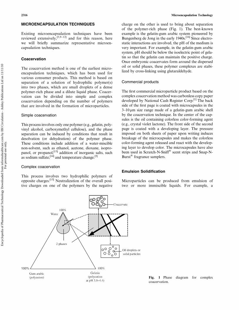
Table 1 Selection of microencapsulation methods for drugs with different properties
Water-soluble drugs Water-insoluble drugs
Liquid drugs w/o/w double emulsion[37] o/w or o/o emulsion[42,43]
w/o/o double emulsion[38] Coacervation[1]
w/o emulsion[27,39] Spray drying[7]
Ionic gelation[39]
Spray drying[41]
Solid drugs Cryogenic solvent extraction method[32,33] Coacervation[46]
s/o/w or s/o/o emulsion[44] Fluid-bed coatingFluid-bed coating[45]
Supercritical fluid[35]
2320 Microencapsulation Technology
Enc
yclo
pedi
a of
Pha
rmac
eutic
al T
echn
olog
y D
ownl
oade
d fr
om in
form
ahea
lthca
re.c
om b
y IB
I C
ircu
latio
n -
Ash
ley
Publ
icat
ions
Ltd
on
11/1
1/10
For
pers
onal
use
onl
y.

acidic microenvironments throughout the release per-iod.[48,56] Acidic pH can cause a number of undesirableevents. First, the acidic conditions can facilitatehydrolysis of peptide bonds and disulfide exchange.[57]
Second, some proteins can undergo conformationaltransition at low pHs and induce non-covalent aggre-gation.[48] Third, it was shown that an acidic environ-ment developed within microspheres contributed todeamidation and covalent dimerization of insulin.[58]
Fourth, lactic acid and glycolic acid units accumulatedwithin degrading microspheres can induce acylation ofpeptides.[59] On the other hand, adsorption of proteinsto the polymer surface is another potential source thatdisturbs structural integrity of the encapsulated pro-tein.[55,60] Among various interactions involved in theprotein adsorption, hydrophobic interactions are gen-erally regarded as the main driving force of adsorptionand aggregation causing incomplete release.[61]
Strategies to address the instability issues
Different approaches have been pursued in an attemptto improve stability of the encapsulated drugs. Table 2
summarizes various approaches according to inacti-vation sources.
Recent approaches include a new microencapsula-tion method that was developed in an attempt toaccommodate most of the above strategies.[70,71] Thisnew method called the ‘‘solvent exchange method’’ isbased on interfacial mass transfer between two con-tacting liquids, which results in reservoir-type micro-capsules. Fig. 3 describes one method of makingmicrocapsules. Series of polymer solution dropletsand aqueous drug solution droplets are separately pro-duced using ink-jet nozzles, and then are induced tocollide in air. Following the collision, the two liquidphases are separated as a core and a membrane withinthe merged microdroplets due to the surface tensiondifference as well as incompatibility of the two liquids.There are several potential advantages of this method.First, the process does not include potentially dam-aging conditions such as an emulsification step butemploys a mild drop generation protocol. Second, inthe mononuclear microcapsules, undesirable interac-tions between protein and organic solvent or polymermatrix are limited only to the interface at the surface
Table 2 Common inactivation sources of encapsulated proteins and the stabilization strategies
Inactivation source Stabilization strategy
Exposure to w/o interfaces Reducing or avoiding denaturation at w/o interfacesby including protective excipients;[55,63] employing anhydrousmicroencapsulation processes.[44]
Exposure to the hydrophobicorganic solvent
Using more hydrophilic solvents such asethyl acetate[17] and methyl ethyl ketone.[64]
Physical stressesduring preparation
Reducing physical stresses by eliminating the emulsificationstep and employing low temperature processes.[32,33]
Acidification of themicroenvironment
Counteracting acidification by coencapsulating antacid excipients;[48]
increasing permeability of the polymer matrix;[65] modifyingdegradation characteristics of the polymer.[65,66]
Interaction between
protein and polymer
Preventing adsorption by modifying hydrophilicity of the
polymer surface;[67] pre-entrapment of protein in the hydrophilic core;[68]
including adsorption competitors.[69]
(From Ref.[62].)
Fig. 3 Microencapsulation based on the solvent exchange method.
Microencapsulation Technology 2321
Enc
yclo
pedi
a of
Pha
rmac
eutic
al T
echn
olog
y D
ownl
oade
d fr
om in
form
ahea
lthca
re.c
om b
y IB
I C
ircu
latio
n -
Ash
ley
Publ
icat
ions
Ltd
on
11/1
1/10
For
pers
onal
use
onl
y.

of the core. A recent study showed that these reservoir-type microcapsules were able to release the encapsulatedlysozyme at a near zero-order kinetics for over 50 days.The released lysozyme remained functionally intact,which suggests that the protein survived the microencap-sulation process without losing its biological activity.Third, the organic solvent for polymers can be chosenwith more flexibility than in conventional methods.Hence, toxicity concerns over residual solvents, in parti-cular, methylene chloride, can be avoided.
Control of Drug Release
Drug release from biodegradable polymer microparti-cles is determined by the polymer degradation kinetics,structural features of the microparticles, and distribu-tion of the drugs within the particle matrix. The ultimategoal of microparticle systems in the controlled drugdelivery is to achieve readily tunable release profiles,which has been pursued in various perspectives.
Polymer chemistry
Polyanhydrides[72] and poly(ortho esters)[73] are alterna-tive biocompatible, biodegradable polymers that haveobtained the FDA approval for the treatment of a braintumor (Gliadel� wafer, approved in 1996) and are cur-rently under the clinical phase II trial as of 2004 forthe treatment of postsurgical pain management,[74]
respectively. Unlike PLGA polymers, which undergobulk degradation, these polymers have in common thattheir erosion processes are confined to the surface layers.Consequently, the drug release is primarily controlled byerosion of the surface. The interior of the matrix remainsessentially neutral in pH because the hydrolysis productsdiffuse away from the device.[73] Microparticles fabri-cated using poly(ortho esters) showed lower initial burstsand sustained release profiles of a model protein.[75]
Hybrids of existing biodegradable polymers were intro-duced in order to increase versatility of the polymericsystems taking advantage of different attributes ofthe participant polymers. For example, copolymers of
polyesters and polyanhydrides were synthesized toachieve better control over the degradation propertiesand drug encapsulation.[76]
Formulation efforts
If a high initial dose is not an intended effect as in thevaccine or antibiotic delivery, the initial burst is, inmost cases, an undesirable form of drug release. How-ever, it is often true that microparticle systems contain-ing hydrophilic drugs, such as proteins, display a largeinitial burst before they can reach a stable release rate.Initial burst is generally ascribed to two possiblecauses. First, drug distribution in the matrix is hete-rogeneous. Drugs that are either loosely associatedwith the surface or embedded in the surface layer areresponsible for the burst release.[77] Second, the micro-particles may have porous structure. Drugs can escapethrough the pores and cracks that form during themicroparticle fabrication process.[78,79]
One way of reducing the surface-bound drugs is tomake a blank layer on the microparticle surface. In thisregard, it is worthy to note the recent efforts to makemultiwall polymeric microparticles. The additionallayers have traditionally been made using the spraycoating method. However, the major disadvantagesare relatively large size of the resulting particles andlow production efficiency. Alternatively, double-walledmicroparticles can be prepared utilizing phase sepa-ration of constituent polymers:[80,81] A mixture of twopolymer solutions, such as polyanhydride and PLA,is emulsified in an aqueous continuous phase. As thesolvent evaporates, the polymer solution concentrationincreases to the point where they are no longer mutuallysoluble and begin to separate. However, since thelocation of individual polymer in the microparticlesmainly depends on the thermodynamics of the phaseseparation, there is not much control over the wallcomposition of the microparticles. On the other hand,a recent study showed that this limitation could beovercome by utilizing multiple concentric nozzles andcontrolling the flow rates and concentrations of thepolymer solutions (Fig. 4).[82] Another way of making
Fig. 4 Scanning electron micrographs of PLGA encapsulating poly[(1,6-bis-carboxyphenoxy)hexane] (PCPH) (A), PCPHencapsulating PLGA (PCPH : PLGA mass ratio ¼ 2 : 1) (B), and PCPH encapsulating PLGA (PCPH : PLGA massratio ¼ 3 : 1) (C). Scale bar ¼ 25 mm. (From Ref.[82].)
2322 Microencapsulation Technology
Enc
yclo
pedi
a of
Pha
rmac
eutic
al T
echn
olog
y D
ownl
oade
d fr
om in
form
ahea
lthca
re.c
om b
y IB
I C
ircu
latio
n -
Ash
ley
Publ
icat
ions
Ltd
on
11/1
1/10
For
pers
onal
use
onl
y.

coated microparticles involves sequential formation of amatrix core containing drugs and a wall polymer coat-ing.[83] An in vitro release study using tetanus toxoidas a model drug showed that the coating was effectivein suppressing the initial burst.[83]
Migration of drugs during drying and storage stepsis sometimes responsible for a heterogeneous drug dis-tribution in the polymer matrix.[78] During the air orvacuum drying process, the residual solvent (usuallywater) flows to the matrix surfaces before evaporation.Here, drugs also diffuse toward the surface along withwater and result in heterogeneous drug distributionin the polymer matrix. In this case, the drying methodcan make a difference: For example, the burst releasewas significantly reduced[77] or almost entirely elimi-nated[84] by the freeze-drying.
High affinity of the encapsulated drug to the con-tinuous phase is another reason for heterogeneousdrug distribution. For efficient internalization of thedrug within the polymer matrix, formulation andfabrication parameters are varied. Addition of a smallfraction of glycerol into the discontinuous phase wasfound to be effective in enhancing internalization ofthe drug (insulin) to the polymer phase.[85] The initialburst decreased from 40% to 10% by this approach.In another example, increasing hydrophilicity of theencapsulating polymer by polyethylene glycol (PEG)modification contributed to reducing the presence ofthe surface-associated drug and the initial burst.[86]
Relatively homogeneous drug distribution can also beachieved by generating a fine primary emulsion.[87,88]
The fine primary emulsion was obtained using a highenergy homogenization method such as sonication[88]
or high shear rate application.[87]
In alternative approaches, improvement of thestructural features of microparticles was soughtthrough various formulation parameters. In general,low molecular weight polymers result in high burstrelease.[79] It is partly because the low molecular weightpolymer is more soluble in the organic solvent andundergoes slow solidification to produce more porousmicroparticles. Polymer concentration,[89] compositionof the copolymer,[90] and hydrophilicity of the poly-mer[91] also affect the degree of initial burst by govern-ing the rate of polymer solidification. In case of theemulsion method, composition of the continuousphase influences particle size, encapsulation efficiency,and/or initial burst.[79,92] High concentration of PVAsuppresses the initial burst because the viscosity ofthe continuous phase prevents migration of the inter-nal aqueous phase toward the continuous phase.[79]
Addition of salt or sugar into the continuous phasecan also contribute to suppression of the initialburst.[93] The presence of salt or sugar increases theosmotic pressure across the polymer phase, which isin essence a semipermeable membrane in the semisolid
state. This increase in osmotic pressure prevents influxof the continuous phase into the dispersed phase andreduces the formation of water channels, which oftenresults in a high initial burst.
Size control
Particle sizes are known to have significant influenceson the release rate.[94,95] The effects vary dependingon the drug release mechanism. When the drug releaseis mainly controlled by diffusion, which is usually thecase of the early stage of release from polymericmicroparticles or hydrogel-based microparticles,smaller microparticles tend to release the drugs at arelatively high rate due to the large surface area andthe short diffusion distance.[96] On the other hand,when the release depends on the polymer degradation,the particle size controls the release rate by affectingadditional features such as drug distribution and poly-mer degradation rate.[97]
In this regard, a variety of efforts have been madeto obtain microparticles of monodisperse and con-trollable sizes. A common feature of these approachesis that the dispersed phase is formed by fragmentationof a liquid jet, which is a more readily controllableprocess as compared to emulsification. A few exampleshave shown promising results in controlling the par-ticle size distribution.
A method developed by Amsden and Goosen[98]
involves extruding a solution through a needle andthen an electric field, which pulls the droplets off theend of the needle. Applicability of this method tomicroparticle fabrication for the controlled releasewas demonstrated using a solution of ethylene vinylacetate and BSA. It was shown that the particle sizecould be controlled by varying size of the needle andstrength of the electric field. However, this methodwas not effective in reducing the mean diameter main-taining a narrow size distribution. This limitation waslater alleviated using a continuous phase flowing per-pendicularly past the needle tip, which aids in over-coming interfacial tension between the polymersolution and the needle tip.[99]
Researchers at the University of Illinois utilize asmall-gauge needle that vibrates at an ultrasonic fre-quency for this purpose.[100] A jet of a polymer solutionpassing the needle breaks up into uniform droplets,which becomes solidified micromatrices after solventremoval. The droplet size can be precisely controlledas a function of orifice size, solution flow rate, andvibration frequency. An optional carrier streamenables further reduction of the particle size. In vitrorelease studies using microparticles of different meandiameters demonstrated the dependence of the diffusion-dependent release profiles on the particle size.[96]
Microencapsulation Technology 2323
Enc
yclo
pedi
a of
Pha
rmac
eutic
al T
echn
olog
y D
ownl
oade
d fr
om in
form
ahea
lthca
re.c
om b
y IB
I C
ircu
latio
n -
Ash
ley
Publ
icat
ions
Ltd
on
11/1
1/10
For
pers
onal
use
onl
y.

The solvent exchange method utilizing the dualmicrodispenser system also demonstrates a goodpotential of controlling the microcapsule size.[70] Here,two ink-jet nozzles carrying a polymer solution and anaqueous drug solution, respectively, are arranged in away that two emerging liquid jets can collide resultingin reservoir-type microcapsules (Fig. 5A). The ink-jetnozzles operate in the same mechanism as the small-gauge needle described above to generate homo-geneous microdroplets. The size of merged droplets,i.e., the microcapsules, is 1.26 times of the singledroplets, when measured right after the collision, indi-cating that there is no volume loss upon the collision(Fig. 5A). On the other hand, the majority of micro-capsules collected in the water bath were close to singledroplets in size, and the membrane existed only as athin membrane (Fig. 5B). It is likely that the polymerlayer shrank as the solvent that constituted themajority of the polymer phase was extracted into theaqueous phases by the solvent exchange.[70]
RECENT APPLICATIONS OF THEMICROPARTICLE SYSTEMS
Use of microparticle systems is not limited to thesustained or local delivery and has a wide range ofapplications. A limited number of examples are intro-duced below.
Since it was first noticed that bioerodible hydro-phobic polyanhydrides [e.g., poly(fumaric-co-sebacicanhydride)] exhibited strong bioadhesiveness, micro-particles made of these polymers have been investi-gated as potential oral drug delivery systems.[101] Thebioadhesiveness in this case comes from the hydrogenbonding interactions between mucin and carboxylicacid groups that form during the polymer erosion.[102]
Furthermore, the small size of microparticles providesadditional advantages by promoting cellular uptake ofthe formulation. Taking advantage of both the small
size of microparticles and the chemical attributes ofthe polymeric system, the bioadhesive microparticleshave shown enhanced oral bioavailability ofdicumarol,[103] insulin, and DNA.[101]
Polymeric microspheres have been used for deliveryof DNA vaccines, which enable prolonged immuneresponses through sustained release of DNA encodinga protein antigen. It has been known that <10 mmparticles are preferentially internalized throughphagocytosis by macrophages and antigen-presentingcells.[91] Poly(lactic-co-glycolic acid) was used as anencapsulating polymer in the initial research withpromising results.[91] On the other hand, disadvantagesof PLGA particles in delivering DNA vaccines wereacidification of microenvironment, which can inacti-vate encapsulated DNA,[49] and slow release rate, whichdoes not catch up with the life span of the target cellssuch as dendritic cells. In order to provide for rapidand tunable release and to avoid internal acidification,use was recently made of pH-triggered biodegradablepolymers based on poly(ortho esters)[50] and poly-b-amino esters.[104] These microparticles loaded withDNA were successfully internalized into the antigen-presenting cells, enhanced immune response, andsuppressed in vivo tumor challenges significantly.[50,51]
CONCLUSIONS
The microencapsulation technology, which started as away of encapsulating dyes and flavors, has nowbecome one of the most intriguing fields in the areaof controlled drug delivery systems. The encapsulationtechniques have been advanced to such a level that notonly small molecular weight drugs but also macro-molecules, such as proteins and genes, can be deliveredvia microparticle carriers. Although the technologicaladvances have led to commercialization of severalmicroparticulate products in recent years, many techni-cal problems are to be overcome yet. Examples of such
Fig. 5 Stroboscopic images of microcapsule formation via midair collision between two component liquids (scalebar ¼ 100mm) (A) and bright-field microscope images of microcapsules (B). The left and streams are 0.25% alginate solutionand 4% PLGA solution, respectively. The nozzle orifice diameter d ¼ 60 mm; volumetric flow rate Q ¼ 0.6ml/min; and forcingfrequency f ¼ 10.6 kHz. (From Ref.[70].)
2324 Microencapsulation Technology
Enc
yclo
pedi
a of
Pha
rmac
eutic
al T
echn
olog
y D
ownl
oade
d fr
om in
form
ahea
lthca
re.c
om b
y IB
I C
ircu
latio
n -
Ash
ley
Publ
icat
ions
Ltd
on
11/1
1/10
For
pers
onal
use
onl
y.
hurdles are maintaining the stability of encapsulateddrugs throughout the lifetime of the products,manipulating release rates according to the applica-tions, and transferring bench scale processes to themanufacturing scale. Some of the answers to those pro-blems have been provided by advances in polymerchemistry, formulation efforts, and recent progressesin new microencapsulation techniques. The microen-capsulation technology will remain as one of the mostimportant areas in drug delivery and various otherapplications.
ACKNOWLEDGMENT
This study was supported in part by the NationalInstitutes of Health through grant GM67044.
ARTICLE OF FURTHER INTEREST
Microsphere Technology and Applications, p. 2328.
REFERENCES
1. Green, B.K.; Schleicher, L. Oil-containing MicroscopicCapsules and Method of Making Them. U.S. Patent2800457, 1957.
2. Bakan, J.A.; Powell, T.C.; Szotak, P.S. Recent advancesusing microencapsulation for taste-masking of bitterdrugs. In Microcapsules and Nanoparticles in Medicineand Pharmacy; Donbrow, M., Ed.; CRC Press: BocaRaton, FL, 1992; 149–156.
3. Arica, B.; Arica, M.Y.; Kas, H.S.; Hincal, A.A.; Hasirci, V.In-vitro studies of enteric coated diclofenac sodium car-boxymethyl cellulose microspheres. J. Microencapsul.1996, 13 (6), 689–699.
4. Muller, R.H.; Peters, K. Nanosuspensions for the formu-lation of poorly soluble drugs. I. Preparation by a size-reduction technique. Int. J. Pharm. 1998, 160 (2), 229–237.
5. Mathiowitz, E.; Kreitz, M.R. Microencapsulation. InEncyclopedia of Controlled Drug Delivery; Mathiowitz,E., Ed.; John Wiley & Sons, Inc.: New York, U.S.A.,1999; Vol. 2, 493–546.
6. Lim, F.; Sun, A.M. Microencapsulated islets as bioartifi-cial endocrine pancreas. Science 1980, 210, 908–910.
7. Kohane, D.S.; Lipp, M.; Kinney, R.C.; Lotan, N.; Langer,R. Sciatic nerve blockade with lipid-protein-sugar particlescontaining bupivacaine. Pharm. Res. 2000, 17 (10), 1243–1249.
8. Ying, M.; Thomasin, C.; Merkle, H.P.; Gander, B.;Corradin, G. A single administration of tetanus toxoidin biodegradable microspheres elicits T cell and antibodyresponses similar or superior to those obtained with alumi-num hydroxide. Vaccine 1995, 13 (7), 683–689.
9. Yeo, Y.; Baek, N.; Park, K. Microencapsulation methodsfor delivery of protein drugs. Biotechnol. Bioprocess Eng.2001, 6 (4), 213–230.
10. Benoit, J.-P.; Marchais, H.; Rolland, H.; Velde, V.V.Biodegradable microspheres: advances in production tech-nology. In Microencapsulation: Methods and IndustrialApplication; Benita, S., Ed.; Marcel Dekker, Inc.:New York, U.S.A., 1996; Vol. 73, 35–72.
11. Thies, C. A survey of microencapsulation processes. InMicroencapsulation: Methods and Industrial Applications;
Benita, S., Ed.; Marcel Dekker, Inc.: New York, U.S.A.,1996; Vol. 73, 1–19.
12. Gouin, S. Microencapsulation industrial appraisal of exist-ing technologies and trends. Trends Food Sci. Technol.2004, 15 (7–8), 330–347.
13. Mohanty, B.; Bohidar, H.B. Systematic of alcohol-inducedsimple coacervation in aqueous gelatin solutions. Bioma-cromolecules 2003, 4 (4), 1080–1086.
14. Weiss, G.; Knoch, A.; Laicher, A.; Stanislaus, F.; Daniels,R. Simple coacervation of hydroxypropyl methyl cellulosephthalate (HPMCP). I. Temperature and pH dependencyof coacervate formation. Int. J. Pharm. 1995, 124 (1), 87–96.
15. Burgess, D.J.; Singh, O.N. Spontaneous formation ofsmall sized albumin/acacia coacervate particles. J. Pharm.Pharmacol. 1993, 45, 586–591.
16. de Jong, H.G.B. Complex colloid systems (Chapter X). InColloid Science; Elsevier: New York, 1949.
17. Sah, H. Microencapsulation techniques using ethyl acetateas a dispersed solvent: effects of its extraction rate on thecharacteristics of PLGA microspheres. J. ControlledRelease 1997, 47 (3), 233–245.
18. Herrmann, J.; Bodmeier, R. Somatostatin containing bio-degradable microspheres prepared by a modified solventevaporation method based on W/O/W-multiple emul-sions. Int. J. Pharm. 1995, 126 (1–2), 129–138.
19. Bodmeier, R.; McGinity, J.W. Solvent selection in thepreparation of PLA microspheres prepared by the solventevaporation method. Int. J. Pharm. 1988, 43, 179–186.
20. Yeo, Y.; Park, K. Control of encapsulation efficiency andinitial burst in polymeric microparticle systems. Arch.Pharmacol. Res. 2004, 27 (1), 1–12.
21. Kempen Diederik, H.R.; Lu, L.; Zhu, X.; Kim, C.; Jabbari,E.; Dhert Wouter, J.A.; Currier Bradford, L.; YaszemskiMichael, J. Development of biodegradable poly(propylenefumarate)/poly(lactic-co-glycolic acid) blend microspheres.II. Controlled drug release and microsphere degradation.J. Biomed. Mater. Res. 2004, 70A (2), 293–302.
22. Dittrich, M.; Hampl, J.; Soukup, F. Branched oligoestermicrospheres fabricated by a rapid emulsion solvent extrac-tion method. J. Microencapsul. 2000, 17 (5), 587–598.
23. Kas, H.S. Chitosan: properties, preparations and appli-cation to microparticulate systems. J. Microencapsul.1997, 14 (6), 689–711.
24. Kumbar, S.G.; Kulkarni, A.R.; Aminabhavi, T.M. Cross-linked chitosan microspheres for encapsulation of diclofe-nac sodium: effect of crosslinking agent. J. Microencapsul.2002, 19 (2), 173–180.
25. Kim, S.E.; Park, J.H.; Cho, Y.W.; Chung, H.; Jeong, S.Y.;Lee, E.B.; Kwon, I.C. Porous chitosan scaffold containingmicrospheres loaded with transforming growth factor-beta1: implications for cartilage tissue engineering. J.Controlled Release 2003, 91, 365–374.
26. Yun, Y.H.; Goetz, D.J.; Yellen, P.; Chen, W. Hyaluronanmicrospheres for sustained gene delivery and site-specifictargeting. Biomaterials 2004, 25 (1), 147–157.
27. Chickering, D.E.; Jacob, J.S.; Desai, T.A.; Harrison, M.;Harris, W.P.; Morrell, C.N.; Chaturvedi, P.; Mathiowitz,E. Bioadhesive microspheres: III. An in vivo transit andbioavailability study of drug-loaded alginate andpoly(fumaric-co-sebacic anhydride) microspheres. J. Con-trolled Release 1997, 48, 35–46.
28. Whateley, T.L. Microcapsules: preparation by interfacialpolymerization and interfacial complexation and theirapplication. In Microencapsulation: Methods and Indus-trial Applications; Benita, S., Ed.; Dekker: New York,U.S.A., 1996; Vol. 73, 349–375.
29. Ting, T.; Gonda, I.; Gripps, E.M. Microparticles of PVAfor nasal delivery. I. Generation by spray-drying andspray-desolvation. Pharm. Res. 1992, 9 (10), 1330–1335.
30. Sam, A.P.; Haan, F.D.; Dirix, C. A Novel Process forManufacturing PLG Microparticles by Spray DesolvationAvoiding the use of Toxic Solvents. InternationalSymposium on Controlled Release of Bioactive Materials.Nice, France, 1994; 198–199.
Microencapsulation Technology 2325
Enc
yclo
pedi
a of
Pha
rmac
eutic
al T
echn
olog
y D
ownl
oade
d fr
om in
form
ahea
lthca
re.c
om b
y IB
I C
ircu
latio
n -
Ash
ley
Publ
icat
ions
Ltd
on
11/1
1/10
For
pers
onal
use
onl
y.

31. Gombotz, W.R.; Healy, M.S.; Brown, L.R. Very LowTemperature Casting of Controlled Release Microspheres.U.S. Patent 5019400, 1991
32. Johnson, O.L.; Cleland, J.L.; Lee, H.J.; Charnis, M.;Duenas, E.; Jaworowicz, W.; Shepard, D.; Shahzamani,A.; Jones, A.J.; Putney, S.D. A month-long effect from asingle injection of microencapsulated human growth hor-mone. Nat. Med. 1996, 2, 795–799.
33. Lee, H.J.; Riley, G.; Johnson, O.; Cleland, J.L.; Kim, N.;Charnis, M.; Bailey, L.; Duenas, E.; Shahzamani, A.;Marian, M.; Jones, A.J.; Putney, S.D. In vivo characteri-zation of sustained-release formulations of human growthhormone. J. Pharmacol. Exp. Ther. 1997, 281 (3), 1431–1439.
34. Santos, I.R.D.; Richard, J.; Pech, B.; Thies, C.; Benoit, J.P.Microencapsulation of protein particles within lipids usinga novel supercritical fluid process. Int. J. Pharm. 2002,242 (1–2), 69–78.
35. Young, T.J.; Johnston, K.P.; Mishima, K.; Tanaka, H.Encapsulation of lysozyme in a biodegradable polymerby precipitation with a vapor-over-liquid antisolvent.J. Pharm. Sci. 1999, 88 (6), 640–650.
36. Sunkara, G.; Kompella, U.B. Drug delivery applicationsof supercritical fluid technology. Drug Delivery Technol.2002, 44 (1), 46–50.
37. Ogawa, Y.; Yamamoto, M.; Okada, H.; Yashiki, T.;Shimamoto, T. A new technique to efficiently entrap leu-prolide acetate into microcapsules of polylactic acid orcopoly(lactic/glycolic) acid. Chem. Pharm. Bull. 1988,36 (3), 1095–1103.
38. Viswanathan, N.B.; Thomas, P.A.; Pandit, J.K.; Kulkarni,M.G.; Mashelkar, R.A. Preparation of non-porous micro-spheres with high entrapment efficiency of proteins by a(water-in-oil)-in-oil emulsion technique. J. ControlledRelease 1999, 58 (1), 9–20.
39. Lim, S.T.; Martin, G.P.; Berry, D.J.; Brown, M.B.Preparation and evaluation of the in vitro drug releaseproperties and mucoadhesion of novel microspheres ofhyaluronic acid and chitosan. J. Controlled Release2000, 66, 281–292.
40. Takka, S.; Acarturk, F. Calcium alginate microparticlesfor oral administration. I. Effect of sodium alginate typeon drug release and drug entrapment efficiency. J. Micro-encapsul. 1999, 16 (3), 275–290.
41. Takada, S.; Uda, Y.; Toguchi, H.; Ogawa, Y. Applicationof a spray drying technique in the production of TRH-containing injectable sustained-release microparticles ofbiodegradable polymers. PDA J. Pharm. Sci. Technol.1995, 49 (4), 180–184.
42. Ruan, G.; Feng, S.-S. Preparation and characterization ofpoly(lactic acid)-poly(ethylene glycol)-poly(lactic acid)(PLA-PEG-PLA) microspheres for controlled release ofpaclitaxel. Biomaterials 2003, 24, 5037–5044.
43. Hickey, A.J.; Tian, Y.; Parasrampuria, D.; Kanke, M.Biliary elimination of bromsulfthalein, phenolphthalein,and doxorubicin released from microspheres followingintravenous administration. Biopharm. Drug Disposition1993, 14 (2), 181–186.
44. Perez, C.; Castellanos, I.J.; Costantino, H.R.; Al-Azzam,W.; Griebenow, K. Recent trends in stabilizing proteinstructure upon encapsulation and release from bioerodiblepolymers. J. Pharm. Pharmacol. 2002, 54 (3), 301–313.
45. Knezevic, Z.; Gosaki, D.; Hraste, M.; Jalsenjak, I. Fluid-bed microencapsulation of ascorbic acid. J. Microencap-sul. 1998, 15 (2), 237–252.
46. Palmieri, G.F.; Lauri, D.; Martelli, S.; Wehrle, P. Methoxy-butropate microencapsulation by gelatin–acacia complexcoacervation. Drug Dev. Ind. Pharm. 1999, 25 (4), 399–407.
47. Okada, H.; Heya, T.; Ogawa, Y.; Shimamoto, T. One-month release injectable microcapsules of luteinizinghormone-releasing hormone agonist (leuprolide acetate)for treating experimental endometriosis in rats. J. Pharma-col. Exp. Ther. 1988, 244 (2), 744–750.
48. Zhu, G.; Mallery, S.R.; Schwendeman, S.P. Stabilizationof proteins encapsulated in injectable PLGA. Nat. Bio-technol. 2000, 18, 52–57.
49. Walter, E.; Moelling, K.; Pavlovic, J.; Merkle, H.P. Micro-encapsulation of DNA using poly(DL-lactide-co-glycolide):stability issues and release characteristics. J. ControlledRelease 1999, 61 (3), 361–374.
50. Wang, C.; Ge, Q.; Ting, D.; Nguyen, D.; Shen, H.-r.; Chen,J.; Eisen, H.N.; Heller, J.; Langer, R.; Putnam, D. Molecu-larly engineered poly(ortho ester) microspheres forenhanced delivery of DNA vaccines. Nat. Mater. 2004,3, 190–196.
51. Little, S.R.; Lynn, D.M.; Ge, Q.; Anderson, D.G.; Puram,S.V.; Chen, J.; Eisen, H.N.; Langer, R. Poly-beta aminoester-containing microparticles enhance the activity ofnon-viral genetic vaccines. Proc. Natl. Acad. Sci. U.S.A.2004, 101 (26), 9534–9539.
52. Schwendeman, S.P. Recent advances in the stabilization ofproteins encapsulated in injectable PLGA delivery sys-tems. Crit. Rev. Ther. Drug Carrier Syst. 2002, 19 (1),73–98.
53. van de Weert, M.; Hennink, W.E.; Jiskoot, W. Proteininstability in PLGA microparticles. Pharm. Res. 2000,17 (10), 1159–1167.
54. Sah, H. Protein instability toward organic solvent/wateremulsification: implications for protein microencapsula-tion into microspheres. PDA J. Pharm. Sci. Technol.1999, 53 (1), 3–10.
55. Lu, W.; Park, T.G. Protein release from poly(lactic-co-gly-colic acid) microspheres: protein stability problems. PDAJ. Pharm. Sci. Technol. 1995, 49 (1), 13–19.
56. Fu, K.; Pack, D.W.; Klibanov, A.M.; Langer, R. Visualevidence of acidic environment within degrading poly(lactic-co-glycolic acid) (PLGA) microspheres. Pharm.Res. 2000, 17 (1), 100–106.
57. Manning, M.C.; Patel, K.; Borchardt, R.T. Stability ofprotein pharmaceuticals. Pharm. Res. 1989, 6 (11), 903–918.
58. Shao, P.G.; Bailey, L.C. Porcine insulin biodegradablepolyester microspheres: stability and in vitro release char-acteristics. Pharm. Dev. Technol. 2000, 5 (1), 1–9.
59. Lucke, A.; Kiermaier, J.; Gopferich, A. Peptide acylationby poly(alpha-hydroxy esters). Pharm. Res. 2002, 19 (2),175–181.
60. Crotts, G.; Park, T.G. Stability and release of bovineserum albumin encapsulated within PLGA microparticles.J. Controlled Release 1997, 44, 123–134.
61. Sluzky, V.; Klibanov, A.M.; Langer, R. Mechanism ofinsulin aggregation and stabilization in agitated aqueoussolutions. Biotechnol. Bioeng. 1992, 40, 895–903.
62. Yeo, Y.; Park, K. Microencapsulation of protein drugs: anovel approach. In Biomaterials Handbook—AdvancedApplications of Basic Sciences and Bioengineering; Wise,D.L., Hasirci, V., Lewandrowski, K.-U., Yaszemski, M.J.,Altobelli, D.E., Trantolo, D.J., Eds.; Marcel Dekker, Inc.,2004; 305–332.
63. Sah, H. Stabilization of proteins against methylene chlor-ide/water interface-induced denaturation and aggregation.J. Controlled Release 1999, 58 (2), 143–151.
64. Sah, H.; Smith, M.; Chern, R. A novel method of prepar-ing PLGA microcapsules utilizing methylethyl ketone.Pharm. Res. 1996, 13 (3), 360–367.
65. Jiang, W.; Schwendeman, S.P. Stabilization and controlledrelease of bovine serum albumin encapsulated in poly(D,L-lactide) and poly(ethylene glycol) microsphere blends.Pharm. Res. 2001, 18 (6), 878–885.
66. Yang, Y.Y.; Wan, J.P.; Chung, T.S.; Pallathadka, P.K.;Ng, S.; Heller, J. POE–PEG–POE triblock copolymericmicrospheres containing protein I. Preparation and char-acterization. J. Controlled Release 2001, 75 (1–2), 115–128.
67. Bouillot, P.; Ubrich, N.; Sommer, F.; Duc, T.M.; Loeffler,J.P.; Dellacherie, E. Protein encapsulation in biodegrad-able amphiphilic microspheres. Int. J. Pharm. 1999,181 (2), 159–172.
2326 Microencapsulation Technology
Enc
yclo
pedi
a of
Pha
rmac
eutic
al T
echn
olog
y D
ownl
oade
d fr
om in
form
ahea
lthca
re.c
om b
y IB
I C
ircu
latio
n -
Ash
ley
Publ
icat
ions
Ltd
on
11/1
1/10
For
pers
onal
use
onl
y.

68. Wang, N.; Wu, X.S.; Li, J.K. A heterogeneously structuredcomposite based on poly(lactic-co-glycolic acid) micro-spheres and poly(vinyl alcohol) hydrogel nanoparticlesfor long-term protein drug delivery. Pharm. Res. 1999,16 (9), 1430–1435.
69. Blanco, M.D.; Alonso, M.J. Development and characteri-zation of protein-loaded poly(lactide-co-glycolide) nano-spheres. Eur. J. Pharm. Biopharm. 1997, 43, 287–294.
70. Yeo, Y.; Chen, A.U.; Basaran, O.A.; Park, K. Solventexchange method: a novel microencapsulation techniqueusing dual microdispensers. Pharm. Res. 2004, 21 (8),1419–1427.
71. Yeo, Y.; Basaran, O.A.; Park, K. A new process formaking reservoir-type microcapsules using ink-jet tech-nology and interfacial phase separation. J. ControlledRelease 2003, 93 (2), 161–173.
72. Kumar, N.; Langer, R.S.; Domb, A.J. Polyanhydrides:an overview. Adv. Drug Delivery Rev. 2002, 54 (7), 889–910.
73. Heller, J.; Barr, J.; Ng, S.Y.; Abdellauoi, K.S.; Gurny, R.Poly(ortho esters): synthesis, characterization, propertiesand uses. Adv. Drug Delivery Rev. 2002, 54 (7), 1015–1039.
74. Heller, J. Poly(ortho esters)—some recent developments.Polym. Mater. Sci. Eng. 2003, 89, 189.
75. Bai, X.-L.; Yang, Y.-Y.; Chung, T.-S.; Ng, S.; Heller, J.Effect of polymer compositions on the fabrication ofpoly(ortho-ester) microspheres for controlled release ofprotein. J. Appl. Polym. Sci. 2001, 80, 1630–1642.
76. Slivniak, R.; Domb, A.J. Stereocomplexes of enantiomericlactic acid and sebacic acid ester-anhydride triblock copoly-mers. Biomacromolecules 2002, 3, 754–760.
77. Igartua, M.; Hernandez, R.M.; Esquisabel, A.; Gascon,A.R.; Calvo, M.B.; Pedraz, J.L. Influence of formulationvariables on the in-vitro release of albumin from biode-gradable microparticulate systems. J. Microencapsul.1997, 14 (3), 349–356.
78. Huang, X.; Brazel, C.S. On the importance and mechan-isms of burst release in matrix-controlled drug deliverysystems. J. Controlled Release 2001, 73, 121–136.
79. Yang, Y.-Y.; Chung, T.-S.; Ping Ng, N. Morphology, drugdistribution, and in vitro release profiles of biodegradablepolymeric microspheres containing protein fabricated bydouble-emulsion solvent extraction/evaporation method.Biomaterials 2001, 22 (3), 231–241.
80. Pekarek, K.J.; Jacob, J.S.; Mathiowitz, E. Double-walledpolymer microspheres for controlled drug release. Nature1994, 367, 258–260.
81. Mathiowitz, E.; Langer, R. Multiwall Polymeric Micro-spheres. U.S. Patent 5912017, 1999
82. Berkland, C.; Pollauf, E.; Pack, D.W.; Kim, K.K. Uniformdouble-walled polymer microspheres of controllable shellthickness. J. Controlled Release 2004, 96, 101–111.
83. Gopferich, A.; Alonso, M.J.; Langer, R. Developmentand characterization of microencapsulated microspheres.Pharm. Res. 1994, 11 (11), 1568–1574.
84. Wang, H.T.; Schmitt, E.; Flanagan, D.R.; Linhardt, R.J.Influence of formulation methods on the in vitro con-trolled release of protein from poly(ester) microspheres.J. Controlled Release 1991, 17, 23–32.
85. Yamaguchi, Y.; Takenaga, M.; Kitagawa, A.; Ogawa, Y.;Mizushima, Y.; Igarashi, R. Insulin-loaded biodegradablePLGA microcapsules: initial burst release controlled byhydrophilic additives. J. Controlled Release 2002, 81 (3),235–249.
86. Li, X.; Deng, X.; Huang, Z. In vitro protein release anddegradation of poly-dl-lactide-poly(ethylene glycol) micro-spheres with entrapped human serum albumin. Pharm.Res. 2001, 18 (1), 117–124.
87. Sah, H.K.; Toddywala, R.; Chien, Y.W. Biodegradablemicrocapsules prepared by a w/o/w technique: effects ofshear force to make a primary w/o emulsion on theirmorphology and protein release. J. Microencapsul. 1995,12 (1), 59–69.
88. Yan, C.; Resau, J.H.; Hewetson, J.; Mest, M.; Rill, W.L.;Kende, M. Characterization and morphological analysisof protein-loaded poly(lactide-co-glycolide) microparticlesprepared by water-in-oil-in-water emulsion technique.J. Controlled Release 1994, 32, 231–241.
89. Yang, Y.-Y.; Chia, H.-H.; Chung, T.-S. Effect of prep-aration temperature on the characteristics and releaseprofiles of PLGA microspheres containing protein fabri-cated by double-emulsion solvent extraction/evaporationmethod. J. Controlled Release 2000, 69 (1), 81–96.
90. Sah, H.; Toddywala, R.; Chien, Y.W. The influence ofbiodegradable microcapsule formulations on the con-trolled release of a protein. J. Controlled Release 1994,30, 201–211.
91. Walter, E.; Dreher, D.; Kok, M.; Thiele, L.; Kiama, S.G.;Gehr, P.; Merkle, H.P. Hydrophilic poly(dl-lactide-co-glycolide) microspheres for the delivery of DNA tohuman-derived macrophages and dendritic cells. J. Con-trolled Release 2001, 76 (1–2), 149–168.
92. Hsu, Y.-Y.; Hao, T.; Hedley, M.L. Comparison of processparameters for microencapsulation of plasmid DNA inpoly(D,L-lactic-co-glycolic) acid microspheres. J. DrugTargeting 1999, 7 (4), 313–323.
93. Jiang, G.; Thanoo, B.C.; DeLuca, P.P. Effect of osmoticpressure in the solvent extraction phase on BSA releaseprofile from PLGA microspheres. Pharm. Dev. Technol.2002, 7 (4), 391–399.
94. Narayani, R.; Rao, K.P. Gelatin microsphere cocktails ofdifferent sizes for the controlled release of anticancerdrugs. Int. J. Pharm. 1996, 143, 255–258.
95. Bezemer, J.M.; Radersma, R.; Grijpma, D.W.; Dijkstra,P.J.; Blitterswijk, C.A.v.; Feijen, J. Microspheres forprotein delivery prepared from amphiphilic multiblockcopolymers: 2. Modulation of release rate. J. ControlledRelease 2000, 67, 249–260.
96. Berkland, C.; King, M.; Cox, A.; Kim, K.; Pack, D.W.Precise control of PLG microsphere size providesenhanced control of drug release rate. J. ControlledRelease 2002, 82, 137–147.
97. Berkland, C.; Kim, K.K.; Pack, D.W. Protein Releasefrom Uniform Poly(D,L-lactide-co-glycolide) Micro-spheres. 31st International Symposium on ControlledRelease of Bioactive Materials. Honolulu, Hawaii, 2004;pp. 472.
98. Amsden, B.G.; Goosen, M.F.A. An examination of factorsaffecting the size, distribution and release characteristics ofpolymer microbeads made using electrostatics. J. Con-trolled Release 1997, 43 (2–3), 183–196.
99. Amsden, B. The production of uniformly sized polymermicrospheres. Pharm. Res. 1999, 16 (7), 1140–1143.
100. Berkland, C.; Kim, K.; Pack, D.W. Fabrication of PLGmicrospheres with precisely controlled and monodispersesize distributions. J. Controlled Release 2001, 73, 59–74.
101. Mathiowitz, E.; Jacob, J.S.; Jong, Y.S.; Carino, G.P.;Chickering, D.E.; Chaturvedi, P.; Santos, C.A.; Vijaya-raghaven, K.; Montgomery, S.; Bassett, M.; Morrell, C.Biologically erodable microspheres as potential oral drugdelivery systems. Nature 1997, 386, 410–414.
102. Santos, C.A.; Freedman, B.D.; Ghosn, S.; Jacob, J.S.;Scarpulla, M.; Mathiowitz, E. Evaluation of anhydrideoligomers within polymer microsphere blends and theirimpact on bioadhesion and drug delivery in vitro. Bioma-terials 2003, 24, 3571–3583.
103. Thanos, C.G.; Liu, Z.; Goddard, M.; Reineke, J.; Bailey,N.; Cross, M.; Burrill, R.; Mathiowitz, E. Enhancing theoral bioavailability of the poorly soluble drug dicumarolwith a bioadhesive polymer. J. Pharm. Sci. 2003, 92 (8),1677–1689.
104. Lynn, D.M.; Langer, R. Degradable poly(beta-aminoesters): synthesis, characterization, and self-assembly withplasmid DNA. J. Am. Chem. Soc. 2000, 122, 10,761–10,768.
Microencapsulation Technology 2327
Enc
yclo
pedi
a of
Pha
rmac
eutic
al T
echn
olog
y D
ownl
oade
d fr
om in
form
ahea
lthca
re.c
om b
y IB
I C
ircu
latio
n -
Ash
ley
Publ
icat
ions
Ltd
on
11/1
1/10
For
pers
onal
use
onl
y.