Embed Size (px)
Citation preview

Hong, Chen, Groisman, Sci. Signal. 11, eaar7921 (2018) 8 May 2018
S C I E N C E S I G N A L I N G | R E S E A R C H A R T I C L E
1 of 11
M I C R O B I O L O G Y
Gene expression kinetics governs stimulus-specific decoration of the Salmonella outer membraneXinyu Hong,1,2,3,4 H. Deborah Chen,2* Eduardo A. Groisman2,3†
Lipid A is the innermost component of the lipopolysaccharide (LPS) molecules that occupy the outer leaflet of the outer membrane in Gram-negative bacteria. Lipid A is recognized by the host immune system and targeted by cationic antimicrobial compounds. In Salmonella enterica serovar Typhimurium, the phosphates of lipid A are chem-ically modified by enzymes encoded by targets of the transcriptional regulator PmrA. These modifications increase resistance to the cationic peptide antibiotic polymyxin B by reducing the negative charge of the LPS. We report the mechanism by which Salmonella produces different lipid A profiles when PmrA is activated by low Mg2+ versus a mildly acidic pH. Low Mg2+ favored modification of the lipid A phosphates with 4-amino-4-deoxy-l-aminoarabinose (l-Ara4N) by activating the regulatory protein PhoP, which initially increased the LPS negative charge by promoting transcription of lpxT, encoding an enzyme that adds an additional phosphate group to lipid A. Later, PhoP activated PmrA posttranslationally, resulting in expression of PmrA-activated genes, including those encoding the LpxT in-hibitor PmrR and enzymes responsible for the incorporation of l-Ara4N. By contrast, a mildly acidic pH favored mod-ification of the lipid A phosphates with a mixture of l-Ara4N and phosphoethanolamine (pEtN) by simultaneously inducing the PhoP-activated lpxT and PmrA-activated pmrR genes. Although l-Ara4N reduces the LPS negative charge more than does pEtN, modification of lipid A phosphates solely with l-Ara4N required a prior transient increase in lipid A negative charge. Our findings demonstrate how bacteria tailor their cell surface to different stresses, such as those faced inside phagocytes.
INTRODUCTIONGram-negative bacteria are surrounded by an outer membrane that confers protection from various toxic agents (1). The outer membrane is composed of phospholipids in the inner leaflet and lipopolysaccha-ride (LPS) in the outer leaflet. The innermost portion of the LPS is lipid A, followed by a central core region and the outermost O-antigen (2, 3). Phosphate residues in the lipid A and inner core are respon-sible for the negative charge of the LPS at neutral pH. This negative charge is normally neutralized by divalent cations, primarily Mg2+. However, certain bacteria chemically modify their LPS in response to specific environmental signals (2, 4), thereby altering permeabil-ity (5, 6), resistance to antibacterial compounds (7–10), and activa-tion of the immune system of host animals (2, 11).
The Gram-negative bacterium Salmonella enterica serovar Typhimurium chemically modifies its lipid A phosphates with 4-amino-4-deoxy- l-aminoarabinose (l-Ara4N) and phosphoethanolamine (pEtN) (Fig. 1A) when experiencing conditions that activate PmrA, a transcriptional activator of genes encoding lipid A–modifying enzymes and an in-hibitor of one such enzyme (4, 12). PmrA decreases the negative charge of lipid A in several ways. First, it promotes transcription of the pbgP operon and the ugd gene, which are responsible for the modification of the lipid A phosphates with l-Ara4N, and of the pmrC gene, which mediates modification of the lipid A phosphates with pEtN (9, 13) (pbgP is also referred to as arnT and pmrK; pmrC is also referred to as eptA). Second, PmrA directly activates expression of the pmrR gene, which encodes an inhibitor of LpxT (12), an enzyme responsible for
the incorporation of an additional phosphate group at position 1 of lipid A (Fig. 1A) (14). Modification of the lipid A phosphates with l-Ara4N reduces the LPS negative charge more than does modifica-tion of the lipid A phosphates with pEtN. The lipid A phosphates are modified with both l-Ara4N and pEtN in Salmonella harvested from macrophages (15).
PmrA is a response regulator that functions with the sensor PmrB as a two-component system. PmrA is activated (phosphorylated) when its cognate sensor PmrB detects Fe3+ (16) or a mildly acidic pH (17) in the periplasm (Fig. 1B) or when the noncognate sensor PhoQ is ac-tivated by its specific signals such as low periplasmic Mg2+ (18, 19). The latter activation entails PhoQ-mediated phosphorylation of its cog-nate response regulator PhoP (20), transcription of the PhoP-activated gene pmrD (18), and protection of phosphorylated PmrA by the PmrD protein (Fig. 1B) (21). Therefore, transcription of PmrA-activated genes is faster when the inducing signal is Fe3+ or a mildly acidic pH than when the inducing signal is low Mg2+ (Fig. 1, B and C) (22).
Modification of the lipid A phosphates with l-Ara4N and pEtN con-fers resistance to the cationic peptide antibiotic polymyxin B by re-ducing the negative charge of the lipid A (7, 13). A pmrA null mutant is 10,000 times more sensitive to polymyxin B than is wild-type Salmonella (16). Polymyxins constitute a “last-resort” antibiotic against multidrug-resistant isolates of Pseudomonas aeruginosa, Acinetobacter baumanii, and Klebsiella pneumoniae (23) but are not used to treat Salmonella infections. However, the better understanding of the PmrA/ PmrB and PhoP/PhoQ two-component systems uncovered in Salmonella and Escherichia coli (4, 24) has provided a framework to understand the molecular basis for resistance to polymyxins in clin-ical isolates of other species where these systems have not been as thoroughly investigated (25).
Here, we provide a molecular explanation for how wild-type Salmonella decorates its lipid A phosphates with different chemicals depending on the signal that activates the PhoP and PmrA proteins (16–18). We establish that the different lipid A profiles reflect the
1Department of Cell Biology, Yale School of Medicine, 295 Congress Avenue, New Haven, CT 06536, USA. 2Department of Microbial Pathogenesis, Yale School of Med-icine, 295 Congress Avenue, New Haven, CT 06536, USA. 3Yale Microbial Sciences Institute, P.O. Box 27389, West Haven, CT 06516, USA. 4Integrated Graduate Program in Physical and Engineering Biology, Yale University, New Haven, CT 06536, USA.*Present address: Translational Medicine Research Centre, Merck Sharp and Dohme, 8 Biomedical Grove, #04-01, Singapore 138665, Singapore.†Corresponding author. Email: [email protected]
Copyright © 2018 The Authors, some rights reserved; exclusive licensee American Association for the Advancement of Science. No claim to original U.S. Government Works
on June 8, 2019http://stke.sciencem
ag.org/D
ownloaded from
Hong, Chen, Groisman, Sci. Signal. 11, eaar7921 (2018) 8 May 2018
S C I E N C E S I G N A L I N G | R E S E A R C H A R T I C L E
2 of 11
distinct expression kinetics of the genes that encode lipid A–modifying proteins when the inducing signals act on PmrB versus PhoQ. Unex-pectedly, the strongest reduction in lipid A negative charge requires
a prior increase in its negative charge. Our data also suggest that en-vironmental signals control a broad array of bacterial cellular structures by altering the abundance of undecaprenyl phosphate, an essential
BA
PmrD
ugd
pmrC pmrA pmrB pmrR
PhoQ
P
PhoP
PhoP
Low Mg2+
Low pH
Antimicrobialpeptides
PmrB
P
PmrA
PmrA
Fe3+
pbgP
pmrD
lpxT
Low pH Al3+
LpxT
PmrR
C
O
OHO
NHOO
O
NHOOO-
OOO-
O O
HO P O
O
OH
O P O
O
OH
P OH
O
OHO-LpxT
1
4
HO
HO-O
OHO
NHOO
O
NHOOO-
OOO-
O O
HO P O
O
OH
O P O
O
OH
OH
O
O-
HO
NH2
Ugd and PbgP
1
4
HO
HO-
Time after experiencing inducing signal
Low Mg2+
O
OHO
NHOO
O
NHOOO-
OOO-
O O
HO P O
O
OH
O P O
O
OHO-
Ugd and PbgP
1
4
HO
HO-O
OHO
NHOO
O
NHOOO-
OOO-
O O
HO P O
O
OH
O P O
O
OH
OH
O
O-
HO
NH2 Ugd and PbgP
1
4
HO
HO-
Acidic pH
O
OHO
NHOO
O
NHOOO-
OOO-
O O
HO P O
O
OH
O P O
O
OH
P OH
O
OH
OH
O
O-
HO
NH2
LpxT
Ugd and PbgP
1
4
HO
HO-
O
OHO
NHOO
O
NHOOO-
OOO-
O O
HO P O
O
OH
O P O
O
OHP OH
O
OH
OH
O
O-
HO
NH2
LpxT
Ugd and PbgP
1
4
HO
HO-
O
OHO
NHOO
O
NHOOO-
OOO-
O O
HO P O
O
OH
O P O
O
OH
O P O
O
OH
NH2
P OH
O
OH
OH
O
O-
HO
NH2
LpxT
Ugd and PbgP
PmrR
PmrR
PmrRPmrR
1
4
HO
HO-
PmrC
O P O
O
OH
NH2PmrC
O P O
O
OH
NH2PmrC O P O
O
OH
NH2
PmrC
PmrC
O P O
O
OH
NH2PmrC
Periplasm
Cytoplasm(l-Ara4N)
(pEtN)
'
' '
' ' '
'
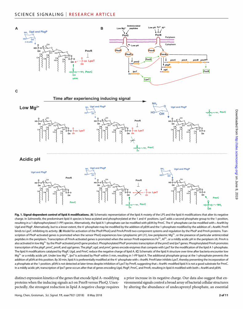
Fig. 1. Signal-dependent control of lipid A modifications. (A) Schematic representation of the lipid A moiety of the LPS and the lipid A modifications that alter its negative charge. In Salmonella, the predominant lipid A species is hexa-acylated and phosphorylated at the 1 and 4′ positions. LpxT adds a second phosphate group to the 1 position, resulting in a 1-diphosphorylated (1-PP) species. Alternatively, the lipid A 1-phosphate can be modified with pEtN by PmrC. The 4′-phosphate can be modified with l-Ara4N by Ugd and PbgP. Alternatively, but to a lesser extent, the 4′-phosphate may be modified by the addition of pEtN and the 1-phosphate modified by the addition of l-Ara4N. PmrR binds to LpxT, inhibiting its activity. (B) Model for activation of the PhoP/PhoQ and PmrA/PmrB two-component systems and regulation by the PhoP and PmrA proteins. Tran-scription of PhoP-activated genes is promoted when the sensor PhoQ experiences low cytoplasmic pH (31), low periplasmic Mg2+, or the presence of particular antimicrobial peptides in the periplasm. Transcription of PmrA-activated genes is promoted when the sensor PmrB experiences Fe3+, Al3+, or a mildly acidic pH in the periplasm (4). PmrA is also activated in low Mg2+ by the PhoP-activated pmrD gene product. Phosphorylated PhoP promotes transcription of the pmrD and lpxT genes. Phosphorylated PmrA promotes transcription of the pbgP, pmrC, pmrR, and ugd genes. The pbgP, ugd, and pmrC genes encode enzymes that compete with LpxT for the modification of the lipid A 1-phosphate. The lipid A modifications catalyzed by PbgP, Ugd, and PmrC reduce the negative charge of lipid A. (C) Schematic of the lipid A structure over time after bacteria encounter low Mg2+ or a mildly acidic pH. Under low Mg2+, lpxT is activated by PhoP within 5 min, resulting in 1-PP lipid A. The additional phosphate group at the 1-phosphate prevents the addition of pEtN at this position. By 30 min, lipid A is preferentially modified at the 4′-phosphate with l-Ara4N. PmrR later inhibits LpxT, thereby preventing the incorporation of a phosphate at the 1 position. pEtN is not detected at later times despite inhibition of LpxT by PmrR, suggesting that l-Ara4N–modified lipid A is not a good substrate for PmrC. In a mildly acidic pH, transcription of lpxT gene occurs after that of genes encoding Ugd, PbgP, PmrC, and PmrR, resulting in lipid A modified with both l-Ara4N and pEtN.
on June 8, 2019http://stke.sciencem
ag.org/D
ownloaded from

Hong, Chen, Groisman, Sci. Signal. 11, eaar7921 (2018) 8 May 2018
S C I E N C E S I G N A L I N G | R E S E A R C H A R T I C L E
3 of 11
lipid molecule that transports sugar substrates for incorporation into various cell envelope structures.
RESULTSPhoP promotes transcription of the lpxT gene in low Mg2+
The PhoP/PhoQ and PmrA/PmrB two-component systems are the major activators of lipid A modifications in Gram-negative bacteria (2). Lipid A modifications that increase the resistance of Salmonella to polymyxin B reduce the negative charge of lipid A; however, all the lipid A–modifying proteins encoded by genes that are activated by PhoP or PmrA, or both, that have been described in the literature either reduce or do not alter the negative charge of lipid A (2). There-fore, we hypothesized that PhoP or PmrA, or both, may repress lpxT expression because the LpxT protein increases lipid A’s negative charge (14). Unexpectedly, we found that the abundance of lpxT mRNA in wild-type Salmonella was 10 times higher after growth in low Mg2+ (10 M) than in high Mg2+ (10 mM) (Fig. 2A), which are inducing and repressing conditions, respectively, for both the PhoP/PhoQ (24) and PmrA/PmrB systems (Fig. 1B) (4). In agreement with the mRNA data, the abundance of the LpxT protein also increased in low Mg2+ compared to high Mg2+ (fig. S1). Transcriptional activation of the lpxT gene in low Mg2+ was not observed in a phoP single mutant or a phoP pmrA double mutant; by contrast, it was normal in a pmrA single mu-tant (Fig. 2A). These results indicate that low Mg2+ promoted lpxT transcription in a phoP-dependent but pmrA-independent manner.
PhoP binding to the lpxT promoter is required for lpxT transcriptional activationAccording to Salmonella genome annotations, the lpxT start codon is located 35 nucleotides (nt) downstream of the yeiR stop codon (Fig. 2B). The short intergenic distance between yeiR and lpxT suggested that these genes form an operon and that PhoP promotes transcription of both yeiR and lpxT. However, yeiR mRNA abundance was not al-tered upon a switch from high to low Mg2+ medium (fig. S2). These results argue that separate promoters transcribe the Salmonella yeiR and lpxT genes under PhoP-inducing conditions, as proposed for the E. coli homologs of these genes (10).
We mapped the lpxT TSS to a G residue 25 nt upstream of the lpxT start codon in wild-type Salmonella grown in low Mg2+ (Fig. 2C). No transcript was detected in cultures of wild-type Salmonella grown in high Mg2+ or in a phoP mutant grown in low Mg2+ (Fig. 2C). These results agree with the lpxT expression behavior of a different Salmonella strain grown under a different PhoP-inducing condition (26). More-over, they support the notion that wild-type Salmonella activates lpxT transcription during growth in low Mg2+ in a phoP-dependent manner and independently of transcription of the yeiR gene.
We identified a putative PhoP binding site within the yeiR cod-ing region (Fig. 2B). This site is a bona fide PhoP binding site because DNase I footprinting analysis demonstrated that phosphorylated PhoP, which is the active form of the PhoP protein (27), protects nu-cleotides −47 to −19 relative to the TSS, overlapping the predicted PhoP binding site (Fig. 2D) (28). In agreement with the notion that PhoP activates lpxT transcription directly (that is, by binding to the lpxT promoter), substitution of 3 nt of the PhoP binding site markedly decreased binding of the PhoP protein to a DNA fragment harbor-ing the lpxT promoter (Fig. 2E). Furthermore, an engineered strain with the 3-nt substitutions in the chromosomal copy of the PhoP bind-ing site in the lpxT promoter failed to increase the abundance of lpxT
mRNA in low Mg2+ (Fig. 2F), behaving like the phoP null mutant (Fig. 2F). A separate engineered strain with a different 3-nt substitution in the PhoP binding site of the lpxT promoter (lpxTA) also failed to increase lpxT mRNA amounts in low Mg2+ (fig. S3).
Collectively, the results described in this section establish that PhoP directly promotes lpxT transcription when Salmonella experiences low Mg2+. This result is paradoxical because low Mg2+ also promotes ex-pression of the pmrR gene, which encodes an LpxT inhibitor, through the stabilization of the PmrA protein by the PhoP transcriptional target PmrD (Fig. 1B) (12). Next, we explored the phenotypic conse-quences of PhoP promoting the transcription of both lpxT and pmrR by examining the kinetics with which the mRNAs corresponding to lipid A phosphate–modifying genes are produced, as well as their im-pact on the resulting lipid A profiles.
The lpxT gene is transcribed before other lipid A phosphate–modifying genes when the inducing signal is low Mg2+
We analyzed the abundance of lpxT, pbgP, pmrC, pmrR, and ugd tran-scripts at different times after wild-type Salmonella was switched from noninducing (high Mg2+) to inducing (low Mg2+) conditions. The lpxT transcript reached a maximum within 5 min of the shift and then decreased, eventually reaching preinduction values by 120 min (Fig. 3). This behavior differed from the expression of the pmrR, pbgP, pmrC, and ugd genes, which peaked between 20 and 60 min and retained >50% of the maximum values at 120 min (Fig. 3).
We reasoned that lpxT transcription precedes that of pbgP, pmrC, pmrR, and ugd in wild-type Salmonella experiencing low Mg2+ be-cause PhoP activates the lpxT promoter directly (Fig. 2) but acts on the pbgP, pmrC, pmrR, and ugd genes indirectly by increasing transcrip-tion from the pmrD promoter, resulting in synthesis of the PmrD protein, the protector of phosphorylated PmrA (Fig. 1B) (18). In sup-port of this notion, pmrD mRNA amounts increased at the same time as those of lpxT, preceding the increase in pbgP, pmrC, pmrR, and ugd mRNAs (Fig. 3). Thus, lpxT is transcribed before other genes that en-code proteins that target the same lipid A position and before the LpxT inhibitor–encoding gene pmrR.
PhoP-dependent transcription of lpxT favors lipid A modification with l-Ara4N during growth in low Mg2+
The rapid and transient lpxT expression exhibited by Salmonella ex-periencing low Mg2+ (Fig. 3) resulted in a rapid and transient presence of two phosphates at position 1 of lipid A (1-PP), the addition of which is catalyzed by LpxT (14). We detected the presence of radiolabeled 1-PP lipid A within 10 min of switching wild-type Salmonella from high to low Mg2+ (Fig. 4A) in the presence of radiolabeled phosphate. The abundance of 1-PP lipid A then decreased, and 1-PP lipid A was no longer observed by 1 hour (fig. S4).
The l-Ara4N modification was first detected at 20 min after the switch to low Mg2+ (Fig. 4A). By 1 hour, the lipid A population contained com-binations of l-Ara4N with other modifications including 2-OH myri-station or palmitation, but not pEtN (fig. S4). Thus, the switch to low Mg2+ resulted in the transient production of 1-PP lipid A, which decreased in abundance and then disappeared as the l-Ara4N modification was detected. These results suggest that the transient increase in 1-PP lipid A taking place shortly after the switch to low Mg2+ favors further mod-ification with l-Ara4N over modification with pEtN.
If the transient increase in 1-PP lipid A mediated by LpxT is re-quired for the subsequent lipid A decoration with l-Ara4N and no
on June 8, 2019http://stke.sciencem
ag.org/D
ownloaded from

Hong, Chen, Groisman, Sci. Signal. 11, eaar7921 (2018) 8 May 2018
S C I E N C E S I G N A L I N G | R E S E A R C H A R T I C L E
4 of 11
pEtN, an lpxT mutant should have both l-Ara4N and pEtN in its lipid A. As predicted, the lipid A of the lpxT mutant was undermodified with the l-Ara4N but hypermodified with various double modifica-tions of l-Ara4N and pEtN (Fig. 4B). The lipid A identities were ver-
ified by mass spectrometry (table S1). Moreover, a pbgP single mutant had little pEtN in its lipid A (Fig. 4C) despite its inability to produce l-Ara4N (8). By contrast, pEtN was the dominant modification in the lipid A of the isogenic pbgP lpxT double mutant (Fig. 4C), reinforcing
A
DE
F
C
B
0
0.1
0.2
0.3High Mg2+ Low Mg2+
lp
xT
mR
NA
ab
un
dan
ce
WT
pm
rA
phoP
phoP p
mrA
WT
pm
rA
phoP
phoP p
mrA
lp
xT
mR
NA
ab
un
dan
ce
0.5
0.4
0.3
0.2
0.1
0
WT
phoP
lpxT
B
WT
phoP
lpxT
B
High Mg2+ Low Mg2+
lpxTB
WT
lpxTBP
erce
nta
ge
(%)
120
100
80
60
40
20
00 5 10
PhoP (µM)15 20 25
WT, high Mg2+
WT, low Mg2+
phoP, low Mg2+
CGAT
G
A T C G 0 1 5 10 20
PhoP (µM)LadderG
GC
CT
TG
TT
GA
AG
CT
TC
GT
TTA
GC
GA
CG
Fig. 2. PhoP promotes lpxT transcription directly by binding to the lpxT pro-moter. (A) Quantification of lpxT transcripts by quan-titative polymerase chain reaction (qPCR) before and 60 min after switching wild- type (WT; 14028s) and pmrA (EG7139), phoP (MS7953s), and phoP pmrA (EG12443) mutant Salmonella from high- Mg2+ medium (10 mM MgCl2) to low-Mg2+ medium (10 M MgCl2) at pH 7.7, normalized to the rrs (16S ribosomal RNA) transcript. (B) Genomic struc-ture and partial nucleotide sequence of the Salmonella yeiR-lpxT chromosomal re-gion. The sequence in red cor-responds to the PhoP box in the lpxT promoter, the under-lined sequence to the yeiR stop codon, +1 to the lpxT tran-scription start site (TSS), and lowercase sequence (atg) to the lpxT start codon. (C) Prim-er extension analysis of lpxT in WT (14028s) and phoP mu-tant (MS7953s) Salmonella grown in N- minimal medium (pH 7.7) containing high Mg2+ or low Mg2+. Lanes C, G, A, and T indicate the sequence of the lpxT promoter. The boxed “G” indicates the TSS of lpxT. (D) Deoxyribonuclease I (DNase I) footprinting analysis of the lpxT promoter region using the indicated amounts of purified phosphorylated PhoP-6×His protein. Lanes A, T, C, and G indicate the se-quence of the lpxT promoter. The sequence highlighted in red corresponds to the PhoP binding site. (E) Filter assay for binding of the purified PhoP protein to the WT lpxT promoter and to the mutant
lpxTB promoter, which contains substitutions in the PhoP binding site. The sequence of the WT lpxT promoter and the nucleotide substitutions in the mutant lpxTB promoter are shown in the box above the graph. The sequence highlighted in red corresponds to the PhoP binding site. The graph shows the percentage of 32P signal compared to the maxi-mum for binding of the indicated amount of PhoP protein to 32P-labeled WT and promoter fragments. Data are representative of three independent experiments. (F) Abundance of
lpxT transcript before and 60 min after switching WT (14028s), phoP (MS7953s), and lpxTB (XH16) Salmonella from N-minimal medium (pH 7.7) containing high Mg2+ to low Mg2+, normalized to the rrs transcript. Mean and SD of three independent experiments are shown.
on June 8, 2019http://stke.sciencem
ag.org/D
ownloaded from

Hong, Chen, Groisman, Sci. Signal. 11, eaar7921 (2018) 8 May 2018
S C I E N C E S I G N A L I N G | R E S E A R C H A R T I C L E
5 of 11
the notion that the addition of a phosphate group to lipid A by LpxT hinders the incorporation of pEtN into lipid A even in organisms that cannot make l-Ara4N. In agreement with this notion, wild-type and pmrC mutant Salmonella exhibited similar lipid A profiles during growth in low Mg2+ (fig. S5). Furthermore, disruption of both pmrA and lpxT abolished all l-Ara4N, pEtN, and 1-PP modifications (fig. S6). Cumulatively, these results indicate that LpxT promotes modifica-tion of lipid A with a single l-Ara4N over pEtN or double modification with both l-Ara4N and pEtN.
Because the lpxT gene is transcribed even in a phoP mutant (Fig. 2A), albeit to a lesser extent than in wild-type Salmonella, we wondered whether the increased lpxT transcription promoted in low Mg2+ by the PhoP pro-tein was responsible for the l-Ara4N–modified lipid A (Fig. 4). Thus, we examined the lipid A profile of the lpxT promoter mutant (lpxTB) that is refractory to PhoP activation (Fig. 2F). The use of this mutant avoids potentially confusing effects resulting from the pleiotropic ef-fects of phoP inactivation, such as the inability to activate the PmrA pro-tein in low Mg2+ (Fig. 1B) (18).
The lpxTB promoter mutant harbored less lipid A species with l-Ara4N and more lipid A species doubly modified with l-Ara4N and pEtN compared to the wild-type strain (Fig. 4B). This phenotype is specific to the PhoP-inducing condition of low Mg2+ because the lpxTB mutant
100500 1500
1
2
3
Time (min)
mR
NA
ab
un
dan
ce
lpxT
pmrD
ugd
pbgP
pmrR
pmrC
Fig. 3. The lpxT gene is expressed earlier relative to other lipid A–modifying genes in low Mg2+. Quantification of the indicated transcripts at different times after switching WT Salmonella (14028s) from high Mg2+ to low Mg2+ at pH 7.7, normalized to the rrs transcript. Data are means ± SD of three independent experiments.
A CB
0 10
2
4
6
8
10
020 30
Time (min)
Lip
id A
1-P
P le
vel (
%)
Time (min) 0 5 10 20 30
1-PPl-Ara4N
WT
lpxT
ugd lpxT
pbgP lpxT
pbgP
ugd
pEtNl-Ara4N
Double modificationscontaining pEtN
WTlp
xT
B
lpxT WT
lpxT
B
lpxT
WTlp
xT
B
lpxT
pH 4.910 mM Mg2+
pH 7.710 µM Mg2+
pH 7.710 mM Mg2+
1-PPpEtN
1 2 3 4 5 6 7 8 9
l-Ara4N
*
*
*
*
*** *
*
*
*
o
oo
o o
o
o
o
DoublemodificationscontainingpEtN
Fig. 4. PhoP-dependent lpxT expression promotes lipid A modification with l-Ara4N in low Mg2+ but not in a mildly acidic pH. (A) Autoradiogram of a thin-layer chromatography (TLC) plate showing lipid A from WT Salmonella (14028s) at different times after they were switched from N-minimal medium (pH 7.7) containing high Mg2+ to low Mg2+ in the presence of radiolabeled phosphate. Data are representative of three independent experiments. The graph below the autoradiogram shows the quantification of lipid A 1-PP from the image, representing mean and SD (normalized to the total signal of each lane). (B) TLC showing lipid A from WT (14028s), lpxT B (XH16), and lpxT (DC72) Salmonella grown in N-minimal medium (pH 7.7) containing high Mg2+, N-minimal medium (pH 7.7) containing low Mg2+, or N-minimal medium (pH 4.9) containing high Mg2+ in the presence of radiolabeled phosphate. (C) TLC showing lipid A from WT (14028s), lpxT (DC72), pbgP (EG9241), pbgP lpxT (XH246), ugd (EG17898), and ugd lpxT (XH251) Salmonella grown in N-minimal medium (pH 7.7) containing high Mg2+ or low Mg2+. Data are representative of two independent exper-iments. The 1-PP band shows different mobility in each of the images because the samples in each panel were run for different amounts of time. Asterisks (*) indicate positions of lipid A containing l-Ara4N, and open circles (o) indicate positions of lipid A containing pEtN.
on June 8, 2019http://stke.sciencem
ag.org/D
ownloaded from

Hong, Chen, Groisman, Sci. Signal. 11, eaar7921 (2018) 8 May 2018
S C I E N C E S I G N A L I N G | R E S E A R C H A R T I C L E
6 of 11
retained normal 1-PP modification after growth under the non-inducing high Mg2+ condition (Fig. 4B). The latter result indicated that the lpxT gene itself was functioning properly in the lpxT promoter mutant (lpxTB). That is, the inability of the lpxT mutant promoter to respond to PhoP did not compromise lpxT expression under non-inducing conditions for PhoP. Moreover, when grown in low Mg2+, the pmrC single mutant and the pmrC lpxTB double mutant displayed the same lipid A profile containing l-Ara4N and no pEtN (fig. S5). These results established that PhoP activation of lpxT transcription is required for normal lipid A modification with l-Ara4N.
Similar transcription timing of lpxT and other lipid A modification genes results in lipid A with pEtN during growth in a mildly acidic pHA mildly acidic pH activates both PmrB and PhoQ, resulting in tran-scription of PmrA- and PhoP-activated genes, respectively (Fig. 1B) (17, 29, 30). However, these sensors differ in that PmrB responds to a mildly acidic pH through protonation of periplasmic histidine residues (17), whereas PhoQ does so through its cytoplasmic region through a mechanism that does not involve histidine residues (31). In addition, full activation of the PhoP/PhoQ system under a mildly acidic pH requires the PhoP-activated gene ugtL, which encodes a protein that stimulates PhoQ autophosphorylation (30).
We reasoned that the kinetics with which a mildly acidic pH in-duces PmrA- and PhoP-activated genes might differ from that taking place in low Mg2+ because low Mg2+ activation requires the PhoP- dependent transcription of the pmrD gene (18), which specifies a pro-tein that protects phosphorylated PmrA from dephosphorylation by its cognate sensor PmrB (21), and results in transcription of genes directly activated by PmrA. By contrast, PmrB-mediated activation of PmrA by a mildly acidic pH does not require PhoP or PmrD (Fig. 1B). That is, when the inducing signal is a mildly acidic pH, the PhoP- dependent lpxT transcription may not precede that of PmrA-activated genes, as observed in low Mg2+.
When wild-type Salmonella was shifted from noninducing condi-tions to medium with a mildly acidic pH, the mRNA amounts of the PmrA- activated genes pbgP, pmrC, and pmrR reached a maximum within 5 min (Fig. 5A). By contrast, lpxT mRNA amounts reached a maximum at 10 min after the shift (Fig. 5A), raising the possibility that PmrR may be available to readily inhibit LpxT activity when the inducing condition is a mildly acidic pH. If true, the lipid A profile of wild-type Salmonella grown in a mildly acidic pH should no longer display the lpxT-dependent accumulation of l-Ara4N observed when the inducing signal was low Mg2+ (Fig. 4). As predicted, wild-type Salmonella, the lpxT promoter mu-tant, and the lpxT deletion mutant produced identical lipid A profiles with an abundance of pEtN-modified lipid A when grown in a mildly acidic pH (Fig. 4B). This result indicates that LpxT is not actively adding phosphate to lipid A when the inducing signal is a mildly acidic pH, re-sulting in a lipid A modified with both pEtN and l-Ara4N.
A mildly acidic pH promotes greater polymyxin B resistance than does low Mg2+
Genetic experiments previously revealed that Salmonella mutants de-fective in the pbgP or ugd genes, which fail to modify lipid A with l-Ara4N, are 100 times more sensitive to polymyxin B than are wild-type Salmonella after growth in low Mg2+ (9). By contrast, a pmrC mu-tant, which is defective in lipid A modification with pEtN, displayed marginally increased polymyxin B sensitivity compared to wild-type Salmonella (9). We have recapitulated these results (fig. S7) and de-
termined that the lpxT null mutant was slightly more resistant to polymyxin B than was wild-type Salmonella (fig. S7).
We hypothesized that growth in low Mg2+ enhanced polymyxin B resistance compared to growth in a mildly acidic pH because the lipid A of wild-type Salmonella grown in low Mg2+ was modified with l-Ara4N and no pEtN (Fig. 4B) and also because inactivation of the genes required for lipid A modification with l-Ara4N decrease poly-myxin B resistance much more than does inactivation of the pmrC gene (fig. S7). Unexpectedly, survival of wild-type Salmonella exposed to polymxyin B was seven times higher after growth in a mildly acidic pH than it was in low Mg2+ (Fig. 5B). As predicted from the results
30
20
10
0
Su
rviv
al (
%)
WTpm
rA
lpxT
B
lpxT WT
pm
rA
lpxT
B
lpxT
Low Mg2+ Acidic pH
A
B
Time (min)
mR
NA
ab
un
dan
ce
2.0
1.5
1.0
0.5
00 50 100 150
ugd
pmrD
pbgP
pmrR
pmrClpxT
Fig. 5. Effects of low Mg2+, acidic pH, and PhoP-dependent transcription of the lpxT gene on polymyxin B resistance. (A) Quantification of the indicated tran-scripts at different times after switching WT Salmonella (14028s) from high Mg2+ at pH 7.7 to high Mg2+ at pH 4.9, normalized to the rrs transcript. Data are means ± SD of three independent experiments. (B) Percent survival of WT (14028s), pmrA (EG7139), lpxT B (XH16), and lpxT (DC72) Salmonella exposed to polymyxin B after growth in N-minimal medium (pH 7.7) with low Mg2+ or that (pH 4.9) with high Mg2+ (acidic pH). Survival values were calculated as relative to the original inoculum. Mean and SD of three independent experiments are shown. Unpaired Student’s t tests were performed comparing mutant strains with the WT under the same growth condi-tion. *P < 0.05, **P < 0.01, and ****P < 0.0001; n.s. indicates P > 0.05 (not significant).
on June 8, 2019http://stke.sciencem
ag.org/D
ownloaded from

Hong, Chen, Groisman, Sci. Signal. 11, eaar7921 (2018) 8 May 2018
S C I E N C E S I G N A L I N G | R E S E A R C H A R T I C L E
7 of 11
presented in the previous section (Fig. 4, B and C), the lpxT promoter mutant and the lpxT null mutant exhibited similar polymyxin B re-sistance as did the wild-type strain when the inducing condition was a mildly acidic pH (Fig. 5B). By contrast, the lpxT promoter mu-tant was more resistant to polymxyin B than was the wild-type strain, and the lpxT null mutant was more resistant than the lpxT promoter mutant when the inducing condition was low Mg2+ (Fig. 5B). Con-trol experiments demonstrated that the pmrA mutant is several orders of magnitude more sensitive than the wild-type strain regardless of the inducing condition (Fig. 5B), in agreement with previous results (18). In summary, Salmonella survival to polymxyin B is higher when its lipid A is modified with both l-Ara4N and pEtN compared to modification with only l-Ara4N (Fig. 6).
PhoP-dependent activation of lpxT is conserved in a subset of Gram-negative speciesA wide range of Gram-negative species have the ability to modify their lipid A phosphates. However, the specific chemical modification and the regu-lation of the genes responsible for such modifications vary among species. For example, Yersinia pestis differs from Salmonella in lacking a pmrC gene and in its pbgP promoter being directly activated by both the PhoP and PmrA proteins (32). In P. aeruginosa, the LpxT enzyme can transfer an additional phosphate to lipid A (33), and the pmrC gene is activated by the ColR/ColS two-component system but not by PmrA/PmrB (34).
To determine whether the PhoP-mediated activation of lpxT tran-scription identified in Salmonella (Fig. 2) is conserved in other Gram- negative species, we examined bacterial genomes for sequences related to the Salmonella lpxT, pbgP, phoP, and pmrC genes, as well as for se-quences resembling a PhoP binding site in the lpxT promoter (table S2). Citrobacter freundii, K. pneumoniae, and E. coli harbor a predicted PhoP binding site upstream of the lpxT coding region, which is highly conserved with the one in the Salmonella lpxT promoter (Fig. 6A). This suggests that lpxT transcription is promoted in a phoP-dependent manner in these species as well. In addition, a 10-nt stretch located
four bases upstream of the lpxT TSS is fully conserved in the four spe-cies, suggesting that it is the binding site for an as-yet-unidentified regulatory molecule that controls lpxT expression. By contrast, Yersinia and Pseudomonas, species in which the pmrC gene is either absent or not regulated by the PmrA/PmrB system, lack sequences resembling a PhoP binding site in their lpxT promoter regions. This analysis sug-gests a correlation between PhoP activation of lpxT transcription and the presence of a PmrA-activated pmrC gene.
We determined that lpxT mRNA abundances were threefold higher in wild-type E. coli grown in low Mg2+ than in those grown in high Mg2+ (Fig. 6B). By contrast, an E. coli phoP null mutant displayed sim-ilarly low amounts of lpxT mRNA under both inducing and repressing conditions (Fig. 6B). Although the expression behavior of the E. coli lpxT gene was qualitatively similar to that of the Salmonella lpxT homolog, the threefold induction exhibited by the E. coli lpxT gene was much lower (Fig. 6B) than the 10-fold increase exhibited by the Salmonella lpxT gene (Fig. 2A) under identical inducing conditions. Given that the E. coli PmrD protein is defective in activating the PmrA protein (35), the lower induction of the lpxT gene of E. coli might reflect the decreased need to activate the lpxT gene ahead of pmrR when the inducing condition is low Mg2+.
DISCUSSIONWe have established that differences in the expression kinetics of lipid A–modifying genes are responsible for the distinct lipid A profiles exhibited by Salmonella experiencing different inducing conditions for these genes. That is, both low Mg2+ and a mildly acidic pH activate the PhoP/PhoQ and PmrA/PmrB systems, causing transcription of their target genes (Figs. 2 and 5). However, these two inducing condi-tions give rise to different lipid A modifications because transcription of the lpxT gene precedes that of other lipid A modification genes in-cluding pbgP, pmrC, and ugd, as well as pmrR, which encodes an in-hibitor of LpxT when the inducing signal is low Mg2+ but not when it
is a mildly acidic pH (Figs. 2 and 5). PhoP- activated lpxT transcription is necessary for lipid A modification with l-Ara4N and no pEtN because preventing this activa-tion results in a lipid A modified with both l-Ara4N and pEtN (Fig. 4B). Given that LpxT is responsible for the incorporation of an additional phosphate into lipid A (14, 36), our results indicate that, para-doxically, a decrease in lipid A negative charge resulting from covalent lipid A modification with l-Ara4N requires a pre-vious lipid A modification that increases its negative charge.
The expression kinetics of lipid A phosphate modification genes determines the distinct chemical modifications of the lipid A phosphatesWhen Salmonella experiences low Mg2+, the PhoP/PhoQ system is activated before the PmrA/PmrB system because, under this inducing condition, PmrA activation is dependent on the PhoP-activated pmrD
A
B
WT phoP WT phoP
High Mg2+ Low Mg2+
lp
xT
mR
NA
ab
un
dan
ce
0.06
0.04
0.02
0.00
Fig. 6. PhoP-dependent transcriptional acti-vation of the lpxT gene is conserved in a sub-set of Gram-negative species. (A) Alignment of the nucleotide sequence of the lpxT promoter region in selected enteric bacterial species re-veals conservation of the PhoP binding site (underlined). The boxed “G” indicates the tran-scription start site of lpxT mapped in Salmonella. Asterisks (*) denote positions that are conserved in all four species: STM, S. enterica serovar Typh-imurium; ECO, E. coli; CFD, C. freundii; KPN, K. pneumoniae. (B) Abundance of lpxT mRNA before and 60 min after WT (MG1655) and phoP (EG12976) E. coli were switched from N-minimal medium (pH 7.7) containing high Mg2+ to low Mg2+, normalized to the rrs transcript. Mean and SD of three independent experiments are shown.
on June 8, 2019http://stke.sciencem
ag.org/D
ownloaded from

Hong, Chen, Groisman, Sci. Signal. 11, eaar7921 (2018) 8 May 2018
S C I E N C E S I G N A L I N G | R E S E A R C H A R T I C L E
8 of 11
gene (Fig. 1B) (18). By being directly activated by the PhoP protein (Fig. 2), lpxT is expressed before the lipid A–modifying genes that are directly controlled by the PmrA protein (Fig. 3), most critically pmrR, which specifies an LpxT inhibitor (12). Therefore, PhoP-dependent lpxT ex-pression induced in low Mg2+ results in 1-PP lipid A (Fig. 4A), which favors further modification of lipid A phosphates with l-Ara4N and the exclusion of pEtN (Fig. 4B).
By contrast, when the activation signal is a mildly acidic pH, PmrA/ PmrB activation does not lag behind that of PhoP/PhoQ (Fig. 1B). This is because PmrB detects a mildly acidic pH in the periplasm (17) and does not depend on the PmrD protein to activate PmrA. In addition, PhoQ responds to mild acidification through its cytoplasmic domain (31). Thus, the early pmrR expression taking place under a mildly acidic pH, which results in the production of the PmrR protein, is anticipated to decrease LpxT activity, resulting in a lipid A with both l-Ara4N and pEtN modifications (Fig. 4B). That is, the lpxT gene is expressed in a mildly acidic pH (Fig. 5A), but the LpxT protein is in-hibited by PmrR (12).
In agreement with the model presented above (Fig. 1B), mutants lacking the lpxT gene or with nucleotide substitutions in the lpxT promoter that render it refractory to activation by PhoP behave like wild-type Salmonella in a mildly acidic pH but show different behaviors when the inducing signal is low Mg2+ (Figs. 4B and 5B). Our findings raise the possibility of an analogous mechanism being responsible for lipid A deacylation by the PhoP-activated PagL protein occurring only when modification with l-Ara4N and pEtN is prevented (37).
Transcription kinetics governs critical cellular processesOur results highlight the fundamental role that gene expression timing plays in physiological responses to stress conditions. Low Mg2+ and a mildly acidic pH promote different lipid A profiles (Fig. 4B) despite stimulating the expression of the same set of lipid A modification genes (Figs. 2 and 5). This is not the only example of expression timing, rather than simply the expression or nonexpression of specific genes at steady- state, dictating phenotypic outcomes. For example, a surge in the tran-scription of phoP is necessary to jump-start Salmonella’s virulence program (20). This surge requires PhoP to positively feedback on its own transcription because a strain constitutively expressing PhoP is attenuated for virulence despite achieving the same steady-state abun-dance of PhoP-activated transcripts (20). Likewise, the increased amounts of phosphorylated PhoP generated after several hours in low Mg2+ enable Salmonella to transcribe PhoP-dependent genes that require high amounts of active PhoP protein (38). That is, the increased amounts of phosphorylated PhoP in this case depend on the Mg2+ importer MgtA removing Mg2+ away from the periplasmic Mg2+-sensing domain of the sensor PhoQ (38). Because transcription elongation into the mgtA coding regions is triggered in low cytoplasmic Mg2+ (39), expres-sion of a subset of PhoP-activated genes takes place only when the bacterium experiences the signals promoting mgtA expression. Fur-thermore, the stimulation of PmrB by Fe3+ activates PmrA to different ex-tents at early and late times due to a negative feedback loop that hinders Fe3+ access to PmrB (12). These examples illustrate how cellular behav-iors in response to particular environmental conditions change over time even when the condition triggering the response does not change.
Transcript abundance does not always reflect the activity of the corresponding proteinmRNA abundance is often used as proxy for the amounts and activ-ities of the corresponding proteins. This is because mRNA determi-
nations are straightforward across a genome, whereas activity assays are not available for the vast majority of gene products. Our data demon-strate two examples in which the presence of particular mRNAs is not accompanied by the activity of the specified gene products. First, when the lpxT gene was transcribed in response to a mildly acidic pH (Fig. 5A), the LpxT-mediated modification of lipid A was not observed (Fig. 4B) presumably because of the presence of the LpxT inhibitor PmrR (12). Likewise, the pmrC gene was highly induced in low Mg2+ (Fig. 3), but the lipid A did not contain detectable pEtN (Fig. 4B) due to preceding PhoP-dependent lpxT transcription (Fig. 3) favoring the incorporation of l-Ara4N over the incorporation of pEtN (Fig. 4B).
Environmental control of LpxT expression affects multiple pathwaysAs discussed above, Salmonella favors lipid A modified with l-Ara4N under low Mg2+ and with both l-Ara4N and pEtN when experiencing a mildly acidic pH (Fig. 1C). The fact that the lipid A from Salmonella harvested from inside macrophages contains both l-Ara4N and pEtN (15) is in agreement with three notions: first, that the macrophage phagosome containing Salmonella is mildly acidic (40); second, that an acidic pH is critical to activate the PhoP/PhoQ system inside macrophages (40); and third, that an acidic pH is the signal promot-ing expression of lipid A phosphate–modifying genes inside macro-phages (15).
Wild-type Salmonella grown in a mildly acidic pH is seven times more resistant to polymyxin B than when grown in low Mg2+ (Fig. 5B). Because polymyxins are not used to treat Salmonella infections, our findings raise the possibility of the lipid A modification with both l-Ara4N and pEtN, which is favored under a mildly acidic pH, me-diating resistance to host-derived products, decreasing recognition by the host immune system, playing a yet-to-be identified function, or a combination of these roles. We note that a pmrA null mutant, which is unable to modify lipid A with either l-Ara4N or pEtN (2), is hyper-virulent in cultured macrophages and in mice inoculated through the intraperitoneal route (41).
Finally, the LpxT protein removes the distal phosphate from its substrate—undecaprenyl diphosphate (C55-PP)—and produces C55-P, which is an essential carrier that shuffles sugar moieties across the inner membrane for various biosynthetic pathways (14, 42). The identifi-cation of environmental conditions and regulatory factors that control lpxT gene expression raises the possibility of the essential C55-P metab-olism being affected by the environmental conditions encountered by bacteria. This hypothesis is supported by the observation that the PhoP and PmrA proteins have been implicated in controlling the expression of the ybjG gene, which, like lpxT, encodes a C55-PP phosphatase that is required for recycling C55-P (43) in E. coli.
MATERIALS AND METHODSBacterial strains, plasmids, primers, and growth conditionsS. enterica serovar Typhimurium strains were derived from the wild-type strain 14028s. Unless otherwise stated, bacteria were grown at 37°C in LB broth or in N-minimal medium (pH 7.7) (44) supplemented with 0.1% casamino acids, 38 mM glycerol, and 10 mM or 10 M of MgCl2. When necessary, antibiotics were added at the following fi-nal concentrations: ampicillin (50 g/ml), chloramphenicol (20 g/ml), kanamycin (50 g/ml), and tetracycline (10 g/ml). P22 transduction of Salmonella strains was performed as described (45). E. coli DH5a
on June 8, 2019http://stke.sciencem
ag.org/D
ownloaded from

Hong, Chen, Groisman, Sci. Signal. 11, eaar7921 (2018) 8 May 2018
S C I E N C E S I G N A L I N G | R E S E A R C H A R T I C L E
9 of 11
was used as a host for the preparation of plasmid DNA. Bacterial strains and plasmids used in this study are listed in table S3. Primers used in this study are listed in tables S4 and S5.
All experiments were carried out with wild-type S. enterica serovar Typhimurium strain 14028s, wild-type E. coli strains MG1655 or W3110, and mutant derivatives. LB (Becton, Dickinson and Co.), Super Optimal Broth (SOB) (Becton, Dickinson and Co.), or Super Optimal broth with Catabolite repression (SOC) (SOB supplemented with 20 mM glucose) media were used for cloning and strain construction, as noted. When necessary, ampicillin was used at 50 g/ml, and tetracycline was used at 12.5 g/ml. Fusaric acid (FA) plates were used for selection against tetracycline-resistant bacteria [LB agar (40 g/liter; Becton, Dickinson and Co.), NaH2PO4·H2O (10 g/liter) (Sigma-Aldrich), chlortetracycline hydrochloride (60 mg/liter) (Sigma-Aldrich), FA (12 mg/liter) (Acros Organics), and 0.1 mM ZnCl2 (Sigma-Aldrich)] (46). For experiments in which gene expression was measured, bacteria were grown in N-minimal medium (pH 7.7 or pH 4.9) (47) and the indicated con-centration of MgCl2. For time course experiments, cells were grown in N-minimal medium (pH 7.7; 10 mM Mg2+) to log phase and then either washed with fresh N-minimal medium (pH 7.7; no Mg2+) twice and resuspended with N-minimal medium (pH 7.7; 10 M Mg2+) or resuspended with N-minimal medium (pH 4.9; 10 mM Mg2+) before being incubated for the indicated times. All incubations were carried out at 37°C with shaking at 250 rpm.
Strain constructionStrains harboring chromosomal point mutations in the lpxT promoter region were constructed by a method based on selection for loss of tetracycline resistance (46). First, the promoter region of lpxT was replaced with a tetracycline resistance (TetR) cassette. This was carried out by transforming wild-type Salmonella (14028s) expressing the phage Red recombinase machinery from plasmid pKD46 with a DNA fragment containing the TetR cassette from transposon Tn10 flanked by ~60-nt regions of nucleotide sequence identity to the lpxT promoter region. The DNA fragment was generated using primers W3423 and W3424 for strain XH16 and primers W3427 and W3428 for strain XH15, with genomic DNA from MS7953s as a template. Electropo-rated cells were recovered in SOC medium at 30°C and plated on LB/Ap/Tet/plates and incubated at 30°C. The resulting strains were grown to mid-log phase in SOB/Ap/10 mM l-arabinose at 30°C and made electrocompetent by washing twice in ice-cold water and once in ice-cold 10% glycerol. Next, a DNA oligo corresponding to the lpxT promoter and containing the desired point mutation(s) was generated using two primers W3425 and W3426 that are reverse complement to each other (W3425 and W3426 for XH16 and W3430 and W3433 for XH15). The resulting product was transformed into electrocompetent cells. Electroporated cells were recovered in SOC at 37°C. Transfor-mants were cured of pKD46 by plating on LB at 42°C and tested for loss of Ap and Tet resistance, and mutations were confirmed by sequencing PCR products generated from purified genomic DNA. To construct the strain specifying a C-terminally FLAG-tagged LpxT protein (XH201), the DNA fragment amplified from pKD3 using primers W4309/W4310 was introduced into wild-type Salmonella 14028s harboring plas-mid pKD46.
When necessary, P22 phage transduction was used to move se-lectable markers. XH246 was generated by using a P22 lysate grown on strain EG9241 to infect strain DC74, which is a derivative of strain DC72 with the Km cassette removed by using plasmid pCP20 (48). XH248 was generated by infecting XH16 with a P22 lysate prepared
on strain EG9460. XH251 was generated by infecting DC72 with a P22 lysate prepared on strain EG17898.
RNA isolation and complementary DNA synthesisA bacterial pellet was isolated from 1 ml of liquid culture and placed immediately on dry ice. The pellet was resuspended in 1 ml of 2:1 RNAprotect reagent (Qiagen)/water and incubated at room tempera-ture for 5 min. After centrifugation at 5000 rpm for 10 min, the pellet was stored at −80°C. The sample was resuspended in 100 l of lysozyme (10 mg/ml; Sigma-Aldrich) and incubated at room temperature for 2 hours with occasional vortexing. Total RNA was isolated using the RNeasy Mini kit (Qiagen) and the recommended on-column DNase I treatment, according to the manufacturer’s instructions. For first-strand complementary DNA (cDNA) synthesis, 1 g of purified RNA in 16 l of ribonuclease (RNase)–free water was mixed with 4 l of VILO Master Mix (Invitrogen) and incubated at 25°C for 10 min, 42°C for 60 min, and 85°C for 5 min. The resulting cDNA was diluted by adding 100 l of water before qRT-PCR.
qRT-PCRFor quantification of cDNA abundance, 2 l of diluted cDNA was mixed with 7.5 l of 2× SYBR Green Master Mix (Applied Biosystems) and 5.5 l of primer mix (each primer diluted to 72.7 M in water). Reac-tions were carried out in a 7500 Real-Time PCR machine (Applied Bio-systems) using the manufacturer-provided software and settings. Transcript quantities were determined by fitting CT (threshold cycle) values to a standard curve generated with Salmonella genomic DNA.
DNase I footprinting assayDNase I foot printing assay was conducted as described (41). DNA fragments corresponding to the lpxT region were generated by PCR using primer W3436 and 32P-labeled primer W3435 with genom-ic DNA from strain 14028s as a template. Unincorporated [-32P]–adenosine 5′- triphosphate was removed by using G-50 microcolumns (GE Healthcare). A total of 2 × 10⁴ cpm of labeled probe (~10 fmol), 200 ng of poly(deoxyinosinic-deoxycytidylic) (Sigma-Aldrich), and purified His-tagged PhoP (PhoP-6×His) (49) were mixed with bind-ing buffer [20 mM Hepes (pH 8.0), 10 mM KCl, 2 mM MgCl2, 0.1 mM EDTA, 0.1 mM dithiothreitol, bovine serum albumin (50 g/ml), and 10% (v/v) glycerol] in a total volume of 20 l and incubated at room temperature for 20 min. DNase I (Gibco) (0.01 U), 10 mM CaCl2, and 10 mM MgCl2 were added and incubated at room temperature for 3 min. Final concentrations of PhoP protein were 0, 1, 5, 10, and 20 M. The reaction was stopped by the addition of 100 l of a phenol chloroform solution, and the aqueous phase was precipitated with ethanol. The precipitate was dissolved in sequence-loading buffer and electrophoresed on a 6% acrylamide/7 M urea gel, together with a sequence ladder generated with the labeled primer by using the T7 Sequenase 2.0 DNA Sequencing kit (Amersham Biosciences), and the gels were dried and autoradiographed.
Filter-binding assayDetermination of binding efficiency of DNAs to the PhoP protein was carried out by the nitrocellulose-binding method (50). 32P-radiolabeled DNAs were incubated with a series of dilutions of the PhoP protein under the same condition used in the electrophoretic mobility shift assay at room temperature for 20 min. Final concentrations of PhoP protein were 0, 1, 2, 5, 10, and 20 M. The samples were filtered through 0.45-m nitrocellulose filters (HAWP, Millipore) under vacuum, and
on June 8, 2019http://stke.sciencem
ag.org/D
ownloaded from

Hong, Chen, Groisman, Sci. Signal. 11, eaar7921 (2018) 8 May 2018
S C I E N C E S I G N A L I N G | R E S E A R C H A R T I C L E
10 of 11
the filters were washed with 5 ml of the binding buffer, air-dried, and quantified using PhosphorImager (GE Healthcare).
Primer extension assayCultures were grown as indicated in the figure legend. Total RNA was extracted as described (51). cDNA synthesis was performed using 2 pmol of 32P-labeled 14997 primer with 10 g of total RNA and 1 U of SuperScript II RNase H2 reverse transcriptase (Life Technologies). The extension products were analyzed by electrophoresis on a 6% acrylamide/7 M urea gel and compared with sequence ladders initiated with the same 32P-labeled primer that was used for primer extension.
Polymyxin B susceptibility assayAntimicrobial peptide susceptibility assays were conducted as de-scribed (7), with modifications. Bacteria were grown in N-minimal medium with 10 M Mg2+ (pH 7.7) or with 10 mM Mg2+ (pH 4.9) for 4 hours. Bacterial cells were then diluted 200× in phosphate-buffered saline (PBS), combined with 1:1 ratio with polymyxin B (final concentra-tion of 5 g/ml). After 1-hour incubation at 37°C with aeration, samples were serially diluted in PBS and plated on LB agar plates for enumera-tion. The percentage survival was calculated as follows: survival (%) = colony-forming units (CFU) of peptide-treated culture/CFU of no- peptide culture × 100.
Isolation of labeled lipid ACultures were diluted to an OD600 (optical density at 600 nm) of 0.05 in 5 ml of fresh medium and labeled with 32Pi (2.5 Ci/ml; Amersham Biosciences) in N-minimal medium, as indicated. For the time course analysis, cells were first grown in 5 ml of N-minimal containing 10 mM Mg2+ to log phase, washed twice with N-minimal medium, and resus-pended with 5 ml of N-minimal medium containing 10 M Mg2+ and 32Pi (2.5 Ci/ml) for the indicated time. Cells were harvested by cen-trifugation, and the isolation of 32P-labeled lipid A was carried out by mild acid hydrolysis, as previously described (8, 52). 32P-lipid A spe-cies (~1000 cpm·per lane) were analyzed by TLC in a solvent system of chloroform, pyridine, 88% formic acid, and water (50:50:16:5, v/v) and visualized using a PhosphorImager (GE Healthcare).
Analysis of lipid A by mass spectrometryBacterial strains were grown as described earlier in 25 ml of medium for 4 hours. Lipid A was purified according to published protocols (53) and analyzed by the Keck Mass Spectrometry & Proteomics Re-source at Yale University.
Western blot assayCells were grown as described above. Crude extracts were prepared in B-PER reagent (Pierce) with lysozyme (100 g/ml) and EDTA-free pro-tease inhibitor (Roche). Samples were loaded onto 4 to 12% NuPAGE gels (Life Technologies) and transferred to nitrocellulose membrane using the iBot machine (Life Technologies). Membranes were blocked with 3% skim milk solution at room temperature for 1 hour. Then, samples were analyzed using anti-FLAG or anti-GroEL antibodies. Rabbit anti-FLAG antibodies were used at 1:4000 dilution. Mouse anti- GroEL antibodies were used as control at 1:12,000 dilution. Secondary horseradish peroxidase–conjugated anti-rabbit or anti-mouse anti-serum (GE Healthcare) was used at 1:4000 dilution. Blots were devel-oped with the Amersham ECL Western Blotting Detection Reagents (GE Healthcare) or SuperSignal West Femto Chemiluminescent sys-tem (Pierce).
SUPPLEMENTARY MATERIALSwww.sciencesignaling.org/cgi/content/full/11/529/eaar7921/DC1Fig. S1. LpxT protein amounts in wild-type Salmonella.Fig. S2. yeiR and lpxT transcript abundances in wild-type Salmonella in low Mg2+.Fig. S3. Abundance of lpxT transcript in wild-type, phoP, and lpxT promoter mutant Salmonella.Fig. S4. Lipid A profiles of wild-type Salmonella in low Mg2+.Fig. S5. Lipid A profiles of wild-type, lpxT promoter, pmrC, and double pmrC lpxT promoter mutant Salmonella.Fig. S6. Lipid A profiles of wild-type, lpxT, pmrA, and pmrA lpxT Salmonella.Fig. S7. Resistance of wild-type, pmrA, pbgP, pmrC, and lpxT Salmonella to polymyxin B.Table S1. Lipid A profiles of isogenic Salmonella strains determined by mass spectrometry.Table S2. Coexistence of lipid A–modifying genes in the genomes of seven Gram-negative bacterial species.Table S3. Bacterial strains and plasmids used in this study.Table S4. Primers used in the construction of strains and plasmids.Table S5. Primers used in this study for the quantification of transcripts in Salmonella.
REFERENCES AND NOTES 1. H. Nikaido, Molecular basis of bacterial outer membrane permeability revisited.
Microbiol. Mol. Biol. Rev. 67, 593–656 (2003). 2. C. R. H. Raetz, C. M. Reynolds, M. S. Trent, R. E. Bishop, Lipid A modification systems in
gram-negative bacteria. Annu. Rev. Biochem. 76, 295–329 (2007). 3. C. R. H. Raetz, C. Whitfield, Lipopolysaccharide endotoxins. Annu. Rev. Biochem. 71,
635–700 (2002). 4. H. D. Chen, E. A. Groisman, The biology of the PmrA/PmrB two-component system:
The major regulator of lipopolysaccharide modifications. Annu. Rev. Microbiol. 67, 83–112 (2013).
5. Y. Shai, Mechanism of the binding, insertion and destabilization of phospholipid bilayer membranes by -helical antimicrobial and cell non-selective membrane-lytic peptides. Biochim. Biophys. Acta 1462, 55–70 (1999).
6. M. Vaara, T. Vaara, M. Sarvas, Decreased binding of polymyxin by polymyxin-resistant mutants of Salmonella typhimurium. J. Bacteriol. 139, 664–667 (1979).
7. E. A. Groisman, J. Kayser, F. C. Soncini, Regulation of polymyxin resistance and adaptation to low-Mg2+ environments. J. Bacteriol. 179, 7040–7045 (1997).
8. M. S. Trent, A. A. Ribeiro, S. Lin, R. J. Cotter, C. R. H. Raetz, An inner membrane enzyme in Salmonella and Escherichia coli that transfers 4-amino-4-deoxy-l-arabinose to lipid A: Induction in polymyxin-resistant mutants and role of a novel lipid-linked donor. J. Biol. Chem. 276, 43122–43131 (2001).
9. H. Lee, F.-F. Hsu, J. Turk, E. A. Groisman, The PmrA-regulated pmrC gene mediates phosphoethanolamine modification of lipid A and polymyxin resistance in Salmonella enterica. J. Bacteriol. 186, 4124–4133 (2004).
10. C. M. Herrera, J. V. Hankins, M. S. Trent, Activation of PmrA inhibits LpxT-dependent phosphorylation of lipid A promoting resistance to antimicrobial peptides. Mol. Microbiol. 76, 1444–1460 (2010).
11. K. Takeda, T. Kaisho, S. Akira, Toll-like receptors. Annu. Rev. Immunol. 21, 335–376 (2003). 12. A. Kato, H. D. Chen, T. Latifi, E. A. Groisman, Reciprocal control between a bacterium’s
regulatory system and the modification status of its lipopolysaccharide. Mol. Cell 47, 897–908 (2012).
13. J. S. Gunn, K. B. Lim, J. Krueger, K. Kim, L. Guo, M. Hackett, S. I. Miller, PmrA–PmrB-regulated genes necessary for 4-aminoarabinose lipid A modification and polymyxin resistance. Mol. Microbiol. 27, 1171–1182 (1998).
14. T. Touzé, A. X. Tran, J. V. Hankins, D. Mengin-Lecreulx, M. S. Trent, Periplasmic phosphorylation of lipid A is linked to the synthesis of undecaprenyl phosphate. Mol. Microbiol. 67, 264–277 (2008).
15. H. S. Gibbons, S. R. Kalb, R. J. Cotter, C. R. H. Raetz, Role of Mg2+ and pH in the modification of Salmonella lipid A after endocytosis by macrophage tumour cells. Mol. Microbiol. 55, 425–440 (2005).
16. M. M. Wösten, L. F. Kox, S. Chamnongpol, F. C. Soncini, E. A. Groisman, A signal transduction system that responds to extracellular iron. Cell. 103, 113–125 (2000).
17. J. C. Perez, E. A. Groisman, Acid pH activation of the PmrA/PmrB two-component regulatory system of Salmonella enterica. Mol. Microbiol. 63, 283–293 (2007).
18. L. F. F. Kox, M. M. S. M. Wösten, E. A. Groisman, A small protein that mediates the activation of a two-component system by another two-component system. EMBO J. 19, 1861–1872 (2000).
19. F. C. Soncini, E. A. Groisman, Two-component regulatory systems can interact to process multiple environmental signals. J. Bacteriol. 178, 6796–6801 (1996).
20. D. Shin, E.-J. Lee, H. Huang, E. A. Groisman, A positive feedback loop promotes transcription surge that jump-starts Salmonella virulence circuit. Science 314, 1607–1609 (2006).
21. A. Kato, E. A. Groisman, Connecting two-component regulatory systems by a protein that protects a response regulator from dephosphorylation by its cognate sensor. Genes Dev. 18, 2302–2313 (2004).
on June 8, 2019http://stke.sciencem
ag.org/D
ownloaded from

Hong, Chen, Groisman, Sci. Signal. 11, eaar7921 (2018) 8 May 2018
S C I E N C E S I G N A L I N G | R E S E A R C H A R T I C L E
11 of 11
22. A. Kato, T. Latifi, E. A. Groisman, Closing the loop: The PmrA/PmrB two-component system negatively controls expression of its posttranscriptional activator PmrD. Proc. Natl. Acad. Sci. U.S.A. 100, 4706–4711 (2003).
23. K. S. Kaye, J. M. Pogue, T. B. Tran, R. L. Nation, J. Li, Agents of Last Resort: Polymyxin resistance. Infect. Dis. Clin. North Am. 30, 391–414 (2016).
24. E. A. Groisman, K. Hollands, M. A. Kriner, E.-J. Lee, S.-Y. Park, M. H. Pontes, Bacterial Mg2+ homeostasis, transport, and virulence. Annu. Rev. Genet. 47, 625–646 (2013).
25. S. Baron, L. Hadjadj, J. M. Rolain, A. O. Olaitan, Molecular mechanisms of polymyxin resistance: Knowns and unknowns. Int. J. Antimicrob. Agents 48, 583–591 (2016).
26. C. Kröger, A. Colgan, S. Srikumar, K. Händler, S. K. Sivasankaran, D. L. Hammarlöf, R. Canals, J. E. Grissom, T. Conway, K. Hokamp, J. Hinton, An infection-relevant transcriptomic compendium for Salmonella enterica serovar Typhimurium. Cell Host Microbe 14, 683–695 (2013).
27. M. E. Castelli, E. G. Véscovi, F. C. Soncini, The phosphatase activity is the target for Mg2+ regulation of the sensor protein PhoQ in Salmonella. J. Biol. Chem. 275, 22948–22954 (2000).
28. I. Zwir, T. Latifi, J. C. Perez, H. Huang, E. A. Groisman, The promoter architectural landscape of the Salmonella PhoP regulon. Mol. Microbiol. 84, 463–485 (2012).
29. L. R. Prost, M. E. Daley, V. Le Sage, M. W. Bader, H. Le Moual, R. E. Klevit, S. I. Miller, Activation of the bacterial sensor kinase PhoQ by acidic pH. Mol. Cell 26, 165–174 (2007).
30. J. Choi, E. A. Groisman, Activation of master virulence regulator PhoP in acidic pH requires the Salmonella-specific protein UgtL. Sci. Signal. 10, eaan6284 (2017).
31. J. Choi, E. A. Groisman, Acidic pH sensing in the bacterial cytoplasm is required for Salmonella virulence. Mol. Microbiol. 101, 1024–1038 (2016).
32. M. D. Winfield, T. Latifi, E. A. Groisman, Transcriptional regulation of the 4-amino-4-deoxy-l-arabinose biosynthetic genes in Yersinia pestis. J. Biol. Chem. 280, 14765–14772 (2005).
33. E. M. Nowicki, J. P. O’Brien, J. S. Brodbelt, M. S. Trent, Characterization of Pseudomonas aeruginosa LpxT reveals dual positional lipid A kinase activity and co-ordinated control of outer membrane modification. Mol. Microbiol. 94, 728–741 (2014).
34. E. M. Nowicki, J. P. O’Brien, J. S. Brodbelt, M. S. Trent, Extracellular zinc induces phosphoethanolamine addition to Pseudomonas aeruginosa lipid A via the ColRS two-component system. Mol. Microbiol. 97, 166–178 (2015).
35. M. D. Winfield, E. A. Groisman, Phenotypic differences between Salmonella and Escherichia coli resulting from the disparate regulation of homologous genes. Proc. Natl. Acad. Sci. U.S.A. 101, 17162–17167 (2004).
36. M. A. Valvano, Undecaprenyl phosphate recycling comes out of age. Mol. Microbiol. 67, 232–235 (2008).
37. K. Kawasaki, T. Manabe, Latency of the lipid a deacylase PagL is involved in producing a robust permeation barrier in the outer membrane of Salmonella enterica. J. Bacteriol. 192, 5837–5840 (2010).
38. S.-Y. Park, E. A. Groisman, Signal-specific temporal response by the Salmonella PhoP/PhoQ regulatory system. Mol. Microbiol. 91, 135–144 (2014).
39. M. J. Cromie, Y. Shi, T. Latifi, E. A. Groisman, An RNA sensor for intracellular Mg2+. Cell 125, 71–84 (2006).
40. C. M. A. Aranda, J. A. Swanson, W. P. Loomis, S. I. Miller, Salmonella typhimurium activates virulence gene transcription within acidified macrophage phagosomes. Proc. Natl. Acad. Sci. U.S.A. 89, 10079–10083 (1992).
41. J. Choi, E. A. Groisman, The lipopolysaccharide modification regulator PmrA limits Salmonella virulence by repressing the type three-secretion system Spi/Ssa. Proc. Natl. Acad. Sci. U.S.A. 110, 9499–9504 (2013).
42. L. D. Tatar, C. L. Marolda, A. N. Polischuk, D. van Leeuwen, M. A. Valvano, An Escherichia coli undecaprenyl-pyrophosphate phosphatase implicated in undecaprenyl phosphate recycling. Microbiology 153, 2518–2529 (2007).
43. G. Manat, S. Roure, R. Auger, A. Bouhss, H. Barreteau, D. Mengin-Lecreulx, T. Touze, Deciphering the metabolism of undecaprenyl-phosphate: The bacterial cell-wall unit carrier at the membrane frontier. Microb. Drug Resist. 20, 199–214 (2014).
44. M. D. Snavely, J. B. Florer, C. G. Miller, M. E. Maguire, Magnesium transport in Salmonella typhimurium: Expression of cloned genes for three distinct Mg2+ transport systems. J. Bacteriol. 171, 4752–4760 (1989).
45. T. Watanabe, Y. Ogata, R. K. Chan, D. Botstein, Specialized transduction of tetracycline resistance by phage P22 in Salmonella typhimurium: I. Transduction of R factor 222 by phage P22. Virology 50, 874–882 (1972).
46. B. R. Bochner, H. C. Huang, G. L. Schieven, B. N. Ames, Positive selection for loss of tetracycline resistance. J. Bacteriol. 143, 926–933 (1980).
47. M. D. Snavely, C. G. Miller, M. E. Maguire, The mgtB Mg2+ transport locus of Salmonella typhimurium encodes a P-type ATPase. J. Biol. Chem. 266, 815–823 (1991).
48. K. A. Datsenko, B. L. Wanner, One-step inactivation of chromosomal genes in Escherichia coli K-12 using PCR products. Proc. Natl. Acad. Sci. U.S.A. 97, 6640–6645 (2000).
49. S. Chamnongpol, E. A. Groisman, Acetyl phosphate-dependent activation of a mutant PhoP response regulator that functions independently of its cognate sensor kinase. J. Mol. Biol. 300, 291–305 (2000).
50. J. Carey, V. Cameron, P. L. De Haseth, O. C. Uhlenbeck, Sequence-specific interaction of R17 coat protein with its ribonucleic acid binding site. Biochemistry 22, 2601–2610 (1983).
51. A. Aguirre, S. Lejona, E. G. Véscovi, F. C. Soncini, Phosphorylated PmrA interacts with the promoter region of ugd in Salmonella enterica serovar typhimurium. J. Bacteriol. 182, 3874–3876 (2000).
52. Z. Zhou, S. Lin, R. J. Cotter, C. R. H. Raetz, Lipid A modifications characteristic of Salmonella Typhimurium are induced by NH4VO3 in Escherichia coli K12. Detection of 4-amino-4-deoxy-l-arabinose, phosphoethanolamine and palmitate. Biochemistry 274, 18503–18514 (1999).
53. J. C. Henderson, J. P. O’Brien, J. S. Brodbelt, M. S. Trent, Isolation and chemical characterization of lipid A from gram-negative bacteria. J. Vis. Exp. e50623 (2013).
Acknowledgments: We thank M. A. Valvano for providing feedback on the manuscript. Funding: This research was supported by NIH grant R01 AI120558 to E.A.G. Author contributions: X.H. and E.A.G. designed the research. X.H. and H.D.C. performed the research. X.H. and E.A.G. analyzed the data. X.H. and E.A.G. wrote the manuscript. Competing interests: The authors declare that they have no competing interests. Data and materials availability: The strains reported in this paper are available to qualified investigators. The mass spectrometry data are available at the University of California, San Diego Metabolomics Workbench (accession no. xhong_20180320_135609; www.metabolomicsworkbench.org).
Submitted 18 December 2017Accepted 30 March 2018Published 8 May 201810.1126/scisignal.aar7921
Citation: X. Hong, H. D. Chen, E. A. Groisman, Gene expression kinetics governs stimulus-specific decoration of the Salmonella outer membrane. Sci. Signal. 11, eaar7921 (2018).
on June 8, 2019http://stke.sciencem
ag.org/D
ownloaded from

membrane outerSalmonellaGene expression kinetics governs stimulus-specific decoration of the
Xinyu Hong, H. Deborah Chen and Eduardo A. Groisman
DOI: 10.1126/scisignal.aar7921 (529), eaar7921.11Sci. Signal.
to different stimuli.polymyxin B. These findings illustrate how bacteria can use a limited set of enzymes to generate a range of adaptations
modifying enzymes were produced. This resulted in distinct lipid A profiles and different effects on resistance to−ADifferent stimuli caused the genes to be activated with different kinetics, thus altering the temporal order in which the lipid
modifying enzymes.−Typhimurium, although they promoted expression of the same set of genes encoding lipid A serovar Salmonella enterica. found that different stimuli elicited distinct lipid A modification profiles in et alHong
to the antibiotic polymyxin B and other cationic antimicrobial compounds.Salmonellalipid A increase the resistance of or confer resistance to antimicrobial compounds. Modifications that reduce the negative charge of the LPS component
The lipopolysaccharide (LPS) coat of Gram-negative bacteria can be modified to evade the host immune responseSame genes, different phenotypes
ARTICLE TOOLS http://stke.sciencemag.org/content/11/529/eaar7921
MATERIALSSUPPLEMENTARY http://stke.sciencemag.org/content/suppl/2018/05/04/11.529.eaar7921.DC1
CONTENTRELATED
http://stke.sciencemag.org/content/sigtrans/11/547/eaat5750.fullhttp://science.sciencemag.org/content/sci/351/6273/608.fullhttp://science.sciencemag.org/content/sci/359/6373/334.fullhttp://science.sciencemag.org/content/sci/359/6377/798.fullhttp://stke.sciencemag.org/content/sigtrans/10/503/eaan1308.fullhttp://stke.sciencemag.org/content/sigtrans/10/461/eaag1775.fullhttp://stke.sciencemag.org/content/sigtrans/10/494/eaan6284.full
REFERENCES
http://stke.sciencemag.org/content/11/529/eaar7921#BIBLThis article cites 52 articles, 22 of which you can access for free
PERMISSIONS http://www.sciencemag.org/help/reprints-and-permissions
Terms of ServiceUse of this article is subject to the
registered trademark of AAAS.is aScience Signaling Association for the Advancement of Science. No claim to original U.S. Government Works. The title
York Avenue NW, Washington, DC 20005. 2017 © The Authors, some rights reserved; exclusive licensee American (ISSN 1937-9145) is published by the American Association for the Advancement of Science, 1200 NewScience Signaling
on June 8, 2019http://stke.sciencem
ag.org/D
ownloaded from