Embed Size (px)
Citation preview

Page 1 of 38
Microbial succession in white button mushroom production systems from compost and
casing to a marketable packed product
Nazareth A. Siyoum,* Karen Surridge,
** Elna J. van der Linde,
*** Lise Korsten
*
*Department of Plant Science, Faculty of Natural and Agricultural Sciences, University of Pretoria, Pretoria, South
Africa.
** Department of Plant Production and Soil Science, Faculty of Natural and Agricultural Sciences, University of
Pretoria, Pretoria, South Africa.
***Plant Protection Research Institute, Agricultural Research Council, P/Bag X134, Queenswood 0121 Pretoria,
South Africa.
*Corresponding author:
Tel. +27 12 420 3295
Fax +27 12 420 4588
Abstract
The aim of the study was to investigate microbial succession in the mushroom supply chain from
compost, casing to fruit body formation and mushroom growth to the point of harvested, packing
and point of sale. The microbial population dynamics of compost, casing and mushrooms were
determined using a plate count technique, denaturing gradient gel electrophoresis (DGGE) and
sequencing of 16S and 18S rDNA. Plating revealed greater abundance of bacteria, fungi and
yeasts in mushroom compost compared to casing and fresh mushroom samples. The viable
count method also showed that bacteria and yeasts increased significantly after harvest and
Page 2 of 38
during cold storage. Sequencing revealed a more diverse culturable bacterial population in
casing and on the mushrooms than in the compost. Phylogenetic analysis revealed a general
trend of grouping of species from the same sources. In contrast, a higher microbial diversity was
recorded in compost when using the DGGE method, which reflects cultural and non- culturable
microorganisms. For compost and casing bacteria studied using DGGE, several species formed
separate lineages, demonstrating highly diverse communities in these samples. Fungi were
shown to be less abundant and less diverse compared to bacteria and yeasts. The study provides
baseline knowledge of microbial populations and -succession trends in mushroom production
systems using viable and non- viable methods. The information provided in this study may be
useful for microbial ecology studies and to identify and develop biocontrol systems for pathogen
control during production or to enhance pinning stimulation by knowing when to apply
Pseudomonas spp. to ensure increased yield. Finally an insight is provided into microbial
survival during cold storage and marketing of mushrooms. Potential antagonistic populations
known to prevent spoilage, quality deterioration and extend shelf life are listed in this paper.
Keywords: Agaricus bisporus, bacteria, casing, compost, fungi, microbial succession, yeasts
Introduction
White button mushrooms (Agaricus bisporus (Lange) Imbach) are commercially produced using
two main substrates, compost and casing. The number of mushrooms harvested is largely
affected by the microbial population dynamics and microbial succession taking place during
composting (Székely et al. 2009). Similarly, the postharvest quality of mushrooms is affected by
microorganisms present on the harvested product (Fett et al. 1995). Therefore, understanding
Page 3 of 38
microbial population dynamics of the substrates as well as on the mushroom surface is important
to enhance production and retain quality.
Compost is a product of the natural microbial breakdown processes of different substrate
materials. Mushroom compost is commercially prepared from grain straw (wheat), animal
manure (from poultry) and gypsum as is commercially used in South Africa (Labuschagne,
1995). Phase I compost where substrates are mixed and self-heated after wetting is characterised
by high microbial activity (Miller et al. 1989), while Phase II compost is the result of aerobic
microbial activities under controlled temperature conditions (Straatsma et al. 1994). Several
microorganisms are known to be associated with general composting processes of organic
materials and were extensively reviewed by Ryckeboer et al. (2003a).
Casing medium that covers Phase III (spawn run) compost in mushroom beds harbours
saprophytic micro-organisms. Although different fungi and bacteria were isolated from
mushroom casing medium by several researchers (Hayes et al. 1969; Eicker and Greuning, 1989;
Fermor et al. 2000), much of the work done focussed on bacteria. The role of bacteria in the
casing medium is of importance in the process of mushroom fruit body initiation as well as in the
initiation of pinning. In this layer, pseudomonads are the most commonly found bacteria
associated with mushroom fruit body formation. Saprophytic pseudomonads isolated from
casing medium included Pseudomonas fluorescens, Pseudomonas gingeri, Pseudomonas putida,
Pseudomonas reactans and Pseudomonas tolaasii (Fett et al. 1995). Fungi in peat casing
medium included Aspergillus, Penicillium, Trichoderma, Geotrichium and Chrysonilia spp.
(Labuschagne, 1995). Some of these species are known to cause mushroom diseases or losses.

Page 4 of 38
Pseudomonads can also be isolated from mushroom fruit bodies; these bacteria may
either be saprophytic e.g. P. reactans or pathogenic including species like Pseudomonas agarici,
P. gingeri and P. tolaasii causing discoloration and other symptoms on growing mushrooms
(Fett et al. 1995). The species P. agarici, P. reactans and P. tolaasii have also been reported
responsible for postharvest browning and decay (Wells et al. 1996).
Since mushrooms are produced in close physical contact with the casing layer and the
compost, the microbial ecology of the substrates could thus be interrelated with the mushroom
fruit body surface microflora (Garbeva et al. 2004). A microbial profile and determination of
population shifts and -succession from the mushroom substrates (compost and casing) to the
harvested and packed mushroom product have not been done before as far as we could
determine. The objectives of this study were therefore 1) to investigate microbial succession of
dominant species and determine its diversity from commercial casing material to the mushroom
fruit bodies and the final consumable product 2) to examine phylogenetic relationships between
micro-organisms in substrates and the mushroom product. This information will be useful in
providing baseline knowledge to develop systems to manipulate microorganisms to enhance
pinning, ensure disease control and extend shelf life.
Materials and Methods
Compost, casing and mushroom samples
Samples (250 g) of compost, casing and mushrooms were collected from a commercial
mushroom farm operating on standard production practices. Compost and fresh casing samples

Page 5 of 38
were collected aseptically in a randomised design in sterile plastic bags on the day mushroom
beds were cased. Casing samples were further collected from the same casing lot and trays at
pinning and again at first flush. Mushrooms were harvested from the same trays to measure
microbial succession from casing to mushroom fruit body formation. Harvested mushrooms
were commercially packed in punnets and wrapped with plastic cling film. Samples were
transported back to the laboratory for immediate processing. The commercially packed product
was also stored to simulate commercial retail and marketing conditions (4 oC storage and
sampling after 4, 8 and 12 days) to ensure a farm-to-fork continuum. All samples were collected
as two subsamples in eight replicates (i.e. from eight mushroom trays per sampling time) except
for mushrooms collected after 12 days of cold storage, in which case only four trays were
sampled. The sampling strategy was based on a larger number of smaller samples for a more
complete inventory of microbial diversity according to Ranjard et al. (2003). A summary of the
sampled production materials, methodologies and findings is shown in the schematic diagram in
Fig 1.
Fig. 1: Schematic diagram showing sampled materials in mushroom production systems and methods used to
analyse samples in order to examine microbial successions in the system

Page 6 of 38
Microbial enumeration
Standard I nutrient agar (STD I) and malt extract agar (MEA) (Merck, South Africa) were used
to isolate and culture bacteria, fungi and yeasts respectively. Ten mL of 0.1 % cycloheximide
(Sigma-Aldrich, South Africa) and a capsule of 250 mg chloramphenicol (CAPS
Pharmaceuticals, South Africa) were added per litre of STD I and MEA media to inhibit fungal
and bacterial growth respectively. The spread plate technique was used to enumerate
microorganisms in the casing and compost substrates as well as the mushrooms. Ten gram of the
compost and casing samples were suspended in 90 mL of biological agar solution (0.1 % in
water) (Merck). A dilution series was subsequently prepared in quarter strength Ringer’s
(Merck) solution, and plated onto STD I and MEA in three replicates. Mushroom samples (25 g)
were blended in a sterile stomacher bag with 225 mL 0.1 % peptone buffer (Merck) using a
stomacher (®400 Circulator, Lasec, South Africa) for 2 min. A series of dilutions were prepared
from the buffer suspension and suitable dilutions were plated. Plates were incubated at 25 oC for
seven days. Colonies were counted (total colonies and diversity) and isolations made to obtain
pure cultures of representative isolates. Bacteria and yeast cultures were preserved on their
respective media at 4 oC and as well as in 15 % glycerol stored at -70
oC. Fungi were preserved
in double sterile distilled water as well as on agar slants.
DNA Extraction
Extraction of DNA from pure cultures was done using a ZR Fungal/Bacterial DNA Kit (Inqaba
Biotec (Pty.) Ltd., South Africa) according to manufacturer instruction. DNA was extracted
using a loop full taken from pure colonies. Total microbial DNAs were extracted for denaturing
gradient gel electrophoresis (DGGE) from compost, casing and mushrooms using a ZR Soil

Page 7 of 38
Microbe DNA Kit (Inqaba Biotec) according to manufacturer instruction, with some
modification. An amount of 150 mL of the above mentioned stomached mushroom suspension
was filtered onto sterile filter paper (0.2 µm pore size; Sartorius Stedim Biotech, Goettingen,
Germany). DNA was extracted from the filtrate by soaking the filter paper in a lysis solution.
To increase DNA yield of all samples, the lysis solutions were disrupted using FastPrep FP 120
(Bio 101 Thermo Electron Corporation, Milford, USA) at 5 m/s for 20 s. DNA was finally
eluted using 100 µL ddH2O. Samples from four mushroom trays (replicates) were used for
molecular analysis.
PCR amplification
Enzymatic amplification of the 16S rDNA region was performed using the primers Prun518r
5’ATT-ACC-GCG-GCT-GCT-GG3’ and PA8f-GC 5’CGC-CCG-CCG-CGC-GCG-GCG-GGC-
GGG-GCG-GGG-GCA-CGG-GGG-GAG-AGT-TTG-ATC-CTG-GCT-CAG3’ designed for
DGGE by Øvereås et al. (1997) and Fjellbirkeland et al. (2001) respectively. The reaction
consisted of a total volume of 25 µL containing the following reagents: 19.25 µL double
sterilised distilled water, 2.5 µL PCR buffer, 1 µL MgCl2 (10 x), 1 µL dNTPs (2.5 µmol/L), 0.25
µL of each primer (10 pmol/L), 0.25 µL Taq DNA polymerase (5 units/µL) and 0.5 µL (~ 25
ng/µL) of sample DNA. PCR amplification was performed in an Eppendorf (Merck) thermal
cycler starting with 10min denaturation at 95 oC followed by 35 cycles of 30 s denaturation at 94
oC, 30 s annealing at 58
oC, 1 min extension at 72
oC, and a final 10 min extension at 72
oC.
Fungal and yeast DNA was amplified using the primers ITS3 (5’-CGC-CCG-CCG-CGC-GCG-
GCG-GGC-GGG-GCG-GGG-GCA-CGG-GGG-GCA-TCG-ATG-AAG-AAC-GCA-GC-3’) and
ITS4 (5’-TCC-TCC-GCT-TAT-TGA-TAT-GC-3’) (White et al. 1990). PCR was performed as

Page 8 of 38
above for bacteria except that the annealing temperature was lowered to 55 oC. The amplicons
were visualized on a 1 % agarose gel stained with 0.01 % ethidium bromide in a Vilber Lourmat
(Omni-Science CC, South Africa) gel imaging system.
Denaturing gradient gel electrophoresis (DGGE)
DGGE was performed using a D-Code (BIO-Rad, South Africa) universal mutation detection
system. PCR products of 10 µL, each mixed with 3 µL loading dye were loaded onto 40–55 %
and 30–60 denaturing gradient polyacrylamide (40 %) gels for bacterial and fungal (or yeast)
DNA respectively. Electrophoresis was performed at 20 V for 10 min to allow gels to acclimatise
and then at 70 V at a temperature of 60 oC for 17 h. The gels were then stained with 4 µL SYBR
Gold nucleic acid gel stain (Whitehead Scientific, South Africa) for one hour in the dark, then
visualized and photographed under UV light in a Vilber Lourmat gel imaging system.
Band excision, purification and sequencing
Dominant bands from the resulting fingerprint pattern on the gels were excised under blue light
on a Jiffy Lites Blue Light Box (Inqaba Biotec) using a clean scalpel. The excised acrylamide
gel fragments containing the bands were placed in sterile 1.5 mL micro tubes, each containing 30
µL of sterile double distilled water and stored at 4 oC for at least 24 h. A 0.5 µL aliquot of the
liquid was then used as a template for re-amplification using the previously mentioned primers.
The DNA yield was verified by visualizing the amplicon on 1 % agarose gel as mentioned
previously.
PCR products were purified with a PCR Purification Kit (250) (QIAquick, South Africa)
according to manufacturer instructions and sequenced at the University of Pretoria’s sequencing

Page 9 of 38
unit. Sequences and electropherograms were edited using Chromas (Chromas Lite 2.01, 1998-
2005 Technelysium (Pty.) Ltd.) available online at
http//www.technelysium.com.au/chromas_lite.html.
Phylogenetic analysis
A BLAST search was done for each sequence on the GenBank database and matching hits, with
the highest percentage identity and e-values closest to 0.0 indicating a statistically acceptable
match, were selected for alignment. Resulting sequences were edited using Contig express
(Vector NTI advance 11.0, Invitrogen 2008) and aligned with Clustal X; inserted gaps were
treated as missing data. Analysis for phylogenetic relationships was performed based on
parsimony using PAUP 4.0b8 (Phylogenetic Analysis Using Parsimony). Random addition of
sequences (100 replicates), tree bisection-reconnection, branch swapping, MULPAR-effective
and MaxTrees were used to perform Heuristic searches. Tree length distributions over 100
randomly generated trees were evaluated to assess phylogenetic signals in the data sets. The
consistency (CI) and retention indices (RI) were determined for all data sets. Phylogenetic trees
were rooted with Nitrospira sp., Agaricus bisporus, and Dipodascus capitatus as outgroups to
the remaining taxa of bacteria, fungi and yeasts respectively. In order to determine confidence in
the branching points (1000 replicates), bootstrap values were generated retaining groups with
greater than 70 % consistency. Bacterial and yeast nucleotide sequences have been deposited in
GenBank database under the accession numbers JX542612 and JX573203-JX573298. Fungal
nucleotide sequences are available in Online Resource 1.

Page 10 of 38
Taxonomic classification
Sequence identity threshold using 16S (bacteria) and 18S (fungi and yeast) rDNA sequence was
set at a similarity of ≥ 99% for species level and ≥ 97% for genus level with reference to
GenBank prototype strain sequences (Drancourt et al. 2000; Orgiazzi et al. 2013). Sequences
having similarity score of rDNA lower than 97% as compared to those deposited in GenBank at
the time of analysis were classified only at ranks higher than genus (Everett, et al. 1999).
Statistical analysis
Data distributions of bacterial, fungal and yeast counts were tested for normality using SPSS,
Version 20.0 (IBM Corp) with Shapiro-Wilk Test at an alpha level of 0.05. All bacterial data
sets were normally distributed. Therefore, bacterial counts of samples of all groups were
compared by performing general analysis of variance (ANOVA) using GenStat release 7.22 DE,
2009 (GenStat Discovery Edition 3). For fungal and yeast counts, few groups (two out of seven
and one out of seven for fungal and yeast counts respectively) were non-normal distribution.
Consequently, a non-parametric method - Kruskal-Walis test was conducted using SPSS on
fungal and yeast data sets in cases of non-normal distributions. Significant difference between
groups was considered at P < 0.05 and alpha level of 0.05. Unless referred as median, all data
presentations were mean values. Mean values were used to compare differences between groups
of normal distribution while median values were used to compare groups of non-normal
distribution. Data sets of non-normal distribution were not included in bar graphs.
Page 11 of 38
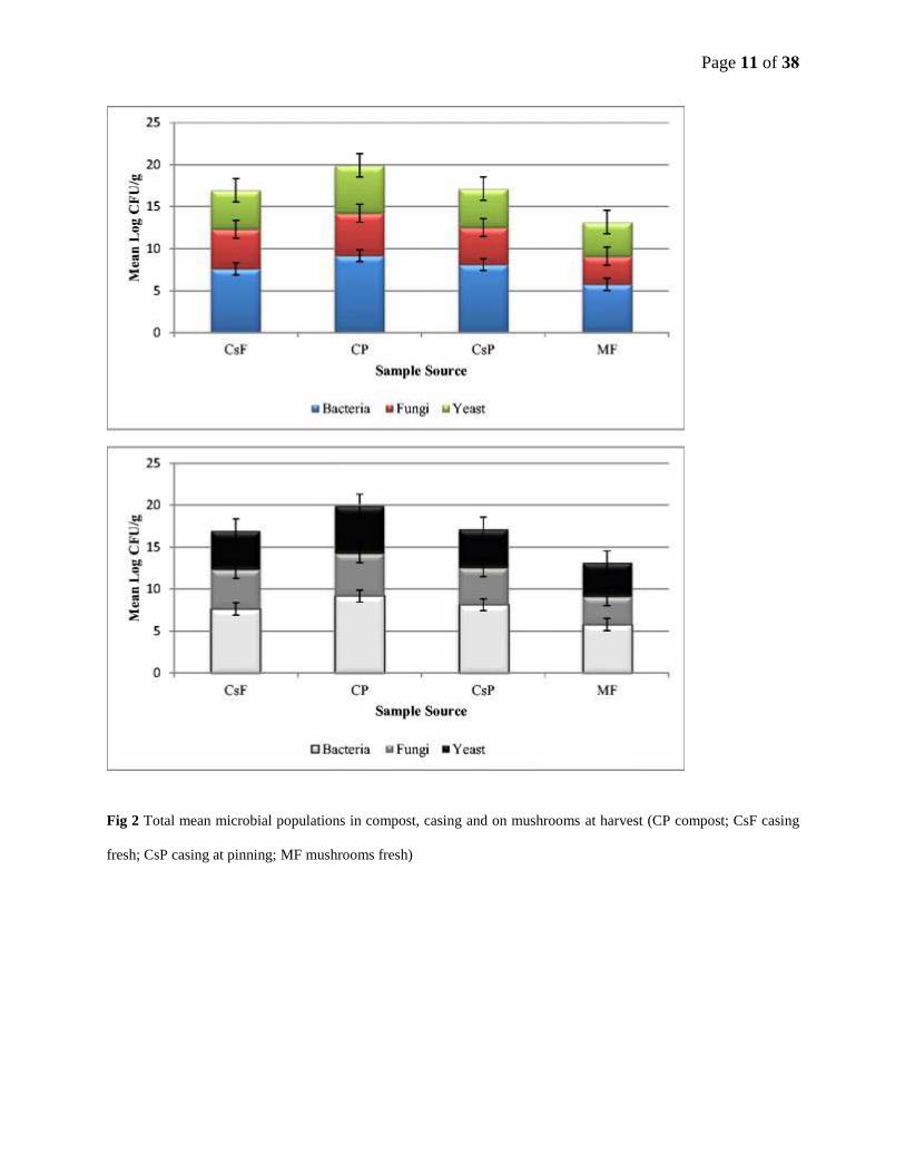
Fig 2 Total mean microbial populations in compost, casing and on mushrooms at harvest (CP compost; CsF casing
fresh; CsP casing at pinning; MF mushrooms fresh)
Page 12 of 38
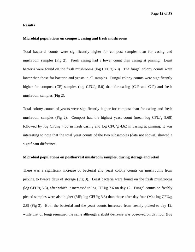
Results
Microbial populations on compost, casing and fresh mushrooms
Total bacterial counts were significantly higher for compost samples than for casing and
mushroom samples (Fig 2). Fresh casing had a lower count than casing at pinning. Least
bacteria were found on the fresh mushrooms (log CFU/g 5.8). The fungal colony counts were
lower than those for bacteria and yeasts in all samples. Fungal colony counts were significantly
higher for compost (CP) samples (log CFU/g 5.0) than for casing (CsF and CsP) and fresh
mushroom samples (Fig 2).
Total colony counts of yeasts were significantly higher for compost than for casing and fresh
mushroom samples (Fig 2). Compost had the highest yeast count (mean log CFU/g 5.68)
followed by log CFU/g 4.63 in fresh casing and log CFU/g 4.62 in casing at pinning. It was
interesting to note that the total yeast counts of the two subsamples (data not shown) showed a
significant difference.
Microbial populations on postharvest mushroom samples, during storage and retail
There was a significant increase of bacterial and yeast colony counts on mushrooms from
picking to twelve days of storage (Fig 3). Least bacteria were found on the fresh mushrooms
(log CFU/g 5.8), after which it increased to log CFU/g 7.6 on day 12. Fungal counts on freshly
picked samples were also higher (MF; log CFU/g 3.3) than those after day four (M4; log CFU/g
2.8) (Fig 3). Both the bacterial and the yeast counts increased from freshly picked to day 12,
while that of fungi remained the same although a slight decrease was observed on day four (Fig

Page 13 of 38
3). The average over 12 days was log CFU/g 7.6 (mean) for bacteria, log CFU/g 6.33 (median)
for yeasts and log CFU/g 2.8 (median) for fungi.
Fig 3 Total mean microbial populations on mushrooms from harvest to 12 days after picking (MF mushrooms fresh;
M4 mushrooms day 4; M8 mushrooms day 8; M12 mushrooms day 12)

Page 14 of 38
Pure culture identification using sequencing
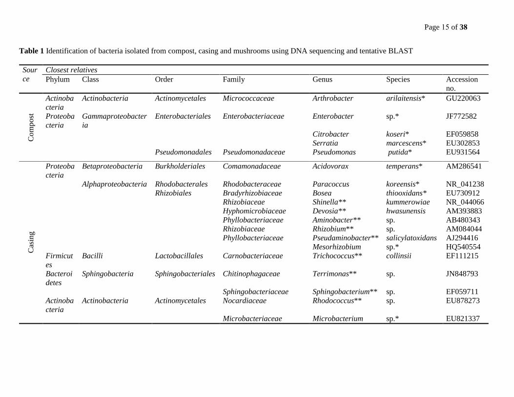
Across board, the culturable bacteria of compost samples showed less diverse phyla than those of
casing and mushroom samples. Bacteria in casing samples were more diverse than those in
mushroom samples. Bacteria isolated from compost samples were members of two phyla,
Actinobacteria and Proteobacteria while those from casing samples were members of four
phyla, Actinobacteria, Proteobacteria, Firmicutes, and Bacteroidetes. Bacteria from mushroom
samples were members of the three phyla, Actinobacteria, Proteobacteria, and Bacteroidetes. In
all compost, casing and mushroom samples, the phylum Proteobacteria was the most prevalent
(Table 1). All fungal cultures isolated belonged to the phylum Ascomycota and the three classes
Pezizomycetes, Sordariomycetes and Eurotiomycetes. From the class Eurotiomycetes, the genus
Penicillium of the family Trichocomaceae was prevalent in compost, casing and mushroom
samples (Table 2). More detailed BLAST results of bacterial and fungal nucleotide sequences
are shown in Online Resource 2.
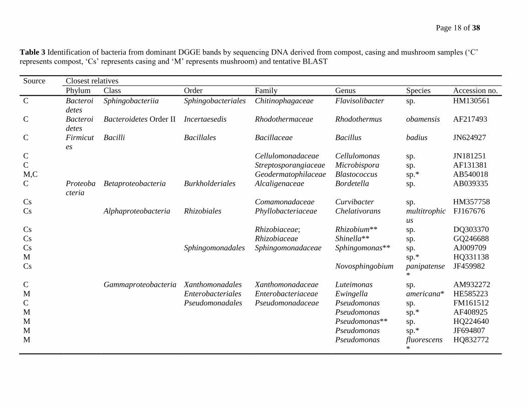
All bacterial DNA sequences of dominant DGGE gel bands matched with four phyla, viz.
Bacteroidetes, Firmicutes, Actinobacteria and Proteobacteria. Members of the first three phyla
were extracted predominantly from compost samples while the last phylum, Proteobacteria
originated mostly from casing and mushroom samples. Of the three classes in this phylum
namely Betaproteobacteria, Alphaproteobacteria and Gammaproteobacteria, the latter was the
most prevalent. The genus Pseudomonas of the family Pseudomonadaceae and order
Pseudomonadales was the most dominant in mushroom samples (Table 3).
Page 15 of 38
Table 1 Identification of bacteria isolated from compost, casing and mushrooms using DNA sequencing and tentative BLAST
Sour
ce
Closest relatives
Phylum Class Order Family Genus Species Accession
no.
Com
post
Actinoba
cteria
Actinobacteria Actinomycetales Micrococcaceae Arthrobacter arilaitensis* GU220063
Proteoba
cteria
Gammaproteobacter
ia
Enterobacteriales Enterobacteriaceae Enterobacter sp.* JF772582
Citrobacter koseri* EF059858
Serratia marcescens* EU302853
Pseudomonadales Pseudomonadaceae Pseudomonas putida* EU931564
Cas
ing
Proteoba
cteria
Betaproteobacteria Burkholderiales Comamonadaceae Acidovorax temperans* AM286541
Alphaproteobacteria Rhodobacterales Rhodobacteraceae Paracoccus koreensis* NR_041238
Rhizobiales Bradyrhizobiaceae Bosea thiooxidans* EU730912
Rhizobiaceae Shinella** kummerowiae NR_044066
Hyphomicrobiaceae Devosia** hwasunensis AM393883
Phyllobacteriaceae Aminobacter** sp. AB480343
Rhizobiaceae Rhizobium** sp. AM084044
Phyllobacteriaceae Pseudaminobacter** salicylatoxidans AJ294416
Mesorhizobium sp.* HQ540554
Firmicut
es
Bacilli Lactobacillales Carnobacteriaceae Trichococcus** collinsii EF111215
Bacteroi
detes
Sphingobacteria Sphingobacteriales Chitinophagaceae Terrimonas** sp. JN848793
Sphingobacteriaceae Sphingobacterium** sp. EF059711
Actinoba
cteria
Actinobacteria Actinomycetales Nocardiaceae Rhodococcus** sp. EU878273
Microbacteriaceae Microbacterium sp.* EU821337

Page 16 of 38
Table 1 (Continued)
Source Closest relatives
Phylum Class Order Family Genus Species Accession
no.
Mush
room
Bacteroidetes Sphingobacteriia Sphingobacteriales Sphingobacteriaceae Sphingobacterium multivorum* HM355636
Actinobacteria Actinobacteria Actinomycetales Microbacteriaceae Microbacterium** foliorum JF303045
Proteobacteria Betaproteobacteria Burkholderiales Alcaligenaceae Alcaligenes faecalis* FJ959394
Gammaproteobacteria Pseudomonadales Pseudomonadaceae Pseudomonas Putida*
fluorescens
JN228297
EF602564
Enterobacteriales Enterobacteriaceae Ewingella americana* HE585223 (Identification: * at species level, ** at genus level)

Page 36 of 38
Table 2 Identification of fungi and yeasts isolated from compost, casing and mushrooms using DNA sequencing and tentative BLAST (‘C’
represents compost, ‘Cs’ represents casing and ‘M’ represents mushroom)
Source Closest relatives
Phylum Class Order Family Genus Species Accession
no.
Fungi
Cs Ascomycota Pezizomycetes Pezizales Pezizaceae Chromelosporium EF589890
Cs Sordariomycetes Hypocreales Bionectriaceae Bionectria sp. AM944351
Cs Microascales Microascaceae Pseudallescheria** boydii GU566282
Cs Sordariales Chaetomiaceae Chaetomium sp. AB506801
Cs Hypocreales Hypocreaceae Trichoderma** sp. AY514867
M Eurotiomycetes Eurotiales Trichocomaceae Paecilomyces** sinensis FJ904847
M,Cs Penicillium madriti* AF033482
C,Cs,M Penicillium** chrysogenum GU565114
C,Cs Penicillium** ochrochloron JF311909
M Penicillium** meleagrinum HM469412
M,Cs Penicillium brevicompactum
*
HM469408
Yea
st
C,M Basidiomyc
ota
Tremellomycetes Cystofilobasidial
es
Cystofilobasidiaceae Cystofilobasidium infirmominiat
um*
AY264716
C,M Urediniomycetes Sporidiobolales Sporidiobolaceae Rhodotorula mucilaginosa
*
EF174513
C Tremellomycetes Tremellales Trichosporonaceae Trichosporon moniliiforme* FR799471
C Trichosporon cutaneum* FJ943422
C Ascomycota Saccharomycetes Saccharomycetal
es
Saccharomycetaceae Candida glaebosa* FJ153208
Cs Candida subhashii EU836707
Cs Candida sp. HQ623537
Cs Candida sp. JF895510
Cs Pichia sp. FJ153190 (Identification: * at species level, ** at genus level)
Page 18 of 38
Table 3 Identification of bacteria from dominant DGGE bands by sequencing DNA derived from compost, casing and mushroom samples (‘C’
represents compost, ‘Cs’ represents casing and ‘M’ represents mushroom) and tentative BLAST
Source Closest relatives
Phylum Class Order Family Genus Species Accession no.
C Bacteroi
detes
Sphingobacteriia Sphingobacteriales Chitinophagaceae Flavisolibacter sp. HM130561
C Bacteroi
detes
Bacteroidetes Order II Incertaesedis Rhodothermaceae Rhodothermus obamensis AF217493
C Firmicut
es
Bacilli Bacillales Bacillaceae Bacillus badius JN624927
C Cellulomonadaceae Cellulomonas sp. JN181251
C Streptosporangiaceae Microbispora sp. AF131381
M,C Geodermatophilaceae Blastococcus sp.* AB540018
C Proteoba
cteria
Betaproteobacteria Burkholderiales Alcaligenaceae Bordetella sp. AB039335
Cs Comamonadaceae Curvibacter sp. HM357758
Cs Alphaproteobacteria Rhizobiales Phyllobacteriaceae Chelativorans multitrophic
us
FJ167676
Cs Rhizobiaceae; Rhizobium** sp. DQ303370
Cs Rhizobiaceae Shinella** sp. GQ246688
Cs
M
Sphingomonadales Sphingomonadaceae Sphingomonas** sp.
sp.*
AJ009709
HQ331138
Cs Novosphingobium panipatense
*
JF459982
C Gammaproteobacteria Xanthomonadales Xanthomonadaceae Luteimonas sp. AM932272
M Enterobacteriales Enterobacteriaceae Ewingella americana* HE585223
C Pseudomonadales Pseudomonadaceae Pseudomonas sp. FM161512
M Pseudomonas sp.* AF408925
M Pseudomonas** sp. HQ224640
M Pseudomonas sp.* JF694807
M Pseudomonas fluorescens
*
HQ832772

Page 19 of 38
M Pseudomonas syringae JN088486
M Pseudomonas migulae* DQ377758
M, Cs Pseudomonas Plecoglossi
cida*
JN624752
M, Cs Pseudomonas putida* AF396076
M, Cs Pseudomonas reactans* JN662499
M, Cs Pseudomonas nitroreduce
ns
JF513151
(Identification: * at species level, ** at genus level)
Page 20 of 38
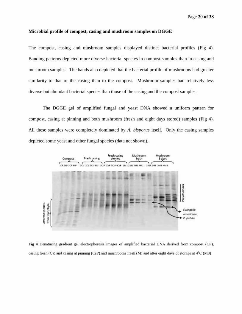
Microbial profile of compost, casing and mushroom samples on DGGE
The compost, casing and mushroom samples displayed distinct bacterial profiles (Fig 4).
Banding patterns depicted more diverse bacterial species in compost samples than in casing and
mushroom samples. The bands also depicted that the bacterial profile of mushrooms had greater
similarity to that of the casing than to the compost. Mushroom samples had relatively less
diverse but abundant bacterial species than those of the casing and the compost samples.
The DGGE gel of amplified fungal and yeast DNA showed a uniform pattern for
compost, casing at pinning and both mushroom (fresh and eight days stored) samples (Fig 4).
All these samples were completely dominated by A. bisporus itself. Only the casing samples
depicted some yeast and other fungal species (data not shown).
Fig 4 Denaturing gradient gel electrophoresis images of amplified bacterial DNA derived from compost (CP),
casing fresh (Cs) and casing at pinning (CsP) and mushrooms fresh (M) and after eight days of storage at 4oC (M8)

Page 21 of 38
Phylogenetic relationships of cultured and non-cultured microorganisms from compost,
casing and mushrooms
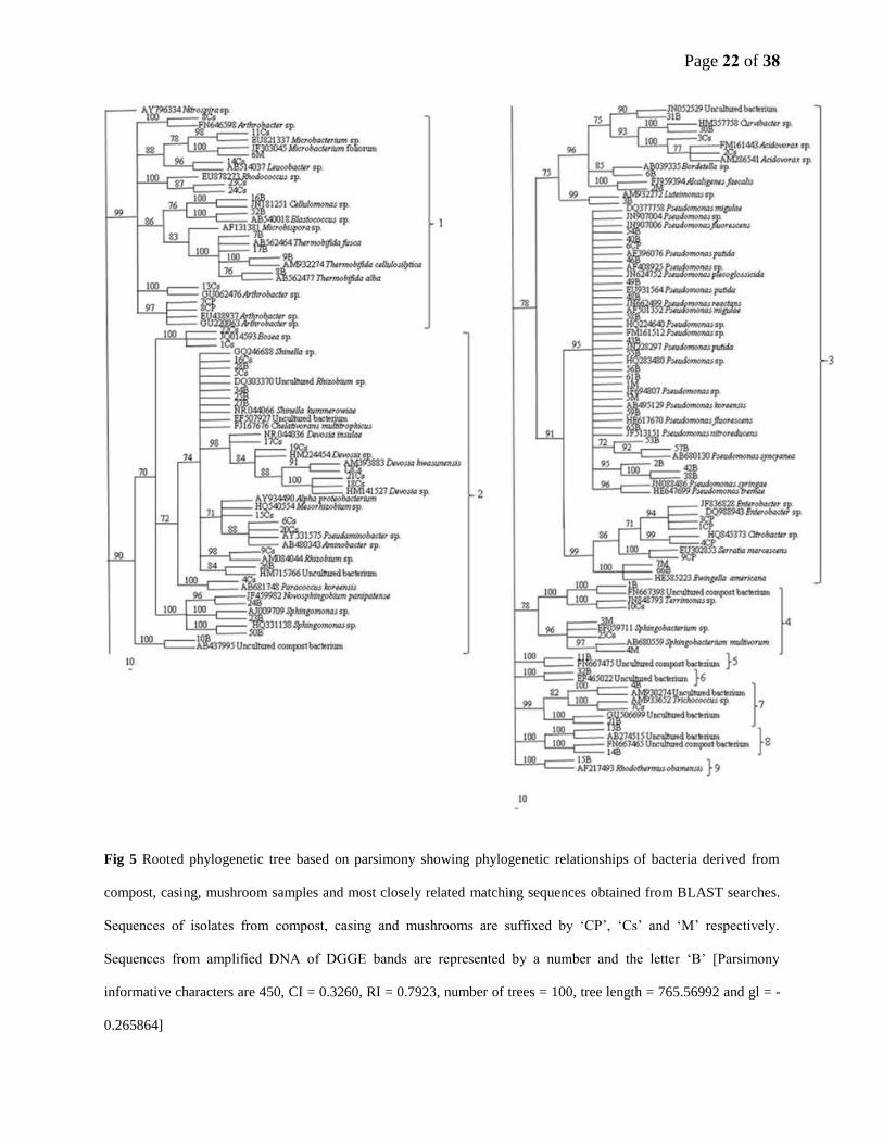
Bacteria from mushrooms showed phylogenetic relationships with those from both casing and
compost (Fig 5). The first four big clades of the phylogenetic tree consisted of bacteria from all
three sources (compost, casing and mushroom). Generally, bacterial sequences from both
cultures and from amplified DNA of DGGE gel bands grouped together in the different clusters
(clades 1-4). However, four separate clades (clades 5, 6, 8, and 9) of amplified DNA from
DGGE gel bands were formed. In the cluster (clade 3) which consisted of different
Pseudomonas species, the majority of its members originated from mushroom samples,
specifically from amplified DNA of the DGGE gel bands.
Almost all DNA sequences of yeasts and fungi were derived from pure culture isolates,
since fungal (and yeast) DGGE profiles of samples were dominated by the A. bisporus itself.
Penicillium spp. were commonly isolated from compost, casing and mushroom samples. The
phylogenetic tree of yeasts formed two clades (Fig 6). Clade 1 consisted of sequences
predominantly from compost and mushrooms while clade 2 contained sequences from casing
except for one from compost.
Page 22 of 38
Fig 5 Rooted phylogenetic tree based on parsimony showing phylogenetic relationships of bacteria derived from
compost, casing, mushroom samples and most closely related matching sequences obtained from BLAST searches.
Sequences of isolates from compost, casing and mushrooms are suffixed by ‘CP’, ‘Cs’ and ‘M’ respectively.
Sequences from amplified DNA of DGGE bands are represented by a number and the letter ‘B’ [Parsimony
informative characters are 450, CI = 0.3260, RI = 0.7923, number of trees = 100, tree length = 765.56992 and gl = -
0.265864]

Page 23 of 38
Fig 6 Rooted phylogenetic tree based on ITS region sequences of yeasts derived from compost, casing and
mushroom samples and most closely related sequences obtained from BLAST searches. Sequences from compost,
casing and mushrooms are suffixed by ‘CPy’, ‘Csy’ and ‘My’ respectively [Parsimony informative characters are
270, CI = 0.7495, RI = 0.9337, number of trees = 100, tree length = 470.22827 and g1 = -0.379514]

Page 24 of 38
Discussion
Most of the microbiological studies on compost and composting were either not specific on
materials for mushroom growing, or if so, they were on the phases before spawning. Few studies
were done on spawned mushroom compost, however, information on diversity was not provided.
Fordyce (1970) reported mesophilic bacteria in the order of 106 - 10
7/g for a three week spawned
compost. Fermor and Wood (1981) also mentioned a reduction and stabilisation of bacterial
numbers in spawned compost following the high number and activity of bacteria in Phase I and
II compost. On both studies, there was no report on diversity. This study therefore provides
baseline microbial population information on mushrooms, compost and casing material and the
resultant mushroom fruit body growth during the production cycle and resultant shelf-life after
picking, packing and cold storage till marketing.
Although compost is known as a nutrient rich medium, bacterial populations at the end of
Phase II composting stage create unfavourable conditions for competitive microorganisms and
provide selectivity to A. bisporus (Fordyce, 1970; Ross and Harris, 1983). Phase II composting
involves conditioning, which is a controlled heat treatment that selectively kills pathogenic
micro-organisms. Other non-pathogenic bacteria may remain static at this stage of composting
and become active later when the temperature is lowered. This trend was confirmed in our study
when comparing the compost casing and mushroom microbial profiles. Bacterial populations of
compost were significantly higher than those of the casing and mushroom samples.
In general, the plating method used demonstrated richer microbial population counts in
compost samples compared with casing and mushroom samples. It was interesting to note the
significant difference in total yeast colony counts between the compost subsamples. This
Page 25 of 38
suggested that yeast populations are influenced by homogeneity of samples more than bacteria.
Considering the lack of information on the identity and role of yeasts, more studies in this regard
should be undertaken.
The casing layer in this study was prepared from peat, which is naturally formed from
partially decomposed plant material. Unlike compost, peat is relatively poor in nutrients and
decomposed organic matter (Eakin, 1969; Abad et al. 1989). Therefore, it is not unexpected that
total bacterial colony count was significantly lower in the casing compared to the compost. Even
so, total bacterial colony counts in casing increased significantly at pinning; although this count
was still significantly lower than that of the compost. This significant increase in total bacterial
counts from casing to pinning is consistent with previous work (Miller et al. 1995; Siyoum et al.
2010). In the cultivation of A. bisporus, compost and casing soil are two major elements; studies
have also demonstrated that populations of pseudomonas in the casing layer on which the
mushroom fruit body develops play an important role (Riahi et al. 2011). In most studies, total
bacterial populations ranged from log CFU/g 8.0 to 8.5 casing material and the majority of
bacterial populations in casing were attributed to pseudomonas species. According to Riahi et al.
(2011) pseudomonads represented more than 80 percent of the bacterial population in the casing
layer. Their results also showed that there was a close relation between growth of mycelium and
number of bacteria in casing soil. Sampling of casing soil at different stages of the mushroom
growing cycle further revealed that the number of bacteria increased simultaneously with
increased growth of mycelium into the casing soil. Riahi et al. (2011) concluded that the
inoculation of native P. putida isolated from casing soil at the primordia formation stage would
be very efficient for increasing mushroom yield and quality.

Page 26 of 38
Fresh casing samples had lower total colony counts of fungi than in compost samples
although the difference was not initially significant. However, at pinning, the total fungal colony
count of the casing was reduced to a significantly lower level than that of the compost.
Verhoeven (1986) described that fungi do not have a significant role to play in peat, unlike
bacteria. Like bacteria and fungi, yeast populations were also significantly higher in compost
samples than in the casing and fresh mushroom samples. The overall lower microbial counts in
casing compared to compost may be attributed to limited energy sources and lack of available
nutrients in peat soil according to Verhoeven (1986).
In our study, there also was a significant increase in total bacteria and yeasts on
mushrooms from the time of harvest to twelve days after cold storage while fungi remained
constant. A similar trend for bacteria, yeasts and moulds was also reported by Chikthimmah
(2006). Postharvest spoilage of mushrooms caused by bacteria is well established (Burton and
Noble, 1993; Fett et al. 1995; Wells et al. 1996). However, the role of yeasts in mushroom
spoilage was not investigated in this study and needs further investigation.
Bacteria isolated from compost were less diverse at phylum and family level than those
isolated from casing and mushrooms. Compost bacteria belonged to two phyla and three
families only while casing bacteria belonged to four phyla and eleven families. The extent of
diversity within bacteria isolated from mushrooms lies between that of the two substrates.
Cultured bacteria of mushrooms belonged to three phyla and five families. Visual examination
of bacterial plates illustrated more uniform colonies for compost and mushroom than for casing
samples, which could be attributed to the less diverse families. Our low diversity result of
culturable compost bacteria is in agreement with Peters et al. (2000) although they investigated

Page 27 of 38
only the thermophilic group. However, work by Ryckeboer et al. (2003b) is not consistent with
our result. The reason for this could be the difference in the type of the compost material
(vegetable, fruit and garden waste in their case).
Results from our DGGE analysis provided a different profile of bacterial diversity in the
compost, casing and mushrooms. Bacterial diversity was highest in compost samples. DNA
sequences that were closely related to known cultured species belonged to four phyla for
compost samples but only to one and two phyla for casing and fresh mushroom samples
respectively. The difference in diversity results between the culturing and the DGGE methods is
understandable. It is known that bacteria that grow in an artificial medium represent only a small
fraction (0.3 % in soil habitat) of the total community in the sampled environment (Amann et al.
1995). Therefore, isolates obtained using culturing methods might not necessarily be the
dominant ones in their habitat. Several researchers have reported that some cultured bacteria
were not part of the dominant groups shown by DGGE/TGGE (Felske et al. 1999; Ellis et al.
2003) and other culture-independent molecular methods (Kaiser et al. 2001; Pearce et al. 2003).
Therefore, in order to have a broader picture of microbial diversity, it is advisable to combine
both culturing and non-culturing methods.
Similarly, fungi and yeasts other than A. bisporus could not be detected on the DGGE gel
for compost, casing at pinning and mushroom samples. These samples were completely
dominated by A. bisporus. In this study, the complete dominance of the mushroom fungal
community by A. bisporus itself was likely due to the method used. The DGGE method revealed
a broader perspective of members of a community. However, using the plating method, fungi
and yeasts were detectable on MEA plates although they were few and of low diversity

Page 29 of 38
dominated by the genus Penicillium. This low level of fungi other than A. bisporus is
satisfactory, especially in compost. Well prepared compost is produced in such a way that it
selectively supports the growth of A. bisporus and is unfavourable for other fungi (Ross and
Harris, 1983; Camp et al. 1990).
When comparing bacterial profiles of the compost, casing and mushroom samples using
DGGE, compost populations were distinct from those of both the casing and mushrooms. This
result is in agreement with Reddy and Patrick (1990), who isolated similar bacteria from the
casing and the mushroom mycelium colonising it. Their results showed that these bacteria were
not similar to those isolated from compost. They also concluded that bacterial isolates from
compost had no effect on formation of mushroom fruit bodies. Profiles for fresh casing and
casing at pinning were similar except that population densities increased at pinning, which is
consistent with previous work (Siyoum et al. 2010). Both fresh mushrooms and mushrooms
stored for eight days revealed few but dominant bacterial species on their profiles in comparison
to casing and compost. These profiles were more similar to that of the casing. Our results
suggest that the fresh mushroom surface could be selective to a specific group of bacteria
dominated by Pseudomonas species. The whole profile also revealed that most bacteria on the
mushroom surface originated very likely from the casing.
It is interesting that almost only Pseudomonas spp. were dominant on the mushroom
profiles. On the casing profile, these species were less abundant (less intense bands) compared
to what was obtained from the mushrooms. It was also clearly shown that Ewingella americana
and certain Pseudomons spp. increased in abundance while a particular species, identified as a
close relative of P. putida, could no longer be detected after the eight day of storage. It is

Page 30 of 38
interesting to note that P. putida was abundant on fresh mushrooms but not on stored ones,
simulating a ready-to-sell product. This bacterium is well known for its role in fruit body
initiation (Hayes et al. 1969; Noble et al. 2009) and our results indicated that it was not part of
the postharvest spoilage syndrome associated with pseudomonads. The presence of
pseudomonads and E. americana in mushrooms is in agreement with reports from different
studies. Members of the genus Pseudomonas are well known as saprophytes and pathogens of
mushrooms (Munsch and Alatossava, 2002; Sivanesan, 2003). The non-pseudomonad bacterium
E. americana was also reported on mushrooms as both a pathogen (Inglis et al. 1996) and as a
member of the indigenous micro-flora (Choudhury and Heinemann, 2006).
Phylogenetic analysis of bacteria from different samples - compost, casing and
mushroom - revealed that generally, species from the same sample source were the most closely
related. Nevertheless, it was also noted that some species from all three different sources were
phylogenetically related to each other. In addition, five minor separate lineages comprising of
compost and casing inhabiting bacterial species were formed on the phylogenetic tree. These
lineages indicate that species from these two sample sources were diverse and several of them
were not affiliated with known cultured bacteria. Ivors et al. (2000) reported a similar result in
which several rDNA sequences from compost samples were unique and not related to previously
identified species. Most of the sequences that were not affiliated with known cultured species in
our study originated from excised DGGE bands of compost and casing samples. These results
suggest that DGGE could reveal species that were not cultured previously and could possibly be
novel.
Page 30 of 38
The phylogenetic tree of yeasts from compost, casing and mushroom samples was
relatively simple, consisting of two lineages. The first lineage consisted of sequences
predominantly from compost samples. Two sequences from mushroom samples were also
affiliated with this lineage. The second lineage contained samples from casing and one sequence
from compost. The phylogenetic tree for yeasts consisted of sequences from cultured species
only, except one sequence from fresh casing sample of DGGE. Our results indicated that yeasts
from compost samples were more diverse than those from casing. Four out of six yeast species
from casing samples were closely related to Candida sp. including the one obtained from DGGE.
The two species were closely related to each other but grouped with the lineage of yeasts from
compost samples, although at a distant level.
In samples tested, yeast species originating from compost and casing samples were not
closely related while yeasts from mushroom samples were closely related to those from compost
samples. Although this result needs further investigation, it provides some background
information on diversity and relationship of yeasts in compost, casings and edible mushrooms
obtained from production, storage and marketing. A significant increase in yeast colonies were
recorded after mushrooms were stored simulating retail practice at 4o C for four, eight and twelve
days. Little information is available on spoilage of mushrooms caused by yeasts. Koorapati et
al. (2004) and Chikthimmah (2006) reported on the control of microbial spoilage after product
irradiation. In their experiment, irradiation reduced all microbial counts including bacteria,
yeasts and moulds, and prolonged mushroom shelf life. However, it was not clear from their
studies whether yeasts also contributed to the spoilage. A general observation in our study
(unpublished data) was that spoilage was associated with superficial slimy yeast growth. Future
studies should focus on determining the role of yeasts in mushroom spoilage at retail-end.

Page 31 of 38
The unique approach reported in this paper that used a combination of methods (cultural
and non-cultural) to better understand microbial dynamics of mushrooms from seeding through
growth, postharvest up to ready-to-eat at the consumer-end of the supply chain contribute
towards a better understanding of what constitutes a healthy edible mushroom. This information
can be used in developing national guidelines for total microbial loads of fresh mushrooms
within a food safety compliance framework. A total microbial load of log CFU/g 4.4 to 9.4 has
been reported for cultivated and wild mushrooms (Venturini et al. 2011). Doores et al. (1987)
found that bacterial populations during postharvest storage at 13 oC increased from an initial load
of log CFU/g 7 to almost log CFU/g 11 over a 10-day storage period. In our studies fresh white
button mushrooms carried an average bacterial load of log CFU/g 5.8 increasing to a maximum
of log CFU/g 7.6 when stored at 4 oC for 12 days. A normal microflora of healthy mushrooms at
the point of harvest can represent a log CFU/g 4 - 5 that reflect a healthy climax bacterial
community. Bacterial microbial loads can be expected to increase to log CFU/g 7-9 depending
on the storage conditions. In the work done by Doores et al. (1987) an increase in Pseudomonas
populations to a log CFU/g 6 were attributed to quality deterioration and brown blotch
development.
Acknowledgments
This research was funded by the South African Mushroom Farmers Association (SAMFA),
National Research Foundation (NRF) and the Technology and Human Resources for Industry
Programme (THRIP) (a partnership programme funded by the Department of Trade and Industry
and managed by the NRF). We would like to thank Dr. L. Meyer, Ms R. Jacobs, for molecular
advice and assistance, as well as Ms A. Redmond for technical assistance. We also would like to
thank the commercial farm for providing research materials.
Page 32 of 38
References
Abad M, Noguera V, Martínez-Corts J (1989) The effect of sedge peat-based media and
controlled-release fertilizer on the growth of begonia, French marigold and geranium. Acta
Hortic 246:199-212
Amann RI, Ludwig W, Schleifer K H (1995) Phylogenetic identification and in situ detection of
individual microbial cells without cultivation. Microbiol Rev 59:143-169
Burton KS, Noble R (1993) The influence of flush number, bruising and storage temperature on
mushroom quality. Postharvest Biol Tec 3:39-47
Camp HJMO, Stumm CK, Straatsma G, Derikx PJL, van Griensven JLD (1990) Hyphal and
mycelia interactions between A. bisporus and Scytalidium thermophilum on agar media. Microb
Ecol 19:303-309
Chikthimmah N (2006) Microbial ecology of mushroom casing soils and preharvest strategies to
enhance safety and quality of fresh mushrooms. Ph.D. Thesis, Pennsylvania State University,
USA
Choudhury PR, Heinemann JA (2006) The general secretory pathway of Burkholderia gladioli
pv. agaricicola BG164R is necessary for cavity disease in white button mushrooms. Appl
Environ Microbiol 72:3558-3565
Doores S, Kramer M, Beelman R (1987) Evaluation and bacterial populations associated with
fresh mushrooms (Agaricus bisporus). In: Cultivating Edible Fungi. Wuest PJ, Royce DL,
Page 33 of 38
Beelman RB (eds) Developments in Crop Science 10, Amsterdam: Elsevier Science Publishers,
pp 283-94
Drancourt M, Bollet C, Carlioz A, Martelin R, Gayral J-P, Raoult D (2000) 16S Ribosomal DNA
sequence analysis of a large collection of environmental and clinical unidentifiable bacterial
isolates. J Clin Microbiol 38:3623-3630
Eakin JH (1969) Let’s know more about peat. Mushroom News 17:12-15
Eicker A, van Greuning M (1989) Economical alternatives for topogenous peat as casing
material in the cultivation of Agaricus bisporus in South Africa. S Afr J Plant Soil 6:129-135
Ellis RJ, Morgan P, Weightman AJ, Fry JC (2003) Cultivation dependent and -independent
approaches for determining bacterial diversity in heavy-metal-contaminated soil. Appl Environ
Microbiol 69:3223-3230
Everett KDE, Bush RM, Andersen AA (1999) Emended description of the order Chlamydiales,
proposal of Parachlamydiaceae fam. nov. and Simkaniaceae fam. nov., each containing one
monotypic genus, revised taxonomy of the family Chlamydiaceae, including a new genus and
five new species, and standards for the identification of organisms. Int J Syst Bacteriol 49:415-40
Felske A, Wolterink A, van Lis R, de Vos WM, Akkermans AD (1999) Searching for
predominant soil bacteria: 16S rDNA cloning versus strain cultivation. FEMS Microbiol Ecol
30:137-145
Page 34 of 38
Fermor TR, Wood DA (1981) Degradation of bacteria by Agaricus bisporus and other fungi. J
Gen Microbiol 126:377-387
Fermor T, Lincoln S, Noble R, Dobrovin-Pennington A, Colauto N (2000) Microbiological
properties of casing. In: Van Griensven LJLD (ed) Science and Cultivation of Edible Fungi.
Balkema, Rotterdam, pp 447-454
Fett WF, Wells JM, Cescutti P, Wijey C (1995) Identification of exopolysaccharides produced
by fluorescent pseudomonads associated with commercial mushroom (Agaricus bisporus)
production. Appl Environ Microbiol 61:513-517
Fjellbirkeland A, Torsvik V, Øvereås L (2001) Methanotrophic diversity in an agricultural soil as
evaluated by denaturing gradient gel electrophoresis profiles of pmoA, mxaF and 16S rDNA
sequence. Anton Leeuwenhoek 79:209-217
Fordyce CJR (1970) Relative numbers of certain microbial groups present in compost used for
mushroom (Agaricus bisporus) propagation. Appl Microbiol 20:196-199
Garbeva P, van Veen JA, van Elsas JD (2004) Microbial diversity in soil: selection of microbial
populations by plant and soil type and implications for disease suppressiveness. Annu Rev
Phytopathol 42:243-70
Hayes WA, Randle P, Last FT (1969) The nature of microbial stimulus affecting sporophore
formation in Agaricus bisporus (Lange) Sing. Ann Appl Biol 64:177-187

Page 35 of 38
Inglis PW, Burden JL, Peberdy JF (1996) Evidence for the association of the enteric bacterium
Ewingella americana with internal stipe necrosis of Agaricus bisporus. Microbiology 142:3253-
3260
Ivors KL, Collopy PD, Beyer DM, Kang S (2000) Identification of bacteria in mushroom
compost using ribosomal RNA sequence. Compost Sci Util 8:247-253
Kaiser O, Puhler A, Selbitschka W (2001) Phylogenetic analysis of microbial diversity in the
rhizoplane of oilseed rape (Brassica napus cv. westar) employing cultivation-dependent and
cultivation-independent approaches. Microb Ecol 42:136-149
Koorapati A, Foley D, Pilling R, Prakash A (2004) Electron-beam irradiation preserves the
quality of white button mushrooms (Agricus bisporus) slices. J Food Sci 69:SNQ25-29
Labuschagne PM (1995) Casing media for Agaricus bisporus cultivation in South Africa. M.Sc.
Thesis, University of Pretoria, South Africa
Miller FC, Harper ER, Macauley BJ (1989) Field examination of temperature and oxygen
relationships in mushroom composting stacks-consideration of stack oxygenation based on
utilisation and supply. Aust J Exp Agr 29:741-750
Miller N, Gallespie JB, Doyle OPE (1995) The involvement of microbiological components of
peat based casing materials. Mushroom Sci 14:313-321
Munsch P, Alatossava T (2002) Several pseudomonads, associated with the cultivated
mushrooms Agaricus bisporus or Pleurotus sp., are hemolytic. Microbiol Res 157:311-315

Page 36 of 38
Noble R, Dobrovin-Pennington A, Hobbs PH, Pederby J, Rodger A (2009) Volatile C8
compounds and pseudomonads influence primordium formation of Agaricus bisporus.
Mycologia 101:583-591
Orgiazzi A, Bianciotto V, Bonfante P, Daghino S, Ghignone S, Lazzari A, Lumini E, Mello A,
Napoli C, Perotto S, Vizzini A, Bagella S, Murat C, Girlanda M (2013) 454 Pyrosequencing
analysis of fungal assemblages from geographically distant, disparate soils reveals spatial
patterning and a core mycobiome. Diversity 5:73-98. doi:10.3390/d5010073
Øvereås L, Forney L, Daae FL, Torsvik V (1997) Distribution of bacterioplankton in meromictic
lake Sælenvennet as determined by denaturing gradient gel electrophoresis of PCR amplified
gene fragments coding for 16S RNA. Appl Environ Microbiol 63:3367-3373
Pearce DA, van der Gast CJ, Lawley B, Ellis-Evans JC (2003) Bacterioplankton community
diversity in a maritime Antarctic lake, determined by culture-dependent and culture-independent
techniques. FEMS Microbiol Ecol 45:59-70
Peters S, Koschinsky S, Schwieger F, Tebbe CC (2000) Succession of microbial communities
during hot composting as detected by PCR-single-strand-conformation polymorphism-based
genetic profiles of small-subunit rRNA genes. Appl Environ Microbiol 66:930-936
Ranjard L, Lehon PH, Mougel C, Schehrer L, Merdinoglu D, Chaussod R (2003) Sampling
strategy in molecular microbial ecology: influence of soil sample size on DNA fingerprinting
analysis of fungal and bacterial communities. Environ Microbiol 5(11):1111-1120
Page 37 of 38
Reddy MS, Patrick ZA (1990) Effect of bacteria associated with mushroom compost and casing
materials on basidiomata formation in Agaricus bisporus. Can J Plant Pathol 12:236-242
Riahi H, Eskash A, Shariatmadari Z (2011) Effect of bacterial and cyanobacterial culture on
growth, quality and yield of Agaricus bisporus. Proceedings of the 7th International Conference
on Mushroom Biology and Mushroom Products (ICMBMP7)
Ross RC, Harris, PJ (1983) An investigation into the selective nature of mushroom compost Sci
Hortic 19:55-64
Ryckeboer J, Mergaert J, Vaes K, Klammer S, de Clercq D, Coosemans J, Insam H, Swings J
(2003a) A survey of bacteria and fungi occurring during composting and self-heating processes.
Ann Microbiol 53:349-410
Ryckeboer J, Mergaert J, Coosemans J, Deprins K, Swings J (2003b) Microbiological aspects of
biowaste during composting in a monitored compost bin. J Appl Microbiol 94:127-137
Sivanesan D (2003) Diversity among bacteria causing blotch disease on the commercial
mushroom, Agaricus bisporus. M.Sc. Thesis, Brock University, Ontario
Siyoum NA, Surridge K, Korsten L (2010) Bacterial profiling of casing materials for white
button mushrooms (Agaricus bisporus) using denaturing gradient gel electrophoresis. S Afr J Sci
doi: 10.4102/sajs. v106i9/10.253
Straatsma G, Samson RA, Olijnsma TW, Camp HJMOD, Gerrits JPG, van Griensven LJLD
(1994) Ecology of thermophilic fungi in mushroom compost, with emphasis on Scytalidium

Page 38 of 38
thermophilum and growth stimulation of Agaricus bisporus mycelium. Appl Environ Microbiol
60:454-458
Székely AJ, Sipos R, Berta B, Vajna B, Hajdú C, Márialigeti K (2009) DGGE and T-RFLP
Analysis of Bacterial Succession during Mushroom Compost Production and Sequence-aided T-
RFLP Profile of Mature Compost. Microb Ecol 57:522–533
Venturini ME, Reyes JE, Rivera CS, Oria R, Blanco D (2011) Microbiological quality and safety
of fresh cultivated and wild mushrooms commercialized in Spain. Food Microbiol 28:1492-1498
Verhoeven JTA (1986) Nutrient dynamics in minerotrophic peat mires. Aquat Bot 25:117-137
Wells JM, Sapers GM, Fett WF, Butterfield JE, Jones JB, Bouzar H, Miller FC (1996)
Postharvest discoloration of the cultivated mushroom Agaricus bisporus caused by Pseudomonas
tolaasii, P. ‘reactans’, and P. ‘gingeri’. Phytopathology 86:1098-1104
White TJ, Bruns T, Lee S, Taylor JW (1990) Amplification and direct sequencing of fungal
ribosomal RNA genes for phylogenetics. In: Innis MA, Gelfand DH, Sninsky JJ, White TJ (eds)
PCR protocols: A Guide to Methods and Applications. Academic Press, New York, pp 315-322