Embed Size (px)
Citation preview
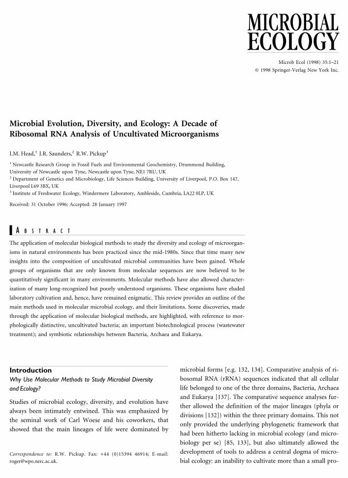
Microbial Evolution, Diversity, and Ecology: A Decade ofRibosomal RNA Analysis of Uncultivated Microorganisms
I.M. Head,1 J.R. Saunders,2 R.W. Pickup3
1 Newcastle Research Group in Fossil Fuels and Environmental Geochemistry, Drummond Building,
University of Newcastle upon Tyne, Newcastle upon Tyne, NE1 7RU, UK2 Department of Genetics and Microbiology, Life Sciences Building, University of Liverpool, P.O. Box 147,
Liverpool L69 3BX, UK3 Institute of Freshwater Ecology, Windermere Laboratory, Ambleside, Cumbria, LA22 0LP, UK
Received: 31 October 1996; Accepted: 28 January 1997
A B S T R A C T
The application of molecular biological methods to study the diversity and ecology of microorgan-
isms in natural environments has been practiced since the mid-1980s. Since that time many new
insights into the composition of uncultivated microbial communities have been gained. Whole
groups of organisms that are only known from molecular sequences are now believed to be
quantitatively significant in many environments. Molecular methods have also allowed character-
ization of many long-recognized but poorly understood organisms. These organisms have eluded
laboratory cultivation and, hence, have remained enigmatic. This review provides an outline of the
main methods used in molecular microbial ecology, and their limitations. Some discoveries, made
through the application of molecular biological methods, are highlighted, with reference to mor-
phologically distinctive, uncultivated bacteria; an important biotechnological process (wastewater
treatment); and symbiotic relationships between Bacteria, Archaea and Eukarya.
IntroductionWhy Use Molecular Methods to Study Microbial Diversityand Ecology?
Studies of microbial ecology, diversity, and evolution have
always been intimately entwined. This was emphasized by
the seminal work of Carl Woese and his coworkers, that
showed that the main lineages of life were dominated by
microbial forms [e.g. 132, 134]. Comparative analysis of ri-
bosomal RNA (rRNA) sequences indicated that all cellular
life belonged to one of the three domains, Bacteria, Archaea
and Eukarya [137]. The comparative sequence analyses fur-
ther allowed the definition of the major lineages (phyla or
divisions [132]) within the three primary domains. This not
only provided the underlying phylogenetic framework that
had been hitherto lacking in microbial ecology (and micro-
biology per se) [85, 133], but also ultimately allowed the
development of tools to address a central dogma of micro-
bial ecology: an inability to cultivate more than a small pro-Correspondence to: R.W. Pickup. Fax: +44 (0)15394 46914; E-mail:
MICROBIALECOLOGY
Microb Ecol (1998) 35:1–21
© 1998 Springer-Verlag New York Inc.

portion (0.1–10%) of the bacteria that can be visualized by
direct count procedures. This has been a considerable handi-
cap to microbial ecology. Ecological inferences based on the
metabolic properties of cultivated bacteria are, by necessity,
unrepresentative of the natural populations from which they
were obtained [15]. Although the biases of cultivation-based
approaches were recognized by Winogradsky [131 cited in
127], it is only recently that means have been developed to
study the uncultivated majority. First Zuckerkandl and Paul-
ing [141], then Woese’s advances in microbial phylogeny
coupled with developments in molecular biology provided
the necessary methods to allow the identity of uncultivated
bacteria to be determined.
Norman Pace’s group, in Indiana [84, 87], was among the
first to appreciate the power of combining Woese’s new
phylogeny with molecular biology, and began what is now
recognised as molecular microbial ecology.
The Phylogenetic Basis of Molecular Microbial Ecology
In principle, the techniques described below can be applied
to any gene. The methods have been used, for example, to
study aromatic hydrocarbon-degrading bacteria [98] and
mercury-resistant microbial populations [86]. Our discus-
sion will, however, concentrate on the approaches involving
analysis of ribosomal RNA sequences [5, 84, 87, 127]. An
outline of many of the procedures commonly used in mo-
lecular microbial ecology are depicted in Fig. 1.
The Nature of rRNA Molecules
Due to the ubiquity of ribosomal RNA molecules (small
subunit, 16S, and 18S, in Eukarya; large subunit 23S and
28S, in Eukarya) in all cellular life forms, comparative analy-
sis of their sequences can be universally applied to infer
relationships among organisms. The rRNA molecules com-
prise highly conserved sequence domains interspersed with
more variable regions [48, 117]. In general, essential rRNA
domains are conserved across all phylogenetic domains, thus
‘‘universal’’ tracts of sequences can be identified [45, 84]. In
addition, it is also possible to identify sequence motifs of
increasing phylogenetic resolution. For example ‘‘signature’’
sequences for the Archaea, Bacteria, and Eukarya have been
recognized [45, 57, 103, 132], as well as short stretches of
sequence characteristic of a number of the bacterial divisions
and subdivisions (a-, b-, d-, g-Proteobacteria, high %G+C
Fig. 1. Commonly used approaches in molecular microbial ecology.
2 I.M. Head et al.

Gram-positive bacteria, and the Flavobacterium-Cytophaga
division [5, 74, 95]. Species- and subspecies-specific se-
quences have also been identified [5].
Inferring Phylogenetic Relationships from rRNA Sequences
The most commonly used form of comparative rRNA se-
quence analysis involves the construction of phylogenetic
trees. There are a number of procedures used to achieve this,
but the first stage in these analyses is always the careful
alignment of the rRNA sequences. This is a relatively
straightforward task for regions that have highly conserved
sequence. However, it is considerably more problematic in
regions of greater sequence variability. Comparison with the
secondary structure model of rRNA can often resolve these
difficulties. The importance of careful alignment cannot be
overstated. In any phylogenetic analysis, we must compare
like with like if we are to be confident that a nucleotide
substitution at any particular position in the sequence is, in
fact, the result of an evolutionary event. Regions of sequence
that cannot be unambiguously aligned are normally not in-
cluded in phylogenetic analyses.
Once the rRNA sequences have been aligned, taking into
consideration secondary structure interactions, phylogenetic
analyses can be undertaken. Three widely used approaches
to inferring phylogenetic trees are distance, parsimony, and
maximum likelihood analyses [109]. The distance methods
are conceptually the most simple. Pairwise comparisons of a
set of aligned sequences are used to construct a distance
matrix. The distances calculated are generally not simple
binary similarities, but include a model of base substitution
to account for multiple substitutions at a single site, for
example, the Jukes and Cantor model [62]. The distance
matrix can then be converted into a bifurcating phylogenetic
tree by grouping the most closely related pairs of sequences.
A useful overview of the principles of molecular sequence-
based taxonomy is given by Schleifer and Ludwig [96]. Ri-
bosomal RNA sequence analyses of this nature have been
greatly facilitated by the availability of an excellent, indis-
pensable, curated database of ribosomal RNA sequences (the
ribosomal database project, RDP), based at the University of
Illinois [72].
The MethodsThe PCR-Clone-Sequence Approach
The earliest attempts to analyze the diversity of naturally
occurring microbial populations relied upon direct extrac-
tion, purification, and sequencing of 5S rRNA from envi-
ronmental samples [see 84 and 87 for examples]. The limited
length of the 5S rRNA molecule (approximately 120 nucleo-
tides) means that there is limited scope for high-resolution
phylogenetic analyses based on 5S rRNA sequences [84].
Consequently, this approach could be used only in the
analysis of microbial communities with limited diversity
[104, 105]. The development of robust and simple DNA
cloning techniques and the polymerase chain reaction (PCR)
have however, allowed higher resolution analyses of more
complex communities using SSU rRNA sequence analysis
[e.g. 46, 125]. The SSU rRNA molecule is approximately 13
times longer than the 5S rRNA, and thus contains consid-
erably more information. The presence of universally con-
served sequences at the 58 and 38 ends [30, 35] allows both
the recovery of rRNA sequences as cDNA [129] and ampli-
fication of nearly complete SSU rRNA genes from DNA
extracted from natural samples [14]. Only the latter proce-
dure will be outlined, as this is currently the most widely
adopted method of sequence retrieval from natural samples.
The starting point for this and related procedures is the
extraction of nucleic acids of sufficient quality to permit
activity of the enzymes used in subsequent procedures (Fig
1). This is not a trivial matter and will be discussed in rela-
tion to limitations of the methods.
The extracted DNA is subjected to PCR amplification
using ‘‘universal’’ primers or primers designed to amplify
rRNA genes from a particular group of organisms. The PCR
product can then be cloned either by ‘‘filling’’ overhanging
38 deoxyadenosine residues and blunt-end ligation proce-
dures, or by using commercially available kits for the cloning
of PCR products. Alternatively, restriction sites can be in-
corporated in the amplification primer. Cloning of the PCR
products can then be achieved by standard cloning method.
Screening clone Libraries for rRNA Genes
Once cloned, the 16S rRNA gene library can be screened by
a variety of methods. Colony hybridization procedures using
rRNA gene–specific oligonucleotide probes of defined phy-
logenetic resolution may be used. However, the specificity of
the probe used is important to avoid false positive signals at
this stage.
Plasmid minipreps and restriction digests can be used to
confirm the presence of cloned DNA of the correct size, or,
alternatively, colony PCR (using, for example, sequencing
primers with priming sites that flank the insert DNA) can be
used as a rapid screening procedure to detect cloned PCR
Molecular Microbial Ecology 3

products and can also rapidly provide template DNA suit-
able for sequencing.
Sequencing of Specific Clones
Automated DNA sequencing systems have greatly facilitated
the rapid screening and analysis of large gene libraries. Initial
screening of rRNA gene–containing clones, by restriction
fragments length polymorphism (RFLP) analysis of purified
plasmid DNA or insert DNA obtained by colony PCR for the
presence of near identical sequences, can greatly reduce the
number of clones that require complete sequencing. Alter-
natively, single-lane sequencing can also be done to allow
higher resolution screening [126].
Complete sequencing of the cloned rRNA genes is facili-
tated by the presence of conserved sequence domains
throughout the molecule, allowing primers to be designed
that permit sequencing of almost the complete rRNA gene
[30]. Once a sequence database has been generated from the
clone library, phylogenetic analyses can be carried out, and
the diversity of the microbial population can be determined,
with reference to previously published sequences [72].
PCR and Diagnostic Oligonucleotide Sequences
The rapidly expanding database of rRNA sequences now
contains several thousand sequences, and represents an in-
valuable resource. By comparison of the more variable re-
gions of the molecule, it is possible to design oligonucleo-
tides of varying phylogenetic resolution. These can be uti-
lized in the detection and enumeration of specific groups of
bacteria. Detection of specific organisms, without cultiva-
tion, can be achieved by PCR alone, or combined with the
use of diagnostic oligonucleotide probes. Enumeration in
PCR-based systems is problematic, and determining relative
abundance [47] may be the best that can be confidently
achieved within the limit of current technology. For absolute
enumeration and spatial localization of specific microorgan-
isms in natural samples,whole cell in situ hybridization tech-
niques hold considerable promise [5, 102].
PCR with Specific Primers
Specific primers have been used to amplify fragments of
rRNA operons and other genes in order to detect the pres-
ence of specific organisms or groups of organisms in clinical
specimens [130], foodstuffs [112], and environmental
samples [58, 60]. The advantage of the method is that it can
be both highly specific and sensitive (detection of as few as
three cells has been reported [115]). The main disadvantage
is that it is difficult to make the system quantitative. Statis-
tical methods based on most probable number estimations
have been used [23], and the use of competitive internal
standards have also been used successfully [29] in an attempt
to obtain quantitative data from PCR-based analyses.
The sensitivity and specificity can be improved by adopt-
ing a ‘‘nested’’ approach to PCR, whereby initial amplifica-
tion is carried out with a pair of primers with broad speci-
ficity. A second round of amplification is conducted on the
product using primers with target sites internal to the first
primer pair and of greater specificity. This has been success-
fully used to detect autotrophic ammonia-oxidizing bacteria
in samples of lakewater, where the populations of these bac-
teria are generally around 104 cells liter−1 [51]. Increased
specificity and sensitivity can also be achieved by probing the
initially amplified product with labeled (isotopically or oth-
erwise) specific oligonucleotides.
Denaturing Gradient Gel Electrophoresis
Denaturing gradient gel electrophoresis (DGGE) [38] is a
method by which fragments of DNA of the same length but
different sequence can be resolved electrophoretically. This
method has recently been applied to the analysis of 16S
rRNA genes from environmental samples [83] and allows
the separation of a heterogeneous mixture of PCR amplified
genes on a polyacrylamide gel. Individual bands may be
excised, reamplified and sequenced [39], or challenged with
a range of oligonucleotide probes [83], to give an indication
of the composition and diversity of the microbial commu-
nity. DGGE is relatively rapid to perform, and many samples
can be run simultaneously. The method is, therefore, par-
ticularly useful when examining time series and population
dynamics. Once the identity of an organism associated with
any particular band has been determined, fluctuations in
individual components of a microbial population, due to
environmental perturbations, can be rapidly assessed. This
would be of particular use when studying microbial popu-
lations in large-scale biotechnological processes such as
wastewater treatment, where the microbial population is
treated, to a large extent, as a ‘‘black box,’’ but where rapid
changes in influent waste composition can have catastrophic
effects on the microbiota, and hence the effluent quality.
Inevitably, there are limitations associated with the tech-
nique. These include assigning particular bands to specific
groups of organisms, particularly where multiple bands oc-
4 I.M. Head et al.

cur; where a band is assigned to a particular organism, fluc-
tuations can be determined only, at most, semiquantitatively
with this PCR-based assay.
Whole-Cell Hybridization
Whole-cell in situ hybridization, with fluorescently-labeled
oligonucleotide probes, for studies in microbial ecology was
first developed in the late 1980s [24]. In recent years, the
technique has been used successfully to analyze many eco-
systems [5]. In short, the procedure involves fixing the
sample (usually with paraformaldehyde or alcohol) to per-
meablize the cells while maintaining their morphological
integrity. The cells are either attached to gelatin-coated mi-
croscope slides or hybridized in suspension and immersed in
hybridization solution containing a fluorescently labeled oli-
gonucleotide. The sample is then incubated for 2–3 h to
allow the probe to bind to complementary rRNA sequences.
The optimal temperature for hybridization must be deter-
mined empirically to avoid binding of the probe to rRNA
sequences with some mismatches with the probe. A more
convenient method for optimizing probe hybridization is by
the inclusion of different concentrations of formamide in
the hybridization buffer [74], with hybridization conducted
at a single temperature. Following hybridization, the sample
is washed to remove unbound probe, and the sample is
viewed by epifluorescence microscopy. Cells showing spe-
cific hybridization with the fluorochrome-labeled probe can
be identified and enumerated. Counterstaining with DAPI
(48,6-diamidino-2-phenylindole) [57] allows total cell
counts to be determined.
Limitations of Molecular Microbial Ecology
While we have undoubtedly gained much new and valuable
knowledge using the techniques described, as with all meth-
ods, there are important limitations that must be minimized,
eradicated, or, at the very least, recognized. The limitations
relate to the extraction of nucleic acids from natural samples,
biases, and artifacts associated with enzymatic amplification
of the nucleic acids, cloning of PCR products, and sensitivity
and target site accessibility in whole-cell hybridization tech-
niques.
Nucleic Acid Extraction
A major limitation of all the methods described, with the
exception of the whole-cell hybridization techniques, is the
quantitative recovery of nucleic acids from environmental
samples. There is always the philosophical argument that if
you do not know the total amount of nucleic acids present
in a sample, then it is difficult to assess the efficiency of
recovery by any extraction technique. This is compounded
by the fact that spores will be less readily lysed than vegeta-
tive cells, and Gram-positive cells are more resistant to cell
lysis than Gram-negative cells. While this is irrefutable, a
reasonable indication of the efficiency of cell lysis in an
environmental sample can be obtained by microscopic enu-
meration of the cells in a sample before and after lysis treat-
ments. There are many published methods for extracting
DNA from natural samples [16, 41, 59, 114], but there have
been few systematic studies that have addressed this issue. It
is possible that the same lysis technique may give different
results with different types of sample such as water, sedi-
ment, or soil, and the degree of cell lysis should be deter-
mined independently. It has been demonstrated that a com-
bination of physical and chemical treatments, such as freez-
ing and thawing, lysis with detergents, and bead beating
lysed approximately ninety-six percent of cells in soil and
also lysed bacterial endospores with high efficiency [81]. It
was noted however, that smaller cells (0.3–1.2 µm) were
more resistant to lysis. This clearly has implications for re-
covery of sequences from environmental samples where
many cells may be in a state of starvation and, hence are
likely to be small. Other workers have found, however, that,
even without harsh physical treatments such as bead beating,
up to 99.8% lysis can be obtained [94], although this did
require long incubations with lysozyme and up to six freeze-
thaw cycles.
PCR and Cloning
Selectivity in PCR amplification of rRNA genes is another
source of bias that can affect the results of molecular bio-
logical measures of diversity. Small differences in the se-
quence of universally conserved regions may result in selec-
tive amplification of some sequences, particularly when
primer annealling is at high stringency. The frequency of
different sequence types in PCR-derived rRNA gene clone
libraries has sometimes been assumed to represent the rela-
tive abundance of different components of a microbial com-
munity. This cannot be claimed with any confidence, as the
copy number of rRNA genes present within the genomes of
different organisms can range from 1 to 14 [21, 139]. Thus,
assuming unbiased amplification, a mixture of equal cell
numbers of Bacillus subtilis (10 rRNA operons) and ‘‘Ther-
mus thermophilus’’ (2 rRNA operons) would produce a li-
Molecular Microbial Ecology 5

brary that indicated a 5 to 1 greater number of B. subtilis in
the original mixture. In this example, the copy number of
the genes in each genome and the size of the genome of both
bacteria are known, and this can be accounted for in our
estimation of species abundance. In natural samples, we
have no such information about the constituent microbial
types. There is also concern that more abundant sequences
are preferentially amplified, and low-abundance sequences
are discriminated against [127]. It has been further suggested
that high %G+C templates are discriminated against due to
lower efficiency of strand separation during the denaturation
step of the PCR reaction [93]. PCR amplification using ar-
tificial mixes of genomic DNA from organisms with differ-
ent genome sizes and numbers of rRNA operons has dem-
onstrated that, in general, the ratio of rRNA genes in the
PCR product mix do, in fact, reflect the ratio in the starting
mixture of DNA [37]. However, when rRNA operons were
clustered on the genome, rather than evenly distributed, the
clustered genes dominated the PCR products [37]. The im-
plication of these results is that we can never confidently
extrapolate from sequence composition in a clone library to
a quantitative population composition in an environmental
sample.
Suzuki and Giovannoni [108] have recently demon-
strated that some primer pairs gave a strong correlation
between the ratio of genes in the starting mix and the ratio
in the final PCR product. Other primer pairs, however, pro-
duced mixtures of rRNA genes in the PCR product that
tended toward a 1:1 ratio independent of the starting ratio of
the genes. This effect was accentuated with increasing num-
bers of cycles. A kinetic model was devised that predicted the
observed PCR bias and demonstrated that preferential rean-
nealing of the template reduced the amplification efficiency
with increasing numbers of PCR cycles (hence concentration
of template). The tendency toward a 1:1 ratio was explained
by the fact that if two templates were originally present, with
one in excess, then the critical template concentration (at
which amplification efficiency was reduced by (preferential
template reannealing) would first be reached in the most
abundant template. The less dominant template would con-
tinue to be amplified more efficiently until it reached a simi-
lar critical concentration. This occurred only with primer
pairs that gave high amplification efficiency. Primers that
gave lower yields of PCR product retained the initial ratios of
different templates.
The authors ultimately suggested that, in environmental
samples, this may not be a problem. It was argued that,
where large numbers of different templates were present at
low concentrations, it was unlikely that any single template
would be present in high enough abundance to result in
preferential template reannealing becoming a problem.
However, the presence of closely related sequences from dif-
ferent taxa was not considered, and it was cautioned that
further work would be required to convincingly resolve this
complex problem [108].
The work of Suzuki and Giovannoni is borne out by the
observation that cloned PCR products generated using dif-
ferent primers resulted in significantly different composition
of clone libraries [90]. Furthermore, this study found that
the same batch of PCR product cloned using either blunt-
end or sticky-end cloning procedures gave different results.
However, it is not clear how internal restriction enzyme
cleavage affected the results, since the clone libraries were
screened by dot blot hybridization procedures and the size of
the insert DNA in the screened clones was not reported.
There has been no systematic attempt to assess cloning bi-
ases and their cause, but the inability to clone PCR products
amplified from DNA extracted from some rhizosphere soil
samples has been observed. A PCR product amplified from
DNA extracted from a soil sample taken only a few milli-
meters further from a plant root could be readily cloned
(A. G. O’Donnell, personal communication).
The fidelity of PCR amplification varies, depending on
the particular thermostable DNA polymerase used (manu-
facturers have quoted misincorporation rates in the range of
0.000002%–1.3% for different thermostable DNA polymer-
ases). Careful analysis of secondary interactions should,
however, normally identify discrepancies due to misincor-
poration of nucleotides during PCR. Nevertheless, there is a
danger that the presence of novel taxa may be assumed as a
result of infidelity in DNA replication.
Furthermore, interoperon differences of up to 5% in
rRNA gene sequence have been noted [78]. This degree of
sequence divergence can be associated with rRNA sequences
from different individual species, as well as intra-operon
variability [19]. This is obviously of concern when making
conclusions about biodiversity from data obtained with
rRNA gene clone libraries. The problem is even more acute
when considering interstrain variability of rRNA sequences,
which have been reported to be up to 16% [19]. This degree
of sequence divergence is associated with different, validly
described species and even genera in some lineages.
The formation of chimeric PCR products has also been
observed [70] in which fragments from two different se-
quences become fused during the amplification process. One
study recently demonstrated that up to 30% of products
6 I.M. Head et al.

generated during coamplification of similar templates were
chimeric [124]. The experimental conditions used may well
have promoted chimera formation to some extent. None-
theless, the results starkly demonstrated the considerable po-
tential for chimera formation during PCR amplification.
A number of computer programs have been developed to
help identify chimeric sequences [66], but these have diffi-
culty in identifying chimeras when the two sequences from
which the chimera is formed show greater than 85% homol-
ogy. The programs may also indicate the presence of chi-
meric sequences even when none exist [66]. The programs
are best used as a guide to the presence of chimeric se-
quences. The authenticity of a sequence should be confirmed
by independent sequence analyses, using the putative chi-
meric fragments. Discrepancies in the secondary structures
also aid in the identification of genuine chimeric molecules.
Whole Cell In Situ Hybridization
While the complex problems of enumeration associated with
quantitative analyses involving PCR do not hold for whole
cell hybridization, a number of other methodological con-
straints do exist. These can be divided into four main cat-
egories: cell permeability problems, target site accessibility,
target site specificity and sensitivity.
The first hurdle that must be overcome for in situ whole-
cell hybridization to be successful is the entry of the probe
into the cell. This is normally achieved by fixation with
denaturants such as alcohols, or cross-linking reagents such
as formaldehyde or paraformaldehyde. These fixation pro-
cedures not only aid in cell permeability, but also help main-
tain the cells’ morphological integrity during hybridization.
Simple fixation methods tend to permeabilize 70–90% of
microscopically visible cells in aquatic samples [1], but, for
some cells, additional treatment with solvents [50], acid
[71], or enzymes [7] may be required.
Even when cell permeabilization has been achieved, there
is no guarantee that probe hybridization to rRNA will occur
within the cell. This is believed to be the result of the target
sequence in the rRNA being inaccessible due to strong in-
teractions with ribosomal proteins or highly stable second-
ary structure elements of the rRNA itself. This problem can
normally be detected by a strong hybridization signal being
obtained with a universal probe that is known to target an
accessible site on the rRNA molecule. If another probe does
not give a hybridization signal in the same cell(s), this gen-
erally indicates poor accessibility of the target site. An ex-
cellent review has been published recently that catalogues a
list of successfully used target sites for rRNA-directed fluo-
rescently labeled oligonucleotide probes in whole-cell hy-
bridization [5]. The same review presents detailed discussion
of the benefits and limitations of whole cell hybridization
techniques.
Sensitivity of in situ hybridization is also an issue. In
general, probes containing a single labeled molecule give a
strong signal only if cells are metabolically active and, hence,
contain large numbers of ribosomes and target rRNA (Fig. 2;
49, 75). A number of approaches have been taken to im-
prove the sensitivity by using multiple singly labeled probes
[2, 69], multiply labeled probes [113, 123], and enzyme-
linked probes or detection systems [4, 140] that allow signal
amplification. In addition, the development of highly sensi-
tive cameras has improved the sensitivity of in situ hybrid-
ization assays. A full discussion of methods for improving
the sensitivity of in situ hybridization techniques is provided
by Amann et al. [5].
As more rRNA sequences become available in sequence
databases, the problem of probe specificity has become ap-
parent, and design of diagnostic probes is becoming more
difficult. While this problem has always existed, it is only
with the rapidly expanding database of sequences that the
problem has become more apparent. These problems are
equally relevant to PCR and other oligonucleotide-
dependent techniques, not only whole-cell hybridization. It
has been pointed out that for an 18mer probe targeting a
variable region of an rRNA molecule, there is a 1:418 chance
of an unrelated target cell being detected. However, because
even in variable regions there may be only a few positions
that vary between taxa, the probability of detecting an un-
related cell is considerably increased (1:45, if only 5 positions
are variable) [1]. It has been suggested that this problem can
be overcome by using multiple specific oligonucleotide
probes targeting different sites on the rRNA molecule and
labeled with different fluorochromes [1]. An elegant solution
based on this approach and taking advantage of additive
color mixing with differently labeled probes has been dem-
onstrated to work well and considerably reduce the detection
of false positives [1].
Analogous to this approach is the use of specific PCR
primers and confirmation of the identity of the amplified
sequence(s) by the use of a specific oligonucleotide probe.
While a single oligonucleotide target sequence may be found
in a number of related taxa, the probability that target sites
for three specifically designed oligonucleotides are found in
a nontarget organism is much reduced.
Molecular Microbial Ecology 7

Some Key Discoveries Revealed by theApplication of the rRNA Approach to Studiesof Microbial Diversity and Ecology
Despite its limitations, this technology is permitting major
advances in our understanding of microbial ecology and
evolution. Its potential lies not only in the identification of
specific organisms in the environment, but also in its ability
to complement other methods (including classical microbi-
ology and process-related studies), to assign them to func-
tional roles, and to assess their significance in environmental
processes. This section considers some examples of systems,
previously refractory to classical techniques alone, where
molecular analysis has played an expanding role.
Morphologically Conspicuous but as Yet Uncultivated Bacteria
Magnetotactic Bacteria
Magnetotactic bacteria were first described over two decades
ago [11] and were perhaps considered microbial oddities.
Some 20 years subsequent to their original description, we
now know that they are common in many freshwater and
marine sediments [73]. In some environments, a single spe-
cies of magnetotactic bacterium has been shown to be the
dominant component of the bacterial population (e.g., the
microoxic zone of a German freshwater lake [101]). Mag-
netotactic bacteria are characterized by the presence of in-
tracellular, single-domain magnetic inclusions, termed mag-
netosomes, that serve to orient the bacteria along the earth’s
magnetic field lines. This may have a role in maintaining the
bacteria in the microoxic zone of sediments, where many
magnetotactic bacteria are found.
The difficulty in cultivating magnetotactic bacteria has
hampered progress in the understanding of these interesting
organisms. However, magnetic enrichment procedures [100]
have been used to obtain relatively purified preparations of
magnetotactic bacteria from freshwater sediments. These
were used to obtain 16S rRNA gene sequences from which
fluorescently-labeled oligonucleotide probes were designed
and used to determine what cells were the source of the
sequences obtained. This revealed the presence of three mor-
phologically similar but phylogenetically distinct magnetic
cocci. In addition, the different magnetic cocci identified
Fig. 2. Whole-cell in situ hybridization of sewage sludge. The images are of identical fields. A, Phase contrast image. B. Epifluoresence
image. The cells were hybridized with a fluorescein-labeled oligonucleotide probe specific for Acinetobacter calcoaceticus. The probe
specifically labeled filaments of cells identified morphologically as Eikelboom type 1863. This suggests that this morphologically identified
group of organisms may be related to Acinetobacter. Bar = 10 µm. Reproduced with permission [121].
8 I.M. Head et al.

using whole-cell hybridization had characteristic tactic be-
havior [100]. All of the magnetotactic cocci belonged to the
a-Proteobacteria, though forming a lineage distinct from the
cultivated magnetotactic spirrila that were also members of
the a-Proteobacteria. The first report of a cultivated mag-
netic coccus was published in 1993 [25], and it proved to be
related to the uncultivated species reported by Spring et al.
[100]. The same study reported the phylogenetic position of
two cultivated magnetotactic vibrios that had identical 16S
rRNA sequences and formed another novel lineage within
the a-Proteobacteria. The 16S rRNA sequence of a multi-
cellular magnetotactic organism that could not be cultivated
from magnetic enrichments was found to be related to sul-
fate-reducing bacteria in the d-Proteobacteria. Interestingly,
unlike the a-Proteobacterial magnetotactic bacteria that
contain magnetosomes formed from magnetite (Fe3O4), the
d-Proteobacterial species contained greigite (Fe3S4) magne-
tosomes, and the authors suggested that greigite-based mag-
netotaxis evolved separately from magnetite-based magneto-
taxis. However, an uncultured, magnetite-containing bacte-
rium from a freshwater lake in Germany was found to
occupy a novel lineage within the bacteria [101], indicating
that there may have been multiple origins of magnetite-
based magnetotaxis.
Epulopiscium fishelsoni
The gut of a number of species of surgeonfish (family
Acanthuridae) are known to harbor large (up to 80 × 600
mm) symbiotic microorganisms. These were originally de-
scribed as eukaryotic protists because of their large size [40].
The ultrastructure of the bacterium, as determined by elec-
tron microscopy, was, however, more characteristic of pro-
karyotic organisms [20]. Like many of the magnetotactic
bacteria, E. fishelsoni remains uncultured, so taxonomic in-
ferences based on the biochemistry or physiology of the
organism have been difficult to establish. Angert et al. [6]
used the PCR to obtain 16S rRNA sequences from E. fish-
elsoni cells purified by micromanipulation. Comparative se-
quence analyses demonstrated that three sequence types ob-
tained from E. fischelsoni purified from Australian surgeon-
fish formed a monophyletic group related to Clostridium
spp. in the low mol% G+C Gram-positive bacteria. Further-
more, oligonucleotide probes designed to target the 16S
rRNA from E. fishelsoni-like symbionts of an Australian sur-
geonfish also hybridized with symbionts present in the gut of
surgeonfish indigenous to the Red Sea. This suggests that
geographically isolated populations of these symbionts were
related.
Microbial Ecology of Morphologically Distinct BacteriaImportant in Wastewater Treatment
A combination of labeling with fluorescent oligonucleotides
and DAPI staining has proved particularly useful in the
analysis of biofilms and sludges associated with biotreatment
processes [e.g. 74, 119, 120, 121] and has already uncovered
inconsistencies in our understanding of these processes. Tra-
ditional thinking, based on viable counts of bacteria, sug-
gests that the bulk of bacterial biomass in activated sludge is
moribund. However probing with fluorescently-labeled oli-
gonucleotides suggested that up to 90% of the biomass pre-
sent was metabolically active [119]. Furthermore, evidence
obtained using culture-based techniques has implied that
removal of phosphate from wastewaters was associated with
bacteria of the genus Acinetobacter. Enumeration of these
bacteria by whole-cell hybridization techniques, however,
indicated that they constituted only a small proportion of
the bacterial population in enhanced biological phosphate
removal plants. This was contrary to results obtained from
parallel culture-based studies [121].
Activated sewage sludge contains a considerable diversity
of microorganisms, both prokaryotic and eukaryotic. Fila-
mentous bacteria in sewage sludge have long been defined
morphologically with little or no information on the physi-
ology or evolutionary relationships of the bacteria observed
[31, 32]. A number of these filamentous forms have been
associated with operational problems in sewage treatment
plants, such as sludge bulking (e.g., Thiothrix spp. and Beg-
giatoa spp.) and foaming (e.g., ‘‘Microthrix parvicella’’) that
cause problems with solids separation. However, a number
of these filamentous species are difficult to distinguish, even
morphologically, and some are known to exhibit nonfila-
mentous growth. Wagner et al. [120] developed a range of
oligonucleotide probes targeting filamentous bacteria char-
acteristic of activated sludge. A suite of probes that could
distinguish Haliscomenobacter hydrossis, Thiothrix nivea,
Leucothrix mucor. Eikelbloom type 021N, some strains of
‘‘Leptothrix discophora,’’ and Sphaerotilus natans were used
to analyze the bacterial communities of ten German acti-
vated sludge plants.
The whole-cell hybridization approach successfully iden-
tified different filamentous bacteria in the activated sludge
samples analyzed. It offered advantages over conventional
morphological identification in that filaments deep in sludge
flocs could be readily visualized, and large cocoid cells
thought to be gonidia of Thiothrix nivea were identified, as
were two morphologically similar, but distinct, filament
Molecular Microbial Ecology 9

types. These would not have been distinguished by conven-
tional methods of examination.
Two of the plants examined in this study were suffering
from sludge bulking at the time of sampling. In one of these,
the only filamentous type present was Thiothrix nivea, as
determined by whole-cell hybridisation with fluorescently-
labeled diagnostic oligonucleotides. T. nivea was, thus, con-
firmed as being responsible for sludge bulking in this par-
ticular plant [120]. Although Thiothrix nivea was detected in
a number of plants not experiencing bulking, no quantita-
tive data on the abundance of the filaments was presented.
Therefore, the magnitude of specific populations could not
be related to solids separation problems.
‘‘Microthrix parvicella’’ is a filamentous Gram-positive
bacterium associated with activated sludge bulking and
foaming. On the basis of morphological descriptions, this
bacterium was the most prevalent filamentous bacterium
associated with instances of sludge bulking in Europe [32].
‘‘M. parvicella’’ is notoriously difficult to maintain in pure
culture [10], and little information has been obtained re-
garding the physiology and taxonomy of the bacterium. Re-
cently, however, pure cultures of ‘‘M. parvicella’’ have been
obtained by painstaking micromanipulation of individual
filaments onto different growth media. After 5 weeks of in-
cubation, colonies were just visible on agar plates. Small
amounts of biomass scraped from agar plates provided suf-
ficient DNA to allow 16S rRNA genes to be amplified and
sequenced. Phylogenetic analyses placed ‘‘M. parvicella’’ with
actinomycetes in the high mol% G+C Gram-positive bacte-
ria [9]. This was particularly interesting since, previously,
filamentous bacteria from the high mol% G+C Gram-
positives had been identified in activated sludge using broad
specificity 23S rRNA–targeted probes [95].
With the availability of rRNA sequence data and specific
probes for these taxa, it will now be possible to objectively
examine the phenomena of sludge bulking and foaming and
to determine the changes in population structure and mor-
phology associated with the problem. Physicochemical mea-
surements of treatment processes combined with relatively
rapid assessment of the microbial communities present will
help overcome the ‘‘black box’’ philosophy that has domi-
nated our understanding of biological wastewater treatment
systems.
Achromatium oxaliferum
Besides chapters in reference texts such as The Prokaryotes
[68] and Bergey’s Manual [67], there have been few recent
publications on A. oxaliferum. This is, perhaps, surprising
for a bacterium that has been known for over a century,
particularly when A. oxaliferum is such a striking organism
(Fig. 3).
A. oxaliferum is a remarkable bacterium. It can be greater
than 100 µm in length and up to 30 µm in diameter. It
deposits intracellular sulfur and is unique among the Bac-
teria in precipitating intracellular calcite inclusions [22, 54].
It is believed to be a sulfide-oxidizing autotroph [68], but no
direct evidence for this has been reported. Until recently, the
taxonomic position of A. oxaliferum was as a genus incertae
sedis of the nonfruiting, nonphotosynthetic gliding bacteria
[67].
The paucity of information about A. oxaliferum is due, in
large part, to the continuing inability to cultivate the bacte-
rium. Fortunately, cells of A. oxaliferum can be readily pu-
rified from crudely screened sediments by virtue of their
high specific gravity conferred by the intracellular calcite
inclusions [22, 55]. This has allowed sufficient purified DNA
to be extracted to allow PCR amplification and sequencing
of 16S rRNA genes [55]. Direct sequencing of PCR products
amplified from A. oxaliferum DNA gave poor sequence data,
but cloning of the PCR product and sequencing a number of
the clones revealed substantial heterogeneity in the se-
quences obtained from purified cells. Three major lineages
were apparent (Fig. 4) from the eight clones partially se-
quenced. The cloned sequences, however, formed a strongly
supported monophyletic group within the g-Proteobacteria.
The subdivision of the g-Proteobacteria within which the A.
oxaliferum sequences fall is dominated by sulfide-oxidizing
autotrophs. These include both symbionts of marine inver-
tebrates and free-living, sulfide-oxidizing autotrophs and
mixotrophs, including Thiothrix spp., Beggiatoa alba, and
Thioploca spp. (Fig. 4). In addition to the sequence diversity
observed in homogeneous population of morphologically
conspicuous bacteria, electron microscopy (EM), employing
a novel freeze-spray fixation procedure [55] that shrinks
back the cell envelope to reveal details of the internal inclu-
sions, demonstrated that distinct morphological types were
also present. The morphological types that were character-
ized by the size and number of calcite inclusions, could not
be distinguished by light microscopy. A. oxaliferum cells
were confirmed as the source of the most dominant se-
quence type in the clone library by whole-cell hybridization
techniques. However, it was impossible to associate specific
EM-defined morphotypes with specific sequence types by
using whole-cell hybridization techniques with sequence-
specific oligonucleotides. Careful EM and molecular biologi-
cal analyses of intact sediment samples will be required to
10 I.M. Head et al.

confirm that different morphotypes are associated with dif-
ferent sequence types, and not simply a result of the physi-
ological condition of the cells.
Symbiotic Relationships
Anaerobic Ciliates
Many protists are known to harbor endosymbiotic Bacteria
and Archaea. For example Paramecium caudatum is infected
by a number of bacterial symbionts of the genus Holospora.
Comparative sequence analysis demonstrated that these bac-
teria were related to other intracellular parasites, the rick-
ettsias, within the a-Proteobacteria [3].
Most anaerobic ciliates lack mitochondria and rely upon
fermentative metabolism and substrate level phosphoryla-
tion to provide energy for the cells. Most of the anaerobic
ciliates, however, contain hydrogenosomes [82] that pro-
duce ATP, carbon dioxide, and hydrogen. Elevated partial
pressures of hydrogen within the symbiont cells are inhibi-
tory, and some mechanism for its removal must exist. To
overcome this problem, many of the anaerobic ciliates have
evolved symbiotic relationships with syntrophic endosymbi-
otic Archaea. It was suggested that the endosymbionts were
hydrogen-oxidizing methanogens because of their autofluo-
rescence, indicative of the presence of cytochrome F420, a
methanogen-specific electron carrier. Furthermore, the an-
Fig. 3. A, Light micrograph of A. oxaliferum
cells showing intracellular calcite inclusions. B,Electron micrograph of A. oxaliferum. The cells
were fixed by freeze spraying. This results in
shrinkage of the cell envelope revealing intra-
cellular inclusions below the cell surface. Scale
bar = 10 µm. Reproduced with permission [54].
Molecular Microbial Ecology 11

aerobic ciliates produce methane; addition of bromoethane
sulfonic acid (BES), an inhibitor of methanogenesis, results
in accumulation of hydrogen. Electron microscopy studies
revealed that the cells of the symbiotic Archaea are inti-
mately associated with the hydrogenosomes [33, 34]. Re-
cently, molecular methods have contributed to understand-
ing the nature of the methanogenic symbionts, the specificity
of the relationships, and how they evolved.
These methods have revealed that the Archaeal endosym-
bionts from a number of anaerobic ciliates are phylogeneti-
cally related to, but distinct from, known free-living metha-
nogens. Archaeal endosymbionts from the Methanomicro-
biales and the Methanobacteriales were identified, but none
from the other main group of methanogenic Archaea
(Methanococcales). A very small number of ciliate taxa have
been investigated to date, and further investigations may
reveal that methanogenic symbionts are even more diverse.
As in many studies of this kind, the identity of the symbionts
determined using molecular methods was different from
that determined using culture-based methods [33, 34], and
the use of in situ hybridization techniques confirmed that
the rRNA gene sequences recovered by the PCR had genu-
inely come from the symbionts.
Interestingly, identical isolates of putative methanogenic
endosymbionts were obtained from phylogenetically distant
protists Metopus striatus (a ciliate) and Pelomyxa palustris
(an amoeba) [33, 34]. Also, different strains of Metopus con-
tortus harbored distinct endosymbiotic Archaea, indicating
that the host-symbiont relationship was not highly specific.
Parallel phylogenetic studies of the host protists demon-
strated that there was no evidence of parallel evolution of
host and symbionts. Thus, it was suggested that the protist-
methanogen symbiosis probably evolved independently on
several occasions [33, 34].
The associations within anaerobic ciliates have been taken
a step further, by examining the phylogeny of the hydrog-
enosome-containing anaerobic ciliates [36]. Sophisticated
phylogenetic analyses further demonstrated that different
ciliate lineages had evolved hydrogenosomes on at least four
independent occasions. The likelihood of the complex
changes required for development of a hydrogenosome from
an endosymbiotic bacterium occurring on four occasions
was considered to be low. The authors suggested that an
ancestral mitochondrion may have acquired the hydrog-
enase enzyme and other enzymes characteristic of the hy-
drogenosome, but not found in mitochondria, on four sepa-
rate occasions. Another hypothesis would be that the origi-
nal endosymbiont had already acquired a hydrogenase and
other hydrogenosome associated enzymes (e.g., pyruvate-
ferrodoxin oxidoreductase) before evolving into the hydrog-
enosome. These hypotheses may now be tested. Without the
use of molecular techniques, it is likely that questions on the
origin and evolution of the endosymbionts and hydrogeno-
somes could not be addressed.
Symbionts of Marine Animals
A diverse range of animals are known to harbor symbiotic
bacteria. A number of particularly interesting examples are
found in marine vertebrates and invertebrates. The symbio-
sis can range from facultative symbiosis, as is found with
some bacteria inhabiting the light organs of flashlight fish, to
obligate symbiosis, as is found among chemoautotrophic,
sulfur-oxidizing symbionts and their gutless invertebrate
hosts.
Symbiotic Bacteria in Light-Organs of Marine Fish. Symbi-
otic, light organ bacteria are known to occur in 22 families
of fish [52]. In most of these, the symbiotic bacteria are
readily cultivated and belong to three species of the genus
Fig. 4. Phylogenetic relationship between A. oxaliferum–derived
16S rRNA sequences and those from other morphologically dis-
tinctive sulfur bacteria.
12 I.M. Head et al.

Photobacterium [56]. They are apparently indistinguishable
from free-living members of the genus Photobacterium
found widely in marine environments. It is believed that
these facultative symbionts colonize the fish light organs,
from the gut (which is connected with the light-organ in
these fish). In support of the hypothesis that colonization by
particular Photobacterium species from the environment oc-
curs, it has been found that the light organs from deep-sea
fish contain Photobacterium phosphoreum, characteristic of
deep waters; whereas fish dwelling in shallow waters contain
Photobacterium fischeri, indigenous to shallow water. In the
flashlight fish of the family Anomalopidae and the deep-sea
anglerfish of the suborder Ceratioidei, it has proved impos-
sible to cultivate the bacteria that inhabit the light organ and
the bioluminescent lure, respectively, of these two groups of
fish. Thus, the nature of the symbionts and their source are
unknown. The polymerase chain reaction was used to am-
plify 16S rRNA genes from the bacterial symbionts from the
light organs of several genera of flashlight fish and deep sea
angler fish, in an attempt to characterize them. The light
organs proved to contain bacteria that shared a common
ancestry with the facultative, symbiotic, luminescent bacteria
of the genus Photobacterium and nonsymbiotic luminescent
Vibrio spp. [52]. The uncultured symbionts however, con-
stituted two new lineages within this radiation of the g-
Proteobacteria. Interestingly, the symbionts from different
flashlight fish constituted a novel monophyletic group that
was distinct from a second, monophyletic group formed by
the symbionts from the anglerfish. There is, therefore, a
possibility that coevolution of the symbionts and the host
fish has occurred.
Symbionts of Marine Invertebrates. A number of marine in-
vertebrates are known to harbor symbiotic bacteria. Inter-
esting symbioses exist between the sulfur-oxidizing, chemo-
autotrophic symbionts of vestimentiferan worms and bi-
valves from submarine hydrothermal sites and sulfidic
muds. Methanotrophic symbionts are found in some mus-
sels.
Sulfur-oxidizing endosymbionts were among the first un-
cultivated bacteria to be analyzed using ribosomal RNA–
based techniques. Total RNA was extracted from a homog-
enized symbiont containing tissue from the gutless tube
worm Riftia pachyptila, the giant clam Calyptogena mag-
nifica, and Solemya velum, a mussel that inhabits sulfidic
tidal mudflats. Bacterial 5S rRNAs were purified from the
homogenized tissues and sequenced [104]. Phylogenetic
comparison with 5S rRNA sequences from a number of
free-living, sulfur-oxidizing bacteria placed all of the bacteria
in the g-Proteobacteria [106]. Subsequent analysis of 16S
rRNA sequences from the symbiont bacteria, purified by
density gradient centrifugation techniques, confirmed this
phylogenetic placement, and also that the relationship be-
tween host and symbiont were rather specific [27, 28]. The
sequence data suggested that symbionts from related inver-
tebrates such of symbionts from within the family Vesico-
myidae formed monophyletic groups, whereas symbionts
from more distantly related hosts including symbionts from
members of the Vesicomyidae (Vesicomya spp., Calyptogena
spp) and the superfamily Lucinacea (Codakia spp., Lucina
spp., Lucinoma spp.), were phylogenetically distinct (Fig. 4.
[28]). One explanation for this observation is that the host-
symbiont relationship based on chemolithotrophic sulfur-
oxidation evolved independently on several occasions.
A combination of 16S rRNA sequence analysis and PCR
analysis has also been used to demonstrate that putative
symbiotic sulfur-oxidizing bacteria, cultivated from the bi-
valve Thyasira flexuosa [138], were, in fact, most likely sur-
face contaminants of the gill tissue from which the bacteria
were isolated; the true symbionts were not closely related to
the cultured isolate [26].
The use of in situ hybridization techniques have also
helped to unravel the mystery of symbiont transmission in
these seemingly obligate associations. In the case of the ves-
timentiferan worm Riftia, this apparently occurs by reinfec-
tion of the larvae by free-living bacteria, since the symbionts
are not associated with the sexual organs of the worm, and
the larvae do not contain the symbiotic bacteria [18, 27, 44].
In the case of vesicomyid clams of the genus Calyptogena, it
was discovered that symbiont 16S rRNA sequences amplified
from the ovaries of the clam were identical to the symbiont
sequences recovered from somatic tissues. Furthermore, in
situ hybridization with oligonucleotide probes confirmed
that the symbionts were specifically associated with ovarian
tissues. This suggested that vertical transmission was the
means by which the symbionts were maintained [17]. This
could not have been discovered without molecular biological
studies since elucidation of such transmission mechanisms
had previously only been possible when both the host and
symbiont could be cultured in the laboratory. In the case of
the hydrothermal vent invertebrates and their bacterial part-
ners, neither could be cultivated in the laboratory.
Ammonia-Oxidizing Bacteria
Autotrophic ammonia-oxidizing bacteria are an ecologically
important and physiologically specialized group. They are
Molecular Microbial Ecology 13

responsible for the oxidation of ammonia to nitrite, the re-
action that drives the process of nitrification in a wide range
of environments [51]. The organisms are obligate chemo-
autotrophs. They all exhibit an essentially identical central
metabolism. Carbon dioxide is fixed via the Calvin cycle, and
ammonia is oxidized to nitrite to produce energy and re-
ducing power. A paucity of diagnostic phenotypic characters
in this group of Bacteria means that the described genera are
distinguished solely on morphological criteria [13, 64]. All
other phenotypic characters investigated such as cellular
fatty acids, physiological characteristics, and cytochromes
are essentially identical among the different genera [12, 43,
28]. Within the morphologically-defined genera species, des-
ignations have been defined largely on the basis of genotypic
data including mol% G+C and DNA-DNA hybridization
[63, 65]. The lack of useful phenotypic characters in the
autotrophic ammonia-oxidizing bacteria makes them ideal
candidates for the application of rRNA-based taxonomy.
Furthermore, the study of autotrophic ammonia-
oxidizing bacteria in natural environments using culture-
based methods is problematic. Molecular biological methods
provide a useful alternative approach. Autotrophic ammo-
nia-oxidizers are notoriously difficult to isolate in pure cul-
ture from environmental samples. Repeated sequential en-
richment in ammonium salts medium is the method most
frequently employed; it results in a collection of isolates that
may not be representative of the diversity that exists in the
environment [8]. Laboratory enrichment would be expected
to favor those species that exhibit the fastest growth, irre-
spective of their activity under natural environmental con-
ditions. For example, studies on the physiology of ammonia-
oxidizing bacteria have been almost exclusively centered on
Nitrosomonas europaea [89], because of the comparative ease
with which this species can be grown in culture. This has
resulted in ammonia oxidation in the environment often
being equated with the activity of nitrosomonads [88, 99].
This sort of generality can lead to gross misconceptions in
microbial ecology. There are now a number of reports in
which the application of molecular biological techniques has
revealed microbial populations whose compositions are not
represented by those isolates found in culture collections
[e.g. 42, 46].
Phylogenetic Diversity of CulturedAutotrophic Ammonia-Oxidizers
Data from 16S rRNA catalogues [135, 136] first demon-
strated that there are two phylogenetically distinct groups of
autotrophic ammonia-oxidizing bacteria. One of these con-
tained Nitrosococcus oceanus and was within the g-
subdivision of the Proteobacteria. The other contained Ni-
trosococcus mobilis and representatives of all of the other
described genera of ammonia-oxidizers and was located
within the b-subdivision of the Proteobacteria. The ammo-
nia-oxidizing bacteria in the b-subdivision formed three
deep branches represented by, respectively, Nitrosococcus
mobilis, Nitrosomonas europaea, and a branch containing Ni-
trosospira, Nitrosovibrio, and Nitrosolobus strains. Each genus
was represented by a single strain, and any taxonomic con-
clusions were further constrained by the cataloguing
method, utilizing only 40% of the total sequence. Further
investigation focused on partial sequences of the 16S ribo-
somal RNA genes of eleven autotrophic ammonia-oxidizing
bacteria [53]. This confirmed the conclusions drawn from
cataloguing data. The autotrophic ammonia oxidizers were
recovered in two major lines of descent within the Proteo-
bacteria: Nitrosomonas spp., Nitrosococcus mobilis, and
strains of Nitrosovibrio, Nitrosospira, and Nitrosolobus in the
b-subdivision (Fig. 5), and Nitrosococcus oceanus strains in
the g-subdivision. The autotrophic ammonia-oxidizing bac-
teria of the b-Proteobacteria formed a coherent group that
could be interpreted as representing a single family. Within
this clade, the genera Nitrosovibrio, Nitrosospira, and Nitro-
solobus exhibited very high levels of homology in their 16S
ribosomal RNA gene sequences; they can be accommodated
within a single genus. Separation of these genera is currently
based entirely on gross morphological differences, and these
can now be considered more appropriate for the identifica-
tion of species within this group. It was therefore proposed
that the b-subdivision ammonia-oxidizers could be accom-
modated in two genera; Nitrosomonas and Nitrospiral which
includes Nitrosolobus, Nitrosovibrio, and Nitrosospira strains
[53].
This interpretation has been challenged recently [110]. It
was suggested that, while Nitrosospira and Nitrosovibrio were
sufficiently similar to warrant their inclusion in a single ge-
nus, the presence of internal membranes in Nitrosolobus spp.
made them sufficiently distinct to be retained as a separate
genus. It could, however, be argued that the internal mem-
branes are a single character that distinguish Nitrosolobus
and Nitrosospira. The few other phenotypic data available for
these organisms argue for their unification in a single genus.
Even morphology of Nitrosolobus, Nitrosopira, and Nitroso-
vibrio is not a stable character. Spherical cells are produced
in cultures of Nitrosospira and Nitrosovibrio, Nitrosolobus is
described as pleomorphic [128]. Moreover, a recent study of
16S rRNA sequences from newly isolated Nitrosospira spp.
14 I.M. Head et al.
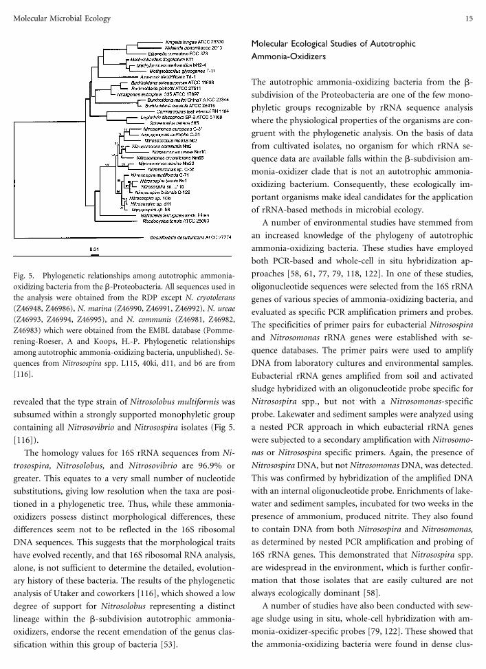
revealed that the type strain of Nitrosolobus multiformis was
subsumed within a strongly supported monophyletic group
containing all Nitrosovibrio and Nitrosospira isolates (Fig 5.
[116]).
The homology values for 16S rRNA sequences from Ni-
trosospira, Nitrosolobus, and Nitrosovibrio are 96.9% or
greater. This equates to a very small number of nucleotide
substitutions, giving low resolution when the taxa are posi-
tioned in a phylogenetic tree. Thus, while these ammonia-
oxidizers possess distinct morphological differences, these
differences seem not to be reflected in the 16S ribosomal
DNA sequences. This suggests that the morphological traits
have evolved recently, and that 16S ribosomal RNA analysis,
alone, is not sufficient to determine the detailed, evolution-
ary history of these bacteria. The results of the phylogenetic
analysis of Utaker and coworkers [116], which showed a low
degree of support for Nitrosolobus representing a distinct
lineage within the b-subdivision autotrophic ammonia-
oxidizers, endorse the recent emendation of the genus clas-
sification within this group of bacteria [53].
Molecular Ecological Studies of AutotrophicAmmonia-Oxidizers
The autotrophic ammonia-oxidizing bacteria from the b-
subdivision of the Proteobacteria are one of the few mono-
phyletic groups recognizable by rRNA sequence analysis
where the physiological properties of the organisms are con-
gruent with the phylogenetic analysis. On the basis of data
from cultivated isolates, no organism for which rRNA se-
quence data are available falls within the b-subdivision am-
monia-oxidizer clade that is not an autotrophic ammonia-
oxidizing bacterium. Consequently, these ecologically im-
portant organisms make ideal candidates for the application
of rRNA-based methods in microbial ecology.
A number of environmental studies have stemmed from
an increased knowledge of the phylogeny of autotrophic
ammonia-oxidizing bacteria. These studies have employed
both PCR-based and whole-cell in situ hybridization ap-
proaches [58, 61, 77, 79, 118, 122]. In one of these studies,
oligonucleotide sequences were selected from the 16S rRNA
genes of various species of ammonia-oxidizing bacteria, and
evaluated as specific PCR amplification primers and probes.
The specificities of primer pairs for eubacterial Nitrosospira
and Nitrosomonas rRNA genes were established with se-
quence databases. The primer pairs were used to amplify
DNA from laboratory cultures and environmental samples.
Eubacterial rRNA genes amplified from soil and activated
sludge hybridized with an oligonucleotide probe specific for
Nitrosospira spp., but not with a Nitrosomonas-specific
probe. Lakewater and sediment samples were analyzed using
a nested PCR approach in which eubacterial rRNA genes
were subjected to a secondary amplification with Nitrosomo-
nas or Nitrosospira specific primers. Again, the presence of
Nitrosospira DNA, but not Nitrosomonas DNA, was detected.
This was confirmed by hybridization of the amplified DNA
with an internal oligonucleotide probe. Enrichments of lake-
water and sediment samples, incubated for two weeks in the
presence of ammonium, produced nitrite. They also found
to contain DNA from both Nitrosospira and Nitrosomonas,
as determined by nested PCR amplification and probing of
16S rRNA genes. This demonstrated that Nitrosospira spp.
are widespread in the environment, which is further confir-
mation that those isolates that are easily cultured are not
always ecologically dominant [58].
A number of studies have also been conducted with sew-
age sludge using in situ, whole-cell hybridization with am-
monia-oxidizer-specific probes [79, 122]. These showed that
the ammonia-oxidizing bacteria were found in dense clus-
Fig. 5. Phylogenetic relationships among autotrophic ammonia-
oxidizing bacteria from the b-Proteobacteria. All sequences used in
the analysis were obtained from the RDP except N. cryotolerans
(Z46948, Z46986), N. marina (Z46990, Z46991, Z46992), N. ureae
(Z46993, Z46994, Z46995), and N. communis (Z46981, Z46982,
Z46983) which were obtained from the EMBL database (Pomme-
rening-Roeser, A and Koops, H.-P. Phylogenetic relationships
among autotrophic ammonia-oxidizing bacteria, unpublished). Se-
quences from Nitrosospira spp. L115, 40ki, d11, and b6 are from
[116].
Molecular Microbial Ecology 15

ters containing up to 3,000 cells, and, in actively nitrifying
plants, up to 20% of the total cells gave a signal with a probe
specific for some, but not all Nitrosomonas spp.
Dual staining experiments with ammonia-oxidizer-
specific probes and a recently developed Nitrobacter-specific
probe revealed that the nitrite-oxidizing bacteria were asso-
ciated with the clusters of ammonia-oxidizers found in sew-
age sludge flocs [79]. In the same study, a wide range of
probes was used to detect different taxa of ammonia-
oxidizing bacteria. This revealed the presence of cells related
to Nitrosomonas spp., but Nitrosospira was not detected in
any of the sewage treatment plants examined. This was also
recently observed in a study of nitrifying bacteria from
aquarium filters [61]. Interestingly, in the same study, it was
noted that samples from freshwater aquaria rarely harbored
ammonia-oxidizers from the b-subdivision of the Proteo-
bacteria [61]. Previous studies indicated that Nitrosospira
was detectable in a range of environments by PCR and prob-
ing procedures, while Nitrosomonas could be detected only
in enrichment cultures from the same samples [58]. It has
been suggested that the inability to detect Nitrosomonas spp.
may have been the result of inefficient DNA extraction from
flocs, and that this limitation would not occur with in situ
hybridization procedures [79]. This explanation, however,
seems unlikely since Nitrosomonas could be detected from
DNA extracted from enrichment cultures where the cells
often exist as flocs. A more likely explanation of a failure to
detect Nitrosomonas would be that the specificity of the
primers used was too great; only on enrichment were low
numbers of organisms closely related to N. europaea and N.
eutropha (the organisms that the PCR primers were designed
to detect) detected. Furthermore, in situ hybridization stud-
ies in our laboratory (T. P. Curtis and I. M. Head, unpub-
lished data) have shown that, while in most nitrifying acti-
vated sludge plants sampled, Nitrosomonas spp. were dom-
inant, in many cases Nitrosospira could be detected. These,
too, were present in large aggregates within the sludge flocs.
It thus seems likely that the dominant groups of autotrophic
ammonia-oxidizing bacteria found in any particular envi-
ronment will be determined by characteristics specific to that
environment.
Conclusions and Future Prospects
We have chosen some examples where the application of 16S
rRNA PCR sequences analysis and probing with specific
fluorescent oligonucleotide probes have generated advances
in microbial ecology in a range of habitats that could not be
achieved using conventional techniques alone. However, the
role of classical microbial ecology should never be underes-
timated. With respect to defining a functional role in their
particular ecosystems, organisms catalogued only by se-
quence will permit assessments of diversity only. However,
the scope for assigning function to form and to groups of
microorganisms remains the challenge of this new technol-
ogy. The first steps to achieving this are already being made
with the application of cloning techniques that allow cloning
of large fragments of DNA extracted from environmental
samples [107]. This will potentially allow determination of
the phylogenetic position of an uncultured organism by se-
quencing cloned rRNA genes. It will also allow identification
of functional genes cloned on the same fragment of DNA,
thus giving an indication of the possible metabolic capabili-
ties of an uncultivated organism identified solely from a
rRNA sequence.
Combining molecular measures of species composition
and the abundance of biogeochemically important groups
with measurement of particular processes and environmen-
tal parameters is also now being more widely adopted [91,
111]. Such integrated studies will be essential in the future if
we are to reap the maximum rewards from nucleic acid–
based studies of microbial ecology. Molecular studies are
now being complemented by appropriate culture-based in-
vestigations [76] that will assist in obtaining cultures of or-
ganisms that are truly representative of those important in
nature. In addition, where culture is not possible or not an
appropriate strategy for the study, quantification of organ-
isms in a particular niche, or an appreciation of their spatial
distribution are exciting goals made possible by coupling
molecular tools with other developing technologies. For in-
stance, through a combination of confocal microscopy and
in situ hybridization, toluene-degrading Pseudomonas putida
were shown to be distributed throughout a multispecies bio-
film. They were found to be active in all regions of the
biofilm, although less active than those from batch culture,
and responsible for 65% of the toluene degraded by the
community [80]. Similarly, the combination of microelec-
trodes, measuring nitrate concentration, and in situ hybrid-
ization to detect nitrifying bacteria has been used to examine
their distribution in biofilms. The microelectrodes showed
that nitrification occurred in a narrow 50-µm zone on top of
the biofilm, whereas the ammonia-oxidizers formed a dense
layer of cell clusters in the upper part of the biofilm, below
which a less dense aggregate of nitrite oxidizers was detected.
In addition, both groups were detected in substantially lower
16 I.M. Head et al.
numbers in the anoxic layers deeper in the biofilm [97]. The
distribution of sulfate-reducing (SRB) and methanogenic
bacteria was also determined in a similar manner, with re-
spect to activity [92]. These examples exemplify the way
forward for molecular microbial ecology. In situ hybridiza-
tion circumvents the need for culture (nitrifying, SRB, and
methanogens can be difficult to culture. [92, 97] and, in the
case of the toluene degrading biofilm, direct culture of P.
putida would provide no relevant information [97]); the use
of confocal microscopy and microelectrodes provides a
means of examination that is relatively noninvasive to the
community; and both illustrate the ability to relate commu-
nity structure to function and activity. Furthermore, the ap-
plication of amplified ribosomal DNA restriction analysis
(ARDRA) may provide a means of examining the succession
and convergence/divergence of microbial communities [76].
The laboratory biofilm is, at present, the main focus for
these studies, but application of these complementary meth-
ods has potential for unravelling the complexities of micro-
bial populations as they exist in nature.
In conclusion, it should be emphasized that molecular
ecology is a valuable tool. It should, however, always be
appreciated that there are many other equally useful ap-
proaches among those used by microbial ecologists. It is only
be selecting a range of appropriate tools in a complementary
fashion that we can hope to unlock some of the mysteries of
microbial ecology.
Acknowledgments
The authors wish to thank the Universities of Newcastle and
Liverpool, Institute of Freshwater Ecology, and the Natural
Environment Research Council for support.
References
1. Amann RT (1995) Fluorescently labelled, rRNA-targeted oli-
gonucleotide probes in the study of microbial ecology. Mol
Ecol 4:543–554
2. Amann RJ, Binder B, Olson RI, Chisholm SW, Devereux R,
Stahl DA (1990) Combination of 16S rRNA-targeted oligo-
nucleotide probes with flow cytometry for analyzing mixed
microbial populations. Appl Environ Microbiol 56:1919–1925
3. Amman R, Springer N, Ludwig W, Gortz H-D, Schleifer K-H
(1991) Identification and phylogeny of uncultured bacterial
endosymbionts. Nature 351:161–164
4. Amann RI, Zarda B, Stahl DA, Schleifer K-H (1992) Identifi-
cation of individual prokaryotic cells with enzyme-labelled
rRNA-targeted oligonucleotide probes. Appl Environ Micro-
biol 58:3007–3011
5. Amann RI, Ludwig W, Schleifer K-H (1995) Phylogenetic
identification and in situ detection of individual microbial
cells without cultivation. Microbiol Rev 59:143–169
6. Angert ER, Clements KD, Pace NR (1993) The largest bacte-
rium. Nature 362:239–241
7. Beimfohr C, Krause A, Amann R, Ludwig W, Schleifer K-H
(1993) In situ identification of lactococci, enterococci, and
streptococci. Syst Appl Microbiol 16:450–456
8. Belser LW (1979) Population ecology of nitrifying bacteria.
Ann Rev Microbiol 33:309–333
9. Blackall LL, Seviour EM, Cunningham MA, Seviour RJ, Hu-
genholz P (1994) ‘‘Microthrix parvicella’’ a novel deep branch-
ing member of the actinomyces subphylum. System Appl Mi-
crobiol 17:513–518
10. Blackall LL, Stratton H, Bradford D, Del Dot T, Sjorup C,
Seviour EM, Seviour RJ (1996) ‘‘Candidatus Microthrix parvi-
cella’’ a filamentous bacterium from activated sludge sewage
treatment plants. Int J Syst Bacteriol 46:344–346
11. Blakemore RP (1975) Magnetotactic bacteria. Science 190:
377–379
12. Blumer M, Chase T, Watson SW (1969) Fatty acids in the
lipids of marine and terrestrial nitrifying bacteria. J Bacteriol
99:366–370
13. Bock E, Koops H-P, Harms H (1986) Cell biology of nitrifying
bacteria. In: Prosser JI (ed) Nitrification. IRL Press, Oxford,
pp 17–38
14. Britschgi TB, Giovannoni SJ (1991) Phylogenetic analysis of a
natural marine bacterioplankton population by rRNA gene
cloning and sequencing. Appl Environ Microbiol 57:1707–
1713
15. Brock TD (1987) The study of microorganisms in situ: prog-
ress and problems. Symp Soc Gen Microbiol 41:1–17
16. Bruce KD, Hiorns WD, Hobman JL, Osborn AM, Strike P,
Ritchie DA (1992) Amplification of DNA from native popu-
lations of soil bacteria by using the polymerase chain reaction.
Appl Environ Microbiol 58:3413–3416
17. Cary SC, Giovannoni SJ (1993) Transovarial inheritance of
endosymbiotic bacteria in clams inhabiting deep-sea hydro-
thermal vents and cold seeps. Proc Natl Acad Sci USA 90:
5695–5699
18. Cary SC, Warren WD, Anderson E, Giovannoni SJ (1993)
Identification and localization of bacterial endosymbionts in
hydrothermal vent taxa with symbiont specific PCR amplifi-
cation and in situ hybridization techniques. Marine Mol Biol
Biotechnol 2:51–62
19. Clayton RA, Sutton G, Hinkle Jr PS, Bult C, Fields C (1995)
Intraspecific variation in small-subunit rRNA sequences in
GenBank: why single sequences may not adequately represent
prokaryotic taxa. Int J Syst Bacteriol 45:595–599
20. Clements KD, Bullivant S (1991) An unusual symbiont from
the gut of surgeonfishes may be the largest known prokaryote.
J Bacteriol 171:5359–5362
Molecular Microbial Ecology 17

21. Cole ST, Girons IS (1994) Bacterial genomics. FEMS Micro-
biol Rev 14:139–160
22. de Boer WE, La Riviere JWM, Schmidt K (1971) Some prop-
erties of Achromatium oxaliferum. Ant van Leeuwen 37:553–
563
23. Degrange V, Bardin R (1995) Detection and counting of Ni-
trobacter populations in soil by PCR. Appl Environ Microbiol
61:2093–2098
24. DeLong EF, Wickham GS, Pace NR (1989) Phylogenetic
stains: ribosomal RNA-based probes for the identification of
single microbial cells. Science 243:1360–1363
25. DeLong EF, Frankel RB, Bazylinski DA (1993) Multiple evo-
lutionary origins of magnetotaxis in bacteria. Science 259:803–
806
26. Distel DL, Wood AP (1992) Characterization of the gill sym-
biont of Thyasira flexuosa (Thyasiridae: Bivalvia) by use of
polymerase chain reaction and 16S rRNA sequence analysis. J
Bacteriol 174:6317–6320
27. Distel DL, Lane DL, Olsen GJ, Giovannoni SJ, Pace B, Pace
NR, Stahl DA, Felbeck H (1988) Sulfur-oxidizing bacterial
endosymbionts: analysis of phylogeny and specificity by 16S
rRNA sequences. J Bacteriol 170:2506–2510
28. Distel DL, Felbeck H, Cavanaugh CM (1994) Evidence for
phylogenetic congruence among sulfur-oxidizing chemoauto-
trophic bacterial endosymbionts and their bivalve hosts. J Mol
Evol 38:533–542
29. Diviacco S, Norio P, Zentilin L, Menzo S, Clementi M, Bi-
amonti G, Riva S, Falaschi A, Giacca M (1992) A novel pro-
cedure for quantitative polymerase chain reaction by co-
amplification of competitive templates. Gene 122:313–320
30. Edwards U, Rogall T, Blocker H, Emde M, Bottger EC (1989)
Isolation and direct complete nucleotide determination of en-
tire genes. Characterization of a gene coding for 16S ribosomal
RNA. Nucl Acids Res 17:7843–7853
31. Eikelboom DH (1975) Filamentous organisms observed in ac-
tivated sludge. Water Res 9:365–388
32. Eikelboom DH (1977) Identification of filamentous bacteria
in bulking activated sludge. Progress in Water Technol 8:153–
162
33. Embley TM, Finlay BJ (1994) The use of small subunit rRNA
sequences to unravel the relationships between anaerobic cili-
ates and their methanogen symbionts. Microbiology 140:225–
235
34. Embley TM, Finlay BJ (1994) Phylogenetic diversity of meth-
anogen endosymbionts of anaerobic ciliates. In: Priest FG,
Ramos-Cormenzana A, Tindall BJ (eds) Bacterial diversity and
systematics. Plenum Press, New York, pp 153–160
35. Embley TM, Finlay BJ, Thomas RH, Dyal PL (1992) The use
of rRNA sequences and fluorescent probes to investigate the
phylogenetic positions of the anaerobic ciliate Metopus palae-
formis and its archaeobacterial endosymbiont. J of Gen Mi-
crobiol 138:1479–1487
36. Embley TM, Finlay BJ, Dyal PL, Hirt RP, Wilkinson M, Willi-
ams AG (1995) Multiple origins of anaerobic ciliates with
hydrogenosomes within the radiation of aerobic ciliates. Proc
R Soc London Ser B 262:87–93
37. Farrelly V, Rainey FA, Stackebrandt E (1995) Effect of genome
size and rrn gene copy number on PCR amplification of 16S
rRNA genes from a mixture of bacterial species. Appl Environ
Microbiol 61:2798–2801
38. Fischer SG, Lerman LS (1979) Length-independent separation
of DNA restriction fragments in two-dimensional gel electro-
phoresis. Cell 16:191–200
39. Ferris MJ, Muyzer G, Ward DM (1996) Denaturing gradient
gel electrophoresis profiles of 16S rRNA-defined populations
inhabiting a hot spring microbial community. Appl Environ
Microbiol 62:340–346
40. Fishelson L, Montgomery WL, Myrberg Jr AA (1985) A
unique symbiosis in the gut of tropical herbivorous surgeon-
fish (Acanthuridae: Teleostei) from the Red Sea. Science 229:
49–51
41. Fuhrman JA, Comeau DE, Hagstrom A, Chan AM (1988)
Extraction from natural planktonic microorganisms of DNA
suitable for molecular biological studies. Appl Environ Micro-
biol 54:1426–1429
42. Fuhrman JA, McCallum K, Davis AA (1992) Novel major
archaebacterial group from marine plankton. Nature 356:148–
149
43. Giannakis C, Miller DJ, Nicholas DJD (1985) Comparative
studies on redox proteins from ammonia-oxidizing bacteria.
FEMS Microbiol Lett 30:81–85
44. Giovannoni SJ, Cary SC (1993) Probing marine systems with
ribosomal RNAs. Oceanography 6:95–104
45. Giovannoni SJ, DeLong EF, Olsen GJ, Pace NR (1988) Phy-
logenetic group-specific oligodeoxynucleotide probes for
identification of single microbial cells. J Bacteriol 170:720–726
46. Giovannoni SJ, Britschgi TB, Moyer CL, Field KG (1990) Ge-
netic diversity in Sargasso Sea bacterioplankton. Nature 345:
60–63
47. Gordon DA, Giovannoni SJ (1996) Detection of stratified mi-
crobial populations related to Chlorobium and Fibrobacter spe-
cies in the Atlantic and Pacific oceans. Appl Environ Microbiol
62:1171–1177
48. Gutell RR, Larsen N, Woese CR (1994) Lessons from an evolv-
ing rRNA: 16S and 23S rRNA structures from a comparative
perspective. Microbiol Rev 58:10–26
49. Hahn D, Amann RI, Ludwig W, Akkermans ADL, Schleifer
K-H (1992) Detection of microorganisms in soil after in situ
hybridization with rRNA-targeted, fluorescently labelled oli-
gonucleotides. J Gen Microbiol 138:879–887
50. Hahn D, Amann RI, Zeyer J (1993) Detection of mRNA in
Streptomyces cells by in situ hybridization with digoxigenin-
labelled probes. Appl Environ Microbiol 59:2753–2757
51. Hall GH (1986) Nitrification in Lakes. In: Prosser JI (ed)
Nitrification. IRL Press, Oxford, pp 127–156
52. Haygood MG, Distel DL (1993) Bioluminescent symbionts of
flashlight fishes and deep-sea anglerfishes form unique lin-
eages related to the genus Vibrio. Nature 363:154–156
53. Head IM, Hiorns WD, Embley TM, McCarthy AJ, Saunders JR
18 I.M. Head et al.

(1993) The Phylogeny of autotrophic ammonia-oxidising bac-
teria as determined by analysis of 16S ribosomal RNA gene
sequences. J Gen Microbiol 139:1147–1153
54. Head IM, Gray ND, Pickup RW, Jones JG (1995) The biogeo-
chemical role of Achromatium oxaliferum. In: Grimalt JO,
Dorronsoro C (eds) Organic geochemistry: developments and
applications to energy, climate, environment, and human his-
tory. (Selected papers from the 17th International Meeting on
Organic Geochemistry, 4–8 September 1995, Donostia-San
Sebastian, The Basque Country, Spain) AIGOA, Donostia-San
Sebastian, pp 895–898
55. Head IM, Gray ND, Clarke KJ, Pickup RW, Jones JG (1996)
The phylogenetic position and ultrastructure of the uncul-
tured bacterium Achromatium oxaliferum Microbiology 142:
2341–2354
56. Herring PJ (1982) Aspects of the bioluminescence of fishes.
Oceanogr Marine Biol 20:415–470
57. Hicks R, Amann RI, Stahl DA (1992) Dual staining of natural
bacterioplankton with 48,6-diamidino-2-phenylindole and
fluorescent oligonucleotide probes targeting kingdom level
16S rRNA sequences. Appl Environ Microbiol 58:2158–2163
58. Hiorns WD, Hastings RC, Head IM, McCarthy AJ, Saunders
JR, Pickup RW, Hall GH (1995) Amplification of 16S ribo-
somal RNA genes of autotrophic ammonia-oxidising bacteria
demonstrates the ubiquity of nitrosospiras in the environ-
ment. Microbiology 141:2793–2800
59. Holben WE, Jansson JK, Chelm BK, Tiedje JM (1988) DNA
probe method for the detection of specific microorganisms in
the soil community. Appl Environ Microbiol 54:703–711
60. Holmes AJ, Owens NJP, Murrell JC (1995) Detection of novel
marine methanotrophs using phylogenetic and functional
gene probes after methane enrichment. Microbiol 141:1947–
1955
61. Hovanec TA, DeLong EF (1996) Comparative analysis of ni-
trifying bacteria associated with freshwater and marine
aquaria. Appl Environ Microbiol 62:2888–2896
62. Jukes TH, Cantor CR (1969) Evolution of protein molecules.
In: Munro HN (ed) Mammalian protein metabolism. Aca-
demic Press, New York, pp 21–132
63. Koops H-P, Harms H (1985) Deoxyribonucleic acid homol-
ogy among 96 strains of ammonia-oxidizing bacteria. Arch
Microbiol 141:214–218
64. Koops H-P, Moller UC (1991) The lithotrophic ammonia-
oxidizing bacteria. In: Balows A, Truper HG, Dworkin M,
Harder W, Schleifer KH (eds) The prokaryotes, 2nd ed.
Fischer-Verlag, New York, pp 2625–2637
65. Koops H-P, Bottcher B, Moller UC, Pommerening-Roeser A,
Stehr G (1991) Classification of eight new species of ammo-
nia-oxidizing bacteria: Nitrosomonas communis sp. nov., Ni-
trosomonas ureae sp. nov., Nitrosomonas aestuarii sp. nov.,
Nitrosomonas marina sp. nov., Nitrosomonas nitrosa sp. nov.,
Nitrosomonas eutropha sp. nov., Nitrosomonas oligotropha sp.
nov., and Nitrosomonas halophila sp. nov. J Gen Microbiol
137:1689–1700
66. Kopczynski ED, Bateson MM, Ward DM (1994) Recognition
of chimeric small subunit ribosomal DNAs composed of genes
from uncultivated microorganisms. Appl Environ Microbiol
60:746–748
67. La Riviere JWM, Schmidt K (1989) The genus Achromatium.
In: Staley JT, Bryant MP, Pfennig N, Holt JG (eds) Bergey’s
manual of systematic bacteriology, vol 3. Williams and
Wilkins, Baltimore, pp 2131–2133
68. La Riviere JWM, Schmidt K (1991) Morphologically conspicu-
ous sulfur-oxidizing Eubacteria. In: Balows A, Truper HG,
Dworkin M, Harder W, Schleifer KH (eds) The prokaryotes,
2nd edn. Fischer-Verlag, New York, pp 3934–3947
69. Lee S, Malone C, Kemp PF (1993) Use of multiple 16S rRNA–
targeted fluorescent probes to increase signal strength and
measure cellular RNA from natural planktonic bacteria. Ma-
rine Ecol Prog Ser 101:193–201
70. Liesack W, Weyland H, Stackebrandt E (1991) Potential risks
of gene amplification by PCR as determined by 16S rDNA
analysis of a mixed culture of strict barophilic bacteria. Microb
Ecol 21:191–198
71. MacNaughton SJ, O’Donnell AG, Embley TM (1994) Perme-
ablization of mycolic acid containing actinomycetes for in situ
hybridization with fluorescently labelled oligonucleotide
probes. Microbiology 140:2859–2865
72. Maidak BL, Larsen N, McCaughey MJ, Overbeek R, Olsen GJ,
Fogel K, Blandy J, Woese CR (1994) The ribosomal database
project. Nucl Acids Res 22:3485–3487
73. Mann S, Sparks NHC, Board RG (1990) Magnetotactic bac-
teria: microbiology, biomineralization, paeleomagnetism, and
biotechnology. Adv Microb Physiol 31:125–181
74. Manz W, Amann R, Ludwig W, Wagner M, Schleifer K-H
(1992) Phylogenetic oligodeoxynucelotide probes for the ma-
jor subclasses of the proteobacteria: problems and solutions.
Syst Appl Microbiol 15:593–600
75. Manz W, Szewzyk U, Ericsson P, Amann R, Schleifer KH,
Stenstrom TA (1993) In situ identification of bacteria in
drinking water and adjoining biofilms by hyridization with
16S ribosomal RNA–directed and 23S ribosomal RNA–
directed fluorescent oligonucleotide probes. Appl Environ Mi-
crobiol 59:2293–2298
76. Massol-Deya A, Weller R, Rios-Hernandez L, Zhou J-Z,
Hickey RF, Tiedje JM (1997) Succession and convergence of
biofilm communities in fixed-film reactors treating aromatic
hydrocarbons in groundwater. Appl Environ Microbiol 63:
270–276
77. McCaig AE, Embley TM, Prosser JI (1994) Molecular analysis
of enrichment cultures of marine ammonia oxidizers. FEMS
Microbiol Lett 120:363–368
78. Mevarech M, Hirsch-Twizer S, Goldman S, Yakobson E, Ei-
senberg H, Dennis PP (1989) Isolation and characterization of
the rRNA gene clusters of Halobacterium marismortui. J Bac-
teriol 171:3479–3485
79. Mobarry BK, Wagner M, Urbain V, Rittman BE, Stahl DA
(1996) Phylogenetic probes for analysing abundance and spa-
tial organization of nitrifying bacteria. Appl Environ Micro-
biol 62:2156–2162
Molecular Microbial Ecology 19

80. Moller S, Pedersen AR, Poulsen LK, Arvin E, Molin S (1996)
Activity and three-dimensional distribution of toluene-
degrading Pseudomonas putida in multispecies biofilm as-
sessed by quantitative in situ hybridization and scanning con-
focal laser microscopy. Appl Environ Microbiol 62:4632–4640
81. More M, Herrick JB, Silva MO, Ghiorse WC, Madsen EJ
(1994) Quantitative cell lysis of indigenous microorganisms
and rapid extraction of microbial DNA from sediment. Appl
Environ Microbiol 60:1572–1580
82. Muller M (1988) Energy metabolism in protozoa without mi-
tochondria. Ann Rev Microbiol 42:465–488
83. Muyzer G, de Waal EC, Uitterrlinden AG (1993) Profiling of
complex microbial populations by denaturing gradient gel
electrophoresis analysis of polymerase chain reaction–
amplified genes coding for 16S rRNA. Appl Environ Microbiol
59:695–700
84. Olsen GJ, Lane DJ, Giovannoni SJ, Pace NR (1986) Microbial
ecology and evolution: a ribosomal RNA approach. Ann Rev
Microbiol 40:337–365
85. Olsen GJ, Woese CR, Overbeek R (1994) The winds of (evo-
lutionary) change: breathing new life into microbiology. J Bac-
teriol 176:1–6
86. Osborn AM, Bruce KD, Strike P, Ritchie DA (1993) Polymer-
ase chain reaction–restriction length polymorphism analysis
shows divergence among mer determinants from Gram-
negative soil bacteria indistinguishable by DNA-DNA hybridi-
sation. Appl Environ Microbiol 59:4024–4030
87. Pace NR, Stahl DA, Lane DJ, Olsen GJ (1986) The analysis of
natural microbial populations by ribosomal RNA sequences.
Adv Microb Ecol 9:1–55
88. Paerl HW (1993) Interaction of nitrogen and carbon cycles in
the marine environment. In: Ford TE (ed) Aquatic microbi-
ology: an ecological approach. Blackwell Scientific Publica-
tions, Boston, pp 343–381
89. Prosser JI (1989) Autotrophic nitrification in bacteria. Adv
Microb Physiol 30:125–181
90. Rainey FA, Ward N, Sly LI, Stackebrandt E (1994) Depen-
dence on the taxon composition of clone libraries for PCR-
amplified, naturally occurring 16S rDNA on the primer pair
and the cloning system used. Experientia 50:796–797
91. Ramsing NB, Fossing H, Ferdelman TG, Andersen F, Tham-
drup B (1996) The distribution of bacterial populations in a
stratified Fjord (Mariager Fjord, Denmark) quantified by in
situ hybridization and related to chemical gradients in the
water column. Appl Environ Microbiol 62:1391–1404
92. Raskin L, Rittman BE, Stahl DA (1996) Competition and co-
existence of sulfate-reducing and methanogenic populations
in anaerobic biofilms. Appl Environ Microbiol 62:3847–3857
93. Reysenbach A, Giver LJ, Wickham GS, Pace NR (1992) Dif-
ferential amplification of rRNA genes by polymerase chain
reaction. Appl Environ Microbiol 58:3417–3418
94. Rochelle PA, Fry JC, Parkes RJ, Weightman AJ (1992) DNA
extraction for 16S rRNA gene analysis to determine genetic
diversity in deep sediment communities. FEMS Microbiol Lett
100:59–66
95. Roller C, Wagner M, Amann R, Ludwig W, Schleifer K-H
(1994) In situ probing of Gram-positive bacteria with high
DNA G+C content using 23S rRNA–targeted oligonucleo-
tides. Microbiology 140:2849–2858
96. Schleifer K-H, Ludwig W (1994) Molecular taxonomy: classi-
fication and identification. In: Priest FG, Ramos-Cormenzana
A, Tindall BJ (eds) Bacterial diversity and systematics. Plenum
Press, New York, pp 1–15
97. Schramm A, Larsen LH, Revsbech NP, Ramsing NB, Amman
R, Schleifer K-H (1996) Structure and function of a nitrifying
biofilm as determined by in situ hybridization and the use of
microelectrodes. Appl Environ Microbiol 62:4641–4647
98. Silva MC, More MI, Batt CA (1995) Development of a mo-
lecular detection method for naphthalene degrading pseudo-
monads. FEMS Microbiol Ecol 18:225–235
99. Sprent JI (1987) The ecology of the nitrogen cycle. Cambridge
University Press, Cambridge
100. Spring S, Amann R, Ludwig W, Schleifer K-H, Petersen N
(1992) Phylogenetic diversity and identification of noncultu-
rable magnetotactic bacteria. Syst Appl Microbiol 15:116–122
101. Spring S, Amann R, Ludwig W, Schleifer K-H, van Gemerden
H, Petersen N (1993) Dominating role of an unusual mag-
netotactic bacterium in the microaerophilic zone of a fresh-
water sediment. Appl Environ Microbiol 59:2397–2403
102. Stahl DA (1995) Application of phylogenetically based hybrid-
ization probes to microbial ecology. Molec Ecol 4:535–542
103. Stahl DA, Amann RI (1991) Development and application of
nucleic acid probes in bacterial systematics. In: Stackebrandt
E, Goodfellow M (eds) Nucleic acid techniques in bacterial
systematics. John Wiley & Sons, Chichester, England, pp 205–
248
104. Stahl DA, Lane DJ, Olsen GJ, Pace NR (1984) Analysis of
hydrothermal vent–associated symbionts by ribosomal RNA
sequences. Science 224:409–411
105. Stahl DA, Lane DJ, Olsen GJ, Pace NR (1985) Characterization
of a Yellowstone hot spring microbial community by 5S ribo-
somal RNA sequences Appl Environ Microbiol 49:1379–1384
106. Stahl DA, Lane DJ, Olsen GJ, Heller DJ, Schmidt TM, Pace NR
(1987) Phylogentic analysis of certain sulfide-oxidizing and
related morphologically conspicuous bacteria by 5S ribo-
nucleic acid sequences. Int J Syst Bacteriol 37:116–122
107. Stein JL, Marsh TL, Wu KY, Shizuya H, DeLong EF (1996)
Characterization of uncultivated prokaryotes—isolation and
analysis of a 40-kilobase-pair genome fragment from a plank-
tonic marine Archaeon. J Bacteriol 178:591–599
108. Suzuki MT, Giovannoni SJ (1996) Bias caused by template
annealing in the amplification of mixtures of 16S rRNA genes
by PCR. Appl Environ Microbiol 62:625–630
109. Swofford DL, Olsen GJ (1990) Phylogeny reconstruction. In:
Hillis DM, Moritz C (eds) Molecular systematics. Sinauer As-
sociates, Sunderland, Mass., pp 411–501
110. Teske A, Alm E, Regan JM, Rittman BE, Stahl DA (1994)
Evolutionary relationships among ammonia- and nitrite-
oxidizing bacteria. J Bacteriol 176:6623–6630
111. Teske A, Wawer C, Muyzer G, Ramsing NB (1996) Distribu-
20 I.M. Head et al.

tion of sulfate-reducing bacteria in a stratified fjord (Mariager
Fjord, Denmark) as evaluated by most-probable-number
counts and denaturing gradient gel electrophoresis of PCR-
amplified ribosomal DNA fragments. Appl Environ Microbiol
62:1405–1415
112. Thomas EJG, King RK, Burchack J, Gannon VPJ (1991) Sen-
sitive and specific detection of Listeria monocytogenes in milk
and ground beef with the polymerase chain reaction. Appl
Environ Microbiol 57:2576–2580
113. Trebesius KR, Amann R, Ludwig W, Muhlegger K, Schleifer
K-H (1994) Identification of whole fixed bacterial cells with
non-radioactive rRNA-targeted transcript probes. Appl Envi-
ron Microbiol 60:3228–3235
114. Tsai Y-L, Olson BH (1991) Rapid method for direct extraction
of DNA from soil and sediments. Appl Environ Microbiol
57:1070–1074
115. Tsai Y-L, Olson BH (1992) Detection of low numbers of bac-
terial cells in soils and sediments by polymerase chain reac-
tion. Appl Environ Microbiol 58:754–757
116. Utaker JB, Bakken L, Jiang QQ, Nes IF (1996) Phylogenetic
analysis of 7 new isolates of ammonia-oxidizing bacteria based
on 16s ribosomal RNA gene sequences. Syst Appl Microbiol
18:549–559
117. Van de Peer Y, Chapelle S, De Wachter R (1996) A quantita-
tive map of nucleotide substitution rates in bacterial rRNA.
Nucl Acids Res 24:3381–3391
118. Voytek MA, Ward BB (1995) Detection of ammonium-
oxidizing bacteria in the beta-subclass of the class proteobac-
teria in aquatic samples with the PCR. Appl and Environ
Microbiol 61:1444–1450
119. Wagner M, Amann R, Lemmer H, Schleifer K-H (1993) Prob-
ing activated sludge with oligonucleotides specific for proteo-
bacteria: inadequacy of culture-dependent methods for de-
scribing microbial community structure. Appl Environ Micro-
biol 59:1520–1525
120. Wagner M, Amann R, Kampfer P, Assmus B, Hartmann A,
Hutzler P, Springer N, Schleifer KH (1994) Identification and
in situ detection of Gram-negative filamentous bacteria in
activated sludge. Syst Appl Microbiol 17:405–417
121. Wagner M, Erhart D, Manz W, Amann R, Lemmer N, Wedi
D, Schleifer K-H (1994) Development of an rRNA-targeted
oligonucleotide probe specific for the genus Acinetobacter, and
its application for in situ monitoring in activated sludge. Appl
Environ Microbiol 60:792–800
122. Wagner M, Rath G, Amann R, Koops H-P, Schleifer K-H
(1995) In situ identification of ammonia-oxidizing bacteria.
Syst Appl Microbiol 18:251–264
123. Wallner G, Amann R, Beisker W (1993) Optimizing fluores-
cent in situ hybridization of suspended cells with rRNA-
targeted oligonucleotide probes for flow cytometric identifi-
cation of microorganisms. Cytometry 14:136–143
124. Wang GC-Y, Wang Y (1996) The frequency of chimeric mol-
ecules as a consequence of PCR co-amplification of 16S rRNA
genes from different bacterial species. Microbiology 142:1107–
1114
125. Ward DM, Weller R, Bateson MM (1990) 16S rRNA se-
quences reveal numerous uncultured microorganisms in a
natural community. Nature 345:63–65
126. Ward DM, Weller R, Bateson MM (1990) 16S rRNA se-
quences reveal uncultured inhabitants of a well-studied ther-
mal community. FEMS Microbiol Rev 75:105–116
127. Ward DM, Bateson MM, Weller R, Ruff-Roberts AL (1992)
Ribosomal RNA analysis of microorganisms as they occur in
nature. Adv Microb Ecol 12:219–286
128. Watson SW, Bock E, Harms H, Koops H-P, Hooper AB
(1989) Nitrifying bacteria. In: Staly JT, Bryant MP, Pfennig N,
Holt JG (eds) Bergey’s manual of systematic bacteriology, vol
3. Williams and Wilkins, Baltimore, pp 1808–1834
129. Weller R, Ward DM (1989) Selective recovery of 16S rRNA
sequences from natural microbial communities in the form of
cDNA. Appl Environ Microbiol 55:1818–1822
130. Wilson KH (1994) Detection of culture-resistant bacterial
pathogens by amplification and sequencing of ribosomal
DNA. Clin Infect Dis 18:958–962
131. Winogradsky S (1949) Microbiologie du sol, problemes et
methodes. Barneoud Freres, France
132. Woese CR (1987) Bacterial evolution. Microbiol Rev 51:221–
271
133. Woese CR (1994) There must be a prokaryote somewhere:
Microbiology’s search for itself. Microbiol Rev 58:1–9
134. Woese CR, Fox GE (1977) Phylogenetic structure of the pro-
karyotic domain: the primary kingdoms. Proc Natl Acad Sci
USA 74:5088–5090
135. Woese CR, Weisburg WG, Paster BJ, Hahn CM, Tanner RS,
Krieg NR, Koops H-P, Harms H, Stackebrandt E (1984) The
phylogeny of purple bacteria: the beta subdivision. Syst Appl
Microbiol 5:327–336
136. Woese CR, Weisburg WG, Hahn CM, Paster BJ, Zablen LB,
Lewis BJ, Macke TJ, Ludwig W, Stackebrandt E (1985) The
phylogeny of purple bacteria: the gamma subdivision. Syst
Appl Microbiol 6:25–33
137. Woese CR, Kandler O, Wheelis ML (1990) Towards a natural
system of organisms: proposal for the domains Archaea, Bac-
teria, and Eucarya. Proc Natl Acad Sci USA 87:4576–4579
138. Wood AP, Kelly DP (1989) Isolation and characterization of
Thiobacillus thyasiris sp. nov., a novel marine facultative au-
totroph and the putative symbiont of Thyasira flexuosa. Arch
Microbiol 152:160–166
139. Young M, Cole ST (1993) Clostridium. In: Sonenshein AL,
Hoch JA, Losick R (eds) Bacillus subtilis and other Gram-
positive organisms. American Society for Microbiology,
Washington, DC, pp 35–52
140. Zarda B, Amann R, Wallner G, Schleifer K-H (1991) Identi-
fication of single cells using digoxigenin-labelled rRNA-
targeted oligonucleotides. J Gen Microbiol 137:2823–2830
141. Zuckerkandl E, Pauling L (1965) Molecules as documents of
evolutionary history. J Theor Biol 8:357–366
Molecular Microbial Ecology 21