Embed Size (px)
Citation preview
R E S E A R CH A R T I C L E
Micro-morphological identification study on Cordyceps sinensis(Berk.) Sacc. and its adulterants based on stereo microscopeand desktop scanning electron microscope
Shuai Kang1,2 | Li-xing Nie2 | Yu-guang Zheng1 | Tian-tian Zuo2 |
Ying Wang2 | Jia Shi2 | Shuang-Cheng Ma2
1College of Pharmacy, Hebei University of
Chinese Medicine, Shijiazhuang, China
2Institute for Quality Control of Chinese
Traditional Medicine and Ethnic Medicine,
National Institutes for Food and Drug Control,
Beijing, China
Correspondence
Yu-guang Zheng, College of Pharmacy, Hebei
University of Chinese Medicine, Shijiazhuang,
China.
Email: [email protected]
Shuang-Cheng Ma, Institute for Control of
Chinese Traditional Medicine and Ethnic
Medicine, National Institutes for Food and
Drug Control, Beijing, China.
Email: [email protected]
Funding information
Digital Technology Service Platform of Chinese
Materia Medica in Qinghai Province, Grant/
Award Number: 2018-0401-ZJC-0013
Review Editor: Paul Verkade
Abstract
The Chinese Materia Medica, Cordyceps sinensis (called “Dongchongxiacao” in
Chinese), used as a tonic for nearly 600 years by Traditional Chinese Medicine, which
has been recorded by Chinese Pharmacopoeia. This drug is rare and precious, which
in turn lead to the emergence of adulterants derived from the same genus of
Cordyceps. The adulterants which can be commonly found in the market are
Cordyceps gunnii (called “Gunichongcao” in Chinese), Cordyceps liangshanensis (called
“Liangshanchongcao” in Chinese), and Cordyceps gracilis (called “Xinjiangchongcao”in Chinese). This study combined a desktop scanning electron microscope and stereo
microscope to distinguish C. sinensis from the above three adulterants especially on
their different characters of caterpillar parts. Referring to the professional entomo-
logical literature, the micro-morphological features including the cuticle of the abdo-
men and the planta of abdomen prolegs were observed, photographed, and
expressed based on the description of macroscopic characters. The identification
method studied in this article is more convenient, quick, and environmental friendly.
K E YWORD S
adulterant, Cordyceps sinensis, desktop scanning electron microscope, micro-morphologicalidentification, stereo microscope
1 | INTRODUCTION
Cordyceps sinensis (Berk.) Sacc. (Family Hypocreaceae), called “Dong-
chongxiacao” in Chinese, is a composite consisting of the stroma of
fungus, parasitized on the larva of some species of insects (Family
Hepialidae), and the dead caterpillar. It is one of the famous and valu-
able Chinese Materia Medica. This herb is distributed in the Qinghai-
Tibet plateau and its surrounding high-altitude areas (Zhang, Liu, &
Huang, 2008). According to Traditional Chinese Medicine, it has the
action of tonifying kidney, replenishing lung, stanching bleeding, and
resolve phlegm (Chinese Pharmacopoeia Commission, 2015). Modern
pharmacological studies showed that C. sinensis mainly contains the
components of polysaccharides, sugar alcohols, amino acids and
nucleosides, sterols, fatty acids, and so forth, and has the immuno-
modulatory, antioxidant, and antitumor activities, and so forth (Chen,
Wang, Nie, & Marcone, 2013; Olatunji et al., 2018; Qiu, Cao, &
Han, 2016; Wang et al., 2017; Yue, Ye, Zhou, Sun, & Lin, 2013).
“Dongchongxiacao” is mainly collected from the wild, and is the only
accepted species in the genus of Cordyceps that has been recorded
officially as a herbal drug in Chinese Pharmacopoeia (Chinese Pharma-
copoeia Commission, 2015). With the overharvesting of this species
and dramatic rise in price, the resource of C. sinensis is decreasing year
by year, which in turn has led to the emergence of adulterants derived
from the same genus of Cordyceps (Zhuo & Da, 2014).
Cordyceps gunnii (Berk.) Berk. is one of the most commonly found
adulterant of “Dongchongxiacao” in the market (Liang, 1983; Liang, 2007),Li-xing Nie is a co-first author.
Received: 1 October 2020 Revised: 9 February 2021 Accepted: 25 February 2021
DOI: 10.1002/jemt.23749
Microsc Res Tech. 2021;1–11. wileyonlinelibrary.com/journal/jemt © 2021 Wiley Periodicals LLC. 1
called Gunichongcao in Chinese and distributed in low altitude areas
(200-800 m) of some southern provinces in China (Wu et al., 1997; Zhu,
Wang, & Han, 2004). Cordyceps liangshanensis Zang, Liu and Hu and
Cordyceps gracilis (Grev.) Dur. et Mont. are the other two adulterants
which were commonly used as “Dongchongxiacao” in Sichuan and
Xinjiang provinces respectively (Hu, 1983; Pu, 1983).
Although these adulterants have some similar chemical compo-
nents with C. sinensis (Chen, Tian, Meng, & Fu, 2003; Wu &
Tang, 2011), but they are different in the source of fungi, the host
larva, producing areas, and growing environment. On the other hand,
some wholesalers sometimes used the dyed and processed adulter-
ants to sold as “Dongchongxiacao”(Zeng, 2009). In particular, C. gunnii
has toxicity and adverse reactions reports (Xu, 2009). These adulter-
ants have important implications for safety and efficacy in Morden
clinical practice. Therefore, it is necessary to carry out research on
identification methods in order to prevent confusion.
Microscopic identification by a light microscope is the most practical
method to authenticate C. sinensis from its adulterants or counterfeits
(Au et al., 2012; Chan et al., 2011; Gao, Wang, Zeng, Mai, & Ma, 2011;
Hu, Kang, & Zhao, 2003; Kang, Luo, Zheng, & Lin, 2011; Liu, Hua, Chu,
Li, & Li, 2011), which is considered as a more convenient, low-cost and
environment-friendly method when compared with chemical compo-
nents analysis (Deng, Cheong, Wang, Zhao, & Li, 2018; Guo, Li, Huang,
Liang, & Chen, 2006; Wang et al., 2013; Zuo et al., 2013) and DNA
sequencing analysis (Duan, Shang, Zhang, & Zheng, 2017; Wong,
Wong, & Shawa, 2015; Zhang, Kang, Wei, & Ma, 2015).
Microscopic identification study of C. sinensis mainly focuses on
fungal characters such as the embedded type of perithecia and the
shape of the ascus, which is found only in the mature individuals
(He & Zhang, 2000; Liang, Liu, & Liu, 1995). However, C. sinensis is
mostly harvested with a short stroma, and mature individuals are diffi-
cult to find in the commodity. Therefore, the studies on microscopic
identification methods have gradually focused on the characters of
caterpillar parts (Chan et al., 2011; Kang, Zhang, & Lin, 2013; Ye
et al., 2016). But most of the features reported in this literature could
not be clearly characterized under the light microscope and correctly
expressed using professional terms.
Hence, this study aimed to research the differences of C. sinensis
and its adulterants on their micro-morphological characteristics of the
caterpillar parts using stereo microscope (SM) and desktop scanning
electron microscope (SEM), and describe the characteristics based on
professional entomological literatures (Gullan & Cranston, 2014; Lu,
Guan, & Wu, 1951; Zhu, 1965; Zhu et al., 2004), then establish a
quick, simple, and environmental friendly method to distinguish
C. sinensis from its three adulterants.
2 | MATERIALS AND METHODS
2.1 | Materials
Different batches of samples from C. sinensis and its adulterants-
C. gunnii, C. liangshanensis, and C. gracilis were investigated
(Table 1). The voucher specimens were identified by the authors
and preserved in the National Institutes for Food and Drug Control
(China).
TABLE 1 List of the tested samplesNo. Latin name Chinese phonetic name Source Collection time
A1 C. sinensis Dongchongxiacao Changdu, Xizang 2015.6
A2 C. sinensis Dongchongxiacao Guoluo, Qinghai 2015.6
A3 C. sinensis Dongchongxiacao Yushu, Qinghai 2015.6
A4 C. sinensis Dongchongxiacao Biru, Xizang 2015.5
A5 C. sinensis Dongchongxiacao Deqin, Yunnan 2015.6
B1 C. gunnii Gunichongcao Huaihua, Hunan 2010.6
B2 C. gunnii Gunichongcao Hanshou, Hunan 2010.7
B3 C. gunnii Gunichongcao Xinyang, Henan 2015.5
B4 C. gunnii Gunichongcao Qiandongnan, Guizhou 2015.5
B5 C. gunnii Gunichongcao Lushan, Jiangxi 2019.6
C1 C. liangshanensis Liangshanchongcao Luzhou, Sichuan 2017.10
C2 C. liangshanensis Liangshanchongcao Luzhou, Sichuan 2017.10
C3 C. liangshanensis Liangshanchongcao Liangshan, Sichuan 2017.9
C4 C. liangshanensis Liangshanchongcao Liangshan, Sichuan 2017.9
C5 C. liangshanensis Liangshanchongcao Liangshan, Sichuan 2017.9
D1 C. gracilis Xinjinagchongcao Aletai, Xinjiang 2010.7
D2 C. gracilis Xinjinagchongcao Altai, Xinjiang 2015,6
D3 C. gracilis Xinjinagchongcao Aletai, Xinjiang 2015,7
D4 C. gracilis Xinjinagchongcao Aletai, Xinjiang 2019,6
D5 C. gracilis Xinjinagchongcao Urumqi, Xinjiang 2019.6
2 KANG ET AL.
2.2 | Reagents and apparatus
A digital single-lens reflex (DSLR) camera (Canon 5D4, Japan) was used
for acquiring photographs of the samples. A stereo microscope
(SM) (Zeiss SteREO Discovery V12, Germany) equipped with a digital
camera (Zeiss Axiocam 506, Germany) and a desktop scanning electron
microscope (SEM) (Phenom Pure desktop SEM, Netherlands) were used
for acquiring photographs of the micro-morphological characters.
3 | METHODS
A soft bristle brush was dipped in little water and 95% alcohol sepa-
rately to remove the dust and residual hyphae membrane present on
the surface of each batch of the sample. The samples were photo-
graphed using the DSLR camera. In addition, the characters of
C. sinensis from its three adulterants, including shape, color, size, and
so forth, were described and compared.
Then the samples were placed on the microscope stage to observe
the characteristics under visible light, which included the cuticle of the
abdomen segments and the planta of the abdomen proleg. Then, the
function of extended depth of focus was used to capture and merged the
images. The blade was held to scrape the body wall and the proleg on the
abdomen of the caterpillar part. Then the material was adhered to the
carbon conductive tape (25 mm W x 5 ml, Tedpella) and treated them by
spray-gold. This was followed by observation under the desktop SEM
with 10 kV, and 25% spot size was selected. A temperature-controlled
SEM stage was used to freeze all samples below 20�C in each case to
maintain the shape of the samples in a high vacuum SEM environment.
4 | RESULTS
4.1 | Description
4.1.1 | C. sinensis
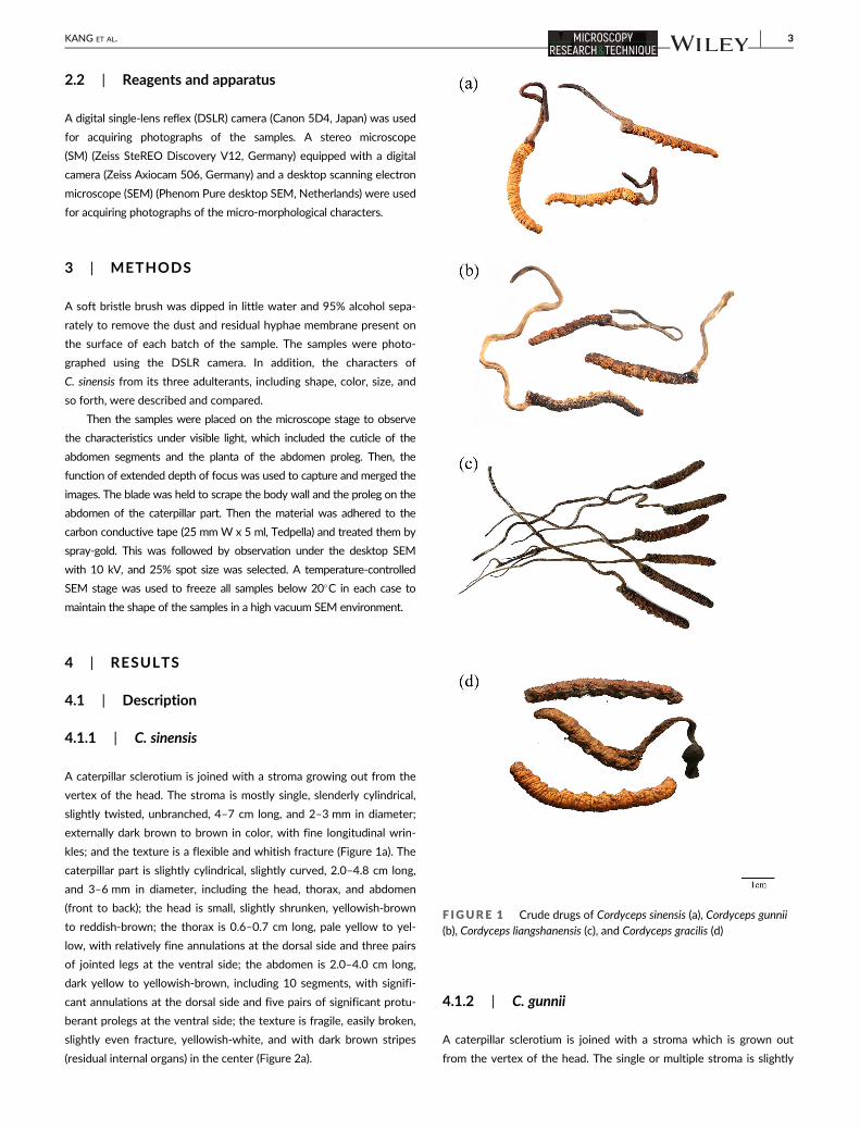
A caterpillar sclerotium is joined with a stroma growing out from the
vertex of the head. The stroma is mostly single, slenderly cylindrical,
slightly twisted, unbranched, 4–7 cm long, and 2–3 mm in diameter;
externally dark brown to brown in color, with fine longitudinal wrin-
kles; and the texture is a flexible and whitish fracture (Figure 1a). The
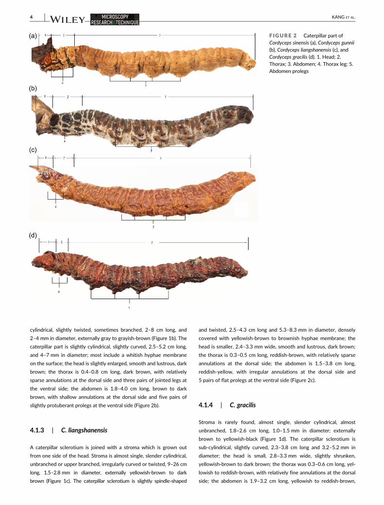
caterpillar part is slightly cylindrical, slightly curved, 2.0–4.8 cm long,
and 3–6 mm in diameter, including the head, thorax, and abdomen
(front to back); the head is small, slightly shrunken, yellowish-brown
to reddish-brown; the thorax is 0.6–0.7 cm long, pale yellow to yel-
low, with relatively fine annulations at the dorsal side and three pairs
of jointed legs at the ventral side; the abdomen is 2.0–4.0 cm long,
dark yellow to yellowish-brown, including 10 segments, with signifi-
cant annulations at the dorsal side and five pairs of significant protu-
berant prolegs at the ventral side; the texture is fragile, easily broken,
slightly even fracture, yellowish-white, and with dark brown stripes
(residual internal organs) in the center (Figure 2a).
4.1.2 | C. gunnii
A caterpillar sclerotium is joined with a stroma which is grown out
from the vertex of the head. The single or multiple stroma is slightly
F IGURE 1 Crude drugs of Cordyceps sinensis (a), Cordyceps gunnii(b), Cordyceps liangshanensis (c), and Cordyceps gracilis (d)
KANG ET AL. 3
cylindrical, slightly twisted, sometimes branched, 2–8 cm long, and
2–4 mm in diameter, externally gray to grayish-brown (Figure 1b). The
caterpillar part is slightly cylindrical, slightly curved, 2.5–5.2 cm long,
and 4–7 mm in diameter; most include a whitish hyphae membrane
on the surface; the head is slightly enlarged, smooth and lustrous, dark
brown; the thorax is 0.4–0.8 cm long, dark brown, with relatively
sparse annulations at the dorsal side and three pairs of jointed legs at
the ventral side; the abdomen is 1.8–4.0 cm long, brown to dark
brown, with shallow annulations at the dorsal side and five pairs of
slightly protuberant prolegs at the ventral side (Figure 2b).
4.1.3 | C. liangshanensis
A caterpillar sclerotium is joined with a stroma which is grown out
from one side of the head. Stroma is almost single, slender cylindrical,
unbranched or upper branched, irregularly curved or twisted, 9–26 cm
long, 1.5–2.8 mm in diameter, externally yellowish-brown to dark
brown (Figure 1c). The caterpillar sclerotium is slightly spindle-shaped
and twisted, 2.5–4.3 cm long and 5.3–8.3 mm in diameter, densely
covered with yellowish-brown to brownish hyphae membrane; the
head is smaller, 2.4–3.3 mm wide, smooth and lustrous, dark brown;
the thorax is 0.3–0.5 cm long, reddish-brown, with relatively sparse
annulations at the dorsal side; the abdomen is 1.5–3.8 cm long,
reddish-yellow, with irregular annulations at the dorsal side and
5 pairs of flat prolegs at the ventral side (Figure 2c).
4.1.4 | C. gracilis
Stroma is rarely found, almost single, slender cylindrical, almost
unbranched, 1.8–2.6 cm long, 1.0–1.5 mm in diameter; externally
brown to yellowish-black (Figure 1d). The caterpillar sclerotium is
sub-cylindrical, slightly curved, 2.3–3.8 cm long and 3.2–5.2 mm in
diameter; the head is small, 2.8–3.3 mm wide, slightly shrunken,
yellowish-brown to dark brown; the thorax was 0.3–0.6 cm long, yel-
lowish to reddish-brown, with relatively fine annulations at the dorsal
side; the abdomen is 1.9–3.2 cm long, yellowish to reddish-brown,
F IGURE 2 Caterpillar part ofCordyceps sinensis (a), Cordyceps gunnii(b), Cordyceps liangshanensis (c), andCordyceps gracilis (d). 1. Head; 2.Thorax; 3. Abdomen; 4. Thorax leg; 5.Abdomen prolegs
4 KANG ET AL.
with significant annulations at the dorsal side and five pairs of signifi-
cant protuberant prolegs at the ventral side (Figure 2d; Table 2).
4.2 | Micro-morphological characteristics
4.2.1 | Cuticle of the abdomen segments
C. sinensis
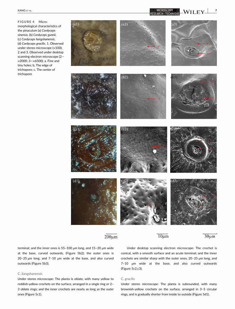
Under stereo microscope: The surface is rough, dark yellow to
yellowish-brown (Figure 3a1). Multiple pinaculums are arranged regularly
in each abdominal segment. The pinaculum is subrounded, 300–500 μm
in diameter, yellow, with a smooth or slightly wrinkled surface, slightly
lustrous, with one or two rounded nests in the center (Figure 4a1).
Under desktop scanning electron microscope: The surface is densely
covered with microthichiae, and scattered or aggregated in granular-like
protuberance; conical microthichiae, irregularly arranged, 2–35 μm long,
1–2 μm wide at the base, with a sharp terminal (Figure 3a2); the protu-
berance agglomerate is 25–60 μm in diameter, with a rough surface and
small microthichiae (Figure 3a3). The pinaculum has many significant tiny
holes on the surface and a ring-shaped trichopore near the center; the
trichopore is 75–105 μm in diameter, with a protuberant edge; the visi-
ble seta residue is in the sunken center (Figure 4a2,a3).
C. gunnii
Under stereo microscope: The surface is smooth, lustrous, dark brown
(Figure 3b1). The pinaculum is subrounded, 770–950 μm in diameter,
with a smooth or slightly wrinkled surface, lustrous, sometimes dam-
aged, brownish-black, with one or two rounded nests in the center
(Figure 4b1).
Under desktop scanning electron microscope: Most of the cuticle
surface is uneven, with glyph-like striation and many fine tiny holes,
scattered with subrounded warty protuberance (Figure 3b2,b3); The
surface of the pinaculum is smooth, with many insignificant tiny holes
and a ring-shaped trichopore near the center; the trichopore was 100–
120 μm in diameter, had a slightly protuberant edge (Figure 4b2,b3).
C. liangshanensis
Under stereo microscope: The surface is rough, lustrous, dark brown
(Figure 3c1). The pinaculum was subrounded, 500–600 μm in diame-
ter, yellowish-brown, with the slightly wrinkled surface, slightly lus-
trous, with one or two rounded nests in the center (Figure 4c1).
Under desktop scanning electron microscope: The surface is
uneven, with the hyphae spread under the cuticle; some hyphae pro-
trude from the surface, long tubular, hollow (Figure 3c2,c3). The pin-
aculum had many insignificant tiny holes on the surface and a ring-
shaped trichopore near the center; the trichopore was 50–75 μm in
diameter, with a protuberant edge (Figure 4c2,c3).
C. gracilis
Under stereo microscope: The surface is smooth, slightly lustrous, yel-
lowish to reddish-brown (Figure 3d1). The pinaculum is reddish-yel-
low, with an unclear edge and one or two rounded nests in the center
(Figure 4d1).
Under desktop scanning electron microscope: The surface is
smooth or uneven, with many significant tiny holes (Figure 3d2,d3);
The surface of the pinaculum is wrinkled, with many significant tiny
holes and a ring-shaped trichopore near the center; the trichopore is
80–120 μm in diameter, with a slightly protuberant edge (Figure 4b2,
b3; Table 3).
TABLE 2 Comparison of the descriptive characteristics of Cordyceps sinensis and its adulterants
C. sinensis C. gunnii C. liangshanensis C. gracilis
Stroma Number Mostly single Single or multiple Mostly single Rare
Branched Unbranched Sometimes
branched
Unbranched or upper branched Unbranched
Color Dark brown to brown Gray to grayish-
brown
Yellowish-brown to dark brown Brown to
yellowish-black
Caterpillar Surface Without hyphae
membrane
With whitish
hyphae
membrane
With yellowish-brown to
brownish hyphae membrane
Without hyphae
membrane
Head Shape Small, slightly shrunk, Enlarged, smooth
and lustrous
Small, smooth and lustrous Small, slightly
shrunken
Color Yellowish brown to
reddish brown
Dark brown Dark brown Yellowish brown to
dark brown
Thorax Color Pale yellow to yellow Dark brown Reddish-brown Yellowish to
reddish-brown
Annulations Fine Sparse Sparse Fine
Abdomen Color Dark yellow to
yellowish brown
Brown to dark
brown
Reddish-yellow Reddish-brown
Annulations Significant Shallow Irregular Significant
Prolegs Significant
protuberant
Slightly protuberant Flat Significant
protuberant
KANG ET AL. 5
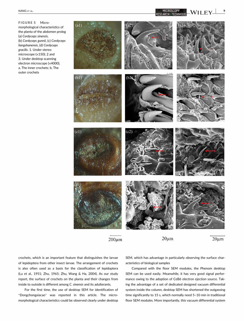
4.2.2 | Planta of the abdomen proleg
C. sinensis
Under stereo microscope: The planta is located on the top of the
abdomen proleg, subrounded, with many brownish-yellow crochets
on the surface, arranged in 5–6 circular rings, gradually shorter from
inside to outside (Figure 5a1).
Under desktop scanning electron microscope: The crochet is coni-
cal, with smooth surface and an obtuse terminal; the inner ones is 35–
65 μm long, and is 10–15 μm wide at the base, and curved outwards
(Figure 5a2); the outer ones is 20–25 μm long, and 8–10 μm wide at
the base, and upstanding or curved outwards (Figure 5a3).
C. gunnii
Under stereo microscope: The planta is oblate, with many brownish-
yellow to brown crochets on the surface, arranged in 4–7 oblate rings;
and the innermost ones are more than twice as long as the outer ones
(Figure 5b1).
Under desktop scanning electron microscope: The crochet is
conical, with longitudinal striation on the surface and an shape
F IGURE 3 Micro-morphological characteristics ofthe cuticle surface (a) Cordycepssinensis, (b) Cordyceps gunnii,(c) Cordyceps liangshanensis,(d) Cordyceps gracilis. 1. Observedunder stereo microscope (×150);2 and 3. Observed under desktopscanning electron
microscope (×6500)
6 KANG ET AL.
terminal; and the inner ones is 55–100 μm long, and 15–20 μm wide
at the base, curved outwards, (Figure 5b2); the outer ones is
20–25 μm long, and 7–10 μm wide at the base, and also curved
outwards (Figure 5b3).
C. liangshanensis
Under stereo microscope: The planta is oblate, with many yellow to
reddish-yellow crochets on the surface, arranged in a single ring or 2–
3 oblate rings; and the inner crochets are nearly as long as the outer
ones (Figure 5c1).
Under desktop scanning electron microscope: The crochet is
conical, with a smooth surface and an acute terminal; and the inner
crochets are similar sharp with the outer ones, 20–25 μm long, and
7–10 μm wide at the base, and also curved outwards
(Figure 5c2,c3).
C. gracilis
Under stereo microscope: The planta is subrounded, with many
brownish-yellow crochets on the surface, arranged in 3–5 circular
rings, and is gradually shorter from inside to outside (Figure 5d1).
F IGURE 4 Micro-morphological characteristics ofthe pinaculum (a) Cordycepssinensis, (b) Cordyceps gunnii,(c) Cordyceps liangshanensis,(d) Cordyceps gracilis. 1. Observedunder stereo microscope (×100);2 and 3. Observed under desktopscanning electron microscope (2—×2000; 3—×6500). a. Fine andtiny holes; b, The edge oftrichopore; c, The center oftrichopore
KANG ET AL. 7
Under desktop scanning electron microscope: The crochet is coni-
cal, with smooth surface and an obtuse terminal; the inner ones is
25–45 μm long, and is 8–15 μm wide at the base, and curved out-
wards (Figure 5d2); the outer ones is 18–25 μm long, and 8–12 μm
wide at the base, and upstanding or curved outwards (Figure 5d3).
5 | DISCUSSION
Traditional macroscopic identification is one of the important
methods for the authentication of Chinese Materia Medica
(Zhao, 2011). However, it is very difficult to distinguish the fine mor-
phological differences between Chinese Materia Medica and its adul-
terants. With the help of stereo microscope and scanning electron
microscope, the micro-morphological characteristics of Chinese
Materia Medica can be observed. At the same time, clear images can
be obtained by the digital camera system. Through the comparison of
the digital micro-morphological characteristics, the origin of Chinese
Materia Medica can be identified more accurately.
In this article, we studied some important identification character-
istics of C. sinensis emphasizing on its caterpillar sclerotium parts, such
as the cuticle of the abdomen and the planta on the abdomen prolegs,
based on the reference of professional entomological literature using
desktop scanning electron microscope and stereo microscope. The
comparison study between C. sinensis and its adulterants indicated
that they are different from each other. The differences are mainly
associated with the different host insects. The host of C. sinensis and
C. gracilis are different species from the genus of Hepialus (Family
Hepialidae). Nevertheless, the host of C. gunnii and C. liangshanensis
are separately derived from two different genus of Hepialidae family
(Liang, 1983; Liang, 2007; Zhang et al., 2008).
In this study,the micro-morphological characteristics of the cat-
erpillar parts have been studied to identify C. sinensis and its
adulterants
The cuticle of the larvae acts as a barrier between the living tis-
sues and the environment. It is thin and flexible in many larvae, but its
structure remains complex and varies among different taxonomic
groups (Gullan & Cranston, 2014). The microtrichiae on the cuticle of
the abdomen is considered as a very important identification charac-
ter to distinguish C. sinensis from its adulterants.
On the other hand, the Lepidopteran caterpillars are characterized
with polypod larvae, cylindrical bodies, short thoracic legs, and
abdominal prolegs (pseudopods). The prolegs on the abdomen are
usually lobe-like and have a planta each on the top. The planta bears
TABLE 3 Comparison of micro-morphological characteristics of Cordyceps sinensis and its adulterants
C. sinensis C. gunnii C. liangshanensis C. gracilis
Cuticle Under SM Surface Rough, lustrousless Smooth, lustrous Rough, lustrous Smooth, slightly
lustrous
Pinaculum size 300–500 μm in
diameter
770–950 μm in diameter 500–600 μm in
diameter
500–600 μm in
diameter
Pinaculum color Yellow Brownish-black Yellowish-brown Yellowish to
reddish-brown
Under
desktop
SEM
Surface Densely covered with
microthichiae
Covered with glyph-like
striation
With the hyphae
spread under the
cuticle
Smooth or uneven
Microthichiae Conical, 2–35 μmlong
– – –
Tiny holes on the
surface
Insignificant Insignificant Insignificant Significant
Trichopore of the
Pinaculum
75–105 μm in
diameter
100-120 μm in diameter 50–75 μm in diameter 80–120 μm in
diameter
Planta Under SM Shape Subrounded Oblate Oblate Subrounded
Crochets 5–6 circular rings 4–7 oblate rings Single ring or 2–3oblate rings
3–5 circular rings
Changes of
crochets
Gradually shorter
from the inside to
the outside
The innermost ones more
than twice as long as
the secondary outer
ring
The inner crochets are
nearly as long as the
outer ones
Gradually shorter
from the inside to
the outside
Under
desktop
SEM
Surface of crochet Smooth With longitudinal
striation
Smooth Smooth
Size of the inner
crochets
35–65 μm long 55–100 μm long 20–25 μm long 25–45 μm long
Shape of the
outer crochets
Upstanding or curved
outwards
Curved outwards Curved outwards Upstanding or
curved outwards
8 KANG ET AL.
crochets, which is an important feature that distinguishes the larvae
of lepidoptera from other insect larvae. The arrangement of crochets
is also often used as a basis for the classification of lepidoptera
(Lu et al., 1951; Zhu, 1965; Zhu, Wang & Ha, 2004). As our study
report, the surface of crochets on the planta and their changes from
inside to outside is different among C. sinensis and its adulterants.
For the first time, the use of desktop SEM for identification of
“Dongchongxiacao” was reported in this article. The micro-
morphological characteristics could be observed clearly under desktop
SEM, which has advantage in particularly observing the surface char-
acteristics of biological samples
Compared with the floor SEM modules, the Phenom desktop
SEM can be used easily. Meanwhile, it has very good signal perfor-
mance owing to the adoption of CeB6 electron ejection source. Tak-
ing the advantage of a set of dedicated designed vacuum differential
system inside the column, desktop SEM has shortened the outgassing
time significantly to 15 s, which normally need 5–10 min in traditional
floor SEM modules. More importantly, this vacuum differential system
F IGURE 5 Micro-morphological characteristics ofthe planta of the abdomen proleg(a) Cordyceps sinensis,(b) Cordyceps gunnii, (c) Cordycepsliangshanensis, (d) Cordycepsgracilis. 1. Under stereomicroscope (×150); 2 and3. Under desktop scanning
electron microscope (×4000).a, The inner crochets; b, Theouter crochets
KANG ET AL. 9
assists in observing the fragile samples a lot. When traditional floor
SEM were used, the sample must be well prepared by following a
series of complicated sample preparation procedures like chemical fix-
ation and gradient dehydration to maintain the plumpness in ultra-
high vacuum level (normally 10–5 Pa). However, in desktop SEM, the
sample chamber was designed to work at 0.1 Pa, and this prevents
the samples from narrowing even without the involvement of those
pre-preparation procedures.
DATA AVAILABILITY STATEMENT
The data that support the findings of this study are available on
request from the corresponding author. The data are not publicly
available due to privacy or ethical restrictions.
ORCID
Shuai Kang https://orcid.org/0000-0001-5465-5086
REFERENCES
Au, D., Wang, L. J., Yang, D. J., Mok, D. K. W., Chan, A. S. C., & Xu, H. X.
(2012). Application of microscopy in authentication of valuable Chi-
nese medicine I—Cordyceps sinensis, its counterfeits, and related prod-
ucts. Microscopy Research and Technique, 75, 54–64.Chan, S., Liu, B. L., Zhao, Z. Z., Lam, M., Lam, K. K., & Chen, H. B. (2011).
Studies on macroscopic and microscopic identification of Cordyceps
sinensis and its counterfeits. China Journal of Chinese Materia Medica,
36, 1141–1143.Chen, P. X., Wang, S. N., Nie, S. P., & Marcone, M. (2013). Properties of
Cordyceps sinensis: A review. Journal of Functional Foods, 5, 550–569.Chen, Z. H., Tian, X. R., Meng, X. X., & Fu, L. (2003). Determination of
nucleosides in Cordyceps gunnii by HPLC. Mycosystema, 22,
489–493.Chinese Pharmacopoeia Commission. (2015). Pharmacopoeia of the Peo-
ple's Republic of China (Vol. 1, pp. 153, 1–284). Beijing: Chinese Medical
Science Press.
Deng, Y., Cheong, K. L., Wang, L. Y., Zhao, J., & Li, S. P. (2018). Analysis of
monosaccharide composition in polysaccharides of Cordyceps spp. by
TLC. Chinese Journal of Pharmaceutical Analysis, 38, 13–21.Duan, Q. Z., Shang, K., Zhang, L., & Zheng, P. (2017). Identification of
Cordyceps sinensis by molecular method. West China Journal of Pharma-
ceutical Sciences, 32, 205–207.Gao, M., Wang, J. S., Zeng, J. L., Mai, M. X., & Ma, Y. D. (2011). The identi-
fication of Cordyceps sinensis and several common counterfeits. Journal
of Chinese Medicinal Materials, 34, 213–216.Gullan, P. J., & Cranston, P. S. (2014). The insects: An outline of entomology
(fifth edition) (pp.1–604). Hoboken: Wiley Blackwell.
Guo, F. Q., Li, A., Huang, L. F., Liang, Y. Z., & Chen, B. M. (2006). Identifica-
tion and determination of nucleosides in Cordyceps sinensis and its
substitutes by high performance liquid chromatography with mass
spectrometric detection. Journal of Pharmaceutical and Biomedical
Analysis, 40, 623–630.He, D. Z., & Zhang, X. C. (2000). Observation on Cordyceps sinensis (Berk)
Sacc. On microscope. Chinese Qinghai Journal of Animal and Veterinary
Sciences, 30, 6–7.Hu, R. Y. (1983). The study of Cordyceps liangshanensis Zang, Liu et Hu.
Chinese Traditional and Herbal Drugs, 14(7), 35–36.Hu, Y. N., Kang, T. G., & Zhao, Z. Z. (2003). Studies on microscopic identifica-
tion of animal drugs’ remnant hair (1) identification of Cordyceps sinensis
and its counterfeits. Natural Medicines, 57(5), 163–171.Kang, S., Luo, H. M., Zheng, J., & Lin, R. C. (2011). Pharmacognostic
authentication study of Cordyceps and its uncertified product—
Daishichongcao. Chinese Journal of Pharmaceutical Analysis, 31, 1035–1039.
Kang, S., Zhang, J., & Lin, R. C. (2013). Macroscopic and microscopic char-
acteristics of Chinese Caterpillar fungus. Acta Pharmaceutica Sinica, 48,
428–434.Liang, Z. Q. (1983). A description of Cordyceps gunnii in China. Acta
Mycologica Sinica, 2, 258–259.Liang, Z. Q. (2007). Flora fungorum sinicorum (Vol. 32, pp. 1–190). Beijing:
Science Press.
Liang, Z. Q., Liu, A. Y., & Liu, Z. Y. (1995). The development of Ascospores
in Cordyceps sinensis (Berk.) Sacc. Acta Mycologica Sinica, 14, 148–152.Liu, H. J., Hua, H. B., Chu, C., Li, Q., & Li, P. (2011). Morphological and
microscopic identification studies of Cordyceps and its counterfeits.
Acta Pharmaceutica Sinica B, 1, 189–195.Lu, J. R., Guan, Z. H., & Wu, W. J. (1951). A key to the families of the Lepi-
dopterous larvae. Acta Entomologica Sinica of China, 1, 321–340.Olatunji, O. J., Tang, J., Tola, A., Auberon, F., Oluwaniyi, O., & Ouyang, Z.
(2018). The genus Cordyceps: An extensive review of its traditional uses,
phytochemistry and pharmacology. Fitoterapia, 129, 293–316.Pu, Y. (1983). "Dongchongxiacao" produced from Altai Mountain. Forestry
of Xinjiang, 1, 38.
Qiu, X. H., Cao, L., & Han, R. C. (2016). The progress, issues and perspec-
tives in the research of Ophiocordyceps sinensis. Journal of Environmen-
tal Entomology, 38, 1–23.Wang, J. Q., Nie, S. P., Cui, S. W., Wang, Z. J., Phillips, A. O., Phillips, G. O.,
… Xie, M. Y. (2017). Structural characterization and immuno-
stimulatory activity of a glucan from natural Cordyceps sinensis. Food
Hydrocolloids, 67, 139–147.Wang, Z. B., Li, N., Wang, M., Wang, Y., Du, L., & Ji, X. F. (2013). Simulta-
neous determination of nucleosides and their bases in Cordyceps
sinensis and its substitutes by matrix solid-phase dispersion extraction
and HPLC. Journal of Separation Science, 36, 2348–2357.Wong, Y. L., Wong, K. L., & Shawa, P. C. (2015). Rapid authentication of
Cordyceps by lateral flow dipstick. Journal of Pharmaceutical and Bio-
medical Analysis, 111, 306–310.Wu, D. L., Wang, W. F., Xue, F. S., Wang, J. G., Jiang, Y. C., Lan, R. L., …
Jiang, K. F. (1997). Studies on Cordyceps hawksii Grey. Acta
Agriculturae Universitatis Jiangxiensis, 19, 31–36.Wu, W. B., & Tang, J. (2011). HPLC determination of nucleosides and
bases in natural Cordyceps gunnii (Berk.) Berker. Hook produced in
Anhui province. Chinese. Journal of Pharmaceutical Analysis, 31, 1668–1771.
Xu, H. (2009). Research progress of Cordyceps hawkesii. Anhui Medical and
Pharmaceutical Journal, 13(2), 209–210.Ye, M., Tong, X. X., Ren, Y., Qiu, Y., Chen, L., … Guo, J. L. (2016). Morphologi-
cal and microscopic characteristics study on insect part of TCM
Cordyceps. Lishizhen Medicine and Materia Medica Research, 27, 631–633.Yue, K., Ye, M., Zhou, Z. J., Sun, W., & Lin, X. (2013). The genus Cordyceps:
A chemical and pharmacological review. Journal of Pharmacy and Phar-
macology, 65, 474–493.Zeng, D. (2009). Counterfeit Cordyceps (Cordyceps gunnii)-doped color
studies on chemical constituents of the material. Chengdu University of
Traditional Chinese Medicine, 1–76.Zhang, W. J., Kang, S., Wei, F., & Ma, S. C. (2015). Identification of
Cordyceps sinensis from Cordyceps gunnii based on ITS analysis. Chinese
Journal of Pharmaceutical Analysis, 35, 1551–1555.Zhang, X. F., Liu, H. Q., & Huang, L. C. (2008). Chinese caterpillar fungus—
History resource research (pp. 329–358). Xian: Shanxi Science and
Technology Press.
Zhao, Z. Z. (2011). Macroscopic identification of Chinese medicinal mate-
rials: Traditional experiences and modern understanding. Journal of
Ethnopharmacology, 134, 556–564.Zhu, H. F. (1965). The host insecrt of Chinese “insect herb”, Hepilalus
armoricanus Oberthür. Acta Entomologica Sinica, 14, 620–621.
10 KANG ET AL.
Zhu, H. F., Wang, L. Y., & Han, H. X. (2004). Fauna Sinica Insecta (Vol. 32,
pp. 1–291). Beijing: Science Press.
Zhuo, G., & Da, B. Q. (2014). Market situation and development strategy
of Cordyceps sinensis in Tibet. Jiangsu Agricultural Sciences, 42,
482–484.Zuo, H. L., Chen, S. J., Zhang, D. L., Zhao, J., Yang, F. Q., & Xia, Z. N.
(2013). Quality evaluation of natural Cordyceps sinensis from different
collecting places in China by the contents of nucleosides and heavy
metals. Analytical Methods, 5, 5450–5456.
How to cite this article: Kang S, Nie L, Zheng Y, et al. Micro-
morphological identification study on Cordyceps sinensis (Berk.)
Sacc. and its adulterants based on stereo microscope and
desktop scanning electron microscope. Microsc Res Tech.
2021;1–11. https://doi.org/10.1002/jemt.23749
KANG ET AL. 11

![Research Article Studies on the Antidiabetic Activities of ... · anamorph of Cordyceps sinensis ,isadvertisedasaChinese herb with antioxidant [ ], immunomodulatory [ ], anti-cancer,](https://img.dokumen.tips/doc/110x75/5ea6fac56ddf6201dd0c04bc/research-article-studies-on-the-antidiabetic-activities-of-anamorph-of-cordyceps.jpg)
![Sportsmen’s energy package Cordyceps sinensis Medicinal ...Cordyceps on the energetic system, physical and functional performance of the sportsmen’s body [28]. Related study has](https://img.dokumen.tips/doc/110x75/5e8710a97052aa19940f5918/sportsmenas-energy-package-cordyceps-sinensis-medicinal-cordyceps-on-the-energetic.jpg)


![BMC Evolutionary Biology BioMed Central · The ascomycete Ophiocordyceps sinensis (Berk.) Sung, Sung, Hywel-Jones and Spatafora (syn. Cordyceps sinensis) [1], ... The singleton sites](https://img.dokumen.tips/doc/110x75/60b6ad30fc316b4a363742fd/bmc-evolutionary-biology-biomed-central-the-ascomycete-ophiocordyceps-sinensis-berk.jpg)