Embed Size (px)
Citation preview

8/3/2019 Michael R. Braden and David E. Nichols- Assessment of the Roles of Serines 5.43(239) and 5.46(242) for Binding a…
http://slidepdf.com/reader/full/michael-r-braden-and-david-e-nichols-assessment-of-the-roles-of-serines 1/10
Assessment of the Roles of Serines 5.43(239) and 5.46(242)for Binding and Potency of Agonist Ligands at the HumanSerotonin 5-HT
2A
Receptor
Michael R. Braden and David E. Nichols
Department of Medicinal Chemistry and Molecular Pharmacology, School of Pharmacy and Pharmaceutical Sciences, PurdueUniversity, West Lafayette, Indiana
Received June 22, 2007; accepted August 22, 2007
ABSTRACT
We assessed the relative importance of two serine residues
located near the top of transmembrane helix 5 of the human5-HT
2Areceptor, comparing the wild type with S5.43(239)A
or S5.46(242)A mutations. Using the ergoline lysergic aciddiethylamide (LSD), and a series of substituted tryptamineand phenethylamine 5-HT2A receptor agonists, we found thatSer5.43(239) is more critical for agonist binding and functionthan Ser5.46(242). Ser5.43(239) seems to engage oxygensubstituents at either the 4- or 5-position of tryptamine li-gands and the 5-position of phenylalkylamine ligands. Evenwhen a direct binding interaction cannot occur, our datasuggest that Ser5.43(239) is still important for receptor acti-vation. Polar ring-substituted tryptamine ligands also seem
to engage Ser5.46(242), but tryptamines lacking such a sub-
stituent may adopt an alternate binding orientation that doesnot engage this residue. Our results are consistent with therole of Ser5.43(239) as a hydrogen bond donor, whereasSer5.46(242) seems to serve as a hydrogen bond acceptor.These results are consistent with the functional topographyand utility of our in silico-activated homology model of theh5-HT2A receptor. In addition, being more distal from theabsolutely conserved Pro5.50, a strong interaction withSer5.43(239) may be more effective in straightening the kinkin helix 5, a feature that is possibly common to all type AGPCRs that have polar residues at position 5.43.
The serotonin 2A (5-HT2A) receptor is a member of themonoamine family A type GPCRs. It seems to play an essen-tial role in cognition, memory, and consciousness (Nichols,2004). Although no crystal structure exists for this or any of the monoamine GPCRs, advances in X-ray crystallographyhave provided structures for the dark-adapted inverse ago-nist form of bovine rhodopsin (Palczewski et al., 2000; Li etal., 2004), creating opportunities to develop homology modelsof general GPCR structure. Given sufficient sequence simi-larity/identity, the accuracy and reliability of comparative
homology models is generally believed to be superior to denovo models (Baker and Sali, 2001)
As an example of this approach, the in silico-activatedhomology model of the h5-HT2A receptor developed in ourlaboratory (Chambers and Nichols, 2002) has been used topredict several ligand-receptor interactions. Our results thusfar have provided qualitative support for the model and haveidentified directions for further investigation of the struc-ture-activity relationships of agonist ligands (Parrish et al.,2005; Braden et al., 2006; McLean et al., 2006a,b). Althoughsuch homology receptor models must be viewed with caution,they can often be validated by receptor mutagenesis ex-periments, along with complementary changes in ligandstructure.
To refine our homology model and provide empirical sup-port for the receptor functional topography it defines, weextended results derived from previous mutagenesis studies
This research was supported by National Institute on Drug Abuse grantDA02189, a grant from the Heffter Research Institute, and the work wasconducted in a facility constructed with support from Research FacilitiesImprovement Program grant C06–14499 from the National Center for Re-search Resources of the National Institutes of Health.
Article, publication date, and citation information can be found athttp://molpharm.aspetjournals.org.
doi:10.1124/mol.107.039255.
ABBREVIATIONS: 1-iPr-5-MeO-T, 1-isopropyl-5-methoxytryptamine; 1-Me-5-HT, 1-methyl-5-hydroxytryptamine (1-methylserotonin); 2CH, 2,5-
dimethoxyphenethylamine; 2CI, 2,5-dimethoxy-4-iodophenethylamine; 2-Et-DOM, 2-ethyl-5-methoxy-4-methylphenyl-isopropylamine; 5-Et-
DOM, 5-ethyl-2-methoxy-4-methylphenylisopropylamine; 5-H-DOM, 2-methoxy-4-methylphenylisopropylamine; 5-HT, 5-hydroxtryptamine
(serotonin); 5-Me-T, 5-methyltryptamine; 5-MeO-DMT, 5-methoxy-N,N-dimethyltryptamine; 5-MeO-T, 5-methoxytryptamine; DMT, N,N-dimeth-
yltryptamine; DOH, 2,5-dimethoxyphenyl-isopropylamine; DOI, 4-iodo-2,5-dimethoxyphenylisopropylamine; DOM, 2,5-dimethoxy-4-methylphe-
nylisopropylamine; PI, phosphoinositide; TM, transmembrane; GPCR, G protein-coupled receptor; LSD, lysergic acid diethylamide; ANOVA,
analysis of variance; WT, wild type.
0026-895X/07/7205-1200–1209$20.00MOLECULAR PHARMACOLOGY Vol. 72, No. 5Copyright © 2007 The American Society for Pharmacology and Experimental Therapeutics 39255/3270410
Mol Pharmacol 72:1200–1209, 2007 Printed in U.S.A.
1200
8/3/2019 Michael R. Braden and David E. Nichols- Assessment of the Roles of Serines 5.43(239) and 5.46(242) for Binding a…
http://slidepdf.com/reader/full/michael-r-braden-and-david-e-nichols-assessment-of-the-roles-of-serines 2/10
of 5-HT2A receptors. Agonist ligands for this receptor are of two chemical types: tryptamines and phenylalkylamines, theformer being analogs of the endogenous ligand serotonin(5-hydroxytryptamine; 5-HT) and the latter comprisingphenethylamines and phenylisopropylamines, all typicallywith polar substituents attached to the aromatic ring. Apolar oxygen atom in the 4- or 5-position of tryptaminesgenerally confers high affinity and activity at this receptor
(McKenna and Towers, 1984; Nichols and Glennon, 1984).Tricyclic ergolines such as LSD can be considered specialcases of rigidified tryptamines and are particularly interest-ing because they lack polar aromatic ring substituents. Ex-tensive studies of the phenylalkylamine pharmacophorehave revealed optimal polar ring substitution when methoxygroups are located at the 2- and 5-positions (Nichols, 1981;Glennon et al., 1986, 1989; Shulgin and Shulgin, 1991;Nichols, 1994, 1997; Glennon, 1999). Although most previouspharmacological investigations of 5-HT2A receptor agonistshave been carried out in rats or in rat-derived preparations,we believe that the mechanistic effects and possible clinicalutility of these compounds must be based on an understand-
ing of the human receptor.Before the advent of computationally derived homologymodels, putative binding sites had been identified using site-directed mutagenesis data. A strong consensus exists for thesmall molecule agonist binding site of these receptors to benestled within the transmembrane domains (TM) of theseven heptahelical bundle, with contacts primarily betweenthe ligand and residues in helices 3, 5, and 6 (van Rhee andJacobsen, 1996; Roth et al., 1998).
The present study focused on serine residues at positions5.43 [Ser5.43(239)] and 5.46 ]Ser5.46(242)] in TM5 of the5-HT2A receptor [see Ballesteros and Weinstein (1995) fornumbering scheme]. These two residues are located one andtwo turns, respectively, above the highly conserved prolineresidue Pro5.50(246) in TM5, toward the extracellular face of the receptor. In other homologous GPCRs with a polar resi-due at position 5.43, mutagenesis to a nonpolar residue dra-matically reduced the affinity and activity of nearly all ago-nists tested (Strader et al., 1989; Ho et al., 1992; Pollock etal., 1992; Wess et al., 1992) or increased the affinity of an-tagonists or was involved in species selectivity (Link et al.,1992).
Mutation of a polar residue at position 5.46 in nonseroto-nin aminergic GPCRs also decreased affinity of agonists(Strader et al., 1989; Wang et al., 1991; Pollock et al., 1992),affected the selectivity of agonists or antagonists (Mansour et
al., 1992), and/or decreased the affinity of antagonists (Gantzet al., 1992; Leurs et al., 1994). Functional activity wasgenerally unaffected (van Rhee and Jacobsen, 1996).
Previous mutagenesis studies have examined theS5.43(239)A mutant rat 5-HT2A receptor, and Ser5.46(242) inthe human and Ala5.46(242) in the rat 5-HT2A receptorshave been reciprocally mutated (Kao et al., 1992; Johnson etal., 1993, 1994, 1997; Shapiro et al., 2000). There is some
disagreement, however, among these studies as to the bind-ing orientation of tryptamines and/or their interactions withthese residues. Furthermore, these residues have not beeninvestigated in parallel in any 5-HT2A receptor, and only onephenylalkylamine was tested in these mutant rat 5-HT2A
receptors. In this study, we wished to clarify discrepanciesin the earlier work and extend the data to include morephenylalkylamines by mutating residues Ser5.43(239) andSer5.46(242) to alanine in the human 5-HT2A receptor andassessing the effects on selected structurally modifiedtryptamine and phenylalkylamine derivatives.
The data from the present study support the hypothesisthat Ser5.43(239) serves as the most critical residue engaged
by substituted tryptamine and phenylalkylamine agonistsin the process of receptor binding and activation. We findthat most oxygen-substituted tryptamines interact withboth Ser5.43(239) and Ser5.46(242), whereas unsubsti-tuted tryptamines probably adopt alternate binding orien-tations. The potent hallucinogen LSD seems to engage onlySer5.46(242) directly, whereas the phenylalkylamines en-gage only Ser5.43(239).
Materials and Methods
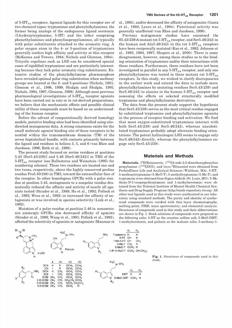
Materials. [3H]Ketanserin, [125I]4-iodo-2,5-dimethoxyphenyliso-propylamine ([125I]DOI), and [myo-3H]inositol were obtained fromPerkinElmer Life and Analytical Sciences (Waltham, MA). 5-HT,5-methoxytryptamine (5-MeO-T), 5-methyltryptamine (5-Me-T), andtryptamine were obtained from Sigma-Aldrich (St. Louis, MO). 5-Me-thoxy- N (1)-isopropyltryptamine and 1-methylserotonin) were ob-tained from the National Institute of Mental Health Chemical Syn-thesis and Drug Supply Program (http://nimh-repository.rti.org). Allother test ligands used in this study were synthesized in our labo-ratory using standard methods. The purity and identity of synthe-sized compounds were verified with thin layer chromatography,melting point, NMR, mass spectrometry, and elemental analysis.Structures of compounds used in this study and their abbreviationsare shown in Fig. 1. Stock solutions of compounds were prepared asthe following salts: 5-HT as the creatine sulfate salt; 5-MeO-DMT,1-methylserotonin, and psilocin as the maleate salts; 5-methoxy-1-
Fig. 1. Structures of compounds used in thisstudy.
TM5 Serines of the h5-HT2A Receptor 1201

8/3/2019 Michael R. Braden and David E. Nichols- Assessment of the Roles of Serines 5.43(239) and 5.46(242) for Binding a…
http://slidepdf.com/reader/full/michael-r-braden-and-david-e-nichols-assessment-of-the-roles-of-serines 3/10
isopropyltryptamine as the oxalate salt; and all other compounds astheir HCl salts. All phenylisopropylamines were prepared as theirracemates.
Cell Culture Methods. The construction of human embryonickidney 293 human embryonic kidney cells with stable expression of wild-type h5-HT2A receptors at high (Hh2Ahi; 8000 fmol/mg) andmoderate (Hh2Alo; 1600 fmol/mg) expression levels has been de-scribed previously (Parrish et al., 2005; Braden et al., 2006). All celltypes were maintained in complete Dulbecco’s modified Eagle’s me-
dium with 30 g/ml Zeocin (Invitrogen) as described previously (Par-rish et al., 2005; Braden et al., 2006). The Hh2Ahi cell line was usedfor competition binding assays and the Hh2Alo cell line was used forfunctional assays.
Establishing Mutant Human 5-HT2A Receptor Cell Lines. Vec-tor construction and site-directed mutagenesis using the QuikChangekit (Stratagene, La Jolla, CA) method were performed as describedpreviously (Braden et al., 2006) with the following sense primers andcorresponding antisense primers (Integrated DNA Technologies, Cor-alville, IA): S5.43(239)A, CTTTGTCCTGATCGGCTCTTTTGTGTCAT-TTTTCATTCCC; S5.46(242)A, CCTGATCGGCTCTTTTGTGTCATTT-TTCATTCCCTTAACC. Mutant inserts verified by primer-directedsequencing (Retrogen, San Diego, CA) were then subcloned into thepBudCE4 vector (Invitrogen, Carlsbad, CA). Human embryonic kidney
293 cells were transfected, colonies selected, and receptor expression verified as described previously (Parrish et al., 2005). The Hh2A/ S5.43(239)A and Hh2A/S5.46(242)A cell lines were chosen for moderateexpression (3800 and 3200 fmol/mg, respectively).
Radioligand Binding Assays. Membrane preparations, satura-tion isotherm, and competition binding assays were performed asdescribed in detail previously (Chambers et al., 2002; Marona-Lewicka et al., 2002). Saturation isotherm binding assays used 0.25to 10 nM [3H]ketanserin or 0.125 to 5.0 nM ()-[125I]DOI. Ligandswere tested in competition binding assays for their ability to displace0.25 nM [125I]DOI. Binding assays at wild-type receptors used thehigh-expressing cell line (Hh2Ahi; 8000 fmol/mg); those at mutantreceptors used the cell lines described above.
Inositol phosphate accumulation assays. Compounds weretested for their ability to stimulate hydrolysis of radiolabeled phos-
phatidyl inositides (PI) by measurement of radiolabeled inositolphosphate accumulation as described previously in detail (Marona-Lewicka et al., 2002; Kurrasch-Orbaugh et al., 2003). Each assayplate was normalized to wells stimulated with water (0%) and aconcentration of serotonin chosen to be maximally stimulating(100%). PI accumulation assays at wild-type receptors used the lowerexpressing cell line (Hh2Alo; 1600 fmol/mg); assays at mutant recep-tors used the cell lines described above.
Computational Modeling/Virtual Docking. Ligand structureswere virtually docked into an in silico-activated h5-HT2AR homologymodel constructed from the published X-ray crystal structure of bovine rhodopsin after undergoing rigid-body molecular dynamicssubsequent to isomerization of the bound 11-cis-retinal to all-trans-retinal (Chambers and Nichols, 2002). Local energy-minimized en-semble structures were obtained as described previously (Parrish etal., 2005; Braden et al., 2006). In brief, energy-minimized structureswere virtually docked using the GOLD software package (CambridgeCrystallographic Data Center, Cambridge, UK). Docking algorithmsfor phenylalkylamines and ergolines were performed without con-straints. Docking algorithms for tryptamines were performed withthe following constraints: 1) a distance constraint of 1.5 to 3.5Åbetween the ligand amine (nonindole) nitrogen and the carbonylcarbon of the side chain of Asp3.32(155); 2) a protein hydrogen bondconstraint for the side-chain oxygen of Ser5.46(242); and 3) a proteinhydrogen bond constraint for the side-chain OH hydrogen of Ser5.43(239). The latter two constraints favorably weight the scoringof docking orientations that place any polar ligand atom in proximityto the protein atoms that could form a possible hydrogen bondwithout constraining a particular ligand atom. Ligand-receptor en-
semble structures were each obtained by merging the highest ranked
docking output ligand orientation structures with the input h5-HT2A
homology model structure using the SYBYL software package (Tri-pos, St. Louis, MO), followed by energy minimization, moleculardynamics, and a final energy minimization simulation. Aggregatesfor molecular dynamics and minimization simulations were definedas residues more than 6 Å from the ligand as well as the backboneatoms. The molecular dynamics and minimization simulations wereperformed with constraints between Asp3.32(155) and the ligandaliphatic amine, and between any polar residue within 2.5 Å of a
polar ligand group, including Ser3.36(159), Thr3.37(160),Ser5.43(239), or Ser5.46(242). Final energy minimization simula-tions also used these constraints.
Data Analysis. Prism software (GraphPad Software Inc., SanDiego, CA) was used to calculate nonlinear regression curves basedon the Cheng-Prusoff equation for a one-site model to obtain K i(affinity) values for radioligand displacement and variable slopesigmoidal dose-response curves for EC50 (potency) and intrinsic ac-tivity from PI hydrolysis. This software package was also used toperform two-way ANOVA calculations with Bonferroni post-testscomparing wild-type and mutant p K i, pEC50, and intrinsic activity
values. Values obtained from the mutant receptors were consideredstatistically distinguishable from wild-type if the models generated
p 0.05. Change in the standard Gibbs free energy (G°) of bindingdue to the mutation was calculated from the K
ivalues at 25°C as
follows: (G°) G°mutant G°wild-type RT ln( K mutant / kwild-type),where R is the gas constant, and T is absolute temperature.Changes in binding affinity and EC50 were transformed to nor-malize the scale by taking the difference of the log10 value (p K iand EC50, respectively) as follows: p K i p K i-mutant– p K i-WT
log K i-mutant (log K i-WT) and pEC50 pEC50-mutant
pEC50-WT logEC50-mutant (logEC50-WT). Changes in intrinsicactivity (Int.Act.) were calculated as follows: Int.Act.
Int.Act.mutant Int.Act.WT. Virtual docking figures were generated using pyMol (DeLano Sci-
entific, San Carlos, CA; http://www.pymol.org). Amino acid residuesare numbered with their position relative to the most highly con-served residue of that transmembrane region and their sequenceposition in the h5-HT2A receptor (Ballesteros and Weinstein, 1995).
Results
Human S5.43(239)A and S5.46(242)A Mutant Recep-
tors Possessed Appropriate Affinity and Expression
Levels. Table 1 presents the results from saturation bind-ing experiments with membrane preparations of stable celllines expressing wild-type and mutant h5-HT2A receptorsusing a radiolabeled antagonist, [3H]ketanserin, or ago-nist, [125I]DOI. As Table 1 indicates, we were able to detectand characterize the mutant receptors with both radioli-gands, and all mutant receptors exhibited acceptable re-ceptor expression. Note that cells with moderate WT h5-
HT2A receptors (1600 fmol/mg) were used in functionalassays, whereas those with high expression (8800 fmol/mg)were used only for binding assays.
Virtual Docking of Tryptamines, Ergolines, and Phe-
nylalkylamines to an in Silico- Activated h5-HT2A Re-
ceptor Homology Model Indicated That Ser5.43(239)
Interacted with Polar Substituents on the Aromatic
Moiety of the Ligand, Whereas Ser5.46(242) Interacted
with the Indole Nitrogen of Tryptamines and Ergo-
lines, but Did Not Interact with Phenylalkylamines.
Fig. 2 illustrates representative binding poses for virtualdocking and subsequent energy minimization simulations of tryptamines, ergolines, and phenylalkylamines in an in
silico-activated homology model of the h5-HT2A receptor. Vir-
1202 Braden and Nichols

8/3/2019 Michael R. Braden and David E. Nichols- Assessment of the Roles of Serines 5.43(239) and 5.46(242) for Binding a…
http://slidepdf.com/reader/full/michael-r-braden-and-david-e-nichols-assessment-of-the-roles-of-serines 4/10
tual docking of LSD or the phenylalkylamines consistentlyoriented the ligand amine near Asp3.32(155) and the polarligand substituents near polar residues in TM3 and TM5.Initial unconstrained docking orientations for tryptamines,however, were highly variable. Few of these orientationspositioned the ligand so that the strong ionic interactioncould occur between the protonated amine of the ligand andthe highly conserved acidic residue at position 3.32,
Asp3.32(155), believed to be the counter-ion in all biogenicamine-binding GPCRs (Ho et al., 1992; Mansour et al., 1992;Kristiansen et al., 2000; Abekawa et al., 2003). Applying adistance constraint between the side-chain nitrogen of theligand and the carbonyl carbon of the side chain of Asp3.32(155) gave more reasonable and less variable high-est-ranked output structures from docking simulations, al-though many structures were still oriented out of the puta-tive small agonist binding pocket. Thus, additional dockingconstraints for tryptamines were introduced to encourageany hydrogen bonding between the ligand and eitherSer5.43(239) and Ser5.46(242) but without forcing a partic-ular receptor-ligand interaction.
We observed final low-energy binding poses for ligandsthat position their polar aromatic substituents in regions andorientations able to interact with h5-HT2A receptor residuesidentified to be potentially important for agonist binding andactivity (Chambers and Nichols, 2002). Of particular rele- vance for this study, the 4- or 5-oxygen atom of ring-substi-
tuted tryptamines is positioned to engage in hydrogen bond-ing with Ser5.43(239). Moreover, virtual docking simulationsare consistent with an interaction between Ser5.46(242) andthe indole nitrogen of the tryptamines, as suggested by oth-ers (Kao et al., 1992; Johnson et al., 1994). As Fig. 2 furtherillustrates, LSD, an ergoline that has structural featuressimilar to those of the tryptamines, is also oriented to allowan interaction between the indole nitrogen and Ser5.46(242);
however, there are no polar groups in LSD positioned tointeract with Ser5.43(239). The 2-methoxy and 5-methoxygroups of phenylalkylamines are positioned to indicate hy-drogen bonding with Ser3.36(159) and Ser5.43(239), respec-tively. Virtual docking simulations, however, indicate no in-teraction between Ser5.46(242) and phenylalkylamines.
The S5.43(239)A Mutation Detrimentally Affected
the Binding and Activity of Tryptamines and Phenyl-
alkylamines Predicted to Interact with This Residue.
Table 2 and Fig. 3 present the results of competition bindingassays with wild-type, S5.43(239)A mutant, and S5.46(242)Amutant h5-HT2A receptors. To aid visual interpretation, aloss in binding affinity is defined as a negative (G°). Bind-
ing of tryptamines with a polar aromatic substituent, such as5-MeO-T, 5-HT, 5-MeO-DMT, psilocin, and 1-Me-5-HT, wasattenuated by the S5.43(239)A mutation to a degree consis-tent with the loss of a hydrogen bond (0.5 to 1.5 kcal/mol;Fersht, 1988), although 1-iPr-5-MeO-T was surprisingly un-affected. Both 5-HT and 5-MeO-DMT suffered the greatest
TABLE 13H-Ketanserin and 125IDOI saturation binding at wild type and mutant receptorsData are represented as the mean S.E.M. of K d and Bmax from nonlinear regression fits of a single binding site model from at least three independent experiments. Celllines used are from isolated colonies of human embryonic kidney 293 cells with stable receptor expression.
Drug
h5-HT2A
WT(moderate) WT (high) S5.43(239)A S5.46(242)A
Ketanserin K d (nM) 0.7 0.1 1.1 0.1 2.2 0.2 0.7 0.1 Bmax (fmol/mg) 1620 80 8800 860 3790 560 3215 530
DOI K d (nM) N.D. 0.78 0.01 2.15 0.31 1.74 0.17 Bmax (fmol/mg) N.D. 625 110 519 65 300 25
Fig. 2. Illustrative cross-eyed stereopair representation of ligand poses from virtual docking experiments with 5-MeO-DMT (A), d-LSD (B), ( R)-DOM(C), and 2CH (D) in the h5-HT2A receptor showing predicted polar interactions between the ligand and receptor residues. Ligands are shown asspace-filling spheres, whereas receptor residues believed to be interacting with the ligand are displayed as sticks. The view is within the membrane,with TM5 on the left, TM6 in the foreground, TM3 in the right background, and the extracellular face of the receptor toward the top of the figure. TMs1, 2, 4, and 7 are not displayed. Energy-minimized ensemble structures indicate that Ser5.46(242) is interacting with the indole nitrogen of LSD andthe tryptamines and Ser5.43(239) is interacting with the 4- and 5-oxygen of tryptamines. Only Ser5.43(239) is able to interact with the 5-oxygen atom
of DOM or 2CH.
TM5 Serines of the h5-HT2A Receptor 1203

8/3/2019 Michael R. Braden and David E. Nichols- Assessment of the Roles of Serines 5.43(239) and 5.46(242) for Binding a…
http://slidepdf.com/reader/full/michael-r-braden-and-david-e-nichols-assessment-of-the-roles-of-serines 5/10
losses of affinity, approximately 14- and 11-fold (1.5 kcal/ mol), respectively. The affinities of the phenylisopro-pylamines DOI, DOM, and DOH were detrimentally affectedby the S5.43(239)A mutation, with G° well within therange given above for hydrogen bonds. The phenethylamines2CI and 2CH also were detrimentally affected, but the G° values were less than 0.5 kcal/mol, and not statistically dis-cernible from wild-type.
Table 3 and Fig. 4 present measures of functional activityof the test compounds using PI hydrolysis assays at wild-typeand mutant receptors. In general, the trends observed inaffinity changes for polar-substituted tryptamines weremaintained in shifts of functional potency, as defined bypEC50. All compounds were affected by this mutation to acertain degree, although in some cases the effect was veryslight (e.g., 5-MeO-T). Again, 5-MeO-DMT and 5-HT were
the polar-substituted tryptamines most affected, their poten-cies being reduced by approximately 20- and 35-fold, respec-tively. Psilocin was the only tryptamine to show a statisti-cally discernible change in intrinsic activity (Table 3; 33%compared with the wild type) as well as a 30-fold decreasein potency.
By contrast, LSD lacks a polar substituent in the areaindicated by virtual docking experiments to be near
Ser5.43(239) and, not surprisingly, this mutation had noeffect on its affinity and only a weak effect on its potency.Likewise, tryptamine and DMT also lack a polar substituentin this region; their binding was relatively unaffected andpotency was only weakly affected. Although affinity of 5-Me-T also was unaffected, we were surprised to observe adramatic 65-fold decrease in potency.
All phenylalkylamines tested showed significant decreasesin potency (pEC50) at the S5.43(239)A mutant receptor com-pared with wild type. DOM and DOH were the most mark-edly affected ligands, with decreases of 60- and 220-fold,respectively. Of all the phenylalkylamines tested at all themutant receptors in this study, we observed a decrease in
efficacy (Int.Act. 35%) only with 2CI at the S5.43(239)Areceptor.
The S5.46(242)A Mutation in the h5-Ht2A Receptor At-
tenuated the Affinity and Activity of Some Tryptamines
Predicted to Interact with This Residue, but Enhanced
the Binding and Activity of Tryptamines with Alkyl Sub-
stitution on the Indole (N1) Nitrogen and Did Not Affect
Phenylalkylamines. As Fig. 3 illustrates, the S5.46(242)Amutation attenuated the affinity only of LSD, 5-HT, and5-MeO-DMT to a degree consistent with the loss of a hydrogenbond (Fersht, 1988). Changes to the binding energetics of psi-locin and 5-MeO-T approachthelower limit of this energy range(0.5–1.5 kcal/mol). The affinity of other tryptamines, particu-
larly those lacking a polar ring substituent, was not signifi-cantly affected by the S5.46(242)A mutation. By contrast, thetryptamines with alkyl substitution on theindole(N1) nitrogen,1-Me-5-HT and 1-iPr-5-MeO-T, showed 12- and 21-fold in-creased affinity, respectively.
Similar trends were generally observed in functional po-tency, as illustrated in Fig. 4. With the exception of 5-MeO-T,the potency of all the tryptamines with polar aromatic sub-stituents, as well as LSD, was weakly to moderately de-creased. No shifts in potency were observed in any of thering-unsubstituted tryptamines 5-Me-T, tryptamine, orDMT. Again, we observed dramatic 45- and 51-fold increasesin potency for the N(1)-alkyl analogs 1-Me-5-HT and 1-iPr-
5-MeO-T, respectively.The S5.46(242)A mutation did not affect the binding of anyof the phenylalkylamines tested to a degree statistically dis-cernible from wild type. At the S5.46(242)A mutant receptor,DOM, DOH, and 2CH suffered generally weak losses in po-tency (2-, 6-, and 5-fold, respectively), whereas the potency of DOI was slightly increased (3-fold). This result is consis-tent with our observation that there is no significant differ-ence between the affinity of phenethylamine agonists at thehuman and rat 5-HT2A receptors, which differ only in thatthe human receptor has a serine at this position, whereas therat receptor has an alanine. The S5.46(242)A mutation didnot lead to a significant change in efficacy for any of the
phenylalkylamines tested.
TABLE 2 Abilities of test compounds to displace ( )-125IDOI at wild-type andmutant h5-HT2A receptorsData are represented as the mean (S.E.M.) of K i values from non-linear regressionfits of a single binding site model from at least three independent experiments.
Drug K i
WT S5.43(239)A S5.46(242)A
nM
5-MeO-DMT 7.54 (1.06) 105 (19)* 36.0 (1.9)*5-HT 4.84 (0.20) 53.2 (8.0)* 20.2 (0.9)*5-MeO-T 1.34 (0.22) 4.17 (0.68)* 2.70 (0.29)Psilocin 11.8 (1.2) 58.0 (3.7)* 23.1 (3.3)5-Me-T 11.7 (0.6) 10.9 (1.3) 25.8 (3.9)Tryptamine 29.7 (4.4) 50.2 (6.6) 36.0 (6.0)DMT 75.1 (6.0) 116 (12) 135 (20)1-Me-5-HT 70.0 (1.2) 320 (37)* 5.73 (0.83)*1-iPr-5-MeO-T 494 (91) 786 (85) 24.1 (0.8)*LSD 0.40 (0.02) 0.41 (0.08) 1.66 (0.32)*DOI 0.64 (0.06) 2.19 (0.26)* 0.83 (0.11)DOM 5.91 (0.97) 46.4 (5.9)* 5.04 (0.55)
2CI 0.73 (0.06) 1.20 (0.22) 1.28 (1.28)2-Et-DOM 91.1 (9.7) 460 (92)* 38.5 (4.1)*5-Et-DOM 22.4 (1.2) 32.2 (1.9) 31.5 (5.0)DOH 245 (28) 2246 (144)* 294 (54)5-H-DOM 169 (12) 343 (41) 164 (7)2CH 377 (67) 782 (19) 440 (23)
* P 0.05 for values of p K i from ANOVA with Bonferroni post tests comparingmutant and wild-type values
Fig. 3. Effects on binding affinities of the S5.43(239)A (open bars) andS5.46(242)A (closed bars) mutations in the h5-HT2A receptor. This bargraph displays the G° values derived from the data of Table 2 (see
Materials and Methods). Negative values in these graphs indicate anadverse effect of the mutation on affinity. Positive values indicate thatbinding is enhanced by the mutation. The dashed line at 0.5 indicates alower threshold for the energetics of a hydrogen bond (Fersht, 1988).
indicates p 0.05 for values of G° from two-way ANOVA with Bonfer-
roni post tests comparing mutant to wild-type values.
1204 Braden and Nichols

8/3/2019 Michael R. Braden and David E. Nichols- Assessment of the Roles of Serines 5.43(239) and 5.46(242) for Binding a…
http://slidepdf.com/reader/full/michael-r-braden-and-david-e-nichols-assessment-of-the-roles-of-serines 6/10
A Complementary Alteration of Phenylalkylamine
Structure Could Produce Binding and Activity Changes
at the WT h5-HT2A Receptor Similar to Those of the
Unmodified Ligand at the S5.43(239)A Mutation and
Could Rescue Some Binding and Activity at the
S5.43(239)A Mutant. As Table 4 indicates, removal of apolar group from the aromatic ring of the phenylisopro-pylamine DOM to yield 2-Et-DOM, 5-Et-DOM, or 5-H-
DOM gave changes in affinities at the wild-type receptorconsistent with the energetics of a lost hydrogen bond(Fersht, 1988). These shifts are similar in magnitude tothose observed when comparing the polar-substituted li-gand homologues in wild-type versus S5.43(239)A mutantreceptors [e.g., the effect of the S5.43(239)A mutation onDOI or DOM in Fig. 3]. Removal of the polar group at the5-position of DOM to give 5-Et-DOM did not detrimentallyaffect binding at the S5.43(239)A mutant receptor. Thismutant was still sensitive to the complete removal of the5-methoxy to give 5-H-DOM (G° 1.2). It was not,however, as sensitive as the wild-type receptor (G°
2.0).
Table 4 also illustrates the difference in functional potencybetween wild-type and S5.43(239)A mutant receptors when apolar group is removed from phenylisopropylamines. Similarto the binding energies, the functional potencies at h5-HT2A
wild-type receptors are sensitive to the removal of a polargroup either at the 2- or 5-position of phenylalkylamines. Bycontrast, the S5.43(239)A mutant receptor is generally lesssensitive or even insensitive to the loss of a polar group, butonly at the ligand 5-position. This insensitivity is clearlyevident in the comparison of DOM and 5-Et-DOM. Althoughwe did not expect 2-Et-DOM and DOM to have significantlydifferent potencies in the S5.43(239)A mutant receptor, theloss of sensitivity to substituting the 2-oxygen (2-Et-DOM;
pEC50 1.2 versus 0.7) is not as great as seen with eithersubstitution (5-Et-DOM; pEC50 1.4 versus 0.0), or re-
moval (5-H-DOM pEC50 2.3 versus 0.6) of the 5-oxygensubstituent.
Discussion
The goal of the current study was to test the hypothesisthat Ser5.43(239) in the h5-HT2A receptor is critical for en-gaging a polar 5-oxygen of phenylalkylamines and a 4- or
5-oxygen substituent of tryptamines, including serotonin, theendogenous agonist ligand for this receptor. Moreover, a par-allel hypothesis was that Ser5.46(242) interacts with theN(1)H of the indole ring in tryptamines, yet does not engagephenylalkylamines. With these experiments, we sought toreplicate, expand, and more clearly elucidate results fromprevious studies mutating these residues in the rat and hu-man 5-HT2A receptors (Kao et al., 1992; Johnson et al., 1993,1994, 1997; Shapiro et al., 2000).
A nondisruptive mutation to an alanine was chosen foreach residue because it was anticipated that this changewould abolish specific ligand-receptor interactions withoutaffecting global receptor structure (Fersht et al., 1987).
Indeed, as Table 1 indicates, only a 2-fold shift in K D wasobserved for [3H]ketanserin at S5.43(239)A receptors (1.1 versus 2.2 nM) and a slight enhancement at S5.46(242)Areceptors (1.1 versus 0.71 nM). A shift of 3-fold in the K Dfor [125I]DOI was observed at S5.43(239)A receptors (0.78 versus 2.19 nM) and approximately 2-fold at S5.46(242)Areceptors (0.78 versus 1.7 nM). The mutant S5.43(239)Aand S5.46(242)A receptors still were able to induce therelease of radiolabeled inositol phosphates in a dose-de-pendent manner that was approximately comparable inmagnitude with that of the wild-type receptor (data notshown), although functional potencies were slightly de-creased for all chemical classes tested at the S5.43(239)A
receptor. Although these findings indicate that theS5.43(239)A mutation may have slightly altered G-protein
TABLE 3 Ability of compounds to activate PI hydrolysis at wild type and mutant h5-HT2A receptorsData are represented as the mean (SEM) of computer-derived estimates of EC50 and Intrinsic Activity values from at least three independent experiments. Except whereindicated (†), all EC50 values were statistically distinguishable from wild-type as defined by P 0.05 values from ANOVA calculations with Bonferroni post-tests betweenmutant and wild-type receptors, whereas * indicates P 0.05 values for Int.Act. values using the same statistical comparison.
DrugWT h5-HT2A S5.43(239)A S5.46(242)A
EC50 PI Hydrolysis Intrinsic Activity EC50 PI Hydrolysis Intrinsic Activity EC50 PI Hydrolysis Intrinsic Activity
nM % 5-HT nM % 5-HT nM % 5-HT
5-MeO-DMT 4.3 (0.78) 98 (4) 150 (25) 88 (7) 31 (4.5) 91 (5)5-HT 5.2 (0.97) 100 100 (8) 100 19 (3.4) 100
5-MeO-T 5.2 (0.48) 99 (4) 26 (1.2) 100 (7) 5.0 (0.45)
†
100 (6)Psilocin 7.3 (0.72) 110 (9) 210 (25) 72 (8)* 45 (8.4) 92 (3)5-Me-T 18 (3.4) 110 (8) 1200 (184) 99 (2) 21 (1.0)† 90 (3)Tryptamine 94 (18) 100 (0) 230 (41)† 97 (3) 95 (17)† 96 (8)DMT 190 (6) 70 (1) 790 (73)† 91 (3) 200 (20)† 95 (4)1-Me-5-HT 310 (30) 100 (3) 1800 (200) 101 (3) 6.9 (1.1) 95 (4)1-iPr-5-MeO-T 2400 (180) 100 (1) 7900 (1400)† 83 (8) 48 (6.7) 97 (3)LSD 0.22 (0.04) 84 (3) 0.66 (0.10) 80 (5) 1.10 (0.12) 86 (2)DOI 3.79 (0.36) 95 (7) 38.9 (8.9) 92 (12) 1.37 (0.06) 92 (2)DOM 2.81 (0.22) 87 (3) 620 (103) 87 (3) 6.46 (1.23) 91 (2)2CI 4.81 (0.89) 87 (7) 71.8 (13.6) 52 (5)* 5.93 (0.58)† 86 (3)2-Et-DOM 49.8 (7.0) 81 (2) 2975 (546) 75 (6) 22.6 (2.8) 93 (2)5-Et-DOM 71.9 (10.8) 84 (10) 571 (78) 81 (6) 62.0 (10.8)† 89 (7)DOH 284 (22) 104 (5) 17669 (2290) 95 (6) 1793 (298) 101 (7)5-H-DOM 533 (64) 106 (5) 2532 (108) 98 (1) 245 (40) 98 (1)2CH 1021 (14) 96 (10) 13053 (416) 100 (4) 4917 (346) 92 (5)
* Int. Act. P 0.05.†
EC50 P 0.05.
TM5 Serines of the h5-HT2A Receptor 1205
8/3/2019 Michael R. Braden and David E. Nichols- Assessment of the Roles of Serines 5.43(239) and 5.46(242) for Binding a…
http://slidepdf.com/reader/full/michael-r-braden-and-david-e-nichols-assessment-of-the-roles-of-serines 7/10
coupling, we do not believe it produces a significant globaldisruption of receptor structure. Thus, our initial hypoth-esis was that any differences in ligand affinity or activitybetween the wild-type and S5.43(239)A or S5.46(242)Amutant receptors would mainly result from a loss of directinteraction between the ligand and those serine residues.
The clearest results were obtained with the potent hallu-cinogen LSD, which was predicted not to interact with
Ser5.43(239). The S5.43(239)A mutation had no effect at allon affinity or intrinsic activity and only a minor 3-fold effecton potency (Figs. 3 and 4). By contrast, the S5.46(242)Amutation caused a 4-fold shift in affinity (Table 2), resultingin a change of the standard free energy of binding (G°) of 0.8 kcal/mol (Fig. 4), within the 0.5 to 1.5 kcal/mol rangedetermined for a hydrogen bond (Fersht, 1988). This muta-tion similarly caused a shift of approximately 6-fold in po-tency for LSD, although it had no effect on intrinsic activity(Fig. 4). Our previous virtual docking studies (Chambers andNichols 2002) placed LSD into the agonist binding domainsuch that there was no interaction with Ser5.43(239),whereas the indole N(1)H engaged Ser5.46(242), located ap-
proximately one turn below Ser5.43(239). The present dataare consistent with this predicted binding pose. The fact thatLSD is such a rigid molecule indicates that the bound ligand-ensemble has little conformational freedom to involveSer5.43(239) in binding. The slight loss of potency in theS5.43(239)A mutant does suggest, however, that even when aligand does not directly engage this residue, it may still beimportant for secondary intramolecular interactions that cre-ate an “efficient” activated receptor state.
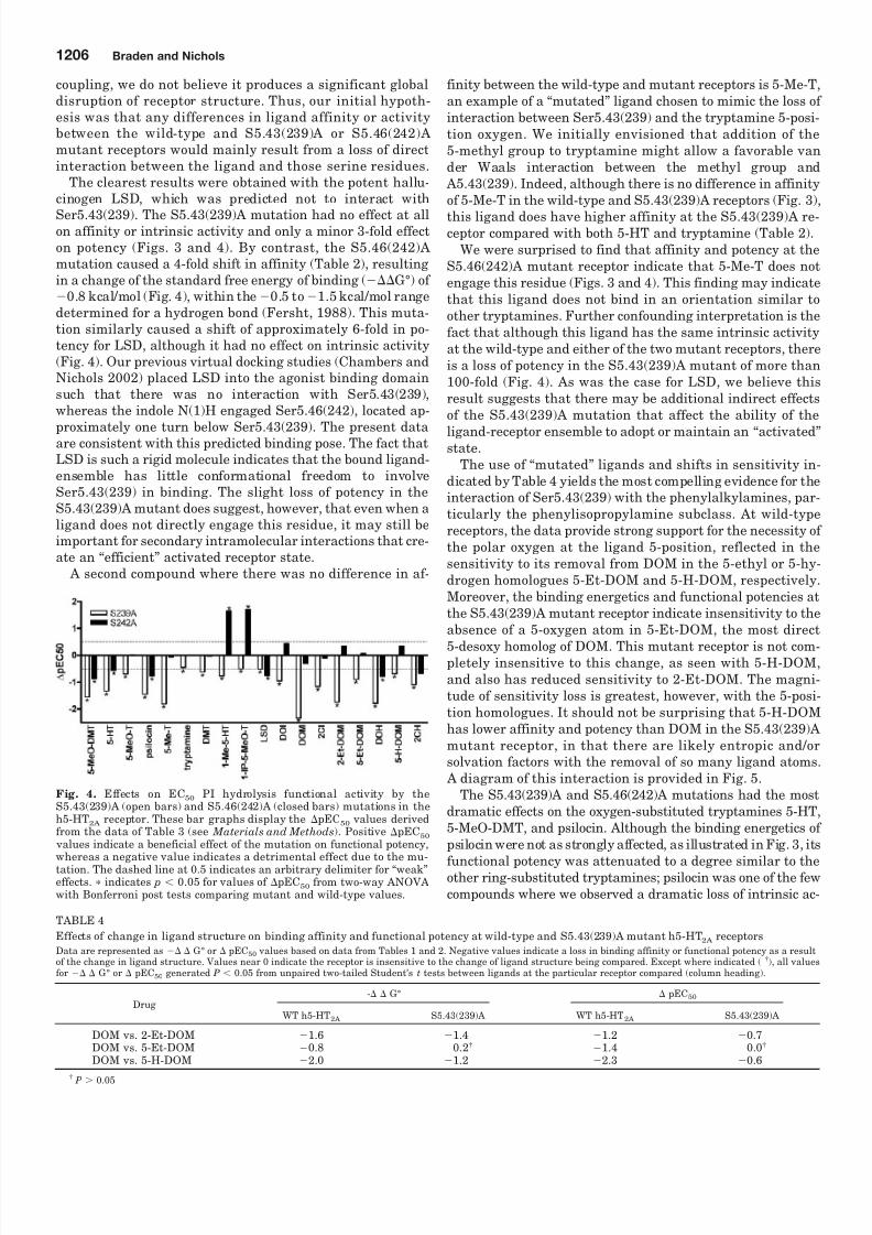
A second compound where there was no difference in af-
finity between the wild-type and mutant receptors is 5-Me-T,an example of a “mutated” ligand chosen to mimic the loss of interaction between Ser5.43(239) and the tryptamine 5-posi-tion oxygen. We initially envisioned that addition of the5-methyl group to tryptamine might allow a favorable vander Waals interaction between the methyl group and A5.43(239). Indeed, although there is no difference in affinityof 5-Me-T in the wild-type and S5.43(239)A receptors (Fig. 3),
this ligand does have higher affinity at the S5.43(239)A re-ceptor compared with both 5-HT and tryptamine (Table 2).
We were surprised to find that affinity and potency at theS5.46(242)A mutant receptor indicate that 5-Me-T does notengage this residue (Figs. 3 and 4). This finding may indicatethat this ligand does not bind in an orientation similar toother tryptamines. Further confounding interpretation is thefact that although this ligand has the same intrinsic activityat the wild-type and either of the two mutant receptors, thereis a loss of potency in the S5.43(239)A mutant of more than100-fold (Fig. 4). As was the case for LSD, we believe thisresult suggests that there may be additional indirect effectsof the S5.43(239)A mutation that affect the ability of the
ligand-receptor ensemble to adopt or maintain an “activated”state.The use of “mutated” ligands and shifts in sensitivity in-
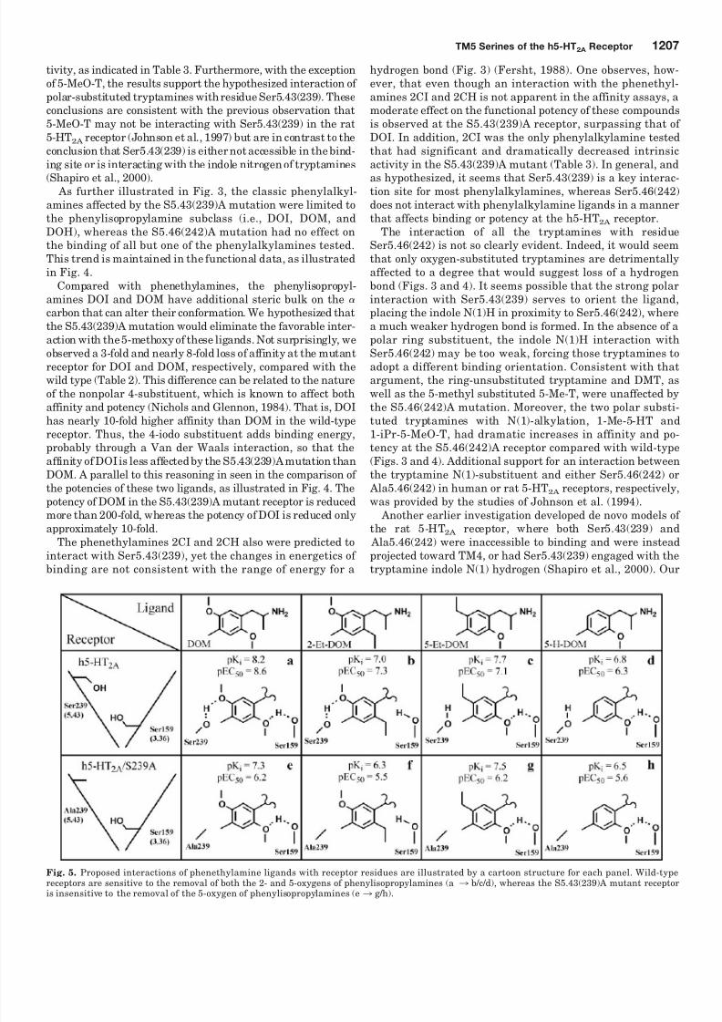
dicated by Table 4 yields the most compelling evidence for theinteraction of Ser5.43(239) with the phenylalkylamines, par-ticularly the phenylisopropylamine subclass. At wild-typereceptors, the data provide strong support for the necessity of the polar oxygen at the ligand 5-position, reflected in thesensitivity to its removal from DOM in the 5-ethyl or 5-hy-drogen homologues 5-Et-DOM and 5-H-DOM, respectively.Moreover, the binding energetics and functional potencies atthe S5.43(239)A mutant receptor indicate insensitivity to theabsence of a 5-oxygen atom in 5-Et-DOM, the most direct
5-desoxy homolog of DOM. This mutant receptor is not com-pletely insensitive to this change, as seen with 5-H-DOM,and also has reduced sensitivity to 2-Et-DOM. The magni-tude of sensitivity loss is greatest, however, with the 5-posi-tion homologues. It should not be surprising that 5-H-DOMhas lower affinity and potency than DOM in the S5.43(239)Amutant receptor, in that there are likely entropic and/orsolvation factors with the removal of so many ligand atoms. A diagram of this interaction is provided in Fig. 5.
The S5.43(239)A and S5.46(242)A mutations had the mostdramatic effects on the oxygen-substituted tryptamines 5-HT,5-MeO-DMT, and psilocin. Although the binding energetics of psilocin were not as strongly affected, as illustrated in Fig. 3, its
functional potency was attenuated to a degree similar to theother ring-substituted tryptamines; psilocin was one of the fewcompounds where we observed a dramatic loss of intrinsic ac-
Fig. 4. Effects on EC50 PI hydrolysis functional activity by theS5.43(239)A (open bars) and S5.46(242)A (closed bars) mutations in theh5-HT2A receptor. These bar graphs display the pEC50 values derivedfrom the data of Table 3 (see Materials and Methods). Positive pEC50
values indicate a beneficial effect of the mutation on functional potency,whereas a negative value indicates a detrimental effect due to the mu-tation. The dashed line at 0.5 indicates an arbitrary delimiter for “weak”effects. indicates p 0.05 for values of pEC50 from two-way ANOVAwith Bonferroni post tests comparing mutant and wild-type values.
TABLE 4Effects of change in ligand structure on binding affinity and functional potency at wild-type and S5.43(239)A mutant h5-HT2A receptorsData are represented as G° or pEC50 values based on data from Tables 1 and 2. Negative values indicate a loss in binding affinity or functional potency as a resultof the change in ligand structure. Values near 0 indicate the receptor is insensitive to the change of ligand structure being compared. Except where indicated ( †), all valuesfor G° or pEC50 generated P 0.05 from unpaired two-tailed Student’s t tests between ligands at the particular receptor compared (column heading).
Drug- G° pEC50
WT h5-HT2A S5.43(239)A WT h5-HT2A S5.43(239)A
DOM vs. 2-Et-DOM 1.6 1.4 1.2 0.7DOM vs. 5-Et-DOM 0.8 0.2† 1.4 0.0†
DOM vs. 5-H-DOM 2.0 1.2 2.3 0.6†
P 0.05
1206 Braden and Nichols
8/3/2019 Michael R. Braden and David E. Nichols- Assessment of the Roles of Serines 5.43(239) and 5.46(242) for Binding a…
http://slidepdf.com/reader/full/michael-r-braden-and-david-e-nichols-assessment-of-the-roles-of-serines 8/10
tivity, as indicated in Table 3. Furthermore, with the exceptionof 5-MeO-T, the results support the hypothesized interaction of polar-substituted tryptamines with residue Ser5.43(239). Theseconclusions are consistent with the previous observation that5-MeO-T may not be interacting with Ser5.43(239) in the rat5-HT2A receptor (Johnson et al., 1997) but are in contrast to theconclusion that Ser5.43(239) is either not accessible in the bind-ing site or is interacting with the indole nitrogen of tryptamines
(Shapiro et al., 2000). As further illustrated in Fig. 3, the classic phenylalkyl-amines affected by the S5.43(239)A mutation were limited tothe phenylisopropylamine subclass (i.e., DOI, DOM, andDOH), whereas the S5.46(242)A mutation had no effect onthe binding of all but one of the phenylalkylamines tested.This trend is maintained in the functional data, as illustratedin Fig. 4.
Compared with phenethylamines, the phenylisopropyl-amines DOI and DOM have additional steric bulk on the
carbon that can alter their conformation. We hypothesized thatthe S5.43(239)A mutation would eliminate the favorable inter-action with the5-methoxy of these ligands. Not surprisingly, we
observed a 3-fold and nearly 8-fold loss of affinity at the mutantreceptor for DOI and DOM, respectively, compared with thewild type (Table 2). This difference can be related to the natureof the nonpolar 4-substituent, which is known to affect bothaffinity and potency (Nichols and Glennon, 1984). That is, DOIhas nearly 10-fold higher affinity than DOM in the wild-typereceptor. Thus, the 4-iodo substituent adds binding energy,probably through a Van der Waals interaction, so that theaffinity of DOIis less affectedby the S5.43(239)Amutation thanDOM. A parallel to this reasoning in seen in the comparison of the potencies of these two ligands, as illustrated in Fig. 4. Thepotency of DOM in the S5.43(239)A mutant receptor is reducedmore than 200-fold, whereas the potency of DOI is reduced onlyapproximately 10-fold.
The phenethylamines 2CI and 2CH also were predicted tointeract with Ser5.43(239), yet the changes in energetics of binding are not consistent with the range of energy for a
hydrogen bond (Fig. 3) (Fersht, 1988). One observes, how-ever, that even though an interaction with the phenethyl-amines 2CI and 2CH is not apparent in the affinity assays, amoderate effect on the functional potency of these compoundsis observed at the S5.43(239)A receptor, surpassing that of DOI. In addition, 2CI was the only phenylalkylamine testedthat had significant and dramatically decreased intrinsicactivity in the S5.43(239)A mutant (Table 3). In general, and
as hypothesized, it seems that Ser5.43(239) is a key interac-tion site for most phenylalkylamines, whereas Ser5.46(242)does not interact with phenylalkylamine ligands in a mannerthat affects binding or potency at the h5-HT2A receptor.
The interaction of all the tryptamines with residueSer5.46(242) is not so clearly evident. Indeed, it would seemthat only oxygen-substituted tryptamines are detrimentallyaffected to a degree that would suggest loss of a hydrogenbond (Figs. 3 and 4). It seems possible that the strong polarinteraction with Ser5.43(239) serves to orient the ligand,placing the indole N(1)H in proximity to Ser5.46(242), wherea much weaker hydrogen bond is formed. In the absence of apolar ring substituent, the indole N(1)H interaction with
Ser5.46(242) may be too weak, forcing those tryptamines toadopt a different binding orientation. Consistent with thatargument, the ring-unsubstituted tryptamine and DMT, aswell as the 5-methyl substituted 5-Me-T, were unaffected bythe S5.46(242)A mutation. Moreover, the two polar substi-tuted tryptamines with N(1)-alkylation, 1-Me-5-HT and1-iPr-5-MeO-T, had dramatic increases in affinity and po-tency at the S5.46(242)A receptor compared with wild-type(Figs. 3 and 4). Additional support for an interaction betweenthe tryptamine N(1)-substituent and either Ser5.46(242) or Ala5.46(242) in human or rat 5-HT2A receptors, respectively,was provided by the studies of Johnson et al. (1994).
Another earlier investigation developed de novo models of the rat 5-HT2A receptor, where both Ser5.43(239) and Ala5.46(242) were inaccessible to binding and were insteadprojected toward TM4, or had Ser5.43(239) engaged with thetryptamine indole N(1) hydrogen (Shapiro et al., 2000). Our
Fig. 5. Proposed interactions of phenethylamine ligands with receptor residues are illustrated by a cartoon structure for each panel. Wild-typereceptors are sensitive to the removal of both the 2- and 5-oxygens of phenylisopropylamines (a 3 b/c/d), whereas the S5.43(239)A mutant receptor
is insensitive to the removal of the 5-oxygen of phenylisopropylamines (e 3 g/h).
TM5 Serines of the h5-HT2A Receptor 1207

8/3/2019 Michael R. Braden and David E. Nichols- Assessment of the Roles of Serines 5.43(239) and 5.46(242) for Binding a…
http://slidepdf.com/reader/full/michael-r-braden-and-david-e-nichols-assessment-of-the-roles-of-serines 9/10
data are not consistent with such an orientation of TM5 inthe human 5-HT2A receptor, although we agree with theconclusion of that study that not all tryptamines may bebinding in the same orientation. Instead, we believe our datasupport the hypothesis that Ser5.43(239) is directly engagedwhen binding tryptamines with polar substituents at eitherthe 4- or 5-position. Although the ergoline LSD lacks a polarsubstituent to interact with Ser5.43(239), it has other struc-
tural features that may orient it within the binding site, sothat it can engage Ser5.46(242). Even when a direct ligand-receptor interaction cannot occur, our data suggest that res-idue Ser5.43(239) may be involved in additional indirect in-teractions essential for potency at the 5-HT2A receptor. Theseindirect interactions are difficult to model and indicate thatour model may not be fully accounting for all possible “active”receptor conformations. Moreover, it may be possible, asStrader et al. (1989) suggested with the -adrenergic recep-tor, that some ligands are able to bind to the mutant recep-tors in orientations that are competitive but not able to affectreceptor activation.
Polar residues have been shown to be critical for agonist
activity in a variety of monoamine GPCRs, as well as mus-carinic acetylcholine receptors; representative TM5 se-quences are illustrated in Fig. 6. In particular, in catechol-amine receptors, the serine(s) at position 5.43 (and/orSer5.42) probably engages the “meta” OH of the catecholmoiety, a key structural feature of catecholamine agonistsrecognized for many years (e.g., Strader et al., 1989).
Our results also are consistent with the role of Ser5.43(239) as a hydrogen bond donor for both phenylal-kylamines and tryptamines, whereas it seems more likelythat Ser5.46(242) serves as a hydrogen bond acceptor fortryptamines and ergolines. This idea is reinforced by thepresence of an alanine at position 5.46 in the human 5-HT2B,5-HT2C, and 5-HT1A, as well as the rat 5-HT2A receptors.Furthermore, our modeling of the h5-HT2A receptor suggeststhat Ser5.46(242) may form an intrahelical hydrogen bond tothe backbone carbonyl of G5.42(238). From a mechanicalpoint of view, if Pro5.50(246) serves to allow the top of TM5to undergo movement upon ligand binding, engaging a polarresidue more distal from Pro5.50(246) would provide greatermechanical force to displace the helix, relative to a residuecloser to Pro5.50(246). The global change helical tilt angleindicative of receptor-activation may be related to the loss of a kink near Pro5.50(246) in helix 5 (Crozier et al., 2007).
Our data also indicate that not all tryptamines interact
with the receptor in the same way, a conclusion reached byprevious studies based on interactions with residueSer5.43(239) in the rat 5-HT2A receptor (Johnson et al., 1997;Shapiro et al., 2000). The present results extend previousinvestigations into the role of these residues in the h5-HT2A
receptor. As with investigations by our and other laboratoriesinto the aromatic contacts of TM6 (Choudhary et al., 1993,1995; Roth et al., 1997; Braden et al., 2006), it seems that the
functional topography of the receptor binding site is suchthat a contact with TM5 is necessary for high affinity andfunctional potency of agonists. These results further supportthe topography and utility of our in silico-activated homologymodel of the h5-HT2A receptor, and we anticipate that thepresent findings can be extended to the understanding of other family A monoamine-binding GPCRs.
Acknowledgments
We thank Stewart Frescas for the synthesis of several of theligands used in this study.
References
Abekawa T, Honda M, Ito K, and Koyama T (2003) Effects of NRA0045, a novel
potent antagonist at dopamine D4, 5-HT2A, and alpha1 adrenaline receptors, andNRA0160, a selective D4 receptor antagonist, on phencyclidine-induced behaviorand glutamate release in rats. Psychopharmacology (Berl) 169:247–256.
Baker D and Sali A (2001) Protein structure prediction and structural genomics. Science 294:93–96.
Ballesteros JA and Weinstein H (1995) Integrated methods for the construction of three-dimensional models and computational probing of structure-function rela-tions in G protein-coupled receptor. Methods Neurosci 25:366–428.
Braden MR, Parrish JC, Naylor JC, and Nichols DE (2006) Molecular interaction of serotonin 5 HT2A receptor residues Phe339(6.51) and Phe340(6.52) with super-potent N -benzyl phenethylamine agonists. Mol Pharmacol 70:1956–1964.
Chambers JJ, Kurrasch-Orbaugh DM, and Nichols DE (2002) Translocation of the5-alkoxy substituent of 2,5-dialkoxyarylalkylamines to the 6-position: effects on5-HT(2A/2C) receptor affinity. Bioorg Med Chem Lett 12:1997–1999.
Chambers JJ and Nichols DE (2002) A homology-based model of the human 5-HT2Areceptor derived from an in silico activated G-protein coupled receptor. J Comput
Aided Mol Des 16:511–520.Choudhary MS, Craigo S, and Roth BL (1993) A single point mutation (Phe340–
LEU340) of a conserved phenylalanine abolishes 4-[125I]iodo-(2,5-dimethoxy)-
phenylisopropylamine and [3
H]mesulergine but not [3
H]ketanserin binding to5-hydroxytryptamine2 receptors. Mol Pharmacol 43:755–761.Choudhary MS, Sachs N, Uluer A, Glennon RA, Westkaemper RB, and Roth BL
(1995) Differential ergoline and ergopeptine binding to 5-hydroxytryptamine2Areceptors: ergolines require an aromatic residue at position 340 for high affinitybinding. Mol Pharmacol 47:450–457.
Crozier PS, Stevens MJ, and Woolf TB (2007) How a small change in retinal leads toG-protein activation: initial events suggested by molecular dynamics calculations.
Proteins 66:559–574.Fersht AR (1988) Relationships between apparent binding energies measured in
site-directed mutagenesis experiments and energetics of binding and catalysis. Biochemistry 27:1577–1580.
Fersht AR, Leatherbarrow RJ, and Wells TN (1987) Structure-activity relationshipsin engineered proteins: analysis of use of binding energy by linear free energyrelationships. Biochemistry 26:6030–6038.
Gantz I, Delvalle J, Wang LD, Tashiro T, Munzert G, Guo YJ, Konda Y, and YamadaT (1992) Molecular basis for the interaction of histamine with the histamine H2receptor. J Biol Chem 267:20840–20843.
Glennon RA (1989) Stimulus properties of hallucinogenic phenalkylamines andrelated designer drugs: formulation of structure-activity relationships. NIDA Res
Monogr 94:43–67.Glennon RA (1999) Arylalkylamine drugs of abuse: an overview of drug discrimina-
tion studies. Pharmacol Biochem Behav 64:251–256.Glennon RA, Titeler M, and Young R (1986) Structure-activity relationships and
mechanism of action of hallucinogenic agents based on drug discrimination andradioligand binding studies. Psychopharmacol Bull 22:953–958.
Ho BY, Karschin A, Branchek T, Davidson N, and Lester HA (1992) The role of conserved aspartate and serine residues in ligand binding and in function of the5-HT1A receptor: a site-directed mutation study. FEBS Lett 312:259–262.
Johnson MP, Audia JE, Nissen JS, and Nelson DL (1993) N(1)-substituted ergolinesand tryptamines show species differences for the agonist-labeled 5-HT2 receptor.
Eur J Pharmacol 239:111–118.Johnson MP, Loncharich RJ, Baez M, and Nelson DL (1994) Species variations in
transmembrane region v of the 5-hydroxytryptamine type 2A receptor alter thestructure-activity relationship of certain ergolines and tryptamines. Mol Pharma-col 45:277–286.
Johnson MP, Wainscott DB, Lucaites VL, Baez M, and Nelson DL (1997) Mutationsof transmembrane IV and V serines indicate that all tryptamines do not bind to therat 5-HT2A receptor in the same manner. Brain Res Mol Brain Res 49:1–6.
Kao HT, Adham N, Olsen MA, Weinshank RL, Branchek TA, and Hartig PR (1992)
Fig. 6. Selected examples of sequence homology in the ligand-specific
portion of transmembrane helix 5 for several type A family GPCRs.
1208 Braden and Nichols

8/3/2019 Michael R. Braden and David E. Nichols- Assessment of the Roles of Serines 5.43(239) and 5.46(242) for Binding a…
http://slidepdf.com/reader/full/michael-r-braden-and-david-e-nichols-assessment-of-the-roles-of-serines 10/10
Site-directed mutagenesis of a single residue changes the binding properties of theserotonin 5-HT2 receptor from a human to a rat pharmacology. FEBS Lett 307:324–328.
Kristiansen K, Kroeze WK, Willins DL, Gelber EI, Savage JE, Glennon RA, and RothBL (2000) A highly conserved aspartic acid (Asp-155) anchors the terminal aminemoiety of tryptamines and is involved in membrane targeting of the 5-HT(2A)serotonin receptor but does not participate in activation via a “salt-bridge disrup-tion” mechanism. J Pharmacol Exp Ther 293:735–746.
Kurrasch-Orbaugh DM, Watts VJ, Barker EL, and Nichols DE (2003) Serotonin5-hydroxytryptamine 2A receptor-coupled phospholipase C and phospholipase A2signaling pathways have different receptor reserves. J Pharmacol Exp Ther 304:229–237.
Leurs R, Smit MJ, Tensen CP, Ter Laak AM, and Timmerman H (1994) Site-directedmutagenesis of the histamine H1-receptor reveals a selective interaction of aspar-agine207 with subclasses of H1-receptor agonists. Biochem Biophys Res Commun201:295–301.
Li J, Edwards PC, Burghammer M, Villa C, and Schertler GF (2004) Structure of bovine rhodopsin in a trigonal crystal form. J Mol Biol 343:1409–1438.
Link R, Daunt D, Barsh G, Chruscinski A, and Kobilka B (1992) Cloning of twomouse genes encoding 2-adrenergic receptor subtypes and identification of asingle amino acid in the mouse 2-C10 homolog responsible for an interspecies
variation in antagonist binding. Mol Pharmacol 42:16–27.Mansour A, Meng F, Meador-Woodruff JH, Taylor LP, Civelli O, and Akil H (1992)
Site-directed mutagenesis of the human dopamine D2 receptor. Eur J Pharmacol227:205–214.
Marona-Lewicka D, Kurrasch-Orbaugh DM, Selken JR, Cumbay MG, Lisnicchia JG,and Nichols DE (2002) Re-evaluation of l isuride pharmacology:5-hydroxytryptamine1A receptor-mediated behavioral effects overlap its otherproperties in rats. Psychopharmacology (Berl) 164:93–107.
McKenna DJ, and Towers GH (1984) Biochemistry and pharmacology of tryptaminesand beta-carbolines. A minireview. J Psychoactive Drugs 16:347–358.
McLean TH, Chambers JJ, Parrish JC, Braden MR, Marona-Lewicka D, Kurrasch-Orbaugh D, and Nichols DE (2006a) C-(4,5,6-trimethoxyindan-1-yl)methanamine:a mescaline analogue designed using a homology model of the 5-HT2A receptor.
J Med Chem 49:4269–4274.McLean TH, Parrish JC, Braden MR, Marona-Lewicka D, Gallardo-Godoy A, and
Nichols DE (2006b) 1-Aminomethylbenzocycloalkanes: conformationally restrictedhallucinogenic phenethylamine analogues as functionally selective 5-HT2A recep-tor agonists. J Med Chem 49:5794–5803.
Nichols DE (1981) Structure-activity relationships of phenethylamine hallucinogens. J Pharm Sci 70:839–849.
Nichols DE (1994) Medicinal chemistry and structure-activity relationships, in Am- phetamine and Its Analogs (Cho AK and Segal DS eds) pp 3–41, Academic Press,New York.
Nichols DE (1997) Role of serotoninergic neurons and 5-HT receptors in the action of
hallucinogens, in Serotoninergic Neurons and 5-HT Receptors in the CNS (Baum-garten HG and Gothert M eds) pp 563–585, Springer-Verlag, Berlin Heidelberg
Nichols DE (2004) Hallucinogens. Pharmacol Ther 101:131–181.Nichols DE and Glennon RA (1984) Medicinal chemistry and structure-activity
relationships of hallucinogens, in Hallucinogens: Neurochemical, Behavioral, andClinical Perspectives (Jacobs BL ed) pp 95–142, Raven Press, New York
Palczewski K, Kumasaka T, Hori T, Behnke CA, Motoshima H, Fox BA, Le T, I,Teller DC, Okada T, et al. (2000) Crystal structure of rhodopsin: a G protein-coupled receptor. Science 289:739–745.
Parrish JC, Braden MR, Gundy E, and Nichols DE (2005) Differential phospholipaseC activation by phenylalkylamine serotonin 5-HT 2A receptor agonists. J Neuro-chem 95:1575–1584.
Pollock NJ, Manelli AM, Hutchins CW, Steffey ME, MacKenzie RG, and Frail DE(1992) Serine mutations in transmembrane V of the dopamine D1 receptor affectligand interactions and receptor activation. J Biol Chem 267:17780–17786.
Roth BL, Shoham M, Choudhary MS, and Khan N (1997) Identification of conservedaromatic residues essential for agonist binding and second messenger productionat 5-hydroxytryptamine2a receptors. Mol Pharmacol 52:259–266.
Roth BL, Willins DL, Kristiansen K, and Kroeze WK (1998) 5-hydroxytryptamine2-family receptors (5-hydroxytryptamine2a, 5-hydroxytryptamine2b,5-hydroxytryptamine2c): where structure meets function. Pharmacol Ther 79:231–257.
Shapiro DA, Kristiansen K, Kroeze WK, and Roth BL (2000) Differential modes of agonist binding to 5-hydroxytryptamine(2A) serotonin receptors revealed by mu-tation and molecular modeling of conserved residues in transmembrane region 5.
Mol Pharmacol 58:877–886.Shulgin A and Shulgin A (1991) PIHKAL: A Chemical Love Story. Transform Press,
Berkeley, CA.Strader CD, Candelore MR, Hill WS, Sigal IS, and Dixon RA (1989) Identification of
two serine residues involved in agonist activation of the beta-adrenergic receptor. J Biol Chem 264:13572–13578.
van Rhee AM and Jacobsen KA (1996) Molecular architecture of G protein-coupledreceptors. Drug Dev Res 37:1–38.Wang CD, Buck MA, and Fraser CM (1991) Site-directed mutagenesis of 2A-
adrenergic receptors: identification of amino acids involved in ligand binding andreceptor activation by agonists. Mol Pharmacol 40:168–179.
Wess J, Maggio R, Palmer JR, and Vogel Z (1992) Role of conserved threonine andtyrosine residues in acetylcholine binding and muscarinic receptor activation. Astudy with M3 muscarinic receptor point mutants. J Biol Chem 267:19313–19319.
Address correspondence to: Dr. David E. Nichols, Dept. of Medicinal Chem-istry and Molecular Pharmacology, School of Pharmacy and PharmaceuticalSciences, 575 Stadium Mall Drive, Purdue University, West Lafayette, IN47907-2091. E-mail: [email protected]
TM5 Serines of the h5-HT2A Receptor 1209