Embed Size (px)
Citation preview

M&I
2 0 1 5

M&I is the annual newsletter of the Department of Microbiology & Immunology at Columbia University
Editor-in-chiefSankar Ghosh, Ph.D.
EditorOliver Jovanovic, Ph.D.
Art Direction and DesignOliver Jovanovic, Ph.D.
Content Authors Uttiya Basu Ph.D.David Fidock, Ph.D.Sankar Ghosh, Ph.D.Oliver Jovanovic, Ph.D.John Seeley, Ph.D.
Cover DesignShomik Ghosh
PhotographyHyunju Oh, Ph.D.
M&I highlights exciting new research discoveries, except iona l facu l t y ach ievements , and depar tment-wide init iat ives, providing a comprehensive summary of the goings-on in the department, which bridges modern molecular biology with research on infectious disease and immunology. Digital versions and back issues are available at: www.microbiology.columbia.edu
M&I EditorDept. of Microbiology & ImmunologyColumbia University701 West 168th St.New York, NY 10032 (212) [email protected]

3 Message4 Highlights9 Department13Features22Notes30 Events
13 Immunotherapy 19 Harold S. Ginsberg
10 News 12 Alumni
4 Educat ion Bui lding 5 David Fidock 7 Utt iya Basu
22 Publ icat ions 26 Lab Notes

I AM PLEASED to present the 2015 issue of M&I, the Department of Microbiology & Immunology’s annual newsletter. Although the publication of this year’s issue was delayed, we are happy that it is finally available for distribution.
In this issue, we have once again highlighted the science being carried out in the department, as well as different departmental activities and departmental news. We have profiled exciting research from two of our faculty members, David Fidock and Uttiya Basu. We have also highlighted the emerging area of tumor immunotherapy, a tremendously exciting area of research that is having dramatic effects in the clinic, extending lives of countless patients with heretofore incurable malignancies such as melanomas. It is worth noting that Columbia University awarded the 2014 Louisa Gross Horwitz Prize, its most distinguished scientific award, to James P. Allison, who is widely credited for having spearheaded the development of this novel approach of treating tumors. Finally, continuing our tradition of articles that highlight important individuals and events in the department’s history, we feature a biographical piece on Harold S. Ginsberg, one of the founding fathers of virology and microbiology, who served as chairman of the department from 1973 to 1985.
The past year saw a major overhaul of the departmental office, with James Lapin assuming the role of Departmental Administrator. Our graduate program continues to flourish and we have four outstanding new students this fall. Unfortunately we lost two important members of the department. Boris Reizis left for NYU, while Fred Chang is leaving for UCSF. We are happy however to welcome amongst our midst Anne Moscona from Cornell, a well-known virologist, who joined us in December. She will be located on the 13th floor of Hammer. Yiping Han, a joint faculty member who was previously located in the dental school, has also moved her laboratory to the 15th floor of Hammer. We are also actively seeking new faculty members and have interviewed a number of candidates.
As always, I want to end by thanking all the people who help ensure that the department runs smoothly, including the other staff in the departmental office (Carol Duigou and Andrew Wong), Oliver Jovanovic, Carla Horne in my office, and Amir Figueroa in the FACS/Microscope facilities. The editing and development of content of the newsletter this year was primarily the work of Oliver, with assistance from John Seeley, a cover design by Shomik Ghosh and additional photography by Hyunju Oh.
I hope you enjoy reading this newsletter and I would like to thank all of you for your efforts in making M&I a great place to be.
Sankar Ghosh, Ph.D.
Message from the Chair

Construction on the new 14 story Medical and Graduate Education Building on Haven Avenue continues, and is expected to be completed by 2016. The landmark building is meant to provide additional classroom and lecture space and resources for students.
L I G H T B OX
Education Building Progress


H I G H L I G H T S > F I D O C K 6
D R . DAV I D F I D O C K
Pinpointing Malarial Artemisinin ResistanceMALARIA CONTINUES TO BE the leading infectious cause of infant deaths worldwide, with over 0.5 million deaths in 2013 resulting from infections with the eukaryotic parasite Plasmodium falciparum. The vast majority of these deaths occur in sub-Saharan Africa, where individuals are often chronically infected with multiple strains. There, sub-sterile immunity is acquired over many years and at best only achieves a stalemate, whereby individuals acquire protection against disease manifestations (such as cerebral malaria or severe anemia) but cannot eliminate the parasites. Historically, drugs have been the foundation of all efforts to treat and control malaria. The first-line drug artemisinin (ART), used in combination with longer-lasting but less potent partner drugs, has helped halve the malarial death rate in the past 15 years. Parasite resistance to ART, however, has now emerged. Early in 2015, the Fidock lab reported a series of genome editing experiments that provided definitive evidence that mutations in the P. falciparum K13 gene were responsible for this gain of resistance. These data were acquired by introducing or removing K13 mutations from clinical isolates obtained from
western Cambodia, the epicenter of ART resistance, as well as from standard laboratory strains. K13-edited parasite lines were then assayed in vitro using the “Ring-stage Survival Assay”, whereby intracellular parasites that had recently invaded red blood cells were briefly exposed to a high concentration of the ART metabolite (dihydro-artemisinin) and their parasite survival measured three days later. Whereas parasites expressing wild-type K13 showed survival rates < 0.7%, isogenic parasite lines expressing mutant K13 showed survival rates up to 40%. These studies also revealed secondary contributions from other genetic loci, as recently collected strains manifested the highest levels of resistance. These studies were made possible by the earlier development of genome editing for P. falciparum, achieved by the Fidock lab using customized zinc-finger nucleases (Straimer et al. 2012 Nature Methods). Researchers and field-based teams can now use these validated K13 mutations in rapid and inexpensive PCR-based approaches to pinpoint where ART resistance has emerged or spread and alter local treatment and containment strategies accordingly.
Genetic modification of the K13-propeller domain. Location of K13-propeller mutations and sequencing results showing the insertion of individual mutations into recombinant parasites used in the in vitro assay that measured artemisinin resistance. Dd2ctrl parasites contain only synonymous, phenotypically silent binding-site mutations and showed 0.7% survival rates, which is equivalent to those of parental Dd2 parasites. Parasites expressing individual K13 mutations showed survival rates more typically in the 10% to 40% range, indicative of resistance. This study was a collaborative effort between the Fidock laboratory in the Department of Microbiology & Immunology at Columbia University and groups working at the NIH (Bethesda, MD), the Pasteur Institutes in Paris and Phnom Penh (Cambodia), the University of Toulouse and Sangamo Biosciences (Richmond, CA).
Reference: Straimer, J., Gnädig, N.F., Witkowski, B., Amaratunga, C., Duru, V., Ramadani, A.P., Dacheux, M., Khim, N., Zhang, L., Lam, S., Gregory, P.D., Urnov, F.D. Mercereau-Puijalon, O., Benoit-Vical, F., Fairhurst, R.M., Ménard D. and Fidock, D.A. (2015) K13-propeller mutations confer artemisinin resistance in Plasmodium falciparum clinical isolates. Science 347: 428-431.


H I G H L I G H T S > B A S U 8
D R . U T T I YA B A S U
Transcriptomics Identify Breg Marker CD9B CELLS PLAY MULTIPLE ROLES in the homeostasis of the human adaptive immune system. Their best known role is probably their generation of diverse types of antibodies that protect us from pathogenic viruses and bacteria, but equally important is the role B cells play in presenting antigens to T cells and producing co-stimulatory molecules to promote various immune responses. A small population of B cells, named regulatory B cells (Bregs), can also suppress immune response pathways, primarily through the production of the anti-inflammatory cytokine IL-10. Bregs are found enriched in phenotypically diverse B-cell subsets. However, the identification and characterization of Breg cells has been impeded by a scarcity of unique cell surface markers which would exclusively and exhaustively identify them. Lack of the proper tools has foiled detailed investigations into their origin, development, and immunological roles. In a recent paper published in Cell Reports, Uttiya Basu, Assistant Professor of Microbiology & Immunology at Columbia University and Jianbo Sun, an associate research scientist in the Basu laboratory, identified a tetraspanin family transmembrane protein known as CD9 as a marker of IL-10 competent Breg cells (also known as B10 cells) in mice. The identification of CD9 as a surface marker of Breg cells was accomplished by a detailed transcriptome analysis of IL10-secreting B cells, in collaboration with Raul Rabadan’s laboratory in Systems Biology at Columbia University. The tetraspanin-family transmembrane protein CD9 can distinguish both mature and immature B10 cells from non-regulatory B cells. Thus, it provides a system to study unperturbed gene expression profiles of Breg progenitors and Bregs without the additional stimulation previously required to prompt IL-10 secretion for Breg studies. This is important, since Breg transcription factor activity and networks may be perturbed by stimulation with factors such as LPS, PMA and ionomycin. With the identification of CD9 as a marker of B10 cells, identification of physiologically relevant transcription factors driving B10 differentiation becomes a real possibility. The exact function of CD9 on the surface of B10 cells remains unknown. Experiments suggest that B10 cells may communicate with other cells or respond to environmental cues via CD9, either of which could promote their differentiation from multi-potential B cells into effector B10 cells. As CD9+ B cells possess immune suppressive function(s) and can be easily enriched, adoptive transfer of Breg
cells into murine autoimmune disease models becomes a real possibility. Similarly, elimination of immunosuppressive IL-10-expressing plasma cells could allow cytotoxic T cell-dependent eradication of therapy-resistant tumors. Therefore, the possibility exists for tumor immunotherapy in which depletion of CD9+ B cells could be an effective alternative to specific depletion of IL-10 producing B cells. Although exciting possibilities exists in terms of translating these experimental observations into therapies, the expression of CD9 or an equivalent molecule on the surface of human Breg cells has yet to be identified, and is one of the goals of the Basu lab as it continues to investigate the complex roles of B cells in the immune system.
Graphical Abstract
Reference: Sun J., Wang J., Pefanis E., Chao J., Rothschild G., Tachibana I., Chen J., Ivanov I.I., Rabadan R., Takeda Y. and Basu U. (2015) Transcriptomics identify CD9 as a marker of murine IL-10 competent regulatory B cells. Cell Reports 13: 1110-1117.


Department NewsD E PA R T M E N T > N E W S
Boris Reizis and Fred Chang Moving
BORIS REIZIS, PH.D. AND FRED CHANG, M.D., PH.D., Professors of Microbiology & Immunology, are moving their laboratories. Dr. Reizis has moved his laboratory to New York University as of November 1, 2015, where he will serve on the faculty of the departments of Pathology and Medicine. Boris served as the department’s Director of Graduate Studies for many years, and recently served as the Co-Director of the department’s NIH NIAID T32 training grant. Dr. Chang is moving his laboratory to UCSF as of January 1, 2016, where he will serve on the faculty of the department of Cell & Tissue Biology. Both will be greatly missed, and we wish them the best of luck at their new institutions.
JAMES LAPIN was appointed Departmental Administrator of the department of Microbiology & Immunology on May 31, 2015. He previously served as Deputy Administrator of the department, and Research Manager of the department of Biomedical Informatics. We would like to welcome him in his new position, where he will work with Andrew Wong, our new Human Resources/Pre-Awards Administrator and Carol Duigou, our Administrative Coordinator. James has a back-ground in non-profit management and is an experienced grant manager. Jessica Sama, Departmental Administrator of the department of Genetics & Development, assisted us in this transition, for which we are thankful.
Department Welcomes James Lapin
Four New Students Join Department
Simon DiezUniversity of South Carolina
Phoebe OldachUniversity of Cambridge
Lekha NairUniversity of Chicago
Katherine KaiserSUNY Stony Brook
10
PROFESSOR RICCARDO DALLA-FAVERA, M.D. was elected to the National Academy of Sciences on April 27, 2015. He is Professor of Microbiology & Immunology, Pathology & Cell Biology, Genetic & Development, Percy & Joanne Uris Professor of Clinical Medicine and Director of the Institute for Cancer Genetics. Dr. Dalla-Favera is a distinguished investigator in the molecular genetics of cancer and has been an internationally recognized leader in the field of lymphoid neoplasia for the last 30 years. He was elected to the Institute of Medicine in 2010, and also received the Burkitt Medal in 2015 for his work on lymphoma.
Professor Riccardo Dalla-Favera Elect-ed to National Academy of Sciences
Departmental RetreatTHE 2014 MICROBIOLOGY & IMMUNOLOGY annual retreat was held at the Wyndham Hamilton Park Hotel and Conference Center in Florham Park, NJ on September 4th and 5th. The keynote speaker was Dr. Andrea Califano, founding chair of the Department of Systems Biology at Columbia University and Director of the J.P. Sulzberger Columbia Genome Center. The retreat featured research talks by faculty members and a poster session presented by students and postdoctoral fellows, as well as a collegial after party. During down time, attendees were welcome to enjoy numerous impromptu activities including basketball, hiking, and foosball. The 2015 Microbiology & Immunology retreat was held at the Wyndham Hamilton Park Hotel and Conference Center on September 3rd and 4th, 2015.

Memorial Lectures
1 1D E PA R T M E N T > N E W S
THE DEPARTMENT IS RECRUITING faculty to increase our representation of research in immunology and host-pathogen interactions. We are seeking outstanding candidates who are pursuing fundamental problems at the molecular, cellular and organismal levels in all areas of immunology and immune response to commensal and pathogenic microbes. The position can be filled at any rank from tenure-eligible Assistant Professor to full Professor with tenure. Candidates are expected to maintain or develop an independent research program and participate in departmental teaching.
The department has had several potential faculty candidates present special seminars on their topic of research, and will continue to do so in the near future. Please try to attend these special seminars, which will be announced by postings and departmental email.
PROFESSOR ANNE MOSCONA has joined our department in a dual appointment with Pediatrics as Professor of Pediatrics and Microbiology & Immunology on December 16, 2015. Dr. Moscona received her M.D. from Columbia University in 1982, then completed her residency and fellowship training at Mt. Sinai School of Medicine, where she developed her research on paramyxovirus and was most recently appointed Professor of Pediatrics and Microbiology & Immunology. At Mt. Sinai, she served as Vice Chair for Research in Pediatrics and Director of the Division of Pediatric Infectious Diseases. Dr. Moscona has received a number of honors and awards, including a Charles H. Revson Foundation Fellowship, Solomon Silver Award in Clinical Medicine, Pediatric Infectious Disease Society Young Investigator Award and a Hirschl/Monique Weill-Caulier Career Scientist Award. Dr. Moscona has been elected to membership in the Society for Pediatric Research, American Society of Clinical Investigation and to fellowship in the American Society for Microbiology. She will be joined by her research partner, Matteo Porotto, who is moving to CUMC as an Associate Professor of Pediatrics.
Anne Moscona Joins DepartmentDepartmental Faculty Recruitment
THE 2015 RICHARD C. PARKER LECTURE, “Dissecting the Role of mTOR in Cell Growth and Proliferation” was presented on May 13, 2015 by Dr. Craig B. Thompson, President and CEO of Memorial Sloan Kettering Cancer Center.
THE 2015 HEIDELBERGER-KABAT LECTURE, “From Inflammation to Immunity: Understanding Cancer and Improving its Treatment” was presented on June 24, 2015 by Michael Karin, Distinguished Professor of Pharmacology, Ben and Wanda Hildyard Chair for Mitochondrial and Metabolic Diseases, and American Cancer Society Research Professor of the University of California, San Diego.
Richard C. Parker Memorial Award
THE RECIPIENTS OF the 2015 Richard C. Parker Graduate Student Award were Naomi Yudanin, Evangelos Pefanis and Jaeyop Lee. Yudanin’s research in the Farber laboratory focused on defining the mechanisms of memory T cell migration to and retention in specific tissue sites and how human T cells are compartmentalized in tissues. Pefanis’ research in the Basu laboratory focused on the role of RNA exosome in AID targeting in the B cell genome. Lee’s research in the Liu laboratory focused on identifying dendritic cell and monocyte progenitors in human cord blood and bone marrow. Their awards were presented on May 13, 2015 at the 29th Richard C. Parker Memorial Lecture, delivered by noted cancer researcher Craig B. Thompson.
1 2
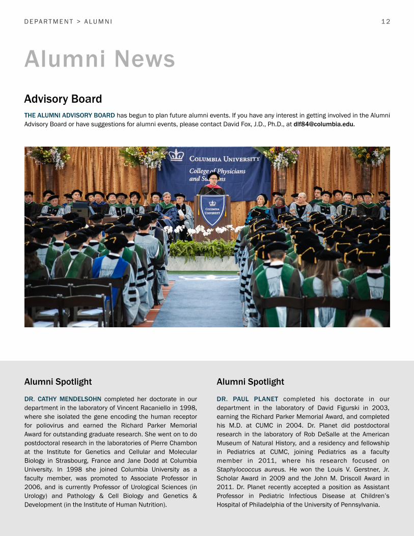
Advisory BoardTHE ALUMNI ADVISORY BOARD has begun to plan future alumni events. If you have any interest in getting involved in the Alumni Advisory Board or have suggestions for alumni events, please contact David Fox, J.D., Ph.D., at [email protected].
Alumni News
D E PA R T M E N T > A L U M N I
Alumni Spotlight Alumni Spotlight
DR. CATHY MENDELSOHN completed her doctorate in our department in the laboratory of Vincent Racaniello in 1998, where she isolated the gene encoding the human receptor for poliovirus and earned the Richard Parker Memorial Award for outstanding graduate research. She went on to do postdoctoral research in the laboratories of Pierre Chambon at the Institute for Genetics and Cellular and Molecular Biology in Strasbourg, France and Jane Dodd at Columbia University. In 1998 she joined Columbia University as a faculty member, was promoted to Associate Professor in 2006, and is currently Professor of Urological Sciences (in Urology) and Pathology & Cell Biology and Genetics & Development (in the Institute of Human Nutrition).
DR. PAUL PLANET completed his doctorate in our department in the laboratory of David Figurski in 2003, earning the Richard Parker Memorial Award, and completed his M.D. at CUMC in 2004. Dr. Planet did postdoctoral research in the laboratory of Rob DeSalle at the American Museum of Natural History, and a residency and fellowship in Pediatrics at CUMC, joining Pediatrics as a faculty member in 2011, where his research focused on Staphylococcus aureus. He won the Louis V. Gerstner, Jr. Scholar Award in 2009 and the John M. Driscoll Award in 2011. Dr. Planet recently accepted a position as Assistant Professor in Pediatric Infectious Disease at Children’s Hospital of Philadelphia of the University of Pennsylvania.

“Rest assured, if the infection does not cure the tumor, Dr. Saw!sh has a most cunning procedure in mind.”

F E AT U R E S > I M M U N OT H E R A P Y 14
IMMUNOTHERAPY, the modulation of the host immune system to treat disease, has become a focus of immunology and cancer research. However, the potential of immunotherapy to cause cancer remission was actually recognized over a century ago – although early experiments in the field seem rather brutal and born of desperation when viewed through a modern lens. In contrast with these early works, the recent successes of immunotherapy in extending the lifespan and quality of life in a subset of cancer patients clearly demonstrate the great progress that has been made, and the potential of immunotherapy as a safe, targeted cancer treatment.
Examples of doctors noticing that concurrent infections could cause tumor remission can be traced back as far as 1813, when it was noticed that gas gangrene (an effect of Clostridial infection) could cause tumor regression (1). By the time a review was published in 1904, a limited collection of reports indicated that influenza, typhoid, streptococcus infection, and sepsis could cause remission in patients with leukemia. Tuberculosis may also have caused some remission, although the reviewer noted that “it is difficult to understand the author’s statement that the ‘leukemia improved while the phthisis made great progress,’ particularly as the patient died” (2).
In spite of this limitation, several attempts to generate therapies from these observations were made. Dr. William B. Coley began using sterilized bacterial cultures to treat inoperable tumors in 1891, and introduced Coley’s toxins as a treatment in the following year. Over the course of his studies, an optimistic assessment found that these treatments may have caused complete tumor regression in up to 190 (out of 312) inoperable cases of cancer. Unfortunately, the effects were unpredictable, and over 11 different formulations were used with varying results. Several collaborators were involved, and it may be of some interest that a Dr. Alexander Lambert, of the College of Physicians and Surgeons, contributed to one of the formulations. In the end, although entirely consistent results could not be obtained, it was noted that, the stronger the response to the toxins, the better the end result. Undesirable side effects including fever, nausea, vertigo, sweating of the palms, and “fugacious eruptions” were all to be expected (3).
From these observations, it would appear possible that the host immune system, rather than infection itself, was responsible for these remissions. Paul Erlich, who shared the 1908 Nobel Prize in Physiology or Medicine, was one of the first to fully articulate a theory of immune surveillance in 1909. Together with other observations (including an oft-cited case study of the remission of an Italian woman’s cervical cancer after “fortuitous inoculation with rabies virus” in 1912 (4)), these studies led to many further observations and experiments with infection.
For instance, in the 1940s, it was noted that a woman with malignant melanoma had a remission for 5.5 years, which was attributed to receiving 14 injections of rabies vaccine because of another (fortuitous) dog bite (5). Through the 1940s and 50s, people continued to observe that concurrent infections could sometimes, but not always, result in leukemia remission; infections of staphylococcus, streptococcus, and chicken pox were noted to have this effect (6). Glandular fever (mononucleosis) was also found to cause temporary remission during infection of leukemic patients (7). Malaria infection might also have been of temporary benefit. It was even observed that cancers were rarer amongst Native Americans (who were presumed to suffer from higher infection rates) and German beekeepers (who had constant reactions to bee stings) (8).
These observations led to several renewed attempts at experimental intervention, along the lines of Coley’s earlier work. Attempts to use rabies vaccine as a treatment led to a partial reduction in tumor size, but not complete cancer remission (5), while injection of attenuated smallpox virus could seemingly prolong the life of melanoma patients (4). Other trials involved transfer of “pooled glandular-fever serum from… recent cases of acute glandular fever” to leukemic patients (7) and injection of a “testicular extract” (8). The effects of these treatments were understandably inconclusive. In one memorable study, feline panleukopenia virus was injected into leukemic patients. “The inoculum… was a 20 per cent saline suspension of minced spleen tissue obtained under sterile precautions from infected rural kittens.” The authors at the time were unable to conclude whether viral replication was involved, as “an unpredictable shortage of rural kittens prevented further virus-recovery studies” (6).
F E AT U R E A R T I C L E
Immunotherapy: Past and Future

F E AT U R E S > I M M U N OT H E R A P Y 1 5
By the 1950s and 1960s, studies were explicitly discussing possible immunological bases for the (rare) successes in cancer therapies. As one author hypothesized in the 1950s, “infections or their toxins may activate, or mobilize, various tissues or systems in the body which may be less active than normal in these patients” (8). In another study from the 1960s, smallpox vaccine was used to cause remission of a cutaneous tumor. The authors specifically looked for “antibodies that could be cytotoxic to the melanoma cells... Obviously, if a method for regularly producing an autoimmune reaction against pigment-producing cells could be developed, some of the patients with otherwise hopelessly disseminated melanoma might be salvageable or their lives might be significantly prolonged” (9). Similarly, when a serum transfer between a regressive and a non-regressive cancer patient performed in the mid-50s had some effect, “the unusual reactions in these 2 patients suggested to us possible immunological factors involved in the regression” (10).
These works clearly established that immunotherapy was considered to have some cancer treatment potential. However, it was unclear whether viral infection of the tumor itself, cytokines produced as a result of infection such as tumor necrosis factor (TNF), or a triggering of adaptive immune responses ultimately caused these variable effects. As it was realized that the limited technical ability of the time would prevent researchers from further testing these hypothesis, research into immunotherapy became dormant for a time.
By the 1970s, however, several important advances in technology began to be made. In the 70s, it was discovered that the Bacillus Calmette–Guérin (BCG) vaccine against tuberculosis could be directly injected into human tumors as a form of immunotherapy (11). The vaccine appears to elicit an immune response against antigens in the tumor cells, causing some remission in patients. BCG instillations continue to be used to treat non-invasive bladder cancer (12).
In the late 70s and 80s, advancements in hybridoma technologies allowed monoclonal antibodies to be produced, leading to a turning point in immunotherapy development. One former faculty member in our department, Sherie
Morrison, contributed to progress on this front. In work done at Columbia and at Stanford University, Morrison co-invented a system to produce antibodies with both mouse and human components. These chimeric antibodies proved to be more efficacious for some uses in humans, and this technology contributed to the development of products such as infliximab (Remicade), an antibody against TNF-a used to treat Crohn’s disease, rheumatoid arthritis, and other autoimmune conditions.
Several effective, targeted cancer treatments rely on the foundation provided by monoclonal antibody technology. Rituximab (Rituxan/MabThera/Zytux), for instance, is a chimeric monoclonal antibody designed to target the CD20 B-cell antigen. It can be used to treat non-Hodgkin’s lymphoma and chronic lymphocytic leukemia, causing clearance of the cancer cells (13). Trastuzumab (Herceptin) is a monoclonal antibody that blocks signaling through HER2 (Human Epidermal Growth Factor Receptor). This prevents growth signaling in the tumor, and is used in the treatment of breast cancer. Bevacizumab (Avastin), another monoclonal antibody, blocks VEGF (Vascular Endothelial Growth Factor) signaling, preventing the growth of new blood vessels that support tumor growth. Cetuximab (Erbitux), a chimeric monoclonal, blocks growth signaling through EGFR (Epidermal Growth Factor Receptor), and is used in the treatment of colorectal cancer, some lung cancers, and head and neck cancer (14).
Interestingly, while these antibodies target the cancer cells directly or aim to block growth signaling, thereby preventing tumor growth, several also seem to have effects on immunity. Trastuzumab increases the activity of cytotoxic T lymphocytes against tumor cells, and boosts natural killer cell function. Cetuximab also seems to aid in the priming of cytotoxic lymphocytes, while bevacizumab affects dendritic cell maturation and subsequent cytotoxic lymphocyte priming (14).
Because of these observations, trials are underway to enhance the immunogenic functions of these antibodies with other immunotherapies. It is hoped that this may cause a synergistic effect on tumor depletion, and may reduce the ability of tumors to escape drug targeting (which may result from mutation and selection for cells that do not express the

F E AT U R E S > I M M U N OT H E R A P Y 16
proteins targeted by the antibody) (14). For instance, animal experiments have been performed using an agonistic CD137 antibody in combination with trastuzumab treatment. CD137, a co-stimulatory molecule, is expressed on natural killer cells. Because trastuzumab partially functions through natural killer cell function, it was hypothesized that the combination of antibodies would exert a greater effect than either treatment alone. This indeed turned out to be the case in an animal tumor model. Furthermore, specificity for HER2-expresssing tumors was still observed, in spite of the addition of the CD137 treatment, suggesting that combinatorial therapies might also be useful to limit potential side effects of treatment (15, 16).
Monoclonal antibodies have also allowed for the generation of what may be, to date, some of the most exciting and effective immunotherapies: those that focus on the blockade of immune checkpoints. The immune system has a variety of checks and balances to allow it to fight infection without causing excessive damage to the host. These “checkpoints” prevent damage of normal self-tissues that may be distant from the site of infection, as well as bystander cells that may be present at an infection (or tumor) site but are unrelated to the pathogen. The prevention of auto-immunity is accomplished through a variety of means, and can include signals extrinsic to immune effector cells (such as release of the immunosuppressive cytokine IL-10), extrinsic cell types
!"#$%&"'()&*&"#$"%+",-#./0)-1&22*
3-&44&1#0)
56'7--56'87
!"#$%&"'()&*&"#$"%+",-#./0)-1&22*
3-&44&1#0)
96:;96;<=;>
96;<=;>
!"#$'938!? !"#$'56'87
@.(()&**$0"
3-)&%.2+#0)A
!"#$'#./0)-)&*(0"*&A
B
C
D
E
F
G
3./0)-&"B$)0"/&"# 3./0)-&"B$)0"/&"#-,.)$"%1C&1D(0$"#-E201D+,&-#C&)+(A
!"#$%&"'()&*&"#$"%+",-#./0)-1&22*
56'7-
96;<=;>938!?
938!?
56'87

F E AT U R E S > I M M U N OT H E R A P Y 17
(such as regulatory T-cells, which suppress the activity of effector cells), as well as mechanisms that are intrinsic to the immune effector cells (such as the upregulation of inhibitors of immune activation within an effector cell). Unfortunately, tumors often take advantage of these checkpoints, and use them create an immunosuppressive environment that prevents host recognition and clearance of the tumor. Therefore, inhibition of some of these checkpoints may remove some of the “brakes” on immunity and increase host immune function to the point that the tumors are once again targeted for destruction. Two important checkpoints that have now been targeted for inhibition are the Cytotoxic T-Lymphocyte-Associated protein 4 (CTLA4) and Programmed cell Death protein 1 (PD-1) (17).
CTLA4 is important in limiting effector T-cell function. Effector T-cells require binding of both their T-cell receptor with a cognate MHC molecule and CD28 with CD80/CD86 (on antigen presenting cells) to be fully licensed to function. CTLA4 competes with CD28 for binding to CD80/CD86, thus limiting T-cell activation. Because CTLA4 is upregulated on effector cells after their activation, it is particularly important in limiting co-stimulation at later time points during the effector response, and functions to prevent prolonged, destructive activation of these cells. CTLA4 is also strongly expressed on regulatory T-cells, and enhances their suppressive effects. The importance of this checkpoint is demonstrated most clearly by the fact that CTLA4 knockout mice die of lethal autoimmunity (17).
Ipilimumab (Yervoy) is a monoclonal antibody that blocks CTLA4 function, stopping its inhibitory effects. In trials, ipilimumab treatment led to long-term survival (beyond 2 years) in a subset (~18%) of melanoma patients. The startling, long-term remissions in some patient subsets led to great excitement; ipilimumab has now been approved by the FDA for melanoma treatment, and is in trial for use in several other cancers. However, why only a subset of patients respond so well to treatment remains a subject of investigation (17).
PD-1 operates in a similar fashion in order to prevent damage to peripheral tissues during an immune response. PD-1 is also expressed on effector T-cells after activation, and is highly expressed on regulatory T-cells. Its ligands, PD-
L1 and PD-L2, can be expressed on tumor cells, preventing effectors from acting on tumors. Two antibodies, pembrolizumab (Keytruda) and nivolumab (Opdivo), have been made to target PDL1 and prevent tumors from taking advantage of this checkpoint pathway. Trials with these inhibitors had similar, if not more striking, results than seen with ipilimumab, and may have fewer side effects. These antibodies were FDA-approved to treat melanoma, and approval has recently been extended for the treatment of non-small cell lung cancer (17-19).
Other approaches to immunotherapy are also being researched, and show promise. Work on adjuvants and cancer vaccines, pioneered by research including the prior BCG studies, continues. Flagellin was recently found to work well as an adjuvant in animal studies, and may aid attempts to create tumor vaccines (20). Meanwhile, the development of new vaccines, such as the Gardasi l human papipollomavirus vaccine, can of course prevent cancers that are linked directly to viral infection.
An increased understanding of basic immunological responses has also enabled the use of recombinant cytokines to enhance desired host immunity. A TNF fusion protein has been created which directs the protein to sites in tumor-associated vasculature. In animal models, the resulting increase in TNF activity caused by administration of the fusion protein increases effector T-cell infiltration and anti-tumor immunity (21). Administration of the cytokine IL-12 appears to enhance the effects of trastuzumab (14), while the cytokine interferon alpha has been approved by the FDA to treat hairy cell leukemia, chronic myelogenous leukemia (CML), follicular non-Hodgkin lymphoma (NHL), melanoma, and AIDS-related Kaposi’s sarcoma (22).
Other experiments with T-cells show that the cells that infiltrate into a tumor can be removed, isolated, and expanded in vitro. Because the cells are expanded outside of the immunosuppressive environment of the tumor, they seem to have improved efficacy in attacking tumors when re-injected into a patient (23). New advances in genetic engineering also make it possible for the specificity of immune cells to be edited in vitro, possibly making them better at attacking specific types of cancer (24).

F E AT U R E S > I M M U N OT H E R A P Y 1 8
Similarly, the viral work that received so much attention in the past is now coming to fruition. On October 27, the FDA approved talimogene laherparepvec (T-VEC), a genetically engineered virus, for use in melanoma treatment. The virus, a modified herpes virus, cannot replicate in healthy cells. In cancer cells, however, the virus causes a lytic infection, destroying the cell, while also causing the release of immunogenic proteins and antigens. Thus, the virus is designed to not only destroy the tumor cells directly, but also to stimulate an immune response against them (25).
Many of the therapies undergoing testing (including the PD-1 and CTLA4-based therapies) show efficacy in only a subset
of the treated patients. The exact reasons for this are unclear, but it is hoped that combining these drugs with other cancer treatments may improve results. Many treatments can also cause systemic effects on immunity, increasing the likelihood that patients will suffer from autoimmune conditions during treatment. This is a challenge that was faced even in the initial studies of infection performed half a century ago, highlighting the challenges in determining the optimal combinations and dosing of treatment. Nevertheless, immunotherapy has progressed greatly since the days of Coley’s toxins, and may provide new efficacious options for cancer therapy.
References
1. Weber, G.F. (2015) Molecular Therapies of Cancer 11: 333-351.2. Dock, G.A.M. (1904) The influence of complicating diseases upon leukaemia. American Journal of the Medical Sciences 127: 563-592.3. Nauts, S.W., Swift, H.C. and Coley, B.L. (1946) The treatment of malignant tumors by bacterial toxins as developed by the late William B. Coley, M.D., reviewed in the light of modern research. Cancer Research 6: 205-216.4. Milton, G.W. and Brown, M.M. (1966) The limited role of attenuated smallpox virus in the management of advanced malignant melanoma. Aust. N.Z. J. Surg. 35: 286-290.5. Pack, G.T. (1950) Note on the experimental use of rabies vaccine for melanomatosis. A.M.A. Archives of Dermatology and Syphilology 62: 694-695.6. Bierman, H.R. et al., (1953) Remissions in leukemia of childhood following acute infectious disease: staphylococcus and streptococcus, varicella, and feline panleukopenia. Cancer 6: 591-605.7. Taylor, A.W. (1953) Effects of glandular fever infection in acute leukaemia. Br. Med. J. 1: 589-593.8. Pelner, L. (1958) Effect of concurrent infections and their toxins on the course of leukemia. Acta Medica Scandinavica 162: 5-24.9. Burdick, K.H. and Hawk, W.A. (1964). Vitiligo in a case of Vaccinia virus-treated melanoma. Cancer 17: 708-712.10. Sumner, W. C. and Foraker, A. G. (1960) Spontaneous regression of human melanoma: clinical and experimental studies. Cancer 13: 79-81.11. Morton, D.L., et al. (1970) Immunological factors in human sarcomas and melanomas: a rational basis for immunotherapy. Ann. Surg. 172: 740-749.12. N. C. Institute (2015) Bladder cancer treatment for health professionals (PDQ®).13. Pazdur, R. (2013). FDA approval for rituximab. FDA.14. Vanneman, M. and Dranoff, G. (2012) Combining immunotherapy and targeted therapies in cancer treatment. Nat. Rev. Cancer 12: 237-251.15. Bird, L. (2012) Immunotherapy: A killer combination. Nature Reviews Immunology 12: 231.16. Seton-Rogers, S. (2012) Immunotherapy: Combinations that work. Nature Reviews Cancer 12: 231.17. Pardoll, D.M. (2012) The blockade of immune checkpoints in cancer immunotherapy. Nature Reviews Cancer 12: 252-264.18. U.S.F.D.A. (2015) FDA approves Keytruda for advanced non-small cell lung cancer.19. U.S.F.D.A. (2015) FDA expands approved use of Opdivo in advanced lung cancer.20. Leavy, O. (2012) Tumour immunology: A close-range dual hit for tumour immunity. Nat. Rev. Immunol. 12: 227. 21. Bordon, Y. (2012) Tumour immunology: Hope in a sticky situation. Nature Reviews Immunology 12: 213.22. Schering Corporation, Inc. (2014) Medication Guide, Intron® A (Interferon alfa-2b, recombinant).23. Restifo, N.P., Dudley, M.E., Rosenberg, S. A. (2012) Adoptive immunotherapy for cancer: harnessing the T cell response. Nature Reviews Immunology 12: 269-281.24. Pollack, A. (2015) A cell therapy untested in humans saves a baby with cancer. The New York Times, November 5, 2015.25. Ledford, H. (2015) First cancer-fighting virus approved. Nature 526: 622-623.


DR. HAROLD S. GINSBERG, former Chair of the Department of Microbiology & Immunology, became widely known as a groundbreaking and pioneering virologist best known for his work with adenoviruses. In retrospect, he demonstrated the same keen insight that led to his later successes at the very start of his career.
Early CareerHarold Ginsberg was born on May 27, 1917 in Daytona Beach, FL. He graduated with an A.B. from Duke University in 1937, and an M.D. from Tulane University in 1941. He
served in the military as a medical officer during World War II starting in 1943, posted in the United Kingdom. Even as a young physician, Dr. Ginsberg noticed a high incidence of hepatitis in soldiers that had received blood transfusions, investigated, and concluded that the pooled blood plasma being used to treat wounded soldiers was causing hepatitis. His early work resulted in major changes in how blood plasma was used, saving numerous lives, and led to the eventual discovery of the Hepatitis B virus. In 1945, he was awarded the Legion of Merit Award by the U.S. Army for his work, and attained the rank of Lieutenant Colonel by the time he left the military in 1946.
Entering AcademiaAfter the war ended, Dr. Ginsberg entered academia as an resident Physician Associate at the Rockefeller Institute in New York City in 1946, where he performed research with Dr. Frank Horsfall, developing techniques to block viral infection and replication with chemotherapy. In 1951, Dr. Ginsberg joined the faculty at Case Western Reserve in Cleveland as an Associate Professor of Preventive Medicine. While in the military, he had developed an interest in Acute Respiratory Disease (ARD), which at the time was a disease of unknown origin, and he began to investigate the cause of ARD at Case Western Reserve. There, he demonstrated that
adenoviruses, normally found in the adenoid, lymph tissue at the back of the nose, were responsible for ARD, as well as atypical pneumonia and phyarngitis. He showed that the viruses could remain dormant, then become infectious, and described how the viruses invaded host cells and caused disease. This was groundbreaking work, as described by Dr. Saul Silverstein, former Chair of Microbiology & Immunology at Columbia University: “He was one of the founding fathers of modern virology and microbiology. His discoveries involving the genetics of adenoviruses paved the way for the development of gene therapy.”
University of PennsylvaniaIn 1961, Dr. Ginsberg joined the University of Pennsylvania as Chair of the Department of Microbiology. There his research focus shifted to understanding the structural components of adenoviruses. Dr. Ginsberg's laboratory isolated a number of temperature-sensitive mutant adenoviruses which not only played a critical role in his subsequent functional characterization of viral capsid proteins, but proved extremely useful to other adenovirus researchers. His laboratory also isolated individual late viral gene products, and made significant contributions to the understanding of viral DNA packaging and host cell changes during viral infections.
Columbia UniversityIn 1973, Dr. Ginsberg accepted the position of Chair and Higgins Professor of the Department of Microbiology at Columbia University, replacing Dr. Harry M. Rose, who had retired. The department blossomed under Dr. Ginsberg's leadership, and he was able to recruit both leading virologists, including Dr. Max Gottesman, Dr. Hamish Young and Dr. Saul Silverstein, and leading immunologists such as Dr. Sherie Morrison and Dr. Kathryn Calame to the department. He led the department by example and praise, and according to his colleagues at the time, inspired the
F E AT U R E S > H A R O L D S . G I N S B E R G 2 0
“ He was one of the founding fathers of modern virology and microbiology. His discoveries involving the genetics of adenoviruses paved the way for the development of gene therapy.”
H I S T O R I C A L F E AT U R E
Harold S. Ginsberg: The Quiet Pioneer

F E AT U R E S > H A R O L D S . G I N S B E R G 21
department with true esprit de corps. His former colleague, Dr. Hamish Young, described Dr. Ginsberg as very department oriented, with a keen interest in the lives of the department’s faculty, students and postdocs, and great commitment to the development of students and postdocs. He was very involved with his own students, and always had an idea for another experiment ready for them. On a personal level, Hamish noted that Dr. Ginsberg was a “fiendish tennis player”, which is consistent with his early athleticism – while a student at Duke University, he earned varsity letters in swimming, tennis and football, and only gave up playing football when it began to conflict with his laboratory courses. As Chair of the Department of Microbiology, former colleagues described Dr. Ginsberg’s style as low-key, but effective. Dr. Ginsberg was apolitical, exhibited a scrupulous fairness in his dealings, and was respected by everyone.
Dr. Ginsberg was deeply interested in knowledge for the sake of knowledge, as noted by his former colleague Dr. Dickson Despommier, “He had a true appreciation for the breadth and depth of how life expresses itself.” While a member of the Department of Microbiology, Dr. Ginsberg continued his research on adenovirus, making a number of important contributions to our understanding of adenovirus genetics, biology and tumor transformation, and developed a variety of useful conditionally lethal adenovirus mutants. In addition, he authored two standard textbooks, Microbiology and Virology, which were widely used by medical and graduate students at the time, and served as editor in chief of the Journal of Virology. During his tenure with the department he also served as Chair of Board of Governors of the American Academy of Microbiology, Chair of the Microbiology Committee of the National Board of Medical Examiners, Chair of the Virology study section of the NIH and Chair of the Microbiology and Immunology Advisory Council of the NIH. Dr. Ginsberg was elected to the Institute of Medicine in 1979 and the National Academy of Sciences in 1982. Dr. Ginsberg was always modest about his pioneering work and numerous achievements, but despite his quiet demeanor, they spoke for themselves.
RetirementIn 1985, Dr. Ginsberg stepped down as Chair of Microbiology in preparation for retirement, and after 15 years of service to the department, retired and become Professor Emeritus in 1988. Even in retirement, he continued to do research with Dr. Robert Chanock at NIH, further exploring the roots of adenovirus pathogenesis and developing systems for the study of adenovirus. The last of Dr. Ginsberg’s 200 publications was in 1993, on a new model system for adenovirus pneumonia. He passed away on February 2, 2003 at Woods Hole at the age of 85, survived by his wife, Marion Reibstein Ginsberg, an attorney and 1949 graduate of Columbia Law School, and their four children. To the very end, Dr. Ginsberg was a scholar and a gentleman.

Pefanis, E., Wang, J., Rothschild, G., Lim, J., Chao, J., Rabadan, R., Economides, A.N. and Basu, U. (2014) Noncoding RNA transcription targets AID to divergently transcribed loci in B cells. Nature 514: 389-393.
Chao, J., Rothschild, G. and Basu, U. (2014) Ubiquitination events that regulate recombination of immunoglobulin Loci gene segments. Front. Immunol. 5: 100.
Haupt, A., Campetelli, A., Bonazzi, D., Piel, M., Chang, F. and Minc, N. (2014) Electrochemical regulation of budding yeast polarity. PLoS Biol. 12: e1002029.
Chang, F. and Huang, K.C. (2014) How and why cells grow as rods. B.M.C. Biology 2: 54.
Chang, F. and Minc, N. (2014) Electrochemical control of cell and tissue polarity. Annu. Rev. Cell Dev. Biol. 30: 317-336.
Basu, R., Munteanu, E.L. and Chang, F. (2014) Role of turgor pressure in endocytosis in fission yeast. Mol. Biol. Cell 25: 679-687. PMCID: PMC3937093
Pan, K., Saunders, T.E., Flor-Parra, I., Howard, M. and Chang, F. (2014) Cortical regulation of cell size by a sizer Cdr2. ELife 24642412. PMCID: PMC3956294
Chang, F., Atilgan, E., Burgess, D. and Minc, N. (2014) Manipulating cell shape by placing cells into micro-fabricated chambers. Methods Mol. Biol. 1136: 281-290.
Schneider, C., Setty, M., Holmes, A.B., Maute, R.L., Leslie, C.S., Mussolin, L., Rosolen, A., Dalla-Favera, R. and Basso, K. (2014) MicroRNA 28 controls cell proliferation and is down-regulated in B-cell lymphomas. Proc. Natl. Acad. Sci. U.S.A. 111: 8185-8190.
Pasqualucci, L., Khiabanian, H., Fangazio, M., Vasishtha, M., Messina, M., Holmes, A.B., Ouillette, P., Trifonov, V., Rossi, D., Tabbo, F., Ponzoni, M., Chadburn, A., Murty, V.V., Bhagat, G., Gaidano, G., Inghirami, G., Malek, S.N., Rabadan, R. and Dalla-Favera, R. (2014) Genetics of follicular lymphoma transformation. Cell Rep. 6: 130-140.
Trimarchi, T., Bilal, E., Ntziachristos, P., Fabbri, G., Dalla-Favera, R., Tsirigos, A. and Aifantis, I. (2014) Genome-wide mapping and characterization of Notch-regulated long noncoding RNAs in acute leukemia. Cell 158: 593-606.
Messina, M., Del Giudice, I., Khiabanian, H., Rossi, D., Chiaretti, S., Rasi, S., Spina, V., Holmes, A.B., Marinelli, M., Fabbri, G., Piciocchi, A., Mauro, F.R., Guarini, A., Gaidano, G.,
Dalla-Favera, R., Pasqualucci, L., Rabadan, R. and Foa, R. (2014) Genetic lesions associated with chronic lymphocytic leukemia chemo-refractoriness. Blood 123: 2378-2388.
Pasqualucci, L. and Dalla-Favera, R. (2014) SnapShot: diffuse large B cell lymphoma. Cancer Cell 25: 132-132 e131.
Burberry, A., Zeng, M.Y., Ding, L., Wicks, I., Inohara, N., Morrison, S.J. and Nunez, G. (2014) Infection mobilizes hematopoietic stem cells through cooperative NOD-like receptor and Toll-like receptor signaling. Cell Host Microbe 15: 779-791.
Chaix, J., Nish, S.A., Lin, W.H., Rothman, N.J., Ding, L., Wherry, E.J. and Reiner, S.L. (2014) Cutting edge: CXCR4 is critical for CD8+ memory T cell homeostatic self-renewal but not rechallenge self-renewal. J. Immunol. 193: 1013-1016.
Dworkin, J. (2014) The medium is the message: interspecies and interkingdom signaling by peptidoglycan and related bacterial glycans. Annu. Rev. Microbiol. 68: 137-154.
Dworkin, J. (2014) Protein targeting during sporulation. In The Bacterial Spore: From Molecules to Systems, Driks, A. and Eichenberger, P., Eds. ASM Press.
Brestoff, J.R., Kim, B.S., Saenz, S.A., Stine, R.R., Monticelli, L.A., Sonnenberg, G.A., Thome, J.J.T., Farber, D.L., Lutfy,K., Seale, P., and Artis, D. (2014) Group 2 innate lymphoid cells promote beiging of white adipose tissue and limit diet-induced obesity. Nature doi: 10.1038/nature14115. Epub 2014 Dec 22.
Thome, J.J.C., Yudanin, N.A., Ohmura, Y., Kubota, M., Grinshpun, B., Sathaliyawala, T., Kato, T., Lerner, H., Shen, Y. and Farber, D.L. (2014) Spatial map of human T cell compartmentalization and maintenance over decades of life. Cell 159: 814-828.
Farber, D.L., Yudanin, N.A. and Restifo, N.P. (2014) Human memory T cells: generation, compartmentalization and homeostasis. Nature Rev. Immunol. 14: 24-35.
Camirand, G., Wang, Y., Lu, Y., Wan, Y.Y., Lin, Y., Deng, S., Guz, G., Perkins, D.L., Finn, P.W., Farber, D.L., Flavell, R.A., Shlomchik, W.D., Lakkis, F.G., Rudd, C.E. and Rothstein, D.M. (2014) CD45 ligation expands Tregs by promoting interactions with DCs. J. Clin. Invest. 124: 4603-4613.
N OT E S > P U B L I C AT I O N S
N O T E S
2014 Faculty Publications
2 2

2 3
Turner, D.L. and Farber, D.L. (2014) Mucosal resident memory CD4 T cells in protection and immunopathology. Front. Immunol. 5: 331.
Turner, D.L., Gordon, C.L. and Farber, D.L. (2014) Tissue-resident T cells, in situ immunity and transplantation. Immunol. Rev. 258: 150-166.
Subramanian, M., Hayes, C.D., Thome, J.J., Thorp, E., Matsushima, G.K., Herz, J., Farber, D.L., Liu, K., Lakshmana, M. and Tabas, I. (2014) An AXL/LRP-1/RANBP9 complex mediates DC efferocytosis and antigen cross-presentation in vivo. J. Clin. Invest. 124: 1296-1308.
Lee, M.C. and Fidock, D.A. (2014) CRISPR-mediated genome editing of Plasmodium falciparum malaria parasites. Genome Med. 6: 63.
Lee, A.H., Symington, L.S. and Fidock, D.A. (2014) DNA repair mechanisms and their biological roles in the malaria parasite Plasmodium falciparum. Microbiol. Mol. Biol. Rev. 78: 469-486.
Lisewski, A.M., Quiros, J.P., Ng, C.L., Adikesavan, A.K., Miura, K., Putluri, N., Eastman, R.T., Scanfeld, D., Regenbogen, S.J., Altenhofen, L., Llinas, M., Sreekumar, A., Long, C., Fidock, D.A. and Lichtarge, O. (2014) Supergenomic network compression and the discovery of EXP1 as a glutathione transferase inhibited by artesunate. Cell 158: 916-928.
Johnston, G.L., Gething, P.W., Hay, S.I., Smith, D.L. and Fidock, D.A. (2014) Modeling within-host effects of drugs on Plasmodium falciparum transmission and prospects for malaria elimination. PLoS Comput. Biol. 10: e1003434.
Jimenez-Diaz, M.B., Ebert, D., Salinas, Y., Pradhan, A., Lehane, A.M., Myrand-Lapierre, M.E., O'Loughlin, K.G., Shackleford, D.M., Justino de Almeida, M., Carrillo, A.K., Clark, J.A., Dennis, A.S., Diep, J., Deng, X., Duffy, S., Endsley, A.N., Fedewa, G., Guiguemde, W.A., Gomez, M.G., Holbrook, G., Horst, J., Kim, C.C., Liu, J., Lee, M.C., Matheny, A., Martinez, M.S., Miller, G., Rodriguez-Alejandre, A., Sanz, L., Sigal, M., Spillman, N.J., Stein, P.D., Wang, Z., Zhu, F., Waterson, D., Knapp, S., Shelat, A., Avery, V.M., Fidock, D.A., Gamo, F.J., Charman, S.A., Mirsalis, J.C., Ma, H., Ferrer, S., Kirk, K., Angulo-Barturen, I., Kyle, D.E., DeRisi, J.L., Floyd, D.M. and Guy, R.K. (2014) (+)-SJ733, a clinical candidate for malaria that acts through ATP4 to induce rapid host-mediated clearance of Plasmodium. Proc. Natl. Acad. Sci. U.S.A. 111: E5455-5462.
Kuhen, K.L., Chatterjee, A.K., Rottmann, M., Gagaring, K., Borboa, R., Buenviaje, J., Chen, Z., Francek, C., Wu, T., Nagle,
A., Barnes, S.W., Plouffe, D., Lee, M.C., Fidock, D.A., Graumans, W., van de Vegte-Bolmer, M., van Gemert, G.J., Wirjanata, G., Sebayang, B., Marfurt, J., Russell, B., Suwanarusk, R., Price, R.N., Nosten, F., Tungtaeng, A., Gettayacamin, M., Sattabongkot, J., Taylor, J., Walker, J.R., Tully, D., Patra, K.P., Flannery, E.L., Vinetz, J.M., Renia, L., Sauerwein, R.W., Winzeler, E.A., Glynne, R.J. and Diagana, T.T. (2014) KAF156 is an antimalarial clinical candidate with potential for use in prophylaxis, treatment, and prevention of disease transmission. Antimicrob. Agents Chemother. 58: 5060-5067.
Prestia, K., Bandyopadhyay, S., Slate, A., Francis, R.O., Francis, K.P., Spitalnik, S.L., Fidock, D.A., Brittenham, G.M. and Hod, E.A. (2014) Transfusion of stored blood impairs host defenses against Gram-negative pathogens in mice. Transfusion 54: 2842-2851.
Kumpornsin, K., Modchang, C., Heinberg, A., Ekland, E.H., Jirawatcharadech, P., Chobson, P., Suwanakitti, N., C h a o t h e i n g , S . , W i l a i r a t , P. , D e i t s c h , K . W. , Kamchonwongpaisan, S., Fidock, D.A., Kirkman, L.A., Yuthavong, Y. and Chookajorn, T. (2014) Origin of robustness in generating drug-resistant malaria parasites. Mol. Biol. Evol. 31: 1649-1660.
Jayabalasingham, B., Voss, C., Ehrenman, K., Romano, J.D., Smith, M.E., Fidock, D.A., Bosch, J. and Coppens, I. (2014) Characterization of the ATG8-conjugation system in 2 Plasmodium species with special focus on the liver stage: possible linkage between the apicoplastic and autophagic systems? Autophagy 10: 269-284.
Henrich, P.P., O'Brien, C., Saenz, F.E., Cremers, S., Kyle, D.E. and Fidock, D.A. (2014) Evidence for pyronaridine as a highly effective partner drug for treatment of artemisinin-resistant malaria in a rodent model. Antimicrob. Agents Chemother. 58: 183-195.
Oeckinghaus, A., Postler, T.S., Rao, P., Schmitt, H., Schmitt, V., Grinberg-Bleyer, Y., Kuhn, L.I., Gruber, C.W., Lienhard, G.E. and Ghosh, S. (2014) kB-Ras proteins regulate both NF-kB-dependent inflammation and Ral-dependent proliferation. Cell Rep. 8: 1793-1807. doi: 10.1016/j.celrep.2014.08.015. Epub 2014 Sep 15.
Hayden, M.S. and Ghosh, S. (2014) Innate sense of purpose for IKKbeta. Proc. Natl. Acad. Sci. U.S.A. 111: 17348-17349.
Hayden, M.S. and Ghosh, S. (2014) Regulation of NF-kappaB by TNF family cytokines. Semin. Immunol. 26: 253-266.
N OT E S > P U B L I C AT I O N S
De, A., Dainichi, T., Rathinam, C.V. and Ghosh, S. (2014) The deubiquitinase activity of A20 is dispensable for NF-kappaB signaling. EMBO Rep. 15: 775-783.
Bhatt, D. and Ghosh, S. (2014) Regulation of the NF-kappaB-mediated transcription of inflammatory genes. Front. Immunol. 5: 71.
Seeley, J.J. and Ghosh, S. (2014) Tolerization of inflammatory gene expression. Cold Spring Harb Symp Quant Biol.
Kao, T.-H., Liao, H.-F., Wolf, D., Tai, K.-Y., Chuang, C.-Y., Lee, H.-S., Kuo, H.-C., Hata, K., Zhang, X., Cheng, X., Goff, S.P., Ooi, S.K., Bestor, T.H., and Lin, S.-P. (2014) Ectopic DNMT3L triggers assembly of a repressive complex for retroviral silencing in somatic cells. J. Virol. 88: 10680-10695.
Arriagada, G., Metzger, M.J., Muttray, A., Sherry, J., Reinisch, C., Street, C., Lipkin, W.I., and Goff, S.P. (2014) Activation of transcription and retrotransposition of a novel retroelement, Steamer, in neoplastic hemocytes of the mollusk Mya arenaria. Proc. Natl. Acad. Sci. U.S.A. 111: 14175-14180.
Schlesinger, S., Meshorer, E. and Goff, S.P. (2014) Asynchronous transcriptional silencing of individual retroviral genomes in embryonic cells. Retrovirology 11: e31.
Wang, G.Z., Wolf, D. and Goff, S.P. (2014) EBP1, a novel host factor involved in primer binding site-dependent restriction of moloney murine leukemia virus in embryonic cells. J. Virol. 88: 1825-1829.
Vitiello, C.L., Kireeva, M.L., Lubkowska, L., Kashlev, M. and Gottesman, M. (2014) Coliphage HK022 Nun protein inhibits RNA polymerase translocation. Proc. Natl. Acad. Sci. U.S.A. 111: e2368-2375. Epub 2014 May 22.
Vitiello, C.L. and Gottesman, M.E. (2014) Bacteriophage HK022 Nun protein arrests transcription by blocking lateral mobility of RNA polymerase during transcription elongation. Bacteriophage 4: e32187.
Zuchegna, C., Aceto, F., Bertoni, A., Romano, A., Perillo, B., Laccetti, P., Gottesman, M.E., Avvedimento, E.V. and Porcellini, A. (2014) Mechanism of retinoic acid-induced transcription: histone code, DNA oxidation and formation of chromatin loops. Nucleic Acids Res. 42: 11040-11055.
Morano, A., Angrisano, T., Russo, G., Landi, R., Pezone, A., Bartollino, S., Zuchegna, C., Babbio, F., Bonapace, I.M., Allen, B., Muller, M.T., Chiariotti, L., Gottesman, M.E., Porcellini, A. and Avvedimento, E.V. (2014) Targeted DNA methylation by
homology-directed repair in mammalian cells. Transcription reshapes methylation on the repaired gene. Nucleic Acids Res. 42: 804-821.
Han, Y.W. (2014) Oral bacteria as drivers for colorectal cancer. J. Periodontol. 85: 1155-1157.
Goto, Y., Obata, T., Kunisawa, J., Sato, S., Ivanov, I.I., Lamichhane, A., Takeyama, N., Kamioka, M., Sakamoto, M., Matsuki, T., Setoyama, H., Imaoka, A., Uematsu, S., Akira, S., Domino, S.E., Kulig, P., Becher, B., Renauld, J.C., Sasakawa, C., Umesaki, Y., Benno, Y. and Kiyono, H. (2014) Innate lymphoid cells regulate intestinal epithelial cell glycosylation. Science 345: 1254009.
Goto, Y., Panea, C., Nakato, G., Cebula, A., Lee, C., Diez, M.G., Laufer, T.M., Ignatowicz, L. and Ivanov, I.I. (2014) Segmented filamentous bacteria antigens presented by intestinal dendritic cells drive mucosal Th17 cell differentiation. Immunity 40: 1-14.
Heise, N., De Silva, N.S., Silva, K., Carette, A., Simonetti, G., Pasparakis, M. and Klein, U. (2014) Germinal center B-cell maintenance and differentiation are controlled by distinct NF-kappaB transcription factor subunits. J. Exp. Med. 211: 2103-2118.
Simonetti, G., Bertilaccio, M.T.S., Ghia, P. and Klein, U. (2014) Mouse models in the study of chronic lymphocytic leukemia pathogenesis and therapy. Blood 124: 1010-1019.
Klein, U. (2014) Programming plasma cell survival. J. Exp. Med. 211: 744. (Insights Article)
Subramanian, M., Hayes, C.D., Thome, J.J., Thorp, E., Matsushima, G.K., Herz, J., Farber, D.L., Liu, K., Lakshmana, M. and Tabas, I. (2014) An AXL/LRP-1/RANBP9 complex mediates DC efferocytosis and antigen cross-presentation in vivo. J. Clin. Invest. 124: 1296-1308.
Franklin, R.A., Liao, W., Sarkar, A., Kim, M.V., Bivona, M.R., Liu, K., Pamer, E.G. and Li, M.O. (2014) The cellular and molecular origin of tumor-associated macrophages. Science 344: 921-925.
Ghosh, H.S., Ceribelli, M., Matos, I., Lazarovici, A., Bussemaker, H.J., Lasorella, A., Hiebert, S.W., Liu, K., Staudt, L.M. and Reizis, B. (2014) ETO family protein Mtg16
N OT E S > P U B L I C AT I O N S 24

regulates the balance of dendritic cell subsets by repressing Id2. J. Exp. Med. 211: 1623-1635.
Schoggins, J.W., MacDuff, D.A., Imanaka, N., Gainey, M.D., Shrestha, B., Eitson, J.L., Mar, K.B., Richardson, R.B., Ratushny, A.V., Litvak, V., Dabelic, R., Manicassamy, B., Aitchison, J.D., Aderem, A., Elliott, R.M., Garcia-Sastre, A., Racaniello, V., Snijder, E.J., Yokoyama, W.M., Diamond, M.S., Virgin, H.W. and Rice, C.M. (2014) Pan-viral specificity of IFN-induced genes reveals new roles for cGAS in innate immunity. Nature 505: 691-695.
Schuler, B.A., Schreiber, M.T., Li, L., Mokry, M., Kingdon, M.L., Raugi, D.N., Smith, C., Hameister, C., Racaniello, V.R. and Hall, D.J. (2014) Major and minor group rhinoviruses elicit differential signaling and cytokine responses as a function of receptor-mediated signal transduction. PLoS One 9: e93897.
Reiner, S.L. and Adams, W.C. (2014) Lymphocyte fate specification as a deterministic but highly plastic process. Nature Rev. Immunol. 14: 699-704.
Chaix, J., Nish, S.A., Lin, W.H., Rothman, N.J., Ding, L., Wherry, E.J. and Reiner, S.L. (2014) Cutting edge: CXCR4 is critical for CD8+ memory T cell homeostatic self-renewal but not rechallenge self-renewal. J. Immunol. 193: 1013-1016.
Huang, S.X., Islam, M.N., O'Neill, J., Hu, Z., Yang, Y.-Y., Chen, Y.-W., Green, M.D., Mumau, M., Vunakc-Novakovic, G., Bhattacharya, J. and Snoeck, H.W. (2014) Highly efficient generation of lung and airway epithelial cells from human pluripotent stem cells. Nature Biotechnol. 32: 84-91.
Kamezaki, K. and Snoeck, H.W. (2014) Differential requirement of wild-type Flt3 in leukemic stem cells which is not reflected in vitro assays for leukemic stem cells. Exp. Hematol. 42: 192-203.
Baker, S.J., Ma'ayan, A., Lieu, Y.K., John, P., Reddy, M.V.R., Chen, E.Y., Duan, Q., Snoeck, H.W. and Reddy, E.P. (2014) B-myb is an essential regulator of hematopoietic stem cell and myeloid progenitor cell development. Proc. Natl. Acad. Sci. U.S.A. 111: 3122-3127.
Kalscheuer, H., Onoe, T., Dahmani, A., Holzl, M., Yamada, K. and Sykes, M. (2014). Xenograft tolerance and immune
function of human T cells developing in pig thymus xenografts. J. Immunol. 192: 3442-3450.
Griesemer, A., Yamada, K. and Sykes, M. (2014). Xenotransplantation: Immunological hurdles and progress toward tolerance. Immunol. Rev. 258: 241-258.
Haspot, F., Li, H.W., Lucas, C.L., Fehr, T., Beyaz, S. and Sykes, M. (2014) Allospecific rejection of MHC class I-deficient bone marrow by CD8 T cells. Am. J. Transplant. 14: 49-58.
Sykes, M. (2014) Transplantation: moving to the next level. Immunol. Rev. 258: 5-11.
Deng, S.K., Gibb, B., Almeida, M.J., Greene, E.C. and Symington, L.S. (2014) RPA antagonizes microhomology-mediated repair of DNA double-strand breaks. Nature Struct. Mol. Biol. 21: 405-412.
Eissler, C.L., Mazon, G., Powers, B.L., Savinov, S.N., Symington, L.S. and Hall, M.C. (2014) The Cdk/Cdc14 module controls activation of the Yen1 Holliday junction resolvase to promote genome stability. Mol. Cell 54: 80-93.
Stafa, A., Donnianni, R.A., Timashev, L.A., Lam, A.F. and Symington, L.S. (2014) Template switching during break-induced replication is promoted by the Mph1 helicase in Saccharomyces cerevisiae. Genetics 196: 1017-1028.
Symington, L.S., Rothstein, R. and Lisby, M. (2014) Mechanisms and regulation of mitotic recombination in Saccharomyces cerevisiae. Genetics 198: 795-835.
Symington, L.S. (2014) DNA repair: Making the cut. Nature 514: 39-40.
Lee, A.H., Symington, L.S. and Fidock, D.A. (2014) DNA repair mechanisms and their biological roles in the malaria parasite Plasmodium falciparum. Microbiol. Mol. Biol. Rev. 78: 469-486.
Stafa, A., Miklenic, M., Zunar, B., Lisnic, B., Symington, L.S. and Svetec, I.K. (2014) Sgs1 and Exo1 suppress targeted chromosome duplication during ends-in and ends-out gene targeting. DNA Repair (Amst). 22: 12-23.
Symington, L.S. (2014) End resection at double-strand breaks: mechanism and regulation. Cold Spring Harb. Perspect. Biol. 6.
N OT E S > P U B L I C AT I O N S 2 5

Basu LabUttiya Basu’s laboratory is interested in the developmental fate regulation of B-lymphocytes, a vital component of the adaptive immune system. Recent research from his laboratory has identified a key regulatory complex known as “RNA exosome” that promotes genomic alterations in the immunoglobulin loci such that high affinity antibodies can be generated via processes like class switch recombination and somatic hypermutation. The RNA exosome is an 11 subunit non-coding RNA degradation/processing complex whose role in various cellular function constitutes a current topic of investigation. Ongoing research is focused on probing the RNA exosome-dependent co-transcriptional regulation of non-coding RNA biogenesis in the immunoglobulin loci. These findings should lead to a better understanding of the mechanism of B cell development during adaptive immunity and initiation of oncogenesis.
Chang LabFred Chang’s laboratory studies fundamental mechanisms underlying cell morphogenesis, including cytokinesis, cell polarity, nuclear positioning and the regulation of actin and microtubules. The lab uses the rod-shaped fission yeast Schizosaccharomyces pombe as a model cell, as well as animal cell models. One of the questions that the lab has been trying to answer is how the site of division is positioned during cytokinesis. In fission yeast, the division site is determined by the position of the nucleus, through a process involving the peripheral membrane protein mid1p. The lab studies how mid1p is localized to a series of dots on the cortex near the nucleus, which then recruit other cytokinesis factors to assemble the contractile ring, a complex process that involves nuclear shuttling, the endoplasmic reticulum, and a cortical gradient of a protein kinase pom1p emanating from the cell tips. This system represents one of the best-understood examples of division site placement in any organism.
Dalla-Favera LabRiccardo Dalla-Favera’s laboratory is focused on three major goals: (1) identifying the lesions and genes involved in the development of human B cell lymphoma, (2) determining the mechanism by which these lesions occur and (3) elucidating the contribution of each lesion to tumor development. The lab identifies novel oncogenes and tumor suppressors involved in the pathogenesis of lymphoma by virtue of their involvement in tumor-associated chromosomal translocations or by “positional cloning” from chromosomal regions involved in tumor-associated deletions. They study the function of the BCL-6 gene, a recently identified proto-oncogene which codes for a transcription factor expressed in B cells and is altered in its regulatory region in a significant fraction of human lymphoma. Finally, they seek to elucidate the role of chromosomal translocations involving the c-myc proto oncogene locus and immuno-globulin loci in the development of Burkitt’s lymphoma.
Ding LabLei Ding’s laboratory studies hematopoietic stem cells (HSCs), which play critical roles in the generation, repair and homeostasis of the blood and immune system via self-renewal and multilineage differentiation. The lab is investigating extrinsic mechanisms that
regulate HSC self-renewal, and how mis-regulation of niche/HSC interactions contributes to diseases such as cancer and anemia. Understanding the HSC niche is a key step in helping design better strategies for in vitro expansion of HSCs, and for treatment of niche related diseases such as leukemia and anemia.
Dworkin LabJonathan Dworkin’s laboratory studies the synthesis and modification of peptidoglycan of the bacterial cell wall, and how peptidoglycan derived muropeptides serve as an inter-bacterial signal. The lab has recently focused on trying to understand how these bacterial molecules are recognized by vertebrates, and found a previously uncharacterized protein, LysMD3, that is present on the surface of human cells and serves as a peptidoglycan receptor. They showed that LysMD3 is involved in activation of NF-κB, a key innate immune transcription factor, as well as cyotokine production in response to bacteria and peptidoglycan. The lab identified the domain of LysMD3 responsible for binding peptidoglycan and interestingly, found related domains in other bacterial, yeast and plant proteins. LysMD3 homologs are also found in flies and nematodes, suggesting that this mechanism of bacterial recognition may be widespread. They are currently trying to understand how this receptor functions in greater detail, including studying the signal transduction cascade that it stimulates.
Emerson LabStephen Emerson’s laboratory studies the transcriptional regulation of hematopoietic stem cell (HSC) proliferation, self-renewal and differentiation. In particular the lab focuses on NF-Y, the inducible transcriptional regulator of Hox-X4 genes, Bmi-1, the polycomb complex protein, and telomerase. The lab also studies the orphan nuclear hormone receptor Nur77 with the laboratory of Jennifer Punt. Within the context of HSC engraftment, they are interested in immune inputs into HSC proliferation, including the effects of interferons and other products of activated T lymphocytes in the context of graft-versus-host disease.
Farber LabDonna Farber’s laboratory studies immunological memory, and in particular memory T cells, which direct and coordinate anamnestic immune responses to pa thogens and can med ia te immunopathology in autoimmune disease and in transplantation. Memory T cells are generated following an initial encounter with antigen and can persist in multiple lymphoid and non-lymphoid peripheral tissue sites. The lab has recently found that lungs contain a resident population of memory T cells that are retained in the lung and mediate optimal protective responses to influenza virus. They are investigating how these tissue resident memory T cells are generated and retained in mucosal sites in mouse virus infection models, and how pancreas-homing autoreactive memory T cells are generated and maintained in a mouse model for Type I diabetes. Biochemical and molecular approaches are being used to elucidate mechanisms controlling the rapid recall response of memory T cells and their homeostasis and maintenance in mouse models with conditional deletion of key signal transduction intermediates. The lab recently began translational studies on
N OT E S > L A B S
N O T E S
Lab Notes
2 6

human memory T cells, with a focus on characterizing tissue-specific immune responses in multiple lymphoid and non-lymphoid human tissue obtained from organ donors in collaboration with the New York organ donor network. Other translational studies in progress include analyses of T cell homeostasis and function in human transplant recipients and in Type I diabetes.
Fidock LabDavid Fidock’s laboratory studies the malarial parasite Plasmodium, with a central focus on what parasite factors determine treatment outcome. They are particularly interested in the genetic basis of antimalarial drug resistance, and use molecular techniques to genetically modify known or candidate determinants of resistance (including pfcrt and pfmdr1) and study their impact on drug potency, uptake, fitness and transmission. While most studies focus on the human parasite Plasmodium falciparum, the Fidock lab also uses the rodent model P. berghei to study pharmacological properties of antimalarial drugs when used to treat drug-resistant strains of malaria. They work with several teams to identify novel antimalarial agents and study their mode of action and mechanisms of resistance, using in vitro resistance selection and genome-wide methods of analysis of mutant lines. Another research area of interest is lipid and fatty acid metabolism and the pathways that are essential as the parasite progresses through its life cycle that alternates between the vertebrate and mosquito host. Finally, the Fidock lab investigates mechanisms of cytokinesis and protein trafficking in blood stage forms of P. falciparum. The laboratory recently identified a highly mutated allele of pfcrt from Cambodia that manifests high-level multi-drug resistance and that displays greater fitness in vitro than the wild-type allele in drug-sensitive parasites.
Ghosh LabSankar Ghosh’s laboratory is striving to understand how the transcription factor NF-κB shapes various aspects of the immune response. Last year the Ghosh lab demonstrated that one component of the NF-κB signaling pathway, IκB-β, plays a surprisingly crucial role in the expression of the pro-inflammatory cytokine TNF. This insight into how NF-κB regulates TNF is likely to be important for understanding the etiology of chronic inflammatory and autoimmune diseases and suggests novel approaches for therapeutic targeting of inflammation. In other work, the Ghosh lab identified γδ T cells as a new target of regulatory T cells and elucidated the mechanism whereby regulatory T cells suppress γδ T cell activation. They went on to show that in the absence of functional regulatory T cells, γδ T cells become hyperactivated, causing the development of colitis. Current efforts in the Ghosh lab seek to more fully characterize the role of NF-κB in regulatory T cells. Current work in the Ghosh lab seeks to further understand the contribution of mitochondria to bacterial clearance and inflammation. Other ongoing projects are focused on the role of non-coding RNAs in inflammation and immunity, the intersection of Ras-like and NF-κB signaling pathways in inflammation and cancer, the role of NF-κB in the skin, and the function of novel Toll like receptors in the recognition of both prokaryotic and eukaryotic pathogens.
Goff LabStephen Goff’s laboratory studies retrovirus replication and the host restriction systems that inhibit virus replication. The lab has identified and characterized a novel host protein, termed ZAP for zinc finger antiviral protein, that blocks gene expression of many viruses, including the murine leukemia viruses, Ebola, Sindbis, and HIV-1, by degrading viral mRNAs. The lab has also characterized a protein complex responsible for the silencing of retroviral DNAs in embryonic stem (ES) cells, and identified a zinc finger protein, ZFP809, as an ES-cell specific recognition molecule that binds the proviral DNA and brings TRIM28 to locally modify chromatin. The lab has recently isolated proteins associated with HIV-1 mRNAs and identified Upf1, a component of the nonsense-mediated decay machinery. Upf1 binds to the 3'UTR of mRNA to measure 3'UTR length and trigger mRNA decay. Finally, the lab has studied the TRIM5a-mediated restriction of retroviruses, showing that the SUMO-Interacting Motifs (SIMs) in TRIM5a, and likely SUMO conjugation of the viral capsid, are important for this restriction.
Gottesman LabMax Gottesman’s laboratory investigates the mechanism of transcription termination in E. coli and how termination affects other cellular processes. Blocking the release of elongating RNA polymerase leads to clashes with the replisome and the formation of DNA double-strand breaks. Transcription termination is linked to translation. NusG protein forms a molecular bridge that couples RNA polymerase and the first translating ribosome. NusG mutants that fail to form this bridge are exquisitely sensitive to the protein synthesis inhibitor, chloramphenicol; slowing translation probably leads to failure to terminate transcription and replisome clashes. The interactions among ribosomes, RNA polymerase and DNA polymerase are being investigated by the lab using genetic and biochemical approaches. In addition, the laboratory has recently begun work on a cryoEM structure of the ribosome-NusG-RNA polymerase complex.
Han LabYiping Han’s laboratory studies the role of oral bacteria in extra-oral infection and inflammation. Increasing evidence suggests that microbial communities specific to particular regions of the body are not isolated from each other, but rather, are mobile and interchangeable. For example, oral bacteria are not limited to the confines of the mouth and are frequently detected at extra-oral sites associated with infections and inflammation. The lab uses culture-independent approaches to investigate the impact of the oral microbiome and oral health on pregnancy complications and gastrointestinal (GI) disorders. Fusobacterium nucleatum is a gram-negative anaerobic oral commensal prevalent in pregnancy complications including preterm birth, stillbirth and neonatal sepsis. It has also been associated with GI disorders, including inflammatory bowel disease, colorectal cancer and appendicitis. The lab identified a unique adhesin, FadA, from F. nucleatum that plays an essential virulent role, and is thus a potential diagnostic and therapeutic target. The lab continues to study the structure, function and regulation of FadA, as well its clinical relevance in patient cohorts. The Han lab was also the first to utilize ultrasound
N OT E S > L A B S 27
to facilitate DNA delivery (sonoporation) into bacteria and construct a double-crossover allelic exchange mutant in F. nucleatum, and developing bacterial genetic tools continues to be a focus.
Ivanov LabIvaylo Ivanov’s laboratory studies the mechanisms by which different resident gut bacteria (a.k.a. commensal bacteria) affect the mucosal and systemic immune systems. The lab has focused on investigating the action of one particular commensal – segmented filamentous bacteria (SFB). SFB are unique in that they are one of the very few commensals that penetrate the mucus layer and attach themselves to epithelial cells. SFB were described more than 100 years ago, but are currently unculturable ex vivo. Dr. Ivanov found that SFB specifically induce Th17 cells in the gut, which are involved in mucosal immune responses. In an effort to examine the mechanisms by which the bacteria modulate the immune system, the lab has recently sequenced the SFB genome. The lab is currently trying to utilize the data from the genome to design strategies for culturing the bacteria and to assess the role of specific bacterial products in immunity.
Klein LabUlf Klein’s laboratory focuses on elucidating the molecular mechanisms that govern the differentiation of B lymphocytes into memory B cells and plasma cells, and on trying to understand how these mechanisms are disrupted in B-cell lymphomas. Recently, it has emerged that lymphomas frequently harbor genetic mutations that lead to the constitutive activation of the nuclear factor-κB (NF-κB) transcription factor complex, thereby promoting oncogenesis. Despite extensive knowledge about the biology of NF-κB, surprisingly little is known on the function of NF-κB in the precursor cells of these tumors. NF-κB signaling can occur via two different routes, mediated by specific NF-κB subunits. In the last year, the lab has characterized the expression pattern of the five NF-κB transcription factor subunits in the various B-cell subsets. Interestingly, they obtained evidence suggesting a differential activation of the separate NF-κB pathways in memory B-cell versus plasma cell precursors. The lab is now studying the in vivo function of the separate NF-κB pathways in the differentiation of memory and plasma cell precursors using conditional knockout systems. They are also undertaking a genome-wide identification of NF-κB pathway-specific targets. The results are expected to provide new insights into the role of NF-κB in normal B-cell differentiation and in lymphomagenesis.
Liu LabKang Liu’s laboratory studies the development and function of dendritic cells (DCs) and monocytes. They recently identified a population of DCs in the steady state mouse brain located along the “gates” of T cell entry into the central nervous system (CNS). They demonstrated that developmentally and functionally, these brain DCs are related to spleen DCs and distinct from microglia. They are currently investigating how CNS infection alters brain DC development and function in shaping CNS T cell immunity. The laboratory also continues to do research using a humanized mouse
model to study molecular mechanisms controlling human DC and monocyte development.
Racaniello LabVincent Racaniello’s laboratory studies picornaviruses, the RNA-containing viruses that cause a variety of human diseases including paralysis (e.g. poliomyelitis), myocarditis, conjunctivitis, and the common cold. Their research focuses on the interaction of viruses with the innate immune system, viral pathogenesis, and viral discovery in wild animals. Innate responses to viral infection are triggered when cellular pattern recognition receptors engage viral macromolecules. The ensuing signal transduction cascade leads to induction of IFN and other cytokines and establishment of an antiviral state. Research in this lab has revealed that RIG-I, MDA-5, and IPS-1 are degraded in cells infected with picornaviruses. Experiments are ongoing to determine whether cleavage of these sensor molecules benefits viral replication. The poliovirus proteinase 2Apro renders this virus relatively resistant to the antiviral effects of IFN. Research is in progress to identify which IFN-induced proteins are the targets of 2Apro. Insertion of the gene encoding poliovirus 2Apro into the genome of the IFN-sensitive picornavirus, encephalomyocarditis virus (EMCV), renders that virus resistant to IFN. Passage of the recombinant EMCV in the presence of IFN has permitted the isolation of viruses that are even more resistant to the antiviral effect of IFN. Identification of the amino acid changes that lead to this phenotype will permit a better understanding of how IFN-stimulated gene products block viral replication, and how viruses evade this innate immune response.
Reiner LabSteven Reiner’s laboratory studies developmental biology and regeneration during the mammalian immune response. Upon engagement in an immune response, a naive T lymphocyte undergoes a program of rapid proliferation and many of its cellular progeny undergo terminal effector differentiation. After an immune response has ended, some antigen-specific daughter cells remain as long-lived replicas of the useful clone, so-called memory cells, which form the basis for successful vaccination. Using lymphocytes as a model system, they have provided evidence that asymmetric cell division may be a way for many mobile, non-polarized cells to generate cell fate diversity among their progeny. They are using static and time-lapsed imaging, genetic, and biochemical methods to better understand the nature and extent of asymmetric cell division in multi-celled beings. This should allow for better understanding how blood stem cells and metastatic cancer stem cells can generate diverse progeny despite their lack of obvious polarity. Studies of lymphocyte differentiation during the immune response should continue to become an increasingly useful model for inquiry into the fundamental problem of regulated gene expression in dividing, differentiating, and highly mobile cells.
Schindler LabChristian Schindler’s laboratory studies how cytokines, like interferons (IFNs), mediate their potent immunomodulatory effects on target tissues. Macrophages and some dendritic cells (DCs) are
N OT E S > L A B S 2 8

an important source and target of IFNs, which the lab had previously demonstrated to transduce signals through the JAK-STAT pathway. Macrophages are widely distributed throughout the body, where they appear to regulate tissue homeostasis in addition to functioning as immune sentinels. Known for their antiviral activity, IFNs have more recently been shown to regulate the innate response towards a number of bacterial pathogens, including Streptococci , Staphylococcus aureus and Legionel la pneumophila. Yet, the mechanism by which these bacteria induce m a c r o p h a g e I F N e x p r e s s i o n h a s n o t b e e n f u l l y elucidated. Intriguingly, studies exploring Legionella pneumophila infection have identified the bacterial regulator 3',5'-cyclic diguanylate (c-diGMP) as an important trigger of IFN expression. The Schindler laboratory is currently exploiting biochemical and genetic approaches to characterize the mechanism by which macrophages sense and respond to c-diGMP.
Shapira LabSagi Shapira’s laboratory is focused on deciphering the genetic and molecular circuitry that is at the interface of host-pathogen interactions. The lab would like to understand how this circuitry controls cellular responses to infection, imparts selective pressure on viruses and affects disease progression. They use animal models of infectious disease, molecular biology, and genomic and computational methods to generate mechanistic models of the dynamic interactions between host and pathogen. The efforts are aimed at developing general strategies for the study of host-pathogen dynamics. A mechanistic understanding of these relationships provides important insights into cellular machinery that control basic cell biology and have broad implications in human translational immunology and infectious disease research.
Sykes LabMegan Sykes’ research is in the areas of hematopoietic cell transplantation, achievement of graft-versus-leukemia effects without GVHD, organ allograft tolerance induction and xenotransplantation. Her research aims to utilize hematopoietic cell transplantation as immunotherapy to achieve graft-versus-tumor effects while avoiding the common complication of such transplants, graft-versus-host disease. Work in this area is currently focused on understanding the iNKT cell-dependent pathway by which intentional rejection of an established hematopoietic allograft promotes the development of anti-tumor immunity. Another aim is to utilize hematopoietic stem cell transplantation for the induction of transplantation tolerance, both to organs from the same species (allografts) and from other species (xenografts). Approaches in the lab to achieving allograft tolerance have been applied in the first successful human studies of allograft tolerance induction and the lab is performing in vitro analyses to understand the mechanisms of allogeneic tolerance in these patients. The lab’s work has also extended into the area of xenogeneic thymic transplantation as an approach to tolerance induction. In this area, the lab is currently focused on understanding and overcoming the immunoregulatory consequences of differentiation of human T cells in a porcine thymic xenograft. The lab has investigated the mechanisms by which non-myeloablative induction of mixed
chimerism reverses the autoimmunity of Type 1 diabetes (T1D) and has recently developed a way of generating robust human immune systems in mice using adult volunteer bone marrow donors. This model is being used to dissect the genetically-determined, HSC-intrinsic immunoregulatory abnormalities that predispose to T1D.
Symington LabLorraine Symington’s laboratory studies the mechanisms for repair of DNA double-strand breaks and genome integrity in the model eukaryote, Saccharomyces cerevisiae (budding yeast). The focus of the lab is identifying the proteins that act in homology-dependent double-strand break (DSB) repair, and understanding how cells decide between homology-dependent repair and direct ligation of DNA ends. In the last year, they solved the longstanding question of how mitotic crossovers are formed by showing a complete defect in this process in the absence of two partially redundant nucleases, Mus81 and Yen1. Furthermore, they identified gross genomic instability in the absence of these two nucleases. The other major accomplishment was showing that Ku, a DNA end-binding protein that is essential for direct ligation of DNA ends, interferes with homology dependent repair by blocking access to the Exo1 nuclease. They identified an essential role for Sae2 (homolog of the BRCA1 interacting protein CtIP) in counteracting the negative effect of Ku on homologous recombination.
N OT E S > L A B S 2 9

Things are always going on at the Department of Microbiology & Immunology: from “Research in Progress” seminars, to weekly happy hours, to guest lectures. This year we had a great group of speakers at our weekly “Seminars in Microbiology & Immunology” series, listed below, as well as our annual retreat at the Wyndham Hamilton Park Hotel & Conference Center in Florham Park, NJ. A full list of events can be found on our website, www.microbiology.columbia.edu
Jan. 21 Thomas Gajweski University of ChicagoFeb. 4 Rafael Casellas NCI/NIHFeb. 18 Nathaniel Landau New York UniversityFeb. 25 Hans-Reimer Rodewald DKFZMar. 11 Thomas Petes Duke UniversityMar. 18 Eric Meffre Yale University Mar. 25 Arlene Sharpe Harvard UniversityApr. 1 Greg Barton UC BerkeleyApr. 8 Erika Pearce Washington University Apr. 29 Stephen Nutt WEHIMay 13 Craig Thompson MSKCC (Parker Lecture)May 20 Max Cooper Emory UniversityJun. 24 Michael Karin UC San Deigo (H-K Lecture)Jul. 1 Miguel Soares IGCOct. 14 Blossom Damania University of North CarolinaOct. 21 Susan Kaech Yale UniversityNov. 4 Laurence Turka Harvard UniversityNov. 11 Tina Henkin Ohio State University
E V E N T S > S E M I N A R S
E V E N T S
2015 Events & Seminars
3 0

M&I2015 Newsletter