Embed Size (px)
Citation preview

WELCOME TO the third edition of our newsletterEstablished in 1986, Metropolitan Veterinary Associates & Emergency Services is a veterinary group that provides referral veterinary services. We concentrate on specialty and emergency cases, allowing us to dedicate high-level care to the following disciplines: behavior, cardiology, dentistry, dermatology, emergency, internal medicine, neurology, ophthalmology, radiology (including CT and MRI) and surgery.
In order to maintain a high level of patient care, MVA moved into a newly renovated 18,000 square foot facility with state-of-the-art diagnostic and therapeutic equipment in 2006. If you haven’t been able to visit our practice, we hope you can join us at one of the upcoming hospital lectures mentioned on page 4.
Please enjoy this newsletter and let us know of any topics of interest you’d like to see explored in future editions.
I N S I D E :p2-3Focus on Canine Cranial Cruciate Ligament Disease
p4Pet Loss Support Group
p4Monthly Lecture Series
InsertMeet our Surgeons
InsertVet Imaging Partners
METROPOLITAN VETERINARY ASSOCIATESP r o v i d i n g e m e r g e n c y c a r e & s p e c i a l i z e d v e t e r i n a r y s e r v i c e s
❤
A n e w s l e t t e r f o r r e f e r r i n g v e t e r i n a r i a n s
SPRING 2010
We’ve made it easier to contact us. Catch us 24 HOURS A DAY at 610/666/1050!
(our primary phone number)
DID YOU KNOW?
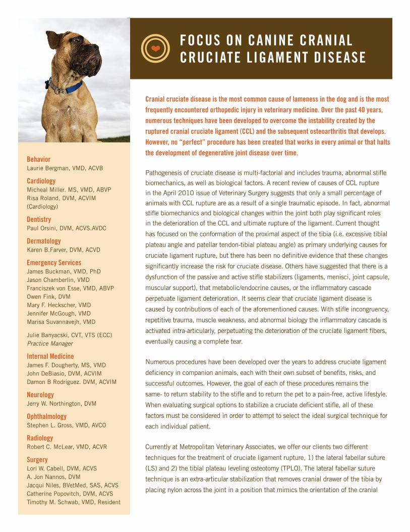
Cranial cruciate disease is the most common cause of lameness in the dog and is the most frequently encountered orthopedic injury in veterinary medicine. Over the past 40 years, numerous techniques have been developed to overcome the instability created by the ruptured cranial cruciate ligament (CCL) and the subsequent osteoarthritis that develops. However, no “perfect” procedure has been created that works in every animal or that halts the development of degenerative joint disease over time.
Pathogenesis of cruciate disease is multi-factorial and includes trauma, abnormal stifle biomechanics, as well as biological factors. A recent review of causes of CCL rupture in the April 2010 issue of Veterinary Surgery suggests that only a small percentage of animals with CCL rupture are as a result of a single traumatic episode. In fact, abnormal stifle biomechanics and biological changes within the joint both play significant roles in the deterioration of the CCL and ultimate rupture of the ligament. Current thought
has focused on the conformation of the proximal aspect of the tibia (i.e. excessive tibial
plateau angle and patellar tendon-tibial plateau angle) as primary underlying causes for
cruciate ligament rupture, but there has been no definitive evidence that these changes
significantly increase the risk for cruciate disease. Others have suggested that there is a
dysfunction of the passive and active stifle stabilizers (ligaments, menisci, joint capsule,
muscular support), that metabolic/endocrine causes, or the inflammatory cascade
perpetuate ligament deterioration. It seems clear that cruciate ligament disease is
caused by contributions of each of the aforementioned causes. With stifle incongruency,
repetitive trauma, muscle weakness, and abnormal biology the inflammatory cascade is
activated intra-articularly, perpetuating the deterioration of the cruciate ligament fibers,
eventually causing a complete tear.
Numerous procedures have been developed over the years to address cruciate ligament
deficiency in companion animals, each with their own subset of benefits, risks, and
successful outcomes. However, the goal of each of these procedures remains the
same- to return stability to the stifle and to return the pet to a pain-free, active lifestyle.
When evaluating surgical options to stabilize a cruciate deficient stifle, all of these
factors must be considered in order to attempt to select the ideal surgical technique for
each individual patient.
Currently at Metropolitan Veterinary Associates, we offer our clients two different
techniques for the treatment of cruciate ligament rupture, 1) the lateral fabellar suture
(LS) and 2) the tibial plateau leveling osteotomy (TPLO). The lateral fabellar suture
technique is an extra-articular stabilization that removes cranial drawer of the tibia by
placing nylon across the joint in a position that mimics the orientation of the cranial
FOCUS ON CANINE CRANIAL CRUCIATE LIGAMENT DISEASE
❤
BehaviorLaurie Bergman, VMD, ACVB
CardiologyMicheal Miller. MS, VMD, ABVPRisa Roland, DVM, ACVIM (Cardiology)
DentistryPaul Orsini, DVM, ACVS.AVDC
DermatologyKaren B.Farver, DVM, ACVD
Emergency ServicesJames Buckman, VMD, PhDJason Chamberlin, VMDFranciszek von Esse, VMD, ABVPOwen Fink, DVMMary F. Heckscher, VMDJennifer McGough, VMDMarisa Suvannavejh, VMD
Julie Banyacski, CVT, VTS (ECC)Practice Manager
Internal MedicineJames F. Dougherty, MS, VMDJohn DeBiasio, DVM, ACVIMDamon B Rodriguez. DVM, ACVIM
NeurologyJerry W. Northington, DVM
OphthalmologyStephen L. Gross, VMD, AVCO
RadiologyRobert C. McLear, VMD, ACVR
SurgeryLori W. Cabell, DVM, ACVSA. Jon Nannos, DVMJacqui Niles, BVetMed, SAS, ACVSCatherine Popovitch, DVM, ACVSTimothy M. Schwab, VMD, Resident
cruciate ligament. The TPLO stabilizes the stifle by rotating the
tibial plateau to approximately 6 degrees relative to the weight-
bearing axis. This changes cranial tibial thrust into caudal tibial
thrust and this is counteracted by the caudal cruciate ligament.
Both procedures are reported to have an 85%-90% good
to excellent prognosis for full return to function with few
complications, however, heated debates continue at all
veterinary surgery conferences as to which is the best procedure
to perform. In the literature, there has been no evidence-
based proof of the superiority of one procedure over the
other. A comparison of the short- and long-term function and
development of osteoarthritis in dogs having either TPLO or
LS was published in the February 2010 Veterinary Surgery.
This study revealed there was no significant difference in
ground reaction forces or development of osteoarthritis in dogs
receiving either surgical technique. However, these animals also
received post-operative physical therapy.
When evaluating animals with cruciate ligament disease at
Metropolitan Veterinary Associates, we evaluate each animal as
an individual, taking into account conformation, breed, activity
level, and biological factors. Our goal is always to address
abnormal biology and biomechanics, decrease pain, restore
function, and slow the progression of degenerative joint disease.
Pre-operative radiographs are evaluated closely to determine
if one procedure would produce better long-term outcomes
over another. We find it important to educate owners on each
procedure and work with them to decide on the best surgical
option for their pet. Other factors taken into consideration
when choosing a surgical technique include the weight and
temperament of the dog, its expected activity level, owner
finances and the presence or absence of cruciate ligament
disease in the contra-lateral stifle. It is import to remind owners
that even if their dog is currently sound in the other stifle, they
have a 40-50% chance that their dog will develop cruciate
ligament rupture in the other stifle within 1-2 years of the first
side becoming clinical.
Post-operatively, animals are monitored closely and
recommendations are made to help them achieve a full
recovery. We have found that post-operative physical therapy
plays an important role in rapid recovery, improved weight
bearing, and full range of motion and each client is encouraged
to have their pet participate in a program near them.
Owners are also advised to maintain their dogs at an ideal
body weight and consider starting their pet on a glucosamine/
chondroitin joint supplement if they have not already done so.
Regardless of the surgical technique used, most animals are
able to return to normal pre-operative activity levels around
12 weeks post-operatively. Dogs that still have a degree
of residual lameness at 12 weeks post-operatively often
continue to improve for 6-8 months post-operatively. Due to
the subsequent development of osteoarthritis, many dogs will
require the tactical use of non-steroidal anti-inflammatory drugs
(NSAID’s) if they have periods of mild lameness or stiffness,
particularly associated with heavier than normal activity levels
or cold, damp weather.
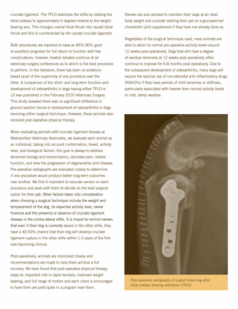
Post-operative radiographs of a giant breed dog after tibial plateau leveling osteotomy (TPLO).

FOR OUR MONTHLY HOSPITAL LECTURESPRESENTER/TOPICTo get a list of topics and speakers please visit our website in the “for referring veterinarians” section
UPCOMING DATES/TIMES6/17/10/6pm 7/15/10/6pm
8/12/10/6pm 9/16/10/6pm
10/14/10/6pm
METROPOLITAN VETERINARY ASSOCIATES2626 Van Buren Avenue Norristown, PA 19403 tel 610/666/1050 fax 610/666/1199 website www.metro-vet.com
SAVE THE DATE 10/28/10 CE FOR VETERINARIANS
JOIN
US
DO YOU WANT TO GO GREEN?
Have this newsletter electronically sent to you by contacting Stacey Connell at 610/666/1050 or email to [email protected]
ALL LECTURES WILL BE HELD AT METROPOLITANDinner provided :: Space is limited Stacey Connell at 610/666/1050 or [email protected]
RSVP
PET LOSS SUPPORT GROUP
Many of our employees have experienced and understand the depth of loss one experiences when a beloved four-legged family member has passed. For that reason, Metropolitan provides a pet loss support group to help grieving owners in need. Our pet group is designed to provide grieving pet parents with a safe, confidential environment to share their feelings with others who have experienced pet loss.
The meetings are held once a month onsite at Metropolitan and are free of charge for your clients (all family members are invited to attend). The group is led by Dr. Cari Thomson and co-led by psychiatrist Dr. Carol Tavani.
Please contact us at 610/666/1050 if you would like to have Pet Loss Support Group brochures mailed to your office. Clients are able to visit our website to find meeting dates and times, general information and recommendations on obtaining help outside of the group setting.
Pet Loss Support Group meetings held monthly for your clients (and are free of charge). Please contact us at 610-666-1050 for more information or for brochures More details to come!

FINALLY, A SAFE NON-INVASIVE DIAGNOSTIC TOOL AT YOUR DOORSTEP THAT CAN ANSWER THESE QUESTIONS:1) How can I obtain quality information to make good clinical decisions?2) How can I help my owners obtain all the information they need?3) How can I enhance my practice when dealing with patients that arrive with:
AtaxiaParesisCervical painTetraparesisLameness
Trauma Urinary control difficultiesSeizuresHead tilt
Neurological symptomsHind limb hypermetriaOncology issuesHind limb weaknessBrain tumors
ANSWER: DIAGNOSE WITH MRI AT VET IMAGING PARTNERS
9-Year-old German Shepherd with history of worsening neurological symptoms.
Results: Severe spinal cord compression, levels L1/L2, L2/L3, caused by intervertebral disc herniation.
7-Year-old Golden Retriever with left sided nasal bleeding over the past two months.Results: A left sided nasal cavity mass extending through the choana and slightly through to the right of midline within the nasal pharynx. Radiotherapy treatment was planned.
877 DOG SCAN (877/364/7226) WWW.VETIMAGINGPARTNERS.COMCONTACT
A N E S T H E S I A : Sandra Perkowski, VMD, PhD, Dipl ACVA
R A D I O L O G Y : Alexia McKnight, DVM, DACVR, McKnight Insight Robert C. McLear, VMD, DACVR, PetRad
COME MEET OUR DOCTORSto schedule your patient’s MRI or to speak with our staff about how we can serve your practice and patients.CA
LL U
S TODAY
OUR RESULTS
SAFE MRI & HIGH QUALITY IMAGES ADJACENT TO METROPOLITAN VETERINARY ASSOCIATE’S BUILDING
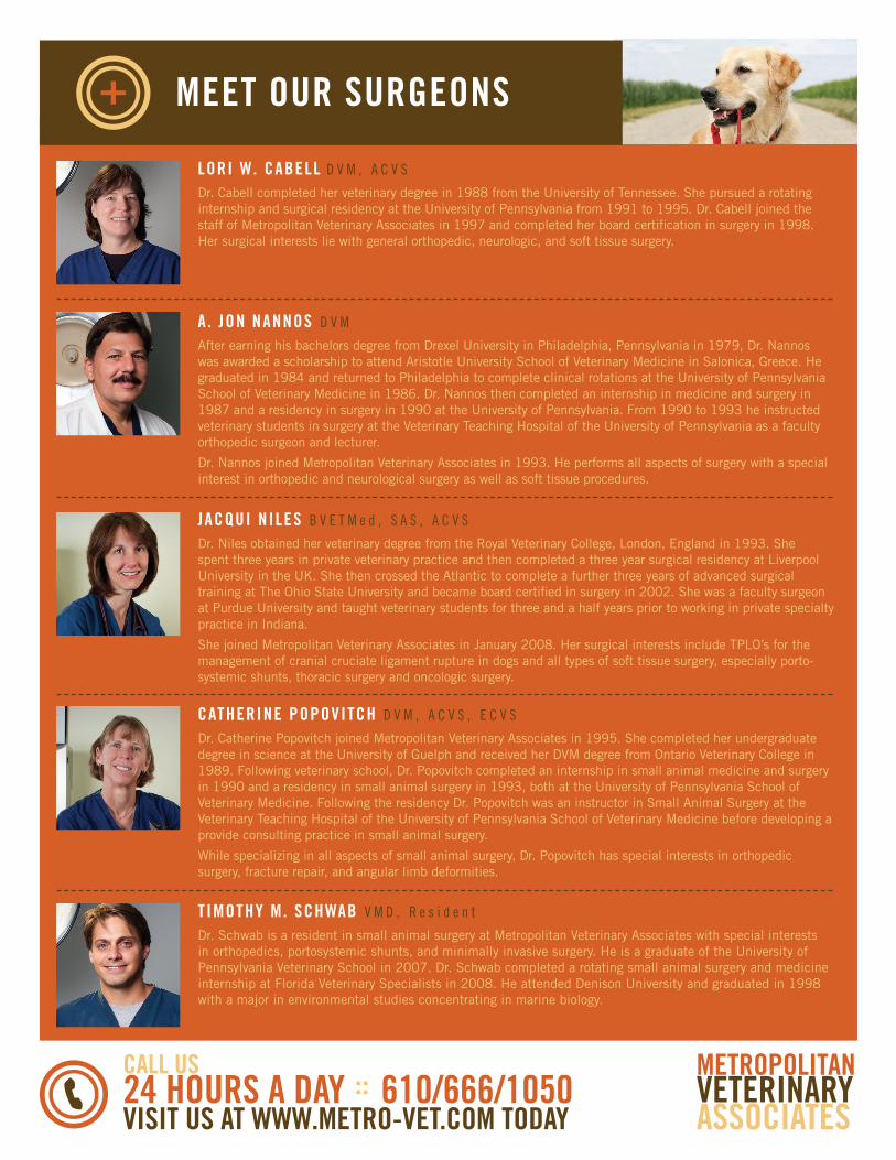
MEET OUR SURGEONS
LORI W. CABELL D V M , A C V S
Dr. Cabell completed her veterinary degree in 1988 from the University of Tennessee. She pursued a rotating internship and surgical residency at the University of Pennsylvania from 1991 to 1995. Dr. Cabell joined the staff of Metropolitan Veterinary Associates in 1997 and completed her board certification in surgery in 1998. Her surgical interests lie with general orthopedic, neurologic, and soft tissue surgery.
TIMOTHY M. SCHWAB V M D , R e s i d e n t
Dr. Schwab is a resident in small animal surgery at Metropolitan Veterinary Associates with special interests in orthopedics, portosystemic shunts, and minimally invasive surgery. He is a graduate of the University of Pennsylvania Veterinary School in 2007. Dr. Schwab completed a rotating small animal surgery and medicine internship at Florida Veterinary Specialists in 2008. He attended Denison University and graduated in 1998 with a major in environmental studies concentrating in marine biology.
JACQUI NILES B V E T M e d , S A S , A C V S
Dr. Niles obtained her veterinary degree from the Royal Veterinary College, London, England in 1993. She spent three years in private veterinary practice and then completed a three year surgical residency at Liverpool University in the UK. She then crossed the Atlantic to complete a further three years of advanced surgical training at The Ohio State University and became board certified in surgery in 2002. She was a faculty surgeon at Purdue University and taught veterinary students for three and a half years prior to working in private specialty practice in Indiana.
She joined Metropolitan Veterinary Associates in January 2008. Her surgical interests include TPLO’s for the management of cranial cruciate ligament rupture in dogs and all types of soft tissue surgery, especially porto-systemic shunts, thoracic surgery and oncologic surgery.
A . JON NANNOS D V M
After earning his bachelors degree from Drexel University in Philadelphia, Pennsylvania in 1979, Dr. Nannos was awarded a scholarship to attend Aristotle University School of Veterinary Medicine in Salonica, Greece. He graduated in 1984 and returned to Philadelphia to complete clinical rotations at the University of Pennsylvania School of Veterinary Medicine in 1986. Dr. Nannos then completed an internship in medicine and surgery in 1987 and a residency in surgery in 1990 at the University of Pennsylvania. From 1990 to 1993 he instructed veterinary students in surgery at the Veterinary Teaching Hospital of the University of Pennsylvania as a faculty orthopedic surgeon and lecturer.
Dr. Nannos joined Metropolitan Veterinary Associates in 1993. He performs all aspects of surgery with a special interest in orthopedic and neurological surgery as well as soft tissue procedures.
CATHERINE POPOVITCH D V M , A C V S , E C V S
Dr. Catherine Popovitch joined Metropolitan Veterinary Associates in 1995. She completed her undergraduate degree in science at the University of Guelph and received her DVM degree from Ontario Veterinary College in 1989. Following veterinary school, Dr. Popovitch completed an internship in small animal medicine and surgery in 1990 and a residency in small animal surgery in 1993, both at the University of Pennsylvania School of Veterinary Medicine. Following the residency Dr. Popovitch was an instructor in Small Animal Surgery at the Veterinary Teaching Hospital of the University of Pennsylvania School of Veterinary Medicine before developing a provide consulting practice in small animal surgery.
While specializing in all aspects of small animal surgery, Dr. Popovitch has special interests in orthopedic surgery, fracture repair, and angular limb deformities.
CALL US 24 HOURS A DAY :: 610/666/1050 VISIT US AT WWW.METRO-VET.COM TODAY
METROPOLITAN VETERINARY ASSOCIATES