Embed Size (px)
Citation preview
Vinnytsya National Medical University n.a. Pirogov
Chair of endoscopic and cardiovascular surgery
Methodical recommendations for practical lessons for 5th course
students
Theme: The varicose vein of the lower limbs. The classification.
The complications. The diagnostics. The special methods
of examination. The methods of surgical treatment.
(2 hours)
Author: MD, PhD Tatarin A.E.
Ratified on meeting of the chair
« __ » ____________ 20 __ . Protocol № _____

1. Concrete aims
Be able to analyze the precursors and risk factors of varicose disease
(congenital or acquired defects of venous wall, congenital or acquired defects
valvular veins, congenital or acquired defects of muscle–fascial system of the
lower extremities, pregnancy, resistant venous hypertension, hormonal and
neuroendocrine changes, etc.).
Explain the causal relationship of etiologic and pathogenetic features of illness
(long standing work > venous hypertension > functional failure of ostial valve
> varicose of great saphenous vein, etc.).
Know modern classification of varicose veins (clinical classification VVLL
(Expert Meeting, Moscow, 2000)).
Know the clinical manifestations (symptoms) of varicose veins.
Master the techniques of clinical diagnosis of varicose veins (holding
functional tests, reading flebogram).
To interpret the results of clinical and laboratory examination of patients with
varicose disease (complex characteristic symptoms, results functional tests,
data of laboratory tests).
To make the algorithm of conservative and surgical treatment of patients with
varicose disease based on the stage of the disease, its severity and the presence
of complications.
Base level of preparation
Educational previous discipline Skills are got
Normal anatomy Describe the anatomical structure of different
types of venous system of the lower extremities.
Normal physiology Describe the principles of normal blood flow in
the venous system body.
Pathological anatomy Describe the characteristics of pathological
changes in the venous wall and valvular veins of
the lower limbs with varicose veins.
Physiopathology Describe mechanisms for violations of blood
flow in the venous system of the organism in the
presence of risk factors for varicose veins and
blood flow characteristics in dilatation of
varicose veins of the lower limbs, depending on
the form and stage of disease.
Propedeutics of the internal
medicine
Master the methods of determining the symptoms
of varicose veins.

Radiology Demonstrate skills of reading angiograms.
Pharmacology Identify classes and groups of pharmacological
drugs used in treatment of varicose veins.
Clinical pharmacology Compare pharmacokinetic characteristics of
groups of drugs used in treatment of varicose
veins, with regard to the shape basic disease and
the presence concomitant disease.
Therapy Portray a schematic algorithms conservative
therapy varicose veins, depending of the form
basic disease and the presence concomitant
disease.
Surgery Portray schematically different methods of
surgical treatment of varicose veins.
2. Organization of the content of teaching material
Venous Anatomy
Veins are part of a dynamic and complex system that returns venous blood
to the heart against the force of gravity in an upright individual. Venous blood flow
is dependent upon multiple factors such as gravity, venous valves, the cardiac and
respiratory cycles, blood volume, and the calf muscle pump. Alterations in the
intricate balance of these factors can result in venous pathology.
Structure of Veins
Veins are thin–walled, highly distensible, and collapsible structures. Their
structure specifically supports their two primary functions of transporting blood
toward the heart and as a reservoir for preventing intravascular volume overload.
The venous intima is composed of a nonthrombogenic endothelium with an
underlying basement membrane and an elastic lamina. The endothelium produces
endothelium–derived relaxing factor and prostacyclin, which help maintain a
nonthrombogenic surface through inhibition of platelet aggregation and by
promoting platelet disaggregation. 1 Circumferential rings of elastic tissue and
smooth muscle located in the media of the vein allow for changes in vein caliber
with minimal changes in venous pressure. When an individual is upright and
standing still, the veins are maximally distended and their diameters may be
several times greater than if the individual was in a horizontal position.

Unidirectional blood flow is achieved with multiple venous valves. The
number of valves is greatest below the knee and decreases in number in the more
proximal veins. The inferior vena cava (IVC), the common iliac veins, the portal
venous system, and the cranial sinuses are valveless. Each valve is made of two
thin cusps consisting of a fine connective tissue skeleton covered by endothelium.
Venous valves close in response to cephalad–to–caudal blood flow at a velocity of
at least 30 cm/s.
Lower Extremity Veins
Lower extremity veins are divided into superficial, deep, and perforating
veins. The superficial venous system lies above the uppermost fascial layer of the
leg and thigh and consists of the greater saphenous vein (GSV) and lesser
saphenous vein (LSV) and their tributaries. The GSV originates from the dorsal
pedal venous arch and courses cephalad anterior to the medial malleolus and enters
the common femoral vein approximately 4 cm inferior and lateral to the pubic
tubercle. The saphenous nerve accompanies the GSV medially and supplies
cutaneous sensation to the medial leg and ankle. The LSV originates laterally from
the dorsal pedal venous arch and courses cephalad in the posterior calf and
penetrates the popliteal fossa, most often between the medial and lateral heads of
the gastrocnemius, to join the popliteal vein. The termination of the LSV is
somewhat variable. It may enter the deep venous system as high as the mid–
posterior thigh. The sural nerve accompanies the LSV laterally along its course and
supplies cutaneous sensation to the lateral malleolar region.
The deep veins follow the course of major arteries in the extremities. In the
lower leg, paired veins parallel the course of the anterior and posterior tibial and
peroneal arteries and join behind the knee to form the popliteal vein. Venous
bridges connect the paired veins in the lower leg. The popliteal vein continues
through the adductor hiatus to become the femoral vein. In the proximal thigh, the
femoral vein joins with the deep femoral vein to form the common femoral vein. In
the groin, the common femoral vein lies medial to the common femoral artery. The
common femoral vein becomes the external iliac vein at the inguinal ligament.
Multiple perforator veins traverse the deep fascia to connect the superficial
and deep venous systems. Clinically important perforator veins are the Cockett and
Boyd perforators. The Cockett perforator veins drain the medial lower leg and are
relatively constant. They connect the posterior arch vein (a tributary of the GSV)
and the posterior tibial vein. They may become varicose or incompetent in venous
insufficiency states. Boyd's perforator veins connect the greater saphenous vein to

the deep veins approximately 10 cm below the knee and 1 to 2 cm medial to the
tibia.
Venous sinuses are thin–walled, large veins located within the substance of
the soleus and gastrocnemius muscles. These sinuses are valveless and are linked
by valved, small venous channels that prevent reflux. A large amount of blood can
be stored in the venous sinuses. With each contraction of the calf muscle bed,
blood is pumped out through the venous channels into the main conduit veins to
return to the heart.
Upper Extremity Veins
As in the lower extremity, there are deep and superficial veins in the upper
extremity. Deep veins of the upper extremity are paired and follow the named
arteries in the arm. Superficial veins of the upper extremity are the cephalic and
basilic veins and their tributaries. The cephalic vein originates at the lateral wrist
and courses over the ventral surface of the forearm. In the upper arm, the cephalic
vein terminates in the infraclavicular fossa, piercing the clavipectoral fascia to
empty into the axillary vein. The basilic vein runs medially along the forearm and
penetrates the deep fascia as it courses past the elbow in the upper arm. It then

joins with the deep brachial veins to become the axillary vein. The median cubital
vein joins the cephalic and the basilic veins on the ventral surface of the elbow.
The axillary vein becomes the subclavian vein at the lateral border of the
first rib. At the medial border of the scalenus anterior muscle, the subclavian vein
joins with the internal jugular vein to become the brachiocephalic vein. The left
and right brachiocephalic veins join to become the superior vena cava, which
empties into the right atrium.
The Varicose vein disease
Varicose veins are a common medical condition present in at least 10% of
the general population. The findings of varicose veins may include dilated and
tortuous veins, telangiectasias, and fine reticular varicosities. Risk factors for
varicose veins include obesity, female sex, inactivity, and family history. Varicose
veins can be classified as primary or secondary. Primary varicose veins result from
intrinsic abnormalities of the venous wall, while secondary varicose veins are
associated with deep and/or superficial venous insufficiency.
Etiology and pathogenesis
Among the etiological factors of the development of varicosity an
important role plays hereditary factors, hormono–endocrine abnormal changes,
particularly pregnancy, various physiological and pathological factors, that cause
elevation of intraabdominal pressure.
In the condition of inherited or acquired structural weakness of a venous
wall and valves the abnormal reflux of venous blood occurs: from superficial veins
of leg the blood flows into deep, rises upward to saphenofemoral junction, where
the second part returns to superficial veins and as a result of the valvular
incompetence dumps downwards. It results in elevation of venous pressure and
varicose transformation.
Pathology
The veins are tortuous, irregularly dilated, protruded, and sometimes are
filled with thrombi. Walls of veins thickened, and vice versa, in the places of
protruding are thinned. Microscopically at the onset of the disease a focal
hyperplasia of elastic fibers and hypertrophy of longitudinal and circular muscle
fibers are revealed. Further a focal plasmorrhagia, fibroelastosis and sclerosis
develops. There comes an atrophy of muscular fibers. The expansion of the lumen

of veins tends to functional incompetence of valves. On the skin (particularly of
legs) a hyperpigmentation and trophic ulcers appear.
In addition to an unsightly appearance, patients with varicose veins often
complain of aching, heaviness, and early fatigue of the affected leg. These
symptoms worsen with prolonged standing and sitting and are relieved by leg
elevation above the level of the heart. A mild amount of edema is often present.
More severe signs include thrombophlebitis, hyperpigmentation,
lipodermatosclerosis, ulceration, and bleeding from attenuated vein clusters.
The compensated varicosity usually does not manifest. Some patients after
physical exertion feel a heavy, dull sensation in legs.
In the stage of subcompensation the patients complain usually of a heavy
sensation and fatigability of legs, their swelling or edema, burning pain in the
region of vari–cosity and night cramps of tibial muscles. During examination of the
patient in standing position it is possible to note a considerable varicosity of
superficial veins of the inferior extremities. The skin of lower legs more often is
unchanged. The functional examination of the valves reveals valvular incompe-
tence of superficial or perforating veins.
In the stage of decompensation the chief complaints of a constant gravity in
legs, pain, prompt fatigue, edema and cramps of tibial muscles. This is associated
with pigmentation, induration and trophic ulcer with localization in the lower third
of leg. A large protruding veins are common for these patients. At functional
examination it is possible to determine valvular incompetence of superficial,
perforating and deep veins.
Tests for definition of valvular incompetence of superficial veins
Troyanov–Trendelenburg's test. The patient lies supine with the elevated
extremity, and superficial veins is emptied. A rubber tourniquet is applied around
the upper third of thigh. The patient stands up. If in a vertical position (with a
tourniquet and after its releasing) the veins are slowly filled from below upward,
the test is considered as negative. At prompt filling of veins mainly from above
downward the test is positive.
Hackenbruch's test. In upward position, the great saphenous vein is
compressed with fingers and the patient is asked to cough. In incompetence of
venous valves, particularly ostial, it is possible to feel a retrograde wave of a blood,
which is transmitted by vessel below the finger.
Tests for evaluation of a valvular incompetence of perforating veins
Pratt's test. After the veins have been emptied when the patient lies supine
and elevates the lower limb vertically, the elastic bandage is applied from toes to
groin. The superficial veins are compressed with a rubber tourniquet in the upper
third of thigh. The patient stand; up. The imposed bandage is released gradually
from above downward, simultaneously another elastic bandage is applied from
inguinal region downward thus between them forms a space 5–6 cm. In site of
incompetent valves of perforating vein observed a protruding of superficial vein.
Sheinis' test. Around the leg, after the emptying of superficial veins by
elevating the extremity, three tourniquets are applied: around the thigh just below
oval fossa, above the knee and around upper leg. The patient is recommended to
stand up. The veins are gradually filled up by a blood. If the vein in any region is
promptly dilated, in this place it is necessary to consider the incompetence of
valves of perforating veins.
Talman's test is the modification of previous. For performance of this test a
rubber tourniquet of 2–3 m in length is used. It is imposed on the leg of patient
after emptying of superficial veins. Further the patient is asked to stand and the
tourniquet is released. The filling of veins informs about the incompetence of
valves of perforating veins, as at Sheinis' test.
Tests for estimation of deep veins patency
Mayo–Pratt's test. When the patient is supine, around the upper third of the
thigh of elevated extremity a tourniquet is applied and superficial veins are
compressed. Then the extremity is imposed by elastic bandage from toes to the
groin. The patient is asked to stand up. If after that he feels the pain in leg, sense of
compressing and fullness, it is possible to think, that deep veins are obstructed.
Delbet–Perthes' test (march test). When the patient is upward in order to
stop the blood flow in superficial veins above the knee joint a rubber tourniquet is
applied. After that, the patient is recommended to walk during 3–5 min. The
constriction of superficial veins indicates on satisfactory patency of deep veins
Clinical course (II – III st)
The signs of chronic venous insufficiency of II–III degree superimpose on
the clinical manifestations of varicosity. First of all it is edema and itching of the
skin, that promptly progress and constantly troubling the patient. Further the
trophic disturbances such as induration, hyperpigmentation, alopecia and
induration of subcutaneous fat develop. Very often this pathology is accompanied
by phlebothrombosis, eczema, erypsipelas etc
The diagnostic program
Anamnesis
Objective examination
General blood and urine analysis
Coagulogram
Functional tests for definition of the state of a valvular system
of superficial, deep and perforating veins
Sonography
Dopplerography

Phlebography
An important component of treatment for patients with varicose veins is the
use of elastic compression stockings. Patients are prescribed 20– to 30–mm Hg
elastic compression stockings to wear during the day. Stronger elastic stockings
may be required for those who still complain of leg aching or fatigue. The majority
of patients can be managed without additional therapy.
Additional interventions are warranted in patients whose symptoms worsen
or are unrelieved despite compression therapy or who have signs of
lipodermatosclerosis. Cosmetic concerns also can lead to intervention. Varicose
veins may be managed by injection sclero–therapy or surgical therapy or a
combination of both techniques. Injection sclerotherapy can be successful in
varicose veins less than 3 mm in diameter and telangiectatic vessels. Sclerotherapy
acts by destroying the venous endothelium. Sclerosing agents include hypertonic
saline, sodium tetradecyl sulfate, and polidocanol. Concentrations of 11.7 to 23.4%
hypertonic saline, 0.125 to 0.250% sodium tetradecyl sulfate, and 0.5%
polidocanol are used for telangiectasias. Larger varicose veins require higher
concentrations: 23.4% hypertonic saline, 0.50 to 0.75% sodium tetradecyl sulfate,
and 0.75 to 1.0% polidocanol. 75 Elastic bandages are wrapped around the leg
postinjection and worn continuously for 3 to 5 days to produce apposition of the
inflamed vein walls and prevent thrombus formation. After bandage removal,
elastic compression stockings should be worn for a minimum of 3 weeks.
Complications from sclerotherapy include allergic reaction, pigmentation,
thrombophlebitis, DVT, and possible skin necrosis.
In patients with symptomatic GSV reflux, the GSV should be removed.
Small incisions are placed medially in the groin and just below the knee, and the
GSV is stripped. Complications associated with GSV stripping include
ecchymosis, lymphocele formation, infection, and transient numbness in the
saphenous nerve distribution. GSV stripping has been documented in a randomized
study to have a lower rate of recurrent varicose veins than GSV ligation alone
(relative risk 0.28, 95% CI 0.13 to 0.59). Alternatively the greater saphenous vein
can be left in situ and destroyed with catheter–based techniques using laser or radio
frequency energy. Long–term follow–up and efficacy of such catheter–based
techniques is currently not available, but studies are ongoing.
Larger varicose veins are best treated by surgical excision using the "stab
avulsion" technique. Stab avulsions are performed by making 2–mm incisions
directly over branch varicosities, and the varicosity is dissected from the
surrounding subcutaneous tissue as far proximally and distally as possible through

the small incisions. In most cases the vein is simply avulsed with no attempt at
ligation. Bleeding is easily controlled with leg elevation and manual pressure.
Chronic Venous Insufficiency (CVI)
CVI is a major and costly medical problem affecting an estimated 600,000
patients in the United States. Patients complain of leg fatigue, discomfort, and
heaviness. Signs of CVI may include varicose veins, pigmentation,
lipodermatosclerosis, and venous ulceration. Importantly, severe CVI can be
present without varicose veins. In addition, chronic venous ulcers carry significant
negative physical, financial, and psychologic implications. A quality–of–life study
reported that 65% of chronic leg ulcer patients had severe pain, 81% had decreased
mobility, and 100% experienced a negative impact of their disease upon their work
capacity. The socioeconomic impact of chronic venous leg ulcers is staggering,
with an estimated 2 million workdays lost per year. The annual health care cost in
the United States to treat CVI is estimated at $1 billion. The signs and symptoms
of CVI can be attributed to venous reflux, venous obstruction, calf muscle pump
dysfunction, or a combination of these factors, as well as loss of venous wall
elasticity. The most important factor appears to be venous reflux in the majority of
patients with CVI. Venous reflux results from abnormalities of the venous valve
and can be classified as primary or secondary. Primary valvular reflux or
incompetence is diagnosed when there is no known underlying etiology of valvular
dysfunction. Secondary valvular reflux is diagnosed when an identifiable etiology
is present. The most frequent secondary etiology is DVT, which can lead to the
dysfunction of venous valves. Signs of CVI include edema, hyperpigmentation,
and ulceration.
The classification of chronic veins insufficiency CVI (SEAR, Hawai, 1994)
C – clinical picture;
E – aetiology;
A – anatomy;
P – pathophysiology.
C – clinical picture
C0а – asymptomatic (are absent the signs at physical examination);
C0s – are absent the signs at physical examination + symptoms;
C1а – telangiectasias or reticular veins;
C1s – telangiectasias or reticular veins + symptoms;
C2а – a varicose vein;

C2s – a varicose vein + symptoms;
C3а – an edemas;
C3s – an edemas + symptoms;
C4а – a skin change (the pigmentation, venous eczema, lipodermosclerosis);
C4s – a skin change + symptoms;
C5а – a skin change and close up ulcer;
C5s – a skin change and close up ulcer + symptoms;
C6а – a skin change and openning ulcer;
C6s – a skin change and openning ulcer + symptoms.
E – aetiology
Eе – congenital (the hoyer's canals, venous angioma);
Ep – primary, reason its is not installed;
Es – secondary (PTFS, posttraumatic).
A – an anatomy
As (superficialis) – a surrface veins;
Ad (deep) – a deep veins;
Ap (perforating) – an perforans veins.
P – pathophysiology
Pr – reflux;
Po – an obstruction;
Pr,o – their combination (reflux + obstruction).
The classification of chronic venous insufficiency (CVI):
0 – clinical symptoms are absent;
1 – a syndrome of "heavy" legs, transitory edema;
2 – a steadfast edema, hypo– or hyperpigmentation, lipodermosclerosis, eczema;
3 – venous trophic ulcer (openning or close up).
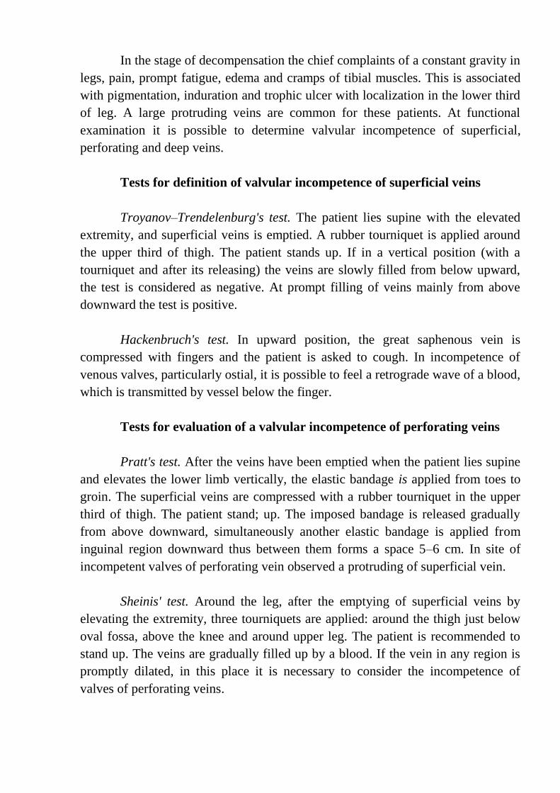
CEAP Class 1. Superficial spider veins (reticular veins) only.
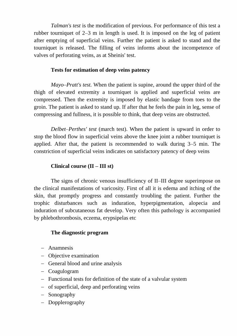
CEAP Class 2. Simple varicose veins only.
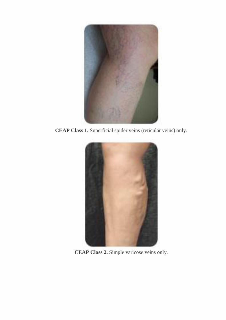
CEAP Class 3. Ankle edema (due to venous disease). The venous congestion,
often due to saphenous vein incompetence, may lead to dependent edema.
Elimination of the venous reflux often gives dramatic results.
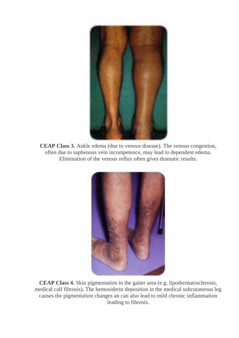
CEAP Class 4. Skin pigmentation in the gaiter area (e.g. lipodermatosclerosis,
medical calf fibrosis). The hemosiderin deposition in the medical subcutaneous leg
causes the pigmentation changes an can also lead to mild chronic inflammation
leading to fibrosis.

CEAP Class 5. A healed venous ulcer. Elevated venous pressures may lead to
relative tissue hypoxia, which causes skin fragility and ulceration. Restoration of
normal venous hemodynamics leads to ulcer healing.
CEAP Class 6. An open venous ulcer. Venous hypertension changed the perfusion
gradients which let to this ulcer. Once the refluxing saphenous vein was ablated
and venous flow normalized, the ulcer began to heal quickly.
Nonoperative Treatment of Chronic Venous Insufficiency
Compression Therapy. Compress ion therapy is the mainstay of CVI
management. Compression can be achieved using a variety of techniques,
including elastic compression stockings, paste gauze boots (Unna's boot),
multilayer elastic wraps/dressings , or pneumatic compression devices. The exact
mechanism by which compression therapy can improve CVI remains uncertain.
Improvement in skin and subcutaneous tissue microcirculatory hemodynamics as
well as a direct effect on subcutaneous pressure have been hypothesized as the
mechanism of compression therapy. Clinically, routine use of elastic and
nonelastic bandages reduces lower extremity edema in patients with CVI. In
addition, supine perimalleolar subcutaneous pressure has been demonstrated to be
increased with elastic compression. With edema reduction, cutaneous metabolism
may improve due to enhanced diffusion of oxygen and other nutrients to the
cellular elements of skin and subcutaneous tissues. Increases in subcutaneous
tissue pressure with elastic compression bandages may counteract transcapillary
Starling forces, which favor leakage of fluid out of the capillary.
Prior to the initiation of therapy for CVI, patients must be educated about
their chronic disease and the need to comply with their treatment plan in order to
heal ulcers and prevent recurrence. A definitive diagnosis of venous ulceration
must be made prior to undergoing treatment. A detailed history should be obtained
from a patient presenting with lower extremity ulcerations, including medications
and associated medical conditions that may promote lower extremity ulceration.
Arterial insufficiency is assessed by physical examination or noninvasive studies.
In addition, systemic conditions that affect wound healing and leg edema such as
diabetes mellitus, immunosuppression, malnutrition, or congestive heart failure
should be improved as much as possible.
Compression therapy is most commonly achieved with gradient elastic
compression stockings. Gradient elastic compression stockings, initially developed
by Conrad Jobst in the 1950s, were made to simulate the gradient hydrostatic
forces exerted by water in a swimming pool. Elastic compression stockings are
available in various compositions, strengths, and lengths, and can be customized
for a particular patient.
Patient compliance with compression therapy is crucial in treating venous
leg ulcers. Many patients are often initially intolerant of compression in areas of
hypersensitivity adjacent to an active ulcer or at sites of previously healed ulcers.
They may also have difficulty applying elastic stockings. To improve compliance,
patients should be instructed to initially wear their stockings only as long as it is
easily tolerable and then gradually increase the amount of time stockings are worn.
Alternatively, patients can be initially fitted with lower–strength stockings
followed by higher–strength stockings over a period of several weeks. Many
commercially available devices, such as silk inner toe liners, stockings with
zippered sides, and metal fitting aids, are available to assist patients in applying
elastic stockings.
Another method of compression was developed by the German
dermatologist Paul Gerson Unna in 1896. Unna's boot has been used for many
years to treat venous ulcers and is available in many versions. A typical Unna's
boot consists of a three–layer dressing and requires application by trained
personnel. A rolled gauze bandage impregnated with calamine, zinc oxide,
glycerin, sorbitol, gelatin, and magnesium aluminum silicate is first applied with
graded compression from the forefoot to just below the knee. The next layer
consists of a 4–inch–wide continuous gauze dressing followed by an outer layer of
elastic wrap, also applied with graded compression. The bandage becomes stiff
after drying and the rigidity may aid in preventing edema formation. Unna's boot is
changed weekly or sooner if the patient experiences significant drainage from the
ulcer bed.
Once applied, Unna's boot requires minimal patient involvement and
provides continuous compression and topical therapy. However, the Unna's boot
has several disadvantages. It is uncomfortable to wear because of its bulkiness,
which may affect patient compliance. In addition, the ulcer cannot be monitored
after the boot is applied, the technique is labor intensive, and the degree of
compression provided is operator–dependent. Occasionally, patients may develop
contact dermatitis to the components of Unna's boot, which may require
discontinuation of therapy.
Other forms of compression dressing, also available to treat CVI, include
multilayered dressings and legging orthosis. The purported advantages of
multilayered dressings include maintenance of compression for a longer period of
time, more even distribution of compression, and better absorption of wound
exudates. However, the efficacy of multilayered dressings is dependent on the
wrapping technique of health care personnel. A commercially available legging
orthosis consisting of multiple adjustable loop–and–hook closure compression
bands provides compression similar to Unna's boot and can be applied daily by the
patient.
Surgical Therapy of Chronic Venous Insufficiency
Troyanov–Trendelenburg's operation/ A great saphenous vein just at
saphenofemoral junction is ligated and cut. Previously before this management

in the same way three veins are followed up widely and ligated in the place of
their draining into a great saphenous vein: superficial external pudendal,
circumflex iliac, inferior epigastric tributaries.
Babcock's operation. A great saphenous vein is removed by means of a vein
stripper which inserted in its distal end.
Narath's operation. Operation is carry out as addition to Babcock's operation in
diffuse or mixed type of varicosity. The varicose tributary is exposed and
removed by either stripping or excision between two incisions.
Cocket's operation. A suprafascial ligation and cutting of perforating veins. An
incision up to 2–5 cm in length is performed on the posteriormedial side of a
lower leg. Perforating veins (3–5 and more) are ligated and cut above the
fascia.
Linton's operation – a subfascial ligation and cutting of perforating veins.
Perforator Vein Ligation
Incompetence of the perforating veins connecting the superficial and deep
venous systems of the lower extremities has been implicated in the development of
venous ulcers. The classic open technique described by Linton in 1938 for
perforator vein ligation has a high incidence of wound complications and has
largely been abandoned. A newer, minimally invasive technique termed subfascial
endoscopic perforator vein surgery (SEPS) has evolved with the improvement in
endoscopic equipment.
DUS is performed preoperatively in patients undergoing SEPS to document
deep venous competence and to identify perforating veins in the posterior
compartment. The patient is positioned on the operating table with the affected leg
elevated at 45° to 60°. An Esmarque bandage and a thigh tourniquet are used to
exsanguinate the limb. The knee is then flexed, and two small incisions are made
in the proximal medial leg away from areas of maximal induration at the ankle.
Laparoscopic trocars are then positioned, and the subfascial dissection is
performed with a combination of blunt and sharp dissection. Carbon dioxide is
then used to insufflate the subfascial space. The thigh tourniquet is inflated to
prevent air embolism. The perforators are then identified and doubly clipped and
divided. After completion of the procedure, the leg is wrapped in a compression
bandage for 5 days postoperatively.

Venous Reconstruction
In the absence of significant deep venous valvular incompetence,
saphenous vein stripping and perforator vein ligation can be effective in the
treatment of CVI. However, in patients with a combination of superficial and deep
venous valvular incompetence, the addition of deep venous valvular reconstruction
theoretically may improve ulcer healing. Numerous techniques of deep venous
valve correction have been reported. These techniques consist of repair of existing
valves, transplant of venous segments from the arm, and transposition of an
incompetent vein onto an adjacent competent vein. Cryopreserved venous valve
allografts placed below incompetent vein segments surgically or percutaneously
are currently in the early phases of development, but do not seem effective.
Successful long–term outcomes of 60 to 80% have been reported for
venous valve reconstructions by internal suture repair. However, in patients who
initially had ulceration, 40 to 50% still had persistence or recurrence of ulcers in
the long term. Valve transplantation involves replacement of a segment of
incompetent femoral vein or popliteal vein with a segment of axillary or brachial
vein with competent valves. Early results are similar to those of venous valve
reconstruction. However, in the long term, the transplanted venous segments tend
to develop incompetence, and long–term outcomes are poorer than those of venous
valve reconstructions. The outcomes for venous transposition are similar to those
of valve transplantation.
Sclerotherapy
The indications for sclerotherapy: a compensated stage of varicosity
associated with a diffuse type; smaller recurrent varicosities;
contraindications to operative treatment.
Contraindications for sclerotherapy are: the decompensative varicosity,
decompensative diseases of heart, lungs, kidneys, acute infectious and
purulent disease, acute thrombophlebitis of deep and superficial veins, preg-
nancy, bronchial asthma, obesity of III degree.
Endoveinous Laser Treatment
Laser treatment is another way to treat varicose veins. Your physician
inserts a tiny fiber into a varicose vein through a catheter. The fiber sends out laser
energy that kills the diseased portion of your varicose vein. The vein closes and
your body eventually absorbs it.

3. A plan and organizational structure of lesson is from discipline.
№
пп
Stages of employment Distributing
of time
Type of control Facilities of studies
1. Preparatory stage 15 %
3 min.
3min.
30min.
Structured written
work, written and
computer tests,
practical tasks, case
studies, oral
interviews for
standardized list of
questions.
Equipment, books,
manuals, guides,
atlases,
recommendations,
medications,
models, research
results, test results
and examinations,
computers with the
appropriate
information,
electronic
directories
1.1. Organization questions
1.2. Forming of motivation
1.3. Control of initial level of
preparation
– Name the factors that
ensure the normal
venous hemodynamics.
– Define varicose veins
of the lower extremities.
– Name the predisposing
factors and immediate
causes of varicose veins.
– Pathogenesis of
varicose veins.
– Classification of
varicose veins.
– Chronic venous
insufficiency and its
extent.
– Clinic varicose veins,
depending on the stage
of the process.
– Clinical characteristics
of varicose veins of the
lower extremities.
– Clinical characteristics
of chronic venous
insufficiency I st.
– Clinical characteristics
of chronic venous
insufficiency II st.

– Clinical characteristics
of chronic venous
insufficiency III st.
– Three groups
functional tests to
determine the patency of
superficial, deep and
perforating veins of the
lower extremities.
– X–ray diagnostic
methods of varicose
veins.
– Ultrasound and
computer diagnostics
varicose veins.
– Differential diagnosis
of varices inguinal area
and femoral hernia.
–Differential diagnosis
of varicose veins and
congenital dysplasia.
– Conservative treatment
of varicose veins.
–Surgical treatment of
varicose veins.
–Causes of recurrence of
varicose veins of the
lower extremities after
safenektomiyi.
– Treatment of recurrent
varicose veins of the
lower extremities after
safenektomy.
–Treatment of eczema
and dermatitis caused by
chronic venous
insufficiency.
–Treatment of trophic
ulcers caused by chronic
venous insufficiency.
–Minimally invasive
treatments for varicose
veins.
–Rehabilitation of
patients with varicose
disease in the early and
late postoperative period.
–Prevention of varicose
veins in threatening
group of patients (hard
physical labor,
pregnancy, etc.).
2. The main stage
(indicate all kinds of
work that students
perform during this
phase)
1. Hold measuring CVP
and CVP.
2. Perform functionality
tests to determine
patency of deep veins
and perforating veins.
3.Identify failure ostial
valve of large cutaneous
vein.
4.Determine the Cocket's
area
5. Read flebohramm.
6.Interpret the sonogram.
7.Hold elastic bandaging
a patient with lower
extremity varicose veins.
8. Identify signs of
lymphedema, venous
eczema,
156 min.
limfodermatosclerosis,
trophic ulcers.
9. To make the algorithm
conservative treatment of
the patient with the
initial stages of the
disease.
10.Identify the
indications and
contraindications for
surgical treatment.
11. Identify the
indications and
contraindications for
minimally invasive
therapy.
12. Collect a set of tools
for performing
safenektomy.
13. To bandage patient in
the early postoperative
period.
14. Fold algorithm
prevention of varicose
veins in threatening
patients.
15 Carry out preventive
conversation with a
patient with risk of
varicose veins.
I6 Evaluate the
effectiveness of the
method of treatment
(conservative and
surgical).
3. Final stage 20 %
3.1. Control of the final level
of preparation
30 min.
3.2. General estimation of 10 min.

educational activity of
student
3.3. Informing of students is
about the theme of next
employment
8 min.
Test for self–control
1. Which of the following conditions causes varicose veins?
1. Tunica media tear
2. Intraluminal occlusion
3. Intraluminal valvular compression
4. Intraluminal valvular incompetence
Answer 4. Varicose veins, dilated tortuous surface veins engorged with blood,
result from intraluminal valvular incompetence. An intraluminal occlusion would
result from plaque or thrombosis. The valves aren’t outside the lumen (intra-
luminal) and a tear would result in a hematoma.
2. Which of the following factors causes varicose veins?
1. Hypertension
2. Pregnancy
3. Thrombosis
4. Trauma
Answer 2. Primary varicose veins have a gradual onset and progressively worsen.
In pregnancy, the expanding uterus and increased vascular volume impede blood
return to the heart. The pressure places increased stress on the veins. Hypertension
has no role in varicose vein formation. Thrombosis and trauma cause valvular
incompetence and so are secondary causes of varicosities — not primary.
3. Which of the following symptoms com-monly occur in a client with varicose
veins?
1. Fatigue and pressure
2. Fatigue and cool feet
3. Sharp pain and fatigue
4. Sharp pain and cool feet
Answer 1. Fatigue and pressure are classic signs of varicose veins, secondary to
increased blood volume and edema. Sharp pain and cool feet are symptoms of
alteration in arterial blood flow.
4. In which of the following veins do varicose veins most commonly occur?
1. Brachial
2. Femoral
3. Renal
4. Saphenous
Answer 4. Varicose veins occur most frequently in the saphenous veins of the
lower extremities. They don’t develop in the brachial, femoral, or renal veins.
5. Which of the following conditions is caused by increased hydrostatic pressure
and chronic venous stasis?
1. Venous occlusion
2. Cool extremities
3. Nocturnal calf muscle cramps
4. Diminished blood supply to the feet
Answer 3. Calf muscle cramps result from increased pressure and venous stasis
secondary to varicose veins. An occlusion is a blockage of blood flow. Cool
extremities and diminished blood supply to the feet are symptoms of arterial blood
flow changes.
6. Which of the following activities should a client with varicose veins avoid?
1. Exercise
2. Leg elevations
3. Prolonged lying
4. Wearing tight clothing
Answer 4. Tight clothing, especially below the waist, increases vascular volume
and impedes blood return to the heart Exercise, leg elevations, and lying down
usually relieve symptoms
of varicose veins.
7. Which of the following tests demon¬strates the backward flow of blood through
incompetent valves of superficial veins?
1. Trendelenburg's test
2. Manual compression test
3. Perthes' test
4. Plethysmography
Answer 1. Trendelenburg's test is the most accurate tool used to determine
retrograde venous filling. The manual compression test is a quick, easy test done
by palpation and usually isn't diagnostic of the backward flow of blood. Perthes’
test easily indicates whether the deeper venous system and communicating veins
are competent Plethysmography allows measurement of changes in venous blood
volume.
8. Which of the following signs and symp¬toms are produced by secondary
varicose veins?
1. Pallor and severe pain
2. Severe pain and edema
3. Edema and pigmentation
4. Absent hair growth and pigmentation
Answer 3. Secondary varicose veins result from an obstruction of the deep veins.
Incompetent valves lead to impaired blood flow, and edema and pigmentation
result from venous stasis. Severe pain, pallor, and absent hair growth are symptoms
of an altered arterial blood flow.
9. Which of the following treatments can be used to eliminate varicose veins?
1. Ablation therapy
2. Cold therapy
3. Ligation and stripping
4. Radiation
Answer 3. ligation and stripping of the vein can rid the vein of varicosity. TTiis
invasive procedure will take care of current varicose veins only; it won’t prevent
others from forming. The other procedures aren't used for varicose veins.
10. Which of the following treatments is rec-ommended for postoperative
management of a client who has undergone ligation and strip¬ping?
1. Sitting
2. Bed rest
3. Ice packs

4. Elastic leg compression
Answer 4. Elastic leg compression helps venous return to the heart, thereby
decreasing venous stasis. Sitting and bed rest are contraindicated because both
promote decreased blood return to the heart and venous stasis. Although ice packs
would help reduce edema, they would also cause vasoconstriction and impede
blood flow.
Literature
1. Merchant RF, Kistner RL, Kabnick LS. Is there an increased risk for DVT with
the VNUS closure procedure? J Vasc Surg 2003; 38(3):628.
2. Dwerryhouse S, Davies B, Harradine K, et al. Stripping the long saphenous vein
reduces the rate of reoperation for recurrent varicose veins: five–year results of a
randomized trial. J Vasc Surg 1999;29(4):589–92.
3.Turton EP, Scott DJ,Richards SP, et al.Duplex–derived evidence of reflux after
varicose vein surgery: neoreflux or neovascularisation? Eur J Vasc Endovasc Surg
1999;(3 VI):230–3.
4. Goldman MP. Closure of the greater saphenous vein with endoluminal
radiofrequency thermal heating of the vein wall in combination with ambulatory
phlebectomy: preliminary 6–month follow–up. Dermatol Surg 2000;26(5):452–6.
5. Chandler JG, PichotO, SessaC, et al. Treatment of primary venous insufficiency
by endovenous saphenous vein obliteration. Vasc Surg 2000;14(3):201–14.
6.Weiss RA,WeissMA. Controlled radiofrequency endovenous occlusion using a
unique radiofrequency catheter under duplex guidance to eliminate saphenous
varicose vein reflux: a 2–year follow–up. Dermatol Surg 2002;28(1):38–42.
7.Weiss RA. Comparison of endovenous radiofrequency versus 810 nmdiode laser
occlusion of large veins in an animalmodel.Dermatol Surg 2002;28(1):56–61.
8.Manfrini S, Gasbarro V, Danielsson G, et al. Endovenous management of
saphenous vein reflux. Endovenous Reflux Management Study Group. J Vasc Surg
2000;32(2):330–42.
9. Merchant RF, DePalma RG, Kabnick LS. Endovascular obliteration of
saphenous reflux: a multicenter study. J Vasc Surg 2002;35(6):1190–6.
10. Dauplaise TL,Weiss RA. Duplex–guided endovascular occlusion of refluxing
saphenous veins. J Vasc Technol 2001;25(2):79–82.
11. Chandler JG, PichotO, Sessa C, et al.Defining the role of extended
saphenofemoral junction ligation: a prospective comparative study. J Vasc Surg
2000;32(5):941–53.
12. Goldman MP, Amiry S. Closure of the greater saphenous vein with
endoluminal radiofrequency thermal heating of the vein wall in combination with
ambulatory phlebectomy: 50 patients with more than 6–month follow–up.
Dermatol Surg 2002;28(1):29–31.
13. Pichot O, Kabnick LS, Creton D, et al. Duplex ultrasound scane findings two
years after great saphenous vein radiofrequency endovenous obliteration. J Vasc
Surg 2004;39:189–95.
14. Lurie F, Creton D, Eklof B, et al. Prospective randomized study of endovenous
radiofrequency obliteration (Closure procedure) versus ligation and stripping in a
selected patient population (EVOLVeS Study). J Vasc Surg 2003;38:207–14.
15. Sybrandy JE,Wittens CH. Initial experiences in endovenous treatment of
saphenous vein reflux. J Vasc Surg 2002;36(6):1207–12.
16. Rautio TT, Perala JM, Wiik HT, et al. Endovenous obliteration with
radiofrequency–resistive heating for greater saphenous vein insufficiency: a
feasibility study. J Vasc Interv Radiol 2002;13(6): 569–75.