Embed Size (px)
Citation preview

Planta (2007) 226:1287–1297
DOI 10.1007/s00425-007-0563-6ORIGINAL ARTICLE
Metabolic processes sustaining the reviviscence of lichen Xanthoria elegans (Link) in high mountain environments
Serge Aubert · Christine Juge · Anne-Marie Boisson · Elisabeth Gout · Richard Bligny
Received: 14 February 2007 / Accepted: 25 May 2007 / Published online: 16 June 2007© Springer-Verlag 2007
Abstract To survive in high mountain environmentslichens must adapt themselves to alternating periods of des-iccation and hydration. Respiration and photosynthesis ofthe foliaceous lichen, Xanthoria elegans, in the dehydratedstate were below the threshold of CO2-detection by infraredgas analysis. Following hydration, respiration totally recov-ered within seconds and photosynthesis within minutes. Inorder to identify metabolic processes that may contribute tothe quick and eYcient reactivation of lichen physiologicalprocesses, we analysed the metabolite proWle of lichenthalli step by step during hydration/dehydration cycles,using 31P- and 13C-NMR. It appeared that the recovery ofrespiration was prepared during dehydration by the accu-mulation of a reserve of gluconate 6-P (glcn-6-P) and bythe preservation of nucleotide pools, whereas glycolyticand photosynthetic intermediates like glucose 6-P and ribu-lose 1,5-diphosphate were absent. The large pools of poly-ols present in both X. elegans photo- and mycobiont arelikely to contribute to the protection of cell constituents likenucleotides, proteins, and membrane lipids, and to preservethe integrity of intracellular structures during desiccation.Our data indicate that glcn-6-P accumulated due to activa-tion of the oxidative pentose phosphate pathway, in
response to a need for reducing power (NADPH) during thedehydration-triggered down-regulation of cell metabolism.On the contrary, glcn-6-P was metabolised immediatelyafter hydration, supplying respiration with substrates duringthe replenishment of pools of glycolytic and photosyntheticintermediates. Finally, the high net photosynthetic activityof wet X. elegans thalli at low temperature may help thisalpine lichen to take advantage of brief hydration opportu-nities such as ice melting, thus favouring its growth inharsh high mountain climates.
Keywords Energy metabolism · Lichens · Metabolic proWling · NMR spectroscopy · Reviviscence · Xanthoria elegans
AbbreviationCDTA 1,2-Cyclohexylenedinitrilotetraacetic acidGlcn-6-P Gluconate 6-PGPG GlycerophosphoglycerolGPC GlycerophosphocholinePCA Perchloric acid
Introduction
Lichens are among the most resistant of living organisms.They survive in extreme environments including the desertsand frigid areas of all Wve continents. In the alpine environ-ment, these symbiotic organisms are exposed to harsh Xuc-tuations in water supply, light intensity, and temperature(Kappen 1988; Körner 2003). Xanthoria elegans (Link),used in this study, is a lichen typical of this habitat, whosegrowth on rocky surfaces mainly depends on atmosphericwater inputs. X. elegans is a saxicolous desiccation-tolerantlichen of the Teloschistaceae family (Helms 2003),
S. Aubert (&) · C. Juge · R. BlignyStation Alpine Joseph Fourier, UMS 2925 UJF CNRS, Université Joseph Fourier, BP 53, 38041 Grenoble cedex 9, Francee-mail: [email protected]
A.-M. Boisson · E. Gout · R. BlignyLaboratoire de Physiologie Cellulaire Végétale, Unité Mixte de Recherche 5168, Institut de Recherche en Technologies et Sciences pour le Vivant, CEA, 17 rue des Martyrs, 38054 Grenoble cedex 9, France
123

1288 Planta (2007) 226:1287–1297
containing an ascomycetous fungus and a unicellular greenalga belonging to the Teloschistes and Trebouxia genera,respectively (Helms 2003). At the site where they wereharvested thalli naturally undergo hydration/dehydrationcycles, growing when they are hydrated by melting snow,rain, or dew.
Metabolic activity of lichen thalli, apparent as gasexchange, becomes almost undetectable when their watercontent decreases below 10–15% of their dry weight(Lange 1980; Schroeter et al. 1991). But lichens may takeup suYcient moisture from atmospheric water vapour(Lange 1980) to stay above this low hydration threshold,and as such maintain signiWcant metabolic activity evenunder quite dry conditions. Hydration is facilitated by highconcentrations of polyols in both the photo- and mycobiont(Rundel 1988) that lower water potential (Lange et al.1990) allowing photosynthetic activity even under increas-ing degrees of desiccation (Nash et al. 1990). Immersion inwater or rewetting in moist air triggers the process of revi-viscence in dry desiccation-tolerant lichens (Smith andMolesworth 1973; Bewley 1979). Respiration and photo-synthesis quickly return to normal upon hydration, indicat-ing that cell damage induced by drying is rapidly repaired(Farrar and Smith 1976). According to several authors(Dudley and Lechowicz 1987; Longton 1988), desiccatedmembranes are leaky, leading to the loss of organic andinorganic solutes from the cell into the surrounding solutionduring rewetting. Thus, cell survival requires a rapid resealingof plasma-membrane disruptions (McNeil and Steinhardt1997; McNeil et al. 2003). Nevertheless, some delay beforerepair and synthesis of lost metabolites should be expected.
Intense solar radiation is another environmental stressthat threatens lichens with the potential damaging eVects ofreactive oxygen species (ROS) production (Weissman et al.2005), in particular when photosynthetic water oxidationstops due to dehydration. In the plant kingdom, the ability towithstand light stress and desiccation of vegetative organsinvolves various mechanisms recently reviewed by Hoekstraet al. (2001), Rascio and La Rocca (2005), and Heber et al.(2005). In hydrated organs, these include increased energydissipation by Xuorescence at PSII, and cyclic electrontransport around PSI (Heber and Walker 1992; Manuel et al.1999; Cornic et al. 2000). On the contrary, when they aredry, poikilohydric organisms do not emit light-induced Xuo-rescence, revealing inactivation of PSII function (Langeet al. 1989). These organisms minimise damage by activat-ing transfer to non-photochemical quenchers and convertingexcess energy to heat (Bilger et al. 1989; Heber et al. 2000).Interestingly, following hydration, a burst of intracellularproduction of ROS occurs in the photo- and mycobiont ofRamalina lacera, which modiWes superoxyde dismutase,catalase, glutathione reductase, and glucose 6-P dehydroge-nase activities (Weissman et al. 2005).
In desiccation-tolerant plants, the repair of cell damageupon hydration, and the subsequent restoration of cell func-tion, involves various complex inductive mechanismsincluding gene transcriptions and an increased protein turn-over (Oliver et al. 2004), therefore it requires a delay rang-ing from a few hours to several days (GaV 1997). Incontrast, we observed that respiration and photosyntheticactivity of several high-mountain lichens restarted almostimmediately after rewetting. Therefore, we hypothesisedthat the rapid recovery of these organisms relies on thepreservation/accumulation of key pools of metabolites dur-ing dehydration. We supposed, for example, that metabolicintermediates like respiratory substrates, nucleosides, andpyridine nucleotides, do not signiWcantly leak from thephoto- and mycobiont during hydration/desiccation cyclesin lichens. To substantiate this hypothesis, we assessed inparallel the respiration and photosynthetic activities and themetabolite proWles of X. elegans thalli during hydration anddehydration. The main pools of metabolites were character-ised in vitro and in vivo using 31P- and 13C-NMR, as aconvenient technique giving a precise overview of the solu-ble organic compounds present in plant materials (Blignyand Douce 2001; Streb et al. 2003).
Materials and methods
Lichen sampling and measurement of dry and wet weight
Xanthoria elegans thalli were collected at 2,800 m on lime-stone rocks situated above the Galibier pass (Hautes-Alpes), an area characterised by a relatively continental anddry climate. The so-called “dry thalli” were taken under thesun, during the hottest hours of the day (rocks surface tem-perature 30–35°C; relative air humidity 20%). They stillcontained ca 8% water by comparison with oven-desiccatedthalli (2 h at 110°C). The so-called “wet thalli” correspondto lichens collected in the same area either at dawn, in thedark, or just after a rain, in the light. The weight of wetthalli was measured in situ after removing interstitial waterby straining between two layers of absorbent paper. Thewet weight versus dry weight ratio was usually ca 2.5.Thalli were stored frozen at ¡20°C until needed.
Measurements of respiration and photosynthesis activities
These activities were measured either through O2 or CO2
exchange. CO2 exchange was measured with an infraredgas analyser (IRGA, LI-COR 6200, Lincoln, USA)equipped with a 1.2 l chamber. The temperature inside theclosed chamber was thermostatically maintained between»2.5°C and 30°C. Thalli (ca 0.5 g dry weight) were leftattached to their support. O2 was monitored polarographically
123

Planta (2007) 226:1287–1297 1289
at 20°C in a 1 ml water cuvet equipped with a Clark-typeoxygen-electrode purchased from Hansatech Ltd (King’sLynn, Norfolk, UK). The O2 concentration in an air- saturatedmedium was taken as 210 �M at 20°C and 780 hPa inthe alpine lab (2,100 m) where measurements were done.To enable the sample to be stirred in the measurementchamber, thalli were fragmented into 1–2 mm2 pieces bygentle grinding with mortar and pestle in liquid nitrogen;50 mg of dry thalli was used for O2-electrode measurements.Controls were made to ensure that lichen fragmentation inliquid nitrogen did not modify the rates of respiration andphotosynthesis after unfreezing and hydration. I A saturat-ing radiation dose of photosynthetically active photon Xuxdensity (PPFD) of 500 �mol m¡2 s¡1, was provided using aShott KL 1500 (Amilabo, Chassieu, France) light generator.The incubation medium contained 0.1 mM potassiumbicarbonate, pH 6.8.
Perchloric Acid (PCA) extract preparation
Thalli (4 g dry weight) were quickly frozen in liquid nitro-gen and ground to a Wne powder with a mortar and pestlewith 1 ml of 70% (vol/vol) PCA. The frozen powder waskept at ¡10°C until use. The thick suspension thus obtainedwas centrifuged at 15,000g for 10 min to remove particu-late matter and the supernatant was buVered with 2 MKHCO3 to about pH 5.2. The supernatant was then centri-fuged at 10,000g for 10 min to remove KClO4; the resultingsupernatant was lyophilised and stored in liquid nitrogen.This freeze-dried material was dissolved in 2.5 ml of watercontaining 10% D2O, and stored frozen.
31P- and 13C-NMR analyses of PCA extracts
Spectra were recorded on a Bruker NMR spectrometer(AMX 400, wide bore; Bruker Instruments, Inc., Billerica,MA, USA) equipped with a 10 mm multinuclear probetuned at 162 or 100.6 MHz for 31P- or 13C-NMR studies,respectively. The deuterium resonance of D2O was used asa lock signal.
13C-NMR acquisition conditions: 90° radio frequencypulses (19 �s) at 6 s intervals; spectral width 20,000 Hz;3,600 scans; Waltz-16 1H decoupling sequence (with twolevels of decoupling: 2.5 W during acquisition time, 0.5 Wduring delay). Free induction decays were collected as 16-Kdata points, zero-Wlled to 32K, and processed with a 0.2 Hzexponential line broadening. 13C-NMR spectra are referencedto hexamethyldisiloxane at 2.7 ppm. Mn2+ was chelated bythe addition of 2 �mol 1,2-cyclohexylenedinitrilotetraaceticacid (CDTA) and the pH was adjusted to 7.5 (samplevolume of 2.5 ml).
31P-NMR acquisition conditions: 70° radio frequencypulses (15 �s) at 3.6-s intervals; spectral width 8,200 Hz;
4,096 scans; Waltz-16 1H decoupling sequence (with twolevels of decoupling: 1 W during acquisition time, 0.5 Wduring delay). Free induction decays were collected as 8-Kdata points, zero-Wlled to 16K and processed with a 0.2-Hzexponential line broadening. 31P-NMR spectra are refer-enced to methylenediphosphonic acid (pH 8.9) at16.38 ppm. Before 31P-NMR analyses, divalent cationswere chelated by the addition of appropriate amounts ofCDTA ranging from 100 to 150 �mol (sample volume of2.5 ml). The much higher amount of CDTA as compared to13C-NMR analysis (see above) corresponded to the neces-sity to chelate all divalent cations including Ca2+, Mg2+, etc.while only Mn2+ had to be chelated for 13C-NMR analysis.The pH was buVered by the addition of 75 �mol Hepes andadjusted to 7.5.
The assignments were made after running a series ofstandard solutions of known compounds at pH 7.5 andadding aliquots of these compounds to the PCA extractsas described previously (Roby et al. 1987). IdentiWedcompounds were quantiWed by comparison of thesurface of their resonance peaks to the surface of reso-nance peaks of standards added to samples before grind-ing according to Aubert et al. (1996); NADP+ andNADPH were quantiWed as described by Pugin et al.(1997). Fully relaxed conditions during spectraacquisition (pulses at 20-s intervals) were used forquantiWcation. The standards utilised were methylphospho-nate and maleate for 31P- and 13C-NMR analyses,respectively.
In vivo 31P-NMR measurements
A perfusion system was utilised to optimise the signal-to-noise ratio as described earlier (Gout et al. 2001). Spectrawere recorded on a Bruker spectrometer (AMX 400, widebore) equipped with a 25-mm probe tuned at 162 MHz.31P-NMR acquisition conditions: 50° radio frequencypulses (70 �s) at 0.6 s intervals; spectral width 9,800 Hz;6,000 scans; Waltz-16 1H decoupling sequence (with twolevels of decoupling: 2.5 W during acquisition time,0.5 W during delay). Free induction decays were collectedas 4-K data points, zero-Wlled to 8K and processed with a2 Hz exponential line broadening. Spectra were refer-enced to a solution of 50 mM methylenediphosphonic acid(pH 8.9 in 30 mM Tris) contained in a 0.8-mm capillaryitself inserted inside the inlet tube along the symmetryaxis of the cell sample (Roby et al. 1987). The assignmentof inorganic phosphate (Pi), phosphate esters, phosphatediesters, and nucleotides to speciWc peaks was carried outaccording to Roberts and Jardetzky (1981), Roby et al.(1987), Aubert et al. (1996), and from spectra of the PCAextracts that contained the soluble low molecular weightconstituents.
123

1290 Planta (2007) 226:1287–1297
Results
As frequently observed with lichens (Smith and Moles-worth 1973; Farrar and Smith 1976; Larson 1981), X. eleganscan lose most of its constitutive water in a dry atmospherewithout apparent damage. The dehydration of this lichentook about 30 min when it was exposed to the summer sun,but it was rehydrated immediately on application of water.Whilst respiration was undetectable in the dry thalli, uponrewetting its initiation and progress to full speed within theseconds was spectacular.
In situ measurement of gas exchanges in X. elegans thalli
No gas exchange was detected using the IRGA apparatus(detection threshold, ca 10 nmol CO2 min¡1) in thalli con-taining less than 10% water, irrespective of the temperatureand the light conditions i.e. dark or light (photon Xuxexceeding 1,500 �mol m¡2 s¡1). This indicated that photo-synthesis and respiration activities were negligible. On thecontrary, CO2 uptake or loss, depending on light or darkconditions, was easily measured across a range of tempera-tures in wet thalli (Fig. 1). Interestingly, chlorophyll Xuo-rescence measured according to Heber et al. (2000) waslargely quenched in dry lichens, whereas it was detectedunder light within the Wrst min following hydration,increasing fast and reaching a steady state after 5–6 min(data not shown). This conWrms that electron transport wasnegligible in dry thalli.
Not surprisingly, CO2 exchange of wet thalli was tem-perature-dependent. In the dark, the respiration of wet thalliincreased exponentially with temperature (Fig. 1), like thatof higher plants (Bligny et al. 1985). For example, at 5°C,
the emission of CO2 was 25 § 5 nmol O2 min¡1 g¡1 lichendry weight, whereas it was 600 § 90 at 20°C. Under thelight, the uptake of CO2 increased with temperature up to amaximum of 800 § 80 nmol O2 min¡1 g¡1 lichen dryweight at 10–15°C, and then decreased continuously above15°C. Net CO2 uptake, considered as net photosynthesis,can be broken-down as gross CO2 uptake due to the photo-synthetic activity of the alga, and the emission of CO2
mainly due to fungal respiration if we consider that algalrespiration was negligible in the light (Gans and Rébeillé1988; Tcherkez et al. 2005). In illuminated wet thalli, theCO2 emitted by respiration compensated for the CO2 assim-ilated by photosynthesis at 28–29°C.
Recovery of respiration and photosynthesis activities of dry X. elegans thalli after hydration
In the following experiments, gas exchange was measuredon immersed thalli fragments with a Clark-type O2-electrode,at 20°C, as indicated in Materials and methods. We pre-ferred this technique because it was very diYcult to stabi-lise the temperature and relative humidity in the IRGAmeasurement chamber during dark/light cycles. The oxy-graphic traces (Fig. 2) show that: (1) the O2 uptake by thallifragments introduced into the dark O2-electrode chamberstarted immediately at full speed irrespective of their initialdry or wet status (traces a1 and b1). Oxygen was consumedat a rate of between 580 and 620 nmol min¡1 g¡1 lichendry weight. These values were comparable with the rates ofCO2 production measured in situ with the IRGA. (2) In thelight, there was a 1-min delay before the rate of O2 produc-tion initially by wet thalli stabilised (Fig. 2, trace b2). Thisdelay was shorter when the duration of the preceding darkperiod was reduced (trace b2) and longer (2–3 min) whenthalli were initially dry (trace a2). In all cases, the stablerate of O2 emission was comparable (between 520 and640 nmol min¡1 g¡1 lichen dry weight); (3) when sampleswere then alternately illuminated and darkened in succes-sive dark/light sequences, respiration and net photosynthe-sis rates remained remarkably stable over several hours; (4)assays done with lyophilised thalli containing less than 4%water or with thalli kept dry in the laboratory over 1 yeargave similar results (not shown), thus showing the remark-able adaptation of this lichen to severe and prolonged desic-cation.
Taken together, these data indicate that the cell struc-tures of the fungal and algal partners did not suVer irrevers-ible desiccation-induced damage. Moreover, the fact thatrespiration recovered instantly suggested that key meta-bolic substrates, nucleotides, and pyridine nucleotides,were not lost during the hydration/desiccation cycle. On thecontrary, the delayed recovery of photosynthesis suggestedthat key Benson–Basham–Calcin cycle intermediates
Fig. 1 Respiration (Wlled circle) and net photosynthesis (open circle)of wet X. elegans thalli in the dark and light at diVerent temperatures.Respiration and net photosynthesis were determined in situ via CO2exchanges measured by IRGA, as described in Materials and methods.Values are calculated from the weight of thalli dried at 110°C. Valuesare means § SD (n = 5)
123

Planta (2007) 226:1287–1297 1291
decreased sizably in the chloroplasts of photobiont duringthalli dehydration. In order to verify these hypotheses, weanalysed the metabolite proWles of dry and wet X. elegansthalli, and thalli during rewetting and drying cycles.
Metabolite proWling of X. elegans thalli
Perchloric acid extracts of X. elegans thalli were analysedusing 31P- and 13C-NMR spectroscopy as described in theMaterials and methods. Representative spectra are shown inFig. 3 and 5, and comparative data are given in Table 1.
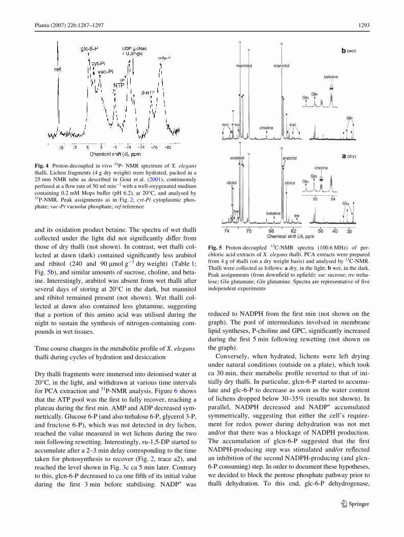
In dry thalli, the two most abundant metabolites mea-sured by 31P-NMR (Fig. 3a) were inorganic phosphate(1.7 �mol g¡1 dry weight) and glcn-6-P (0.91 �mol g¡1 dryweight). Other P-compounds identiWed were, from down-Weld to upWeld: mannitol 1-P, glycerate 3-P, AMP, NADP+,P-choline, the two phosphodiesters glycerylphosphoryl–glycerol and -choline (GPG and GPC), nucleosides (mainlyATP and ADP), and nucleoside diphosphate sugars,UDP-glc and UDP-N-acetylglucosamine (UDP-glcNAc).Polyphosphates were also detected in PCA extracts;however, they often precipitate during PCA extraction andwe quantiWed these compounds from in vivo NMR analy-ses. Spectra like the one shown on Fig. 4 indicate that thallicontained a 6.5-�mol g¡1 dry weight Pi equivalent ofpolyphosphates, which constitutes by far, the largest phos-phate pool in this lichen. In vivo assays also indicated thatthe distribution of Pi between alkaline (pH 7.5 cytoplasm)and acidic (pH 5.0–5.5 vacuoles) compartments wasroughly 1:2.
Fig. 2 O2 exchange in X. elegans thalli successively incubated in thedark and saturating light. These traces were obtained using thalli frag-ments incubated in an oxygen-electrode chamber. Values on the tracesrefer to nmol of O2 consumed or produced min¡1 g¡1 lichen dryweight. Thalli (th) were introduced into the chamber as dry fragmentsin dark (a1) or light (a2), or as wet fragments in dark (b1) or light (b2).The incubation medium contained 0.1 mM bicarbonate at pH 6.8.Open arrows light on; solid arrows light oV
Fig. 3 Proton-decoupled 31P-NMR spectra (161.93 MHz) ofperchloric acid extracts of X. elegans thalli. Extracts were preparedfrom 4 g of thalli (on a dry weight basis) and analysed by 31P-NMR.Thalli were collected as follows: a dry, in the light; b wet, in the dark;c wet, in the light. Peak assignments (from downWeld to upWeld): mnt-1-P mannitol 1-P; glcn-6-P gluconate 6-P; glc-6-P glucose 6-P; tre-6-P trehalose 6-P; gly-3-P glycerol 3-P; PGA phosphoglycerate; fru-6-P
fructose 6-P; ru-1,5-DP ribulose 1,5-diphosphate; AMP adenosinemonophosphate; P-cho P-choline; GPG glycerophosphoglycerol;GPC glycerophosphocholine; PEP phosphoenolpyruvate; UDP-glcuridine 5�-diphosphate-�-D-glucose; UDP-glcNAc uridine 5�-diphos-pho-N-acetylglucosamine; poly-P polyphosphates. Spectra are repre-sentative of Wve independent experiments
123

1292 Planta (2007) 226:1287–1297
The metabolite proWle of wet thalli harvested in the dark(at dawn) is strikingly diVerent from that of dry ones(Fig. 3b). First, wet thalli contained large pools of varioussugar phosphates, including glucose 6-P, trehalose 6-P, andfructose 6-P. Glyceraldehyde 3-P, also phosphoenolpyr-uvate. Second, glcn-6-P was much less abundant(0.18 �mol g¡1 dry weight) in wet than in dry thalli. Third,wet thalli contained NADPH (NADP+ was not detected), P-choline, GPC, and ATP (but less ADP and AMP), and theirUDP-glc pool was ca three times larger. PGA, GPG, andUDP-glcNAc pools were similar in dry and wet thalli. Themetabolite proWles of wet thalli harvested in the light(Fig. 3c) resemble those of wet thalli harvested in the dark,
except for a striking double peak, corresponding to theBenson–Basham–Calcin cycle intermediate ribulose 1,5-diphosphate (ru-1,5-DP), located in the chloroplasts of thealgal partner. The Xuctuations of Pi between samples mayoriginate from partial hydrolyses of polyphosphates duringPCA extraction.
13C-NMR spectra show much fewer diVerences due tothe water status of thalli. Typically, dry-thalli spectra(Fig. 5a) contain major resonance peaks corresponding topolyols; namely arabitol, mannitol, and ribitol (360, 240,and 110 �mol g¡1 dry weight, respectively). The othercompounds detected included two sugars, sucrose and tre-halose, two amino acids, glutamine and glutamate, choline
Table 1 Metabolic proWles of dry and wet X. elegans thalli
Dry thalli were collected in the light and wet thalli either in the dark (at dawn) or in the light. Metabolites were identiWed and quantiWed from PCAextracts, using maleate and methylphosphonate as internal standards for 13C- and 31P-NMR analyses, respectively, as described in Materials andmethods. Values are given as �mol g-1 lichen dry weight. Abbreviations as indicated in the legends of Fig. 3 and 5; nd, not detected (< 0.05 �mol).Values were obtained from a series of independent experiments and are given as mean § SD (n = 5). Statistical t test was applied to the data andP values are indicated (*P < 0.05) for wet lichens (dark) versus dry lichens (light) and for wet lichens (light) versus dry lichens (light)
Metabolite Dry lichens (light) Wet lichens (dark) P value (t test) Wet lichen (light) P value (t test)
Sucrose 27 § 3 29 § 3 7.01E-01 27 § 3 3.49E-01
Trehalose 9 § 2 5 § 2 1.81E-02* 8 § 2 2.70E-01
Ribitol 110 § 10 90 § 9 3.37E-02* 105 § 10 5.81E-01
Arabitol 360 § 30 240 § 20 9.69E-06* 340 § 30 3.01E-01
Mannitol 240 § 20 290 § 30 2.36E-01 250 § 20 3.13E-01
Glutamate 45 § 5 40 § 4 5.17E-01 43 § 5 6.19E-01
Glutamine 120 § 12 71 § 7 2.43E-05* 125 § 12 6.50E-02
Choline 85 § 9 80 § 8 9.34E-02 88 § 9 7.15E-01
Betaine 170 § 15 150 § 14 2.32E-01 160 § 15 2.23E-01
Polyphosphates 6.5 § 1 6.4 § 1 3.81E-01 6.2 § 1 4.37E-01
Pi 2.5 § 0.4 2.7 § 0.4 3.93E-01 3.6 § 0.5 1.63E-03*
Mannitol 1-P 0.06 § 0.01 0.02 § 0.01 1.62E-03* 0.5 § 0.01 1.14E-08*
Glcn-6-P 0.91 § 0.09 0. 18 § 0.02 4.38E-06* 0.20 § 0.02 1.79E-06*
Glucose 6-P nd 0.52 § 0.05 nd 0.81 § 0.08 1.43E-04*
Trehalose 6-P nd 0.22 § 0.02 nd 0.22 § 0.02 3.71E-03*
Glycerol 3-P nd 0.15 § 0.02 nd 0.17 § 0.02 1.05E-03*
Ru-1,5-DP nd nd nd 0.27 § 0.03 nd
PGA 0.27 § 0.03 0.25 § 0.03 7.06E-01 0.27 § 0.03 5.49E-01
Fructose 6-P nd 0.8 § 0.01 nd 0.13 § 0.02 7.15E-04*
AMP 0.12 § 0.02 0.04 § 0.01 nd nd nd
NADPH 0.02 § 0.01 0.08 § 0.01 1.19E-04* 0.08 § 0.01 2.49E-05*
NADP+ 0.05 § 0.01 nd nd nd nd
P-choline 0.15 § 0.02 0.40 § 0.04 1.26E-04* 0.33 § 0.03 1.78E-04*
GPG 0.30 § 0.03 0.25 § 0.02 5.52E-03* 0.25 § 0.02 1.63E-02*
GPC 0.28 § 0.03 0.47 § 0.05 3.55E-03* 0.47 § 0.05 4.94E-08*
PEP nd 0.07 § 0.01 nd 0.06 § 0.01 3.45E-04*
ATP 0.38 § 0.04 0.48 § 0.05 1.40E-01 0.53 § 0.05 3.81E-02*
ADP 0.14 § 0.02 0.08 § 0.01 3.04E-03* 0.05 § 0.01 1.96E-05*
UDP-glc 0.13 § 0.02 0.33 § 0.04 1.68E-05* 0.33 § 0.04 3.15E-07*
UDP-glcNAc 0.39 § 0.04 0.43 § 0.04 1.04E-01 0.46 § 0.05 4.58E-03*
123
Planta (2007) 226:1287–1297 1293
and its oxidation product betaine. The spectra of wet thallicollected under the light did not signiWcantly diVer fromthose of dry thalli (not shown). In contrast, wet thalli col-lected at dawn (dark) contained signiWcantly less arabitoland ribitol (240 and 90 �mol g¡1 dry weight) (Table 1;Fig. 5b), and similar amounts of sucrose, choline, and beta-ine. Interestingly, arabitol was absent from wet thalli afterseveral days of storing at 20°C in the dark, but mannitoland ribitol remained present (not shown). Wet thalli col-lected at dawn also contained less glutamine, suggestingthat a portion of this amino acid was utilised during thenight to sustain the synthesis of nitrogen-containing com-pounds in wet tissues.
Time course changes in the metabolite proWle of X. elegans thalli during cycles of hydration and desiccation
Dry thalli fragments were immersed into deionised water at20°C, in the light, and withdrawn at various time intervalsfor PCA extraction and 31P-NMR analysis. Figure 6 showsthat the ATP pool was the Wrst to fully recover, reaching aplateau during the Wrst min. AMP and ADP decreased sym-metrically. Glucose 6-P (and also trehalose 6-P, glycerol 3-P,and fructose 6-P), which was not detected in dry lichen,reached the value measured in wet lichens during the twomin following rewetting. Interestingly, ru-1,5-DP started toaccumulate after a 2–3 min delay corresponding to the timetaken for photosynthesis to recover (Fig. 2, trace a2), andreached the level shown in Fig. 3c ca 5 min later. Contraryto this, glcn-6-P decreased to ca one Wfth of its initial valueduring the Wrst 3 min before stabilising. NADP+ was
reduced to NADPH from the Wrst min (not shown on thegraph). The pool of intermediates involved in membranelipid syntheses, P-choline and GPC, signiWcantly increasedduring the Wrst 5 min following rewetting (not shown onthe graph).
Conversely, when hydrated, lichens were left dryingunder natural conditions (outside on a plate), which tookca 30 min, their metabolic proWle reverted to that of ini-tially dry thalli. In particular, glcn-6-P started to accumu-late and glc-6-P to decrease as soon as the water contentof lichens dropped below 30–35% (results not shown). Inparallel, NADPH decreased and NADP+ accumulatedsymmetrically, suggesting that either the cell’s require-ment for redox power during dehydration was not metand/or that there was a blockage of NADPH production.The accumulation of glcn-6-P suggested that the WrstNADPH-producing step was stimulated and/or reXectedan inhibition of the second NADPH-producing (and glcn-6-P consuming) step. In order to document these hypotheses,we decided to block the pentose phosphate pathway prior tothalli dehydration. To this end, glc-6-P dehydrogenase,
Fig. 4 Proton-decoupled in vivo 31P- NMR spectrum of X. elegansthalli. Lichen fragments (4 g dry weight) were hydrated, packed in a25 mm NMR tube as described in Gout et al. (2001), continuouslyperfused at a Xow rate of 50 ml min¡1 with a well-oxygenated mediumcontaining 0.2 mM Mops buVer (pH 6.2), at 20°C, and analysed by31P-NMR. Peak assignments as in Fig. 2; cyt-Pi cytoplasmic phos-phate; vac-Pi vacuolar phosphate; ref reference
Fig. 5 Proton-decoupled 13C-NMR spectra (100.6 MHz) of per-chloric acid extracts of X. elegans thalli. PCA extracts were preparedfrom 4 g of thalli (on a dry weight basis) and analysed by 13C-NMR.Thalli were collected as follows: a dry, in the light; b wet, in the dark.Peak assignments (from downWeld to upWeld): suc sucrose; tre treha-lose; Glu glutamate; Gln glutamine. Spectra are representative of Wveindependent experiments
123

1294 Planta (2007) 226:1287–1297
which converts glc-6-P into glcn-6-P, was inhibited usingglucosamine 6-P, a competitive inhibitor of the enzyme(Glaser and Brown 1955). As such, wet thalli were incu-bated for 1 h in the dark in the presence of 5 mM glucosa-mine, which was taken up by lichen cells andphosphorylated to N-glc-6-P (Table 2). As previouslyobserved with tobacco cells (Pugin et al. 1997), glcn-6-Pand NADPH decreased substantially, and NADP+
increased, whereas glc-6-P remained constant. Whenthese thalli were subsequently dehydrated, NADPH wasno longer detected, like in the control dry lichen(Table 1). However, in contrast to control lichen dehy-drated in the absence of N-glc, glcn-6-P did not increase(Table 2). In addition, lichens treated with glucosamineshowed a marked delay (1–2 min) after rehydration beforerespiration attained full speed while no delay wasobserved in control lichens (not shown). Photosynthesiswas similarly delayed. These results suggest that the accu-mulation of glcn-6-P was boosted by the cell dehydratingcell’s demand for redox power and that it prompted therecovery of metabolism after rehydration.
Discussion
In this study, we present novel Wndings on the time-coursechanges of diVerent pools of soluble metabolites in the foli-aceous lichen X. elegans during hydration/dehydrationcycles. Respiration, net photosynthesis, and its metaboliteproWle were analysed simultaneously during hydration ofthis lichen. The purpose of this approach was to identifywhich pools of metabolites were retained in dry lichens,and as such play key roles in their energetic metabolism.Furthermore, we aimed to determine how these metabolitespermit the instantaneous restart of respiration and the veryeYcient recovery of photosynthesis on rewetting.
The Wrst surprise was that dry thalli did not contain hex-ose phosphates, triose phosphates, and other intermediatesof glycolysis, except PGA, which could have been used tofuel respiration during the Wrst seconds following hydration(Fig. 3), in the absence of tricarboxylic cycle intermediates,like pyruvate, malate, succinate, or citrate (Fig. 5). How-ever, in contrast with wet thalli and with most living mate-rial examined so far, they contained a very large pool ofglcn-6-P (Fig. 3), that can be converted into fructose 6-Pand glyceraldehyde 3-P in the pentose phosphate pathwayand subsequently contribute to fuel respiration. In fact, theconsumption of just half the glcn-6-P pool during the Wrst 3min following hydration (ca 620 nmol g¡1 lichen dry weight,Fig. 6) was suYcient to sustain respiration (580 nmolO2 min¡1 g¡1 lichen dry weight). The rest of metabolisedglcn-6-P may contribute, via the pentose phosphate pathway,to the recovery of glc-6-P and ru-1,5-DP, which increasedsymmetrically to the decrease of glcn-6-P. Finally, thereplenishment of the ATP pool after hydration was a clearindicator of the recovery of the energetic metabolism. ATP,
Fig. 6 Time-course evolution of glcn 6-P, glucose 6-P, ribulose 1,5-diphosphate, ATP, ADP, and AMP in X. elegans thalli followinghydration in the light. At time zero, thalli fragments were incubated ina well aerated liquid medium containing 0.2 mM Mops buVer (pH 6.2),at 20°C. Metabolites were quantiWed as indicated in Material andmethods. Values are means § SD (n = 5)
Table 2 ModiWcations of gluconate 6-P, NADPH, and NADP+ in-duced in X. elegans thalli by glucosamine treatment
Lichens were incubated for 1 h in the dark in the presence of 5 mMN-glc (wet thalli) and a fraction of them was subsequently let dryingunder natural conditions (dry thalli), prior to PCA extraction. Metabo-lites were identiWed and quantiWed as indicated in Table 1. Values aregiven as �mol g-1 lichen dry weight. Abbreviations as indicated in thelegends of Fig. 3; nd, not detected (<0.05 �mol). Values were obtainedfrom a series of independent experiments and are given as mean § SD(n = 5). The levels of N-glucosamine 6-P, gluconate 6-P and NADP+were not statistically diVerent in dry and wet lichens (P < 0.05, t test)
Metabolite Wet thalli Dry thalli
N-glucosamine 6-P 0.38 § 0.04 0.37 § 0.04
Glucose 6-P 0.52 § 0.06 nd
glcn-6-P 0.04 § 0.01 0.03 § 0.01
NADPH 0.02 § 0.01 nd
NADP+ 0.07 § 0.01 0.08 § 0.01
123

Planta (2007) 226:1287–1297 1295
which decreased by nearly 30% during dehydration, recoveredtotally at the expense of AMP and ADP during the Wrstminute following hydration, indicating that adenylatekinase and ATP synthase activities restarted very rapidly(Roberts et al. 1997). Taken together, these results suggestthat the stores of glcn-6-P accumulated during dehydrationcontributed to the rapid and sustained recovery of respira-tory and photosynthetic activities in X. elegans thalli afterrehydration.
The accumulation of glcn-6-P in X. elegans during dehy-dration, when the relative water content of thalli droppedbelow ca 30%, could have originated from an increase ofglucose-6-phosphate dehydrogenase activity, in relation tothe production of ROS, as observed in other lichens(Weissman et al. 2005; Kranner and Grill 1994). Indeed,the reducing power (NADPH) is required to limit the poten-tially damaging eVect of the ROS burst due to impairedelectron transport chains in water-stressed cells (Rascio andLa Rocca 2005). For example, the production of reactionoxygen in the chloroplasts is avoided by reduction of gluta-thione, via the ascorbate–glutathione cycle (Asada 1994;Foyer et al.1994), which requires NADPH. This mecha-nism has also been observed in the alpine plant Soldanellaalpina and in Pisum sativum exposed to cold-induced pho-toinhibition causing glcn-6-P to accumulate (Streb et al.2003). On the contrary, when the pentose phosphate path-way was blocked after incubating wet thalli in the presenceof glucosamine, NADP+ was no longer reduced to NADPHand glcn-6-P did not accumulate during dehydration(Table 2). In addition, a delay in the recovery of respirationand photosynthesis was observed when glucosamie-treatedthalli were subsequently rehydrated, as compared to controllichens. These results are consistent with a role of the pen-tose phosphate pathway in the adaptation of X. elegans tohydration/dehydration cycles. Finally, the fact that no othermetabolite of the pentose phosphate pathway was detectedin dry thalli, in particular ribulose 5-P, suggests that thefunctioning of 6-phosphogluconate dehydrogenase wasblocked before that of glucose-6-phosphate dehydrogenaseduring cell dehydration.
The protection of many fungi and vascular plants againstreactive oxygen species can also involve polyols whichconstitute alternative metabolic reserves, behave as osmo-protectants, and, like mannitol, are potent quenchers ofROS (Jennings et al. 1998). In this context, we observedthat mannitol, when added to a solution of ATP, protectedATP from oxidation during desiccation when exposed tosunlight (result not shown). More generally, polyols, sug-ars, and other compounds like glutamate, glycine–betaine,etc. stabilise proteins and protect intimate cellular struc-tures against the potentially deleterious eVects of dehydra-tion (Hoekstra et al. 2001). Like many other lichens(Vincente and Legaz 1988; Honneger 1991), X. elegans
contained high amounts of polyols in both photobiont (ribi-tol) and mycobiont (mannitol and arabitol). These polyolpools did not rapidly change after rehydration. Neverthe-less, in accordance with previous results obtained by Farrar(1988), wet thalli collected at dawn contained less arabitoland ribitol than dry thalli (Fig. 5). Arabitol originates fromribitol, a sugar alcohol synthesised by the photobiont,which moves from alga to fungus as demonstrated in X.aureola and X. calcicola (Richardson and Smith 1968;Lines et al. 1989). Table 1 shows that wet lichens underdark conditions contain lower levels of arabitol than drylichens. Interestingly, under very long dark periods, arabitolwas completely metabolised in wet lichens suggesting thatit contributed to sustaining fungal respiration, while manni-tol-content remained nearly constant.
The mechanisms of lichen tolerance to dehydration/rehy-dration cycles include an adaptive response of algal and fun-gal cells to ROS-induced peroxidation and de-esteriWcationof glycerolipids that permeate membranes, and to mechanicalconstraints, which lead to cell membrane disruption. Hence,the capacity to rapidly reseal disrupted membranes plays acentral role in these mechanisms (McNeil and Steinhardt1997; McNeil et al. 2003). In this context, our results haveshown that, during rehydration, there was a threefoldincrease in P-choline and GPC nearly doubled (Fig. 5;Table 1). The increase of these two precursors of phosphati-dylcholine synthesis (van der Rest et al. 2002) may reXect thesynthesis of phosphatidylcholine-rich membrane systems,like the plasma membrane or tonoplast, and thus a role in themaintenance of cell structural integrity. On the contrary, thestability of GPG suggests that thylakoid membranes, whichcontain most of the cell’s phosphatidylglycerol (Joyard et al.1993) remained intact in the chloroplasts of photobiontduring dehydration/rehydration cycles.
In conclusion, our data suggest that the very rapid recov-ery of X. elegans respiration and photosynthetic activitiesfollowing rehydration was facilitated by the accumulationof stores of glcn-6-P during dehydration and by coordinatedevents associated with preventing oxidative damage and pro-tecting cell components and structures. Glcn-6-P appearedto accumulate in response to several factors including aneed for reducing power in the cell, to limit desiccation-generated ROS and the blockade of glcn-6-P metabolisation.The sizeable pools of polyols present in both phyco- andmycobionts served to protect cell constituents like nucleo-tides and proteins, and to preserve the integrity of intracel-lular structures. In lichen thalli, like in other poikilohydricorganisms such as seeds, progressive dehydration modiWesand Wnally stops metabolic activities. But, as opposed to seedswhere dormancy is advantageous to avoid undesirable ger-mination during transiently-favourable conditions (Bewley1997), the ability of lichen thalli to restart respiration and pho-tosynthesis without delay permits them to take advantage
123
1296 Planta (2007) 226:1287–1297
of all reviviscence opportunities oVered by the presence ofboth water and light, particularly at low temperatures(Fig. 1). This is the case, for example, when winter sunmelts ice to temporarily reveal rocky slopes. In such situa-tions, high net photosynthetic activities achieved at lowtemperatures will enable synthesis of carbohydrates withinminutes following rehydration. Finally, this work high-lights Glcn-6-P accumulation during dehydration as a meta-bolic adaptation of lichens to the anhydrobiotic cyclesimposed by the high-mountain climate.
Acknowledgments The authors are indebted to Pr Ulrich Heber(Univ. Wuerzburg, Germany) for his kind encouragements and for hisparticipation in Xuorescence assays. We thank Peter Streb (Univ.Orsay-Paris XI, France), Fabrice Rébeillé and Philippe Choler for theircritical reading of the manuscript, Matt Robson for style revision, andJuliette Asta for having introduced us to the world of lichens. We alsothank the reviewers for helpful comments and we are also grateful toOdile and Roland Donzel (Café de la Ferme, Col du Lautaret) for theirfriendly help and Jean-luc Lebail for his dedicated technical assistancewith the NMR spectrometer and perfusion system.
References
Asada K (1994) Production and action of active oxygen species in pho-tosynthetic tissues. In: Foyer CH, Mullineaux PM (eds) Causes ofphotooxidative stress and amelioration of defence systems inplants. CRC Press, Boca Raton, pp 77–104
Aubert S, Bligny R, Gout E, Alabouvette J, Marty-Mazars D, Marty F,Douce R (1996) Autophagy in higher plant cells submitted tocarbon deprivation: control by the supply of mitochondria withrespiratory substrates. J Cell Biol 133:1251–1263
Bewley JD (1979) Physiological aspects of desiccation tolerance.Annu Rev Plant Physiol 30:195–238
Bewley JD (1997) Seed germination and dormancy. Plant Cell 9:1055–1066
Bilger W, Rimke S, Schreiber U, Lange OL (1989) Inhibition of ener-gy transfer to photosystem II in lichens by dehydration: diVerentproperties of reversibility with green and blue-green photobionts.J Plant Physiol 134:261–268
Bligny R, Rebeille F, Douce R (1985) O2-triggered changes of mem-brane fatty acid composition have no eVect on Arrhenius discon-tinuities of respiration in sycamore (Acer pseudoplatanus L.)cells. J Biol Chem 260:9166–9170
Bligny R, Douce R (2001) NMR and plant metabolism. Curr OpinPlant Biol 4:191–196
Cornic G, Bukhov NG, Wiese C, Bligny R, Heber U (2000) Flexiblecoupling between light-dependent electron and vectorial protontransport in illuminated leaves of C3 plants. Role of photosystemI-dependent proton pumping. Planta 210:468–477
Dudley S, Lechowicz MJ (1987) Losses of polyol through leaching insubarctic lichens. Plant Physiol 83:813–815
Farrar JF (1988) Physiological buVering. In: Galun M (ed) Handbookof lichenology II. CRC Press, Boca Raton, pp 101–105
Farrar JF, Smith DC (1976) Ecological physiology of the lichen hypo-gymnia physodes. III. The importance of the rewetting phase.New Phytol 77:115–125
Foyer CH, Lelandais M, Kunert KJ (1994) Photooxidative stress inplants. Physiol Plant 92: 696–717
GaV DF (1997) Mechanisms of desiccation-tolerance in resurrectionvascular plants. In: Basra AS, Basra RK (eds) Mechanisms of
environmental stress resistance in plants. Harwood AcademicPublishers, Reading, pp 43–58
Gans P, Rébeillé P (1988) Light inhibition of mitochondrial respirationin a mutant of Chlamydomonas reinhardtii devoid of ribulose-1,5-bisphosphate carboxylase/oxygenase activity. Arch BiochemBiophys 260:109–117
Glaser BL, Brown DH (1955) PuriWcation and properties of D-glucose6-phosphate dehydrogenase. J Biol Chem 216:67–79
Gout E, Boisson AM, Aubert S, Douce R, Bligny R (2001) Origin ofthe cytoplasmic pH changes during anaerobic stress in higherplant cells. Carbon-13 and phosphorous-31 nuclear magnetic res-onance studies. Plant Physiol 125:912–925
Heber U, Walker D (1992) Concerning a dual function of coupled elec-tron cyclic transport in leaves. Plant Physiol 100:1621–1626
Heber U, Bilger W, Bligny R, Lange OL (2000) Phototolerance of li-chens, mosses and higher plants in an alpine environment: analy-sis of photoreactions. Planta 211:770–780
Heber U, Lange OL, Shuvalov VA (2005) Conservation and dissipa-tion of light energy as complementary processes: homoiohydricand poikilohydric autotrophs. J Exp Bot 57:1211–1223
Helms GWF (2003) Taxonomy and symbiosis in associations of Phy-sciaceae and Trebouxia. Dissertation zur Erlangung des Doktor-grades der Biologischen Fakultät der Georg-August UniversitätGöttingen 141 p
Hoekstra FA, Golovina EA, Buitink J (2001) Mechanisms of plant des-iccation tolerance. Trends Plant Sci 6:431–438
Honegger R (1991) Functional aspects of the lichen symbiosis. AnnuRev Plant Physiol Plant Mol Biol 42:553–578
Jennings DB, Ehrenshaft M, Pharr DM, Williamson JD (1998) Rolesof mannitol and mannitol dehydrogenase in active oxygen-medi-ated plant defense. Proc Natl Acad Sci USA 95:15129–15133
Joyard J, Block MA, Malherbe A, Maréchal E, Douce R (1993) Originof the synthesis of galactolipids and sulfolipid head groups. In: TSMoore Jr (ed) Lipid metabolism in plants. CRC Press, Boca Ra-ton, pp 231–258
Kappen L (1988) Ecophysiological relationships in diVerent climaticregions. In: Galun M (ed) Handbook of lichenology II. CRCPress, Boca Raton, pp 37–100
Körner C (2003) Alpine plant life: functional ecology of high mountainecosystems. Springer, Berlin
Kraner I, Grill D (1994) Rapid change of the glutathione status and theenzymes involved in the reduction of glutathionr-disulWde duringthe initial stage of wetting of lichens. Crypt Bot 4:203–206
Lange OL (1980) Moisture content and CO2 exchange of lichens. Oec-ologia 45:82–87
Lange OL, Bilger W, Rimke S, Schreiber U (1989) Chlorophyll Xuo-rescence of lichens containing green and blue green algae duringhydratation by water vapour uptake and by addition of liquid wa-ter. Bot Acta 102:306–313
Lange OL, Pfanz H, Kilian E, Meyer A (1990) EVect of low water po-tential on photosynthesis in intact lichens and there liberated algalcomponents. Planta 182:467–472
Larson DW (1981) DiVerential wetting in some lichens and mosses:the role of morphology. Bryologist 84:1–15
Lines CEM, RatcliVe RG, Rees TAV, Southon TE (1989) A 13C NMRstudy of photosynthate transport and metabolism in the lichenXanthoria calcicola Oxner. New Phytol 111:447–456
Longton RE (1988) The biology of polar bryophytes and lichens. Studiesin polar research. Cambridge University Press, Cambridge, p 391
Manuel N, Cornic G, Aubert S, Choler P, Bligny R, Heber U (1999)Protection against photoinhibition in the alpine plant Geummontanum. Oecologia 119:149–158
McNeil PL, Steinhardt RA (1997) Loss, restoration, and maintenanceof plasma membrane integrity. J Cell Biol 137:1–4
McNeil PL, Katsuya M, Vogel SS (2003) The endomembrane requirementfor cell surface repair. Proc Natl Acad Sci USA 100:4592–4597
123

Planta (2007) 226:1287–1297 1297
Nash TH, Reiner A, Demmig-Adams B, Kaiser WM, Lange OL (1990)The eVect of atmospheric desiccation and osmotic water stress onphotosynthesis and dark respiration of lichens. New Phytol116:269–276
Oliver MJ, Dowd SE, Zaragoza J, Mauget SA, Payton PR (2004) Therehydration transcriptome of the desiccation-tolerant bryophyteTortula ruralis: transcript classiWcation and analysis. BMC Ge-nomics 5:89
Pugin A, Frachisse J-M, Tavernier E, Bligny R, Gout E, Douce R,Guern J (1997) Early events induced by the elicitor cryptogein intobacco cells: involvement of a plasma membrane NADPH oxi-dase and activation of glycolysis and the pentose phosphate path-way. Plant Cell 9:2077–2091
Rascio N, La Rocca N (2005) Resurrection plants: the puzzle of sur-viving extreme vegetative desiccation. Crit Rev Plant Sci 24:209–225
Richardson DHS, Smith DC, Lichen Physiology (1968) IX. Carbohy-drate movement from the Trebouxia symbiont of Xanthoria aur-eola to the fungus. New Phytol 67:61–68
Roberts JKM, Jardetzky O (1981) Monitoring of cellular metabolismby NMR. Biochim Biophys Acta 639:53–76
Robert JKM, Aubert S, Gout E, Bligny R, Douce R (1997) Cooperationand competition among adenylate kinase, nucleoside diphospho-kinase, electron transport, and ATP synthase in plant mitochon-dria studied by 31P-nuclear magnetic resonance. Plant Physiol113:1–7
Roby C, Martin J-B, Bligny R, Douce R (1987) Biochemical changesduring sucrose deprivation in higher plant cells. II. Phosphorus-31nuclear magnetic resonance studies. J Biol Chem 262:5000–5007
Rundel PW (1988) Water relations. In: Galun M (ed) Handbook oflichenology II, CRC Press, Boca Raton, pp 17–36
Schroeter B, Jacobsen P, Kappen L (1991) Thallus moisture and mi-croclimatic control of CO2 exchange of Peltigera aphthosa (L)Willd on Disco Island (West Greenland). Symbiosis 11:131–146
Smith DC, Molesworth S (1973) Lichen physiology. XIII. EVects ofrewetting dry lichens. New Phytol 72:525–533
Streb P, Aubert S, Gout E, Bligny R (2003) Cold- and light-inducedchanges of metabolite and antioxidant levels in two high moun-tain plant species Soldanella alpina and Ranunculus glacialis anda lowland species Pisum sativum. Physiol Plant 118:96–104
Tcherkez G, Cornic G, Bligny R, Gout E, Ghashgaie J (2005) In vivorespiratory metabolism of illuminated leaves. Plant Physiol138:1596–1606
Van der Rest B, Boisson A-M, Gout E, Bligny R, Douce R (2002)Glycerophosphocholine metabolism in higher plant cells. Evi-dence of a new glyceryl-phosphodiester phosphodiesterase. PlantPhysiol 130:244–255
Vicente C, Legaz ME (1988) Lichen enzymology. In: Galun M (ed)Handbook of lichenology I. CRC Press, Boca Raton, pp 239–284
Weissman L, Garty J, Hochman A (2005) Characterization of enzy-matic antioxidant in the lichen Ramalina lacera and their re-sponse to rehydration. Appl Environ Microbiol 71:6508–6514
123