Embed Size (px)
Citation preview

EClinicalMedicine 8 (2019) 57–71
Contents lists available at ScienceDirect
EClinicalMedicinej ourna l homepage: ht tps : / /www. journa ls .e lsev ie r .com/
ec l in ica lmed ic ine
Research Paper
Meta-analytic Evidence for the Plurality of Mechanisms in Transdiagnostic StructuralMRI Studies of Hallucination Status
Colleen P.E. Rollins a,⁎, Jane R. Garrison a,b, Jon S. Simons b, James B. Rowe c, Claire O'Callaghan a,Graham K. Murray a,d, John Suckling a
a Department of Psychiatry, University of Cambridge, Cambridge, UKb Department of Psychology and Behavioural & Clinical Neuroscience Institute, University of Cambridge, Cambridge, UKc Department of Clinical Neurosciences, University of Cambridge, Cambridge, UKd Cambridgeshire and Peterborough NHS Foundation Trust, UK
⁎ Corresponding author at: Department of PsychiaCambridge CB2 0SP, UK
E-mail address: [email protected] (C.P.E. Rollins).
https://doi.org/10.1016/j.eclinm.2019.01.0122589-5370/© 2019 Published by Elsevier Ltd. This is an op
a b s t r a c t
a r t i c l e i n f oArticle history:Received 1 November 2018Received in revised form 15 January 2019Accepted 27 January 2019Available online 21 February 2019
Background:Hallucinations are transmodal and transdiagnostic phenomena, occurring across sensorymodalitiesand presenting in psychiatric, neurodegenerative, neurological, and non-clinical populations. Despite their cross-category occurrence, little empirical work has directly compared between-group neural correlates of hallucina-tions.Methods:Weperformedwhole-brain voxelwisemeta-analyses of hallucination status across diagnoses using an-isotropic effect-size seed-based d mapping (AES-SDM), and conducted a comprehensive systematic review inPubMed and Web of Science until May 2018 on other structural correlates of hallucinations, including corticalthickness and gyrification.Findings: 3214 abstractswere identified. Patientswith psychiatric disorders and hallucinations (eight studies) ex-hibited reduced gray matter (GM) in the left insula, right inferior frontal gyrus, left anterior cingulate/paracingulate gyrus, left middle temporal gyrus, and increased in the bilateral fusiform gyrus, while patientswith neurodegenerative disorders with hallucinations (eight studies) showed GM decreases in the left lingualgyrus, right supramarginal gyrus/parietal operculum, left parahippocampal gyrus, left fusiform gyrus, right thal-amus, and right lateral occipital gyrus. Group differences between psychiatric and neurodegenerative hallucina-tion meta-analyses were formally confirmed using Monte Carlo randomizations to determine statisticalsignificance, and a jackknife sensitivity analysis established the reproducibility of results across nearly all studycombinations. For other structuralmeasures (28 studies), themost consistent findings associatedwith hallucina-tion status were reduced cortical thickness in temporal gyri in schizophrenia and altered hippocampal volume inParkinson's disease and dementia. Additionally, increased severity of hallucinations in schizophrenia correlatedwith GM reductionswithin the left superior temporal gyrus, rightmiddle temporal gyrus, bilateral supramarginaland angular gyri.Interpretation: Distinct patterns of neuroanatomical alteration characterize hallucination status in patients withpsychiatric and neurodegenerative diseases, suggesting a plurality of anatomical signatures. This approach hasimplications for treatment, theoretical frameworks, and generates refutable predictions for hallucinations inother diseases and their occurrence within the general population.Funding: None.
© 2019 Published by Elsevier Ltd. This is an open access article under the CC BY-NC-ND license(http://creativecommons.org/licenses/by-nc-nd/4.0/).
Keywords:HallucinationStructural MRITransdiagnosticMeta-analysisSystematic reviewPsychiatricNeurodegenerative
1. Introduction
Hallucinations are transdiagnostic and transmodal perceptions ofstimuli that do not exist in the physical world [1]. They are prevalentin both psychiatric disorders, such as schizophrenia (60–80%) [2] and
try, University of Cambridge,
en access article under the CC BY-NC
bipolar disorder (BD 10–23%) [3], and neurodegenerative diseases,such as Parkinson's disease (PD; 22–38%) [4], dementia with Lewy Bod-ies (DLB; 80%) [5], and Alzheimer's disease (AD; 13–18%) [6], as well asin other psychiatric and neurological disorders, and among the generalpopulation (4.5–12.7%) [7]. Irrespective of diagnosis, the presence ofhallucinations marks an increased risk of adverse outcomes, such as re-duced likelihood of recovery in schizophrenia [8], more severe cognitivedeficits in PD [9], increased mortality in AD [10], increased suicidal be-haviour in adults with psychosis [11], and transition to later mental
-ND license (http://creativecommons.org/licenses/by-nc-nd/4.0/).

Research in context
Evidence before This Study
There is increasing recognition that hallucinations occur be-yond the archetype of schizophrenia, presenting in other psychi-atric disorders, neurological and neurodegenerative conditions,and among the general population. Not only are hallucinationsa transdiagnostic phenomenon, but also the subjective experi-ence of hallucinating is diverse, varying in modality, content, fre-quency, and affect. It has been suggested that no one type ofhallucination is pathognomic to any one disorder, but ratherthat hallucinations may exist on a spectrum from health to ill-ness, epidemiologically or experientially continuous between in-dividuals who do and do not meet criteria for a mental illness.However, limited research has been done to directly comparethe underlying neuroanatomy of hallucinations between differ-ent disorders. With this aim, we conducted a meta-analysis andsystematic review of structural MRI studies comparing individ-uals who experience hallucinationswith thosewho do not, to in-vestigate the brain morphology related to the transdiagnosticpresentation of hallucinations. We searched PubMed and Webof Sciencewith no start date limit, up toMay 2018, using the key-word combination (hallucinat*) AND (MRI OR magnetic reso-nance imaging OR morphology OR voxel?based ORmorphometr* OR neural correlate OR structur*). We includedonly studies with a within-diagnosis no-hallucination control totease out structural changes specific to hallucinations from ef-fects of the broader pathology. Neuroimaging meta-analyseswere conducted on studies performing whole-brain voxelwisegray matter differences, while studies assessing other structuralcorrelates were qualitatively synthesized.
Added Value of this Study
This is the first meta-analysis to illustrate the brain structuralcorrelates of hallucination occurrence derived from T1-weightedmagnetic resonance imaging (MRI) in a comparative manneracross clinical groups. We identified two distinct gray mattersubstrates for hallucination presence in psychiatric compared toneurodegenerative diseases, which we hypothesize constituteat least two distinct mechanisms. In addition, we qualitativelyassessed other structural neuroimaging studies over a variety ofmorphometric indices. We therefore provide a complete charac-terization of current knowledge of the brain morphology associ-ated with hallucinations across clinical status and modality.
Implications of all the Available Evidence
Our findings show at least two structural substrates that linkto the hallucinatory experience. This informs theoretical work onhallucinationswhichhave to date been limited in generating uni-fying direction-specific predictions of brain structure and func-tion. Understanding the plurality of anatomical signatures ofhallucinations may also inform treatment strategies. We predictthat other disorders in which patients experience hallucinationscan be categorized by our approach based on the broader pheno-type; for example, hallucinations in personality disorder may beof the psychiatric type, and similarly for early onset hallucina-tions in the general population, whilst later onset will be neuro-degenerative. Moreover, by differentiating the mechanisms ofhallucinations we recommend the contextualizing of researchby the appropriate phenotype.
58 C.P.E. Rollins et al. / EClinicalMedicine 8 (2019) 57–71
illness in children and young adults [12,13]. Although hallucinations areoften distressing, they may also be benign or contribute to meaningfulpersonal experiences [14,15].
Historically, hallucinations were considered a cardinal symptom ofschizophrenia, but they are not pathognomic: one-third of patients donot hallucinate [2], and the experience is often heterogeneous amongthose who do [1]. This has been confirmed across clinical and non-clinical populations, revealing diverse phenomenology involving mo-dality, content, affect, onset, and frequency [1,15,16]. Inter-individualdifferences among hallucinations prompt a number of conceptual,mechanistic, and clinical questions: Does phenomenological heteroge-neity translate into neurobiological plurality? Howwould this influencetheoretical models of hallucinations and inform treatments? Does theepidemiological and experiential diversity of hallucinations reflect acontinuum model, in which symptoms like hallucinations are distrib-uted over a spectrum of individuals who do and do not meet criteriafor mental illness, and thus arise from a common mechanism instanti-ated to different degrees of severity [17]? Establishing the validity ofthis conceptual framework against alternatives is important for howwe understand and treat hallucinations.
Despite the plurality of hallucinations, there is little empirical workcomparing between-group neural correlates of hallucinations. Prior re-views and meta-analyses on the brain structural and functional corre-lates of hallucinations have generally limited their scope to a singlediagnosis or modality [18–20], or both [21–25]. Only two reviewshave investigated hallucinations transdiagnostically or in more thanone modality: one without quantitative meta-analytic comparison[26], the other focussed on acute functional correlates of hallucinations[27]. Two meta-analyses have explored the structural correlates of hal-lucinations, but assessed correlates of hallucination severity rather thanpresence/absence, and limited their scope to auditory verbal hallucina-tions (AVH) in schizophrenia [23,24]. We therefore planned meta-analyses to evaluate MRI-derived volumetric structural gray matter(GM) correlates of hallucination status across populations,complemented with a comprehensive review of other structural mea-sures, including cortical thickness, gyrification, and structure-specificmorphometrics.
A significant issue in neuroimaging studies of hallucinations hasbeen the lack of a clinical control group, thus confounding abnormalitiesspecific to hallucination status with those of the broader phenotype.Equally challenging has been a tangled conceptual landscape, with nu-merousmodels proposed as cognitive or neurobiological accounts of au-ditory or visual hallucinations [5,26,28–41] (Fig. 1). Though aninfluential model of auditory hallucinations is the inner speech model[45], which proposes that AVHs arise from misattributing inner speechto a non-self source, alternative models posit the causal agent to bememory-related processes [28], spontaneous activation in auditoryand related memory areas [29], inappropriate proximal salience [30],skewed balance of top-down/bottom-up control dynamics betweensecondary sensory cortices and frontal regions [26,33] or of inhibition/excitation at the physiological level [34], or themismatch between pro-cesses comparing predictive representations of the external world tosensory evidence [31,35,37].While thesemodels attempt to explain au-ditory hallucinations in schizophrenia and non-clinical populations, aseparate array of models have been proposed for visual hallucinationsin neurodegenerative disorders like PD and AD [5,32,36,46]. Auditoryand visual hallucination models overlap in alluding to deficits in realitymonitoring, memory, salience, inhibition, and excitation. Additionally,hallucinations have been subcategorized by different neurocognitivemechanisms [40], or by differential contribution of a range of pharmaco-logical systems [41]. Obtaining differentiating evidence is difficult asthese models are not mutually exclusive, each drawing upon a similarrepertoire of constituents, making it non-trivial to derive correspondingpredictions [42]. However, specific morphological variation can differ-entiate patients who do and do not hallucinate [43], indicating thatstructural MRI can provide insights into why individuals hallucinate.

Fig. 1. Landscape of theoretical models of hallucinations. The major cognitive, psychological, and neurobiological theories for auditory and visual hallucinations are depicted. Separatetheories have been proposed to underlie auditory versus visual hallucinations, although they share many common themes. Different theories within each modality category are notmutually exclusive and may overlap in their predictions. Dotted lines delineate proposals of divisions between, extensions to, or limitations of current theories. Key references: Innerspeech model [45]; Intrusive memory hypothesis [28]; Resting state hypothesis [29]; Abnormal salience monitoring hypothesis [30]; Expectation-perception model [31]; Realitymonitoring deficit theory, Dream imagery intrusion theory, Activation input modulation theory [36,46]; Disinhibition hypothesis [5]; Perception and attention deficit model [32]; Top-down bottom-up models [26,33]; Excitatory-inhibitory imbalance [34]; Predictive processing accounts [35,37]; proposal of divide between self-monitoring accounts and spontaneousactivity accounts for auditory verbal hallucinations (AVH) [38]; proposal of subtypes for AVH [40]; proposal for differential contribution of pharmacological subsystems to differenttypes of AVH [41]; commentary on need to address interaction between and hierarchy of different modalities of hallucinations [40].
59C.P.E. Rollins et al. / EClinicalMedicine 8 (2019) 57–71
Voxel-based morphometry (VBM) is a common method for unbi-ased, automated quantification of GM differences between groups.Conducting a meta-analysis of VBM studies is an objective approach tosynthesize the extant literature and identify replicable findings [44].Knowledge of neuroanatomical signatures of hallucinations present incertain populations and absent in others would clarify the continuummodel by identifying whether there exist common neural correlatesand contribute towards a clearer neurobiological picture of the originsand mechanisms of hallucinations. Considering the cultural and histor-ical influences on hallucination interpretation [14], an organic model ofhallucinations could moreover substantiate accurate diagnostic criteria.Thismeta-analysis and systematic reviewquantitatively compared peo-ple with and without hallucinations in terms of brain structure to iden-tify the neuroanatomy related to the transdiagnostic presence ofhallucinations.
2. Methods
2.1. Search Strategy and Selection Criteria
A systematic review of the literature for the structural correlates ofhallucinations was conducted in October 2017, with update notifica-tions received until May 2018. Following PRISMA guidelines [47], arti-cles were identified by searching PubMed and Web of Science usingthe keyword combination (hallucinat*) AND (MRI OR magnetic reso-nance imaging OR morphology OR voxel?based OR morphometr* ORneural correlate OR structur*) with no date limit. Reviews and meta-analyses on neuroimaging of hallucinations were cross-referenced toensure no relevant studies were missed [20,23–26].
Studies were included in the meta-analyses if they: (a) employedstructural MRI in a whole-brain investigation of voxelwise differencesinGM reported in standard stereotaxic space; (b) included a direct com-parison between groups with and without hallucinations within thesame diagnostic category. Corresponding authors were contacted to re-quest coordinate information if not reported in the original article, or to
clarifymethodological issues. CPER evaluated all studies and JS, GKM, orJRG confirmed the selection criteria, with uncertainties discussed toconsensus. Region of interest (ROI) VBM studies and studies usingnon-voxelwise structural MRI methods that otherwise matched inclu-sion criterion (b) were included in the systematic review.
2.2. Data Analysis
Voxel-wise meta-analyses were undertaken using anisotropiceffect-size seed-based d Mapping (AES-SDM; https://www.sdmproject.com/) [48,49] following recommended guidelines [44](Supplementary S1). AES-SDM uses peak coordinates and effect sizesfrom primary studies to create maps of meta-analytic effect size andvariance of the signed GM differences. Similar to other voxel-basedmeta-analytic methods [50], loci from primary studies are estimatedas smoothed spheres and meta-analytic maxima calculated byweighting the encompassed voxels [48]. Additionally, AES-SDM incor-porates the effect sign (increases or decreases) and the t-statistic asso-ciated with each peak, increasing both sensitivity and accuracy [48].AES-SDM also allows inclusion of non-significant studies, reducingbias towards positive results. AES-SDM is detailed elsewhere (https://www.sdmproject.com/software/tutorial.pdf), and summarized in Sup-plementary Methods.
Anticipating differences in mechanisms of hallucinations betweenpsychiatric illnesses and neurodegenerative diseases based on distinc-tions in phenomenology, modality, prevalence [51], and the significantparticipant age separation among primary studies (t(25) = 17.324, pb 0.001), we performed a meta-analysis including schizophrenia, firstepisode schizophrenia (FES), first episode psychosis (FEP), and youngadults at clinical risk for psychosis (at-risk mental state long-term,ARMS-LT), and BD, and a second of neurodegenerative disorders, in-cluding PD and AD. Of the 16 studies included in these two cross-sectional meta-analyses, three (see Table 1) did not make an explicitcomparison between a hallucination (H) and no-hallucinations (NH)group, though themajority of patients in each group respectively either
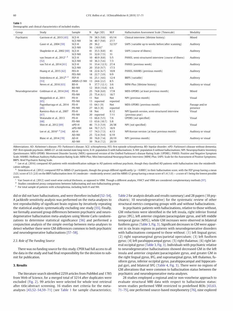
Table 1Demographic and clinical characteristics of included studies.
Group Study Sample N Age (SD) M/F Hallucination Assessment Scale (Timescale) Modality
Psychiatric Garrison et al., 2015 [43] SCZ-HSCZ-NH
7934
38.5 (9.8)40.7 (9.8)
65/1427/7
Clinical interview (lifetime history) Mixed
Gaser et al., 2004 [59] SCZ-HSCZ-NH
2956
36.2(10.9)e
52/33e SAPS (variable up to weeks before/after scanning) Auditory
Shapleske et al., 2002 [60] SCZ-HSCZ-NH
4131
35.5 (8.8)32.0 (7.5)
4131
SAPS (course of illness) Auditory
van Swam et al., 2012c, d
[52]SCZ-HSCZ-NH
1010
40.9 (8.8)36.3 (5.6)
5/57/3
PANSS, semi-structured interview (course of illness) Auditory
van Tol et al., 2014 [61] SCZ-HSCZ-NH
3120
33.4 (12.5)35.0 (9.7)
27/417/3
PANSS (previous week) Auditory
Huang et al., 2015 [62] FES-HFES-NH
1818
22.6 (6.7)22.7 (3.9)
10/89/9
PANSS, HAHRS (previous month) Auditory
Smieskova et al., 2012b, d
[53]FEP-HARMS-LT-NH
1613
25.1 (4.6)24.6 (2.2)
12/48/5
BRPS (variable) Auditory
Neves et al., 2016 [63] BD-HBD-NH
912
37.7 (12.1)39.9 (15.0)
3/66/6
MINI-Plus (lifetime history) Auditory or visual
Neurodegenerative Goldman et al., 2014 [64] PD-HPD-NH
2525
74.8 (6.0)75.4 (6.1)
17/818/7
MDS-UPDRS (at least previous month) Mixed
Meppelink et al., 2011[65]
PD-HPD-NH
1113
Notreported
Notreported
NPI (previous month) Visual
Pagonbarraga et al., 2014[66]
PD-HPD-NH
1527
64.1 (9)66.3 (8)
Notreported
MDS-UPDRS (previous month) Passage and/orpresence
Ramirez-Ruiz et al., 2007[67]
PD-HPD-NH
1820
Notreported
8/127/11
NPI Spanish version, semi-structured interview(previous year)
Visual
Watanabe et al., 2013[68]
PD-HPD-NH
1313
66.6 (5.5)63.6 (10.7)
7/65/8
UPDRS (not specified) Visual
Shin et al., 2012 [69] nPD-HnPD-NH
4664
71.3 (5.9)70.7 (5.7)
26/3818/9
NPI (not specified) Visual
Lee et al., 2016a, d [54] AD-HAD-NH
1725
74.3 (7.3)72.4 (9.4)
4/136/19
NPI Korean version (at least previous month) Auditory or visual
Blanc et al., 2014 [70] AD-HAD-NH
3939
76.0 (7.4)76.4 (7.2)
20/1920/19
NPI (previous month) Auditory or visual
Abbreviations: AD: Alzheimer's disease; PD: Parkinson's disease; SCZ: schizophrenia; FES: first episode schizophrenia; BD: bipolar disorder; nPD: Parkinson's disease without dementia;FEP:first episode psychosis; ARMS-LT: at riskmental state long-term; X-H: population Xwith hallucinations; X-NH: population Xwithout hallucinations; NPI: Neuropsychiatric InventoryQuestionnaire; MDS-UPDRS: Movement Disorder Society (MDS)-sponsored version of the Unified Parkinson's disease Rating Scale (UPDRS); PANSS: Positive and Negative SymptomScale; HAHRS: Hoffman Auditory Hallucination Rating Scale; MINI-Plus; Mini International Neuropsychiatric Interview (MINI) Plus; SAPS: Scale for the Assessment of Positive Symptoms;BRPS: Brief Psychiatric Rating Scale.
a Lee et al. (2016) compared AD patients with misidentification subtype to AD patients without psychosis, though they classified AD patients with hallucination into the misidentifi-cation subtype.
b Smieskova et al. (2012) compared FEP to ARMS-LT participants, though the groups differed significantly (p b 0.0001) in their hallucination score, with the FEP group having a mean(S.D.) score of 3.5 (2.0) on the BRPS hallucination item10 (moderate –moderately severe) and theARMS-LT grouphaving amean score of 1.4 (1.0)— a score of 1 being the lowest possiblescore.
c Van Swam et al. (2012) used voxel-wise cortical thickness, as opposed to VBM. Though a different analysis, VWCT and VBM are considered complementary methods [97].d Studies considered proxy comparisons between hallucinating and non-hallucinating groups.e For total sample of patients with schizophrenia, including both H and NH.
60 C.P.E. Rollins et al. / EClinicalMedicine 8 (2019) 57–71
did or did not have hallucinations, andwere therefore included [52–54].A jackknife sensitivity analysis was performed on the meta-analyses totest reproducibility of significant brain regions by iteratively repeatingthe statistical analysis systematically excluding one study [55]. Finally,we formally assessed group differences between psychiatric and neuro-degenerative hallucination meta-analyses using Monte Carlo randomi-zations to determine statistical significance [56] and performed aconjunction analysis of the simple overlap between meta-analyses todetect whether there were GM differences common to both psychiatricand neurodegenerative hallucinations [57–58].
2.3. Role of The Funding Source
Therewas no funding source for this study. CPERhad full access to allthe data in the study and had final responsibility for the decision to sub-mit for publication.
3. Results
The literature search identified 2259 articles from PubMed and 1785from Web of Science, for a merged total of 3214 after duplicates wereexcluded (Fig. 2). 99 articles were selected for whole text retrievalafter title/abstract screening. 16 studies met criteria for the meta-analyses [43,52–54,59–71] (see Table 1 for sample characteristics;
Table 2 for analysis details and results summary) and 28 papers (18 psy-chiatric; 10 neurodegenerative) for the systematic review of otherstructural metrics comparing groups with and without hallucinations.
In psychiatric patients with hallucinations, relative to thosewithout,GM reductions were identified in the left insula, right inferior frontalgyrus (IFG), left anterior cingulate/paracingulate gyrus, and left middletemporal gyrus (MTG), while GM increases were observed in bilateralfusiform gyrus (Table 3, Fig. 3). Significant decreases in GMwere appar-ent in six brain regions in patients with neurodegenerative disorderswith hallucinations compared to those without: (1) left lingual gyrus;(2) right supramarginal gyrus/parietal operculum; (3) left fusiformgyrus; (4) left parahippocampal gyrus; (5) right thalamus; (6) right lat-eral occipital gyrus (Table 3, Fig. 3). Individuals with psychiatric relativeto neurodegenerative hallucinations showed decreased GM in the leftinsula and anterior cingulate/paracingulate gyrus, and greater GM inthe right lingual gyrus, IFG, and supramarginal gyrus, left thalamus, fu-siform gyrus, inferior occipital gyrus, parahippocampal and hippocam-pal gyri, and bilateral SFG (Table 4, Fig. 3). There were no regions ofGM alterations that were common to hallucination status between thepsychiatric and neurodegenerative meta-analyses.
28 studies employed a regional and/or non-voxelwise approach toevaluate structural MRI data with respect to hallucination status:seven studies performed VBM restricted to predefined ROIs [43,63,71–75], one performed source-based morphometry [76], nine explored

Fig. 2. PRISMA flowchart for identification and selection of studies. Some studies performed analyses of multiple structural features and are therefore represented more than once.Abbreviations: H: population with hallucinations; NH: population without hallucination; VBM: voxel-based morphometry.
61C.P.E. Rollins et al. / EClinicalMedicine 8 (2019) 57–71
cortical thickness (CT) and/or surface area [77–85], three investigatedgyral/sulcal properties [43,86,87], and 11 assessed structure-specificshape parameters [43,83,85,88–95]. Results are summarized inTables 5–6. Overall, findings were heterogeneous, with few direct repli-cations. In schizophrenia, the most consistent findings were reductionsin CT in the vicinity of the left or right temporal gyrus for patients withhallucinations compared to those without [77,78,96], coincident withthe reductions in GM in left MTG observed in the meta-analysis(Fig. 3). However, two studies reported increases in GM in temporal re-gions with hallucinations [90,92]. Hallucinations in PD and DLB werecharacterized by distributed patterns of cortical thinning [83,84] and re-lated to hippocampal volume, though the direction of this associationwas mixed [83,85].
4. Discussion
Distinctive patterns of neuroanatomical alteration characterizehallucination status in patients with psychiatric and neurodegenera-tive diseases, with the former associated with fronto-temporal defi-cits and the latter with medial temporal, thalamic and occipitaldeficits. These results broadly align with prior meta-analyses inves-tigating GM correlates of hallucination severity of AVH in schizo-phrenia, which found negative correlations between hallucinationseverity in schizophrenia and GMV within the bilateral STG, bilateralHG, and bilateral insula [23,24,26] (Supplementary S3–4) and quali-tative reviews on structural imaging studies of visual hallucinations(VH) in neurodegenerative illnesses, which found GM atrophy
associated with VH in patients with PD in parietal, hippocampal,and occipito-temporal regions, primarily the lingual and fusiformgyri [25,98]. The distributed pattern of structural changes seen inboth hallucination signatures is suggestive of impairment in the co-ordination of information flow. Indeed, AVH in schizophrenia hasbeen associated with increased functional activation in the STG,insula, anterior cingulate, and pre/post central gyrus [21,22], re-duced resting connectivity between default mode regions [99], dis-ruptions to the salience network [30], and altered interactionsbetween resting-state networks [99]. Compared to AVH, VH inschizophrenia have been associated with increased seed-based func-tional connectivity between the amygdala and visual cortex [100],among the hippocampus, mPFC, and caudate nuclei and white mat-ter connectivity between the hippocampus and visual areas [91], aswell as decreased global sulcation in the right hemisphere [87]. VHin PD have been associated with increased functional activity in thelingual gyrus, cuneus, and fusiform gyrus [27], andhyperconnectivity in the default mode network [101]. Cortical thick-ness studies lend further support for divergent structural patterns,showing localized decreases in CT in temporal regions in schizophre-nia spectrum disorders and more widespread decreases in dementiaand PD (Table 6).
We reviewed the brain structural abnormalities associated withhallucinations, yet how changes to the brain's topological substratetranslate to changes in an individual's experiential landscape remainunknown. Our findings are consistent with multiple models of hallu-cinations (Fig. 1). For instance, volume loss in temporal regions could

Table 2Imaging characteristics and key results of included studies.
Group Study T Software Covariates FWHM(mm)
Statistical Threshold Originalstereotaxicspace
nFoci
Main result
Psychiatric Garrisonet al., 2015[43]
1.5 SPM8 TIV 8 p b 0.001, uncorrected;minimum cluster size= 100 voxels
MNI 2 H N NH: bilateral occipital lobe
Gaser et al.,2004 [59]
1.5 SPM99 SANS total score,SAPS total scorewithout auditoryhallucinationsub-items, gender
8 p b 0.001, uncorrected,k = 100 voxels
Talairach 4 H b NH: L transverse temporal (Heschl's)gyrus R middle/inferior frontal gyrus, Lmidde temporal gyrus, L paracingulategyrus,
Shapleskeet al., 2002[60]
1.5 AFNI Age, handedness −4.2 Absolute value ofstandard error b 1.96
Talairach 1 H b NH: L insular cortex
van Swamet al., 2012[52]
3 BrainVoyagerQX 1.9
None Notreported
p b 0.05, cluster sizeN15 voxels, correctedfor multiplecomparisons(Bonferroni p b 0.0063)
MNI 7 H N NH: L middle frontal gyrus, L posteriorcingulate gyrus, L frontal insula, Lparahippocampal gyrus, L postcentralsulcus, R visual cortexHbNH: posteriorinferior temporal sulcus, postcentral gyrus
van Tol et al.,2014 [61]
3 SPM8 Age, sex 8 p b 0.05, FWE-corrected(cluster level),voxel-wise threshold ofp b 0.005 uncorrected
MNI 3 H b NH: L putamen
Huang et al.,2015 [62]
3 SPM8 Age, gender, yearsof education
8 p b 0.001, uncorrected Talairach 0 n.s.
Smieskovaet al., 2012[53]
3 SPM8 Age, gender, totalGMV
8 p b 0.001, uncorrected(cluster-formingthreshold); p b 0.05FWE-corrected
MNI 3 H b NH: L parahippocampal gyrusH N NH: L superior frontal gyrus, L caudate
Neves et al.,2016 [63]
1.5 SPM8 Total GMV 8 p b 0.05, whole-brainFWE-corrected
Notreported
0 n.s.
Neurodegenerative Goldmanet al., 2014[64]
1.5 SPM8 TIV 8 p b 0.01, uncorrected;cluster extent thresholdk = 10
Talairach 18 H b NH: bilateral cuneus, bilateral fusiformgyrus, bilateral inferior parietal lobule,bilateral precentral gyrus, bilateral middleoccipital gyrus, R lingual gyrus, bilateralcingulate gyrus, L paracentral lobule
Meppelinket al., 2011[65]
3 SPM5 Total GM 10 p b 0.05, brain-volumecorrected cluster-level
MNI 0 n.s.
Pagonbarragaet al., 2014[66]
1.5 SPM5 Age, gender, globalGMV
12 p b 0.001, uncorrected;cluster size = 207voxels (determined by1000 Monte Carlosimulations)
MNI 4 H b NH: R vermis, R precuneusH N NH: posterior lobe of cerebellum, L inf.frontal cortex
Ramirez-Ruizet al., 2007[67]
1.5 SPM2 TIV, MMSE,Hamilton score,Hoehn and Yahrscore
12 p b 0.05, correctedcluster p-level
Talairach 3 H b NH: bilateral sup. parietal lobe, Llingual gyrus
Watanabeet al., 2013[68]
3 SPM8 TIV, age, sex 8 p b 0.01, FWE corrected;cluster size N50 voxelsand z-scores ≥3.00
MNI 15 H b NH: bilateral middle frontal gyrus, Lcingulate gyrus, R inferior parietal lobule,bilateral cuneus, L fusiform gyrus, Lposterior lobe, L inferior occipital gyrus, Linferior frontal gyrus, L declive, R lingualgyrus
Shin et al.,2012 [69]
3 SPM8 Age, sex, PDduration,intracerebralvolume, K-MMSEscore
6 p b 0.05, FWE corrected;uncorrected p b
0.001 at the voxel level,minimum cluster size= 100 voxels
Talairach 5 H b NH: R inferior frontal gyrus, Lthalamus, L uncus, L parahippocampalgyrus
Lee et al.,2016 [54]
3 SPM8 Age, gender,education, TIV, CDRscore, NPInon-psychoticscores
8 p b 0.001, uncorrected;extent threshold ofcontiguous 100 voxels(k N 100)
MNI 6 H b NH: R inferior parietal lobule, R lingualgyrus, L cuneus, R middle frontal gyrus, Rsuperior occipital gyrus, R middletemporal gyrus
Blanc et al.,2014 [70]
1.5 SPM12b Age, total GMV 8 p b 0.001, uncorrected;minimum cluster size= 25 voxels
MNI 3 H b NH: R insula/inferior frontal gyrus, Lsuperior frontal gyrus, bilateral lingualgyrus
Abbreviations: AD: Alzheimer's disease; PD: Parkinson's disease; SCZ: schizophrenia; FES: first episode schizophrenia; BD: bipolar disorder; nPD: Parkinson's disease without dementia;FEP:first episode psychosis; ARMS-LT: at riskmental state long-term; X-H: population Xwith hallucinations; X-NH: population Xwithout hallucinations; NPI: Neuropsychiatric InventoryQuestionnaire; MDS-UPDRS: Movement Disorder Society (MDS)-sponsored version of the Unified Parkinson's disease Rating Scale (UPDRS); PANSS: Positive and Negative SymptomScale; HAHRS: Hoffman Auditory Hallucination Rating Scale; MINI-Plus; Mini International Neuropsychiatric Interview (MINI) Plus; SAPS: Scale for the Assessment of Positive Symptoms;BRPS: Brief Psychiatric Rating Scale; SANS: Scale for the Assessment of Negative Symptoms; FWE: family-wise error; TIV: total intracranial volume; GM: gray matter; GMV: gray mattervolume; CDR: Clinical Dementia Rating scale; MMSE: Mini-Mental State Examination; K-MMSE: Korean version of MMSE; L: left; R: right.
62 C.P.E. Rollins et al. / EClinicalMedicine 8 (2019) 57–71

Table 3Regions of significant differences in gray matter between patients with hallucinations compared to those without for psychiatric and neurodegenerative disorders.
Group Contrast Region Peak local maximum Jackknifesensitivityanalysisa
MNIcoordinate
Cluster size (no. ofvoxels)
SDMZ-score
Uncorrectedp-value
Psychiatric H b NH L insula −46,2,−2 820 −1.885 0.0000464 7/8R inferior frontal gyrus, pars triangularis/frontal pole 48,36,8 281 −1.464 0.0008257 7/8L anterior cingulate gyrus/paracingulate gyrus 0,36,−2 132 −1.259 0.0028023 7/8L middle temporal gyrus −58,−42,−2 30 −1259 0.0028023 7/8
H N NH R fusiform gyrus 44,−64,−18 574 1.455 0.0000877 7/8L lateral occipital cortex/fusiform gyrus −40,−82,−16 345 1.454 0.0000981 7/8
Neurodegenerative H b NH L lingual gyrus/intracalcarine cortex 0,−86,−4 1275 −2.621 0.0000103 8/8L fusiform gyrus/inferior temporal gyrus −36,−18,−26 50 −1.860 0.0009702 7/8R supramarginal gyrus/parietal operculum 54,−36,30 75 −1.609 0.0034835 6/8L parahippocampal gyrus −38,−32,−10 42 −1.740 0.0018579 7/8R thalamus 2,−2, 12 14 −1.637 0.0030603 7/8R lateral occipital cortex 36,−80,14 10 −1.511 0.0043970 6/8
Abbreviations: H: Hallucinations; NH: No hallucinations; L: left; R: right.a The jackknife sensitivity analysis tests the reproducibility of significant brain regions by iteratively repeating the statistical analysis, but systematically excluding one study from each
replication [55]. Fractions show the number of study combinations in which the region was preserved out of the total number of dataset combinations.
63C.P.E. Rollins et al. / EClinicalMedicine 8 (2019) 57–71
reflect the misattribution of inner speech to a non-self source (innerspeechmodel) [45], or relate to abnormalities in cortical feedback forpredictive signal processing (predictive processing account) [102],or could be the result (or cause) of heightened resting state activityin the auditory cortex (resting state hypothesis) [29], or a combina-tion of some or all of these mechanisms. That substantial heteroge-neity was observed in ROI VBM hypothesis-driven studies furtheremphasizes the limits of current theories.
Our meta-analyses suggest that there are at least two broad bio-logical categories of hallucination mechanism: a psychiatric mecha-nism and a neurodegenerative mechanism. In support, structuralsignatures of hallucinations in the psychiatric meta-analysis overlapwith comparisons of patients to non-disordered controls. For in-stance, a meta-analysis of GM changes in patients with psychosiscompared to healthy controls shows reductions in bilateral insulaand anterior cingulate cortex [103], coinciding with regions identi-fied in the meta-analysis of hallucinations in neurodevelopmentaldisorders, while thalamic, hippocampal, and occipital GM reductionsin PD [104] partly coincide with the changes seen in neurodegenera-tive hallucinations. The relation between disorder-specific GMchanges and hallucination category suggests that hallucinationsshare networks of brain regions with the pathologies of the disorderin which they are embedded.
Knowledge of the structural correlates of hallucination types mayhelp understand their cognitive phenotypes. For instance, hallucina-tions are linked to reality monitoring, the cognitive capacity to distin-guish between self-generated and external sources of information[105]. Impaired in schizophrenia, reality monitoring is associatedwith the structure and function of the anterior cingulate cortex [43,105]. The cingulate gyrus is part of a network involving the IFG, ven-tral striatum, auditory cortex, right posterior temporal lobe whosefunctional connectivity is related to the subjective extent to whicha hallucination feels real [106]. Indeed, we propose that connectivityis key: together with the insula, the anterior cingulate constitutesnodes of the salience network, dysfunctions in which have been pro-posed as central to experiencing hallucinations [30]. Structural defi-cits in the insula in psychosis might also underpin atypicalinteractions between the DMN and salience network observed inhallucinations [39]. The left STG/MTG have been robustly implicatedin the manifestation of AVH [23,102], emphasizing the importance ofspeech perception and processing in hallucinations in schizophreniaspectrum psychosis.
If hallucinations experienced by those with schizophrenia spec-trum and bipolar psychosis are an example of a broader mechanism,then we predict that other neurodevelopmental disorders will have
similar patterns of associated GM loss. For example, hallucinationshave a prevalence of 43% in personality disorder [107], suggestedto be a neurodevelopmental disorder [108], and are predicted tohave a mechanism similar to other psychiatric disorders.
Abnormalities in the occipital cortex in neurodegenerative diseasessuggest that deficits in sensory regions contribute to hallucinations ofthe associated sensory modality since VH are more common in PDthan in schizophrenia [19]. Hallucinations in PD and ADwere character-ized by GM reduction in the thalamus and PHG. The thalamus mediatesinformation in the cortical hierarchies via corticothalamo-cortico cir-cuits and contributes to working memory maintenance [109], whilethe PHG is implicated in processing contextual associations in the ser-vice ofmemory formation and generating expectations about spatial re-lations [110]. Their involvement supports memory-related processes inhallucinations, thoughmay equally relate to neurodegenerative pathol-ogies. The anterior cingulate was implicated in hallucinations occurringin psychiatric disorders, but not neurodegenerative etiology. As the an-terior cingulate is involved in self-referential processing, this is consis-tent with the observation that psychotic hallucinations address theindividual and vary across continental location and historical time pe-riod [14,111]. Conversely, hallucinations in PD have a more passivequality and form historically stable categories of visual percepts [4].
Anatomic heterogeneity related to hallucination presence/absencehas important consequences for the plurality of treatment options. Aspecific example is repetitive transcranial magnetic stimulation(rTMS) used to reduce hallucination frequency and severity in schizo-phrenia, albeit with some reservations [112, 113]. A number of param-eters including frequency of stimulation and anatomical sitecontribute to the outcome of rTMS, and so anatomical heterogeneity isa possible source for the ambiguity of efficacy in therapeutic trials[114]. Antipsychotic (dopamine receptor anatagonist) medication isthe mainstay of treatment for hallucinations in schizophrenia, and issometimes required in PD, with evidence of therapeutic effect in each[115,116]. Psychological treatments include cognitive behavioral ther-apy and avatar therapy. However, little is known to help guide a choiceof which treatmentwill be tolerable and effective for a given individual;efforts to develop personalized treatment for hallucinations requires anunderstanding of the underlying mechanism that we suggest variesacross diagnosis.
The multimodality of hallucinations is under-documented andunder-researched, with b2% of studies included in this review prob-ing hallucinations beyond audition or vision [66]. However, 30–50%of schizophrenia or PD patients report hallucinations in more thanone modality [2,117]: olfactory hallucinations are present in10–13.7% [51,118] and tactile sensations frequently co-occur with

Fig. 3. Meta-analysis results for individuals with hallucinations compared to those without hallucinations in psychiatric (A) and in neurodegenerative disorders (B). A. For psychiatricdisorders, the meta-analysis revealed gray matter decreases in the left insula, right inferior frontal gyrus (pars triangularis)/frontal pole, left anterior cingulate gyrus/paracingulategyrus, left middle temporal gyrus, and gray matter increases in the bilateral fusiform gyrus in patients with hallucinations relative to those without. B. For neurodegenerativedisorders, the meta-analysis revealed decreases in the left lingual gyrus/intracalcarine cortex, left fusiform gyrus, right supramarginal gyrus, left parahippocampal gyrus, rightthalamus, and right lateral occipital cortex. C. Formal comparison between meta-analyses revealed reduced GM in the left insula and left anterior cingulate/paracingulate gyrus forindividuals with psychiatric relative to neurodegenerative hallucinations, and greater GM in the right lingual gyrus, IFG, and supramarginal gyrus, left thalamus, fusiform gyrus, inferioroccipital gyrus, parahippocamapal and hippocampal gyri, and bilateral SFG. Abbreviations: STG: superior temporal gyrus; MTG: middle temporal gyrus; IFG: inferior frontal gyrus;PHG: parahippocampal gyrus; ICC: intracalcarine cortex; SFG: superior frontal gyrus.
64 C.P.E. Rollins et al. / EClinicalMedicine 8 (2019) 57–71
auditory hallucinations [1]. Despite the dimensionality of hallucina-tions, many questionnaires and theoretical models target unimodalaccounts. Non-clinical individuals who hallucinate or hear voicesare receiving increasing interest in scientific research [7], yet onlyone study in this review assessed a structural correlate (corticalthickness) of hallucinations in this population [82]. Similarly, nostudies investigated brain structure or function of hallucinations inborderline personality disorder, in spite of a high point prevalenceof 43% [107]. Although hallucinations are recognized to occur across
diagnostic boundaries, the current scope of transdiagnostic researchon hallucinations remains narrow.
The prevalence of auditory hallucinations in the general populationvaries across the lifespan with peaks in early life (b30 years) and be-tween 50 and 59 years [119]. Results from these meta-analyses predictthat early onset of hallucinations will have a pattern of frontotemporalstructural deficits similar to psychiatric disorders withneurodevelopmental origins, whilst later onset will show a neurode-generative pattern of GM change in the occipital cortex, medial
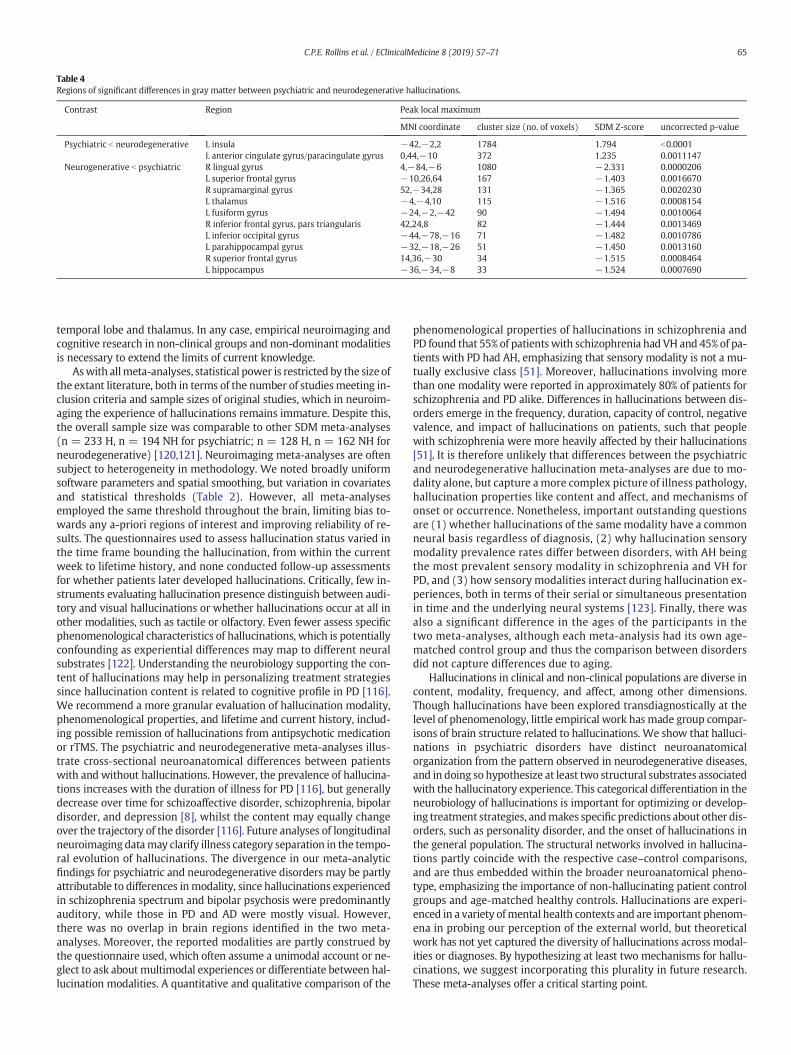
Table 4Regions of significant differences in gray matter between psychiatric and neurodegenerative hallucinations.
Contrast Region Peak local maximum
MNI coordinate cluster size (no. of voxels) SDM Z-score uncorrected p-value
Psychiatric b neurodegenerative L insula −42,−2,2 1784 1.794 b0.0001L anterior cingulate gyrus/paracingulate gyrus 0,44,−10 372 1.235 0.0011147
Neurogenerative b psychiatric R lingual gyrus 4,−84,−6 1080 −2.331 0.0000206L superior frontal gyrus −10,26,64 167 −1.403 0.0016670R supramarginal gyrus 52,−34,28 131 −1.365 0.0020230L thalamus −4,−4,10 115 −1.516 0.0008154L fusiform gyrus −24,−2,−42 90 −1.494 0.0010064R inferior frontal gyrus, pars triangularis 42,24,8 82 −1.444 0.0013469L inferior occipital gyrus −44,−78,−16 71 −1.482 0.0010786L parahippocampal gyrus −32,−18,−26 51 −1.450 0.0013160R superior frontal gyrus 14,36,−30 34 −1.515 0.0008464L hippocampus −36,−34,−8 33 −1.524 0.0007690
65C.P.E. Rollins et al. / EClinicalMedicine 8 (2019) 57–71
temporal lobe and thalamus. In any case, empirical neuroimaging andcognitive research in non-clinical groups and non-dominant modalitiesis necessary to extend the limits of current knowledge.
Aswith allmeta-analyses, statistical power is restricted by the size ofthe extant literature, both in terms of the number of studiesmeeting in-clusion criteria and sample sizes of original studies, which in neuroim-aging the experience of hallucinations remains immature. Despite this,the overall sample size was comparable to other SDM meta-analyses(n = 233 H, n = 194 NH for psychiatric; n = 128 H, n = 162 NH forneurodegenerative) [120,121]. Neuroimaging meta-analyses are oftensubject to heterogeneity in methodology. We noted broadly uniformsoftware parameters and spatial smoothing, but variation in covariatesand statistical thresholds (Table 2). However, all meta-analysesemployed the same threshold throughout the brain, limiting bias to-wards any a-priori regions of interest and improving reliability of re-sults. The questionnaires used to assess hallucination status varied inthe time frame bounding the hallucination, from within the currentweek to lifetime history, and none conducted follow-up assessmentsfor whether patients later developed hallucinations. Critically, few in-struments evaluating hallucination presence distinguish between audi-tory and visual hallucinations or whether hallucinations occur at all inother modalities, such as tactile or olfactory. Even fewer assess specificphenomenological characteristics of hallucinations, which is potentiallyconfounding as experiential differences may map to different neuralsubstrates [122]. Understanding the neurobiology supporting the con-tent of hallucinations may help in personalizing treatment strategiessince hallucination content is related to cognitive profile in PD [116].We recommend a more granular evaluation of hallucination modality,phenomenological properties, and lifetime and current history, includ-ing possible remission of hallucinations from antipsychotic medicationor rTMS. The psychiatric and neurodegenerative meta-analyses illus-trate cross-sectional neuroanatomical differences between patientswith and without hallucinations. However, the prevalence of hallucina-tions increases with the duration of illness for PD [116], but generallydecrease over time for schizoaffective disorder, schizophrenia, bipolardisorder, and depression [8], whilst the content may equally changeover the trajectory of the disorder [116]. Future analyses of longitudinalneuroimaging datamay clarify illness category separation in the tempo-ral evolution of hallucinations. The divergence in our meta-analyticfindings for psychiatric and neurodegenerative disorders may be partlyattributable to differences inmodality, since hallucinations experiencedin schizophrenia spectrum and bipolar psychosis were predominantlyauditory, while those in PD and AD were mostly visual. However,there was no overlap in brain regions identified in the two meta-analyses. Moreover, the reported modalities are partly construed bythe questionnaire used, which often assume a unimodal account or ne-glect to ask aboutmultimodal experiences or differentiate between hal-lucination modalities. A quantitative and qualitative comparison of the
phenomenological properties of hallucinations in schizophrenia andPD found that 55% of patients with schizophrenia had VH and 45% of pa-tients with PD had AH, emphasizing that sensory modality is not a mu-tually exclusive class [51]. Moreover, hallucinations involving morethan one modality were reported in approximately 80% of patients forschizophrenia and PD alike. Differences in hallucinations between dis-orders emerge in the frequency, duration, capacity of control, negativevalence, and impact of hallucinations on patients, such that peoplewith schizophrenia were more heavily affected by their hallucinations[51]. It is therefore unlikely that differences between the psychiatricand neurodegenerative hallucination meta-analyses are due to mo-dality alone, but capture amore complex picture of illness pathology,hallucination properties like content and affect, and mechanisms ofonset or occurrence. Nonetheless, important outstanding questionsare (1) whether hallucinations of the same modality have a commonneural basis regardless of diagnosis, (2) why hallucination sensorymodality prevalence rates differ between disorders, with AH beingthe most prevalent sensory modality in schizophrenia and VH forPD, and (3) how sensory modalities interact during hallucination ex-periences, both in terms of their serial or simultaneous presentationin time and the underlying neural systems [123]. Finally, there wasalso a significant difference in the ages of the participants in thetwo meta-analyses, although each meta-analysis had its own age-matched control group and thus the comparison between disordersdid not capture differences due to aging.
Hallucinations in clinical and non-clinical populations are diverse incontent, modality, frequency, and affect, among other dimensions.Though hallucinations have been explored transdiagnostically at thelevel of phenomenology, little empirical work has made group compar-isons of brain structure related to hallucinations. We show that halluci-nations in psychiatric disorders have distinct neuroanatomicalorganization from the pattern observed in neurodegenerative diseases,and in doing so hypothesize at least two structural substrates associatedwith the hallucinatory experience. This categorical differentiation in theneurobiology of hallucinations is important for optimizing or develop-ing treatment strategies, andmakes specific predictions about other dis-orders, such as personality disorder, and the onset of hallucinations inthe general population. The structural networks involved in hallucina-tions partly coincide with the respective case–control comparisons,and are thus embedded within the broader neuroanatomical pheno-type, emphasizing the importance of non-hallucinating patient controlgroups and age-matched healthy controls. Hallucinations are experi-enced in a variety ofmental health contexts and are important phenom-ena in probing our perception of the external world, but theoreticalwork has not yet captured the diversity of hallucinations across modal-ities or diagnoses. By hypothesizing at least two mechanisms for hallu-cinations, we suggest incorporating this plurality in future research.These meta-analyses offer a critical starting point.

Table 5Summary of systematic review from GMV ROI studies of regional brain volume comparing individuals with and without hallucinations.
Group Study Sample(M/F)
Age(SD)
Hallucinationmodality(assessmentscale)
ROI(s) Analysis(imagingsoftware)
Main result
Psychiatric Garrison et al.,2015 [43]
79(65/14)SCZ-H34(27/7)SCZ-NH
38.5(9.8)40.7(9.8)
Mixed(clinicalinterview)
Medial profrontal cortex (mPFC) VBM(SPM12)
Gray matter volumeSCZ-H N SCZ-NH: mPFC region surrounding theanterior PCS
Cierpka et al., 2017[71]
10 (6/4)SCZ-H10 (8/2)SCZ-NH
36.5(9.0)32.1(6.2)
Auditory(BRPS,PANSS,PsyRatS)
Cerebellum VBM(SPM8)
Gray matter volumeSCZ-H b SCZ-NH: right lobule VIIIa
Kubera et al., 2014[76]
10 (6/4)SCZ-H10 (8/2)SCZ-NH
36.5(9.0)32.1(6.2)
Auditory(BRPS,PANSS,PsyRatS)
n/a SBM(GIFT)
Gray matter volumeSCZ-H b SCZ-NH: component consisting MFG;IFG; STG; insula; IPL; rectal gyrus; transversetemporal gyrus; supramarginal gyrus; lingualgyrus; postcentral gyrus; fusiform gyrus;subcallosal gyrus; MTG; ITG; orbital gyrus
Neves et al., 2016[63]
9 (3/6)BD-H12 (6/6)BD-NH
37.7(12.1)39.9(15.0)
Auditory orvisual(MINI-Plus)
Orbitofrontal cortex and ventralprefrontal areas, cingulate gyrus,fusiform gyrus, superior temporalsulcus, amygdala, insula, thalamus
VBM(SPM8)
Gray matter volumeBD-H b BD-NH: right posterior insular cortex
Stanfield et al.,2009 [72]
17 (n/a)BD-H49 (n/a)BD-NH
36.4(11.1)a
Auditory(OPCRITsymptomchecklist)
Temporal lobe VBM(SPM99)
Gray matter densityBD-H b BD-NH: left middle temporal gyrus
Neurodegenerative Janzen et al., 2012[73]
13 (6/7)PD-H13 (7/6)PDD-H16 (9/7)PD-NH
66.0(6.9)67.7(7.1)64.3(8.0)
Visual(UPDRS)
Pedunculopontine nucleus (PPN),thalamus
VBM(SPM8)
Gray matter volumePD-H + PDD-H b PD-NH: PPN, thalamusPD-H b PD-NH: PPN
Sanchez-Castenadaet al., 2010 [74]
6 (4/2)DLB-H6 (4/2)DLB-NH8 (6/2)PDD-H7 (4/3)PDD-NH
70.2(12.4)71(10.7)75.3(4.9)70.6(7.1)
Visual (NPI) Frontal (BA 6, 8, 9, 10, 44, 45, and47), occipital (BA 18,19), parietal(BA 7, 39, 40), and temporal (20)regions
VBM(SPM5)
Gray matter volumeDLB-H b DLB-NH: right inferior frontal gyrus(BA 45)PDD-H b PDD-NH: left orbitofrontal lobe (BA10)
Colloby et al., 2017[75]
41(26/15)DLB-H47(33/14)AD-NH
78.6(6.2)79.0(8.8)
Visual (NPI) Substantia innomiata (SI) VBM(SPM8)
Gray matter volumen.s.
Abbreviations: AD: Alzheimer's disease; PD: Parkinson's disease; PDD: Parkinson's diseasewith dementia; SCZ: schizophrenia; FES: first episode schizophrenia; BD: bipolar disorder; nPD:Parkinson's diseasewithout dementia; FEP: first episode psychosis; ARMS-LT: at risk mental state long-term; DLB: dementiawith Lewy bodies; X-H: population Xwith hallucinations; X-NH: population X without hallucinations; NPI: Neuropsychiatric Inventory Questionnaire; MDS-UPDRS: Movement Disorder Society (MDS)-sponsored version of the Unified Parkinson'sdisease Rating Scale (UPDRS); PANSS: Positive and Negative Symptom Scale; HAHRS: Hoffman Auditory Hallucination Rating Scale;MINI-Plus;Mini International Neuropsychiatric Inter-view (MINI) Plus; SAPS: Scale for the Assessment of Positive Symptoms; BRPS: Brief Psychiatric Rating Scale; OPCRIT: Operational Criteria Checklist for Psychotic Illness and Affective Ill-ness; MFG: medial frontal gyrus; IFG: inferior frontal gyrus; STG: superior temporal gyrus: IPL: inferior parietal lobule; MTG: middle temporal gyrus; ITG: inferior temporal gyrus; SBM:source-based morphometry.
a Hallucination and no-hallucinations groups combined.
66 C.P.E. Rollins et al. / EClinicalMedicine 8 (2019) 57–71
Contributors
JS conceived and directed the project. CPER planned the searchcriteria, completed the literature search, data extraction, quality assess-ment, data analyses and summary, created the figures, and wrote thefirst draft of the manuscript, with input from JRG, JS, and GKM. JRG, JS,and GKM confirmed the results of data extraction. All authors criticallyreviewed the manuscript and contributed to its writing and revision.
Declaration of Interests
Ms. Rollins reports a scholarship from Gates Cambridge during theconduct of the study. Professor Rowe reports grants from WellcomeTrust during the conduct of the study, grants from NIHR, McDonnell
Foundation, PSP Association, Parkinsons UK, Medical Research Council,Evelyn Trust, and AZ-Medimmune, personal fees from Asceneuron,and other fromGuarantors of Brain outside the submitted work. Profes-sor Suckling, Dr. Murray, Dr. Garrison, Dr. Simons, and Dr. O'Callaghanhave nothing to disclose.
Acknowledgements
The authors are supported by the following funding sources: GatesCambridge (CPER); Wellcome Trust (JBR, 103838; CO, 200181/Z/15/Z);National Health and Medical Research Council Neil Hamilton Fairley Fel-lowship (CO, 1091310); Wellcome Trust Collaborative award (JRG), anda joint award from the Medical Research Council and theWellcome Trust(JSS).
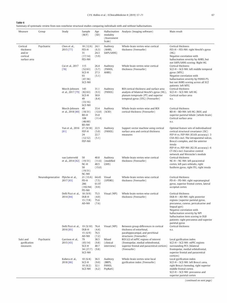
Table 6Summary of systematic review from non-voxelwise structural studies comparing individuals with and without hallucinations.
Measure Group Study Sample(M/F)
Age(SD)
Hallucinationmodality(AssessmentScale)
Analysis (Imaging software) Main result
Corticalthicknessand/orcorticalsurface area
Psychiatric Chen et al.,2015 [77]
18 (12/6)FES-H31(17/14)FES-NH
24.1(6.3)24.3(5.9)
Auditory(AHRS,SAPS/SANS)
Whole-brain vertex-wise corticalthickness (Freesurfer)
Cortical thicknessFES-H b FES-NH: right Heschl's gyrus(HG)Negative correlation withhallucination severity by AHRS, butnot SAPS/SANS scoring: Right HG
Cui et al., 2017[78]
115(52/63)SCZ-H93(47/36)SCZ-NH
26.4(5.7)27.3(5.1)
Auditory(PANSS,AHRS)
Whole brain vertex-wise corticalthickness (Freesurfer)
Cortical thicknessSCZ-H b SCZ-NH: left middle temporalgyrus (MTG)Negative correlation withhallucination severity by PANSS P3,but not AHRS scoring across all SCZpatients: left MTG
Morch-Johnsenet al., 2017 [79]
145(82/63)SCZ-H49(33/16)SCZ-NH
31.1(9.3)30.9(8.4)
Auditory(PANSS)
ROI cortical thickness and surface areaanalysis of bilateral Heschl's gyrus (HG),planum temproale (PT) and superiortemporal gyrus (STG) (Freesurfer)
Cortical thicknessSCZ-H b SCZ-NH: left HGCortical surface arean.s.
Morch-Johnsenet al., 2018 [80]
49(18/31)BD-H108(48/60)BD-NH
33.4(12.0)35.0(11.4)
Auditory(SCID)
Whole-brain vertex-wise and ROIcortical thickness (Freesurfer)
Cortical thicknessBD-H N BD-NH: left HG (ROI) andsuperior parietal lobule (whole-brain)Cortical surface arean.s.
Yun et al., 2016[81]
27 (9/18)FEP-H24(12/12)FEP-NH
22.5(5.0)22.7(5.1)
Auditory(PANSS)
Support vector machine using corticalsurface area and cortical thicknessmeasures
Optimal feature sets of individualizedcortical structural covariance (ISC)FEP-H vs. FEP-NH (83.6% accuracy): 3CSA-ISCs incl. The intraparietal sulcus,Broca's complex, and the anteriorinsulaFEP-H vs. FEP-NH (82.3% accuracy): 6CT-ISCs incl. Executive controlnetwork and Wernicke's module
van Lutterveldet al., 2014 [82]
50(19/31)NC-H50(19/31)NC-NH
40.8(11.6)40.5(15.0)
Auditory(modifiedLSHS)
Whole-brain vertex-wise corticalthickness (Freesurfer)
Cortical thicknessNC-H b NC-NH: left paracentralcortex, left pars orbitalis, rightfusiform gyrus, right ITG, right insula
Neurodegenerative Ffytche et al.,2017 [83]
21 (15/6)PD-H286(192/94)PD-NH
64.43(7.5)61.97(9.9)
Visual(UPDRS)
Whole-brain vertex-wise corticalthickness (Freesurfer)
Cortical thicknessPD-H b PD-NH: right supramarginalgyrus, superior frontal cortex, lateraloccipital cortex
Delli Pizzi et al.,2014 [84]
18 (9/9)DLB-H15 (7/8)AD-NH
75.5(4.0)75.6(7.6)
Visual (NPI) Whole brain vertex-wise corticalthickness (Freesurfer)
Cortical thicknessDLB-H b AD-NH: right posteriorregions (superior parietal gyrus,precuneus, cuneus, pericalcarine andlingual gyri)Negative correlation withhallucination severity by NPIhallucination item scoring in DLBpatients: right precuneus and superiorparietal gyrus
Delli Pizzi et al.,2016 [85]
19 (9/10)DLB-H15 (6/9)AD-NH
76.4(4.4)76.5(7.2)
Visual (NPI) Between group differences in corticalthickness of entorhinal,parahippocampal, and perirhinalstructures (Freesurfer)
Cortical thicknessn.s.
Sulci andgyrificationmeasures
Psychiatric Garrison et al.,2015 [43]
79(65/14)SCZ-H34 (27/7)SCZ-NH
38.5(9.8)40.7(9.8)
Mixed(clinicalinterview)
ROI LGI of mPFC regions of interest(frontopolar, medial orbitofrontal,superior frontal and paracentral cortices)(Freesurfer)
Local gyrification indexSCZ-H b SCZ-NH: mPFC regionssurrounding PCS (bilateralfrontopolar, medial orbitofrontal,superior frontal and paracentralcortices)
Kubera et al.,2018 [86]
10 (6/4)SCZ-H10 (8/2)SCZ-NH
36.5(9.0)32.1(6.2)
Auditory(BRPS,PANSS,PsyRatS)
Whole-brain vertex-wise localgyrification index (Freesurfer)
Local gyrification indexSCZ-H b SCZ-NH: left Broca's area,right Broca's homolog, right superiormiddle frontal cortexSCZ-H N SCZ-NH: precuneus andsuperior parietal cortex
(continued on next page)
67C.P.E. Rollins et al. / EClinicalMedicine 8 (2019) 57–71
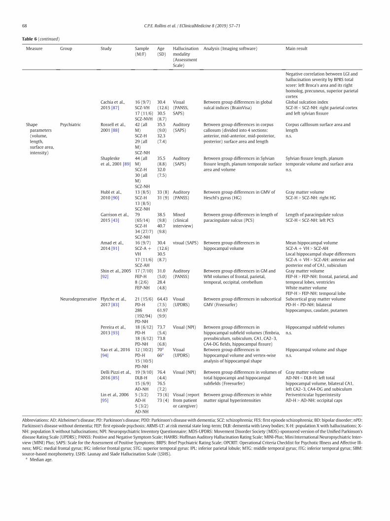
Table 6 (continued)
Measure Group Study Sample(M/F)
Age(SD)
Hallucinationmodality(AssessmentScale)
Analysis (Imaging software) Main result
Negative correlation between LGI andhallucination severity by BPRS totalscore: left Broca's area and its righthomolog, precuneus, superior parietalcortex
Cachia et al.,2015 [87]
16 (9/7)SCZ-VH17 (11/6)SCZ-NVH
30.4(12.6)30.5(8.7)
Visual(PANSS,SAPS)
Between group differences in globalsulcal indices (BrainVisa)
Global sulcation indexSCZ-H b SCZ-NH: right parietal cortexand left sylvian fissure
Shapeparameters(volume,length,surface area,intensity)
Psychiatric Rossell et al.,2001 [88]
42 (allM)SCZ-H29 (allM)SCZ-NH
35.5(9.0)32.3(7.4)
Auditory(SAPS)
Between group differences in corpuscallosum (divided into 4 sections:anterior, mid-anterior, mid-posterior,posterior) surface area and length
Corpus calllosum surface area andlengthn.s.
Shapleskeet al., 2001 [89]
44 (allM)SCZ-H30 (allM)SCZ-NH
35.5(8.8)32.0(7.5)
Auditory(SAPS)
Between group differences in Sylvianfissure length, planum temporale surfacearea and volume
Sylvian fissure length, planumtemporale volume and surface arean.s.
Hubl et al.,2010 [90]
13 (8/5)SCZ-H13 (8/5)SCZ-NH
33 (8)31 (9)
Auditory(PANSS)
Between group differences in GMV ofHeschl's gyrus (HG)
Gray matter volumeSCZ-H N SCZ-NH: right HG
Garrison et al.,2015 [43]
79(65/14)SCZ-H34 (27/7)SCZ-NH
38.5(9.8)40.7(9.8)
Mixed(clinicalinterview)
Between group differences in length ofparacingulate sulcus (PCS)
Length of paracingulate sulcusSCZ-H b SCZ-NH: left PCS
Amad et al.,2014 [91]
16 (9/7)SCZ-A +VH17 (11/6)SCZ-AH
30.4(12.6)30.5(8.7)
visual (SAPS) Between group differences inhippocampal volume
Mean hippocampal volumeSCZ-A + VH N SCZ-AHLocal hippocampal shape differencesSCZ-A + VH N SCZ-AH: anterior andposterior end of CA1, subiculum
Shin et al., 2005[92]
17 (7/10)FEP-H8 (2/6)FEP-NH
31.0(5.0)28.4(4.8)
Auditory(PANSS)
Between group differences in GM andWM volumes of frontal, parietal,temporal, occipital, cerebellum
Gray matter volumeFEP-H N FEP-NH: frontal, parietal, andtemporal lobes, ventriclesWhite matter volumeFEP-H N FEP-NH: temporal lobe
Neurodegenerative Ffytche et al.,2017 [83]
21 (15/6)PD-H286(192/94)PD-NH
64.43(7.5)61.97(9.9)
Visual(UPDRS)
Between group differences in subcorticalGMV (Freesurfer)
Subcortical gray matter volumePD-H b PD-NH: bilateralhippocampus, caudate, putamen
Pereira et al.,2013 [93]
18 (6/12)PD-H18 (6/12)PD-NH
73.7(5.4)73.8(6.8)
Visual (NPI) Between group differences inhippocampal subfield volumes (fimbria,presubiculum, subiculum, CA1, CA2–3,CA4-DG fields, hippocampal fissure)
Hippocampal subfield volumesn.s.
Yao et al., 2016[94]
12 (10/2)PD-H15 (10/5)PD-NH
70a
66aVisual(UPDRS)
Between group differences inhippocampal volume and vertex-wiseanalysis of hippocampal shape
Hippocampal volume and shapen.s.
Delli Pizzi et al.,2016 [85]
19 (9/10)DLB-H15 (6/9)AD-NH
76.4(4.4)76.5(7.2)
Visual (NPI) Between group differences in volumes oftotal hippocampi and hippocampalsubfields (Freesurfer)
Gray matter volumeAD-NH b DLB-H: left totalhippocampal volume, bilateral CA1,left CA2–3, CA4-DG and subiculum
Lin et al., 2006[95]
5 (3/2)AD-H5 (3/2)AD-NH
73 (6)73 (4)
Visual (reportfrom patientor caregiver)
Between group differences in whitematter signal hyperintensities
Periventricular hyperintesityAD-H N AD-NH: occipital caps
Abbreviations: AD: Alzheimer's disease; PD: Parkinson's disease; PDD: Parkinson's diseasewith dementia; SCZ: schizophrenia; FES: first episode schizophrenia; BD: bipolar disorder; nPD:Parkinson's diseasewithout dementia; FEP: first episode psychosis; ARMS-LT: at risk mental state long-term; DLB: dementiawith Lewy bodies; X-H: population Xwith hallucinations; X-NH: population X without hallucinations; NPI: Neuropsychiatric Inventory Questionnaire; MDS-UPDRS: Movement Disorder Society (MDS)-sponsored version of the Unified Parkinson'sdisease Rating Scale (UPDRS); PANSS: Positive and Negative Symptom Scale; HAHRS: Hoffman Auditory Hallucination Rating Scale;MINI-Plus;Mini International Neuropsychiatric Inter-view (MINI) Plus; SAPS: Scale for the Assessment of Positive Symptoms; BRPS: Brief Psychiatric Rating Scale; OPCRIT: Operational Criteria Checklist for Psychotic Illness and Affective Ill-ness; MFG: medial frontal gyrus; IFG: inferior frontal gyrus; STG: superior temporal gyrus: IPL: inferior parietal lobule; MTG: middle temporal gyrus; ITG: inferior temporal gyrus; SBM:source-based morphometry. LSHS: Launay and Slade Hallucination Scale (LSHS).
a Median age.
68 C.P.E. Rollins et al. / EClinicalMedicine 8 (2019) 57–71

69C.P.E. Rollins et al. / EClinicalMedicine 8 (2019) 57–71
Appendix A. Supplementary data
Supplementary data to this article can be found online at https://doi.org/10.1016/j.eclinm.2019.01.012.
References
[1] Woods A, Jones N, Alderson-Day B, Callard F, Fernyhough C. Experiences of hearingvoices: analysis of a novel phenomenological survey. Lancet Psychiatry 2015;2(4):323–31.
[2] Lim A, Hoek HW, Deen ML, Blom JD, Investigators G. Prevalence and classificationof hallucinations in multiple sensory modalities in schizophrenia spectrum disor-ders. Schizophr Res 2016;176(2–3):493–9.
[3] Baethge C, Baldessarini RJ, Freudenthal K, Streeruwitz A, Bauer M, Bschor T. Hallu-cinations in bipolar disorder: characteristics and comparison to unipolar depres-sion and schizophrenia. Bipolar Disord 2005;7(2):136–45.
[4] Diederich NJ, Fenelon G, Stebbins G, Goetz CG. Hallucinations in Parkinson disease.Nat Rev Neurol 2009;5(6):331–42.
[5] Onofrj M, Taylor JP, Monaco D, et al. Visual hallucinations in PD and Lewy body de-mentias: old and new hypotheses. Behav Neurol 2013;27(4):479–93.
[6] Zhao QF, Tan L, Wang HF, et al. The prevalence of neuropsychiatric symptoms inAlzheimer's disease: systematic review and meta-analysis. J Affect Disord 2016;190:264–71.
[7] Maijer K, Begemann MJH, Palmen S, Leucht S, Sommer IEC. Auditory hallucinationsacross the lifespan: a systematic review and meta-analysis. Psychol Med 2018;48(6):879–88.
[8] Goghari VM, Harrow M. Twenty year multi-follow-up of different types of halluci-nations in schizophrenia, schizoaffective disorder, bipolar disorder, and depression.Schizophr Res 2016;176(2–3):371–7.
[9] Wakamori T, Agari T, Yasuhara T, et al. Cognitive functions in Parkinson's disease:relation to disease severity and hallucination. Parkinsonism Relat Disord 2014;20(4):415–20.
[10] Wilson RS, Krueger KR, Kamenetsky JM, et al. Hallucinations and mortality inAlzheimer disease. The American Journal of Geriatric Psychiatry 2005;13(11):984–90.
[11] Kjelby E, Sinkeviciute I, Gjestad R, et al. Suicidality in schizophrenia spectrum dis-orders: the relationship to hallucinations and persecutory delusions. European psy-chiatry 2015;30(7):830–6.
[12] Jardri R, Bartels-Velthuis AA, Debbane M, et al. From phenomenology to neuro-physiological understanding of hallucinations in children and adolescents.Schizophr Bull 2014;40(Suppl. 4):S221–32.
[13] Baumeister D, Sedgwick O, Howes O, Peters E. Auditory verbal hallucinations andcontinuum models of psychosis: a systematic review of the healthy voice-hearerliterature. Clin Psychol Rev 2017;51:125–41.
[14] Laroi F, Luhrmann TM, Bell V, et al. Culture and hallucinations: overview and futuredirections. Schizophr Bull 2014;40(Suppl. 4):S213–20.
[15] Waters F, Fernyhough C. Hallucinations: a systematic review of points of similarityand difference across diagnostic classes. Schizophr Bull 2017;43(1):32–43.
[16] Daalman K, van Zandvoort M, Bootsman F, Boks M, Kahn R, Sommer I. Auditoryverbal hallucinations and cognitive functioning in healthy individuals. SchizophrRes 2011;132(2–3):203–7.
[17] van Os J, Reininghaus U. Psychosis as a transdiagnostic and extended phenotype inthe general population. World Psychiatry 2016;15(2):118–24.
[18] BohlkenMM, Hugdahl K, Sommer IE. Auditory verbal hallucinations: neuroimagingand treatment. Psychol Med 2017;47(2):199–208.
[19] Waters F, Collerton D, Ffytche DH, et al. Visual hallucinations in the psychosis spec-trum and comparative information from neurodegenerative disorders and eye dis-ease. Schizophr Bull 2014;40(Suppl. 4):S233–45.
[20] Carter R, Ffytche DH. On visual hallucinations and cortical networks: a trans-diagnostic review. J Neurol 2015;262(7):1780–90.
[21] Jardri R, Pouchet A, Pins D, Thomas P. Cortical activations during auditory verbalhallucinations in schizophrenia: a coordinate-based meta-analysis. Am J Psychiatry2011;168(1):73–81.
[22] Kuhn S, Gallinat J. Quantitative meta-analysis on state and trait aspects of auditoryverbal hallucinations in schizophrenia. Schizophr Bull 2012;38(4):779–86.
[23] Modinos G, Costafreda SG, van Tol MJ, McGuire PK, Aleman A, Allen P.Neuroanatomy of auditory verbal hallucinations in schizophrenia: a quantita-tive meta-analysis of voxel-based morphometry studies. Cortex 2013;49(4):1046–55.
[24] Palaniyappan L, Balain V, Radua J, Liddle PF. Structural correlates of auditory hallu-cinations in schizophrenia: a meta-analysis. Schizophr Res 2012;137(1–3):169–73.
[25] Pezzoli S, Cagnin A, Bandmann O, Structural Venneri A. Functional neuroimaging ofvisual hallucinations in Lewy body disease: a systematic literature review. Brain Sci2017;7(7).
[26] Allen P, Laroi F, McGuire PK, Aleman A. The hallucinating brain: a review of struc-tural and functional neuroimaging studies of hallucinations. Neurosci Biobehav Rev2008;32(1):175–91.
[27] Zmigrod L, Garrison JR, Carr J, Simons JS. The neural mechanisms of hallucinations:a quantitative meta-analysis of neuroimaging studies. Neurosci Biobehav Rev2016;69:113–23.
[28] Waters FA, Badcock JC, Michie PT, Maybery MT. Auditory hallucinations in schizo-phrenia: intrusive thoughts and forgotten memories. Cogn Neuropsychiatry 2006;11(1):65–83.
[29] Northoff G, Qin P. How can the brain's resting state activity generate hallucina-tions? A 'resting state hypothesis' of auditory verbal hallucinations. SchizophrRes 2011;127(1–3):202–14.
[30] Palaniyappan L, Liddle PF. Does the salience network play a cardinal role in psycho-sis? An emerging hypothesis of insular dysfunction. Journal of Psychiatry & Neuro-science 2012;37(1):17–27.
[31] Nazimek JM, Hunter MD, Woodruff PW. Auditory hallucinations: expectation-perception model. Med Hypotheses 2012;78(6):802–10.
[32] Collerton D, Perry E, McKeith I. Why people see things that are not there: a novelperception and attention deficit model for recurrent complex visual hallucinations.Behav Brain Sci 2005;28(6):737–57 [discussion 57-94].
[33] Hugdahl K. "Hearing voices": auditory hallucinations as failure of top-down controlof bottom-up perceptual processes. Scand J Psychol 2009;50(6):553–60.
[34] Jardri R, Hugdahl K, Hughes M, et al. Are hallucinations due to an imbalance be-tween excitatory and inhibitory influences on the brain? Schizophr Bull 2016;42(5):1124–34.
[35] Fletcher PC, Frith CD. Perceiving is believing: a Bayesian approach to explaining thepositive symptoms of schizophrenia. Nat Rev Neurosci 2009;10(1):48–58.
[36] Muller AJ, Shine JM, Halliday GM, Lewis SJ. Visual hallucinations in Parkinson's dis-ease: theoretical models. Mov Disord 2014;29(13):1591–8.
[37] Powers III AR, Kelley M, Corlett PR. Hallucinations as top-down effects on percep-tion. Biol Psychiatry Cogn Neurosci Neuroimaging 2016;1(5):393–400.
[38] Cho R, WuW. Mechanisms of auditory verbal hallucination in schizophrenia. FrontPsych 2013;4:155.
[39] Alderson-Day B, Diederen K, Fernyhough C, et al. Auditory hallucinations and theBrain's resting-state networks: findings and methodological observations.Schizophr Bull 2016;42(5):1110–23.
[40] Jones SR. Do we need multiple models of auditory verbal hallucinations? Examin-ing the phenomenological fit of cognitive and neurological models. Schizophr Bull2010;36(3):566–75.
[41] Rolland B, Jardri R, Amad A, Thomas P, Cottencin O, Bordet R. Pharmacology of hal-lucinations: several mechanisms for one single symptom? Biomed Res Int 2014;2014 {Rolland, 2014 #81}. (307106).
[42] Curcic-Blake B, Ford JM, Hubl D, et al. Interaction of language, auditory and mem-ory brain networks in auditory verbal hallucinations. Prog Neurobiol 2017;148:1–20.
[43] Garrison JR, Fernyhough C, McCarthy-Jones S, HaggardM, Australian SchizophreniaResearch B, Simons JS. Paracingulate sulcus morphology is associated with halluci-nations in the human brain. Nat Commun 2015;6:8956.
[44] Muller VI, Cieslik EC, Laird AR, et al. Ten simple rules for neuroimaging meta-analysis. Neurosci Biobehav Rev 2018;84:151–61.
[45] Allen P, Aleman A, McGuire PK. Inner speech models of auditory verbal hallucina-tions: evidence from behavioural and neuroimaging studies. International Reviewof Psychiatry 2007;19(4):407–15 Abingdon, England.
[46] Diederich NJ, Goetz CG, Stebbins GT. Repeated visual hallucinations in Parkinson'sdisease as disturbed external/internal perceptions: focused review and a new inte-grative model. Movement Disorders 2005;20(2):130–40.
[47] Moher D, Liberati A, Tetzlaff J, Altman DG, Group P. Preferred reporting items forsystematic reviews and meta-analyses: the PRISMA statement. BMJ 2009;339:b2535.
[48] Radua J, Mataix-Cols D, Phillips ML, et al. A new meta-analytic method for neuro-imaging studies that combines reported peak coordinates and statistical paramet-ric maps. European Psychiatry 2012;27(8):605–11.
[49] Radua J, Rubia K, Canales-Rodriguez EJ, Pomarol-Clotet E, Fusar-Poli P, Mataix-ColsD. Anisotropic kernels for coordinate-based meta-analyses of neuroimaging stud-ies. Front Psych 2014;5:13.
[50] Eickhoff SB, Laird AR, Grefkes C, Wang LE, Zilles K, Fox PT. Coordinate-based activa-tion likelihood estimation meta-analysis of neuroimaging data: a random-effectsapproach based on empirical estimates of spatial uncertainty. Hum Brain Mapp2009;30(9):2907–26.
[51] Llorca PM, Pereira B, Jardri R, et al. Hallucinations in schizophrenia and Parkinson'sdisease: an analysis of sensory modalities involved and the repercussion on pa-tients. Sci Rep 2016;6:38152.
[52] van Swam C, Federspiel A, Hubl D, et al. Possible dysregulation of cortical plasticityin auditory verbal hallucinations-a cortical thickness study in schizophrenia. JPsychiatr Res 2012;46(8):1015–23.
[53] Smieskova R, Fusar-Poli P, Aston J, et al. Insular volume abnormalities associatedwith different transition probabilities to psychosis. Psychol Med 2012;42(8):1613–25.
[54] Lee YM, Chung YI, Park JM, et al. Decreased gray matter volume is associated withthe subtypes of psychotic symptoms in patients with antipsychotic-naive mild ormoderate Alzheimer's disease: a voxel-based morphometry study. Psychiatry Res2016;249:45–51.
[55] Radua J, Mataix-Cols D. Voxel-wise meta-analysis of grey matter changes inobsessive-compulsive disorder. The British Journal of Psychiatry 2009;195(5):393–402.
[56] Radua J, van den Heuvel OA, Surguladze S, Mataix-Cols D. Meta-analytical compar-ison of voxel-based morphometry studies in obsessive-compulsive disorder vsother anxiety disorders. Arch Gen Psychiatry 2010;67(7):701–11.
[57] Radua J, Romeo M, Mataix-Cols D, Fusar-Poli P. A general approach for combiningvoxel-based meta-analyses conducted in different neuroimaging modalities. CurrMed Chem 2013;20(3):462–6.
[58] Wise T, Radua J, Via E, Cardoner N, Abe O, Adams TM, et al. Common and distinctpatterns of grey-matter volume alteration in major depression and bipolar disor-der: evidence from voxel-based meta-analysis. Mol Psychiatry 2017;22(10):1455–63.
70 C.P.E. Rollins et al. / EClinicalMedicine 8 (2019) 57–71
[59] Gaser C, Nenadic I, Volz HP, Buchel C, Sauer H. Neuroanatomy of "hearing voices": afrontotemporal brain structural abnormality associated with auditory hallucina-tions in schizophrenia. Cerebral Cortex 2004;14(1):91–6 New York, NY: 1991.
[60] Shapleske J, Rossell SL, Chitnis XA, et al. A computational morphometric MRI studyof schizophrenia: effects of hallucinations. Cerebral Cortex 2002;12(12):1331–41New York, NY: 1991.
[61] van Tol MJ, van der Meer L, Bruggeman R, Modinos G, Knegtering H, Aleman A.Voxel-based gray and white matter morphometry correlates of hallucinations inschizophrenia: the superior temporal gyrus does not stand alone. NeuroImageClinical 2014; 4: 249–57.
[62] Huang P, Xi Y, Lu ZL, et al. Decreased bilateral thalamic gray matter volume in first-episode schizophrenia with prominent hallucinatory symptoms: a volumetric MRIstudy. Sci Rep 2015;5:14505.
[63] NevesMD, Duarte DG, AlbuquerqueMR, et al. Neural correlates of hallucinations inbipolar disorder. Rev Bras Psiquiatr 2016;38(1):1–5.
[64] Goldman JG, Stebbins GT, Dinh V, et al. Visuoperceptive region atrophy indepen-dent of cognitive status in patients with Parkinson's disease with hallucinations.Brain 2014;137(Pt 3):849–59.
[65] Meppelink AM, de Jong BM, Teune LK, van Laar T. Regional cortical greymatter lossin Parkinson's disease without dementia is independent from visual hallucinations.Movement Disorders 2011;26(1):142–7.
[66] Pagonabarraga J, Soriano-Mas C, Llebaria G, Lopez-SolaM, Pujol J, Kulisevsky J. Neu-ral correlates of minor hallucinations in non-demented patients with Parkinson'sdisease. Parkinsonism Relat Disord 2014;20(3):290–6.
[67] Ramirez-Ruiz B, Marti MJ, Tolosa E, et al. Cerebral atrophy in Parkinson's diseasepatients with visual hallucinations. Eur J Neurol 2007;14(7):750–6.
[68] Watanabe H, Senda J, Kato S, et al. Cortical and subcortical brain atrophy inParkinson's disease with visual hallucination. Movement Disorders 2013;28(12):1732–6.
[69] Shin S, Lee JE, Hong JY, Sunwoo MK, Sohn YH, Lee PH. Neuroanatomical substratesof visual hallucinations in patients with non-demented Parkinson's disease. JNeurol Neurosurg Psychiatry 2012;83(12):1155–61.
[70] Blanc F, Noblet V, Philippi N, et al. Right anterior insula: core region of hallucina-tions in cognitive neurodegenerative diseases. PloS one 2014;9(12):e114774.
[71] Cierpka M, Wolf ND, Kubera KM, et al. Cerebellar contributions to persistent audi-tory verbal hallucinations in patients with schizophrenia. Cerebellum 2017;16(5–6):964–72 London, England.
[72] Stanfield AC, Moorhead TW, Job DE, et al. Structural abnormalities of ventrolateraland orbitofrontal cortex in patients with familial bipolar disorder. Bipolar Disord2009;11(2):135–44.
[73] Janzen J, van't Ent D, Lemstra AW, Berendse HW, Barkhof F, Foncke EM. Thepedunculopontine nucleus is related to visual hallucinations in Parkinson's disease:preliminary results of a voxel-based morphometry study. J Neurol 2012;259(1):147–54.
[74] Sanchez-Castaneda C, Rene R, Ramirez-Ruiz B, et al. Frontal and associative vi-sual areas related to visual hallucinations in dementia with Lewy bodies andParkinson's disease with dementia. Movement Disorders 2010;25(5):615–22.
[75] Colloby SJ, Elder GJ, Rabee R, O'Brien JT, Taylor JP. Structural grey matter changes inthe substantia innominata in Alzheimer's disease and dementia with Lewy bodies:a DARTEL-VBM study. Int J Geriatr Psychiatry 2017;32(6):615–23.
[76] Kubera KM, Sambataro F, Vasic N, et al. Source-based morphometry of graymatter volume in patients with schizophrenia who have persistent auditoryverbal hallucinations. Prog Neuropsychopharmacol Biol Psychiatry 2014;50:102–9.
[77] Chen X, Liang S, Pu W, et al. Reduced cortical thickness in right Heschl's gyrus as-sociatedwith auditory verbal hallucinations severity in first-episode schizophrenia.BMC Psychiatry 2015;15:152.
[78] Cui Y, Liu B, SongM, et al. Auditory verbal hallucinations are related to cortical thin-ning in the left middle temporal gyrus of patients with schizophrenia. Psychol Med2017:1–8.
[79] Morch-Johnsen L, Nesvag R, Jorgensen KN, et al. Auditory cortex characteristics inschizophrenia: associations with auditory hallucinations. Schizophr Bull 2017;43(1):75–83.
[80] Morch-Johnsen L, Nerland S, Jorgensen KN, et al. Cortical thickness abnormalities inbipolar disorder patients with a lifetime history of auditory hallucinations. BipolarDisord 2018;20(7):647–57.
[81] Yun JY, Kim SN, Lee TY, Chon MW, Kwon JS. Individualized covariance profile ofcortical morphology for auditory hallucinations in first-episode psychosis. HumBrain Mapp 2016;37(3):1051–65.
[82] van Lutterveld R, van den Heuvel MP, Diederen KM, et al. Cortical thickness in in-dividuals with non-clinical and clinical psychotic symptoms. Brain 2014;137(Pt10):2664–9.
[83] Ffytche DH, Pereira JB, Ballard C, Chaudhuri KR, Weintraub D, Aarsland D. Risk fac-tors for early psychosis in PD: insights from the Parkinson's progression markersinitiative. J Neurol Neurosurg Psychiatry 2017;88(4):325–31.
[84] Delli Pizzi S, Franciotti R, Tartaro A, et al. Structural alteration of the dorsal visualnetwork in DLB patients with visual hallucinations: a cortical thickness MRIstudy. PloS one 2014;9(1):e86624.
[85] Delli Pizzi S, Franciotti R, Bubbico G, Thomas A, OnofrjM, Bonanni L. Atrophy of hip-pocampal subfields and adjacent extrahippocampal structures in dementia withLewy bodies and Alzheimer's disease. Neurobiol Aging 2016;40:103–9.
[86] Kubera KM, Thomann PA, Hirjak D, et al. Cortical folding abnormalities in patientswith schizophrenia who have persistent auditory verbal hallucinations. EuropeanNeuropsychopharmacology 2018;28(2):297–306.
[87] Cachia A, Amad A, Brunelin J, et al. Deviations in cortex sulcation associated withvisual hallucinations in schizophrenia. Mol Psychiatry 2015;20(9):1101–7.
[88] Rossell SL, Shapleske J, Fukuda R, Woodruff PW, Simmons A, David AS. Corpuscallosum area and functioning in schizophrenic patients with auditory—verbal hal-lucinations. Schizophr Res 2001;50(1–2):9–17.
[89] Shapleske J, Rossell SL, Simmons A, David AS,Woodruff PW. Are auditory hallucina-tions the consequence of abnormal cerebral lateralization? A morphometric MRIstudy of the sylvian fissure and planum temporale. Biol Psychiatry 2001;49(8):685–93.
[90] Hubl D, Dougoud-Chauvin V, Zeller M, et al. Structural analysis of Heschl's gyrus inschizophrenia patients with auditory hallucinations. Neuropsychobiology 2010;61(1):1–9.
[91] Amad A, Cachia A, Gorwood P, et al. The multimodal connectivity of the hippocam-pal complex in auditory and visual hallucinations. Mol Psychiatry 2014;19(2):184–91.
[92] Shin SE, Lee JS, Kang MH, Kim CE, Bae JN, Jung G. Segmented volumes of cerebrumand cerebellum in first episode schizophrenia with auditory hallucinations. Psychi-atry Res 2005;138(1):33–42.
[93] Pereira JB, Junque C, Bartres-Faz D, Ramirez-Ruiz B, Marti MJ, Tolosa E. Regionalvulnerability of hippocampal subfields and memory deficits in Parkinson's disease.Hippocampus 2013;23(8):720–8.
[94] Yao N, Cheung C, Pang S, et al. Multimodal MRI of the hippocampus in Parkinson'sdisease with visual hallucinations. Brain Struct Funct 2016;221(1):287–300.
[95] Lin SH, Yu CY, Pai MC. The occipital white matter lesions in Alzheimer's disease pa-tients with visual hallucinations. Clin Imaging 2006;30(6):388–93.
[96] Morch-Johnsen L, Nesvag R, Jorgensen KN, et al. Auditory cortex characteristics inschizophrenia: associations with auditory hallucinations. Schizophr Bull 2017;43(1):75–83.
[97] Hutton C, Draganski B, Ashburner J, Weiskopf N. A comparison between voxel-based cortical thickness and voxel-based morphometry in normal aging.NeuroImage 2009;48(2):371–80.
[98] Lenka A, Jhunjhunwala KR, Saini J, Pal PK. Structural and functional neuroimagingin patients with Parkinson's disease and visual hallucinations: a critical review. Par-kinsonism Relat Disord 2015;21(7):683–91.
[99] Alderson-Day B, McCarthy-Jones S, Fernyhough C. Hearing voices in the restingbrain: a review of intrinsic functional connectivity research on auditory verbal hal-lucinations. Neurosci Biobehav Rev 2015;55:78–87.
[100] Ford JM, Palzes VA, Roach BJ, Potkin SG, van Erp TG, Turner JA, et al. Visual halluci-nations are associated with hyperconnectivity between the amygdala and visualcortex in people with a diagnosis of schizophrenia. Schizophr Bull 2015;41(1):223–32.
[101] Yao N, Shek-Kwan Chang R, Cheung C, et al. The default mode network is disruptedin Parkinson's disease with visual hallucinations. Hum Brain Mapp 2014;35(11):5658–66.
[102] Horga G, Schatz KC, Abi-Dargham A, Peterson BS. Deficits in predictive coding un-derlie hallucinations in schizophrenia. J Neurosci 2014;34(24):8072–82.
[103] Goodkind M, Eickhoff SB, Oathes DJ, et al. Identification of a common neurobiolog-ical substrate for mental illness. JAMA Psychiat 2015;72(4):305–15.
[104] Zeighami Y, Ulla M, Iturria-Medina Y, et al. Network structure of brain atrophy inde novo Parkinson's disease. Elife 2015;4.
[105] Simons JS, Garrison JR, Johnson MK. Brain mechanisms of reality monitoring.Trends Cogn Sci 2017;21(6):462–73.
[106] Raij TT, Valkonen-KorhonenM, Holi M, Therman S, Lehtonen J, Hari R. Reality of au-ditory verbal hallucinations. Brain 2009;132(Pt 11):2994–3001.
[107] Niemantsverdriet MBA, Slotema CW, Blom JD, et al. Hallucinations in borderlinepersonality disorder: prevalence, characteristics and associations with comorbidsymptoms and disorders. Sci Rep 2017;7(1):13920.
[108] Raine A. Antisocial personality as a neurodevelopmental disorder. Annu Rev ClinPsychol 2018;14:259–89.
[109] Bolkan SS, Stujenske JM, Parnaudeau S, et al. Thalamic projections sustain prefron-tal activity during working memory maintenance. Nat Neurosci 2017;20(7):987–96.
[110] Aminoff EM, Kveraga K, BarM. The role of the parahippocampal cortex in cognition.Trends Cogn Sci 2013;17(8):379–90.
[111] Luhrmann TM, Padmavati R, Tharoor H, Osei A. Differences in voice-hearing expe-riences of people with psychosis in the U.S.A., India and Ghana: interview-basedstudy. The British Journal of Psychiatry 2015;206(1):41–4.
[112] Slotema CW, Blom JD, van Lutterveld R, Hoek HW, Sommer IE. Review of the effi-cacy of transcranial magnetic stimulation for auditory verbal hallucinations. BiolPsychiatry 2014;76(2):101–10.
[113] Otani VH, Shiozawa P, Cordeiro Q, Uchida RR. A systematic review and meta-analysis of the use of repetitive transcranial magnetic stimulation for auditory hal-lucinations treatment in refractory schizophrenic patients. Int J Psychiatry ClinPract 2015;19(4):228–32.
[114] Nathou C, Simon G, Dollfus S, Etard O. Cortical anatomical variations and effi-cacy of rTMS in the treatment of auditory hallucinations. Brain Stimul 2015;8(6):1162–7.
[115] Sommer IE, Slotema CW, Daskalakis ZJ, Derks EM, Blom JD, van der Gaag M. Thetreatment of hallucinations in schizophrenia spectrum disorders. Schizophr Bull2012;38(4):704–14.
[116] Ffytche DH, Creese B, Politis M, et al. The psychosis spectrum in Parkinson disease.Nat Rev Neurol 2017;13(2):81–95.
[117] McCarthy-Jones S, Smailes D, Corvin A, et al. Occurrence and co-occurrence of hal-lucinations by modality in schizophrenia-spectrum disorders. Psychiatry Res 2017;252:154–60.
[118] Stevenson RJ, Langdon R, McGuire J. Olfactory hallucinations in schizophrenia andschizoaffective disorder: a phenomenological survey. Psychiatry Res 2011;185(3):321–7.

71C.P.E. Rollins et al. / EClinicalMedicine 8 (2019) 57–71
[119] Krakvik B, Laroi F, Kalhovde AM, et al. Prevalence of auditory verbal hallucinationsin a general population: a group comparison study. Scand J Psychol 2015;56(5):508–15.
[120] Xu Y, Yang J, Hu X, Shang H. Voxel-based meta-analysis of gray matter volume re-ductions associated with cognitive impairment in Parkinson's disease. J Neurol2016;263(6):1178–87.
[121] Fusar-Poli P. Voxel-wise meta-analysis of fMRI studies in patients at clinical highrisk for psychosis. Journal of Psychiatry & Neuroscience 2012;37(2):106–12.
[122] Plaze M, Paillere-Martinot ML, Penttila J, et al. "Where do auditory hallucinationscome from?"—a brain morphometry study of schizophrenia patients with inneror outer space hallucinations. Schizophr Bull 2011;37(1):212–21.
[123] Dudley R, Aynsworth C, Cheetham R, McCarthy-Jones S, Collerton D. Prevalenceand characteristics of multi-modal hallucinations in peoplewith psychosis who ex-perience visual hallucinations. Psychiatry Res 2018;269:25–30.