Embed Size (px)
Citation preview

ww.sciencedirect.com
med i c a l j o u r n a l a rm e d f o r c e s i n d i a 7 0 ( 2 0 1 4 ) 7 9e8 2
Available online at w
journal homepage: www.elsevier .com/locate/mjafi
Case Report
“Mesenteric cyst: A rare intra-abdominal tumour”
Col A.K. Pithawa a,*, Brig A.S. Bansal b, Brig S.P.S. Kochar c
a Senior Advisor (Surgery & Prosthetic Surgery), Military Hospital Kirkee, Pune 411020, IndiabEx Brig (Med), 16 Corps, C/O 56 APO, IndiacConsultant & HOD (Gynaecology & Obstetrics) Base Hospital, Delhi Cantt-10, India
a r t i c l e i n f o
Article history:
Received 12 December 2011
Accepted 10 June 2012
Available online 23 October 2012
Keywords:
Rare intra-abdominal tumour
Non-specific feature
Complete surgical excision
* Corresponding author. Tel.: þ91 8872411007E-mail address: [email protected]
0377-1237/$ e see front matter ª 2012, Armhttp://dx.doi.org/10.1016/j.mjafi.2012.06.010
disease or any congenital anomaly. On clinical examination
Introductionleft iliac fossa, cystic in consistency, non-tender, with well-
defined margins. It was slightly mobile from side to side.
Mesenteric cysts are rare benign intra-abdominal tumours
with an incidence of 1 case per 250,000 hospital admission.1
Because of variable and non-specific clinical symptoms and
signs, they are discovered either accidentally during an
abdominal radiological examination for other reason or
during laparotomy for the management of one of the
complications. The aetiology of such cysts remains
unknown but several theories regarding their development
exist. Complete surgical excision of the cyst is the treat-
ment of choice. Due to the rarity of this entity and the lack
of specific symptoms, correct pre-operative diagnosis is
difficult. Knowledge of these lesions is important due to the
various complications associated with suboptimal surgical
management.
(mobile).(A.K. Pithawa).
ed Forces Medical Service
Case report
A 7-year-old boy, native ofWest Bengal presentedwith history
of dull aching pain on left side of abdomen of five months
duration, particularly after meals. A lump was noticed on left
side of abdomen by parent of child, which was increasing
gradually. There was no history of fever, vomiting, jaundice,
maleana, haematemesis, bleeding per rectum, dysuria, hae-
maturia, chronic cough, haemoptysis, bony pains, seizures or
worm infestation. There was no family history of similar
vital parameters were found within normal limit with no
pallor, icterus, pedal oedema, lymphadenopathy. Per
abdomen examination revealed a well-defined oval shape,
intra-abdominal lump, extending from left hypochondrium to
Laboratory tests found haemoglobin count of 14.2 g%, PCV of
33%,WBC count of 9200/cm, and platelet count of 1,76,000/cm.
His blood differential showed 68% neutrophils, 24% lympho-
cytes, 6% eosinophils, and 2% basophils. His liver function
tests, basic metabolic panel, amylase and lipase levels, and
urinalysis were within normal limits. A chest radiograph
showed no infiltrates in lungs. Ultrasound abdomen revealed
an intra-abdominal cystic mass, measuring11.5 � 7.2 � 6.5 cm
in dimension, with thick fluid of finely granular echogenecity
on left side of abdomen with an enhancing peripheral rim.
Based on clinical features and ultrasound study of abdomen
diagnosis of “Mesenteric Cyst” was made. CECT abdomenwas
contemplated after USG abdomen, however, could not be
done as CECT facility was not available at that time in
hospital. Therefore, patient was prepared for exploratory
s (AFMS). All rights reserved.
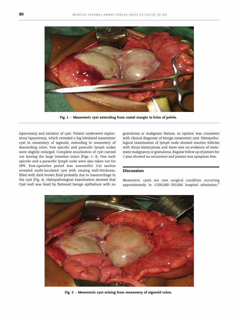
Fig. 1 e Mesenteric cyst extending from costal margin to brim of pelvis.
med i c a l j o u rn a l a rm e d f o r c e s i n d i a 7 0 ( 2 0 1 4 ) 7 9e8 280
laparotomy and excision of cyst. Patient underwent explor-
atory laparotomy, which revealed a big lobulated mesenteric
cyst in mesentery of sigmoid, extending in mesentery of
descending colon. Few epicolic and paracolic lymph nodes
were slightly enlarged. Complete enucleation of cyst carried
out leaving the large intestine intact (Figs. 1e3). One each
epicolic and a paracolic lymph node were also taken out for
HPE. Post-operative period was uneventful. Cut section
revealed multi-loculated cyst with varying wall-thickness,
filled with dark brown fluid probably due to haemorrhage in
the cyst (Fig. 4). Histopathological examination showed that
Cyst wall was lined by flattened benign epithelium with no
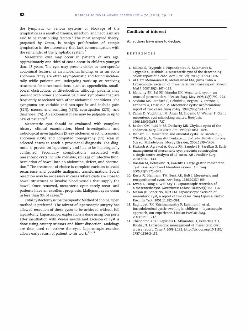
Fig. 2 e Mesenteric cyst arising from
granuloma or malignant feature, so opinion was consistent
with clinical diagnosis of benign mesenteric cyst. Histopatho-
logical examination of lymph node showed reactive follicles
with Sinus histiocytosis and there was no evidence of meta-
staticmalignancy or granuloma. Regular followupofpatient for
1 year showed no recurrence and patient was symptom free.
Discussion
Mesenteric cysts are rare surgical condition occurring
approximately in 1/200,000e350,000 hospital admission.2
mesentery of sigmoid colon.

Fig. 3 e Mesenteric cyst excised completely.
med i c a l j o u r n a l a rm e d f o r c e s i n d i a 7 0 ( 2 0 1 4 ) 7 9e8 2 81
Italian anatomist Benevenni first described this entity per-
forming an autopsy in an 8-year-old boy in 1507,while Roki-
tansky published the first accurate description of a chylous
mesenteric cyst in 1842 and Tillaux performed the first
successful surgery for a cystic mass in the mesentery in
1880.3
A mesenteric cyst is defined as any cyst located in the
mesentery; it may or may not extend into the retro-
peritoneum, which has a recognizable lining of endothelium
ormesothelial cell. Mesenteric cyst can occur anywhere in the
mesentery of gastrointestinal tract from duodenum to
Fig. 4 e HPE shows cyst wall lined by flattened benign epith
rectum. In a review series of 162 patients, 60% of mesenteric
cysts occurred in the small-bowelmesentery, 24% in the large-
bowel mesentery, and 14.5% in the retroperitoneum while it
was indefinite in 1.5% of cases.4 Mesenteric cysts can be
simple or multiple, unilocular or multilocular, and they may
contain hemorrhagic, serous, chylous, or infected fluid. They
can range in size from a few millimetres to few cm in diam-
eter, however, at times may be so large that it may mimic
tubercular ascites.5
Exact aetiology of mesenteric cyst has yet to be ascer-
tained, but failure of the lymph nodes to communicate with
elium. With no evidence of malignancy or granuloma.
med i c a l j o u rn a l a rm e d f o r c e s i n d i a 7 0 ( 2 0 1 4 ) 7 9e8 282
the lymphatic or venous systems or blockage of the
lymphatics as a result of trauma, infection, and neoplasm are
said to be contributing factors.6 The most accepted theory,
proposed by Gross, is benign proliferation of ectopic
lymphatics in the mesentery that lack communication with
the remainder of the lymphatic system.7
Mesenteric cyst may occur in patients of any age.
Approximately one-third of cases occur in children younger
than 15 years. The cyst may present either as non-specific
abdominal feature, as an incidental finding, or as an acute
abdomen. They are often asymptomatic and found inciden-
tally while patients are undergoing work-up or receiving
treatment for other conditions, such as appendicitis, small-
bowel obstruction, or diverticulitis, although patients may
present with lower abdominal pain and symptoms that are
frequently associated with other abdominal conditions. The
symptoms are variable and non-specific and include pain
(82%), nausea and vomiting (45%), constipation (27%), and
diarrhoea (6%). An abdominal mass may be palpable in up to
61% of patients.8
Mesenteric cyst should be evaluated with complete
history, clinical examination, blood investigations and
radiological investigations (X-ray abdomen erect, ultrasound
abdomen (USG) and computed tomography (CT) scan in
selected cases) to reach a provisional diagnosis. The diag-
nosis is proven on laparotomy and has to be histologically
confirmed. Secondary complications associated with
mesenteric cysts include volvulus, spillage of infective fluid,
herniation of bowel into an abdominal defect, and obstruc-
tion.9 The treatment of choice is complete excision to avoid
recurrence and possible malignant transformation. Bowel
resection may be necessary in cases where cysts are close to
bowel structures or involve blood vessels that supply the
bowel. Once removed, mesenteric cysts rarely recur, and
patients have an excellent prognosis. Malignant cysts occur
in less than 3% of cases.10
Total cystectomy is the therapeuticMethod of choice. Open
method is preferred. The advent of laparoscopic surgery has
allowed resection of these cysts to be achieved without full
laparotomy. Laparoscopic exploration is done using four ports
after insufflation with Veress needle and excision of cyst is
done using cautery scissors and blunt dissection. Endobags
are then used to retrieve the cyst. Laparoscopic excision
allows early return of patient to his work.11e14
Conflicts of interest
All authors have none to declare.
r e f e r e n c e s
1. Miliras S, Trygonis S, Papandoniou A, Kalamaras S,Trygonis C, Kiskinis D. Mesenteric cyst of the descendingcolon: report of a case. Acta Chir Belg. 2006;106:714e716.
2. Al Haifi Mohammed B, Abdulsamad MA, Juma Talib A.Laparoscopic excision of mesenteric cyst: case report. KuwaitMed J. 2007;39(2):167e169.
3. Mohanty SK, Bal RK, Maudar KK. Mesenteric cyst e anunusual presentation. J Pediatr Surg. May 1998;33(5):792e793.
4. Saviano MS, Fundaro S, Gelmini R, Begossi G, Perrone S,Farinetti A, Criscuolo M. Mesenteric cystic neoformation:report of two cases. Surg Today. 1999;29(2):174e177.
5. Zamir D, Yuchtman M, Amar M, Shoemo U, Weiner P. Giantmesenteric cyst mimicking ascites. Harefuah.1996;130(10):683e727.
6. Beahrs OM, Judd Jr ES, Dockerty MB. Chylous cysts of theabdomen. Surg Clin North Am. 1950;30:1081e1096.
7. Richard RR. Mesenteric and omental cysts. In: Grosfeld JL,O’Neill Jr JA, Coran AG, Fonkalsrud EW, eds. Pediatric Surgery.6th ed. Philadelphia: Mosby Elsevier; 2006:1399e1406.
8. Prakash A, Agrawal A, Gupta RK, Sanghvi B, Parelkar S. Earlymanagement of mesenteric cyst prevents catastrophes:a single centre analysis of 17 cases. Afr J Paediatr Surg.2010;7:140e143.
9. Hassan M, Dobrilovic N, Korelitz J. Large gastric mesentericcyst: case report and literature review. Am Surg.2005;71(7):571e573.
10. Kurtz RJ, Heimann TM, Beck AR, Holt J. Mesenteric andretroperitoneal cysts. Ann Surg. 1986;203(1):109.
11. Kwan E, Hung L, Wai-Key Y. Laparoscopic resection ofa mesenteric cyst. Gastrointest Endosc. 2004;59(1):154e156.
12. Mason JE, Soper NS, Burt LM. Laparoscopic excision ofmesenteric cyst, a report of two cases. Surg Laparosc EndoscPercutan Tech. 2001;11:382e384.
13. Raghupati RK, Krishnamurthy P, Rajamani J, et al.Intraabdominal cystic swelling in children e laparoscopicapproach, our experience. J Indian Paediatr Surg.2003;8:213e217.
14. Theodorodis TD, Zepiridis L, Athanotos D, Kellartzis TD,Bontis JN. Laparoscopic management of mesenteric cyst:a case report. Cases J. 2009;2:132. http://dx.doi.org/10.1186/1757-1626-2-132.


![Case Reportchange, and calcification [1]. In our case, the cyst wall was calcified, leading to the dilemma in diagnosis, since very few cases of calcified mesenteric cysts have been](https://img.dokumen.tips/doc/110x75/60f80b90704bdc1a0e053a9c/case-report-change-and-calcification-1-in-our-case-the-cyst-wall-was-calcified.jpg)