Embed Size (px)
Citation preview
MELPHALANMelphalan was considered by previous IARC Working Groups in 1974 and 1987 (IARC, 1975, 1987a). Since that time, new data have become available, these have been incorporated into the Monograph, and taken into consideration in the present evaluation.
1. Exposure Data
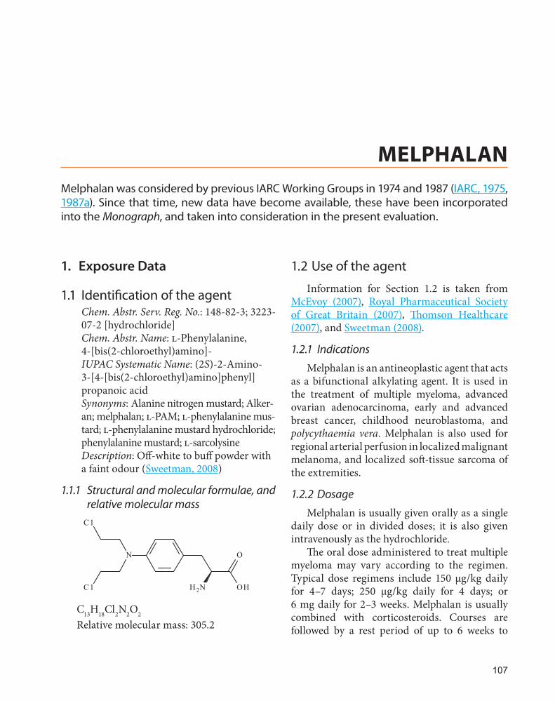
1.1 Identification of the agentChem. Abstr. Serv. Reg. No.: 148-82-3; 3223-07-2 [hydrochloride]Chem. Abstr. Name: l-Phenylalanine, 4-[bis(2-chloroethyl)amino]-IUPAC Systematic Name: (2S)-2-Amino-3-[4-[bis(2-chloroethyl)amino]phenyl]propanoic acidSynonyms: Alanine nitrogen mustard; Alker-an; melphalan; l-PAM; l-phenylalanine mus-tard; l-phenylalanine mustard hydrochloride; phenylalanine mustard; l-sarcolysineDescription: Off-white to buff powder with a faint odour (Sweetman, 2008)
1.1.1 Structural and molecular formulae, and relative molecular mass
OHH2N
ON
C1
C1
C13H18Cl2N2O2
Relative molecular mass: 305.2
1.2 Use of the agent
Information for Section 1.2 is taken from McEvoy (2007), Royal Pharmaceutical Society of Great Britain (2007), Thomson Healthcare (2007), and Sweetman (2008).
1.2.1 Indications
Melphalan is an antineoplastic agent that acts as a bifunctional alkylating agent. It is used in the treatment of multiple myeloma, advanced ovarian adenocarcinoma, early and advanced breast cancer, childhood neuroblastoma, and polycythaemia vera. Melphalan is also used for regional arterial perfusion in localized malignant melanoma, and localized soft-tissue sarcoma of the extremities.
1.2.2 Dosage
Melphalan is usually given orally as a single daily dose or in divided doses; it is also given intravenously as the hydrochloride.
The oral dose administered to treat multiple myeloma may vary according to the regimen. Typical dose regimens include 150 µg/kg daily for 4–7 days; 250 µg/kg daily for 4 days; or 6 mg daily for 2–3 weeks. Melphalan is usually combined with corticosteroids. Courses are followed by a rest period of up to 6 weeks to
107
IARC MONOGRAPHS – 100A
allow recovery of haematological function, and are then repeated, or maintenance therapy may be instituted, usually with a daily dose of 1–3 mg, or up to 50 µg/kg.
Similar oral dose regimens (150–200 µg/kg daily for 5 days, repeated every 4–8 weeks) have been used to treat ovarian adenocarcinoma and advanced breast cancer, although with the devel-opment of newer agents, melphalan is now used infrequently for the treatment of these tumours. The dose administered by mouth to treat poly-cythaemia vera is initially 6–10 mg daily, reduced after 5–7 days to 2–4 mg daily until satisfactory response, then further reduced to 2–6 mg per week for maintenance.
Melphalan has also been given intravenously; a single dose of 1 mg/kg, repeated in 4 weeks if the platelet and neutrophil counts permit, has been used to treat ovarian adenocarcinoma. In multiple myeloma, melphalan has been administered as a single agent intravenously at a dose of 400 µg/kg or 16 mg/m2, infused over 15–20 minutes; the first four doses are given at 2-week intervals, and further doses are given at 4-week intervals depending on toxicity. High-dose melphalan has been given intravenously in some malignan-cies: doses of 100–240 mg/m2 have been used for neuroblastoma, and 100–200 mg/m2 in multiple myeloma, generally followed by autologous stem-cell transplantation. Melphalan may be given by local arterial perfusion in the management of melanoma and soft-tissue sarcoma. A typical dose range for upper extremity perfusions is 0.6–1 mg/kg, whereas for lower extremity perfu-sions, dose ranges of 0.8–1.5 mg/kg (in mela-noma) or 1–1.4 mg/kg (in sarcoma) are typically used.
Melphalan is available as 2 mg tablets for oral administration, and as a 50 mg (melphalan hydrochloride) solution for injection for paren-teral administration.
Melphalan is also used intravenously as part of isolated hyperthermic limb perfusions for
patients with malignant melanoma in transit and limb sarcomas.
1.2.3 Trends in use
The availability of several newer chemothera-peutic and biological therapies has substantially reduced the use of melphalan in patients with solid tumours, whereas the use of high-dose intravenous melphalan followed by autologous stem-cell infusion has increased following rand-omized studies that demonstrated a disease-free, and in some studies, an overall survival advan-tage in patients with multiple myeloma.
2. Cancer in Humans
Epidemiological studies of patients with ovarian carcinoma (Reimer et al., 1977; Einhorn et al., 1982; Greene et al., 1982, 1986; Kaldor et al., 1990), multiple myeloma (Gonzalez et al., 1977; Law & Blom, 1977; Bergsagel et al., 1979; Dent et al., 2000) or breast cancer (Fisher et al., 1985; Curtis et al., 1992) have consistently shown very large excesses of acute myeloid leukaemia in the decade following therapy with melphalan.
Since then, a large number of epidemio-logical studies have contributed to the weight of evidence for the carcinogenicity of melphalan, in particular, two large case–control studies detailed below.
A case–control study conducted in a cohort of 82700 women who received adjuvant chemo-therapy for breast cancer, and who were followed up for at least 18 months after completion of treat-ment, identified 90 cases of leukaemia, of which 80 were acute myeloid leukaemia/myelodysplatic syndromes. The relative risk (RR) of developing acute myeloid leukaemia was highest in patients who received alkylating agents with radiation treatment (RR, 17.4) compared to those who were treated with alkylating agents (RR, 10) or radiation treatment alone (RR, 2.4). The relative risk was 10-fold greater for patients who received
108

Melphalan
melphalan (RR, 31.4) compared to those who received cyclophosphamide (Curtis et al., 1992).
A case–control study compared the rela-tive risk of leukaemia in patients treated with chemotherapy or radiation to patients who only underwent surgery (Kaldor et al., 1990). Approximately 90 cases [The Working Group noted that older nomenclature was used, and it was difficult to precisely assess the diagnosis in some patients] of acute myeloid leukaemia or myelodysplatic syndromes were identified among ~99000 patients with ovarian cancer. All of the alkylating agents (including chloram-bucil, cyclophosphamide, thiotepa, treosulfan, and melphalan) increased the risk of devel-oping leukaemia in a dose-dependent manner. Relative risks for melphalan ranged from 12–23 depending on the total dose used.
3. Cancer in Experimental Animals
Melphalan has been tested in mice by oral, intraperitoneal, and dermal application; in rats by intraperitoneal injection, and in monkeys by oral administration (Table 3.1).
In mice, the administration of melphalan produced forestomach papillomas, lymphosar-comas, and skin and lung tumours (IARC, 1975, 1987a; Satoh et al., 1993; Eastin et al., 2001). In rats, melphalan caused mammary gland tumours, and peritoneal sarcomas (IARC, 1975, 1987a). Results in monkeys were inconclusive (Thorgeirsson et al., 1994; Schoeffner & Thorgeirsson, 2000).
4. Other Relevant Data
4.1 Absorption, distribution, metabolism, and excretion
In humans, following oral administration, melphalan is absorbed from the gastrointestinal tract with a wide range in bioavailability (range
25–89%, mean 56%) (Sweetman, 2008). It also exhibits considerable variability with respect to the time of its appearance in the plasma (range ~0–6 hours), and in the peak plasma concentra-tion achieved (range 70–4000 ng/mL, depending upon the dose) (GlaxoSmithKline, 2007). This variability may be due to incomplete intestinal absorption, variable first-pass metabolism, and/or rapid hydrolysis. Upon absorption, approxi-mately 60–90% of plasma melphalan is bound to plasma proteins such as albumin, and to a lesser degree, α1-acid glycoprotein, with approximately 30% being bound irreversibly (GlaxoSmithKline, 2007). Melphalan does not undergo metabolic activation and is inactivated in the plasma, primarily by non-enzymatic hydrolysis to mono-hydroxymelphalan and dihydroxymelphalan. Apart from these hydrolysis products, no other melphalan metabolites have been detected in humans (GlaxoSmithKline, 2007). Melphalan enters cells through active transport, mostly by the high-affinity l-amino acid transport system, which carries glutamine and leucine (Nieto & Vaughan, 2004).
As a consequence of its inconsistent absorp-tion, melphalan can also exhibit considerable variability in its elimination. For example, when given intravenously, melphalan exhibited a fairly consistent half-life (14–40 minutes in a study of ten patients given a 20 mg/m2 dose) but was absorbed variably, and found to have a half-life of 36–552 minutes in a study of 13 patients admin-istered a 0.6 mg/m2 oral dose (Hall & Tilby, 1992). About 10% of the drug is excreted unchanged in the urine within 24 hours, and about 30% of administered melphalan (including metabolites) is excreted in the urine within 9 days of oral administration (McEvoy, 2007; Sweetman, 2008). Approximately 20–50% of the dose is eliminated via faeces (McEvoy, 2007).
109

IARC M
ON
OG
RAPH
S – 100A
110
Table 3.1 Studies of cancer in experimental animals exposed to melphalan
Species, strain (sex) Duration Reference
Route Dosing regimen Animals/group at start
Incidence of tumours Significance Comments
Mouse, S (NR) 22 wk Roe & Salaman (1956)
Skin 0, 1.44, 3 mg total dose administered by skin painting once weekly for 10 wk 60, 25, 25
Skin (papillomas): 5/53, 2/19, 4/22
[P < 0.05] for 3 mg Croton oil used as promoter (experimental Weeks 5–22)
Lung (adenomas): 10/17 (1.7/mouse), 13/18 (3.6/mouse), 11/13 (5.1/mouse)
Mouse, A/J (M, F) 39 wk Shimkin et al. (1966)
i.p. 0, 0.27, 1.07, 4.27, 17.1 mg/kg bw total dose 3 times/wk for 4 wk 1400, 60, 60, 60, 60
Lung: 307/777, 23/58, 37/56, 43/56, 40/41 Tumours/mouse : 0.5, 0.6, 1.0, 2.1, 4.0
[P < 0.0001] for 1.07, 4.27 and 17.1 mg/kg bw
Mouse, Swiss (M, F) 18 mo Weisburger et al. (1975)
i.p. 0.75, 1.5 mg/kg bw 3 times/wk for 6 mo 25 (M) + 25 (F)
Lung: Dosage groups were combined for each sex; 254 mice served as untreated controls
M–11/44 P = 0.012F–10/23 P = 0.001Lymphosarcomas: M–13/44
P < 0.001
Mouse, Tg.AC (M, F) 27 wk Eastin et al. (2001)
Oral gavage 0, 0.25, 1.0 & 4.0 mg/kg bw/wk for 26 wk 15/sex/group
Forestomach (squamous cell papillomas): M–8/14, 14/15, 15/15, 14/15; F–12/15, 14/14, 13/14, 13/14
[P ≤ 0.03] for 0.25, 1.0, 4.0 mg/kg bw doses in male mice
Mouse, Tg.AC (M, F) 27 wk Eastin et al. (2001)
Topical application (in methanol, 3.3 mL/kg bw), 0, 0.25, 1.0, 4.0 mg/kg bw/wk for 26 wk 15/sex/group
Skin tumours (in area of the vulva): F–1/15, 3/15, 7/15, 12/15
[P ≤ 0.02] for 1.0 & 4.0 mg/kg bw in female mice
Mouse, A/J (F) 24 wk Satoh et al. (1993)
i.p. 0, 4.0 µmol/kg bw/d, 3 d/wk 12, 13
Lung (adenomas): 4/12 & 13/13 (incidence) Tumours per mouse: 0.42 ± 0.67, 12.15 ± 1.68
[P = 0.0005] (incidence) [P < 0.001] for multiplicity

Melphalan
111
Species, strain (sex) Duration Reference
Route Dosing regimen Animals/group at start
Incidence of tumours Significance Comments
Rat, (F) 17 mo Presnov & Iushkov (1964)
i.p 0, 10 mg/kg bw, single application 40, 60
Mammary gland (fibroadenomas): 0/30, 9/33
A mixture of melphalan and medpalan (D isomer of melphalan) was used
Rat, Sprague-Dawley CD (M, F) 18 mo Weisburger et al. (1975)
i.p. 0.9, 1.8 mg/kg bw 3 times/wk for 6 mo 25/sex
Peritoneum (sarcomas): Dosage groups were combined for each sex; 360 rats served as untreated controls
M–11/20 P < 0.001F–10/23 P < 0.001
Monkey, (NR) 16 yr Thorgeirsson et al. (1994), Schoeffner & Thorgeirsson (2000)
Nasogastric tube (in DMSO, volume NR) 0.1 mg/kg bw/5 d/wk
Malignant tumours: 5/12
Incidence in control animals could not be determined
bw, body weight; d, day or days; DMSO, dimethylsulfoxide; F, female; i.p., intraperitoneal; M, male; mo, month or months; NR, not reported; w, week or weeks; yr, year or years
Table 3.1 (continued)

IARC MONOGRAPHS – 100A
4.2 Mechanisms of carcinogenesis
4.2.1 Induction of DNA damage
Melphalan is a direct-acting, bifunctional alkylating agent that binds to cellular macromol-ecules (Osborne et al., 1995). As a phenylalanine derivative of nitrogen mustard, it is capable of producing a variety of DNA adducts including mono-adducts at the N7 of guanine and the N3 of adenine as well as interstrand cross-links, and pre-mutagenic lesions that are believed to play a critical role in its toxic and carcinogenic effects (Povirk & Shuker, 1994; Lawley & Phillips, 1996; GlaxoSmithKline, 2007).
Melphalan has been shown to bind to DNA, RNA, and protein in cells in vitro (Tilby et al., 1987, 1995; Povirk & Shuker, 1994; Osborne et al., 1995), and DNA-binding has been seen in rats treated in vivo (Van den Driessche et al., 2004a, b). The formation of DNA cross-links in vitro and in vivo has also been reported based on the measurement of specific adducts (Osborne & Lawley, 1993) or by changes in DNA migra-tion in DNA strand breakage or electrophoretic assays (Ringborg et al., 1990; Popp et al., 1992; Souliotis et al., 2003, 2006; Cordelli et al., 2004; Dimopoulos et al., 2005).
4.2.2 Mutational consequences of DNA damage
Melphalan has been tested for genotoxicity in an assortment of short-term assays, both in vitro and in vivo. Increased frequencies of domi-nant lethal mutations, chromosomal aberra-tions, micronuclei, and DNA strand breaks have been observed in several studies following treat-ment of rodents with melphalan (IARC, 1987b; Shelby et al., 1989; Generoso et al., 1995; Morita et al., 1997; Tsuda et al., 2000; Sgura et al., 2005, 2008; Ranaldi et al., 2007). In the mouse-specific locus mutation and heritable translocation tests, increases in mutations were seen in both sperma-tagonial as well as postspermatogonial germ cells
(Russell et al., 1992a, b; Generoso et al., 1995; Witt & Bishop, 1996). The observed mutations originated primarily from large deletions in the postspermatogonial cells whereas other types of mutagenic mechanisms predominated in the spermatogonial cells (Witt & Bishop, 1996).
Melphalan also induced chromosomal aber-rations, sister chromatid exchange, micronuclei, mutations at the HPRT gene, and DNA damage in human cells in vitro (IARC, 1987b; Mamuris et al., 1989a, b; Sanderson et al., 1991; Routledge et al., 1992; Sorsa et al., 1992; Vock et al., 1999; Efthimiou et al., 2007; Escobar et al., 2007). It also induced transformation of C3H 10T1/2 and other cells (IARC, 1987b; Miller et al., 1994; Kowalski et al., 2001). In cultured rodent cells, it induced chromosomal aberrations, sister chromatid exchange, gene mutations, and DNA damage (IARC, 1987b; Austin et al., 1992; Preuss et al., 1996; Allan et al., 1998). In addition, it induced aneuploidy and sex-linked recessive lethal muta-tions in Drosophila, and mutation in bacteria (IARC, 1987b).
Increased frequencies of chromosomal aber-rations and sister chromatid exchange occurring in the peripheral blood lymphocytes have been reported in multiple studies of patients treated therapeutically with melphalan (IARC, 1987b; Raposa & Várkonyi, 1987; Mamuris et al., 1989b, 1990; Popp et al., 1992; Amiel et al., 2004). In addition, DNA-binding (DNA mono-adducts and interstrand cross-links) to TP53 and N-RAS, two important cancer-related genes, has been seen in the peripheral blood cells of patients administered melphalan (Souliotis et al., 2003, 2006; Dimopoulos et al., 2005, 2007). Decreased migration of DNA in strand breakage assays indicative of DNA cross-link formation has been observed in melphalan-treated cancer patients (Popp et al., 1992; Spanswick et al., 2002). Haematotoxicity and immunosuppression have also been reported in patients treated with this anticancer agent (Goldfrank et al., 2002; GlaxoSmithKline, 2007).
112

Melphalan
Acute myeloid leukaemia that develops in patients previously treated with alkylating agents such as melphalan frequently exhibits distinctive characteristics that allow it to be distinguished from acute myeloid leukaemia induced by other agents (such as topoisomerase II inhibitors) or acute myeloid leukaemia that occurs spontane-ously (Rödjer et al., 1990; Pedersen-Bjergaard & Rowley, 1994; Jaffe et al, 2001; Pedersen-Bjergaard et al., 2006). One of the hallmarks of leukae-mias induced by alkylating agents is that they frequently exhibit a clonal loss of either chromo-some 5 or 7 (−5, −7) or a loss of part of the long arm of one of these chromosomes (5q−, 7q−). For example, a deletion within the long arm of chro-mosome 5 involving the bands q23 to q32 is often seen (Jaffe et al., 2001). Leukaemias developed in patients treated with melphalan (often in combi-nation with other agents) frequently exhibit these clonal chromosomal changes (Rödjer et al., 1990).
In addition, mutations in TP53 are frequently seen in leukaemias with the −5/5q− karyotype, and mutations involving the AML1 gene as well as mutations in TP53 and RAS are often seen in a subset of leukaemias that exhibit the −7/7q− karyotype (Christiansen et al., 2001, 2005; Pedersen-Bjergaard et al., 2006). These treatment-related acute myeloid leukaemias also frequently exhibit increased methylation of the p15 promoter (Pedersen-Bjergaard et al., 2006). While there is some evidence that melphalan may directly induce damage targeting chromosomes 5 or 7 (Mamuris et al., 1989a, b, 1990; Amiel et al., 2004), this drug has also been reported to induce nonspecific chromosomal alterations in a variety of experimental models, and in the lymphocytes of treated patients (described above). The detec-tion of elevated levels of chromosomal aberra-tions in the peripheral blood lymphocytes of melphalan-treated patients is of particular note, as multiple prospective studies have now shown that individuals with increased levels of chromo-somal aberrations in these cells are at increased
risk of developing cancer later in life (Hagmar et al., 1998, 2004; Liou et al., 1999; Smerhovsky et al., 2001).
4.3 Synthesis
Melphalan is a direct-acting alkylating agent that is carcinogenic via a genotoxic mechanism.
5. Evaluation
There is sufficient evidence in humans for the carcinogenicity of melphalan. Melphalan causes acute myeloid leukaemia.
There is sufficient evidence in experimental animals for the carcinogenicity of melphalan.
Melphalan is carcinogenic to humans (Group 1).
References
Allan JM, Engelward BP, Dreslin AJ et al. (1998). Mammalian 3-methyladenine DNA glycosylase protects against the toxicity and clastogenicity of certain chemotherapeutic DNA cross-linking agents. Cancer Res, 58: 3965–3973. PMID:9731510
Amiel A, Yukla M, Yogev S et al. (2004). Deletion of 5q31 and 7q31 in patients with stable melphalan treated multiple myeloma. Cancer Genet Cytogenet, 152: 84–87. doi:10.1016/j.cancergencyto.2003.10.015 PMID:15193449
Austin MJ, Han YH, Povirk LF (1992). DNA sequence analysis of mutations induced by melphalan in the CHO aprt locus. Cancer Genet Cytogenet, 64: 69–74. doi:10.1016/0165-4608(92)90326-4 PMID:1458453
Bergsagel DE, Bailey AJ, Langley GR et al. (1979). The chemotherapy on plasma-cell myeloma and the inci-dence of acute leukemia. N Engl J Med, 301: 743–748. doi:10.1056/NEJM197910043011402 PMID:481481
Christiansen DH, Andersen MK, Desta F, Pedersen-Bjergaard J (2005). Mutations of genes in the receptor tyrosine kinase (RTK)/RAS-BRAF signal transduc-tion pathway in therapy-related myelodysplasia and acute myeloid leukemia. Leukemia, 19: 2232–2240. doi:10.1038/sj.leu.2404009 PMID:16281072
Christiansen DH, Andersen MK, Pedersen-Bjergaard J (2001). Mutations with loss of heterozygosity of p53 are
113

IARC MONOGRAPHS – 100A
common in therapy-related myelodysplasia and acute myeloid leukemia after exposure to alkylating agents and significantly associated with deletion or loss of 5q, a complex karyotype, and a poor prognosis. J Clin Oncol, 19: 1405–1413. PMID:11230485
Cordelli E, Cinelli S, Lascialfari A et al. (2004). Melphalan-induced DNA damage in p53(+/-) and wild type mice analysed by the comet assay. Mutat Res, 550: 133–143. PMID:15135647
Curtis RE, Boice JD Jr, Stovall M et al. (1992). Risk of leukemia after chemotherapy and radiation treat-ment for breast cancer. N Engl J Med, 326: 1745–1751. doi:10.1056/NEJM199206253262605 PMID:1594016
Dent SF, Klaassen D, Pater JL et al. (2000). Second primary malignancies following the treatment of early stage ovarian cancer: update of a study by the National Cancer Institute of Canada–Clinical Trials Group (NCIC-CTG). Ann Oncol, 11: 65–68. doi:10.1023/A:1008356806417 PMID:10690389
Dimopoulos MA, Souliotis VL, Anagnostopoulos A et al. (2005). Extent of damage and repair in the p53 tumor-suppressor gene after treatment of myeloma patients with high-dose melphalan and autologous blood stem-cell transplantation is individualized and may predict clinical outcome. J Clin Oncol, 23: 4381–4389. doi:10.1200/JCO.2005.07.385 PMID:15883412
Dimopoulos MA, Souliotis VL, Anagnostopoulos A et al. (2007). Melphalan-induced DNA damage in vitro as a predictor for clinical outcome in multiple myeloma. Haematologica, 92: 1505–1512. doi:10.3324/haematol.11435 PMID:18024399
Eastin WC, Mennear JH, Tennant RW et al. (2001). Tg.AC genetically altered mouse: assay working group over-view of available data. Toxicol Pathol, 29: Suppl60–80. doi:10.1080/019262301753178483 PMID:11695563
Efthimiou M, Andrianopoulos C, Stephanou G et al. (2007). Aneugenic potential of the nitrogen mustard analogues melphalan, chlorambucil and p-N,N-bis(2-chloroethyl)aminophenylacetic acid in cell cultures in vitro. Mutat Res, 617: 125–137. PMID:17324445
Einhorn N, Eklund G, Franzén S et al. (1982). Late side effects of chemotherapy in ovarian carcinoma: a cytogenetic, hematologic, and statistical study. Cancer, 49: 2234–2241. doi:10.1002/1097-0142(19820601)49:11<2234::AID-CNCR2820491106>3.0.CO;2-Q PMID:7074540
Escobar PA, Smith MT, Vasishta A et al. (2007). Leukaemia-specific chromosome damage detected by comet with fluorescence in situ hybridization (comet-FISH). Mutagenesis, 22: 321–327. doi:10.1093/mutage/gem020 PMID:17575318
Fisher B, Rockette H, Fisher ER et al. (1985). Leukemia in breast cancer patients following adjuvant chemo-therapy or postoperative radiation: the NSABP experi-ence. J Clin Oncol, 3: 1640–1658. PMID:3906049
Generoso WM, Witt KL, Cain KT et al. (1995). Dominant lethal and heritable translocation tests with
chlorambucil and melphalan in male mice. Mutat Res, 345: 167–180. doi:10.1016/0165-1218(95)90052-7 PMID:8552138
GlaxoSmithKline (2007). Prescribing information leaflet: Alkeran (melphalan) tablets. Available at: http://us.gsk.com/products/assets/us_alkeran_tablets.pdf
Goldfrank L, Flomenbaum N, Lewin N et al. (2002). Antineoplastic agents. In: Goldfrank’s Toxicologic Emergencies, Chap 47. McGraw-Hill Professional.
Gonzalez F, Trujillo JM, Alexanian R (1977). Acute leukemia in multiple myeloma. Ann Intern Med, 86: 440–443. PMID:403840
Greene MH, Boice JD Jr, Greer BE et al. (1982). Acute nonlymphocytic leukemia after therapy with alkylating agents for ovarian cancer: a study of five randomized clinical trials. N Engl J Med, 307: 1416–1421. doi:10.1056/NEJM198212023072302 PMID:6752720
Greene MH, Harris EL, Gershenson DM et al. (1986). Melphalan may be a more potent leukemogen than cyclophosphamide. Ann Intern Med, 105: 360–367. PMID:3740675
Hagmar L, Bonassi S, Strömberg U et al. (1998). Chromosomal aberrations in lymphocytes predict human cancer: a report from the European Study Group on Cytogenetic Biomarkers and Health (ESCH). Cancer Res, 58: 4117–4121. PMID:9751622
Hagmar L, Strömberg U, Bonassi S et al. (2004). Impact of types of lymphocyte chromosomal aberrations on human cancer risk: results from Nordic and Italian cohorts. Cancer Res, 64: 2258–2263. doi:10.1158/0008-5472.CAN-03-3360 PMID:15026371
Hall AG & Tilby MJ (1992). Mechanisms of action of, and modes of resistance to, alkylating agents used in the treatment of haematological malignancies. Blood Rev, 6: 163–173. doi:10.1016/0268-960X(92)90028-O PMID:1422285
IARC (1975). Melphalan, medphalan & merphalan. IARC Monogr Eval Carcinog Risk Chem Man, 9: 167–180. PMID:791831
IARC (1987a). Overall evaluations of carcinogenicity: an updating of IARC Monographs volumes 1 to 42. IARC Monogr Eval Carcinog Risks Hum Suppl, 7: 1–440. PMID:3482203
IARC (1987b). Genetic and related effects: An updating of selected IARC monographs from Volumes 1 to 42. IARC Monogr Eval Carcinog Risks Hum Suppl, 6: 1–729. PMID:3504843
Jaffe ES, Harris NL, Stein H, Vardiman JW, editors (2001). Pathology and Genetics of Tumours of Haematopoietic and Lymphoid Tissues. Lyon: IARC Press.
Kaldor JM, Day NE, Pettersson F et al. (1990). Leukemia following chemotherapy for ovarian cancer. N Engl J Med, 322: 1–6. doi:10.1056/NEJM199001043220101 PMID:2104664
Kowalski LA, Assi KP, Wee RK, Madden Z (2001). In vitro prediction of carcinogenicity using a bovine
114

Melphalan
papillomavirus DNA–carrying C3H/10T 1/2 cell line (T1). II: Results from the testing of 100 chemi-cals. Environ Mol Mutagen, 37: 231–240. doi:10.1002/em.1032 PMID:11317341
Law IP & Blom J (1977). Second malignancies in patients with multiple myeloma. Oncology, 34: 20–24. doi:10.1159/000225175 PMID:405642
Lawley PD & Phillips DH (1996). DNA adducts from chemotherapeutic agents. Mutat Res, 355: 13–40. PMID:8781575
Liou SH, Lung JC, Chen YH et al. (1999). Increased chro-mosome-type chromosome aberration frequencies as biomarkers of cancer risk in a blackfoot endemic area. Cancer Res, 59: 1481–1484. PMID:10197617
Mamuris Z, Dumont J, Dutrillaux B, Aurias A (1990). Specific chromosomal mutagenesis observed in stimu-lated lymphocytes from patients with S-ANLL. Int J Cancer, 46: 563–568. doi:10.1002/ijc.2910460402 PMID:2210879
Mamuris Z, Gerbault-Seureau M, Prieur M et al. (1989b). Chromosomal aberrations in lymphocytes of patients treated with melphalan. Int J Cancer, 43: 80–86. doi:10.1002/ijc.2910430117 PMID:2910833
Mamuris Z, Prieur M, Dutrillaux B, Aurias A (1989a). The chemotherapeutic drug melphalan induces breakage of chromosomes regions rearranged in secondary leukemia. Cancer Genet Cytogenet, 37: 65–77. doi:10.1016/0165-4608(89)90076-9 PMID:2917334
McEvoy GK, editor (2007). American Hospital Formulary Service Drug Information. Bethesda, MD: American Society of Health-System Pharmacists.
Miller RC, Richards M, Baird C et al. (1994). Interaction of hyperthermia and chemotherapy agents; cell lethality and oncogenic potential. Int J Hyperthermia, 10: 89–99. doi:10.3109/02656739409009335 PMID:7511674
Morita T, Asano N, Awogi T et al.Collaborative Study of the Micronucleus Group Test. Mammalian Mutagenicity Study Group. (1997). Evaluation of the rodent micro-nucleus assay in the screening of IARC carcinogens (groups 1, 2A and 2B) the summary report of the 6th collaborative study by CSGMT/JEMS MMS. Mutat Res, 389: 3–122. PMID:9062586
Nieto Y & Vaughan WP (2004). Pharmacokinetics of high-dose chemotherapy. Bone Marrow Transplant, 33: 259–269. doi:10.1038/sj.bmt.1704353 PMID:14647243
Osborne MR & Lawley PD (1993). Alkylation of DNA by melphalan with special reference to adenine deriva-tives and adenine-guanine cross-linking. Chem Biol Interact, 89: 49–60. doi:10.1016/0009-2797(93)03197-3 PMID:8221966
Osborne MR, Lawley PD, Crofton-Sleigh C, Warren W (1995). Products from alkylation of DNA in cells by melphalan: human soft tissue sarcoma cell line RD and Escherichia coli WP2. Chem Biol Interact, 97: 287–296. doi:10.1016/0009-2797(95)03623-T PMID:7545551
Pedersen-Bjergaard J, Christiansen DH, Desta F, Andersen MK (2006). Alternative genetic pathways and coop-erating genetic abnormalities in the pathogenesis of therapy-related myelodysplasia and acute myeloid leukemia. Leukemia, 20: 1943–1949. doi:10.1038/sj.leu.2404381 PMID:16990778
Pedersen-Bjergaard J & Rowley JD (1994). The balanced and the unbalanced chromosome aberrations of acute myeloid leukemia may develop in different ways and may contribute differently to malignant transforma-tion. Blood, 83: 2780–2786. PMID:8180374
Popp W, Vahrenholz C, Schürfeld C et al. (1992). Investigations of the frequency of DNA strand breakage and cross-linking and of sister chromatid exchange frequency in the lymphocytes of patients with multiple myeloma undergoing cytostatic therapy with melphalan and prednisone. Carcinogenesis, 13: 2191–2195. doi:10.1093/carcin/13.11.2191 PMID:1423893
Povirk LF & Shuker DE (1994). DNA damage and mutagenesis induced by nitrogen mustards. Mutat Res, 318: 205–226. PMID:7527485
Presnov MA & Iushkov CF (1964). Development of mastropathies and fibroadenomas of the mammary gland in rats following intra-abdominal injections of sarcolysin Vopr Onkol, 10: 66–72. PMID:14229401
Preuss I, Thust R, Kaina B (1996). Protective effect of O6-methylguanine-DNA methyltransferase (MGMT) on the cytotoxic and recombinogenic activity of different antineoplastic drugs. Int J Cancer, 65: 506–512. doi:10.1002/(SICI)1097-0215(19960208)65:4<506::AID-IJC19>3.0.CO;2-7 PMID:8621235
Ranaldi R, Palma S, Tanzarella C et al. (2007). Effect of p53 haploinsufficiency on melphalan-induced geno-toxic effects in mouse bone marrow and peripheral blood. Mutat Res, 615: 57–65. PMID:17109898
Raposa T & Várkonyi J (1987). The relationship between sister chromatid exchange induction and leukemo-genicity of different cytostatics. Cancer Detect Prev, 10: 141–151. PMID:3568006
Reimer RR, Hoover R, Fraumeni JF Jr, Young RC (1977). Acute leukemia after alkylating-agent therapy of ovarian cancer. N Engl J Med, 297: 177–181. doi:10.1056/NEJM197707282970402 PMID:406560
Ringborg U, Hansson J, Jungnelius U et al. (1990). Melphalan-induced DNA cross-linking and inhibi-tion of DNA and RNA synthesis in human melanoma and lymphoblast cells. Anticancer Res, 10: 2A297–301. PMID:2346304
Rödjer S, Swolin B, Weinfeld A, Westin J (1990). Cytogenetic abnormalities in acute leukemia complicating melphalan-treated multiple myeloma. Cancer Genet Cytogenet, 48: 67–73. doi:10.1016/0165-4608(90)90218-Y PMID:2372790
Roe FJ & Salaman MH (1956). Further tests for tumour-initiating activity: N,
115
IARC MONOGRAPHS – 100A
N-di-(2-chloroethyl)-p-aminophenylbutyric acid (CB1348) as an initiator of skin tumour formation in the mouse. Br J Cancer, 10: 363–378. PMID:13364128
Routledge MN, Garner RC, Kirsch-Volders M (1992). Comparison of 32P-postlabelling and cytogenetic anal-ysis of human blood treated in vitro with melphalan. Mutagenesis, 7: 329–333. doi:10.1093/mutage/7.5.329 PMID:1470027
Royal Pharmaceutical Society of Great Britain (2007). British National Formulary, No. 54. London: BMJ Publishing Group Ltd./RPS Publishing.
Russell LB, Hunsicker PR, Cacheiro NL, Rinchik EM (1992b). Genetic, cytogenetic, and molecular analyses of mutations induced by melphalan demonstrate high frequencies of heritable deletions and other rearrange-ments from exposure of postspermatogonial stages of the mouse. Proc Natl Acad Sci USA, 89: 6182–6186. doi:10.1073/pnas.89.13.6182 PMID:1352884
Russell LB, Hunsicker PR, Shelby MD (1992a). Melphalan, a second chemical for which specific-locus mutation induction in the mouse is maximum in early sper-matids. Mutat Res, 282: 151–158. doi:10.1016/0165-7992(92)90089-Z PMID:1378547
Sanderson BJ, Johnson KJ, Henner WD (1991). Dose-dependent cytotoxic and mutagenic effects of antineo-plastic alkylating agents on human lymphoblastoid cells. Environ Mol Mutagen, 17: 238–243. doi:10.1002/em.2850170404 PMID:2050131
Satoh M, Kondo Y, Mita M et al. (1993). Prevention of carcinogenicity of anticancer drugs by metallothionein induction. Cancer Res, 53: 4767–4768. PMID:8402657
Schoeffner DJ & Thorgeirsson UP (2000). Susceptibility of nonhuman primates to carcinogens of human rele-vance. In Vivo, 14: 149–156. PMID:10757072
Sgura A, De Amicis A, Stronati L et al. (2008). Chromosome aberrations and telomere length modulation in bone marrow and spleen cells of melphalan-treated p53+/- mice. Environ Mol Mutagen, 49: 467–475. doi:10.1002/em.20405 PMID:18481314
Sgura A, Stronati L, Gullotta F et al. (2005). Use of chro-mosome painting for detecting stable chromosome aberrations induced by melphalan in mice. Environ Mol Mutagen, 45: 419–426. doi:10.1002/em.20107 PMID:15685603
Shelby MD, Gulati DK, Tice RR, Wojciechowski JP (1989). Results of tests for micronuclei and chromosomal aberrations in mouse bone marrow cells with the human carcinogens 4-aminobiphenyl, treosulphan, and melphalan. Environ Mol Mutagen, 13: 339–342. doi:10.1002/em.2850130410 PMID:2737185
Shimkin MB, Weisburger JH, Weisburger EK et al. (1966). Bioassay of 29 Alkylating Chemicals by the Pulmonary-Tumor Response in Strain A Mice. J Natl Cancer Inst, 36: 915–935.
Smerhovsky Z, Landa K, Rössner P et al. (2001). Risk of cancer in an occupationally exposed cohort with
increased level of chromosomal aberrations. Environ Health Perspect, 109: 41–45. doi:10.2307/3434919 PMID:11171523
Sorsa M, Autio K, Abbondandolo A et al. (1992). Evaluation of in vitro cytogenetic techniques in nine European laboratories in relation to chromosomal endpoints induced by three model mutagens. Mutat Res, 271: 261–267. PMID:1378199
Souliotis VL, Dimopoulos MA, Episkopou HG et al. (2006). Preferential in vivo DNA repair of melphalan-induced damage in human genes is greatly affected by the local chromatin structure. DNA Repair (Amst), 5: 972–985. doi:10.1016/j.dnarep.2006.05.006 PMID:16781199
Souliotis VL, Dimopoulos MA, Sfikakis PP (2003). Gene-specific formation and repair of DNA monoadducts and interstrand cross-links after therapeutic exposure to nitrogen mustards. Clin Cancer Res, 9: 4465–4474. PMID:14555520
Spanswick VJ, Craddock C, Sekhar M et al. (2002). Repair of DNA interstrand crosslinks as a mechanism of clinical resistance to melphalan in multiple myeloma. Blood, 100: 224–229. doi:10.1182/blood.V100.1.224 PMID:12070031
Sweetman SC, editor (2008). Martindale: The Complete Drug Reference. London: Pharmaceutical Press. Available at: http://www.medicinescomplete.com/mc/
Thomson Healthcare (2007). Physicians’ Desk Reference, 61st ed. Montvale, NJ: Thomson.
Thorgeirsson UP, Dalgard DW, Reeves J, Adamson RH (1994). Tumor incidence in a chemical carcinogenesis study of nonhuman primates. Regul Toxicol Pharmacol, 19: 130–151. doi:10.1006/rtph.1994.1013 PMID:8041912
Tilby MJ, McCartney H, Cordell J et al. (1995). A mono-clonal antibody that recognizes alkali-stabilized melphalan-DNA adducts and its application in immu-nofluorescence microscopy. Carcinogenesis, 16: 1895–1901. doi:10.1093/carcin/16.8.1895 PMID:7634420
Tilby MJ, Styles JM, Dean CJ (1987). Immunological detection of DNA damage caused by melphalan using monoclonal antibodies. Cancer Res, 47: 1542–1546. PMID:3815354
Tsuda S, Matsusaka N, Madarame H et al. (2000). The alkaline single cell electrophoresis assay with eight mouse organs: results with 22 mono-functional alkylating agents (including 9 dialkyl N-nitrosoamines) and 10 DNA crosslinkers. Mutat Res, 467: 83–98. PMID:10771273
Van den Driessche B, Lemière F, Van Dongen W et al. (2004b). Qualitative study of in vivo melphalan adduct formation in the rat by miniaturized column-switching liquid chromatography coupled with electrospray mass spectrometry. J Mass Spectrom, 39: 29–37. doi:10.1002/jms.540 PMID:14760610
Van den Driessche B, Van Dongen W, Lemière F, Esmans EL (2004a). Implementation of data-dependent acqui-sitions in the study of melphalan DNA adducts by
116

Melphalan
miniaturized liquid chromatography coupled to elec-trospray tandem mass spectrometry. Rapid Commun Mass Spectrom, 18: 2001–2007. doi:10.1002/rcm.1578 PMID:15329868
Vock EH, Lutz WK, Ilinskaya O, Vamvakas S (1999). Discrimination between genotoxicity and cytotoxicity for the induction of DNA double-strand breaks in cells treated with aldehydes and diepoxides. Mutat Res, 441: 85–93. PMID:10224325
Weisburger JH, Griswold DP, Prejean JD et al. (1975). The carcinogenic properties of some of the principal drugs used in clinical cancer chemotherapy. Recent Results Cancer Res, 521–17. PMID:138176
Witt KL & Bishop JB (1996). Mutagenicity of anticancer drugs in mammalian germ cells. Mutat Res, 355: 209–234. PMID:8781584
117