Embed Size (px)
Citation preview

621
Veterinarni Medicina, 60, 2015 (11): 621–628 Original Paper
doi: 10.17221/8530-VETMED
Melatonin protects against burn-induced hepatic oxidative injury by inducing HO-1 via the Nrf2 pathway
G. Bekyarova, M. Tzaneva, M. Hristova
Medical University, Varna, Bulgaria
ABSTRACT: Melatonin exerts beneficial effects on early liver injury by modulating hepatic oxidative stress. In order to understand the protective effect of melatonin against burn-induced hepatic injury we investigated the expression of 4-hydroxynonenal (4-HNE), a main product of lipid peroxidation and mediator of oxidative injury, the inducible heme-oxygenase-1 (HO-1), an antioxidant enzyme, and the anti-oxidative stress regulator erythroid 2-related factor 2 (Nrf2) in a burn rat model. Expression and localisation of HO-1, 4-HNE and Nrf2 in liver were investigated using light immunochemistry. Thermal skin injury caused a significant elevation in hepatic 4-HNE and degenerative liver changes. Concurrently, there was increased expression of HO-1, a rate-limiting enzyme for haem degradation and an oxidative stress marker in sinusoidal endothelial cells (SECs) and hepatocytes without changes in Nrf2 expression in the liver. Melatonin (20 mg/kg b.w.) augmented the increase in HO-1 expression, upregulated Nrf2 expression and also led to decreased 4-HNE levels and reduced levels of histopathological changes in rat liver. In conclusion, our results suggest that melatonin ameliorates burn-induced liver injury through the inhibition of oxidative stress, upregulation of the antioxidant enzyme HO-1 and activation of the antioxidant Nrf2 pathway. Stimulation of cellular protective mechanisms by activating the antioxidant stress response through Nrf2 is a new mechanism for protection against liver damage in burns.
Keywords: melatonin; Nrf2; HO-1; 4-HNE; oxidative stress; liver
Thermal injury represents one of the most severe forms of trauma and is a serious clinical problem in emergency medicine. A severe burn is a devastating injury affecting every organ system and leading to complications with poor outcome. The liver plays an important role in metabolism, inflammation, ho-meostasis and host defence mechanisms (Klein et al. 2004), and is a major organ responsible for ini-tiating multiorgan dysfunction syndrome (MODS) following burn injury. The mechanism of burn-in-duced injury is not completely understood; how-ever, several mechanisms, including hypoxia, free radicals, inflammation, and apoptosis are thought to be involved (Jeschke et al. 2001). The excessive production of free radicals such as superoxide anion, hydrogen peroxide and hydroxyl radicals as well as the occurrence of lipid peroxidation products such as malondialdehyde (MDA) due to oxidative stress are associated with burn-induced liver dysfunction (Horton 2003; Bekyarova et al. 2009; Bekyarova et al. 2012). Burn reduces the antioxidant status thus
leading to a failure of the antioxidant defence against free-radical damage (Piantadosi et al. 2006; Parihar et al. 2008; Vinha et al. 2013).
In many cell types, numerous cellular responses to oxidative stress are mediated by signalling proteins that act through the antioxidant response element (ARE) and the transcription factor nuclear factor ery-throid 2-related factor 2 (Nrf2) (Dulak and Jozkowicz 2003; Lee et al. 2005). Nrf2 is a key transcription fac-tor regulating ARE-mediated transcription of genes specialized in the detoxification of certain reactive species (HO-1), as well as phase-2 detoxifying en-zymes and related stress-response proteins that play a central role in the cellular defence against oxidative injury (Osburn et al. 2006; Yang et al. 2011). HO-1 upregulation by pharmacological treatment protects against the deleterious effects of free radicals and oxidative stress, and blocks apoptosis in various cell types and animal models (Ashino et al. 2011).
Melatonin (N-acetyl-5-methoxytryptamine) is a powerful antioxidant that scavenges superoxide

622
Original Paper Veterinarni Medicina, 60, 2015 (11): 621–628
doi: 10.17221/8530-VETMED
radicals as well as other radical oxygen species (ROS) and radical nitrogen species (RNS), protects against oxidative injury and gives rise to a cascade of metabolites (AFMK and AMK) that share its an-tioxidant and anti-inflammatory properties (Reiter et al. 2009; Galano et al. 2013; Mauriz et al. 2013; Zhang and Zhang 2014). Melatonin induces the expression of antioxidant enzymes such as super-oxide dismutase SOD, catalase and gluthatione peroxidase under conditions of elevated oxidative stress (Rodriguez et al. 2004), through activation of Nrf2 (Jung et al. 2009). Further, melatonin ame-liorates oxidative stress through Nrf2 induction in experimental models of hepatic injury (Osburn et al. 2006; Aleksunes and Manautou 2007; Crespo et al. 2010; Yang et al. 2011). Activation of Nrf2 by melatonin has been described to result in an in-creased expression of the antioxidant enzyme haem oxygenase-1 (HO-1) and a restriction of hepatic in-jury (Jung et al. 2009; Crespo et al. 2010). Although experimental and clinical studies have shown that melatonin restrains oxidative liver injury (Ohta et al. 2000; Oz et al. 2006), we failed to find any re-ports on a possible protective effect of melatonin on burn-induced hepatic damage by activation of the Nrf2/HO-1 system. Therefore, we designed this study to investigate whether the protective effect of melatonin against burn-induced liver injury is asso-ciated with the activation of the Nrf2/HO-1 signal-ling pathway. Histological changes were examined and the expression of 4-hydroxynonenal (4-HNE) was determined as a marker of oxidative stress. Expression and localisation of НО-1 and Nrf2 were analysed using light immunohistochemistry.
MATERIAL AND METHODS
Experimental study. The experimental proce-dures were approved by the Home Office for Care and Use of Laboratory Animals and were performed with careful consideration of the ethics of animal experimentation according to the International Guiding Principles for Animal Research approved in Bulgaria. Same-age male rats weighing between 220 and 250 g were fasted for 12 h, but were al-lowed free access to water before injury. Animals were housed in a 20 °C room and offered standard rat chow and water ad libitum. They were kept in dark-light cycles (DL = 12 : 12 h) in individual wire bottomed cages. The lights were turned off at
8:00 p.m. and turned on at 8:00 a.m. to achieve a satisfactory photoperiod.
After mild ether inhalation, general anaesthe-sia was intraperitoneally (i.p.) established using thiopental 30 mg/kg body weight (b.w.) In order to accomplish a 30% third-degree burn, scalding hot water (90 °C) was applied on the back of the animals for a 10 s duration. Following burn injury, 4 ml physiological saline was i.p. applied for imme-diate resuscitation. No animals died within the first 24 h post-burn period. Twenty one male rats were randomly assigned to three groups: group of vehicle treated without burns (C) (n = 7); group of vehicle treated with burns (B) (n = 7); and group of mela-tonin (10 ml/kg b.w.) treated with burns (B + M) (n = 7). Melatonin (N-acetyl-5-methoxytryptamine; Merck, Germany) was dissolved in absolute etha-nol and diluted with physiological saline. The con-centration of ethanol in the final solution was 5%. Melatonin solution was administered i.p. immedi-ately and 12 h after thermal skin injury. The groups without burns and with burns received vehicle only (5% ethanol, 5 ml/kg, i.p.). All animals received buprenorphine (0.3 mg/kg b.w. i.p.) twice daily for post-burn pain control. Twenty four hours after the burns the animals were anaesthetised with thiopen-tal, sacrificed and livers were sampled.
Histological examination. Liver slices were fixed in 10% phosphate-buffered saline (PBS)-formalin and embedded in paraffin. Liver samples were subse-quently sectioned (5 μm), stained with haematoxylin and eosin (H&E), and examined under a microscope (Olympus, Japan) to evaluate liver injuries.
Immunohistochemical staining of 4-HNE, HO-1 and Nrf2. Rat liver specimens were fixed in 10% neutral buffered formalin and embedded in paraffin. The deparaffinised and dehydrated sections (5 μm thick) were treated with 1% hydro-gen peroxide for peroxidase activity inhibition for 5 min. Then they were rinsed in 0.1M phosphate buffered saline (PBS) (pH 7.4) and treated in normal goat serum for 20 min. Subsequently, the sections were incubated with polyclonal primary antibodies for 24 h at room temperature. Rabbit anti-HO-1 antibody (Santa Cruz, USA), anti-Nrf2 antibody (Santa Cruz, USA) and 4-HNE antibody (Abcam, UK) were used. After rinsing with PBS the sec-tions were incubated for 20 min in goat anti-rab-bit immunoglobulins at room temperature. Then, they were rinsed in PBS again, treated with rabbit peroxidase-antiperoxidase complex for 20 min at
623
Veterinarni Medicina, 60, 2015 (11): 621–628 Original Paper
doi: 10.17221/8530-VETMED
room temperature and then rinsed in PBS. Finally, peroxidase activity was estimated using the diami-nobenzydine-tetrachloride H2O2-method.
Negative controls were incubated with non-im-mune sera instead of primary antibody.
The morphometric method described by Bekya- rova et al. (2013) was used to semiquantitatively assess the content of HO-1, Nrf2 and 4-HNE. The immunohistochemical staining was determined as: strong = score 3; moderate = score 2; weak = score 1; and lack of staining = score 0. The content of HO-1, Nrf2 and 4-HNE in the liver cells was defined as the number of each type of the cell content mul-tiplied by their scoring factor and divided by the total number of cells. Morphometry was carried out on 50 cells of each sample.
Statistical analysis. All experimental data were expressed as mean± standard error of the mean (SEM). Significant differences were determined by one-way analysis of variance (ANOVA). P < 0.05 was considered as a statistically significant difference.
RESULTS
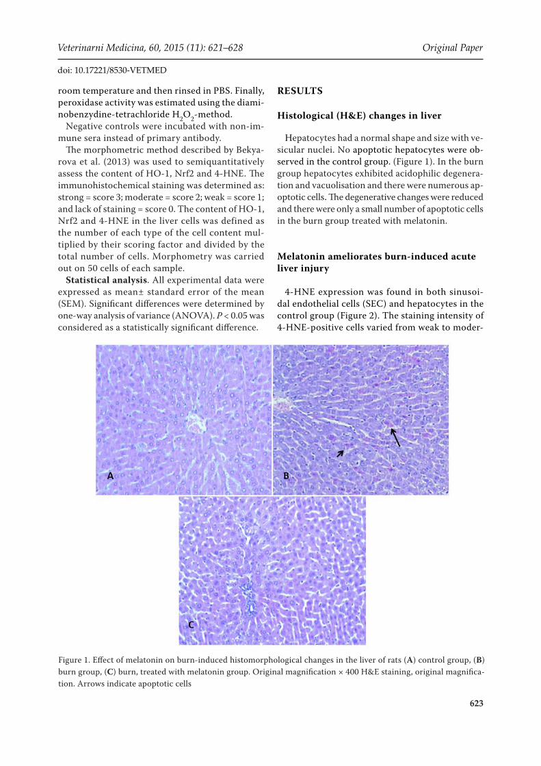
Histological (H&E) changes in liver
Hepatocytes had a normal shape and size with ve-sicular nuclei. No apoptotic hepatocytes were ob-served in the control group. (Figure 1). In the burn group hepatocytes exhibited acidophilic degenera-tion and vacuolisation and there were numerous ap-optotic cells. The degenerative changes were reduced and there were only a small number of apoptotic cells in the burn group treated with melatonin.
Melatonin ameliorates burn-induced acute liver injury
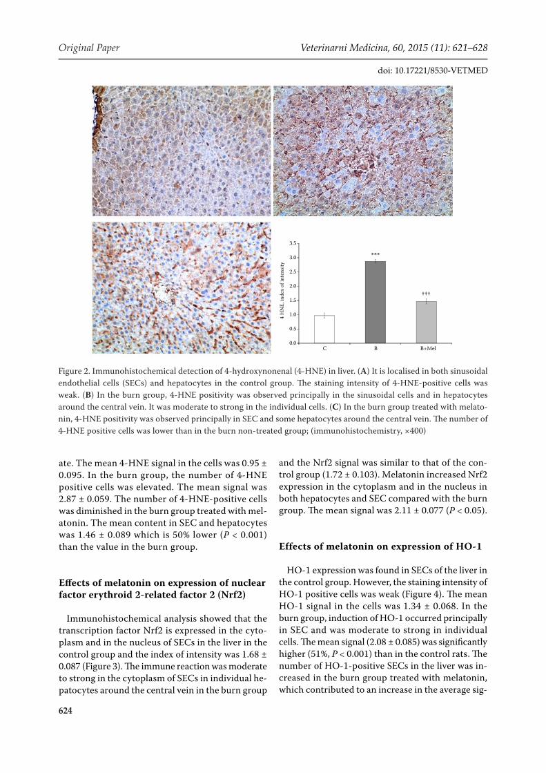
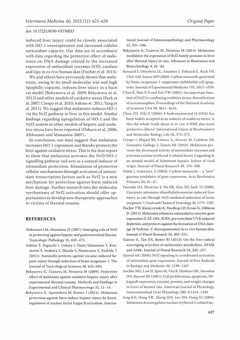
4-HNE expression was found in both sinusoi-dal endothelial cells (SEC) and hepatocytes in the control group (Figure 2). The staining intensity of 4-HNE-positive cells varied from weak to moder-
Figure 1. Effect of melatonin on burn-induced histomorphological changes in the liver of rats (A) control group, (B) burn group, (C) burn, treated with melatonin group. Original magnification × 400 H&E staining, original magnifica-tion. Arrows indicate apoptotic cells
624
Original Paper Veterinarni Medicina, 60, 2015 (11): 621–628
doi: 10.17221/8530-VETMED
ate. The mean 4-HNE signal in the cells was 0.95 ± 0.095. In the burn group, the number of 4-HNE positive cells was elevated. The mean signal was 2.87 ± 0.059. The number of 4-HNE-positive cells was diminished in the burn group treated with mel-atonin. The mean content in SEC and hepatocytes was 1.46 ± 0.089 which is 50% lower (P < 0.001) than the value in the burn group.
Effects of melatonin on expression of nuclear factor erythroid 2-related factor 2 (Nrf2)
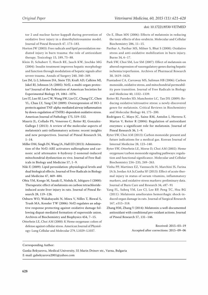
Immunohistochemical analysis showed that the transcription factor Nrf2 is expressed in the cyto-plasm and in the nucleus of SECs in the liver in the control group and the index of intensity was 1.68 ± 0.087 (Figure 3). The immune reaction was moderate to strong in the cytoplasm of SECs in individual he-patocytes around the central vein in the burn group
and the Nrf2 signal was similar to that of the con-trol group (1.72 ± 0.103). Melatonin increased Nrf2 expression in the cytoplasm and in the nucleus in both hepatocytes and SEC compared with the burn group. The mean signal was 2.11 ± 0.077 (P < 0.05).
Effects of melatonin on expression of HO-1
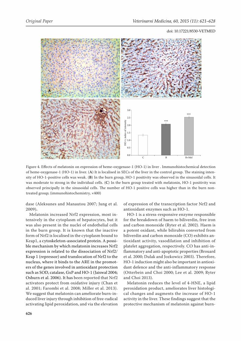
HO-1 expression was found in SECs of the liver in the control group. However, the staining intensity of HO-1 positive cells was weak (Figure 4). The mean HO-1 signal in the cells was 1.34 ± 0.068. In the burn group, induction of HO-1 occurred principally in SEC and was moderate to strong in individual cells. The mean signal (2.08 ± 0.085) was significantly higher (51%, P < 0.001) than in the control rats. The number of HO-1-positive SECs in the liver was in-creased in the burn group treated with melatonin, which contributed to an increase in the average sig-
Figure 2. Immunohistochemical detection of 4-hydroxynonenal (4-HNE) in liver. (A) It is localised in both sinusoidal endothelial cells (SECs) and hepatocytes in the control group. The staining intensity of 4-HNE-positive cells was weak. (B) In the burn group, 4-HNE positivity was observed principally in the sinusoidal cells and in hepatocytes around the central vein. It was moderate to strong in the individual cells. (C) In the burn group treated with melato-nin, 4-HNE positivity was observed principally in SEC and some hepatocytes around the central vein. The number of 4-HNE positive cells was lower than in the burn non-treated group; (immunohistochemistry, ×400)
0.0
0.5
1.0
1.5
2.0
2.5
3.0
3.5
B+MelBC
4 H
NE,
inde
x of
inte
nsity
***
†††

625
Veterinarni Medicina, 60, 2015 (11): 621–628 Original Paper
doi: 10.17221/8530-VETMED
nal of HO-1 protein in the liver to 2.53 ± 0.077, a value which was 23% (P < 0.001) higher than in the burn group which did not receive melatonin.
DISCUSSION
The present study demonstrates that melatonin exerts a protective effect against oxidative liver damage in a burn rat model. The expression of Nrf2 and HO-1 was significantly increased in burned rats treated with melatonin as compared with con-trol rats and burned, non-treated rats.
Thermal skin injury is a strong inducer of oxi-dative stress, and lipid peroxidation is the most prominent among the many biological targets of oxidative stress (Bekyarova et al. 2009; Bekyarova et al. 2012). 4-HNE is one of the most toxic molecules of lipid peroxidation and a biomarker of oxidative injury. This lipid peroxidation by-product has high
reactivity towards the thiol and amino groups of amino acids. 4-HNE modifies essential cellular proteins, decreases glutathione levels and alters cellular redox homeostasis (Niki 2009). The accu-mulation of 4-HNE in the liver may increase the likelihood of tissue injury during oxidative stress and causes degenerative changes in liver. In the cur-rent study increased levels of lipid peroxidation and oxidative injury were demonstrated by increased expression of 4-HNE in the liver.
ROS are crucial for activation of the Nrf2 transcription factor (Chen and Niki 2006). It is suggested that mild oxidative stress and in par-ticular 4-HNE might induce nuclear translocation of Nrf2. Nrf2 protects the cell against oxidative stress through ARE-mediated induction of sev-eral phase-2 detoxifying and antioxidant enzymes, particularly HO-1 (Lee et al. 2005), but also su-peroxide dismutase, γ-glutamylcysteine synthase, glutathione S-transferase and gluthatione peroxi-
Figure 3. Effects of melatonin on expression of nuclear factor erythroid 2-related factor 2 (Nrf2). Immunohistochemical detection of nuclear factor erythroid 2-related factor 2 (Nrf2) in liver (A) It is localised in the cytoplasm and in the nucleus of SECs of the liver in the control group. The staining intensity is moderate. (B) In the burn group, Nrf2 positivity was observed in the cytoplasm of the SECs in individual hepatocytes. It was moderate to strong in the individual cells. (C) In the burn group treated with melatonin, Nrf2 positivity was observed in the cytoplasm and in the nucleus in both hepatocytes and SECs; (immunohistochemistry, ×400)
0.0
0.5
1.0
1.5
2.0
2.5
B+MelBC
Nrf
2, in
dex
of in
tens
ity
†
626
Original Paper Veterinarni Medicina, 60, 2015 (11): 621–628
doi: 10.17221/8530-VETMED
dase (Aleksunes and Manautou 2007; Jung et al. 2009).
Melatonin increased Nrf2 expression, most in-tensively in the cytoplasm of hepatocytes, but it was also present in the nuclei of endothelial cells in the burn group. It is known that the inactive form of Nrf2 is localised in the cytoplasm bound to Keap1, a cytoskeleton-associated protein. A possi-ble mechanism by which melatonin increases Nrf2 expression is related to the dissociation of Nrf2/Keap 1 (repressor) and translocation of Nrf2 to the nucleus, where it binds to the ARE in the promot-ers of the genes involved in antioxidant protection such as SOD, catalase, GxP and HO-1 (Jaiswal 2004; Osburn et al. 2006). It has been reported that Nrf2 activators protect from oxidative injury (Chan et al. 2001; Farombi et al. 2008; Miller et al. 2013). We suggest that melatonin can ameliorate burn-in-duced liver injury through inhibition of free-radical activating lipid peroxidation, and via the elevation
of expression of the transcription factor Nrf2 and antioxidant enzymes such as HO-1.
HO-1 is a stress-responsive enzyme responsible for the breakdown of haem to biliverdin, free iron and carbon monoxide (Ryter et al. 2002). Haem is a potent oxidant, while bilirubin converted from biliverdin and carbon monoxide (CO) exhibits an-tioxidant activity, vasodilation and inhibition of platelet aggregation, respectively. CO has anti-in-flammatory and anti-apoptotic properties (Brouard et al. 2000; Dulak and Jozkowicz 2003). Therefore, HO-1 induction might also be important in antioxi-dant defence and the anti-inflammatory response (Otterbein and Choi 2000; Lee et al. 2009; Ryter and Choi 2013).
Melatonin reduces the level of 4-HNE, a lipid peroxidation product, ameliorates liver histologi-cal changes and augments the increase of HO-1 activity in the liver. These findings suggest that the protective mechanism of melatonin against burn-
Figure 4. Effects of melatonin on expression of heme-oxygenase-1 (HO-1) in liver . Immunohistochemical detection of heme-oxygenase-1 (HO-1) in liver. (A) It is localised in SECs of the liver in the control group. The staining inten-sity of HO-1-positive cells was weak. (B) In the burn group, HO-1 positivity was observed in the sinusoidal cells. It was moderate to strong in the individual cells. (C) In the burn group treated with melatonin, HO-1 positivity was observed principally in the sinusoidal cells. The number of HO-1 positive cells was higher than in the burn non-treated group; (immunohistochemistry, ×400)
0.0
0.5
1.0
1.5
2.0
2.5
3.0
B+MelBC
HO
-1, i
ndex
of i
nten
sity ***
†††
627
Veterinarni Medicina, 60, 2015 (11): 621–628 Original Paper
doi: 10.17221/8530-VETMED
induced liver injury could be closely associated with HO-1 overexpression and increased cellular antioxidant capacity. Our data are in accordance with data regarding the protective effect of mela-tonin on DNA damage related to the increased expression of antioxidant enzymes SOD, catalase and Gpx in ex vivo human skin (Fischer et al. 2013).
We and others have previously shown that mela-tonin, owing to its small molecular size and high lipophilic capacity, reduces liver injury in a burn rat model (Bekyarova et al. 2009; Bekyarova et al. 2013) and other models of oxidative stress (Park et al. 2007; Crespo et al. 2010; Ashino et. 2011; Yang et al. 2011). We suggest that melatonin induces HO-1 via the Nrf2 pathway in liver in this model. Similar findings regarding upregulation of HO-1 and the Nrf2 system in other models of hepatic and oxida-tive stress have been reported (Osburn et al. 2006; Aleksunes and Manautou 2007).
In conclusion, our data suggest that melatonin increases HO-1 expression and thereby protects the liver against oxidative stress. This is the first report to show that melatonin activates the Nrf2/HO-1 signalling pathway and acts as a natural inducer of antioxidant protection. Stimulation of protective cellular mechanisms through activation of antioxi-dant transcription factors such as Nrf2 is a new mechanism for protection against burn-induced liver damage. Further research into the molecular mechanisms of Nrf2 activation should offer op-portunities to develop new therapeutic approaches to victims of thermal trauma.
REFERENCES
Aleksunes LM, Manautou JE (2007): Emerging role of Nrf2 in protecting against hepatic and gastrointestinal disease. Toxicologic Pathology 35, 459–473.
Ashino T, Sugiuchi J, Uehara J, Naito-Yamamoto Y, Ken-motsu S, Iwakura Y, Shioda S, Numazawa S, Yoshida T (2011): Auranofin protects against cocaine-induced he-patic injury through induction of heme oxygenase-1. The Journal of Toxicological Sciences 36, 635–643.
Bekyarova G, Tzaneva M, Hristova M (2009): Protective effect of melatonin against oxidative hepatic injury after experimental thermal trauma. Methods and Findings in Experimental and Clinical Pharmacology 31, 11–14.
Bekyarova G, Apostolova M, Kotzev I (2012): Melatonin protection against burn-induce hepatic injury by down-regulation of nuclear factor kappa B activation. Interna-
tional Journal of Immunopathology and Pharmacology 25, 591–596.
Bekyarova G, Tzaneva M, Hristova M (2013): Melatonin modulates the expression of Bcl2 family proteins in liver after thermal injury in rats. Advances in Bioscience and Biotechnology 4, 41–44.
Brouard S, Otterbein LE, Anrather J, Tobiasch E, Bach FH, Choi AM, Soares MP (2000): Carbon monoxide generated by heme oxygenase-1 suppresses endothelial cell apop-tosis. Journal of Experimental Medicine 192, 1015–1029.
Chan K, Han X D and Kan YW (2001): An important func-tion of Nrf2 in combating oxidative stress: detoxification of acetaminophen. Proceedings of the National Academy of Sciences USA 98, 4611–4616.
Chen ZH, Niki E (2006): 4-hydroxynonenal (4-HNE) has been widely accepted as an inducer of oxidative stress. Is this the whole truth about it or can 4-HNE also exert protective effects? International Union of Biochemistry and Molecular Biology Life 58, 372–373.
Crespo I, Miguel BS, Laliena A, Alvarez M, Culebras JM, Gonzalez-Gallego J, Tunon MJ (2010): Melatonin pre-vents the decreased activity of antioxidant enzymes and activates nuclear erythroid 2-related factor 2 signaling in an animal model of fulminant hepatic failure of viral origin. Journal of Pineal Research 49, 193–200.
Dulak J, Jozkowicz A (2003): Carbon monoxide – a “new” gaseous modulator of gene expression. Acta Biochimica Polonica 50, 31–47.
Farombi EO, Shrotriya S, Na HK, Kim SH, Surh YJ (2008): Curcumin attenuates dimethylnitrosamine-induced liver injury in rats through Nrf2-mediated induction of heme oxygenase-1. Food and Chemical Toxicology 46, 1279–1287.
Fischer TW, Kleszczynski K, Hardkop LH, Kruse N, Zillikens D. (2013): Melatonin enhances antioxidative enzyme gene expression (CAT, GPx, SOD), prevents their UVR-induced depletion, and protects against the formation of DNA dam-age (8-hydroxy-2’-deoxyguanosine) in ex vivo human skin. Journal of Pineal Research 54, 303–312.
Galano A, Tan DX, Reiter RJ (2013): On the free radical scavenging activities of melatonin’s metabolites, AFMK and AMK. Journal of Pineal Research 54, 245–257.
Jaiswal AK (2004): Nrf2 signaling in coordinated activation of antioxidant gene expression. Journal of Free Radicals in Biology and Medicine 36, 1199–1207.
Jeschke MG, Low JF, Spies M, Vita R, Hawkins HK, Herndon DN, Barrow RE (2001): Cell proliferation, apoptosis, NF-kappaB expression, enzyme, protein, and weight changes in livers of burned rats. American Journal of Physiology, Gastrointestinal Liver Physiology 280, G1314–1320
Jung KH, Hong SW, Zheng HM, Lee DH, Hong SS (2009): Melatonin downregulates nuclear erythroid 2-related fac-
628
Original Paper Veterinarni Medicina, 60, 2015 (11): 621–628
doi: 10.17221/8530-VETMED
tor 2 and nuclear factor-kappaB during prevention of oxidative liver injury in a dimethylnitrosamine model. Journal of Pineal Research 47, 173–183.
Horton JW (2003): Free radicals and lipid peroxidation me-diated injury in burn trauma: the role of antioxidant therapy. Toxicology 15, 189, 75–88.
Klein D, Schubert T, Horch RE, Jauch KW, Jeschke MG (2004): Insulin treatment improves hepatic morphology and function through modulation of hepatic signals after severe trauma. Annals of Surgery 240, 340–349.
Lee JM, Li J, Johnson DA, Stein TD, Kraft AD, Calkins MJ, Jakel RJ, Johnson JA (2005): Nrf2, a multi-organ protec-tor? Journal of the Federation of American Societies for Experimental Biology 19, 1061–1076.
Lee IT, Luo SF, Lee CW, Wang SW, Lin CC, Chang CC, Chen YL, Chau LY, Yang CM (2009): Overexpression of HO-1 protects against TNF-alpha-mediated airway inflammation by down-regulation of TNFR1-dependent oxidative stress. American Journal of Pathology 175, 519–532.
Mauriz JL, Collado PS, Veneroso C, Reiter RJ, Gonzalez-Gallego J (2013): A review of the molecular aspects of melatonin’s anti-inflammatory actions: recent insights and new perspectives. Journal of Pineal Research 54, 1–14.
Miller DM, Singh IN, Wang JA, Hall ED (2013): Administra-tion of the Nrf2-ARE activators sulforaphane and car-nosic acid attenuates 4-hydroxy-2-nonenal-induced mitochondrial dysfunction ex vivo. Journal of Free Rad-icals in Biology and Medicine 57, 1–9.
Niki E (2009): Lipid peroxidation: physiological levels and dual biological effects. Journal of Free Radicals in Biology and Medicine 47, 469–484.
Ohta YM, Kongo M, Sasaki E, Nishda K, Ishiguro I (2000): Therapeutic effect of melatonin on carbon tetrachloride-induced acute liver injury in rats. Journal of Pineal Re-search 28, 119–126.
Osburn WO, Wakabayashi N, Misra V, Nilles T, Biswal S, Trush MA, Kensler TW (2006): Nrf2 regulates an adap-tive response protecting against oxidative damage fol-lowing diquat-mediated formation of superoxide anion. Archives of Biochemistry and Biophysics 454, 7–15.
Otterbein LE, Choi AM (2000): K Heme oxygenase: colors of defense against cellular stress. American Journal of Physiol-ogy: Lung Cellular and Molecular 279, L1029–L1037.
Oz E, Ilhan MN (2006): Effects of melatonin in reducing the toxic effects of dox-orubicin. Molecular and Cellular Biochemistry 286, 11–15.
Parihar A, Parihar MS, Milner S, Bhat S (2008): Oxidative stress and anti-oxidative mobilization in burn injury. Burns 34, 6–17.
Park SW, Choi SM, Lee SM (2007): Effect of melatonin on altered expression of vasoregulatory genes during hepatic ischemia/reperfusion. Archives of Pharmacal Research 30, 1619–1624.
Piantadosi CA, Carraway MS, Suliman HB (2006): Carbon monoxide, oxidative stress, and mitochondrial permeabil-ity pore transition. Journal of Free Radicals in Biology and Medicine 40, 1332–1339.
Reiter RJ, Paredes SD, Manchester LC, Tan DX (2009): Re-ducing oxidative/nitrosative stress: a newly-discovered genre for melatonin. Critical Reviews in Biochemistry and Molecular Biology 44, 175–200.
Rodriguez C, Mayo JC, Sainz RM, Antolın I, Herrera F, Martın V, Reiter R (2004): Regulation of antioxidant enzymes: a significant role for melatonin. Journal of Pineal Research 36, 1–9.
Ryter SW, Choi AM (2013): Carbon monoxide: present and future indications for a medical gas. Korean Journal of Internal Medicine 28, 123–140.
Ryter SW, Otterbein LE, Morse D, Choi AM (2002): Heme oxygenase/carbon monoxide signaling pathways: regula-tion and functional significance. Molecular and Cellular Biochemistry 234–235, 249–263.
Vinha PP, Martinez EZ, Vannucchi H, Marchini JS, Farina JA Jr, Jordao AA Jr,Cunha SF (2013): Effect of acute ther-mal injury in status of serum vitamins, inflammatory markers, and oxidative stress markers: preliminary data. Journal of Burn Care and Research 34, e87–91
Yang FL, Subeq YM, Lee CJ, Lee RP, Peng TC, Hsu BG (2011): Melatonin ameliorates hemorrhagic shock-in-duced organ damage in rats. Journal of Surgical Research 167, e315–318.
Zhang HM, Zhang Y (2014): Melatonin: a well-documented antioxidant with conditional pro-oxidant actions. Journal of Pineal Research 57, 131–146.
Received: 2015–03–19Accepted after corrections: 2015–09–06
Corresponding Author:
Ganka Bekyarova, Medical University, 55 Marin Drinov str., Varna, BulgariaE-mail: [email protected]