Embed Size (px)
Citation preview

Medicinal Chemistry Reviews - Online, 2005, 2, 303-323 303
1567-2034/05 $50.00+.00 © 2005 Bentham Science Publishers Ltd.
Unnatural Protein Engineering: Producing Proteins with UnnaturalAmino Acids
Thomas J. Magliery*
Yale University, Department of Molecular Biophysics & Biochemistry, P.O. Box 208114, New Haven, CT 06520-8114,USA
Abstract: Less than a decade ago, the ability to generate proteins with unnatural modifications was a Herculean taskavailable only to specialty labs. Recent advances make it possible to generate reasonable quantities of protein with un-natural amino acids both in vitro and in vivo . The combination of solid-phase peptide synthesis and enzymatic or che-moselective ligation now permits construction of entirely-synthetic proteins as large as 25 kD. Incorporation of recombi-nant fragments (expressed protein ligation) allows unnatural modifications near protein termini in proteins of virtually anysize. Site-specific modification of small quantities of protein at any position can be achieved using chemically-acylatedtRNA. Microelectroporation now extends this method to cells like mammalian neurons, and combination with RNA-display makes unnatural proteins compatible with combinatorial methods. Widespread or residue-specific methods ofamino acid replacement are especially suitable for the production of biomaterials, and bacteria have been engineered toexpand the repertoire of amino acids available for this technique. Excitingly, the wholesale addition of engineered tRNAsand synthetases to bacteria and yeast now makes site-specific incorporation of unnatural amino acids possible in livingcells with no chemical intervention. Methods of expanding the genetic code at the nucleic acid level, including 4-basecodons and unnatural base pairs, are becoming useful for the addition of multiple amino acids to the genetic code. Theserecent advances in unnatural protein engineering are reviewed with an eye toward future challenges, including methods ofcreating nonpeptidic molecules using templated synthesis.
Keywords: Unnatural amino acids; protein ligation; aminoacyl-tRNA synthetase; genetic code; amino acid replacement; site-specific incorporation.
INTRODUCTION
One of the great paradoxes of protein science is that theremarkable diversity of protein structure and function arisesfrom just twenty amino acids, but that this small amino acidrepertoire also limits our ability to study proteins. (Occa-sionally, selenocysteine [1] or pyrrolysine [2, 3] are alsotranslationally incorporated.) Nature did not provide many ofthe kinds of amino acids that would be most useful to bio-chemists and biophysicists. Tryptophan fluorescence is poor,and there are no amino acids with unpaired spins. The aminoacids tend to differ in stereoelectronic (such as pKA) andsteric properties so significantly that it is difficult to interpretthe effects of exchange of one residue for another. Naturehas solved the problem of specificity in posttranslationalmodification using enzymatic recognition of extended motifsrather than unique reactive handles. There is nothing usefulfor trapping interacting proteins. There are neither alkenesnor alkynes. There are no useful electrophiles—not a ketoneor an aldehyde. The heaviest atom to be found is a sulfur,and of course isotopes useful for NMR are only at naturalabundance.
Those taking a chemical approach to biology have knownabout this issue for some time. Without doubt, to this day,
*Address correspondence to this author at the Yale University, Departmentof Molecular Biophysics & Biochemistry, P.O. Box 208114, New Haven,CT 06520-8114, USA; Tel: 203-432-9841; Fax: 203-432-5175; [email protected]
the most important tool for engineering approaches to under-standing protein structure and function is site-directedmutagenesis, which allows us to swap one natural aminoacid for another at a specific site in a protein [4, 5]. Whilethere is no access to unnatural side chains, there is the sub-stantial importance of ease of creation of the mutant proteinat essentially any scale necessary. Modern synthesis of DNAoligonucleotides can provide 100-mers or more, and PCR-based methods make it simple to stitch together genes ofessentially any sequence. In appropriate vectors, these genescan be used to express or overexpress proteins in bacteria,yeast, insect cells, mammalian cells or even living animals.
Until recently, no method of incorporating unnaturalamino acids into proteins could compete in specificity, sim-plicity or scale with the ability to trade natural amino acidswith site-directed mutagenesis. Chemical synthesis of pep-tides, allowing every monomer to be altered at will, was onlypractical for the creation of small quantities of peptides atlengths barely corresponding to the smallest proteins or do-mains known. Co-opting the protein synthesis machineryallowed access to proteins of virtually any size, but therewere significant tradeoffs to be accepted. Near-natural aminoacid homologs (like selenomethionine) could sometimes besubstituted for natural amino acids in living cells, but fewsuitable amino acids were known, and the substitution couldoccur at any position with the related natural residue, al-though it often occurred with less than 100% replacement.On the other hand, chemical acylation of amber stop codon-
304 Medicinal Chemistry Reviews - Online, 2005, Vol. 2, No. 4 Thomas J. Magliery
suppressing tRNA with unnatural amino acids permitted site-specific incorporation of virtually any amino acid, but it re-quires a substantial synthetic undertaking and resulted inpoor yields of protein from in vitro translation mixtures. Inorder to achieve this same type of incorporation in livingcells, microinjection of RNA and chemically-acylated tRNAwas required, essentially restricting the user to cells like thelarge Xenopus oocyte and vanishingly small amounts ofprotein.
This is not to say that these methods were not useful, butjust that they were not practical for the typical biochemistryor biophysics lab to undertake, and of course these are thefields that stand to benefit most from unnatural amino acidincorporation. Fortunately, the last several years have seentremendous strides in methods to incorporate unnaturalamino acids into proteins. Proteins of useful size can now begenerated synthetically or semi-synthetically, using strate-gies to ligate smaller polypeptides. The scale and utility ofchemically-acylated tRNA methods has improved, includingthe ability to produce proteins in mammalian cells, such asneurons. The number of amino acids that can be inserted bywidespread (residue-specific) replacement has expanded,partially through enzyme engineering approaches, and themethods are sufficiently reliable that nearly 100% incorpo-ration can be achieved. But perhaps most significantly, theprotein biosynthetic machineries of bacteria and yeast havebeen engineered to allow site-specific insertion of an ex-panding array of unnatural amino acids without chemicalacylation of tRNA. This has raised the question of expandingthe genetic code at the DNA level, and a number of usefulinsertion signals including 4-base codons and unnatural basepairs have been introduced. Finally, this sort of reprogram-ming of the templated synthesis of proteins has inspired thetemplated synthesis of other kinds of molecules, as well.
My colleagues and I wrote several reviews about the stateof unnatural protein engineering in the last several years [6-8], and this article is intended to be an update to those re-views. A number of excellent reviews of this topic have ap-peared more or less recently, and they offer some substantialinsights that are beyond the focus of this article [9-12].
CHEMICAL SYNTHESIS AND SEMI-SYNTHESIS OFPROTEINS
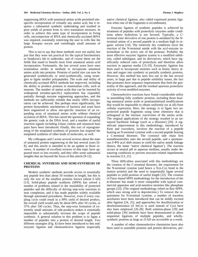
Modern synthetic methods provide access to essentiallyany peptide less than about 50 residues in length, but this isonly the size of the smallest proteins known (about 6 kD)[13]. Solid-phase peptide synthesis (SPPS) has solved anumber of problems related to the insolubility of protectedpeptides and the difficulty of driving step-wise reactions tonear completion, and it has made peptides widely availablethrough automated procedures. However, even if every cou-pling cycle could result in a 99% yield of desired product,the overall yield would only be about 60% after 50 cycles, or37% after 100 cycles. Thus, the step-wise formation of ex-tremely small amounts of side-products makes it essentiallyimpossible to substantially increase the scope of peptidesynthesis. A general solution to this problem is to ligate anumber of peptides into a protein of desired length. Twodifferent strategies (Fig. 1) have been introduced to this end:enzymic ligation and chemoselective ligation (especially
native chemical ligation, also called expressed protein liga-tion when one of the fragments is recombinant).
Enzymic ligation of synthetic peptides is achieved bytreatment of peptides with proteolytic enzymes under condi-tions where hydrolysis is not favored. Typically, a C-terminal ester derivative of one protein is amidated by the N-terminal amine of a second peptide in a medium high in or-ganic solvent [14]. The relatively dry conditions favor thereaction of the N-terminal amide with the acyl-enzyme in-termediate in the active site of the protease. Probably themost effective enzymic ligation system is a modified subtil-isin, called subtiligase, and its derivatives, which have sig-nificantly reduced rates of proteolysis and therefore allowreaction in aqueous media [15-19]. Enzymatic ligation hasbeen used to incorporate moieties such as biotin, heavy at-oms and sugars into synthetic or partially synthetic proteins.However, this method has seen less use in the last severalyears, in large part due to peptide solubility issues, the factthat proteases have sequence requirements that limit the gen-erality of this approach, and the residual spurious proteolyticactivity of even modified enzymes.
Chemoselective reactions have found considerable utilityin assembling fully synthetic proteins, including those bear-ing unnatural amino acids or posttranslational modificationsthat would be impossible to obtain uniformly (or at all) fromcellular expression. Here, the strategy is to ligate two un-protected peptide fragments using some chemistry that isorthogonal to the intrinsic reactivities of the amino acids.The original applications of the strategy resulted in an un-natural backbone linkage (such as a thioester) [20]. A sig-nificant improvement in this methodology, introduced byKent and coworkers, involves the reaction of a peptidebearing an N-terminal cysteine with a second peptide bearinga C-terminal thioester. The cysteinyl side chain firsttransthioesterifies onto the second peptide, and a spontane-ous SàN acyl shift ensues to yield the native amide linkage(hence, the name ‘native chemical ligation’). The reactionoccurs at neutral pH in aqueous medium, usually under de-naturing conditions to prevent structure-related impedimentsto reaction [13, 21].
Three difficulties associated with this methodology arethe creation of the C-terminal thioester, the requirement forthe N-terminal cysteine (and hence a Xxx-Cys bond in themature protein) and the need to sequentially ligate severalpeptides to yield proteins of useful length [22]. The creationof Fmoc-based SPPS methodology for the introduction of theα-thioester has made it more compatible with typical com-mercial apparatus and acid-sensitive moieties like phosphategroups [23]. (The original methodology relied on Boc-SPPS,which uses strong acid in deprotection.) To remove the re-quirement for N-terminal cysteine, a number of tracelessauxiliaries have been introduced that can be mildly excisedafter ligation [24, 25], and approaches for desulfurization ordeseleniumization (if SeCys is used instead of Cys) havealso been employed [26-28]. Both solution-phase [29] andsolid-phase [30] methods have been demonstrated to allowsequential ligations of multiple peptides, and wholly-synthetic proteins as large as 25 kD are accessible as a result.
A number of other chemoselective chemistries have alsobeen used to assemble proteins and protein derivatives, per-

Unnatural Protein Engineering Producing Proteins Medicinal Chemistry Reviews - Online, 2005, Vol. 2, No. 4 305
haps most notably the hydrazone/oxime chemistry of Offord& Rose, wherein an N-terminal aldehyde or ketone (whichcan be created by selective oxidation of N-terminal Ser orThr) is attacked with a C-terminal hydrazide or a hydroxy-lamine [31, 32]. The hydrazide can be introduced into a pep-tide in a way that is similar to the use of subtiligase, whereina protease is used to ligate a hydrazine donor at high con-centration in partially organic media.
Still, when one runs an E. coli lysate out on a gel, there isa conspicuous preponderance of proteins above the 25 kDmarker, and this is even more true in eukaryotes. Muir andcoworkers have developed an extremely clever and usefulmethod of protein semisynthesis based on native chemicalligation, wherein the α-thioester is produced recombinantlyfrom a partially-incapacitated protein splicing system [12,33]. Basically, the protein fragment of interest is expressedwith an intein domain fused at its C-terminus (there are
commercially-available kits to do this). That intein domainhas a mutation that prevents the intramolecular splicing re-action, and so the α-thioester can be transthioesterified withsome other thiol intermolecularly. A synthetic peptide withan N-terminal Cys can then be used to transthioesterify ontothe recombinant fragment, and amide formation followsfrom acyl rearrangement. This allows one to essentially ap-pend a fragment altered at will by chemical synthesis ontothe C-terminus of a recombinant protein of virtually any size.
Broadly considered, expressed protein ligation (EPL) canbe viewed as any native chemical ligation in which one ofthe reactants (the α-thioester or the N-terminal cysteinylfragment) is synthetic and one is recombinant. As mentionedabove, it is possible to introduce either of the appropriatefunctionalities synthetically, and it is possible to recombi-nantly produce α-thioesters. It is also possible to producerecombinant proteins with cysteinyl N-termini, either
Fig. (1). Peptide ligation strategies. Schematic representation of enzymic ligation by reverse proteolysis, chemoselective ligation by hydra-zone formation, native chemical ligation, and expressed protein ligation (EPL). Hydrazone formation could be carried out on wholly syn-thetic fragments, but the steps to ligate fragments from expressed proteins are shown.

306 Medicinal Chemistry Reviews - Online, 2005, Vol. 2, No. 4 Thomas J. Magliery
through removal of initiating methionine by endogenousaminopeptidases [34], or through protease treatment suchthat scission occurs at an Xxx-Cys bond [35]. Thus, it is pos-sible to introduce unnatural amino acids within about 50residues of the N- or C-termini of proteins using a singleligation. There are clearly some limitations, however. Theprotein has to tolerate the critical Cys at the junction site, orsome strategy for alteration of the Cys has to be undertaken.If one wants to make modifications further in than 50 resi-dues, then multiple ligations are required to build the protein,and this rapidly becomes difficult. Also, the ligation achievesits best yields in the absence of steric complications and athigh concentrations of reactants, and therefore the identity ofXxx at the Xxx-Cys junction is important [29], and it is help-ful if the reaction can occur under denaturing conditionsfollowed by protein “refolding.”
Nevertheless, this method has been very useful in insert-ing functionalities that cannot be introduced translationallyinto large, interesting proteins. Fluorescence probes, post-translational modifications, isotopes and various unnaturalamino acids have been used fairly extensively now [12, 22].It is worth noting that this method may always be superiorfor some kinds of modifications, such as the site-specificincorporation of isotopes (for NMR, for example). There willnever be a modified aminoacyl-tRNA synthetase that is ableto distinguish Lys from [15N]-Lys (although a protected[15N]-Lys is in principle not out of the question). Finally, allof these methods produce protein in vitro, which makes itconsiderably less useful for the investigation of cellularfunction (although some progress has been made in the in-troduction of protein into cells) [36, 37].
IN VITRO INCORPORATION OF UNNATURALAMINO ACIDS
Crick’s adapter hypothesis stated that tRNAs are merelyadapter molecules that connect an amino acid with a codonbased on the anticodon of the tRNA [38]. (That is, for themost part, the ribosome does not prevent misacylated tRNAsfrom delivering their cargo.) Essentially, this says that thefidelity of the genetic code depends on both thecodon:anticodon interaction and the acylation of the tRNAwith the proper amino acid, which is carried out by enzymescalled aminoacyl-tRNA synthetases, or aaRSs. The corollaryof this hypothesis is then that changing the genetic code re-quires two things: a tRNA for specifying the codon, and amethod of acylating the tRNA with an amino acid of choice.
The most straightforward way of selecting a codon as aninsertion signal is to choose from one of the 64 three-basecodons in the standard genetic code. One could select one ofthe 61 sense codons as an insertion signal, and the usage ofsome of these in a given organism is sufficiently rare that theamino acid of choice would be inserted at relatively few po-sitions in the proteome. However, a better approach has beento select one of the three stop codons [39], and in particularthe amber (UAG) stop codon has been successful in the Es-cherichia coli translation system. Amber is both the leastused stop codon in E. coli, and a number of efficient ‘sup-pressor’ tRNAs (i.e., those with a CUA anticodon) areknown [40]. (Note that throughout this review all nucleicacid sequences are written 5’-to-3’.) There has been some
recent success in generating other robust insertion signals(see below).
The second issue is how to acylate the tRNA bearing theselected anticodon with an amino acid of choice. This is afield with a long history, dating back to the Raney nickelreduction of Cys-tRNACys to Ala-tRNACys that proved theadapter hypothesis [38, 41]. (Ala is inserted at a Cys codonin an in vitro translation mix using the misacylated tRNA.)The state-of-the-art technology for chemical misacylation oftRNA represents the work of the Hecht, Chamberlin andSchultz groups (Fig. 2) [42-49]. The tRNA scaffold missingthe last two (3’) invariant CA nucleotides is generated by invitro transcription. Then, an unprotected pdCpA molecule issynthesized, and it is acylated with the cyanomethyl ester ofthe amino acid of choice (where the amine is typically pro-tected with a photolabile NVOC group). The pdCpA-aa-NVOC is then ligated into the tRNA-CA using T4 RNA ligase(and the lack of the nucleophilic 2’ hydroxyl on C does notdisturb either this ligation or the ability of the tRNA to act intranslation). Finally, the α-amine is deprotected (with pho-tolysis for NVOC) to yield the mature acylated tRNA. Thischemistry is widely applicable (Fig. 3), although some aminoacids are not compatible with the cyanomethyl activation,and photosensitive amino acids cannot be used with the
Fig. (2). Cell-free synthesis using chemically-acylated tRNA.The site of unnatural amino acid incorporation is specified at thegenetic level by site-directed mutagenesis (here, to an amber TAGstop codon). Chemical acylation of tRNA is carried out by in vitrotranscription of tRNA-CA, chemical acylation of pdCpA dinucleo-tide and ligation of the acylated pdCpA to the immature tRNA.This is then added to an in vitro transcription/translation reactionalong with the DNA encoding the gene of interest.

Unnatural Protein Engineering Producing Proteins Medicinal Chemistry Reviews - Online, 2005, Vol. 2, No. 4 307
NVOC protecting group. Also, not all amino acids are ac-cepted by the translational machinery, likely in part due tothe action of elongation factor Tu [50]. The chief liability ofthis method is that synthesis of both pdCpA and protected,activated amino acid is challenging, and most biochemistrylabs are simply not equipped to do this. Ninomiya et al. re-cently showed that the transesterification reaction betweenthe cyanomethyl-activated amino acid and pdCpA can beimproved using cationic micelles [51], and pdCpA is nowavailable commercially, but the method is still expensive anddifficult.
Another approach to in vitro aminoacylation of tRNAwith unnatural amino acid is to evolve a ribozyme for thispurpose [52, 53]. The method is particularly attractive, sincethe tRNA specificity can be altered rationally. Although thisapproach is less general than synthetic acylation in the sensethat one might have to evolve a separate ribozyme for agiven amino acid, it may provide a tool that is simple to useand applicable to a wide array of amino acids. This approachstill requires amino acid activation (as the cyanomethyl es-ter), but it avoids the pdCpA synthesis and ligation steps, andcan be accomplished without α-amine protection. Recently,a resin-immobilized ribozyme was introduced which hasbroad tRNA aminoacylation specificity toward cyanomethylesters of phenylalanyl analogs [54]. This system makes invitro acylation with unnatural amino acids considerably eas-ier and faster than purely synthetic routes.
The final issue is how to get a tRNA bearing an anti-codon (CUA) and unnatural amino acid of choice to functionin translation. Either one has to get the acylated tRNA intothe cell (see below), or one has to generate the protein invitro using an transcription/translation extract mixture. Ofcourse, the nature of the tRNA scaffold depends upon thechoice of protein biosynthetic apparatus, since one must em-ploy a tRNA that is neither acylated nor deacylated by en-
dogenous aaRSs, but that is competent to act in translation(i.e., binds to elongation factors, is accepted by the ribosome,etc.). The most effective system to date is based on E. coliextract, using amber-suppressing tRNA derived from yeasttRNAPhe or, for small, polar amino acids, E. coli tRNAAsn
[44, 55]. The efficiency of amber suppression, and in vitrotranslation generally, have been improved considerably re-cently by inactivation of the release factor that recognizesamber stop codons, and by optimization of the cell-freesynthesis system, respectively [56-58]. The latter is accom-plished by continuous dialysis of the transcription/translationreaction to replenish necessary small molecules and removeinhibitory byproducts, resulting in yields as high as 6 mgmL-1 (for normal translation, not amber suppression).
A last point worth mentioning is that the template(mRNA) for protein synthesis also needs to be present, obvi-ously, with the amber mutation in the appropriate position.This is straightforward for cell-free transcription/translation,since one can simply add a plasmid or even synthetic DNAcoding for the protein to the mixture. Site-directed mutage-nesis can be used to create the mutation in the plasmid, anduse of the T7 promoter allows one to simply add T7 RNApolymerase to the cell-free synthetic system to achieve largeamounts of mRNA. The importance of this issue will beclear with in vivo systems or alternate insertion signals, dis-cussed below.
Chemical acylation of tRNA offers the considerable ad-vantage over synthetic methods that it is applicable to (solu-ble) proteins of virtually any size with the desired amino acidinserted site-specifically at any position in the protein (re-gardless of distance from the termini). It does not requiredenaturing conditions to produce the protein, so refolding isless likely to be necessary. Unlike in vivo residue-selectiveor specific methods (below), it is totally site-specific (al-though it allows the insertion of only a single unnatural
Fig. (3). Examples of amino acids incorporated by chemical acylation of tRNA. Over a hundred amino acids and analogs have been in-corporated into proteins by chemical acylation of tRNA. Some types of analogs include sterically restricted amino acids (1-2), peptide back-bone mimetics (like α-hydroxy acids, 3, and α-amino alkyl derivatives, 4), amino acids that differ in electronic properties (glutamine analog5), reactive amino acids (for photoaffinity labeling, 6, or photodecaging, 7), fluorescence probes (8) and spin-labels (9).
NH2
OH
O
HN
OH
O
Me
MeNH2
OH
O
O
NO2
NH2
OH
O
NH2
OH
O
N
O
O
NH2
OH
O
N
N
H
OH
OH
O
MeNH2
OH
O
O
NH2
OH
O
S
N
O
1 4 7
2 5 8
3 6 9

308 Medicinal Chemistry Reviews - Online, 2005, Vol. 2, No. 4 Thomas J. Magliery
amino acid using the amber stop codon). It is slightly morelimited than synthetic methods in terms of generality withrespect to amino acid, but it is much broader in scope thanany in vivo method right now. However, the liabilities of thismethod are substantial: it is synthetically challenging, theyields are generally poor (and thus it is labor-intensive andexpensive to generate more than microgram quantities ofprotein), and, like any in vitro method, it does not allow di-rect investigation of cellular function.
mRNA Display
One of the serious issues with in vitro production of un-natural proteins is the question of what one can actually dowith the unnatural protein once it is in hand. It is difficult toproduce large amounts of protein, so it is consequently diffi-cult (or expensive, or both) to carry out material-hungry bio-physical characterization like CD spectroscopy, NMR, or X-ray crystallography. (All of these experiments have beendone, however.) One can carry out tests for binding or en-zymatic activity, which require only a small amount of pro-tein, but combinatorial methods are essentially not an option.The protein is not linked to its genetic material and thuswould be impossible to identify in a mixture.
An elegant solution to the in vitro production of mRNA-linked proteins has substantially expanded the scope of invitro unnatural protein engineering. Essentially, one can pro-duce mRNA that ends with a puromycin (Pur) moiety, whichacts as an aminoacyl-tRNA surrogate at the end of transla-tion, covalently linking the protein to the encoding mRNA[59, 60]. This puromycin-linked RNA can be produced to-tally synthetically, or transcribed from DNA in vitro andthen linked to a short RNA-Pur fusion using T4 RNA ligase(and a DNA splint). This mRNA-Pur fusion can then beadded to a cell-free translation system. The resulting protein,which is labeled with its encoding RNA, can then be sub-jected to screening (for example, binding to an immobilizedligand) and identified by RT-PCR from as little as, in princi-ple, a single molecule [61].
Of course, this protein has a large RNA appended to it,which is a complicating factor. One would like to know thatone is selecting for a property of the protein, not the RNA, soit is necessary to reverse transcribe the RNA to produce an“inert” double-stranded nucleic acid. But this can hardly bethought of as an inconsequential spectator: a 100-residueprotein (13 kD) would bear 300 residues of dsRNA/DNAhybrid (195 kD), and at minimum this is likely to have aprofound effect on the solubility of the fusion (i.e., there isessentially no selection for soluble protein since the highly-charged fusion is virtually guaranteed to be soluble). Also,the cell-free system that has been the most efficient is basedon rabbit reticulocytes rather than E. coli extract, and there-fore one must find a tRNA scaffold and insertion signal thatis applicable for delivery of an unnatural amino acid. Fortu-nately, it was known that THG73, a modified tRNAGln fromTetrahymena thermophila, is an efficient suppressor of am-ber in other eukaryotic systems, and other insertion signalshave been explored (see below for both). Unnatural aminoacids like biocytin (lysyl biotin) and N-methyl-Phe havebeen inserted this way, and selection of biocytin-containingfusions using streptavidin has been demonstrated, making
this a promising approach [62, 63]. A related technology,ribosome display (in which protein and mRNA remain on-board stalled ribosomes), is also potentially amenable to thissort of approach [64].
RECODING – BEYOND AMBER SUPPRESSION
Amber stop codon suppression has been the workhorse ofsite-specific biosynthetic unnatural protein engineering, butthere has been significant development of other insertionsignals. The motivation for this is quite simple: one wouldlike to be able to insert more than one unnatural amino acidinto a single protein site-specifically (as one can do withtotal synthesis). There are also more subtle motivations. Am-ber suppression is not generally as efficient as normal inser-tion of amino acids, presumably in large part due to compe-tition with the release factor, and some sites are not amena-ble to any significant level of suppression. Moreover, there isthe problem of read-through, which is caused by the mis-reading of the amber stop codon by another tRNA (usuallywith an anticodon similar to CUA). This results in competi-tive protein products in which the selected site is occupiedby one of the natural amino acids. Also, although ambersuppression is possible in eukaryotic systems, amber stopcodons are used much more frequently in eukaryotes. Essen-tially, there are four alternatives to the amber stop codon:another stop codon, a sense codon, an extended (e.g., 4-base)codon, or an unnatural codon. All of these options have beenexplored in recent years to varying degrees of success.
The most efficient stop codon in E. coli is ochre (UAA),which is recognized by both release factors, and therefore isnot a good candidate for suppression. There are, however,natural contexts in which opal (UGA) is suppressed in bothprokaryotes and eukaryotes, such as with selenocysteine in-sertion [65]. In fact, the Schultz group has recently engi-neered an opal suppressor for use in E. coli [66]. Moreover,suppression of both amber and ochre stop codons has beendemonstrated in mammalian cells [67]. Attempts have beenmade to use suppressors of rare codons in E. coli expressionsystems, and depletion of endogenous tRNAs from the cell-free extract has made this more tractable [68-72]. However,readthrough from tRNAs with near-anticodons and the needto heavily mutagenize genes to remove the selected sensecodons makes this strategy problematic. Recently, Frankel &Roberts selected for tRNAs with NNC anticodons to sup-press NNN codons in the rabbit reticulocyte expression sys-tem. They found that suppression of GUA was comparableto amber suppression [73]. A related approach may comefrom recent cell-free translation systems reconstituted en-tirely from recombinant proteins, since all of the tRNA andaaRS identities and concentrations can be altered at will toproduce more “blanks” in the genetic code [74].
Naturally occurring frameshift suppressors in yeast andSalmonella are known, typically functioning by decoding a4-base codon using a tRNA with an extended anticodon loop(8 nt instead of 7 nt) [65]. Suppression of 4-base and 5-basecodons has been demonstrated in E. coli and in E. coli cell-free translation systems. Magliery et al. selected for tRNASer
with randomized, extended anticodon loops (8 or 9 nt insteadof 7 nt) that would suppress randomized four-base codons inthe gene for β-lactamase, and identified very efficient anti-
Unnatural Protein Engineering Producing Proteins Medicinal Chemistry Reviews - Online, 2005, Vol. 2, No. 4 309
codon loop/4-base codon pairs for the codons AGGA,UAGA, CCCU and CUAG [75]. A similar approach af-forded fairly efficient 5-base codon suppressors of CUAGU,CUACU, AGGAU [76]. These studies established a numberof factors leading to efficient suppression of codons: Wat-son-Crick pairing at all positions; N+4 nt in the anticodonloop of a tRNA suppressing a codon with N nt, where 2 ntare on either side of the anticodon; and the presence of a U5’ to the anticodon and an A 3’ to it. In general, suppressiblecodons are based on rare 3-base codons. Four-base codonshave been used both for site-directed mutagenesis and toinsert unnatural amino acids into proteins [77-79]. Sisido’sgroup has incorporated multiple unnatural amino acids intoproteins site-specifically using AGGU or GGGU and CGGGcodons [80, 81]. Five-base codons of the type CGGNN werealso investigated, and CGGAC was found to be the mostefficiently suppressed [82]. Extended codons have a distinctadvantage of the amber stop codon in that “readthrough” bynatural tRNAs results in an out-of-frame product that typi-cally terminates rapidly. Thus, material purified with a C-terminal tag is virtually guaranteed to contain unnaturalamino acid at the selected site (except for the possibility ofspontaneous frameshifts).
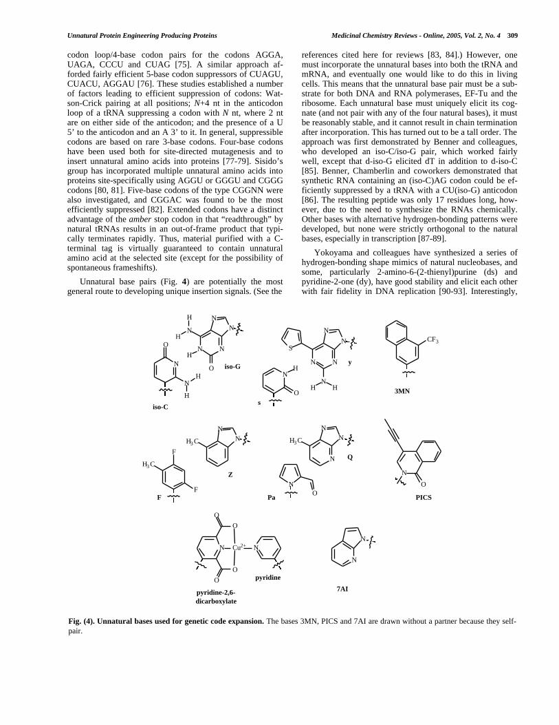
Unnatural base pairs (Fig. 4) are potentially the mostgeneral route to developing unique insertion signals. (See the
references cited here for reviews [83, 84].) However, onemust incorporate the unnatural bases into both the tRNA andmRNA, and eventually one would like to do this in livingcells. This means that the unnatural base pair must be a sub-strate for both DNA and RNA polymerases, EF-Tu and theribosome. Each unnatural base must uniquely elicit its cog-nate (and not pair with any of the four natural bases), it mustbe reasonably stable, and it cannot result in chain terminationafter incorporation. This has turned out to be a tall order. Theapproach was first demonstrated by Benner and colleagues,who developed an iso-C/iso-G pair, which worked fairlywell, except that d-iso-G elicited dT in addition to d-iso-C[85]. Benner, Chamberlin and coworkers demonstrated thatsynthetic RNA containing an (iso-C)AG codon could be ef-ficiently suppressed by a tRNA with a CU(iso-G) anticodon[86]. The resulting peptide was only 17 residues long, how-ever, due to the need to synthesize the RNAs chemically.Other bases with alternative hydrogen-bonding patterns weredeveloped, but none were strictly orthogonal to the naturalbases, especially in transcription [87-89].
Yokoyama and colleagues have synthesized a series ofhydrogen-bonding shape mimics of natural nucleobases, andsome, particularly 2-amino-6-(2-thienyl)purine (ds) andpyridine-2-one (dy), have good stability and elicit each otherwith fair fidelity in DNA replication [90-93]. Interestingly,
Fig. (4). Unnatural bases used for genetic code expansion. The bases 3MN, PICS and 7AI are drawn without a partner because they self-pair.
N
O
NH
H
N N
O
H
NH
H
NN
iso-C
iso-G
F
F
H3 C
NN
H3 C
Z
F
N
O
PICS
N
N
N
O
O
O
O
Cu2+ N
pyridine
pyridine-2,6-dicarboxylate
7AI
CF3
3MN
N
O
H
s
N N
N
N
S
NH H
y
NO
N
NN
H3 C
Pa
Q

310 Medicinal Chemistry Reviews - Online, 2005, Vol. 2, No. 4 Thomas J. Magliery
this pair was used for in vitro site-specific unnatural aminoacid incorporation [94]. DNA was synthesized to contain aCTs sequence that could be transcribed into a yUG codon byT7 RNA polymerase. A yeast tyrosyl tRNA with a CUs anti-codon was prepared with a combination of synthesis andligation, and this was aminoacylated with 3-chlorotyrosineusing yeast tyrosyl synthetase (which accepts ClTyr as asubstrate). The ClTyr-tRNACUs was added to an E. coli cell-free transcription/translation system along with the DNAtemplate and T7 RNA polymerase, and full-length proteinwas produced in good yield (40% compared to a naturalcodon) with high fidelity (>90% of the amino acid at the sitewas ClTyr).
Kool and coworkers suggested the idea of circumventingproblems with using hydrogen-bonding schemes for baserecognition by instead employing hydrophobic base pairs.For example, difluorotoluene (dF) was found to base-pairwith adenine [95]. The base could also elicit dA in DNAreplication, or 4-methylbenzimidazole (dZ) to form a stable,all-hydrophobic base pair [96]. However, both dF and dZalso elicit natural bases, and so are not suitably orthogonal.Improvements by the Kool and Yokoyama groups have ledto the development of pyrrole-2-carbaldehyde (dPa) and 9-methylimidazo[(4,5)-b]pyridine (dQ), which forms a stablebase pair in DNA that is effectively replicated and extended,although dPa is still compromised by spurious insertion ofdA [97]. These bases are essentially shape-complementary tothe natural purine:pyrimidine pairs, and they all possessedminor-groove hydrogen-bonding functionalities.
Romesberg, Schultz and coworkers have explored a se-ries of hydrophobic bases that are, in general, neither shape-complementary nor in possession of any hydrogen-bondingfunctionality. It has been possible to develop stable basepairs of this type, sometimes incorporated into DNA withhigh fidelity, but chain termination after insertion has been asignificant problem (e.g. d3MN, dPICS and d7AI) [98, 99].Interestingly, the most efficient pairs have been “self-pairs,”which is to say a base that elicits itself in DNA synthesis,which is perfectly reasonable for genetic code expansion. Aninteresting approach to solve the chain-termination problemis to explore the use of other natural polymerases, or to engi-neer new polymerases [100, 101]. For example, the d7AIself-pair is efficiently incorporated by Klenow fragment ofDNA polymerase I, and it is efficiently extended by mam-malian polymerase β. Finally, a number of nucleobases thatpair due to metal ion ligation have also been explored [102-105].
Despite the obvious advantages of the unnatural baseapproach, it is still fairly far from being broadly useful, evenfor in vitro applications. At minimum, an unnatural basesynthetically incorporated into a DNA template needs tospecify the efficient in vitro transcription of unnatural base-containing mRNA and tRNA. (This is similar to what Hiraoet al. have accomplished, although the tRNA preparationwas somewhat complicated [94].) It would be considerablymore convenient if the unnatural base could be inserted intoDNA with PCR and/or plasmid replication, which requiresapplicable thermophilic and cellular polymerases. In the caseof cellular replication, it also requires that the unnatural nu-cleosides or nucleotides get into cells and be (or remain)
appropriately phosphorylated. Currently, amber suppressionand four-base codon suppression are the best approaches inE. coli, and other stop codons or sense codons may beequally useful in eukaryotic systems.
IN VIVO INCORPORATION OF UNNATURALAMINO ACIDS
Microinjection/Microelectroporation
So far, I have discussed the use of chemically-acylatedtRNA in cell-free transcription/translation reactions. This is astraightforward way of generating large, soluble proteinswith a site-specific unnatural amino acid mutation, and pro-teins can be produced in reasonable quantities with effort.However, to harness the power of unnatural protein engi-neering to investigate cellular function, the protein must beproduced in living cells. Moreover, membrane-bound pro-teins are not amenable to the cell-free approach, and theDougherty group has elegantly investigated ion channelfunction by adapting the chemical acylation approach for usein vivo . Most of the work so far has been carried out on thenicotinic acetylcholine receptor (nAChR) using Xenopusoocytes, which are sufficiently large that one can microinjectreagents into them [106, 107]. Here, one must inject bothmRNA and chemically-acylated tRNA, and that tRNA mustbe orthogonal to Xenopus aaRSs but competent to act in thetranslational apparatus. A modified version of Tetrahymenathermophila tRNAGln(CUA) called THG73, a tRNA thatnaturally inserts glutamine in response to UAG (which is nota stop codon in Tetrahymena), was both efficient and not asubstrate for the endogenous aaRSs of the oocyte [108].
An exciting development in this field has allowed exten-sion of this scheme to mammalian cells [109]. Both CHO-K1cells and rat hippocampal neurons were subject to delivery ofDNA, mRNA and tRNA using microelectroporation. Byadministering chemically acylated amber suppressor (theTHG73 tRNA) and nAChR mRNA to neurons in this fash-ion, functional receptors site-specifically modified with un-natural amino acids were produced and probed electrophysi-ologically. This method has the substantial advantage overother in vivo methods that virtually any amino acid can beinserted, but only very small quantities of protein can beproduced, requiring very sensitive assays (like voltageclamping). RajBhandary has also shown that acylated tRNAcan be introduced into mammalian cells (COS-1 cells) usingtransfection reagents [67].
Widespread/Residue-Specific Incorporation
In the context of protein science, the term amino acidstypically means proteinogenic amino acids—the 20 commonmonomers that are incorporated into proteins. Recently,other (rarely) translationally incorporated amino acids likeselenocysteine and pyrrolysine have been discovered [1-3].However, there are many other amino acids that act in thebiological milieu, including natural non-proteinogenic aminoacids, amino acids created by posttranslational modification,and amino acids (natural and unnatural) that act on cells inone way or another. Citrulline and ornithine are intermedi-ates in the biosynthesis of arginine, and are not incorporatedinto proteins, despite their relative similarity to arginine and

Unnatural Protein Engineering Producing Proteins Medicinal Chemistry Reviews - Online, 2005, Vol. 2, No. 4 311
lysine, respectively. Posttranslational modification results inphosphorylated and sulfated amino acids, lipidated and gly-cosylated amino acids, methylated and acetylated amino ac-ids, hydroxylated amino acids, and biotinylated amino acids(biocytin). On the other hand, L-canavanine, an oxy-analogof arginine, is produced by legumes and is toxic to bacteriaand animals due to misincorporation into proteins (amongother effects).
This raises an interesting question: how does the cellmaintain the fidelity of the genetic code with so many aminoacids around? The simple answer is that the aminoacyl-tRNA synthetases are exquisitely good at their job, and man-age to correctly distinguish their amino acid substrate (thereis generally one aaRS per amino acid per organism) from allthe other amino acids in the cytosol (or organelle). The errorrate in protein synthesis is only about 1 in every 10,000 resi-dues, and part of this can be blamed on incorrect tRNA se-lection at the ribosome and errors in transcription. ManyaaRSs employ specific mechanisms to “edit” (hydrolyze)improperly-acylated tRNAs, in addition to their exquisiterecognition of amino acid and tRNA in the first instance.
But the more complicated answer is that the fidelity of allaaRSs is limited, especially when confronted with near-analogs of natural amino acids which have never been en-countered in a biological context. The fact is that aaRSs onlyneed to have evolved ways of excluding other cellular aminoacids (proteinogenic or not), and this opens the door to awhole array of residue-selective (or, better, residue-specific)protein modifications achieved by adding unnatural aminoacids to the growth medium. This fact has revolutionizedprotein biophysics: high-level incorporation of selenome-thionine affords a general solution to the phase problem inX-ray crystallography through multiwavelength anomalousdiffraction (MAD) [110, 111]. But it is also a route to bothspecific unnatural modifications of proteins and the genera-tion of unnatural biomaterials.
For widespread replacement of an amino acid to be use-ful, it has to occur as specifically as possible, which is to sayit should replace only one amino acid at any level, and thatlevel should be 100%. Needless to say, it is not generallypossibly to grow cells under normal conditions and achieve100% replacement of one of the natural amino acids; somecritical, lethal replacements are bound to occur. In general,one gets the best incorporation of an amino acid analog with(1) an auxotrophic strain for the related natural amino acid,with sufficient growth to remove all the natural amino acidfrom the growth medium followed by (2) high-level induc-tion of protein expression with concomitant addition of theunnatural amino acid [112]. Nearly quantitative replacementis possible under these conditions. The second issue with thismethod is that any given protein is likely to contain at leastone of each of the natural amino acids, and so one must mu-tate the protein appropriately to achieve labeling at the de-sired position(s). This may not always be practical. Finally,the array of appropriate amino acid analogs essentially has tobe determined empirically, although some engineering hasbeen helpful, and thus the selection of analogs does notcompare to what is possible through synthetic methods.
Nevertheless, some interesting and useful amino acidshave been incorporated this way (Fig. 5). The 4-, 5-, and 6-
fluoro derivatives of tryptophan and o-, m- and p-fluoro de-rivatives of phenylalanine permit site-specific labeling for19F NMR [113-115]. Besides SeMet, other Met analogs havebeen incorporated, such as 2-aminohexanoic acid (nor-leucine), ethionine, telluromethionine and S-nitrosohomo-cysteine [112, 116]. Thiaproline is inserted by ProRS [117].These sorts of amino acids permit the characterization ofproteins with what Budisa and coworkers call “atomic muta-tions,” changes of as little as a single atom in a whole mac-romolecule. Tirrell, Fournier and colleagues became inter-ested in the use of unnatural amino acids to produce homo-geneous, biopolymeric materials (periodic proteins) [118].For example, 3-thienylalanine, azidohomoalanine, trifluoroi-soleucine, hexafluoroleucine, homoallylglycine, and homo-propargylglycine have been inserted into such proteins [119-123].
Some unnatural amino acids are only incorporated effi-ciently if MetRS is overexpressed, such as the olefinic andacetylenic amino acids cis- and trans-2-amino-4-hexenoicacid, 2-butynylglycine and allylglycine [124, 125]. Engi-neering of a PheRS (by Ibba & Hennecke [126]) resulted in alarger binding pocket, which has allowed the insertion ofpara-substituted fluoro-, chloro-, bromo-, iodo-, azido-,cyano- and ethynyl-phenylalanine [127]. A related approachis mutation of an aaRS to abrogate hydrolytic editing ofmisacylated products, which has found purchase with ValRSand LeuRS since these enzymes require editing to removeother hydrophobic amino acids that are erroneously esteri-fied. For example, low-level incorporation of aminobutyrateat Val codons was achieved by ablation of ValRS editingactivity [128]. Tang & Tirrell have used LeuRS with reducedediting activity to introduce six new hydrophobic amino ac-ids, both saturated and unsaturated [129].
Widespread methods of amino acid replacement haveseveral positive points worth highlighting. Once a suitableamino acid is found, the method is technically simple, andlarge amounts of protein can be produced cheaply. So far,the engineering approaches applied to expand the scope ofwidespread amino acid replacement have been relativelysimple and have yielded excellent results. However, themethod is considerably less general with respect to aminoacid than synthetic approaches. While it is an in vivo method,it requires growth conditions that make it much less usefulfor examining cellular processes, and it results in (often le-thal) incorporation of unnatural amino acids all over theproteome under normal growth conditions. Indeed, the mostserious problem is that it is not actually a method of ex-panding the genetic code, since it requires the sacrifice ofone of the natural amino acids. At best, this is likely to beinconvenient in any specific protein. However, when this isnot a concern, as with the homogeneous preparation of un-natural biomaterials, this method is particularly suitable.
Site-Specific Incorporation
The drawbacks and advantages of the in vivo chemicalacylation and widespread replacement methods provide ablueprint for what one ultimately wants in a method of un-natural protein engineering. Ideally, the method will be suit-able for use in living cells, so that it is technically simple andinexpensive to produce large amounts of protein or examinecellular processes (i.e. “unnatural cell biology”). It should

312 Medicinal Chemistry Reviews - Online, 2005, Vol. 2, No. 4 Thomas J. Magliery
require little or no organic synthesis (except perhaps of thefree amino acid), and should be as simple as transformingplasmids and adding amino acid to the growth medium. Onthe other hand, it should be broadly applicable to as manyamino acids as possible. It should expand, not just recode,the genetic code, and it should do so in a site-specific man-ner, ideally so specifically that only the protein of interestwill contain the unnatural amino acid, and only at the se-lected site.
How this can be accomplished returns us to the corollaryto Crick’s adapter hypothesis: essentially, we can invade thegenetic code with a unique tRNA (and anticodon) and ami-noacyl-tRNA synthetase. Specifically, we need the follow-ing:
1. A host organism. Due to the extensive engineering re-quired, well-understood systems like E. coli and yeasthave proven good starting points.
2. An insertion signal (codon). In E. coli, the amber stopcodon is a good first choice. Expansion into four-baseand unnatural codons is necessary to think about addingmultiple amino acids to the genetic code of a single or-ganism.
3. An “orthogonal” tRNA bearing the insertion signal, thatis neither acylated nor deacylated (“edited”) by any en-dogenous aaRS.
4. An orthogonal aaRS, that acylates only the orthogonaltRNA, only with an unnatural amino acid. For simplicity,an aaRS without hydrolytic editing is a good choice.
5. An unnatural amino acid that is not toxic nor incorpo-rated into proteins by endogenous aaRSs, but that isreadily uptaken by the cell (or produced in it).
Fig. (5). Examples of amino acids incorporated by residue-specific replacement.
H2NOH
O
HN
FH2N
OH
O
X
H2NOH
O
SeCH3
seleno-Met
H2NOH
O
CH3
H2NOH
O
S
CH3
H2NOH
O
SNO
OH
O
S
NH H2NOH
O
S
H2NOH
O
N3
H2NOH
O
H2NOH
O
H2NOH
O
CF3
CF3
H2NOH
O
CF3
H2NOH
O
H2NOH
O
H2NOH
O
H2NOH
O
H2NOH
O
N3
H2NOH
O
CN
H2NOH
O
5-fluoro-Trp
45
6
para-halo-Phe
om
p
norleucine ethionine
S-nitroso-homoCys thia-Pro 3-thienyl-Ala azido-homoAla homo-allyl-Gly
homo-propargyl-Gly hexafluoro-Leu trifluoro-Ile
X = F, Cl, Br, I
trans- and cis-2-amino-4-hexenoic acid
2-butynyl-Gly allyl-Gly para-azido-Phe para-cyano-Phe para-ethynyl-Phe

Unnatural Protein Engineering Producing Proteins Medicinal Chemistry Reviews - Online, 2005, Vol. 2, No. 4 313
Generating an Orthogonal tRNA/Synthetase Pair
The initial work by the Schultz group to this end focusedon evolving such an “unnatural organism” in two stages:first, generate an orthogonal tRNA/aaRS pair, and, second,change the specificity of the aaRS with respect to aminoacid. All initial work was carried out in E. coli using amber-suppressing tRNA. We first employed an engineering ap-proach to generate an orthogonal tRNA/synthetase pairbased on the exceedingly well-characterized E. coli glu-taminyl-tRNA synthetase (GlnRS)/tRNAGln(CUA) pair.(GlnRS naturally acylates an efficient amber suppressor inE. coli.) Based on the co-crystal structure [130], three muta-tions in tRNAGln were identified that together abrogated acti-vation by GlnRS [131]. This tRNA was not appreciablyacylated by any aaRS in an E. coli transcription/translationreaction, but it was competent for amino acid delivery whenchemically acylated. A mutant GlnRS was then engineeredusing a DNA shuffling [132, 133] strategy coupled with aselection for amber suppression (survival in galactose in astrain with an amber mutation in the gene for β-galactosidase) [134]. Despite a remarkable change in speci-ficity toward the orthogonal tRNA, the mutant GlnRS wasstill capable of efficiently acylating the wild-type tRNAGln,as well. If the amino acid specificity of this enzyme had beensubsequently altered, the unnatural amino acid would havebeen inserted at Gln codons throughout the proteome in ad-dition to amber codons.
Work by the Schimmel group suggested that another ap-proach might be easier to obtain an orthogonal aaRS/tRNApair: importation of a heterologous pair from another organ-ism [135, 136]. Based on the fact that Saccharomyces cere-visiae tRNAGln (SctRNAGln) was not acylated by E. coliGlnRS (EcGlnRS), Liu & Schultz demonstrated that a modi-fied SctRNAGln(CUA) and ScGlnRS constitute a functional,orthogonal pair in E. coli [137]. (Hereafter, the notationOotRNAAaa(NNN) and OoAaaRS shall be used analogouslyto the examples in the previous sentence, where Oo denotesthe organism, Aaa denotes the amino acid, and NNN denotesthe anticodon.) However, despite nearly 10 man-years ofwork engineering this pair, we were never able to alter theamino acid specificity of ScGlnRS, even though virtually allof the selection and library-construction technology wasworked out on this system (D.R. Liu, T.J. Magliery, M.Pastrnak, S.W. Santoro & P.G. Schultz, unpublished, in ad-dition to the cited references) [137-139].
Based on a similar observation from the Schimmel group[140, 141], Wang et al. showed that the Methanococcus jan-naschii tRNATyr(CUA) and MjTyrRS constituted an or-thogonal pair in E. coli, as well [142]. However, while theMjTyrRS is considerably more active than ScGlnRS, theMjtRNATyr(CUA) was not “as orthogonal” asSctRNAGln(CUA), as measured by the survival of E. coliexpressing these tRNAs in a strain bearing an amber mutantof β-lactamase on varying concentration of ampicillin. (Thatis: E. coli with MjtRNATyr(CUA) survive at higher ampicillinconcentrations due to greater amber suppression from spuri-ous acylation by E. coli endogenous synthetases.) Wang &Schultz used a semi-rational design strategy to increase theorthogonality of this tRNA [143], employing a variation onthe double-sieve selection introduced by Liu & Schultz (see
below) [137]. First, a library of MjtRNATyr(CUA) variantswas co-produced in a strain with a multi-amber mutant ofbarnase, a toxic RNase in E. coli. Surviving cells boretRNAs less capable of being acylated by endogenous syn-thetases. The selected tRNAs were then co-produced in astrain with an amber mutant of β-lactamase and expressingMjTyrRS. Survival at this step ensures that mutant tRNAsare still competent for protein biosynthesis. The resultingorthogonal tRNA and has been by far the most useful so far,due to the high activity of the synthetase and good orthogo-nality of the tRNA, in addition to the relatively large, hydro-phobic Tyr binding pocket in TyrRS. Recently, solution ofthe MjTyrRS crystal structure permitted design of a mutantsynthetase with better recognition of the MjtRNATyr(CUA),which may be useful in further work altering the amino acidspecificity of the synthetase [144].
A number of other pairs have also been introduced.Pastrnak et al. engineered an orthogonal pair based on theamber-suppressing SctRNAAsp(CUA) and a AspàLys mu-tant of ScAspRS known to acylate that tRNA [138]. (Actu-ally, the homologous mutation was known in the E. coli en-zyme and tRNA. The anticodon is a key recognition elementof AspRS, making the amber suppressor instantly orthogo-nal.) However, the amount of amber suppression was weak,although more reasonable in an RF1-deficient E. coli strain,since RF1 competes with amber suppressors for recognitionof UAG codons. The RajBhandary group also selected for amutant ScTyrRS that was orthogonal in E. coli but capableof acylating the orthogonal amber-suppressing mutant of E.coli initiator tRNAfMet [145]. Anderson & Schultz haveshown that Methanobacterium thermoautotrophicum LeuRSand derivatives of Halobacterium NRC-1 tRNALeu constituteorthogonal pairs capable of suppression of amber, opal(UGA) and 4-base (AGGA) codons in E. coli [66]. Santoroet al. also showed that Pyrococcus horikoshii GluRS and aconsensus-derived archaeal tRNAGlu(CUA) are an orthogo-nal pair in E. coli [146]. However, there has been no successaltering the amino acid specificity of any of the synthetasesdescribed in this paragraph. Recently, the Schultz group hasselected a mutant Pyrococcus horikoshii LysRS derivativethat is capable inserting homoglutamine in E. coli in re-sponse to the four-base codon AGGA using a consensus-derived archaeal tRNALys(UCCU) [147]. This is only thesecond orthogonal pair shown to be useful in E. coli, and thefact that it allows four-base suppression means that it is nowpossible to simultaneously, site-specifically introduce twounnatural amino acids into bacterial proteins.
The first orthogonal pair for use in eukaryotes was char-acterized by the RajBhandary group, consisting of EcGlnRSand an amber-suppressing derivative of EctRNAfMet used inyeast [145]. RajBhandary and coworkers also demonstratedthat coexpression of EcGlnRS and EctRNAGln(CUA) is nec-essary and sufficient for amber suppression in mammalianCOS-1 and CV-1 cells, although the orthogonality of thesynthetase was not assessed (beyond the fact that it is notlethal) [148]. However, the amino acid specificity ofEcGlnRS has not subsequently been altered. The Yokoyamagroup found that a mutant E. coli tyrosyl-tRNA synthetasethat inserts 3-iodo-Tyr and EctRNATyr(CUA) constitutes anorthogonal pair in wheat germ extract [149]. When expres-sion of this pair was attempted in mammalian cells (using a

314 Medicinal Chemistry Reviews - Online, 2005, Vol. 2, No. 4 Thomas J. Magliery
D-arm mutant of the tRNA to create an internal promoter),no amber suppression was detected [150]. It was found thatthe same synthetase was capable of acylating a Bacillusstearothermophilus tRNATyr(CUA), allowing amber sup-pression in mammalian CHO cells. Again inspired by workfrom the Schimmel group [151-153], Chin et al. demon-strated that E. coli tyrosyl-tRNA synthetase andEctRNATyr(CUA) constitute an orthogonal pair in yeast, andthey have successfully engineered mutants of EcTyrRS thatacylate with unnatural amino [154, 155]. Recently, Zhang etal. have introduced a mutant Bacillus subtilis TrpRS thatinserts 5-hydroxy-Trp, and an opal-suppressing mutant ofBstRNATrp(UCA), for selective incorporation in mammaliancells [156].
Altering Amino Acid Specificity
Alteration of the amino acid specificity of the aaRSs hasbeen the most challenging aspect of the development of thismethodology. AaRSs are exceedingly good at what they do,and it has proven no simple matter to coax them to do other-wise. The problem is complicated considerably by at leastfive facts. (1) The small amino acid substrates are contactedby a large number of residues concentrated in three-dimensional space but often spread over the primary struc-ture of the synthetase. This makes it technically difficult toconstruct reasonable mutagenic libraries. (2) In spite of this,mutations far from the active site of aaRSs are known toaffect aminoacylation kinetics. (3) Mutations to the syn-thetase can also affect recognition of tRNA, thereby alteringthe orthogonality of the enzyme. (4) It is extremely difficultto alter the specificity of an aaRS without reducing its ami-noacylation activity, but a certain level of activity is requiredto support high-level protein synthesis. Thus, engineering ofsynthetases with weak aminoacylation activity toward nativesubstrates has proven exceedingly difficult. (5) Selecting forunnatural amino acid specificity requires that the survival ofthe organism be tied to the insertion of an unnatural aminoacid, and no direct way to do this has been developed.
A small number of attempts have been made to alter thespecificity of aaRSs by inspection or semi-empirically, withvarying results. From genetic screens, it was known that theEcPheRS Ala294àSer mutant resisted incorporation of p-F-Phe. Ibba & Hennecke showed that the Ala294àGly mutantof this enzyme, with an expanded binding pocket, is capableof inserting p-Cl-Phe and p-Br-Phe, and the Tirrell group hasshown that para-iodo, azido- cyano- and ethynyl-Phe arealso accepted [126, 127]. Furter used the associated p-F-Pheresistant E. coli strain to engineer the first bacterium able tosite-selectively insert an unnatural amino acid [157]. YeastPheRS (which accepts p-F-Phe) and tRNAPhe(CUA) wereexpressed in this strain. When p-F-Phe was added to the me-dium, about 75% of the amber-encoded sites in the targetprotein (DHFR) were found to contain p-F-Phe (the rest wasPhe and Lys), and about 7% of Phe sites contained p-F-Phe.This indicates both that EcPheRS(A294S) accepts p-F-Pheweakly, and that the SctRNAPhe(CUA) is also acylated withPhe and Lys (presumably by EcLyrsRS, in the latter case).While not ideal, this system is probably suitable to obtainprotein site-specifically fluorinated for NMR. Kiga et al.converted EcTyrRS into an enzyme capable of accepting 3-iodotyrosine as a substrate by selecting three sites for
mutagenesis based on structure and examining the aminoa-cylation properties of 50 mutants in vitro [149]. Interest-ingly, while >95% of amber-encoded sites were occupied by3-I-Tyr in a wheat-germ extract translation reaction, Tyr-containing protein was produced if 3-I-Tyr was left out ofthe reaction mixture. This is potentially a problem whenmoving this technology into cells, since the concentration ofunnatural amino acid cannot be controlled arbitrarily. Anumber of attempts have been made to change GlnRS intoGluRS, but the resulting enzymes still prefer Gln (althoughby a considerably smaller amount than wild-type GlnRS)[158, 159]. Additionally, some attempts to computationallypredict mutations to alter amino acid specificity have beenreported [160, 161].
Liu & Schultz introduced the concept of a double-sieveselection to isolate aaRS mutants capable of uniquely in-serting unnatural amino acids [137]. First, selection of a li-brary of aaRS variants is carried out in the presence of thetRNA(CUA) and an amber mutant of β-lactamase, with am-picillin and unnatural amino acid supplementation of themedium. (Minimal medium aids the uptake of unnaturalamino acid.) Use of a permissive site in β-lactamase ensuresthat survivors of the selection contain aaRSs that are capableof acylating the tRNA(CUA)—thus producing β-lactamaseand conferring resistance to ampicillin—but which aminoacid was esterified onto the tRNA is unknown. The syn-thetases from the survivors of this positive selection are thenexpressed in bacteria bearing the tRNA(CUA) and a multi-amber gene for barnase, which is toxic in E. coli. No unnatu-ral amino acids are added to the medium, so survival at thisstage ensures that no cellular amino acids (proteinogenic orotherwise) are a substrate for the mutant synthetases. Survi-vors of both selections are then known to be active towardsome amino acid, but not endogenous amino acids; thus, theymust be active toward the unnatural amino acid. The initialimplementation of this strategy, with theScGlnRS/SctRNAGln(CUA) orthogonal pair, a large libraryof mostly commercially-available amino acids, and syn-thetase libraries created by random mutation (DNA shuf-fling), resulted in no useful synthetases.
Two critical technical improvements to the methodologywere (1) the replacement of the β-lactamase positive selec-tion with chloramphenicol acetyltransferase-based selection,and (2) use of a directed, semi-rational library constructionmethod [8, 75, 138, 162]. Due to the fact that ampicillin isbacteriocidal and periplasmically active, it was impossible toknow the level of ampicillin that was appropriate for selec-tion, and rescue of cells without β-lactamase activity waspossible in trans. Chloramphenicol is bacteriostatic and actscytosolically, allowing a broad range of chloramphenicolconcentration to be suitable for selection. Random mutage-nesis of wild-type GlnRS had two negative consequences.First, most of the library members were very active towardGln, requiring exceptional performance from the negativeselection. Second, the probability of mutation in the proxim-ity of the amino acid substrate was low. Instead, guided bythe ternary X-ray crystal structure of EcGlnRS, tRNAGln anda Gln-AMP analog [130], 5-10 residues proximal to the sub-strate were first mutated to Ala and then randomized to all20 amino acids. The resulting libraries contained few syn-thetases with activity toward Gln, allowing one to forego the

Unnatural Protein Engineering Producing Proteins Medicinal Chemistry Reviews - Online, 2005, Vol. 2, No. 4 315
negative selection in early rounds, and were certain to havean altered amino acid binding pocket.
Unfortunately, libraries designed for use with carbox-amide N-alkylated Gln analogs, analogs elaborated from theγ-carbon, and the α-hydroxy acid analog of Gln were notfound to contain any synthetases capable of activating theunnatural amino acids at useful levels (T.J. Magliery, M.Pastrnak & P.G. Schultz, unpublished, in addition to [75]).However, using the very same library construction and se-lection methodology, the Schultz group has been able to alterthe specificity of MjTyrRS, allowing the delivery of severaluseful tyrosyl analogs, such as O-methyltyrosine, p-benzoylphenylalanine, 3-(2-napthyl)alanine, m-acetylphenyl-alanine, and O-allyltyrosine (Fig. 6) [162-166]. Importantly,the site-specificity associated with these systems is excellent.Typically, no protein can be detected in the absence of thesynthetase, tRNA or unnatural amino acid, and mass spec-
trometry indicates modification at only the single amber-encoded position. It is very likely that the high activity of theMjTyrRS and the excellent orthogonality of the synthetaseand tRNA account for why this enzyme has been so muchmore amenable to substrate specificity changes thanScGlnRS, ScAspRS, MtLeuRS or PhGluRS. It is also proba-bly no coincidence that the synthetase with the most hydro-phobic binding pocket was the easiest to alter. Incidentally,all of the libraries of MjTyrRS were designed using the Ba-cillus stearothermophilus structure, but the structure of theMethanococcus jannaschii protein has recently been solved[144]. There are a number of differences in the tyrosinebinding pocket that will improve further library design withthe enzyme, and may even suggest ways to improve some ofthe existing mutant synthetases, such as that for O-methyltyrosine (Fig. 7).
Fig. (6). Unnatural amino acids incorporated site-specifically in living cells with modified aminoacyl-tRNA synthetases. Except forhomoGln, all of these are inserted by mutants of PheRS or TyrRS, which explains their structural similarity.
H2NOH
O
F
H2NOH
O
OH
I
H2NOH
O
OCH3
H2NOH
O
O
H2NOH
O
H2NOH
O
O
H2NOH
O
O
H2NOH
O
O
H2NOH
O
NH2
H2NOH
O
H2NOH
O
N3
H2NOH
O
I
H2NOH
O
O
H2NOH
O
O
NH2 O
NH
HOHO
O
O
NH2
OH
O
OH
H2NOH
O
Br
para-fluoro-Phe para-bromo-Phe para-iodo-Phe 3-iodo-Tyr para-amino-Phe
para-isopropyl-Phe O-methyl-Tyr or para-methoxy-Phe
3-(2-napthyl)-Ala para-acetyl-Phemeta-acetyl-Phe
O-allyl-Tyr O-propargyl-Tyr para-benzoyl-Phe para-azido-Phe
homo-Gln b-GlcNAc-Ser

316 Medicinal Chemistry Reviews - Online, 2005, Vol. 2, No. 4 Thomas J. Magliery
Santoro et al. extended the selection technology by de-veloping a fluorescence-based screen for amber suppression[167]. An amber mutant of T7 RNA polymerase was used todrive the transcription of green fluorescent protein (GFP). Incombination with fluorescence-activated cell sorting(FACS), this system can be used as both a positive screen foramber suppression (where one sorts for fluorescent cells inthe presence of unnatural amino acids) and a negative screenagainst amber suppression (sorting for dim cells in the ab-sence of unnatural amino acids). A multivalent system,where chloramphenicol-based selection is possible in addi-tion to screening, has been especially effective. Amino acidssuch as p-aminophenylalanine, p-isopropylphenylalanine, O-allyltyrosine, and p-azidophenylalanine have been added tothe repertoire through this approach, again using theMjTyrRS orthogonal pair [167, 168]. An interesting exten-sion of this work was the engineering of a bacterium capableof biosynthesizing p-aminophenylalanine using the PapABCenzymes of Streptomyces venezuelae to convert chorismate[169]. With the mutant synthetase and alteredMjtRNATyr(CUA), this bacterium has a bona fide 21-aminoacid genetic code, as it is not dependent upon the addition ofthe unnatural amino acid to the medium. Such an organismdoes not require minimal medium for culturing, and so maybe suitable for more ambitious organismal engineering pro-jects (with the caveat that containment of the bacterium isexceedingly important).
Recently, Chin et al. reported an important type of dou-ble-sieve selection useful in yeast employing the Ec-TyrRS/EctRNATyr(CUA) orthogonal pair [154]. An amberstop codon was inserted in the gene for the transcriptionfactor GAL4, such that only the DNA-binding domain would
be produced without amber suppression, but full-length pro-tein including the activation domain would be produced withamber suppression. For positive selection in the presence ofunnatural amino acids, complementation of auxotrophy withthe HIS3 allele (under the control of the GAL4 promoter)was used, since the activity of the dehydratase that it encodescan be modulated dose-dependently with 3-aminotriazole.Negative selection (in the absence of unnatural amino acids)was accomplished by modifying the “reverse two-hybrid”system, wherein the URA3 gene product converts 5-fluoroorotic acid to a toxic product. Amino acids includingpara-acetyl-, benzoyl-, azido, and iodo-phenylalanine havebeen added to the yeast repertoire this way, as has O-methyl-and O-propargyl-tyrosine [155, 170].
Site-specific in vivo methods of inserting unnaturalamino acids into proteins are an exciting development for theprotein biochemistry and biophysics community. In eitherbacteria or yeast, specific unnatural modifications can bemade to proteins by simply creating an amber mutant usingoligonucleotide-directed mutagenesis of the gene of interest,co-transforming a plasmid bearing that gene with a plasmidfor orthogonal tRNA and mutant synthetase production, andgrowing the cells in media supplemented with the unnaturalamino acid. The modifications are highly specific, and thetarget protein can be expressed at low levels or overex-pressed to obtain sufficient quantities for in vitro methodslike NMR or X-ray crystallography. Moreover, there is al-ready a useful array of unnatural functionalities available forboth bacterial or yeast work, including affinity labels, uniquereactive handles and heavy atoms.
On the other hand, the Achilles’ heel of this method isthat for every amino acid that one is interested in, one must
Fig. (7). The active site of TyrRS. Amino acids near the substrate tyrosine of Bacillus stearothermophilus (left) and Methanococcus jan-naschii (right) TyrRS. Nearly all of the MjTyrRS derivatives used for insertion of unnatural amino acids were generated by randomization ofthe Tyr32 (Tyr34), Glu107 (Asn123), Asp158 (Asp176), Ile159 (Phe177) and Leu162 (Leu180), where the corresponding amino acids inBsTyrRS are noted parenthetically. These amino acids were selected from the BsTyrRS structure, which was available at the time, but recentsolution of the MjTyrRS structure suggests some possible improvements. Notably, Glu107 is further from the ligand than expected, andnearby His70 and His177 have no analogs in BsTyrRS. Another nearby amino acid, Leu65 (Leu68), has not been altered. Rendered usingPyMOL (Warren Delano, 2002, http://www.pymol.org) from PDB entries 4TS1 and 1J1U.
Unnatural Protein Engineering Producing Proteins Medicinal Chemistry Reviews - Online, 2005, Vol. 2, No. 4 317
engineer a new synthetase. The technology for this is robust,and the range of functionalities does not appear to be limited,but so far only large, hydrophobic analogs of tyrosine havebeen readily accessible. This situation can likely be im-proved by finding further synthetase/tRNA pairs (especiallyusing non-amber insertion signals) that are highly active andorthogonal. However, it is likely to be inherently difficult tomodify active sites that are designed to bind to polar aminoacids, since hydrogen-bonding is much more difficult toachieve geometrically than hydrophobic packing. There willalso be certain interesting amino acids that are not amenableto this approach, such as near-analogs of natural amino acidsthat are misactivated by endogenous synthetases, or aminoacids that are not imported into the cell. Finally, the ability todo “unnatural cell biology” on mammalian cells is sure to beone of the most exciting and useful possibilities, but it willbe increasingly difficult to develop selectable systems inhigher eukaryotic cells. Fortunately, it may be possible to“transplant” tRNA/ synthetase pairs engineered in yeast di-rectly into mammalian cells.
UTILITY OF UNNATURAL PROTEIN ENGIN-EERING
Throughout this review, I have highlighted many of thekinds of amino acids that have been successfully incorpo-rated into proteins using both in vitro and in vivo methodolo-gies. Excellent reviews have been written on the uses of un-natural amino acids, and I refer the reader to those for a morecomprehensive discussion of this topic [7, 8, 10-12, 22, 171,172]. Here, I will only provide a brief overview and mentionsome of the more recent applications of unnatural proteinengineering.
Biophysical Probes
Isotopically labeled amino acids [173], spin-label aminoacids and fluorophores [33, 42, 174, 175] have been incorpo-rated into proteins. Aryl iodides and bromides may be usefulas heavy-atoms for X-ray crystallography [127, 155]. Ex-pressed protein ligation (EPL) has been especially useful forthe segmental isotopic labeling of proteins for NMR, allow-ing solution study of very large proteins without the spectraloverlap problems inherent in such work [176]. Incorporationof two fluorophores with overlapping emission and absorp-tion spectra has allowed fluorescence resonance energytransfer (FRET) studies of protein dynamics. For example,dual-dye incorporation into a version of c-Crk-II permittedreal-time monitoring of phosphorylation by c-Abl kinase[177]. Incorporation of biotin-containing amino acids, whichare strongly bound by streptavidin, has been used both tomap transmembrane topology [178] and to demonstrate thepower of mRNA-protein fusion selections [73]. Incorpora-tion of trimethylammoniumalkyl groups (a ‘tethered ago-nist’) has aided the understanding of acetylcholine recogni-tion by the nicotinic receptor [179].
Fluorinated Amino Acids
Fluorinated amino acids are interesting both because theyare a biophysical probe (19F is NMR active with I=1
/2 and at100% abundance) and have profound effects on the solubil-ity, and consequently stability, of proteins. Tirrell and co-
workers have shown that coiled-coils can achieve higherchemical and thermal stability upon replacement of hydro-phobic amino acids with fluorinated amino acids, and someof these proteins retain activity (fluorous-GCN4 binds toDNA, for example) [122, 180, 181]. This is also interestingsince Kumar and colleagues have shown that fluorous inter-faces can direct protein-protein interactions, particularly inmembrane proteins [182-184]. Fluorophenylalanines havealso been shown to affect the UV absorption properties ofproteins, which may be a useful new biophysical probe[115]. Different stereochemical substitution of fluorine inproline affects the barrier to cis-trans isomerization as wellas the position of the equilibrium [185]. Frieden has usedfluorinated amino acids to investigate the kinetics of side-chain stabilization during protein folding by NMR [186].
Unnatural Fluorescent Proteins
Tryptophan analogs have been introduced to alter theintrinsic fluorescence of proteins. 4-Aminotryptophan, forexample, results in pH-dependent fluorescence [187]. Re-placement by that amino acid of the two Trp positions of thecyan variant of GFP resulted in an extreme red-shifted vari-ant of GFP dubbed “gold” fluorescent protein [188]. Both toexpand the spectral properties of GFP variants and to betterunderstand structure-activity relationships, the tyrosine in theGFP chromophore was replaced with various unnatural tyro-syl analogs in vivo [189]. Two of these in particular, with p-amino-Phe and p-methoxy-Phe, may be useful as replace-ments for EGFP (which allows FRET with fluorescein) andBFP (which can be used in FRET with EGFP, but is limitedby poor quantum yield and rapid photobleaching), respec-tively.
Unique Reactive Handles
Expanding the reactivity of proteins, particularly in asite-specific way, is potentially the most useful alterationthat can be made to proteins. Chemical modification of natu-ral proteins is basically limited to use of the nucleophilicgroups: cysteine’s thiol, amines on lysine and at the N-terminus, histidine’s imidazole, and the alcohols, dependingupon pH. However, it is often difficult to prevent cross-reaction with other nucleophilic groups in the protein, andextensive mutation (for example, to yield a single-Cys ver-sion of a protein) is often required. It is possible to introducean electrophile into a protein by mild oxidation of N-terminal Ser or Thr, but more general approaches to uniquereactive handles have many potential applications [190].
The introduction of ketones and aldehydes allows che-moselective formation of hydrazones and oximes using hy-drazides and hydroxylamines, respectively (Fig. 8). This hasbeen demonstrated with in vitro [191] and in vivo [166, 192]protein production, and it has been used extensively by theBertozzi group for synthesizing glycopeptide mimetics andengineering the cell surface [193, 194]. It has been extendedwith EPL and in vivo methods to large glycoprotein mimet-ics using a keto functionality [195-198]. Recently, theSchultz group demonstrated that hydrazone formation is pos-sible in living cells to label ketone-containing protein withfluorophore hydrazides [192]. Both cytosolic and outer-membrane proteins were labeled in this fashion. Ketones
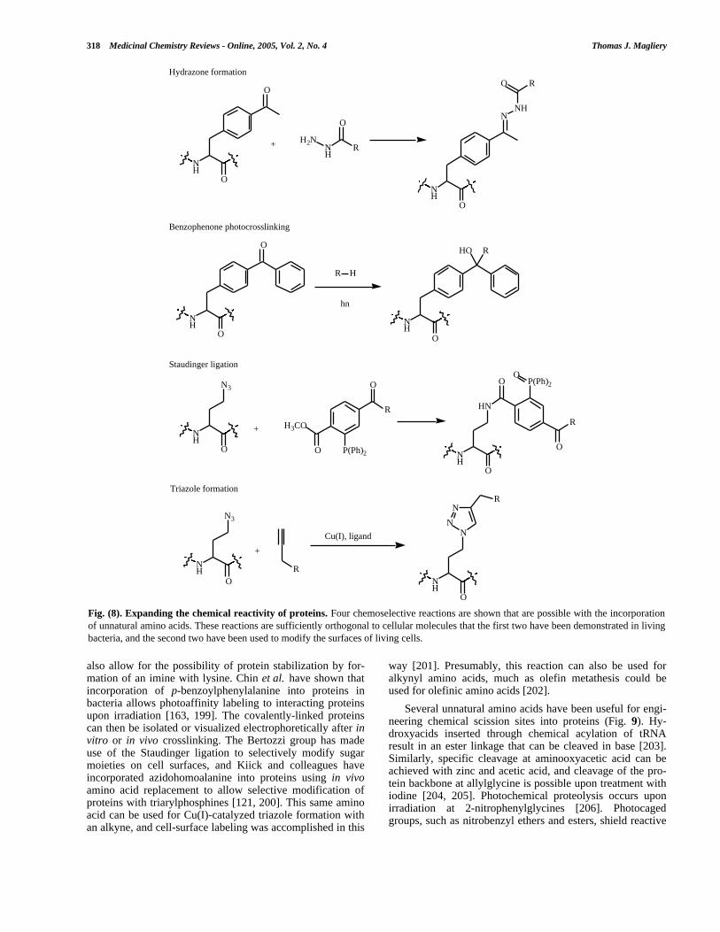
318 Medicinal Chemistry Reviews - Online, 2005, Vol. 2, No. 4 Thomas J. Magliery
also allow for the possibility of protein stabilization by for-mation of an imine with lysine. Chin et al. have shown thatincorporation of p-benzoylphenylalanine into proteins inbacteria allows photoaffinity labeling to interacting proteinsupon irradiation [163, 199]. The covalently-linked proteinscan then be isolated or visualized electrophoretically after invitro or in vivo crosslinking. The Bertozzi group has madeuse of the Staudinger ligation to selectively modify sugarmoieties on cell surfaces, and Kiick and colleagues haveincorporated azidohomoalanine into proteins using in vivoamino acid replacement to allow selective modification ofproteins with triarylphosphines [121, 200]. This same aminoacid can be used for Cu(I)-catalyzed triazole formation withan alkyne, and cell-surface labeling was accomplished in this
way [201]. Presumably, this reaction can also be used foralkynyl amino acids, much as olefin metathesis could beused for olefinic amino acids [202].
Several unnatural amino acids have been useful for engi-neering chemical scission sites into proteins (Fig. 9). Hy-droxyacids inserted through chemical acylation of tRNAresult in an ester linkage that can be cleaved in base [203].Similarly, specific cleavage at aminooxyacetic acid can beachieved with zinc and acetic acid, and cleavage of the pro-tein backbone at allylglycine is possible upon treatment withiodine [204, 205]. Photochemical proteolysis occurs uponirradiation at 2-nitrophenylglycines [206]. Photocagedgroups, such as nitrobenzyl ethers and esters, shield reactive
Fig. (8). Expanding the chemical reactivity of proteins. Four chemoselective reactions are shown that are possible with the incorporationof unnatural amino acids. These reactions are sufficiently orthogonal to cellular molecules that the first two have been demonstrated in livingbacteria, and the second two have been used to modify the surfaces of living cells.
NH
O
O
NH
R
O
H2N
NH
O
NNH
ROHydrazone formation
NH
O
O
R H
hn
NH
O
HO R
Benzophenone photocrosslinking
NH
O
N3
R
O
P(Ph)2
H3CO
O NH
O
HN
O P(Ph)2
R
O
OStaudinger ligation
+
+
NH
O
N3
R
Cu(I), ligand
+
NH
O
N
N
N
RTriazole formation

Unnatural Protein Engineering Producing Proteins Medicinal Chemistry Reviews - Online, 2005, Vol. 2, No. 4 319
moieties until irradiative deprotection, and can be used tocontrol enzymatic activity or protein-protein interaction in atime-resolved manner [207-209]. These types of functionali-ties are only available through synthetic methods at the mo-ment but will prove especially useful for cell biologicalstudies when they are available for translational incorpora-tion.
Posttranslational Modifications
Understanding the roles of proteins in cellular functionnecessarily means understanding the extent and meaning ofposttranslational modification, particularly in eukaryotes.However, it is difficult to obtain specifically, homogene-ously modified protein, not least because many posttransla-
tional modifications do not occur in the bacteria in whichproteins are often most conveniently expressed. EPL hasbeen useful for the homogeneous preparation of phosphory-lated proteins, and has been used to understand the role ofphosphorylation in TGF β signaling [210-213]. The Colegroup has incorporated phosphonoserine, a non-hydrolyzableanalog of phosphoserine, using EPL, which creates an “al-ways on” protein state [214-216]. As mentioned above, theBertozzi group has used a chemoselective mimetic approachto generate neoglycoproteins, employing hydrazone, oximeand Staudinger chemistry [197]. Zhang et al. have transla-tionally incorporated β-GlcNAc-serine in bacteria, whereproteins are not normally glycosylated, permitting homoge-neous preparation of large amounts of glycoprotein [217].Lipidation, such as introduction of a geranylgeranyl group
Fig. (9). Chemical scission and deprotection of proteins. Several methods have been introduced to specifically cleave the backbone ofproteins at the site of unnatural amino acid (or analog) incorporation. Also, photodeprotection of “caged” Ser and Tyr has been demonstrated,including in Xenopus oocytes.
Ester hydrolysis at a-hydroxy acid
NH
O
O
OR1
R2
Base, H2 O
NH
OH
O
R1
HO
O
R2
+
NH
HN
O
O
R1
O
Zn, HOAc
NH
NH2
O
HO
R1
O+
Acid hydrolysis at aminooxyacetic acid
NH
HN
O
O
R2
I2, H2O
NH
H2N
O
R2
O
I
O
+
Iodine-mediated scission at allylglycine
NH
HN
O
OR1
hn
NO2
NH
NH2
O
OR1
NO
O
+
Photochemical scission at 2-nitrophenylglycine
NH
O
O
NO2
NH
OH
ONO
Photodeprotection of "caged" nitrobenzyl ethers, esters
+
O
H

320 Medicinal Chemistry Reviews - Online, 2005, Vol. 2, No. 4 Thomas J. Magliery
into the GTPase Rab7, has also been accomplished with EPL[218, 219].
TEMPLATED SYNTHESIS OF NON-PEPTIDICMOLECULES
While extensive discussion of this topic is beyond thescope of this review, it is worth noting that, in the limit,chemists would like to be able to program and control thesynthesis of nonpeptidic molecules as well. Just as physicalassociation of nucleic acid permits facile production, selec-tion and identification of proteins, it could ultimately do thesame for peptidomimetics, like polyesters, or even less-related molecules. Much of the pioneering work in this fieldhas been carried out by the Orgel group, largely studying thenonenzymatic synthesis of RNA from DNA and other tem-plates [220, 221]. Recently, the Liu group has reported theDNA-templated polymerization of peptide nucleic acid alde-hydes [222]. Liu and colleagues have also devised systemsfor the DNA-programmed multistep syntheses of smallmolecules [223], and have demonstrated that in vitro selec-tions on DNA-linked small molecules are possible as well[224, 225]. Harbury and colleagues recently have introduceda technique called “DNA display” for the programmed syn-thesis of peptides and other polymers using DNA-directedspatial separation [226-228].
CONCLUSION
Less than a decade ago, unnatural protein engineeringwas mostly an idea, accessible to only a few labs of special-ists, and decidedly in the methodology-development stage ofits existence. Today, unnatural protein engineering is a real-ity accessible to any biochemistry or biophysics lab at rea-sonable cost and without heroic synthetic efforts. In particu-lar, expressed protein ligation, widespread amino acid re-placement, and site-specific translational incorporation inliving cells allow the protein scientist to endow a target pro-tein with unique reactive handles, biophysical probes, andposttranslational modifications in a controlled, homogenousmanner. These possibilities herald the beginning of a newphase in the study and engineering of proteins, a phase thatwill vastly aid our understanding of cellular function, and aphase in which molecules with entirely novel properties canbe created. There is, of course, still need for significant im-provements to the existing technologies, including improv-ing the scope of peptide synthesis and working around cur-rent limitations in ligation chemistry; dramatically improvingthe ease of chemical synthesis of acylated tRNA, perhapsusing ribozymes; identifying or engineering many more or-thogonal tRNA/synthetase pairs and radically expanding therepertoire of unnatural amino acids that can be inserted inliving cells; perfecting alternative coding schemes, includingunnatural codons; developing systems for facile use inmammalian cells or whole organisms; and working towardsthe templated creation of molecules that are far differentfrom proteins.
ACKNOWLEDGEMENTS
T.J.M. is a fellow of the National Institutes of Health(GM065750). The author thanks Lynne Regan (Yale Univer-sity, New Haven, CT) for additional support during the
preparation of this manuscript. Thanks to P.G. Schultz (TheScripps Research Institute, La Jolla, CA) for allowing me tocite unpublished results, and to S.W. Santoro (Harvard Uni-versity, Cambridge, MA) for critical reading of this manu-script.
REFERENCES
[1] Bock, A.; Forchhammer, K.; Heider, J.; Baron, C. Trends Biochem.Sci. 1991, 16, 463.
[2] Hao, B.; Gong, W.; Ferguson, T.K.; James, C.M.; Krzycki, J.A.;Chan, M.K. Science 2002, 296, 1462.
[3] Srinivasan, G.; James, C.M.; Krzycki, J.A. Science 2002, 296,1459.
[4] Braman, J. In Vitro Mtuagenesis Protocols, 2nd ed.; Humana Press:Totowa, NJ, 2002.
[5] Zoller, M.J.; Smith, M. Methods Enzymol. 1983, 100, 468.[6] Anthony-Cahill, S.J.; Magliery, T.J. Curr. Pharm. Biotechnol.
2002, 3, 299.[7] Magliery, T.J.; Pastrnak, M.; Anderson, J.C.; Santoro, S.W.; Meg-
gers, E.; Herberich, B.; Wang, L.; Schultz, P.G. In TranslationMechanisms, Lapointe, J.; Brakier-Gingras, L., Ed. Landes Bio-science: Georgetown, TX, 2003; pp. 95-114.
[8] Magliery, T.J.; Liu, D.R. In The Genetic Code and the Origin ofLife, Ribas de Pouplana, L., Ed. Landes Bioscience: Georgetown,TX, 2004.
[9] Wang, L.; Schultz, P.G. Chem. Commun. 2002, 1.[10] Link, A.J.; Mock, M.L.; Tirrell, D.A. Curr. Opin. Biotechnol. 2003,
14, 603.[11] Hohsaka, T.; Sisido, M. Curr. Opin. Chem. Biol. 2002, 6, 809.[12] Muir, T.W. Annu. Rev. Biochem. 2003, 72, 249.[13] Dawson, P.E.; Kent, S.B.H. Annu. Rev. Biochem. 2000, 69, 923.[14] Vogel, K.; Chmielewski, J. J. Am. Chem. Soc. 1994, 116, 11163.[15] Abrahmsen, L.; Tom, J.; Burnier, J.; Butcher, K.A.; Kossiakoff, A.;
Wells, J.A. Biochemistry 1991, 30, 4151.[16] Atwell, S.; Wells, J.A. Proc. Natl. Acad. Sci. USA 1999, 96, 9497.[17] Braisted, A.C.; Judice, J.K.; Wells, J.A. Methods Enzymol. 1997,
289, 298.[18] Chang, T.K.; Jackson, D.Y.; Burnier, J.P.; Wells, J.A. Proc. Natl.
Acad. Sci. USA 1994, 91, 12544.[19] Jackson, D.Y.; Burnier, J.; Quan, C.; Stanley, M.; Tom, J.; Wells,
J.A. Science 1994, 266, 243.[20] Schnolzer, M.; Kent, S.B.H. Science 1992, 256, 221.[21] Dawson, P.E.; Muir, T.W.; Clarklewis, I.; Kent, S.B.H. Science
1994, 266, 776.[22] Hofmann, R.M.; Muir, T.W. Curr. Opin. Biotechnol. 2002, 13,
297.[23] Sewing, A.; Hilvert, D. Angew. Chem., Int. Ed. 2001, 40, 3395.[24] Low, D.W.; Hill, M.G.; Carrasco, M.R.; Kent, S.B.H.; Botti, P.
Proc. Natl. Acad. Sci. USA 2001, 98, 6554.[25] Canne, L.E.; Bark, S.J.; Kent, S.B.H. J. Am. Chem. Soc. 1996, 118,
5891.[26] Yan, L.Z.; Dawson, P.E. J. Am. Chem. Soc. 2001, 123, 526.[27] Gieselman, M.D.; Zhu, Y.T.; Zhou, H.; Galonic, D.; van der Donk,
W.A. Chembiochem. 2002, 3, 709.[28] Zhu, Y.; van der Donk, W.A. Org. Lett. 2001, 3, 1189.[29] Hackeng, T.M.; Griffin, J.H.; Dawson, P.E. Proc. Natl. Acad. Sci.
USA 1999, 96, 10068.[30] Canne, L.E.; Botti, P.; Simon, R.J.; Chen, Y.J.; Dennis, E.A.; Kent,
S.B.H. J. Am. Chem. Soc. 1999, 121, 8720.[31] Gaertner, H.F.; Rose, K.; Cotton, R.; Timms, D.; Camble, R.; Of-
ford, R.E. Bioconj. Chem. 1991, 3, 262.[32] Rose, K. J. Am. Chem. Soc. 1994, 116, 30.[33] Muir, T.W.; Sondhi, D.; Cole, P.A. Proc. Natl. Acad. Sci. USA
1998, 95, 6705.[34] Villain, M.; Vizzavona, J.; Rose, K. Chem. Biol. 2001, 8, 673.[35] Erlanson, D.A.; Chytil, M.; Verdine, G.L. Chem. Biol. 1996, 3,
981.[36] Morris, M.C.; Depollier, J.; Mery, J.; Heitz, F.; Divita, G. Nat.
Biotechnol. 2001, 19, 1173.[37] Morris, M.C.; Chaloin, L.; Heitz, F.; Divita, G. Curr. Opin. Bio-
technol. 2000, 11, 461.[38] Crick, F.H.C. Symp. Soc. Exp. Biol. 1958, 12, 138.

Unnatural Protein Engineering Producing Proteins Medicinal Chemistry Reviews - Online, 2005, Vol. 2, No. 4 321
[39] Crick, F.H.C.; Barret, L.; Brenner, S.; Watts-Tobin, R. Nature1961, 192, 1227.
[40] Bossi, L.; Roth, J.R. Nature 1980, 286, 123.[41] Chapeville, F.; Lipmann, F.; von Ehrenstein, G.; Weisblum, B.;
Ray, W.J., Jr.; Benzer, S. Proc. Natl. Acad. Sci. USA 1962, 48,1086.
[42] Steward, L.E.; Collins, C.S.; Gilmore, M.A.; Carlson, J.E.; Ross,J.B.A.; Chamberlin, A.R. J. Am. Chem. Soc. 1997, 119, 6.
[43] Thorson, J.S.; Cornish, V.W.; Barrett, J.E.; Cload, S.T.; Yano, T.;Schultz, P.G. In Protein Synthesis: Methods and Protocols , Martin,R., Ed. Humana Press: Totowa, NJ, 1998; Vol. 77, pp. 43-73.
[44] Noren, C.J.; Anthony-Cahill, S.J.; Griffith, M.C.; Schultz, P.G.Science 1989, 244, 182.
[45] Bain, J.D.; Glabe, C.G.; Dix, T.A.; Chamberlin, A.R.; Diala, E.S. J.Am. Chem. Soc. 1989, 111, 8013.
[46] Payne, R.C.; Nichols, B.P.; Hecht, S.M. Biochemistry 1987, 26,3197.
[47] Baldini, G.; Martoglio, B.; Schachenmann, A.; Zugliani, C.; Brun-ner, J. Biochemistry 1988, 27, 7951.
[48] Robertson, S.A.; Noren, C.J.; Anthonycahill, S.J.; Griffith, M.C.;Schultz, P.G. Nucl. Acids Res. 1989, 17, 9649.
[49] Robertson, S.A.; Ellman, J.A.; Schultz, P.G. J. Am. Chem. Soc.1991, 113, 2722.
[50] LaRiviere, F.J.; Wolfson, A.D.; Uhlenbeck, O.C. Science 2001,294, 165.
[51] Ninomiya, K.; Kurita, T.; Hohsaka, T.; Sisido, M. Chem. Commun.2003, 2242.
[52] Bessho, Y.; Hodgson, D.R.; Suga, H. Nat. Biotechnol. 2002, 20,723.
[53] Murakami, H.; Saito, H.; Suga, H. Chem. Biol. 2003, 10, 655.[54] Murakami, H.; Kourouklis, D.; Suga, H. Chem. Biol. 2003, 10,
1077.[55] Cload, S.T.; Liu, D.R.; Froland, W.A.; Schultz, P.G. Chem. Biol.
1996, 3, 1033.[56] Short, G.F.; Golovine, S.Y.; Hecht, S.M. Biochemistry 1999, 38,
8808.[57] Yokoyama, S. Curr. Opin. Chem. Biol. 2003, 7, 39.[58] Kigawa, T.; Yabuki, T.; Yoshida, Y.; Tsutsui, M.; Ito, Y.; Shibata,
T.; Yokoyama, S. FEBS Lett. 1999, 442, 15.[59] Takahashi, T.T.; Austin, R.J.; Roberts, R.W. Trends Biochem. Sci.
2003, 28, 159.[60] Roberts, R.W.; Szostak, J.W. Proc. Natl. Acad. Sci. USA 1997, 94,
12297.[61] Liu, R.; Barrick, J.E.; Szostak, J.W.; Roberts, R.W. Methods Enzy-
mol. 2000, 318, 268.[62] Frankel, A.; Li, S.W.; Starch, S.R.; Roberts, R.W. Curr. Opin.
Struct. Biol. 2003, 13, 506.[63] Frankel, A.; Millward, S.W.; Roberts, R.W. Chem. Biol. 2003, 10,
1043.[64] Hanes, J.; Pluckthun, A. Proc. Natl. Acad. Sci. USA 1997, 94,
4937.[65] Atkins, J.F.; Weiss, R.B.; Thompson, S.; Gesteland, R.F. Annu.
Rev. Genet. 1991, 25, 201.[66] Anderson, J.C.; Schultz, P.G. Biochemistry 2003, 42, 9598.[67] Kohrer, C.; Xie, L.; Kellerer, S.; Varshney, U.; RajBhandary, U.L.
Proc. Natl. Acad. Sci. USA 2001, 98, 14310.[68] Kanda, T.; Takai, K.; Hohsaka, T.; Sisido, M.; Takaku, H. Bio-
chem. Biophys. Res. Commun. 2000, 270, 1136.[69] Kanda, T.; Takai, K.; Yokoyama, S.; Takaku, H. FEBS Lett. 1998,
440, 273.[70] Kanda, T.; Takai, K.; Yokoyama, S.; Takaku, H. Bioorg. Med.
Chem. 2000, 8, 675.[71] Kanda, T.; Takai, K.; Yokoyama, S.; Takaku, H. J. Biochem. (To-
kyo, Jpn.) 2000, 127, 37.[72] Jackson, R.J.; Napthine, S.; Brierley, I. RNA 2001, 7, 765.[73] Frankel, A.; Roberts, R.W. RNA 2003, 9, 780.[74] Shimizu, Y.; Inoue, A.; Tomari, Y.; Suzuki, T.; Yokogawa, T.;
Nishikawa, K.; Ueda, T. Nat. Biotechnol. 2001, 19, 751.[75] Magliery, T.J.; Anderson, J.C.; Schultz, P.G. J. Mol. Biol. 2001,
307, 755.[76] Anderson, J.C.; Magliery, T.J.; Schultz, P.G. Chem. Biol. 2002, 9,
237.[77] Hohsaka, T.; Ashizuka, Y.; Murakami, H.; Sisido, M. J. Am. Chem.
Soc. 1996, 118, 9778.
[78] Kramer, G.; Kudlicki, W.; Hardesty, B. In Protein Synthesis:Methods and Protocols, Martin, R., Ed. Humana Press: Totowa,NJ, 1998; Vol. 77, pp. 105-116.
[79] Ma, C.H.; Kudlicki, W.; Odom, O.W.; Kramer, G.; Hardesty, B.Biochemistry 1993, 32, 7939.
[80] Hohsaka, T.; Ashizuka, Y.; Taira, H.; Murakami, H.; Sisido, M.Biochemistry 2001, 40, 11060.
[81] Hohsaka, T.; Ashizuka, Y.; Sasaki, H.; Murakami, H.; Sisido, M. J.Am. Chem. Soc. 1999, 121, 12194.
[82] Hohsaka, T.; Ashizuka, Y.; Murakami, H.; Sisido, M. Nucl. AcidsRes. 2001, 29, 3646.
[83] Kool, E.T. Acc. Chem. Res. 2002, 35, 936.[84] Henry, A.A.; Romesberg, F.E. Curr. Opin. Chem. Biol. 2003, 7,
727.[85] Switzer, C.; Moroney, S.E.; Benner, S.A. J. Am. Chem. Soc. 1989,
111, 8322.[86] Bain, J.D.; Switzer, C.; Chamberlin, A.R.; Benner, S.A. Nature
1992, 356, 537.[87] Piccirilli, J.A.; Krauch, T.; Moroney, S.E.; Benner, S.A. Nature
1990, 343, 33.[88] Rappaport, H.P. Biochemistry 1993, 32, 3047.[89] Rappaport, H.P. Nucl. Acids Res. 1988, 16, 7253.[90] Ohtsuki, T.; Kimoto, M.; Ishikawa, M.; Mitsui, T.; Hirao, I.; Yoko-
yama, S. Proc. Natl. Acad. Sci. USA 2001, 98, 4922.[91] Fujiwara, T.; Kimoto, M.; Sugiyama, H.; Hirao, I.; Yokoyama, S.
Bioorg. Med. Chem. Lett. 2001, 11, 2221.[92] Hirao, I.; Mitsui, T.; Fujiwara, T.; Kimoto, M.; To, T.; Okuni, T.;
Sato, A.; Harada, Y.; Yokoyama, S. Nucl. Acids Res. Suppl. 2001,17.
[93] Ishikawa, M.; Hirao, I.; Yokoyama, S. Nucl. Acids Symp. Ser.1999, 125.
[94] Hirao, I.; Ohtsuki, T.; Fujiwara, T.; Mitsui, T.; Yokogawa, T.;Okuni, T.; Nakayama, H.; Takio, K.; Yabuki, T.; Kigawa, T.; Ko-dama, K.; Nishikawa, K.; Yokoyama, S. Nat. Biotechnol. 2002, 20,177.
[95] Moran, S.; Ren, R.X.F.; Rumney, S.; Kool, E.T. J. Am. Chem. Soc.1997, 119, 2056.
[96] Morales, J.C.; Kool, E.T. Nat. Struct. Biol. 1998, 5, 950.[97] Mitsui, T.; Kitamura, A.; Kimoto, M.; To, T.; Sato, A.; Hirao, I.;
Yokoyama, S. J. Am. Chem. Soc. 2003, 125, 5298.[98] Ogawa, A.K.; Wu, Y.Q.; McMinn, D.L.; Liu, J.Q.; Schultz, P.G.;
Romesberg, F.E. J. Am. Chem. Soc. 2000, 122, 3274.[99] Henry, A.A.; Yu, C.Z.; Romesberg, F.E. J. Am. Chem. Soc. 2003,
125, 9638.[100] Tae, E.J.L.; Wu, Y.Q.; Xia, G.; Schultz, P.G.; Romesberg, F.E. J.
Am. Chem. Soc. 2001, 123, 7439.[101] Xia, G.; Chen, L.J.; Sera, T.; Fa, M.; Schultz, P.G.; Romesberg,
F.E. Proc. Natl. Acad. Sci. USA 2002, 99, 6597.[102] Zimmermann, N.; Meggers, E.; Schultz, P.G. Bioorg. Chem. 2004,
32, 13.[103] Zimmermann, N.; Meggers, E.; Schultz, P.G. J. Am. Chem. Soc.
2002, 124, 13684.[104] Meggers, E.; Holland, P.L.; Tolman, W.B.; Romesberg, F.E.;
Schultz, P.G. J. Am. Chem. Soc. 2000, 122, 10714.[105] Atwell, S.; Meggers, E.; Spraggon, G.; Schultz, P.G. J. Am. Chem.
Soc. 2001, 123, 12364.[106] Nowak, M.W.; Kearney, P.C.; Sampson, J.R.; Saks, M.E.; Labarca,
C.G.; Silverman, S.K.; Zhong, W.G.; Thorson, J.; Abelson, J.N.;Davidson, N.; Schultz, P.G.; Dougherty, D.A.; Lester, H.A. Science1995, 268, 439.
[107] Beene, D.L.; Dougherty, D.A.; Lester, H.A. Curr. Opin. Neurobiol.2003, 13, 264.
[108] Saks, M.E.; Sampson, J.R.; Nowak, M.W.; Kearney, P.C.; Du,F.Y.; Abelson, J.N.; Lester, H.A.; Dougherty, D.A. J. Biol. Chem.1996, 271, 23169.
[109] Monahan, S.L.; Lester, H.A.; Dougherty, D.A. Chem. Biol. 2003,10, 573.
[110] Hendrickson, W.A.; Horton, J.R.; Lemaster, D.M. EMBO J. 1990,9, 1665.
[111] Yang, W.; Hendrickson, W.A.; Crouch, R.J.; Satow, Y. Science1990, 249, 1398.
[112] Budisa, N.; Steipe, B.; Demange, P.; Eckerskorn, C.; Kellermann,J.; Huber, R. Eur. J. Biochem. 1995, 230, 788.
[113] Luck, L.A.; Vance, J.E.; Oconnell, T.M.; London, R.E. J. Biomol.NMR 1996, 7, 261.

322 Medicinal Chemistry Reviews - Online, 2005, Vol. 2, No. 4 Thomas J. Magliery
[114] Minks, C.; Huber, R.; Moroder, L.; Budisa, N. Biochemistry 1999,38, 10649.
[115] Minks, C.; Huber, R.; Moroder, L.; Budisa, N. Anal. Biochem.2000, 284, 29.
[116] Jakubowski, H. J. Biol. Chem. 2000, 275, 21813.[117] Budisa, N.; Minks, C.; Medrano, F.J.; Lutz, J.; Huber, R.; Moroder,
L. Proc. Natl. Acad. Sci. USA 1998, 95, 455.[118] McGrath, K.P.; Fournier, M.J.; Mason, T.L.; Tirrell, D.A. J. Am.
Chem. Soc. 1992, 114, 727.[119] Kothakota, S.; Mason, T.L.; Tirrell, D.A.; Fournier, M.J. J. Am.
Chem. Soc. 1995, 117, 536.[120] Wang, P.; Tang, Y.; Tirrell, D.A. J. Am. Chem. Soc. 2003, 125,
6900.[121] Kiick, K.L.; Saxon, E.; Tirrell, D.A.; Bertozzi, C.R. Proc. Natl.
Acad. Sci. USA 2002, 99, 19.[122] Tang, Y.; Tirrell, D.A. J. Am. Chem. Soc. 2001, 123, 11089.[123] van Hest, J.C.M.; Kiick, K.L.; Tirrell, D.A. J. Am. Chem. Soc.
2000, 122, 1282.[124] Kiick, K.L.; van Hest, J.C.M.; Tirrell, D.A. Angew. Chem. Int. Ed.
2000, 39, 2148.[125] Kiick, K.L.; Weberskirch, R.; Tirrell, D.A. FEBS Lett. 2001, 502,
25.[126] Ibba, M.; Hennecke, H. FEBS Lett. 1995, 364, 272.[127] Kirshenbaum, K.; Carrico, I.S.; Tirrell, D.A. Chembiochem. 2002,
3, 235.[128] Doring, V.; Mootz, H.D.; Nangle, L.A.; Hendrickson, T.L.; de
Crecy-Lagard, V.; Schimmel, P.; Marliere, P. Science 2001, 292,501.
[129] Tang, Y.; Tirrell, D.A. Biochemistry 2002, 41, 10635.[130] Rath, V.L.; Silvian, L.F.; Beijer, B.; Sproat, B.S.; Steitz, T.A.
Structure 1998, 6, 439.[131] Liu, D.R.; Magliery, T.J.; Schultz, P.G. Chem. Biol. 1997, 4, 685.[132] Patten, P.A.; Howard, R.J.; Stemmer, W.P.C. Curr. Opin. Biotech-
nol. 1997, 8, 724.[133] Stemmer, W.P.C. Proc. Natl. Acad. Sci. USA 1994, 91, 10747.[134] Liu, D.R.; Magliery, T.J.; Pasternak, M.; Schultz, P.G. Proc. Natl.
Acad. Sci. USA 1997, 94, 10092.[135] Whelihan, E.F.; Schimmel, P. EMBO J. 1997, 16, 2968.[136] Wang, C.C.; Schimmel, P. J. Biol. Chem. 1999, 274, 16508.[137] Liu, D.R.; Schultz, P.G. Proc. Natl. Acad. Sci. USA 1999, 96, 4780.[138] Pastrnak, M.; Magliery, T.J.; Schultz, P.G. Helv. Chim. Acta 2000,
83, 2277.[139] Magliery, T.J. Ph.D. Thesis, University of California, Berkeley,
CA, 2001.[140] Steer, B.A.; Schimmel, P. J. Biol. Chem. 1999, 274, 35601.[141] Steer, B.A.; Schimmel, P. Proc. Natl. Acad. Sci. USA 1999, 96,
13644.[142] Wang, L.; Magliery, T.J.; Liu, D.R.; Schultz, P.G. J. Am. Chem.
Soc. 2000, 122, 5010.[143] Wang, L.; Schultz, P.G. Chem. Biol. 2001, 8, 883.[144] Kobayashi, T.; Nureki, O.; Ishitani, R.; Yaremchuk, A.; Tukalo,
M.; Cusack, S.; Sakamoto, K.; Yokoyama, S. Nat. Struct. Biol.2003, 10, 425.
[145] Kowal, A.K.; Kohrer, C.; RajBhandary, U.L. Proc. Natl. Acad. Sci.USA 2001, 98, 2268.
[146] Santoro, S.W.; Anderson, J.C.; Lakshman, V.; Schultz, P.G. Nucl.Acids Res. 2003, 31, 6700.
[147] Anderson, J.C.; Wu, N.; Santoro, S.W.; Lakshman, V.; King, D.S.;Schultz, P.G. Proc. Natl. Acad. Sci. USA 2004, 101, 7566.
[148] Drabkin, H.J.; Park, H.J.; Rajbhandary, U.L. Mol. Cell Biol. 1996,16, 907.
[149] Kiga, D.; Sakamoto, K.; Kodama, K.; Kigawa, T.; Matsuda, T.;Yabuki, T.; Shirouzu, M.; Harada, Y.; Nakayama, H.; Takio, K.;Hasegawa, Y.; Endo, Y.; Hirao, I.; Yokoyama, S. Proc. Natl. Acad.Sci. USA 2002, 99, 9715.
[150] Sakamoto, K.; Hayashi, A.; Sakamoto, A.; Kiga, D.; Nakayama,H.; Soma, A.; Kobayashi, T.; Kitabatake, M.; Takio, K.; Saito, K.;Shirouzu, M.; Hirao, I.; Yokoyama, S. Nucl. Acids Res. 2002, 30,4692.
[151] Wakasugi, K.; Quinn, C.L.; Tao, N.; Schimmel, P. EMBO J. 1998,17, 297.
[152] Edwards, H.; Schimmel, P. Mol. Cell Biol. 1990, 10, 1633.[153] Edwards, H.; Trezeguet, V.; Schimmel, P. Proc. Natl. Acad. Sci.
USA 1991, 88, 1153.[154] Chin, J.W.; Cropp, T.A.; Chu, S.; Meggers, E.; Schultz, P.G. Chem.
Biol. 2003, 10, 511.
[155] Chin, J.W.; Cropp, T.A.; Anderson, J.C.; Mukherji, M.; Zhang,Z.W.; Schultz, P.G. Science 2003, 301, 964.
[156] Zhang, Z.; Alfonta, L.; Tian, F.; Bursulaya, B.; Uryu, S.; King,D.S.; Schultz, P.G. Proc. Natl. Acad. Sci. USA 2004, 101, 8882.
[157] Furter, R. Protein Sci. 1998, 7, 419.[158] Agou, F.; Quevillon, S.; Kerjan, P.; Mirande, M. Biochemistry
1998, 37, 11309.[159] Hong, K.W.; Ibba, M.; Soll, D. FEBS Lett. 1998, 434, 149.[160] Datta, D.; Wang, P.; Carrico, I.S.; Mayo, S.L.; Tirrell, D.A. J. Am.
Chem. Soc. 2002, 124, 5652.[161] Zhang, D.; Vaidehi, N.; Goddard, W.A., 3rd; Danzer, J.F.; Debe,
D. Proc. Natl. Acad. Sci. USA 2002, 99, 6579.[162] Wang, L.; Brock, A.; Herberich, B.; Schultz, P.G. Science 2001,
292, 498.[163] Chin, J.W.; Martin, A.B.; King, D.S.; Wang, L.; Schultz, P.G.
Proc. Natl. Acad. Sci. USA 2002, 99, 11020.[164] Wang, L.; Brock, A.; Schultz, P.G. J. Am. Chem. Soc. 2002, 124,
1836.[165] Zhang, Z.W.; Wang, L.; Brock, A.; Schultz, P.G. Angew. Chem.,
Int. Ed. 2002, 41, 2840.[166] Wang, L.; Zhang, Z.W.; Brock, A.; Schultz, P.G. Proc. Natl. Acad.
Sci. USA 2003, 100, 56.[167] Santoro, S.W.; Wang, L.; Herberich, B.; King, D.S.; Schultz, P.G.
Nat. Biotechnol. 2002, 20, 1044.[168] Chin, J.W.; Santoro, S.W.; Martin, A.B.; King, D.S.; Wang, L.;
Schultz, P.G. J. Am. Chem. Soc. 2002, 124, 9026.[169] Mehl, R.A.; Anderson, J.C.; Santoro, S.W.; Wang, L.; Martin,
A.B.; King, D.S.; Horn, D.M.; Schultz, P.G. J. Am. Chem. Soc.2003, 125, 935.
[170] Deiters, A.; Cropp, T.A.; Mukherji, M.; Chin, J.W.; Anderson, J.C.;Schultz, P.G. J. Am. Chem. Soc. 2003, 125, 11782.
[171] Cornish, V.W.; Mendel, D.; Schultz, P.G. Angew. Chem., Int. Ed.1995, 34, 621.
[172] Dougherty, D.A. Curr. Opin. Chem. Biol. 2000, 4, 645.[173] Ellman, J.A.; Volkman, B.F.; Mendel, D.; Schultz, P.G.; Wemmer,
D.E. J. Am. Chem. Soc. 1992, 114, 7959.[174] Cornish, V.W.; Benson, D.R.; Altenbach, C.A.; Hideg, K.; Hub-
bell, W.L.; Schultz, P.G. Proc. Natl. Acad. Sci. USA 1994, 91,2910.
[175] Taki, M.; Hohsaka, T.; Murakami, H.; Taira, K.; Sisido, M. Phos-phorous, Sulfur Silicon Relat. Elem. 2002, 177, 1929.
[176] Cowburn, D.; Muir, T.W. Methods Enzymol. 2001, 339, 41.[177] Cotton, G.J.; Muir, T.W. Chem. Biol. 2000, 7, 253.[178] Gallivan, J.P.; Lester, H.A.; Dougherty, D.A. Chem. Biol. 1997, 4,
739.[179] Li, L.T.; Zhong, W.G.; Zacharias, N.; Gibbs, C.; Lester, H.A.;
Dougherty, D.A. Chem. Biol. 2001, 8, 47.[180] Tang, Y.; Ghirlanda, G.; Vaidehi, N.; Kua, J.; Mainz, D.T.; God-
dard, W.A.; DeGrado, W.F.; Tirrell, D.A. Biochemistry 2001, 40,2790.
[181] Tang, Y.; Ghirlanda, G.; Petka, W.A.; Nakajima, T.; DeGrado,W.F.; Tirrell, D.A. Angew. Chem., Int. Ed. 2001, 40, 1494.
[182] Bilgicer, B.; Fichera, A.; Kumar, K. J. Am. Chem. Soc. 2001, 123,4393.
[183] Bilgicer, B.; Xing, X.; Kumar, K. J. Am. Chem. Soc. 2001, 123,11815.
[184] Bilgicer, B.; Kumar, K. Tetrahedron 2002, 58, 4105.[185] Renner, C.; Alefelder, S.; Bae, J.H.; Budisa, N.; Huber, R.;
Moroder, L. Angew. Chem., Int. Ed. 2001, 40, 923.[186] Frieden, C. Biochemistry 2003, 42, 12439.[187] Budisa, N.; Rubini, M.; Bae, J.H.; Weyher, E.; Wenger, W.; Gol-
bik, R.; Huber, R.; Moroder, L. Angew. Chem., Int. Ed. 2002, 41,4066.
[188] Bae, J.H.; Rubini, M.; Jung, G.; Wiegand, G.; Seifert, M.H.J.;Azim, M.K.; Kim, J.S.; Zumbusch, A.; Holak, T.A.; Moroder, L.;Huber, R.; Budisa, N. J. Mol. Biol. 2003, 328, 1071.
[189] Wang, L.; Xie, J.M.; Deniz, A.A.; Schultz, P.G. J. Org. Chem.2003, 68, 174.
[190] Offord, R.E.; Gaertner, H.F.; Wells, T.N.; Proudfoot, A.E. MethodsEnzymol. 1997, 287, 348.
[191] Cornish, V.W.; Hahn, K.M.; Schultz, P.G. J. Am. Chem. Soc. 1996,118, 8150.
[192] Zhang, Z.W.; Smith, B.A.C.; Wang, L.; Brock, A.; Cho, C.;Schultz, P.G. Biochemistry 2003, 42, 6735.
[193] Mahal, L.K.; Yarema, K.J.; Bertozzi, C.R. Science 1997, 276,1125.
Unnatural Protein Engineering Producing Proteins Medicinal Chemistry Reviews - Online, 2005, Vol. 2, No. 4 323
[194] Hang, H.C.; Bertozzi, C.R. Acc. Chem. Res. 2001, 34, 727.[195] Macmillan, D.; Bertozzi, C.R. Tetrahedron 2000, 56, 9515.[196] Tolbert, T.J.; Wong, C.H. J. Am. Chem. Soc. 2000, 122, 5421.[197] Grogan, M.J.; Pratt, M.R.; Marcaurelle, L.A.; Bertozzi, C.R. Annu.
Rev. Biochem. 2002, 71, 593.[198] Liu, H.T.; Wang, L.; Brock, A.; Wong, C.H.; Schultz, P.G. J. Am.
Chem. Soc. 2003, 125, 1702.[199] Chin, J.W.; Schultz, P.G. Chembiochem. 2002, 3, 1135.[200] Saxon, E.; Bertozzi, C.R. Science 2000, 287, 2007.[201] Link, A.J.; Tirrell, D.A. J. Am. Chem. Soc. 2003, 125, 11164.[202] Blackwell, H.E.; Sadowsky, J.D.; Howard, R.J.; Sampson, J.N.;
Chao, J.A.; Steinmetz, W.E.; O'Leary, D.J.; Grubbs, R.H. J. Org.Chem. 2001, 66, 5291.
[203] Ellman, J.A.; Mendel, D.; Schultz, P.G. Science 1992, 255, 197.[204] Eisenhauer, B.M.; Hecht, S.M. Biochemistry 2002, 41, 11472.[205] Wang, B.; Brown, K.C.; Lodder, M.; Craik, C.S.; Hecht, S.M.
Biochemistry 2002, 41, 2805.[206] England, P.M.; Lester, H.A.; Davidson, N.; Dougherty, D.A. Proc.
Natl. Acad. Sci. USA 1997, 94, 11025.[207] Pollitt, S.K.; Schultz, P.G. Angew. Chem., Int. Ed. 1998, 37, 2104.[208] Miller, J.C.; Silverman, S.K.; England, P.M.; Dougherty, D.A.;
Lester, H.A. Neuron 1998, 20, 619.[209] Mendel, D.; Ellman, J.A.; Schultz, P.G. J. Am. Chem. Soc. 1991,
113, 2758.[210] Flavell, R.R.; Huse, M.; Goger, M.; Trester-Zedlitz, M.; Kuriyan,
J.; Muir, T.W. Org. Lett. 2002, 4, 165.[211] Wu, J.W.; Hu, M.; Chai, J.J.; Seoane, J.; Huse, M.; Li, C.; Rigotti,
D.J.; Kyin, S.; Muir, T.W.; Fairman, R.; Massague, J.; Shi, Y.G.Mol. Cell 2001, 8, 1277.
[212] Huse, M.; Muir, T.W.; Xu, L.; Chen, Y.G.; Kuriyan, J.; Massague,J. Mol. Cell 2001, 8, 671.
[213] Huse, M.; Holford, M.N.; Kuriyan, J.; Muir, T.W. J. Am. Chem.Soc. 2000, 122, 8337.
[214] Cole, P.A.; Courtney, A.D.; Shen, K.; Zhang, Z.S.; Qiao, Y.F.; Lu,W.; Williams, D.M. Acc. Chem. Res. 2003, 36, 444.
[215] Lu, W.; Shen, K.; Cole, P.A. Biochemistry 2003, 42, 5461.[216] Lu, W.; Gong, D.Q.; Bar-Sagi, D.; Cole, P.A. Mol. Cell 2001, 8,
759.[217] Zhang, Z.W.; Gildersleeve, J.; Yang, Y.Y.; Xu, R.; Loo, J.A.;
Uryu, S.; Wong, C.H.; Schultz, P.G. Science 2004, 303, 371.[218] Rak, A.; Pylypenko, O.; Durek, T.; Watzke, A.; Kushnir, S.;
Brunsveld, L.; Waldmann, H.; Goody, R.S.; Alexandrov, K. Sci-ence 2003, 302, 646.
[219] Alexandrov, K.; Heinemann, I.; Durek, T.; Sidorovitch, V.; Goody,R.S.; Waldmann, H. J. Am. Chem. Soc. 2002, 124, 5648.
[220] Kozlov, I.A.; De Bouvere, B.; Van Aerschot, A.; Herdewijn, P.;Orgel, L.E. J. Am. Chem. Soc. 1999, 121, 5856.
[221] Orgel, L.E. Nature 1992, 358, 203.[222] Rosenbaum, D.M.; Liu, D.R. J. Am. Chem. Soc. 2003, 125, 13924.[223] Gartner, Z.J.; Kanan, M.W.; Liu, D.R. J. Am. Chem. Soc. 2002,
124, 10304.[224] Gartner, Z.J.; Liu, D.R. J. Am. Chem. Soc. 2001, 123, 6961.[225] Doyon, J.B.; Snyder, T.M.; Liu, D.R. J. Am. Chem. Soc. 2003, 125,
12372.[226] Halpin, D.R.; Harbury, P.B. PLoS Biol. 2004, 2, E173.[227] Halpin, D.R.; Harbury, P.B. PLoS Biol. 2004, 2, E174.[228] Halpin, D.R.; Lee, J.A.; Wrenn, S.J.; Harbury, P.B. PLoS Biol.
2004, 2, E175.
Received: February 02, 2004 Accepted: October 10, 2004
_____________________________This article is an update of the original article published in Current Pharmaceutical
Biotechnology, Vol. 3, No. 4, December 2002, 299-315.


![[PPT]Slide 1 - Department of Chemistry & Biochemistry |cbc-wb01x.chemistry.ohio-state.edu/~chisholm/archive... · Web viewFuel Cells: Fundamentals, Types, and Fuel Storage Carly Reed](https://img.dokumen.tips/doc/110x75/5aa3c74d7f8b9aa0108f0245/pptslide-1-department-of-chemistry-biochemistry-cbc-wb01x-chisholmarchiveweb.jpg)