Embed Size (px)
Citation preview
A Legal Resource Service A Legal Nurse Consulting Company
Medical Record Review and Interpretation
Sample Case Report
Medical Malpractice Thyroid Surgery
Bobbi Black RN CLNC & Associates
201 Oak Blvd., P.O. Box 67 Huxley, Iowa 50124
Phone: 515-597-4203 Fax: 515-597-3287
Sample: Confidential Attorney Work Product; Do Not Reproduce

The Legal Nurse Consultant was requested to review and provided detailed comprehensive report in regards to the hospital admission and surgery events. The interesting portion of this case was that it involved an post-op admission to a pediatric unit versus a post-operative unit and the typically customary equipment was not available when the patient exhibited complications.
Contents of Report
1. Summary of Events 1
2. Discussion of Diagnosis 3
3. Case Research / Literature 8
4. Conclusion / Suggestion of Focus 13
5. Fact Chronology 18
6. Glossary of Terms 23
7. Medications 26
8. Health Care Providers 29
9. Documents 32
10. References 35

Medical Malpractice Sample Case: Thyroid Surgery Date of Event: June 10 1998 J—S. Medical Record Review Date of Birth: January 23, 1922 J. S. is a 77 year old retired architect. He is married with 4 children and 6 grandchildren. His past medical and surgical history included:
• Epitaxis (excessive nose bleeds) 1980 • Cholecystectomy and Colon Resection for obstruction 1992 • Colostomy reversal 1995 • Rib Fracture from a fall 1996 • Thyroid Tumor identified March 4th 1998. Surgical Biopsy recommendation pending Cardiac
Clearance • EKG abnormal March 24th 1998 referred for Cardiac work-up • Cardiac Catheterization resulting in possible Myocardial Infarct and Occluded Diagonal
Coronary Artery March 24th 1998 treated with medication • Cardiac Catheterization repeated May 19th 1998. No ischemia. Clearance to proceed with
surgical procedure for neck mass. • Neck Mass re-assessed May 19th 1998. Thyroidectomy scheduled. • EKG repeated June 8th 1998 results Sinus Bradycardia • Basic Metabolic Profile June 18th 1998 – normal ranges • Ranitidine (Zantac) and Metoclopramide (Reglan) at bedtime June 9th 1998.
On June 10, 1998 7:00 a.m. Mr. S. was admitted to S. Hospital and under the care and treatment of Surgeon S. General Surgeon. At 10:22 a.m. he was taken to the Operating Room holding area and beginning at 12:22 p.m. he underwent a Total Thyroidectomy for a malignant tumor. General Anesthesia was administered and monitored by Dr. A—, Anesthesiologist. Fentanyl, Atropine and Droperidol was given. During the procedure Dr. S noted the mass to be "large" and "infiltrating". Frozen Section biopsy revealed Papillary Adenocarcinoma and a 3 ½ hour, Thyroidectomy was completed. Mr. S was taken to the PACU for recovery at 2: 45 p.m. and noted in stable condition however, his blood pressure was elevated reading 220/92. Dr. A—ordered Lobetalol which was given and by 3:20 p.m. his blood pressure readings were within a normal range 146/80. Oxygen was increased to 6 liters and saturation readings were at 100%. At 4:45 p.m. vital signs were again outside the normal range with a blood pressure of 180/112 pulse was 90 and respirations were 16, which was increase to 2 liters. EKG monitor revealed a normal sinus rhythm. Fentanyl was given followed by Morphine PCA 3 mg at 5:00 p.m. Still in the PACU (recovery room) at 5:10 p.m., Mr. S was complaining of pain, however the location, duration and intensity are incomplete to the documentation provided. He is medicated with Droperidol and a titrated dose of Morphine Sulfate, as ordered by Dr. S – General Surgeon. who was at the bedside at the time. Mr. S—'s oxygen saturation remained at 100%. At 5:20 p.m. Nurse P noted "bloody drainage" saturating the dressing and Dr. S. was notified at 5:35 p.m. however his assessment and evaluation is incomplete to the medical documentation provided. Mr. S—'s oxygen saturation during this time period, was still within a normal range however had dropped to 98%. The Morphine PCA continued. Oxygen supplement was now on 1 liter. From 5:35 p.m. to 6:25 p.m. Mr. S—'s oxygen saturation levels fluctuated between 98% down to 92%. The Morphine PCA was continued and Droperidol was given again at 6:05 p.m. for complaints of discomfort. Nurse P telephoned Dr. A—Anesthesiologist for transfer orders which were issued. At
1

6:35 p.m. Mr. S—was discharge from PACU (recovery room). Blood pressure was 136/90, Pulse/heart rate 80, Respiration 24 Oxygen saturation was 94%. The medical records indicate a 45 time span from discharge to arrival at the floor. At 7:20 p.m. it is noted that Mr. S—arrived to the surgical 4 W floor. Physician Admission orders:
• Diet Regular low salt • Activity as tolerate • Pain Medication – Morphine PCA • Demerol 75 mg p.r.n.
Initial nursing admission assessment was not obtained until 8:15 p.m. which revealed Mr. S—was alert and oriented, denied pain, large amount of bloody drainage was on the dressing of the neck incision. Vital signs, status of the oxygen supplement and oxygen saturation levels are incomplete to the documentation provided. Family was at the bedside. At. 9:45 Mr. S—'s daughter reported to nursing staff that her father was uncomfortable. Nurse J responded to the bedside. Her assessment findings revealed Mr. S—'s respiration were deep and rhythmical without effort however there were rales in the left base. The surgical dressing was saturated with blood. Demerol 75 mg was given as ordered. Nurse J documents "no hematoma was visible and the reinforced the existing dressing with an ABD dressing. 9:30 p.m. nursing assessment, Nurse J documents that Mr. S—is sleeping comfortably. At 10: 30 p.m. Nurse J responded to a call from the room. Mr. S—was noted with difficulty breathing. O2 Pulse Oximeter was noted as not at bedside therefore Nurse J left to retrieve. When returning Mr. S—was apneic, Blood pressure, pulse and heart rated were undetected. Chest compression began and code was called. Health Care Providers, Responding to Code:
• Respiratory Therapy; S. Mc--- Respiratory Therapist • Recorder; D. E—RN • Anesthesia; Dr. A – MD • Emergency Room Physician; E. Ph--, MD • Surgeon; A. M---, MD ( Dr. S. partner / on call general surgeon)
Mr. S. remained in asystole (no heart rate) from 10:30 p.m. to 10:40 at which time he was bradycardic. Respiratory support was provided by bag. Dr. A—responded at 10:34. Immediately attempting intubation he noted a large blood mass in the air way. He requested a Tracheostomy tray, which was not present at the bedside and requested for, from the 3rd floor. In the meantime, Dr. A— suctioned the airway removing large 'clot" and again attempted intubation without success. Receiving the tracheostomy - tray approximately 10 minutes later he performed a tracheostomy providing airway and supplementing oxygen. Mr. S—continued in asystole and unable to breath on his own. Resuscitation efforts continued until 11:05 at which time Dr. Ph—pronounced as the time of death. Autopsy results: Cause of Death: Airway Obstruction. Massive blood clot infiltrating neck soft tissues and structure which appears to put undo pressure on airway thus causing blockage and collapse.
2
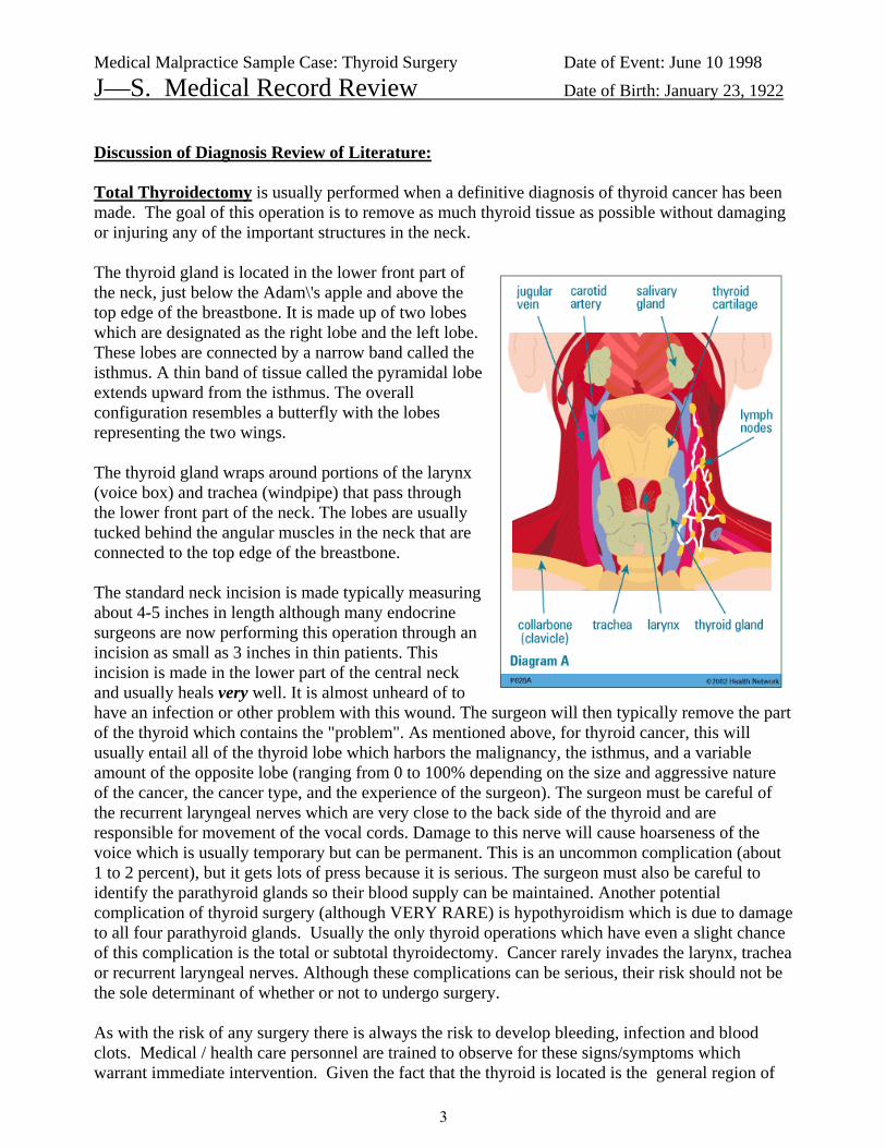
Medical Malpractice Sample Case: Thyroid Surgery Date of Event: June 10 1998 J—S. Medical Record Review Date of Birth: January 23, 1922 Discussion of Diagnosis Review of Literature: Total Thyroidectomy is usually performed when a definitive diagnosis of thyroid cancer has been made. The goal of this operation is to remove as much thyroid tissue as possible without damaging or injuring any of the important structures in the neck. The thyroid gland is located in the lower front part of the neck, just below the Adam\'s apple and above the top edge of the breastbone. It is made up of two lobes which are designated as the right lobe and the left lobe. These lobes are connected by a narrow band called the isthmus. A thin band of tissue called the pyramidal lobe extends upward from the isthmus. The overall configuration resembles a butterfly with the lobes representing the two wings. The thyroid gland wraps around portions of the larynx (voice box) and trachea (windpipe) that pass through the lower front part of the neck. The lobes are usually tucked behind the angular muscles in the neck that are connected to the top edge of the breastbone. The standard neck incision is made typically measuring about 4-5 inches in length although many endocrine surgeons are now performing this operation through an incision as small as 3 inches in thin patients. This incision is made in the lower part of the central neck and usually heals very well. It is almost unheard of to have an infection or other problem with this wound. The surgeon will then typically remove the part of the thyroid which contains the "problem". As mentioned above, for thyroid cancer, this will usually entail all of the thyroid lobe which harbors the malignancy, the isthmus, and a variable amount of the opposite lobe (ranging from 0 to 100% depending on the size and aggressive nature of the cancer, the cancer type, and the experience of the surgeon). The surgeon must be careful of the recurrent laryngeal nerves which are very close to the back side of the thyroid and are responsible for movement of the vocal cords. Damage to this nerve will cause hoarseness of the voice which is usually temporary but can be permanent. This is an uncommon complication (about 1 to 2 percent), but it gets lots of press because it is serious. The surgeon must also be careful to identify the parathyroid glands so their blood supply can be maintained. Another potential complication of thyroid surgery (although VERY RARE) is hypothyroidism which is due to damage to all four parathyroid glands. Usually the only thyroid operations which have even a slight chance of this complication is the total or subtotal thyroidectomy. Cancer rarely invades the larynx, trachea or recurrent laryngeal nerves. Although these complications can be serious, their risk should not be the sole determinant of whether or not to undergo surgery. As with the risk of any surgery there is always the risk to develop bleeding, infection and blood clots. Medical / health care personnel are trained to observe for these signs/symptoms which warrant immediate intervention. Given the fact that the thyroid is located is the general region of
3

the airway should prompt close alert to the usual complications of surgery in order to avoid further complications which might involve the airway and/or the vital respiratory system. Respiratory Assessment: Normal Breath sounds can be eight alveolar or vesicular. When assessing breath sounds it should be both sides should be evaluated and documented as whether they are equal or not. Abnormal breath sounds:
• Wheeze (whistling sound ) - This is heard most commonly in asthmatics and CHF. • Rales - a fine crackle that can be heard during inspiration or expiration. • Rhonchi - coarse crackle sound that is more wet than a rale, suctioning recommended. • Expiratory grunt - a sign of distress, hypoxia, and/or increased work of breathing. • Stridor - usually caused by edema around the vocal cords or from an obstruction or tumor. • Bronchospasm - continuous breath sounds of both rhonchi and wheezing; usually
bronchodilator will help to alleviate this problem.
The 4 major components of the lung exam (inspection, palpation, percussion and auscultation) are also used to examine the heart and abdomen.
Inspection/Observation: A great deal of information can be gathered from simply watching a patient breathe.
1. General comfort and breathing pattern of the patient. Do they appear distressed, diaphoretic, labored? Are the breaths regular and deep?
2. Use of accessory muscles of breathing (e.g. scalenes, sternocleidomastoids). Their use signifies some element of respiratory difficulty.
3. Color of the patient, in particular around the lips and nail beds. Blue indicates difficulty in air exchange.
4. The position of the patient. Those with extreme pulmonary dysfunction will often sit up-right. In cases of real distress, they will lean forward, resting their hands on their knees in what is known as the tri-pod position.
5. Breathing through pursed lips, often seen in cases of emphysema. 6. Ability to speak. At times, respiratory rates can be so high and/or work of breathing so great
that patients are unable to speak in complete sentences. If this occurs, note how many words they can speak (i.e. the fewer words per breath, the worse the problem!).
7. Any audible noises associated with breathing as occasionally, wheezing or the gurgling caused by secretions in large airways are audible to the "naked" ear.
8. The direction of abdominal wall movement during inspiration. Normally, the descent of the diaphragm pushes intra-abdominal contents down and the wall outward. In cases of severe diaphragmatic flattening (e.g. emphysema) or paralysis, the abdominal wall may move inward during inspiration, referred to as paradoxical breathing. If you suspect this to be the case, place your hand on the patient's abdomen as they breathe, which should accentuate its movement.
9. Any obvious chest or spine deformities. These may arise as a result of chronic lung disease (e.g. emphysema), occur congenitally, or be otherwise acquired. In any case, they can impair a patient's ability to breathe normally.
Palpation: Palpation plays a relatively minor role in the examination of the normal chest as the structure of interest (the lung) is covered by the ribs and therefore not palpable. Specific situations where it may be helpful include:
1. Accentuating normal chest excursion.
4

2. Tactile Fremitus: Normal lung transmits a palpable vibratory sensation to the chest wall.
• Lung consolidation: Consolidation occurs when the normally air filled lung parenchyma becomes engorged with fluid or tissue, most commonly in the setting of pneumonia. • Pleural fluid: Fluid, known as a pleural effusion, can collect in the potential space that
exists between the lung and the chest wall, displacing the lung upwards. Fremitus over an effusion will be decreased. If a large enough segment of parenchyma is involved, it can alter the transmission of air and sound. In the presence of consolidation, fremitus becomes more pronounced.
3. Investigating painful areas: If the patient complains of pain at a particular site it is obviously important to carefully palpate around that area.
Percussion: This technique makes use of the fact that striking a surface which covers an air-filled structure (e.g. normal lung) will produce a resonant note while repeating the same maneuver over a fluid or tissue filled cavity generates a relatively dull sound.
Auscultation: or listening over any of the four areas of the chest. Lower lobes occupy the bottom ¾ of the posterior filed, right middle lob is heard in the right axilla, lingula in the left axilla, upper lobes in the anterior chest and at the top 1/4 of the posterior fields. This can be quite helpful in trying to pin down the location of pathologic processes that may be restricted by anatomic boundaries (e.g. pneumonia). Many disease processes (e.g. pulmonary edema, bronchoconstriction) are diffuse, producing abnormal findings in multiple fields.
Breathing difficulties involve a sensation of difficult or uncomfortable breathing or a feeling of not getting enough air. There is no standard definition for "difficulty" breathing therefore each patient must be assessed based on their individual characteristics. These assessments are very dependent on the complaints of the patient as well as the patient's past history for breathing problems; such as allergy, cold symptoms chronic lung diseases etc. If he feels anxious or restless reporting shortness of breath assessment and evaluation is warranted. Typical Causes:
• Panic Attack • Airway Obstruction / Foreign objects • Heart attack / MI / Congestive Heart Failure • Obesity • Chronic lung diseases; Allergies / Asthma /Emphysema / COPD / Coronary diseases / • Acute Diseases; Pneumonia
Signs and symptoms:
• Abnormal Vital Signs • Shortness of breath • Cyanosis or 'bluish" coloring of lips, nail beds.
Apnea means absence of spontaneous breathing from any cause. Apnea can be intermittent and temporary, as occurs with obstructive sleep apnea or prolonged. Prolonged apnea is also called respiratory arrest. Any episode of apnea, even a temporary one, in which a person turns blue, has a seizure, becomes limp, or remains drowsy or unconscious, requires prompt medical attention.
5

Common causes of apnea in adults include:
• obstructive sleep apnea • choking • drug overdose • cardiac arrest • arrythmias (irregular heart beat • metabolic disorders • nervous system disorders.
Breathing difficulty, whether sudden or long term, should always be taken seriously. Any interruption in spontaneous breathing is a medical emergency.
Primary Respirataory Arrest is caused by an airway obstruction, decreased respiratory drive and/or respiratory muscle weakness. Airway obstruction may be partial or complete. Most common causes of Upper Airway Obstruction is displacement of the tongue into the oropharynx, due to loss of muscle tone, blood, mucus, vomitus, phyaryngolaryngeal inflammation or foreign objects, spasm or edema of the vocal cords. Lower Airway Obstruction are more commonly caused from aspiration of gastric contents, widespread severe brochospasm or extensive airspace-filling processes i.e. pneumonia, pulmonary edema or hemorrhage.
Secondary Repsiratory Arrest results from a circulataory insufficiency i.e. MI.
Complete respiratory arrrest is clinically described as the absence of spontaneious ventilatory movement in an unconcscious person. Most often this is associated with cyanosis (bluish color) but it may develop acutely in a conscious patient secondary for foreign object obstruction. If respiratory arrent is prolong cardiac arrest quickly follows because of the progressive hypoxemia which impair the cardiac function. Impending respiratory respt is characterized by a depressed sensorium and feeble, gasping or irregular respiration. Often this is accompanied by tachycardia (fast heart rate) diaphoresis (excessive sweating) and relative hypertension (high blood pressure) usually due to agitation and CO2 (carbon monoxide) accumulation.
Suspicion of Respiratory depression usually necessitate arterial blood gases inorder to confirm or deny the finding. Hypoexmia (low O2) or Hypercarbia (high CO2) will usually confirm the diagnosis. If uncorrected progressive C02 retension and hypoxemia can result in a systemic metabolic upset which can be difficult to reverse. .
Algorithm ACLS according to the Manual of Critical Nursing, 4th ed. Mosby Assess Responsiveness /Not Responsive
1. Activate EMS system 2. Call for Defibrillator
Asses breathing (Open, look listen) / Not breathing 1. Give Two breaths 2. Assess Circulation
Start CPR / No VF/VT Present on Monitor
1. 1.Intubate 2. Confirm placement tube 3. Confirm ventilations 4. Determine rhythm and cause
Non-Responsive
6

1. Assess ABG's 2. Perform CPR until defibrillator attached 3. VF/VT present on monitor
• Defibrillate up to 3 times if needed for persistent VF/VT (200J, 200-300J, 360J • Assess Electrical Activity
If Asystole 1. Continue CPR 2. Intubate at once 3. Obtain IV Access 4. Confirm Asystole in MORE THAN ONE LEAD
Consider possible Causes 1. Hypoxia 2. Hypokalemia 3. Hypokalemia 4. Pre-existing Acidosis 5. Drug Overdose 6. Hypothermia
Consider Immediate Transcutaneous Pacing 1. Epinephrine - 1mg IV push repeat every 3-5 minutes 2. Atropine - 1 mg IV repeat every 3-5 minutes up to a total of 0.3 to 0.4 mg per kg.
Consider termination of efforts.
7

10/15/2004 8:17:08 AMCase Research : LiteratureAuthority Name Extract TextACC/AHA Committee onCoronary Angiography
A report of the American College of Cardiology/American Heart Association Task Force on Practice Guidelines (Committee onCoronary Angiography).
Recommendations for Coronary Angiography in Perioperative Evaluation Before (or After) Noncardiac SurgeryClass I: Patients with suspected or known CAD
1. Evidence for high risk of adverse outcome based on noninvasive test results (Level of Evidence: C)2. Angina unresponsive to adequate medical therapy. (Level of Evidence: C)3. Unstable angina, particularly when facing intermediate* or high-risk* noncardiac surgery. (Level of Evidence: C)4. Equivocal noninvasive test result in a high-clinical-risk** patient undergoing high-risk* surgery. (Level of Evidence: C)Cardiac risk according to type of noncardiac surgery. High risk: emergent major operations, aortic and major vascular, peripheralvascular, anticipated prolonged surgical Procedures associated with large fluid shifts and/or blood loss; intermediate risk: carotidendarterectomy, major head and neck, intraperitoneal and/or intrathoracic, orthopedic surgery, prostate surgery; low risk:endoscopic Procedures, superficial Procedures, cataract surgery, breast surgery.
Cardiac risk according to clinical predictors of perioperative death, MI, or CHF. High clinical risk: unstable angina, recent MI andevidence of important residual ischemic risk, decompensated CHF, high degree of atrioventricular block, symptomatic ventriculararrhythmias with known structural heart disease, severe symptomatic valvular heart disease, multiple intermediate risk markerssuch as prior MI, CHF, and diabetes; intermediate clinical risk: CCS class I or II angina, prior MI by history or ECG, compensatedor prior CHF, diabetes mellitus.
American Journal ofEmergency Medicine01-May-1999; 17(3): 308-9
Acute Postoperative Neck Hematoma
SIMON PAUL ROY MDDepartment of Emergency Medicine, Boston Medical Center, Boston, MA.Manuscript received January 9, 1998accepted January 23, 1998.Address reprint requests to Dr Roy, Department of Emergency Medicine, Boston Medical Center, 818 Harrison Ave, Boston, MA02118.Acute postoperative cervical hematomas can produce intimidating airway emergencies. Conventional airway techniques may notbe the most appropriate way of effectively dealing with these crises. A counterintuitive approach, rapid hematoma evacuationwithout establishing an airway, will be discussed and compared with standard methods. To act quickly and definitively, theemergency department (ED) physician must have reviewed all airway options, including the evacuation technique, before beingfaced with the emergency situation.
DISCUSSIONThis case demonstrates a rarely encountered situation in the ED with a specific therapy. Acute postoperative hematomas areunique in that they have not been incorporated into the surrounding soft tissues. This enables one to easily "shell out" the contents
Confidential Attorney Work Product. Do Not Reproduce. BlackBPage 1
8

10/15/2004 8:17:08 AMCase Research : LiteratureAuthority Name Extract Text** and rapidly relieve mass effect. In the case of a cervical hematoma with airway compromise, this adds an acute technique beyond
conventional airway methods: hematoma evacuation without securing an airway. Ideally, if time permits and the patient is stable,evacuation should occur in the operating room. Overall management will be surgical but the role of the ED physician will betwofold. First, the temptation to engage in airway techniques or hematoma evacuation in the ED when the patient is stable must beavoided. Second, rapid mobilization of surgical services and transportation to the operating room is vital to avoid the need toperform difficult technical maneuvers in the ED as the patient deteriorates.
Airway anatomy can be distorted by hematomas via various mechanisms. Direct pressure on the airway can cause deviation fromthe midline, as well as lumen narrowing. [1] [2] Interestingly, perilaryngeal edema may also be present. Bexton and Radford [3]reported such a case in a 57-year-old woman after thyroid surgery. The patient required emergency hematoma evacuation forsevere respiratory compromise, but failed to significantly improve after the procedure. Upon Intubation, marked cord edema wasnoted. Bexton postulated that cervical hematomas impair regional venous drainage, resulting in edema secondary to venousHypertension. The vast majority of case reports, however, demonstrate rapid symptomatic relief after decompression.
If the physician is faced with a rapidly deteriorating airway situation, ED hematoma evacuation without securing an airway mustbe considered. This is standard teaching in surgical texts regarding the bedside management of hematomas after thyroid surgery,[4] [5] but is rarely mentioned in emergency medicine literature. Fiberoptic nasotracheal Intubation may be considered but requirestime and some degree of patient cooperation. If these features are present, direct transport to the operating room is moreappropriate. Rapid-sequence approaches with paralytic agents are always concerning in any spontaneously breathing patient withdistorted neck/airway anatomy. Simple spray Anesthesia followed by orotracheal Intubation is likely to produce gagging andvomiting, with aspiration risks, in "full stomach" ED patients.
It has been reported in the surgical and anesthesiology literature that evacuation of acute postoperative hematomas withoutestablishing an airway in the unstable patient is both effective and safe. It also appears that any attempt at securing an airway maybe difficult and dangerous. Gomez et al [6] described a case series of 13 carotid endarterectomy patients who developedpostoperative neck swelling with respiratory distress. The hematoma was evacuated under local Anesthesia without securing anairway in 7 cases without adverse effects. The remaining 6 patients underwent general Anesthesia with endotracheal Intubationfollowed by hematoma evacuation. Four of 6 developed severe cardiac 309 and/or neurologic complications (myocardialinfarctions and strokes) after multiple attempts at Intubation. O'Sullivan et al [7] reported a series of 6 patients requiringpostoperative cervical hematoma drainage. Five patients underwent attempts at endotracheal Intubation prior to operative drainage.In all five, the attempts met with extreme difficulty in identifying anatomic landmarks. Desaturation and bradycardia were frequentsequelae. The sixth patient's airway was managed by tracheostomy. It appears that the physiological and psychological stressesplaced on patients undergoing multiple, challenging, and traumatic attempts at Intubation markedly increase overall morbidity andmortality.
Although simple evacuation appears far better tolerated than Intubation followed by drainage, there is commonly reluctance toperform the former because of concerns regarding releasing uncontrollable hemorrhage. In the O'Sullivan [7] and Gomez [6]
Confidential Attorney Work Product. Do Not Reproduce. BlackBPage 2
9

10/15/2004 8:17:09 AMCase Research : LiteratureAuthority Name Extract Text** series', the majority of bleeding was noted to be from subcutaneous capillaries. In no case was a major arterial bleed identified.
This must clearly be differentiated from the patient with a cervical hematoma secondary to nonoperative neck trauma, wherehematoma drainage under local Anesthesia without airway securement is not an option because of a possible major vascular injury.
It appears that with the present cost containment strategies in the United States (including timely discharges), early postoperativepatients may frequently present to the ED for evaluation of incisional hemorrhage/hematoma. Carotid endarterectomy andthyroidectomy patients are typically discharged home less than 48 hours after operation and not infrequently within 24 hours. Theemergency practitioner should be aware of all possible management strategies in these situations, especially with respect to theairway.
REFERENCES
1. Bukht D, Langford RM: Airway obstruction after surgery in the neck. Anaesthesia 1983;38:389-390 Citation2. Hare R: Respiratory obstruction after thyroidectomy. Anaesthesia 1982;37:11363. Bexton MDR, Radford R: An unusual cause of respiratory obstruction after thyroidectomy. Anaesthesia 1982;37:596 Citation4. Kahky MP, Weber RS: Complications of surgery of the thyroid and parathyroid glands. Surg Clin North Am 1993;73:307-321Abstract5. Shaha A, Jaffe BM: Complications of thyroid surgery performed by residents. Surgery 1988;104:1109-1114 Abstract6. Gomez ER, Kunkel JM, Jarstfer S. B., R.N.: Wound hematomas after carotid endarterectomy. Am Surgeon 1985;51:111-1137. O'Sullivan JC, Wells DG, Wells GR: Difficult airway management with neck swelling after carotid endarterectomy. AnaesthIntens Care 1986;14:460-464
American Journal of Surgery01-Jun-2002; 183(6): 630-41
CONCLUSIONS: Understanding perioperative pathophysiology and implementation of care regimes to reduce the stress of anoperation, will continue to accelerate rehabilitation associated with decreased hospitalization and increased satisfaction and safetyafter discharge. Developments and improvements of multimodal interventions within the context of "fast track" surgery programsrepresents the major challenge for the medical professionals working to achieve a "pain and risk free" perioperative course.
An Advisory Statement Fromthe Advanced Life SupportWorking Group of theInternational LiaisonCommittee on Resuscitation :ILCOR Advisory Statements
Unequivocal Advanced Life Support InterventionsValid scientific evidence supports only three interventions as unequivocally effective in adult cardiac resuscitation:•�Basic cardiopulmonary resuscitation (CPR)•�Defibrillation--if the rhythm is ventricular fibrillation or pulseless ventricular tachycardia•�Oxygenation and ventilation of the lungs through a patent secure airway such as a tracheal tubeThe universal algorithm presents these interventions simplistically and recommends a specific sequence that rescuers shouldfollow.Basis for RecommendationsThe sequence of interventions is based, whenever possible, on sound scientific information. But there is a paucity of convincinghuman data on some aspects of resuscitation. Until such time as new information becomes available, the working group made nochanges to well-established Procedures but suggested some modifications on educational rather than scientific grounds.
Confidential Attorney Work Product. Do Not Reproduce. BlackBPage 3
10

10/15/2004 8:17:09 AMCase Research : LiteratureAuthority Name Extract Text** Only Two Arrest Rhythms
Cardiac arrest rhythms can be divided into two subsets: ventricular fibrillation/pulseless ventricular tachycardia (VF/VT) andnon-VF/VT. Non-VF/VT incorporates both Asystole and pulseless electrical activity (PEA). The only difference in managementbetween the two arrest rhythms is the need for rescuers to perform defibrillation for patients in VF/VT. Otherwise the actions andinterventions are essentially the same: basic CPR, tracheal Intubation, epinephrine administration, and correction of reversiblecauses.Basic CPR and the Precordial ThumpBasic life support (BLS) should be performed until advanced life support (ALS) becomes available. In the event of a monitoredarrest, a precordial thump is considered a Class I recommendation by the ILCOR.Defibrillation should be performed as soon as VF/VT is recognized. VF is defined as a pulseless, chaotic, disorganized rhythmcharacterized by an undulating irregular pattern that varies in size and shape with a ventricular waveform of >150 beats perminute.Tracheal Intubation s a Class I recommendation. If tracheal Intubation is not possible, the laryngeal mask airway or Combitube areacceptable initial alternatives in adults.Intravascular Access is a Class I recommendation. If intravascular access is not attainable, epinephrine may be administered viathe tracheal tube using at least double the intravascular dosage.Epinephrine should be administered using a dosage of at least 1 mg (0.01 mg/kg) every 3 minutes.Correction of Reversible CausesThe universal algorithm specifically directs rescuers to seek and treat reversible causes of the cardiac arrest. This recommendationis based on the appreciation that many people, especially those in non-VF/VT, have an identifiable cause for the cardiac arrest.Many of these causes can be reversed with specific interventions. As a teaching aide-memoire, the algorithm lists the mostcommon reversible causes of cardiac arrest. Thus, we have moved from the former rhythm-based treatment approach to a moreclinically relevant etiologic approach.Special ConsiderationsThe use of buffers, antiarrhythmics, Atropine, and pacing can be considered in certain special resuscitation situations. ILCOR hasprepared an advisory statement on conditions that may require modifications in resuscitation Procedures or techniques, based onthe specific etiology of the arrest.Using the Universal AlgorithmResuscitation algorithms are simple visual teaching tools and memory aids. They convey only a small portion of the knowledgeneeded to counter cardiopulmonary emergencies. TThe Ultimate Simplicity of ALS ResuscitationOur knowledge of effective therapy for cardiac arrest can be summarized as follows:•�Perform CPR at all times for pulseless patients (with the obvious exception of rhythm analysis and defibrillation shocks).•�Defibrillate VF/VT until it is no longer present.•�Gain control of the airway and provide adequate oxygenation and ventilation.•�Give intravenous boluses of epinephrine.•�Correct reversible causes.
Confidential Attorney Work Product. Do Not Reproduce. BlackBPage 4
11

10/15/2004 8:17:09 AMCase Research : LiteratureAuthority Name Extract Text** To remember and provide these steps as rapidly and effectively as possible will serve our patients well.
Miller: Anesthesia, 5th ed.,Copyright © 2000 ChurchillLivingstone, Inc.
Propofol affects the respiratory system in a manner qualitatively similar to the action of barbiturates. Apnea occurs after aninduction dose of propofol; the incidence and duration of apnea appear dependent on dose, speed of injection, and concomitantpremedication. An induction dose of propofol results in a 25 to 30 percent incidence of apnea. [419] [420] The apnea occurringwith propofol, however, may be prolonged to more than 30 seconds. The incidence of prolonged apnea (>30 seconds) is furtherincreased by addition of an opiate, either as premedication or just prior to induction, and it is greater with propofol than with othercommonly used intravenous induction agents. [ The onset of apnea is usually preceded by marked tidal volume reduction andtachypnea. Following a 2.5-mg/kg induction dose of propofol, the respiratory rate is significantly decreased for 2 minutes, andminute volume is significantly reduced for up to 4 minutes, a finding that indicates a more prolonged effect of propofol on tidalvolume than on respiratory rate.
Pulse oximetry forperioperative monitoring(Cochrane Review)
Background: Monitoring with pulse oximetry might improve patient outcome by enabling an early diagnosis and consequently,correction of perioperative events that might cause postoperative complications or even death. Only a few randomised clinicaltrials of pulse oximetry have been performed during anaesthesia and in the recovery room which describe perioperativehypoxaemic events, postoperative cardiopulmonary complications and cognitive dysfunction.
Reviewers' conclusions: The studies confirmed that pulse oximetry can detect hypoxaemia and related events. However, we havefound no evidence that pulse oximetry affects the outcome of anaesthesia. The conflicting subjective and objective results of thestudies, despite an intense, methodical collection of data from a relatively large population, indicate that the value of perioperativemonitoring with pulse oximetry is questionable in relation to improved reliable outcomes, effectiveness and efficiency.
Rubin, Adam D., M.D. StaffPhysician, Resident Dept.Otolarygology - Head andNeck Surgery Univ. ofMichigan Medical Center
Intraoperative bleeding causes tissue-staining and increases difficulty recognizing important structures. The risk of otheranatomical complications increases with intraoperative bleeding. Deliberate dissection and fastidious hemostasis are essential.
Postoperative bleeding can be a devastating complication of thyroid surgery. An unrecognized or rapidly expanding hematoma cancause airway compromise and asphyxiation. The incidence of hemorrhage after thyroid surgery is low (0.3-1%), but the surgeonmust be aware of this potentially fatal complication.Evaluation:The physical exam is the only workup necessary. One should not waste time with imaging studies when bleeding is suspected.Fiberoptic laryngoscopy does not help view the distal airway. Imaging studies (eg, CT scans, ultrasound) may be useful in the caseof mild neck swelling without airway compromise. The surgeon should carefully assess the airway prior to sending a patient toradiology.Treatment:If a neck hematoma is causing airway compromise, the incision should be opened at the patient's bedside to release the collectionof blood. The patient should then be taken immediately to the operating room. In the setting of hematoma without impendingairway obstruction, the patient should be taken to the operating room urgently.
Confidential Attorney Work Product. Do Not Reproduce. BlackBPage 5
12

Thyroid Case; Conclusion of Facts
1. Mr. S was diagnosed with a suspicious mass on his thyroid; Dr. SH recommended surgery
2. Initial Pre-operative Screen H&P determine a cardiac abnormality suggestive of MI; extensive work-up was recommended by Dr. C. Cardiologist;
3. 3 months later Mr. S. was given cardiac clearance; surgery was scheduled to proceed.
4. On (date/time) Mr. S underwent complete Thyroidectomy, performed by Dr. S.H. Lopressor was given prior to surgery. Anesthesia records reveal that Mr. S was in sinus bradycardia; during the procedure O2 saturation levels were documented at 99%; he was given Fentanyl and Lobetalol; It was documented ; he was transferred to recover in stable condition.
5. Recovery room nurses document a blood pressure of 180/100; rales in bilateral bases (lungs) Fentanyl and morphine were ordered and given for pain.
6. Following 45 minutes of recovery time; Mr. S. was ready for transfer; med/surg bed was not available; he was transferred to a pediatric unit (private room);again described in stable condition. Blood pressure was 200/92; the surgical wound was described as thickened with a collection of blood and red drainage was coming from the wound; Dr. S and Dr. A were both notified.
7. Noted as the Med / Surg Nursing admission assessments describe Mr. S as "groggy" responding to commands; vital signs were noted as stable
8. Mr. S. daughter reported the patient was not feeling well at 9:45 p.m. Morphine was given.
9. At 10:30 Mr. S was found with breathing difficulties; Nurse J. left the room to obtain a pulse oximetry.
10. 10:30 p.m. full code blue was called; Mr. S was in asystole. Anesthesia's 1st attempt to re-intubate – failed due to blood mass in the airway; Tracheostomy tray was requested however not available; nurse again left the room to retrieve; Anesthesia suction large amounts of clot from airway; tracheostomy was performed 10 minutes later providing patent airway and ventilation support; resuscitation efforts continued.
11. At 11:05 Mr. S. expired.
13

Medical Malpractice Sample Case: Thyroid Surgery Date of Incident: June 10 1998 J—S. Medical Record Review Date of Birth: January 23, 1922
Key Points / Suggestion of Focus A. Potential Defendants: Dr. S – General Surgeon:
• Failed to document a complete and accurate assessment as well as an evaluation in regards to the cause of the bleeding, in the PACU
• Failed to document a plan considering all potential complications of thyroid surgery.
PACU Nursing staff reported respiratory difficulty, excessive bleeding and abnormal vital signs. The medical records provided indicate that Dr. S. was at the bedside however the medical documentation provided does not support evidence that Dr. S assessed and evaluated the patient at that time. Why was the incision bleeding? Did he feel the blood was coming from a vessel or did he feel this was residual fluid or "run off" from the surgery? Dr. A—Anesthesiologist
• Failed to document a complete assessment and evaluation of Mr. S – status after the phone call from the nursing staff of increased pain, no relief from pain medication and dropping O2 saturation levels.
The medical records support evidence that Dr. A—discharged this patient depending on the nursing reports. Mr. S was a "fresh" post operative patient. He had a major surgery in the vicinity of the airway and the nurses reported excessive bleeding. What assessment findings assured Dr. A that Mr. S was stable enough to transfer without physically examining the patient? Nursing Staff; Floor 4 W Nurse A, 4W, Nurse B, 4 W, Nurse C PACU; House Supervisor, Admission
• Failed to assess and evaluated the health status of Mr. S and document a complete physical assessment upon receiving the transfer
• Failed to recognize, interpret and timely record signs and symptoms particularly those requiring the notification of the physician
• Failed to timely assess evaluate and document a complete assessment, in response to Mr. S complaints
• Failed to recognize and consider all potential complications of a post-operative thyroid surgery patient with a neck incision.
a. Based upon objective and subjective data, assessments should be complete, systematic and
continuous. There is almost a two hour lapse from the time of arrival to the time a complete review of systems is documented. Mr. S - had complaints of continued pain, which also was reported by his daughter. He had a episode of bleeding in PACU, he was agitated and struggling. . Nursing staff also failed to recognize the deviation in Mr. S's normal and/or expected pattern of social history which repeatedly describes Mr. S as calm. During this short period of admission documentation continually described as anxious and agitated which warrants investigation. What assessment findings assure the nursing staff that no intervention was necessary?
14

b. Vital Signs are a key assessment to major systems and should be taken at the time of arrival in order to establish the patient status. Typically, during the post-op phase vital signs are checked on regular hourly basis, typically these would be 1 hour x 2, 2hours x 2; 4 hours x 2 once felt stable; per shift. Mr. S's vital signs are not documented for approximately 2 hours after arrival. What assessment findings assured these nurses Mr. S was stable at the time of arrival.
c. Excessive blood drainage from the surgical site. The physician was not notified.
d. Pulse oximetry is usually normal equipment on a surgical post op floor. The medical
records indicate pulse oximetry had to be retrieved from another floor. e. Available Tracheotomy Tray is typically considered routine and/or common bedside
equipment to the post-operative Thyroid patient. Tracheotomy Tray was not available on 4W and had to be retrieved from another floor when needed.
B. Suggestion of Focus:
1. In regards to the floor admission it will be important to investigate as to why Mr. S. was admitted
to a Pediatric floor versus a Medical / Surgical post-operative unit. The fact that typical post-op equipment was not immediately available raises many questions. Was this a typical over-flow unit for surgical patients? Were these nurses oriented and trained to the surgical patient? How familiar were they, in regards to caring for patient with head/neck surgery?
2. It appears that Mr. S was admitted to a pediatric floor versus a med/surg floor it will be important
to request the nursing protocol for routine assessments of the post-operative patient as well as all nursing protocol specific for the thyroid surgical post-operative patient. It will also be important to discuss with the floor supervisor the manner of orientation used to prepare these nurses to care for post-op patients.
3. It will be important to discuss with the Medical Expert Anesthesiologist the fact that bleeding and
decrease O2 saturation levels were reported to Dr. A and Mr. S was discharged via the telephone without physical examination.
4. Complications of Total Thyroidectomy: Given the fact that this type of surgery is done in the neck
close to the airway anatomy it will be important to discuss with the Medical Expert Surgeon the significance in regard to airway precaution specifically as to the availability of tracheostomy tray during immediate post-operative phase in order to determine if all precautions were taken. The medical literature strongly supports the potential for airway complications when the surgical anatomy involves the neck / airway region. No Tracheotomy Tray was available at the bedside or even on the floor at the time Mr. S's airway obstructed.
5. A Medical Expert Surgeon will to be needed to review Dr. S. Admission Orders to 4W Med/Surg
Floor which are vague and incomplete and fail to support a plan for a thyroid surgical patient with a bleeding incision.
• Dr. S writes orders for diet, activity and pain however fails document/order parameters for assessments specifically vital signs, warning signs of thyroid surgery complications including the bleeding for which he was notified by the PACU. This indicates that routine vital signs and assessments were to be done typically these would be 1 hour x 2, 2hours x 2; 4 hours x 2 then per shift.
15

• Pulse oximetry is vital in monitoring the oxygen saturation levels. Mr. S's O2 saturation levels began dropping before discharge of PACU. Physicians, both anesthesia and general surgery were notified. Why did they feel in was not necessary to order oxygen saturation monitoring? • In regard to the decrease O2 saturation level at the time of PACU discharge: Lab and diagnostic studies were not ordered. ABGs can be used to monitor oxygen levels in the blood stream. It will be important to discuss with the Medical Experts Anesthesia and Surgery the significance of ABG monitoring as relevant to thyroidectomy post-op status of saturation levels in the 80's.
Medical Expert Anesthesia and General Surgery or Otolaryngology will be needed in order to determine the course of events that occurred prior and during the code in regards to intubation
• Again, Tracheostomy tray at the bedside for a surgical thyroid patient • The presence of blood in the airway. Airway obstruction and respiratory collapse. • Several minutes of hypoxia and the common end result
Medical Surgical Nurse Expert
• Again, Tracheostomy tray at bedside for a thyroid patient • Pulse oximetry and regular monitoring • Admission and routine assessments of a surgical patient • Monitoring for respiratory distress. It will be important to discuss the significance of a complete
respiratory assessment specifically in post-operative thyroid surgery patient with a recent history of incisional site bleeding.
Additionally
• It will be important to review and discuss with the PACU nurses the 45 minutes from the time of PACU discharge and the arrival time to the floor. This is quite unusual even in very large facilities. As there is no documentation during this time period describing the course of events, raises the question; why did it take so long to move Mr. S from the PACU to the floor?
• Mr. S was transferred from the PACU to the floor between 5:00 and 6:00 p.m. Many hospitals
close down the PACU, once all the surgery patients are all transferred. It may be significant to further investigate the G Hospitals PACU protocol for transferring patients along with the patient log for 6/10/98 in order to determine how Mr. S fell into the rotation of transfer. Simply: Was he the last patient of the day?
• A Medical Expert review of the Fentanyl that was given during Mr. S's PACU stay will be needed
to determine any significance to Mr. S's outcome. Fentanyl can depress the respiratory system. Suggestion for further discovery items:
1. A copy of all instruments in writing from 6/10/98 to present regarding any corrective action taken, based on patient care evaluation studies of Dr. S. and Dr. A.
2. A copy of all Medical Staff Rules and Regulation regarding post-operative patient care. 3. A copy of all Medical Staff Rules and Regulations regarding post-operative orders 4. A copy of all written rules, regulations, policies, procedures, bylaws memoranda or any
instruments in writing regarding the roles, functions duties or responsibilities of the attending surgical/medical staff . Also; Anesthesia Staff.
16

5. A copy of policy, procedure and protocols regarding admission by transfer from the PACU to the Acute Care Floor or post operative Acute Care area within S. Hospital
6. A copy of the complete names of every RN, LVN, Nursing Assistant or Care Technician that rendered care to Mr. S on 6/10/98
7. A blank copy of all flow sheets, chart grafts and forms used in the records of J—S— 8. A copy of all Nursing Policy and Procedure specifically for patient call lights 9. The table of contents of all policies, procedures and nursing protocol specific for patient safety. 10. A copy of PACU Nursing policy, procedures and protocol specific for reporting abnormal clinical
findings. 11. A copy of PACU Nursing Policy, Procedure and Nursing Protocols; specific for mechanism of
release of patients from the PACU 12. A copy of 4 W Nursing Policy, Procedure and Nursing Protocol specifically for the post-operative
Thyroid patient. 13. A copy all policy, procedures and protocol regarding airway maintenance 14. A copy of G Hospital Policy, Procedure and Protocol for Cardiopulmonary Resuscitation 15. A copy of policy, procedure and protocol regarding the administration of Fentanyl and Droperidol. 16. A copy of the Table of Contents of the Pharmacy Medication Safety Manual.
Possible Witnesses Interviews
1. Dr. Ph—Emergency Room Physician 2. K.L. CRNA, Nursing Anesthetist 3. D. E Code Recorder.
17
Sample Case
Fact Chronology: Thyroid Surgery
18

Plaintiff Med Mal Sample Case- Fact Report
Date & Time Source(s) Fact Text
Tue03/24/19981:06 p.m.CT
Tab 1 0010S. Hospital, EKG report (pre-op)S.H. Surgeon, M.D.
Impression: Abnormal Left Axis Deviation (QRS axis >301) Septal Myocardial Infarction (Probably recent) T-Wave Abnormality, Possible Antero-lateral Ischemia
Tue03/24/19981:45 p.m.CT
Tab 1 Page 0021Surgeon Office NotesS.H. Surgeon, M.D.
Tab 6 Page 0019Cardiolgist, Cath ReportC. W. Cardiologist, M.D.
Request delay in non-cardiac surgery due to findings of Cardiac Catheterization.
Cardiac CatheterizationImpression: MI (Myocardial Infarct) secondary to Diagonal Coronary Artery Occlusion. Unclearif recent or remote.Recommendations: Delay non-cardiac surgery 6-8 weeks Start Lopressor and Nitroglycerine Patch Follow-up with Stress Cardiolyte Test, on May 10, 1998 at C. W. Cardiologist, M.D. office.
Mon06/01/1998
Tab 1 Page 0007S. Hospital Consent for Operations andAnestheticsJ. S. witnessed by J.V., R.N.
I authorize Dr. S and associates designated by Dr. S to perform: "Biopsy of Mass Right Neck, possibly remove Thyroid Gland"
Mon06/08/19981:00 p.m.CT
Tab 1 Page 0039S. Hospital, EKG ReportC. W. Cardiologist, M.D.
Impression: Sinus Bradycardia Abnormal Left Axis Deviation ST and T wave abnormality Abnormal EKG
Mon06/08/19982:45 p.m.CT
Tab 1 Page 0004-5S. Hospital, Pre-procedure Instruction SheetD.S., R.N.
Pre-procedure instructions given for "Biopsy of mass right neck with possible excision".Ht 5'11'', Wt. 153#Vital Signs: (t) 97.7 (B/p) 158/68 (p) 46, (r)18J. S. is instructed to take Lopressor with a sip of water and apply Nitroglycerine Patch
Mon06/08/19983:00 p.m.CT
Tab 1 Page 0014S. Hospital, Anesthesia Orders
A. B. Anesthesia, M.D.
MedicationsRanitidine (Zantac) 150 mg orally at bedtime and at 0600 with a sip of waterMetoclopramide (Reglan) 10 mg orally at 0600 on call to OR with a sip of water
Past Medical History: Cardiac Cath of 4/98 findings of 45% ejection fraction BMP (Basic Metabolic Profile) 12 Lead EKG on admission
Confidential Attorney Work Product. Do Not Reproduce.
19

Plaintiff Med Mal Sample Case- Fact Report
Date & Time Source(s) Fact Text
Tue06/09/199810:45 a.m.CT
Tab 1 Page 0033S. Hospital, Nurses NotesS. B., R.N.
Labs faxed to S.H. Surgeon, M.D.
Wed06/10/19989:55 a.m. CT
Tab 1 Page 0033S. Hospital, Pre-procedure Instruction SheetS. Hospital, Nurses NotesJ. H., R.N.
Admission Nursing Assessment: Skin warm and dry, alert and oriented x 3 NPO since midnightVital Signs: (B/p 170/40 (T) 96.2, (P)43 (R) 18Lopressor given.
Wed06/10/199810:22 a.m.CT
Tab 1 Page 0033S. Hospital, Nurses NotesJ. H., R.N.
To OR via stretcher.
Wed06/10/199810:45 a.m.CT
Tab 1 page 0015S. Hospital, Anesthesia OrdersA. B. Anesthesia, M.D.
1. Admit to PACU (post Anesthesia care unit)2. Oxygen 2-6 liters3. Record Vital Signs every 5 minutes x 2 and then every 15 minutes4. Pain - Medications:Morphine Sulfate 1-3 mg IV every 2-3 minutes, maximum dose 16 mgFentanyl 1/2 cc every 2-3 hours to 2 cc5. Nausea - Medications:Droperidol 1/4/cc IVMetoclopramide (Reglan) 10 mg IV6. Respiratory Depression - notify Anesthesia Dept Immedicately7.Discharge to floor if Discharge Criteria of 8 or greater. If less notify Anesthesia Dept.8. Out patient per discharge criteria9. Give Labetalol (a/k/a Trandate) now and again in 10 minutes if B/p greater than 180 (systolic)
Wed06/10/199811:40 a.m.CT
Tab 1 Page 0045S. Hospital, Anesthesia RecordA. B. Anesthesia, M.D.
Monitors placedInduction with intubation without incident
Wed06/10/199812:02 p.m.CT
Tab 1 Page 0045S. Hospital, Anesthesia RecordA. B. Anesthesia, M.D.
Fentanyl 50 mg givenO2 on at 6 liters
EKG reveals Sinus BradycardiaSaO2 100
Confidential Attorney Work Product. Do Not Reproduce.
20
Plaintiff Med Mal Sample Case- Fact Report
Date & Time Source(s) Fact Text
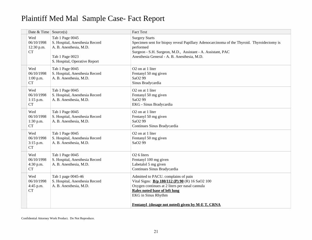
Wed06/10/199812:30 p.m.CT
Tab 1 Page 0045S. Hospital, Anesthesia RecordA. B. Anesthesia, M.D.
Tab 1 Page 0023S. Hospital, Operative Report
Surgery StartsSpecimen sent for biopsy reveal Papillary Adenocarcinoma of the Thyroid. Thyroidectomy isperformedSurgeon - S.H. Surgeon, M.D., Assistant - A. Assistant, PACAnesthesia General - A. B. Anesthesia, M.D.
Wed06/10/19981:00 p.m.CT
Tab 1 Page 0045S. Hospital, Anesthesia RecordA. B. Anesthesia, M.D.
O2 on at 1 literFentanyl 50 mg givenSaO2 99Sinus Bradycardia
Wed06/10/19981:15 p.m.CT
Tab 1 Page 0045S. Hospital, Anesthesia RecordA. B. Anesthesia, M.D.
O2 on at 1 literFentanyl 50 mg givenSaO2 99EKG - Sinus Bradycardia
Wed06/10/19981:30 p.m.CT
Tab 1 Page 0045S. Hospital, Anesthesia RecordA. B. Anesthesia, M.D.
O2 on at 1 literFentanyl 50 mg givenSaO2 99Continues Sinus Bradycardia
Wed06/10/19983:15 p.m.CT
Tab 1 Page 0045S. Hospital, Anesthesia RecordA. B. Anesthesia, M.D.
O2 on at 1 literFentanyl 50 mg givenSaO2 99
Wed06/10/19984:30 p.m.CT
Tab 1 Page 0045S. Hospital, Anesthesia RecordA. B. Anesthesia, M.D.
O2 6 litersFentanyl 100 mg givenLabetalol 5 mg givenContinues Sinus Bradycardia
Wed06/10/19984:45 p.m.CT
Tab 1 page 0045-46S. Hospital, Anesthesia RecordA. B. Anesthesia, M.D.
Admitted to PACU. complains of painVital Signs: B/p 180/112 (P) 90 (R) 16 SaO2 100Oxygen continues at 2 liters per nasal cannulaRales noted base of left lungEKG in Sinus Rhythm
Fentanyl (dosage not noted) given by M-E T, CRNA
Confidential Attorney Work Product. Do Not Reproduce.
21

Plaintiff Med Mal Sample Case- Fact Report
Date & Time Source(s) Fact Text
Wed06/10/19985:00 p.m.CT
Tab 1 Page 0061S. Hospital, PACU Nurses RecordPacu Nurse, R.N.
Complaint of pain noted, titrating Morphine Sulfate 3 mg, IVDroperidol .625 mg given
B/p 220/92 (P) 95 (R) 15SaO2 100
Wed06/10/19985:10 p.m.CT
Tab 1 Page 0061S. Hospital, PACU Nurses RecordPacu Nurse, R.N.
S.H. Surgeon, M.D. at bedside, observing red drainage.Morphine Sulfate 3 mg given
Confidential Attorney Work Product. Do Not Reproduce.
22
Sample Case; Thyroid Surgery
Glossary of Terms
23

Glossary of Terms
Full Name Description
Apnea Temporary cessation of breathing and, therefore, of the body's intake of oxygen and release of carbondioxide. It is a serious symptom, esp. in patients with other potentially life-threatening conditions.
Arterial Blood Gas Literally, any of the gases present in blood; operationally and clinically, they include the determination oflevels of pH, oxygen (O2), and carbon dioxide (CO2) in the blood. ABGs are important in the diagnosis and treatment of disturbances of acid-base balance, pulmonarydisease, electrolyte balance, and oxygen delivery. Values of the gases themselves are usually expressed as thepartial pressure of carbon dioxide or oxygen, although derived values are reported in other units. Severalother blood chemistry values are important in managing acid-base disturbances, including the levels of thebicarbonate ion, HCO3, blood pH, sodium, potassium, and chloride.
Asystole Cardiac standstill; absence of electrical activity and contractions of the heart evidenced on the surfaceelectrocardiogram as a flat (isoelectric) line during cardiac arrest.
Cardiac Catheterization. Cardiac catheterization (also called cardiac cath or coronary angiogram) is a procedure that allows yourdoctor to "see" how well your heart is functioning. The test involves inserting a long, narrow tube, called acatheter, into a blood vessel in your arm or leg, and guiding it to your heart with the aid of a special X-raymachine. Contrast dye is injected through the catheter so that X-ray movies of your valves, coronary arteriesand heart chambers can be created.
Purpose:*Evaluate or confirm the presence of heart disease (such as coronary artery disease, Heart valve disease ordisease of the aorta).*Evaluate heart muscle function.*Determine the need for further treatment (for example, angioplasty or bypass surgery)
Cardiac defibrillation Cardiac defibrillation is a common emergency procedure. It is the process of stopping fibrillation (rapid,uncoordinated heart contraction) by delivering an electric shock to the heart with a machine (defibrillator).
Electrocardiogram (EKG) A test that measures the electrical activity of the heart. This includes the rate and regularity of beats as well asthe size and position of the chambers, any damage to the heart, and effects of drugs or devices to regulate theheart.
Hypertension High blood pressure; transitory or sustained elevation of systemic arterial blood pressure to a level likely toinduce cardiovascular damage or other adverse consequences
Hypoxia An oxygen deficiency. 2. A decreased concentration of oxygen in the inspired air. cerebral hypoxia Lack of oxygen supply to the brain, usually as a result of either diminished blood flow(e.g., cardiopulmonary arrest) or diminished oxygenation of the blood
Confidential Attorney Work Product. Do Not Reproduce.
24

Glossary of Terms
Full Name Description
Intubation The insertion of a tube into any hollow organ. Intubation of the trachea provides an open airway and thus is an essential step in advanced life support. Italso permits the instillation of certain critical care drugs, such as lidocaine, epinephrine, and Atropine, whichthe lungs can absorb directly when other forms of internal access are unavailable.
Endotracheal intubation: The insertion of an endotracheal tube through the nose or mouth into the trachea tomaintain the airway, to administer an anesthetic gas or oxygen, or to aspirate secretions.
Ischemia A temporary deficiency of blood flow to an organ or tissue. The deficiency may be caused by diminished blood flow either through a regional artery or throughout thecirculation.
myocardial ischemia An inadequate supply of blood and oxygen to meet the metabolic demands of the heartmuscle.
Oxygen Therapy The administration of oxygen at higher levels than are normally found in the atmosphere to patients needingenhanced tissue oxygen uptake. Oxygen can be administered via nasal prongs, Venturi masks, nonrebreathing devices, positive pressuremasks, endotracheal tubes, Ambu bags, mist tents, or in airtight or hyperbaric chambers, depending on theneeds of the patient. Each of these modes of therapy has its own benefits and limitations.
Papillary Adenocarcinoma noted as fingerlike processes of vascular connective tissue covered by neoplastic epithelium, projecting intocyst or the cavity of glands, frequently in the thyroid.
Pulmonary edema edema of lungs usually resulting from mitral stenosis or left ventricluar failure
Pulse Pressure The difference between systolic and diastolic pressures.The systolic pressure is normally about 40 points greater than the diastolic. A pulse pressure over 50 points orunder 30 points is considered abnormal.
Respiratory insufficiency failure to adequately provide oxygen to the cells of the body and to remove excess carbon dioxide from them
Thyroid Gland part of the endocrine system, consisting of irregularly spheroidal follicles and lying in fromt and to the sidesof the upper part of the trachea. It secretes thyroid hormone and calcitonin.
Confidential Attorney Work Product. Do Not Reproduce.
25

Sample Case; Thyroid Surgery
Medication Profile
26

Medications
Full Name Description
Atropine DRUG CLASS: Antiarrhythmics; Anticholinergics; Antidotes; Cycloplegics; Mydriatics; Ophthalmics;Preanesthetics
Indications: Anesthesia, adjunct; Bradycardia; Cycloplegia; Heart block; Mydriasis; Toxicity, cholinergicdrugs; Toxicity, mushroom; Toxicity, organophosphate; Inflamation, uvea, adjunct; Pylorospasm;Suppression, vagal activity; Spasm, gastrointestinal; Colic, biliary; Colic, ureteral
Droperidol DRUG CLASS: Anesthetics, general; Antiemetics/antivertigo; Anxiolytics; Sedatives/hypnotics
Indications: Nausea; Vomiting
Warnings: Cases of QT prolongation and serious arrhythmias (e.g., torsades de pointes) have been reported inpatients treated with Droperidol. Based on these reports, all patients should undergo a 12-lead ECG prior toadministration of Droperidol to determine if a prolonged QT interval
For patients in whom the potential benefit of Droperidol treatment is felt to outweigh the risks of potentiallyserious arrhythmias, ECG monitoring should be performed prior to treatment and continued for 2-3 hoursafter completing treatment to monitor for arrhythmias.
Fentanyl DRUG CLASS: Analgesics, narcotic; Anesthetics, general; Antiemetics/antivertigo; Anxiolytics;Sedatives/hypnotics
DRUG CLASS: Analgesics, narcotic; Anesthetics, general; Antiemetics/antivertigo; Anxiolytics;Sedatives/hypnotics
Labetalol DRUG CLASS: Antiadrenergics, beta blocking
Indications: Hypertension, essential
Lopressor DRUG CLASS: Antiadrenergics, beta blocking
Indications: Angina pectoris; Hypertension, essential; Infarction, myocardial, acute; Heart failure, congestive
Metoclopramide (Reglan) DRUG CLASS: Antiemetics/antivertigo; Gastrointestinals; Stimulants, gastrointestinal
Indications: Intubation, intestinal; Gastroparesis, diabetic; Nausea, postoperative; Nausea, secondary tocancer chemotherapy; Gastroesophageal Reflux Disease; Vomiting, postoperative; Vomiting, secondary tocancer chemotherapy
Morphine Sulfate DRUG CLASS: Analgesics, narcotic
Confidential Attorney Work Product. Do Not Reproduce.
27

Medications
Full Name Description
**Indications: Anesthesia, adjunct; Pain, moderate to severe
Nitroglycerine Patch DRUG CLASS: Vasodilators
Indications: Angina pectoris; Heart failure associated with myocardial infarction; Hypertension,perioperative; Surgery, adjunct
Ranitidine (Zantac) DRUG CLASS: Antihistamines, H2; Gastrointestinals
Indications: Acid/peptic disorder; Esophagitis, erosive; Hypersecretion, gastrointestinal; Mastocytosis;Gastroesophageal Reflux Disease; Ulcer, peptic; Zollinger-Ellison syndrome
Confidential Attorney Work Product. Do Not Reproduce.
28

Sample Case; Thyroid Surgery
Health Care Providers
29

Cast of Characters
Full Name Title Works For
A. B. Anesthesia, M.D. Anesthesiologist Anesthesia Associates
A--R--, R.N. Registered Nurse S. Hospital, Operating Room
S. B., R.N. Registered Nurse S. Hospital, Pre-admission Unit
C. W. Cardiologist, M.D. Cardiologist Cardiology Associate
J. LN, CRNA Certified Nurse Anesthetist S. Hospital, Anesthesia Dept.
K--J, CRNA Certified Registered Nurse Anesthetist Anesthesia Associates
M-E T, CRNA Certified Nurse Anesthetist S. Hospital, Anesthesia Dept.
D.S., R.N. Registered Nurse S. Hospital, Pre-admission Unit
DDoctor, M.D. Family Practice Physician Family Practice Associates
R--C--, DO Family Practice Specialist VA Hospital, Primary Care Dept.
J. H., R.N. Registered Nurse S. Hospital, Pre-admissions Unit
J. J., R.N. Registered Nurse S. Hospital, Floor Nurse
J.V., R.N. Registered Nurse S. Hospital, Pre-admissions Unit
P. L, R.N. Registered Nurse S. Hospital, Floor Nurse
M--B--, R.N. Registered Nurse S. Hospital, Pre-surgical Admission Unit
M--L, R.N. Registered Nurse S. Hospital Floor Nurse
M--ML--, R.N. Registered Nurse S. Hospital, Perioperative Holding Unit
Pacu Nurse, R.N. Registered Nurse S. Hospital, Post Anesthesia Care Unit
A. Assistant, PAC Physicians Assistant S.H. Surgeon, M.D., and Surgeon and Surgeon, P.C.
D. Pathologist, M.D. Pathologist S. Hospital, Pathology Department
J. S. Patient
S--P, R.N. Registered Nurse S. Hospital Floor Nurse
Nurse Supervisor, R.N. Registered Nurse, Floor Supervisor S. Hospital, 3-11 Shift House Supervisor
S.H. Surgeon, M.D. General Surgeon Surgeon and Surgeon, P.C.
Confidential Attorney Work Product. Do Not Reproduce.
30

Cast of Characters
Full Name Title Works For
T--B--, M.D. Internal Medicine Specialist Internal Medicine Clinic, P.C.
T--J, R.N. Registered Nurse S. Hospital Floor Nurse
S.L, U.C. Unit Clerk S. Hospital, Ancillary Services for Floor
Confidential Attorney Work Product. Do Not Reproduce.
31

Sample Case; Thyroid Surgery
Documents
32

Sample Case; Documents
Bates - Begin Bates - End Full Name Date Author(s)
Tab 1 0003 Tab 1 0003 S. Hospital, Surgery Admission Orders Wed 06/10/1998 S.H. Surgeon, M.D.
Tab 1 0004 Tab 1 0005 S. Hospital, Pre-procedure Instruction Sheet Mon 06/08/1998 D.S., R.N.
Tab 1 0007 Tab 1 0007 S. Hospital Consent for Operations andAnesthetics
Wed 06/10/1998 J. S., J.V., R.N.
Tab 1 0008 Tab 1 0008 S. Hospital Preparation List Wed 06/10/1998 M--B--, R.N.
Tab 1 0010 Tab 1 0010 S. Hospital, EKG report (pre-op) Tue 03/24/1998 S.H. Surgeon, M.D.
Tab 1 0014 Tab 1 0014 S. Hospital, Anesthesia Orders Mon 06/08/1998 -Wed 06/10/1998
A. B. Anesthesia, M.D.
Tab 1 0015 Tab 1 0015 S. Hospital, Post Operative Admission Orders Wed 06/10/1998 S.H. Surgeon, M.D.
Tab 1 0017 Tab 1 0017 Letter from Cardiologist to Surgeon Tue 05/19/1998 S.H. Surgeon, M.D. / C. W. Cardiologist, M.D.
Tab 1 0021 Tab 1 0024 S. Hospital, 3 Floor Physician Orders Wed 06/10/1998 S. Hospital Medical Staff
Tab 1 0022 Tab 1 0023 S. Hospital Physician Progress Notes Wed 06/10/1998 S. Hospital Medical Staff
Tab 1 0023 Tab 1 0023 S. Hospital, Operative Report Wed 06/10/1998 S.H. Surgeon, M.D.
Tab 1 0033 Tab 1 0035 S. Hospital, Nurses Notes Tue 06/09/1998 -Wed 06/10/1998
S. Hospital Nursing Staff, Pre-admission
Tab 1 0039 Tab 1 0040 S. Hospital, EKG Report Mon 06/08/1998 C. W. Cardiologist, M.D.
Tab 1 0043 Tab 1 0043 S. Hospital, Perioperative Nursing Record Wed 06/10/1998 A--R--, R.N.
Tab 1 0045 Tab 1 0046 S. Hospital, Anesthesia Record Wed 06/10/1998 A. B. Anesthesia, M.D.
Tab 1 0051 Tab 1 0051 S. Hospital Holding Assessment Sheet Wed 06/10/1998 M--ML--, R.N.
Tab 1 0057 Tab 1 0060 S. Hospital, Vital Signs Flow Sheet Sat 06/10/2000 3 Floor Nursing Staff
Tab 1 0057 Tab 1 0057 S. Hospital 3 Floor Nurses Notes Wed 06/10/1998 3 Floor Nursing and Ancillary Staff
Tab 1 0061 Tab 1 0063 S. Hospital, PACU Nurses Record Wed 06/10/1998 Pacu Nurse, R.N.
Tab 1 0070 Tab 1 0071 S. Hospital, Code Sheet Wed 06/10/1998 Code participants
Tab 10 0006 Tab 10 0006 S.Vascular Services, Vascular Lab UltrasoundReport
Wed 06/10/1998 C. W. Cardiologist, M.D.
Confidential Attorney Work Product. Do Not Reproduce.
33

Sample Case; Documents
Bates - Begin Bates - End Full Name Date Author(s)
Tab 2 0021 Tab 2 0030 Surgeon Office Notes Sun 03/01/1998 -Mon 06/08/1998
S.H. Surgeon, M.D.
Tab 2 0062 Tab 2 0062 S. Hospital Death / Discharge Suumary Wed 06/10/1998 S.H. Surgeon, M.D.
Tab 2 0065 Tab 2 0065 S. Hospital Cardiac Monitor Strip Wed 06/10/1998 ? this most like is mechanically generated.
Tab 4 0001 Tab 4 0001 S. Hospital Discharge Summary Mon 11/24/1986 DDoctor, M.D.
Tab 4 0031 Tab 4 0031 S. Hospital Pathology Report Mon 11/24/1986 D. Pathologist, M.D.
Tab 4 0032 Tab 4 0032 S. Hospital Emergency Room Record Sun 11/23/1986 DDoctor, M.D.
Tab 5 0065 Tab 5 0065 S. Hospital, Arrest Sheet Wed 06/10/1998 unsigned
Tab 6 Tab 6 Internal Medicine Office Notes Tue 12/02/1986 -Mon 12/09/1996
T--B--, M.D.
Tab 6 0019 Tab 6 0019 Cardiolgist, Cath Report Thu 03/26/1998 C. W. Cardiologist, M.D.
Tab 7 001 Tab 7 002 CV Diagnositic, Inc, Stress-Tech MyocardialPerfusion Scan
Mon 05/18/1998 C. W. Cardiologist, M.D.
Tab 9 Tab 9 VA Hospital Summary Wed 04/01/1981 - Fri05/08/1981
R--C--, DO
Confidential Attorney Work Product. Do Not Reproduce.
34

10/15/2004 8:52:22 AMReferencesName CitationACC/AHA Committee on Coronary Angiography Recommendations for Coronary Angiography in Perioperative Evaluation Before
(or After) Noncardiac Surgery
American Journal of Emergency Medicine 01-May-1999; 17(3): 308-9 Acute postoperative neck hematoma.
American Journal of Surgery 01-Jun-2002; 183(6): 630-41 Multimodal strategies to improve surgical outcome.
An Advisory Statement From the Advanced Life Support Working Group of theInternational Liaison Committee on Resuscitation : ILCOR Advisory Statements
The Universal Advanced Life Support Algorithm
Berry and Kohn's Operating Room Technique, 8th ed., Mosby 1996 Post-anesthesia and Post-operative care, pp 300, 310, 545-546
Lippencott Manual of Nursing Practice, 5th ed., 1991 Thyroidectomy, pp 540-541
Miller: Anesthesia, 5th ed., Copyright © 2000 Churchill Livingstone, Inc. Propofol, Effects on the Respiratory System, pp 252-253
Mosby's Manual of Critical Care Nursing, 4th ed Advance Cardiac Life Support, Algorithm
Mosby's Medical - Surgical Nursing Text, 4th ed., 1991 Chapter 35, Assessment of the Endocrine System, pp. 997
Pulse oximetry for perioperative monitoring (Cochrane Review) Pedersen T, Dyrlund Pedersen B, Møller AM. Pulse oximetry for perioperativemonitoring (Cochrane Review). In: The Cochrane Library, Issue 1 2003. Oxford:
Rubin, Adam D., M.D. Staff Physician, Resident Dept. Otolarygology - Headand Neck Surgery Univ. of Michigan Medical Center
Complications of Thyroid Surgery
Taber's Cyclopedic and Medical Dictionary Reference Text
Wilson: Williams Textbook of Endocrinology, 9th ed., Copyright © 1998 W. B.Saunders Company
Complications of Thyroidectomy, pp 496
Confidential Attorney Work Product. Do Not Reproduce. BlackBPage 1
35