Embed Size (px)
Citation preview

Medical Image Analysis 36 (2017) 135–146
Contents lists available at ScienceDirect
Medical Image Analysis
journal homepage: www.elsevier.com/locate/media
DCAN: Deep contour-aware networks for object instance segmentation
from histology images
Hao Chen
a , ∗, Xiaojuan Qi a , Lequan Yu
a , Qi Dou
a , Jing Qin
b , Pheng-Ann Heng
a
a Department of Computer Science and Engineering, The Chinese University of Hong Kong, Hong Kong, China b School of Nursing, The Hong Kong Polytechnic University, Hong Kong, China
a r t i c l e i n f o
Article history:
Received 6 June 2016
Revised 9 November 2016
Accepted 10 November 2016
Available online 16 November 2016
Keywords:
Histopathological image analysis
Deep contour-aware network
Deep learning
Transfer learning
Object detection
Instance segmentation
a b s t r a c t
In histopathological image analysis, the morphology of histological structures, such as glands and nuclei,
has been routinely adopted by pathologists to assess the malignancy degree of adenocarcinomas. Accu-
rate detection and segmentation of these objects of interest from histology images is an essential prereq-
uisite to obtain reliable morphological statistics for quantitative diagnosis. While manual annotation is
error-prone, time-consuming and operator-dependant, automated detection and segmentation of objects
of interest from histology images can be very challenging due to the large appearance variation, existence
of strong mimics, and serious degeneration of histological structures. In order to meet these challenges,
we propose a novel deep contour-aware network (DCAN) under a unified multi-task learning framework
for more accurate detection and segmentation. In the proposed network, multi-level contextual features
are explored based on an end-to-end fully convolutional network (FCN) to deal with the large appearance
variation. We further propose to employ an auxiliary supervision mechanism to overcome the problem
of vanishing gradients when training such a deep network. More importantly, our network can not only
output accurate probability maps of histological objects, but also depict clear contours simultaneously for
separating clustered object instances, which further boosts the segmentation performance. Our method
ranked the first in two histological object segmentation challenges, including 2015 MICCAI Gland Segmen-
tation Challenge and 2015 MICCAI Nuclei Segmentation Challenge . Extensive experiments on these two chal-
lenging datasets demonstrate the superior performance of our method, surpassing all the other methods
by a significant margin.
© 2016 Elsevier B.V. All rights reserved.
1
h
d
o
a
2
d
a
o
g
n
s
i
f
h
m
f
i
c
o
i
2
c
h
1
o
i
s
p
h
1
. Introduction
With the advent of whole slide imaging scanners, tissue
istopathology slides can be digitized and stored in the form of
igital images. Meanwhile, histopathological analysis performed
n these digital images has been demonstrated as an effective
nd reliable tool for cancer diagnosis and prognosis ( Gurcan et al.,
009 ). In the routine of histopathological examination, accurate
etection and segmentation of certain histological structures, such
s glands and cancer nuclei, is one of crucial prerequisite steps to
btain reliable morphological statistics that characterize the ag-
ressiveness of tumors. Take the gland for example, a typical gland
ormally is composed of a lumen area forming the interior tubular
tructure and epithelial cell nuclei surrounding the cytoplasm, as
llustrated in Fig. 1 (left). In contrast, malignant tumours arising
rom glandular epithelium, also known as adenocarcinomas, ex-
∗ Corresponding author.
E-mail address: [email protected] (H. Chen).
i
a
u
l
ttp://dx.doi.org/10.1016/j.media.2016.11.004
361-8415/© 2016 Elsevier B.V. All rights reserved.
ibit irregularly degenerated form, see Fig. 1 (right). Therefore, the
orphology of glands has been widely used in clinical practice
or assessing the malignancy degree of several adenocarcinomas,
ncluding breast ( Elston et al., 1991 ), prostate ( Gleason, 1992 ), and
olon ( Fleming et al., 2012 ). Another example is the counting of
bject instances such as cell nuclei, which has diagnostic signif-
cance for some cancerous diseases ( Naik et al., 2008; Xu et al.,
015 ). This requires an accurate detection and segmentation of
ell nuclei, as examples shown in Fig. 2 . The nucleus morphism
as an important diagnostic value for cancer grading ( Stierer et al.,
991; Dunne and Going, 2001; Elston et al., 1991 ); and the count
f mitosis has been regarded as one of the most prognostic factors
n breast cancer ( Roux et al., 2013; Veta et al., 2015 ).
The assessments derived from pathological examinations
erve as the gold standard for cancer diagnosis in many clinical
rotocols. With the recent advances of techniques in digital-
zed scanning, large-scale histopathology images become readily
ccessible and need to be analyzed promptly. However, man-
al searching and segmenting histopathological structures from
arge-scale histopathology images in a conventional way can be
136 H. Chen et al. / Medical Image Analysis 36 (2017) 135–146
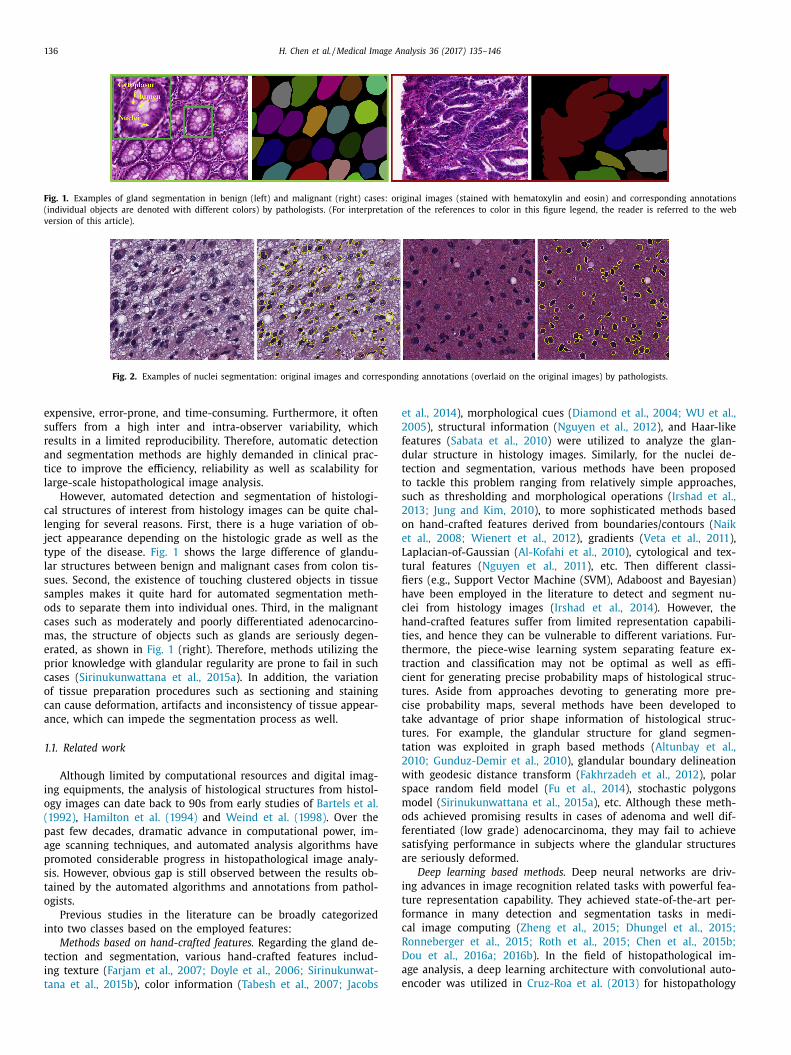
Fig. 1. Examples of gland segmentation in benign (left) and malignant (right) cases: original images (stained with hematoxylin and eosin) and corresponding annotations
(individual objects are denoted with different colors) by pathologists. (For interpretation of the references to color in this figure legend, the reader is referred to the web
version of this article).
Fig. 2. Examples of nuclei segmentation: original images and corresponding annotations (overlaid on the original images) by pathologists.
e
2
f
d
t
t
s
2
o
e
L
t
fi
h
c
h
t
t
t
c
t
c
t
t
t
2
w
s
m
o
f
s
a
i
t
f
c
R
D
a
e
expensive, error-prone, and time-consuming. Furthermore, it often
suffers from a high inter and intra-observer variability, which
results in a limited reproducibility. Therefore, automatic detection
and segmentation methods are highly demanded in clinical prac-
tice to improve the efficiency, reliability as well as scalability for
large-scale histopathological image analysis.
However, automated detection and segmentation of histologi-
cal structures of interest from histology images can be quite chal-
lenging for several reasons. First, there is a huge variation of ob-
ject appearance depending on the histologic grade as well as the
type of the disease. Fig. 1 shows the large difference of glandu-
lar structures between benign and malignant cases from colon tis-
sues. Second, the existence of touching clustered objects in tissue
samples makes it quite hard for automated segmentation meth-
ods to separate them into individual ones. Third, in the malignant
cases such as moderately and poorly differentiated adenocarcino-
mas, the structure of objects such as glands are seriously degen-
erated, as shown in Fig. 1 (right). Therefore, methods utilizing the
prior knowledge with glandular regularity are prone to fail in such
cases ( Sirinukunwattana et al., 2015a ). In addition, the variation
of tissue preparation procedures such as sectioning and staining
can cause deformation, artifacts and inconsistency of tissue appear-
ance, which can impede the segmentation process as well.
1.1. Related work
Although limited by computational resources and digital imag-
ing equipments, the analysis of histological structures from histol-
ogy images can date back to 90s from early studies of Bartels et al.
(1992) , Hamilton et al. (1994) and Weind et al. (1998) . Over the
past few decades, dramatic advance in computational power, im-
age scanning techniques, and automated analysis algorithms have
promoted considerable progress in histopathological image analy-
sis. However, obvious gap is still observed between the results ob-
tained by the automated algorithms and annotations from pathol-
ogists.
Previous studies in the literature can be broadly categorized
into two classes based on the employed features:
Methods based on hand-crafted features. Regarding the gland de-
tection and segmentation, various hand-crafted features includ-
ing texture ( Farjam et al., 2007; Doyle et al., 2006; Sirinukunwat-
tana et al., 2015b ), color information ( Tabesh et al., 2007; Jacobs
t al., 2014 ), morphological cues ( Diamond et al., 2004; WU et al.,
005 ), structural information ( Nguyen et al., 2012 ), and Haar-like
eatures ( Sabata et al., 2010 ) were utilized to analyze the glan-
ular structure in histology images. Similarly, for the nuclei de-
ection and segmentation, various methods have been proposed
o tackle this problem ranging from relatively simple approaches,
uch as thresholding and morphological operations ( Irshad et al.,
013; Jung and Kim, 2010 ), to more sophisticated methods based
n hand-crafted features derived from boundaries/contours ( Naik
t al., 2008; Wienert et al., 2012 ), gradients ( Veta et al., 2011 ),
aplacian-of-Gaussian ( Al-Kofahi et al., 2010 ), cytological and tex-
ural features ( Nguyen et al., 2011 ), etc. Then different classi-
ers (e.g., Support Vector Machine (SVM), Adaboost and Bayesian)
ave been employed in the literature to detect and segment nu-
lei from histology images ( Irshad et al., 2014 ). However, the
and-crafted features suffer from limited representation capabili-
ies, and hence they can be vulnerable to different variations. Fur-
hermore, the piece-wise learning system separating feature ex-
raction and classification may not be optimal as well as effi-
ient for generating precise probability maps of histological struc-
ures. Aside from approaches devoting to generating more pre-
ise probability maps, several methods have been developed to
ake advantage of prior shape information of histological struc-
ures. For example, the glandular structure for gland segmen-
ation was exploited in graph based methods ( Altunbay et al.,
010; Gunduz-Demir et al., 2010 ), glandular boundary delineation
ith geodesic distance transform ( Fakhrzadeh et al., 2012 ), polar
pace random field model ( Fu et al., 2014 ), stochastic polygons
odel ( Sirinukunwattana et al., 2015a ), etc. Although these meth-
ds achieved promising results in cases of adenoma and well dif-
erentiated (low grade) adenocarcinoma, they may fail to achieve
atisfying performance in subjects where the glandular structures
re seriously deformed.
Deep learning based methods. Deep neural networks are driv-
ng advances in image recognition related tasks with powerful fea-
ure representation capability. They achieved state-of-the-art per-
ormance in many detection and segmentation tasks in medi-
al image computing ( Zheng et al., 2015; Dhungel et al., 2015;
onneberger et al., 2015; Roth et al., 2015; Chen et al., 2015b;
ou et al., 2016a; 2016b ). In the field of histopathological im-
ge analysis, a deep learning architecture with convolutional auto-
ncoder was utilized in Cruz-Roa et al. (2013) for histopathology

H. Chen et al. / Medical Image Analysis 36 (2017) 135–146 137
i
p
f
i
p
t
m
r
t
i
p
a
e
a
l
a
h
w
w
o
n
i
m
o
s
t
g
a
i
c
h
p
e
m
p
i
fi
m
s
1
(
m
c
g
i
T
t
d
i
l
p
n
w
o
e
t
o
a
a
i
a
c
p
s
N
p
s
g
i
w
c
t
t
g
i
S
m
e
r
r
l
r
S
t
m
1
w
F
c
r
w
t
S
2
c
f
c
c
o
f
c
i
w
l
t
2
a
2
t
r
b
i
s
t
1 Project page: http://www.cse.cuhk.edu.hk/ ∼hchen/research/2015miccai _ gland.
html .
mage classification. Stacked sparse autoencoders (SSAE) were ex-
loited with unsupervised pre-training and following fine-tuning
or nuclei detection from breast cancer histopathology images
n Xu et al. (2015) . Although along with merit of unsupervised
re-training, which can handle the situation of limited medical
raining data, the auto-encoders usually achieved inferior perfor-
ance on image recognition tasks compared to convolutional neu-
al networks (CNNs). The success of latter one is mostly attributed
o the more elegant structures for dealing with images. Regard-
ng the convolutional neural networks, Cire ̧s an et al. (2013) em-
loyed deep convolutional neural networks for mitosis detection
nd achieved the best performance in two grand challenges ( Roux
t al., 2013; Veta et al., 2015 ). To further improve the efficiency
nd effectiveness, Chen et al. (2016a ) developed a cascaded deep
earning framework, i.e., a coarse model for retrieving candidates
nd a fine-discrimination model for singling out mitoses from
ard mimics. A spatially constrained convolutional neural net-
ork was present in Sirinukunwattana et al. (2016b ) incorporated
ith neighboring ensemble prediction, demonstrating the efficacy
f deep learning based features from CNNs. Recently, holistically-
ested networks were exploited for object segmentation problems
n the medical domain, which have demonstrated excellent perfor-
ance ( Xu et al., 2016; Roth et al., 2016; Isabella et al., 2016 ). An-
ther relevant study to our work is the U-net that employed a U-
hape deep convolutional network for biomedical image segmen-
ation problems, which set state-of-the-art performance on several
rand challenges lately ( Ronneberger et al., 2015 ). Although hier-
rchical features from low-level to high-level were well explored
n Ronneberger et al. (2015) , the very deep U-shape architecture
ould have the issue of vanishing gradients during the training,
ence decrease the segmentation performance. Moreover, the im-
licit utilization of boundaries by weighting losses among differ-
nt classes may not handle the clustered objects properly. Several
ethods have been developed for efficient boundary detection ex-
licitly ( Bertasius et al., 2015a; 2015b ). Different from above stud-
es, we formulated the object segmentation problem under a uni-
ed multi-task learning framework, which harnessed the comple-
entary object and contour information simultaneously for better
egmentation performance.
.2. Contributions
In this paper, we present a novel deep contour-aware network
DCAN) to meet the aforementioned challenges in object seg-
entation from histology images. We first propose a multi-level
ontextual fully convolutional network (FCN), which can effectively
enerate multi-scale feature representations in an end-to-end way
n order to deal with the large variations of histological structures.
he proposed FCN enables us to take an image as input and output
he probability map with the same resolution of the input image
irectly using one single forward propagation. In this regard, it
s quite efficient and has great potential to be applied to ana-
yze large-scale histology images. Furthermore, to overcome the
roblem of vanishing gradients when training such a deep neural
etwork employing limited training samples, we propose to add
eighted auxiliary classifiers to encourage the back-propagation
f gradient flow. Such an auxiliary supervision mechanism can
fficiently alleviate vanishing gradients and hence speedup the
raining process as well as enhance the discrimination capability
f the deep neural network.
More importantly, to handle the challenging cases of touching
nd clustered objects, we propose to elegantly integrate object
ppearance (e.g., textures, colors, etc.) and contour information
nto a multi-task learning framework to form a deep contour-
ware network, in which the complementary appearance and
ontour information can further improve the discriminative ca-
ability of intermediate features, and hence more accurately
eparate the touching or clustered objects into individual ones.
ote that our method does not make any assumptions on the
rior shape of histological objects; it can be applied to biopsy
amples with various histopathological structures with different
rades, including both benign and malignant cases. In addition,
n order to further mitigate the issue of insufficient training data,
hich is a common challenge for medical image analysis tasks, a
ross-domain transfer learning approach is developed to improve
he training performance. We evaluated the proposed DCAN on
wo challenging object segmentation tasks from histology images:
land segmentation and nuclei segmentation, and ranked the first
n both challenges held conjunction with MICCAI 2015 ( Gland
egmentation Challenge and Nuclei Segmentation Challenge ), yielding
uch better performance than state-of-the-art methods.
A preliminary version of this work was presented in Chen
t al. (2016c ). 1 In this paper, we provided comprehensive literature
eview on segmentation related tasks from histology images and
efined the methods by giving much more details on the under-
ying principles as well as running mechanisms. We reported the
esults of our method on another dataset of 2015 MICCAI Nuclei
egmentation Challenge. We also provided detailed discussions to
horoughly analyze the advantages as well as limitations of our
ethods.
.3. Overview
The rest of the article is organized as follows: In Section 2 ,
e first describe the architecture of the multi-level contextual
CN with auxiliary supervision, and then elaborate the proposed
ontour-aware component and the transfer learning strategy. We
eport the experiments and results in Section 3 . In Section 4 ,
e provide the implementation details and computation cost of
he proposed DCAN. We further discuss and analyze our work in
ection 5 . Finally, conclusions are drawn in Section 6 .
. Methods
In this section, we present the formulation of the proposed
ontour-aware network in detail. We first introduce the end-to-end
ully convolutional network, in which we elegantly fuse multi-level
ontextual information with auxiliary supervision in order to effi-
iently generate accurate likelihood maps of objects. Next, we elab-
rate the deep contour-aware network derived from the FCN by
using the complementary information of object appearances and
ontours for more robust and precise segmentation. In addition,
n order to mitigate the challenge of inadequate training samples,
e leverage the transfer learning strategy to exploit the knowledge
earned from cross domains to further improve the performance of
he proposed network.
.1. Multi-level contextual FCN with auxiliary supervision
Fully convolutional networks greatly advanced the state-of-the-
rt performance of image segmentation related tasks ( Chen et al.,
015d; Long et al., 2015 ). These achievements are mainly attributed
o the powerful capability of deep neural networks in feature rep-
esentations for dense inference. In a FCN, the whole network can
e trained in an end-to-end (image-to-image) way, which takes an
mage as input and outputs the probability map directly with the
ame resolution of the input image. Its architecture basically con-
ains two components: downsampling path and upsampling path.

138 H. Chen et al. / Medical Image Analysis 36 (2017) 135–146
Size (pixels)0 100 200 300 400 500 600 700 800
rebmu
N
0
50
100
150
200
250
300Benign
XY
Size (pixels)0 100 200 300 400 500 600 700 800
rebmu
N
0
20
40
60
80
100
120Malignant
XY
Fig. 3. The large variation of gland size (the size denotes the smallest rectangle that encloses the gland object).
64
128
128
Input
256256 512
max-poolconv
deconvclassifier fusion
C1 C2 C3
Softmax
643
512 512 512 1024
U1U2U3
Fig. 4. The schematic illustration of multi-level contextual FCN with auxiliary supervsion.
p
u
fi
l
o
t
m
t
a
T
a
t
d
I
s
w
i
o
p
e
F
m
r
f
c
w
a
L
The downsampling path consists of convolutional and max-pooling
layers, which are extensively used in CNNs for image classification
tasks ( Ciresan et al., 2012; Krizhevsky et al., 2012 ). The upsam-
pling path is composed of convolutional and deconvolutional layers
(backwards strided convolution Long et al., 2015 ), which upsam-
ple the feature maps and output the score masks. The underlying
principle of FCN is that the downsampling path is responsible for
extracting the high level abstraction information, while the upsam-
pling path aims to predict the score map in a pixel-wise way, i.e.,
dense inference.
However, in traditional FCN, the classification scores are usu-
ally figured out based on the intensity information from a given
receptive field (i.e., the region in the original input image that in-
fluences the recognition results LeCun et al., 1998 ). The size of re-
ceptive field should contain the targeting objects along with some
contextual information that contributes to the recognition. Unfor-
tunately, the network with single receptive field size cannot sat-
isfactorily deal with the large variation of histological structures.
For example, as shown in Fig. 3 , a small receptive field (e.g., 150
× 150) is suitable for most glands of benign cases, while malig-
nant cases usually demand a relatively larger receptive field since
the gland shape in adenocarcinomas is degenerated and elongated.
In this regard, enclosing multi-level contextual information rang-
ing from small receptive fields to larger ones can help to recognize
structures with large variations and hence improve the segmenta-
tion performance.
We propose to improve the FCN by harnessing multi-level con-
textual feature representations, which include different levels of
contextual information, i.e., intensities appearing in various sizes
of receptive field. Fig. 4 presents the schematic illustration of
FCN with multi-level contextual feature representations. Specifi-
cally, our FCN contains a number of convolutional layers, 5 max-
ooling layers for downsampling and 3 deconvolutional layers for
psampling. A non-linear mapping layer, i.e., element-wise recti-
ed linear unit (ReLU) ( Krizhevsky et al., 2012 ), is followed for each
ayer with trained parameters. As the network goes deeper, the size
f global receptive field is becoming larger. Built upon this charac-
eristic, the upsampling layers are designed by taking the require-
ent of different receptive field sizes into account. They upsample
he feature maps and make predictions based on the contextual
ppearance information extracted from the given receptive field.
hen these predictions are fused together by a summing operation
nd final segmentation results based on multi-level contextual fea-
ures can be obtained from the softmax classification layer.
Directly training such a deep network, however, may face the
ifficulty of optimization due to the issue of vanishing gradients.
nspired by previous studies on training neural networks with deep
upervision ( Lee et al., 2015; Xie and Tu, 2015; Chen et al., 2016b ),
e added three weighted auxiliary classifiers C1–C3 (see Fig. 4 for
llustration) into the network to further strengthen the training
ptimization process. This mechanism can effectively alleviate the
roblem of vanishing gradients, as the auxiliary supervision can
ncourage the back-propagation of gradient flow ( Lee et al., 2015 ).
urthermore, the incorporated losses on auxiliary supervision from
id-level to high-level layers are helpful to enhance the feature
epresentation capability throughout the network.
Finally, the FCN with multi-level contextual features extracted
rom input I can be trained by minimizing the overall loss L , i.e., a
ombination of auxiliary loss L a (I;W ) with corresponding discount
eights w a and main loss L e (I;W ) between the predicted results
nd ground truth annotations:
(I;W ) = λψ(W ) +
∑
a
w a L a (I;W ) + L e (I;W ) (1)

H. Chen et al. / Medical Image Analysis 36 (2017) 135–146 139
Fig. 5. The overview of the proposed deep contour-aware network. (For interpretation of the references to color in this figure, the reader is referred to the web version of
this article).
w
w
w
2
s
c
H
l
d
w
f
t
o
T
F
m
c
n
t
f
f
i
(
d
s
m
t
s
e
a
a
r
i
N
F
a
e
p
t
f
t
n
u
g
n
g
2
c
i
n
a
s
L
w
a
x
n
o
t
l
n
w
a
t
fi
m
here W denotes the parameters of neural network including
eights and biases; ψ( W ) is the regularization term ( L 2 norm)
ith hyperparameter λ for balancing the tradeoff with other terms.
.2. Deep contour-aware network
By leveraging the multi-level contextual features with auxiliary
upervision ( Xie and Tu, 2015; Chen et al., 2016b ), the network
an produce satisfactory probability maps of histological structures.
owever, it is still quite hard to separate the touching and over-
apped object instances by relying only on the likelihood of objects
ue to the essential ambiguity in clustered regions. This is because
hile the downsampling path can acquire high level abstraction
eatures, it leads to spatial information loss alone with the abstrac-
ion. As we know, in segmentation tasks, the boundary information
f objects provides good complementary cues for splitting objects.
o this end, we propose to integrate contour information into the
CN to form a deep contour-aware network to more accurately seg-
ent the objects in histology images, and, in particular, separate
lustered objects into individual ones.
Fig. 5 shows an overview of the proposed deep contour-aware
etwork. Instead of treating the histological structure segmenta-
ion and contour detection as single and independent tasks, we
ormulate them as a multi-task learning framework, which can in-
er the information of objects and contours simultaneously. Specif-
cally, the feature maps are upsampled with two different branches
as indicated by the green and blue arrows shown in Fig. 5 ) in or-
er to output the segmentation masks of objects and contours, re-
pectively. In each branch, the mask is predicted by the FCN with
ulti-level contextual features as introduced in Section 2.1 . During
he training process, the parameters of downsampling path W s are
hared and updated for both of these two tasks, while the param-
ters of upsampling layers for two individual branches (denoted
s W o and W c ) are updated independently for inferring the prob-
bility of objects and contours, respectively. Therefore, the feature
epresentations through the hierarchical structure can encode the
nformation of segmented objects and contours at the same time.
ote that multi-task network is still trained in an end-to-end way.
This joint multi-task learning process has several advantages.
irst, it can increase the discriminative capability of intermedi-
te feature representations with multiple regularizations on dis-
ntangling subtly correlated tasks ( Zhang et al., 2014 ), hence im-
rove the robustness of the segmentation. Second, in the applica-
ion of histological structure segmentation, the multi-task learning
ramework can provide the complementary contour information
hat serves well to further separate the clustered objects. Last but
ot least, when dealing with large-scale histopathological data, this
nified framework can be quite efficient. With one forward propa-
ation, it can generate the results of objects and contours simulta-
eously instead of resorting to additional post-separating steps by
enerating contours based on low-level cues ( Gunduz-Demir et al.,
010; WU et al., 2005 ).
In the training process, the discount weights w a from auxiliary
lassifiers are decreased until marginal values with the number of
terations increasing. Therefore, we dropped these terms in the fi-
al loss for simplicity. Finally, the training of network is formulated
s a per-pixel classification problem regarding the ground truth
egmentation masks including both objects and contours:
total (x ; θ ) = λψ(θ ) −∑
x ∈X log p o (x, y o (x ) ;W o , W s )
−∑
x ∈X log p c (x, y c (x ) ;W c , W s ) (2)
here the first part is the L 2 regularization term and latter two
re the data error loss terms defined with cross-entropy functions;
is the pixel position in image space X ; p o ( x, y o ( x ); W o , W s ) de-
otes the predicted probability for true label y o ( x ) (i.e., the index
f 1 in one-hot vector encoding) of objects after softmax classifica-
ion; and p c ( x, y c ( x ); W c , W s ) is the predicted probability for true
abel y c ( x ) of object contours. The parameters θ = { W s , W o , W c } of
etwork are optimized by minimizing the total loss function L total
ith stochastic gradient descent ( Williams and Hinton, 1986 ).
With the predicted probability maps of objects p o ( x ; W o , W s )
nd contours p c ( x ; W c , W s ) from the deep contour-aware network,
he complementary information are fused together to generate the
nal segmentation masks m ( x ):
(x ) =
{1 if p o (x ;W o , W s ) ≥ t o and p c (x ;W c , W s ) < t c
0 otherwise (3)

140 H. Chen et al. / Medical Image Analysis 36 (2017) 135–146
w
t
p
n
p
a
P
h
d
e
t
t
g
1
t
w
a
t
t
e
c
a
3
3
i
F
i
s
t
j
t
l
a
l
a
w
j
I
p
o
c
t
o
m
t
3
c
w
a
a
n
i
o
F
where t o and t c are the thresholds (both set as 0.5 in our experi-
ments empirically). Then, post-processing steps including smooth-
ing with a disk filter (the radius was set as 3), filling holes and re-
moving small spurious segments are performed on the fused seg-
mentation results. Finally, each connected component is labeled
with a unique value for representing one segmented object.
2.3. Transfer learning with rich feature hierarchies from
cross-domains
When employing deep neural networks in medical image anal-
ysis tasks, one of the main challenges is the insufficiency of the
high quality training data with accurate annotations ( Greenspan
et al., 2016 ), due to the expensive cost and complicated acquisi-
tion procedures. Compared with the limited data in medical do-
main, much more training data can be obtained in natural im-
age recognition tasks. Previous studies ( Chen et al., 2015a; Shin
et al., 2016; Tajbakhsh et al., 2016; Chen et al., 2016d ) have evi-
denced that transfer learning can alleviate the problem of insuffi-
cient training data in deep convolutional networks. The learned pa-
rameters (convolutional filters) in the lower layers of convolutional
neural networks are inherently generic, while those in higher lay-
ers are more specific to different tasks ( Yosinski et al., 2014 ). Thus,
transferring the rich feature hierarchies with embedded knowledge
learned from cross domains could help to reduce the issue of over-
fitting on limited medical dataset and further boost the perfor-
mance.
Therefore, we utilized an off-the-shelf model from DeepLab
of Chen et al. (2015d ), which was trained on the PASCAL VOC
2012 dataset ( Everingham et al., 2010 ). Compared to the small
scale dataset (a few hundred images) in the histopathological im-
age analysis, the PASCAL VOC dataset contains more than ten thou-
sand images with pixel-level annotations. Leveraging the effec-
tive generalization ability of transfer learning in deep neural net-
works, we initialized the layers in downsampling path with pre-
trained weights from the DeepLab model while the rest layers ran-
domly initialized with Gaussian distribution. Then we fine tuned
the whole network on our medical task in an end-to-end way
with stochastic gradient descent. In our experiments, we observed
the training process converged much faster (about four hours) by
virtue of the knowledge learned from rich dataset than the random
initialization setting trained from scratch.
3. Experiments and results
We evaluated the proposed method on two challenging object
segmentation tasks from histology images: gland segmentation and
nuclei segmentation. We participated two challenges held conjunc-
tion with MICCAI 2015: Gland Segmentation (GlaS) Challenge Con-
test 2 and Segmentation of Nuclei in Digital Pathology Images 3 , and
ranked the first in both of the challenges. We first introduce the
datasets employed in these two challenges and then report the ex-
perimental results in detail.
3.1. Datasets and pre-processing
Dataset of gland segmentation challenge. The histology im-
ages were acquired by a Zeiss MIRAX MIDI slide scanner from
colorectal cancer tissues with a resolution of 0.62 μm/pixel
( Sirinukunwattana et al., 2016a ). The dataset consisted of a wide
range of histologic grades from benign to malignant subjects. It is
2 2015 MICCAI gland segmentation challenge: http://www2.warwick.ac.uk/fac/sci/
dcs/research/combi/research/bic/glascontest/ . 3 2015 MICCAI nuclei segmentation challenge: http://miccai.cloudapp.net:80 0 0/
competitions/37 .
h
r
d
F
orth noting that poorly-differentiated cases were also included
o more comprehensively evaluate the performance of partici-
ated algorithms. The training data were composed of 85 (be-
ign/malignant = 37/48) images with ground truth annotations
rovided by experienced pathologists. The testing data contained
total of 80 (benign/malignant = 37/43) images for evaluation.
articipants were required to detect and segment the glands from
istology images into individual ones. The ground truths of testing
ata were held out by the challenge organizers for independent
valuation.
Dataset of nuclei segmentation challenge. The training data con-
ained more than 500 manually segmented nuclei in 15 image
iles, which were extracted from a set of Glioblastoma and lower-
rade Glioma whole slide tissue images. The testing data included
8 images for evaluation and the ground truths were held out by
he challenge organizers for independent evaluation. Participants
ere asked to detect and segment all the nuclei in the testing im-
ges and the results were compared with the ground truth anno-
ations provided by experienced pathologists.
In order to increase the robustness and reduce overfitting in
raining process, we utilized the strategy of data augmentation to
nlarge the training dataset. The augmentation transformations in-
luded translation, rotation, and elastic distortion (e.g., pincushion
nd barrel distortions).
.2. Results of the gland segmentation challenge
.2.1. Qualitative evaluation
Some typical segmentation results of the testing images, includ-
ng both benign cases and malignant cases, are shown in Fig. 6 .
or diagnosing the role of complementary contour information,
.e., the contour-aware branch network, we performed an ablation
tudy and compared the performance of the proposed DCAN and
hat of the network relying only on the prediction of gland ob-
ects without employing contour information. From the segmen-
ation results, we can observe that the FCN leveraging the multi-
evel contextual features without contour-aware component can
lso effectively segment the gland objects in both benign and ma-
ignant cases. However, compared with the results of DCAN, there
re some touching gland objects that cannot be separated very
ell. The situation is further deteriorated when the touching ob-
ects are clustered together, as shown in the third column of Fig. 6 .
n contrast, thanks to the contributions of the contour-aware com-
onent, the DCAN is capable of separating these touching gland
bjects into individual instances clearly (as the yellow arrows indi-
ate in the figures), and hence achieves more accurate segmenta-
ion results. These examples qualitatively demonstrate the superi-
rity of the DCAN obtained by exploring the complementary infor-
ation of object appearances and contours under a unified multi-
ask learning framework.
.2.2. Quantitative evaluation and comparison
The evaluation metrics employed in the grand challenge in-
lude F1 score, object-level Dice index and Hausdorff distance,
hich reflect the performance of gland detection, segmentation
nd shape similarity, respectively. We submitted two entries to
ssess the performance of the proposed methods. The first one,
amely CUMedVision1 , is the multi-level contextual FCN without
ntegrating contour-aware component, as shown in Fig. 4 . The sec-
nd one, namely CUMedVision2 , is the proposed DCAN, as shown in
ig. 5 . In order to probe the performance of submitted methods on
istology images with different histologic grades, the results are
eported on three categories of testing data: overall data, benign
ata and malignant data.
Gland detection. For the gland detection evaluation, the metric
1 score is utilized, which is the harmonic mean of precision P and

H. Chen et al. / Medical Image Analysis 36 (2017) 135–146 141
Fig. 6. Segmentation results of benign cases (left two columns) and malignant cases (right two columns). From top to bottom shows the original images, segmentation
results of our network without integrating contour information, and segmentation results of our contour-aware network, respectively. Different colors denote individual
gland objects. (For interpretation of the references to color in this figure legend, the reader is referred to the web version of this article).
Table 1
The detection results (F1 score) of different methods in 2015 MICCAI GlaS Challenge
(only top 10 entries are shown here).
Method Overall Benign Malignant
CUMedVision1 0 .856 0 .872 0 .788
CUMedVision2 0 .887 0 .919 0 .771
CVML 0 .637 0 .691 0 .439
ExB1 0 .868 0 .915 0 .698
ExB2 0 .868 0 .920 0 .686
ExB3 0 .875 0 .919 0 .714
Freiburg2 ( Ronneberger et al., 2015 ) 0 .849 0 .916 0 .616
Freiburg1 ( Ronneberger et al., 2015 ) 0 .804 0 .893 0 .521
LIB 0 .704 0 .800 0 .388
vision4GlaS ( Kainz et al., 2015 ) 0 .620 0 .651 0 .478
r
F
w
p
l
t
w
t
t
t
h
m
i
t
o
c
m
t
s
g
2
s
m
l
a
p
i
D
s
i
n
c
e
m
i
a
m
d
i
t
n
f
n
t
t
m
D
d
w
g
o
w
i
v
t
D
4 https://en.wikipedia.org/wiki/Ranking .
ecall R , defined as
1 =
2 P R
P + R
, P =
N t p
N t p + N f p
, R =
N t p
N t p + N f n
(4)
here N tp , N fp , and N fn denote the number of true positives, false
ositives, and false negatives, respectively. According to the chal-
enge evaluation, the ground truth for each segmented object is
he object in the manual annotation that has maximum overlap
ith that segmented object. A segmented gland object that in-
ersects with at least 50% of its ground truth is considered as a
rue positive, otherwise it’s considered as a false positive. A ground
ruth gland object that has no corresponding segmented object or
as less than 50% of its area overlapped by its corresponding seg-
ented object is considered as a false negative.
Table 1 shows the detection results of various methods partic-
pated in the challenge. We just listed the top 10 entries in the
able. Note that all the top 5 entries utilized CNN-based meth-
ds. For examples, the team ExB utilized a two-path network by
ropping global and local patches from the image and the seg-
entation results were generated by fusing these features from
hese two branches. The team Freiburg designed a very deep U-
haped network and has achieved the best results in several other
rand detection and segmentation challenges ( Ronneberger et al.,
015 ). Other teams, such as LIB and SUTECH , utilized hand-crafted
tructural and textural features. However, the performance of these
ethods is much worse than deep learning based methods, as il-
ustrated in Table 1 .
Our submitted entry CUMedVision2 with the proposed DCAN
chieved the best results on overall testing data, and competitive
erformance (ours vs. the first = 0.919 vs. 0.920) on benign test-
ng data, which demonstrated the effectiveness of the proposed
CAN in gland detection. Meanwhile, the other entry CUMedVi-
ion1 employing the multi-level contextual FCN without integrat-
ng contour-aware component achieved the best results on malig-
ant testing data, highlighting the advantages of the multi-level
ontextual FCN with auxiliary supervision. From Table 1 , read-
rs can notice that all methods yielded a relatively lower perfor-
ance on the malignant testing data than the benign data. This
s mainly due to the difficulties incurred by the seriously degener-
ted structures in malignant subjects. Nevertheless, our two sub-
itted entries ranked first and second on the malignant testing
ata with an obvious advantage over other methods, demonstrat-
ng the multi-level contextual features learned from our architec-
ure can more efficiently deal with the large variations of malig-
ant cases. The CUMedVision2 achieved lower but comparable per-
ormance (0.771 vs. 0.788) compared to CUMedVision1 on malig-
ant testing data. By carefully investigating the results, especially
hose of malignant cases, we observed that some inaccurate con-
ours in interior structures may cause the deformed glands frag-
ented in some seriously degenerated cases. Overall, the proposed
CAN achieved the best performance on the evaluation of gland
etection according to the standard competitive ranking, 4 which
as employed by the challenge organizers.
Gland segmentation. Given a set of pixels G annotated as a
round truth object and a set of pixels S as a segmented gland
bject, Dice index is often employed for segmentation evaluation,
hich is defined as D (G, S) = 2(| G ∩ S| ) / ( | G | + | S| ) . However, Dice
ndex is not suitable for evaluation of segmentation results of indi-
idual objects. To the end, an object-level Dice index is utilized in
he challenge. It is defined as
object (G, S) =
1
2
[
n S ∑
i =1
ω i D (G i , S i ) +
n G ∑
j=1
˜ ω j D ( ̃ G j , ̃ S j )
]
(5)

142 H. Chen et al. / Medical Image Analysis 36 (2017) 135–146
Table 2
The segmentation results (object-level Dice index) of different methods in 2015
MICCAI GlaS Challenge.
Method Overall Benign Malignant
CUMedVision1 0 .850 0 .873 0 .826
CUMedVision2 0 .868 0 .907 0 .827
CVML 0 .647 0 .663 0 .630
ExB1 0 .858 0 .892 0 .823
ExB2 0 .851 0 .902 0 .799
ExB3 0 .855 0 .900 0 .810
Freiburg2 ( Ronneberger et al., 2015 ) 0 .853 0 .903 0 .801
Freiburg1 ( Ronneberger et al., 2015 ) 0 .852 0 .903 0 .798
LIB 0 .744 0 .817 0 .668
vision4GlaS ( Kainz et al., 2015 ) 0 .705 0 .738 0 .673
Table 3
The shape similarity results (Hausdorff distance) of different methods in 2015 MIC-
CAI GlaS Challenge.
Method Overall Benign Malignant
CUMedVision1 94 .515 67 .862 122 .187
CUMedVision2 74 .731 43 .025 106 .979
CVML 160 .506 128 .250 192 .555
ExB1 79 .761 53 .979 105 .986
ExB2 89 .180 49 .330 129 .422
ExB3 83 .483 52 .617 114 .722
Freiburg2 ( Ronneberger et al., 2015 ) 79 .842 49 .115 111 .959
Freiburg1 ( Ronneberger et al., 2015 ) 79 .326 49 .118 111 .037
LIB 130 .345 78 .436 184 .510
vision4GlaS ( Kainz et al., 2015 ) 133 .158 102 .277 164 .005
w
d
o
l
r
o
t
b
a
t
o
t
r
r
p
e
b
p
r
p
t
3
3
a
g
m
r
s
t
i
D
a
t
d
c
a
d
3
e
c
o
t
m
m
where S i denotes the i th segmented object, G i denotes a ground
truth object that maximally overlaps S i , ˜ G j denotes the j th ground
truth object, ˜ S j denotes a segmented object that maximally over-
laps ˜ G j , ω i = | S i | / ∑ n S m =1
| S m
| , ˜ ω j = | ̃ G j | / ∑ n G n =1
| ̃ G n | , n S and n G are
the total number of segmented objects and ground truth objects,
respectively.
Table 2 reports the segmentation results of various methods
participating in the challenge according to the object-level Dice in-
dex. It is observed that our results of CUMedVision2 achieved the
best performance on all three categories of testing data, outper-
forming all the other methods.
Shape similarity. The shape similarity is measured by using the
Hausdorff distance between the shape of segmented object and
that of the ground truth object. It is defined as
H(G, S) = max
{sup
x ∈ G inf y ∈ S
‖ x − y ‖ , sup
y ∈ S inf x ∈ G
‖ x − y ‖
}(6)
Likewise, an object-level Hausdorff is defined for the challenge
H object (G, S) =
1
2
[
n S ∑
i =1
ω i H(G i , S i ) +
n G ∑
j=1
˜ ω j H( ̃ G j , ̃ S j )
]
(7)
The shape similarity results of different methods are shown
in Table 3 . In comparison to the CUMedVision1 , the CUMedVision2
Table 4
The final ranking of participants in 2015 MICCAI GlaS Ch
Method Ranking score
Overall Ben
CUMedVision2 1 1
ExB1 2 6
Freiburg2 ( Ronneberger et al., 2015 ) 5 2
ExB3 3 5
Freiburg1 ( Ronneberger et al., 2015 ) 4 4
ExB2 6 3
CUMedVision1 7 7
LIB 8 8
vision4GlaS ( Kainz et al., 2015 ) 9 9
CVML 10 10
ith contour-aware branch can dramatically reduce the Hausdorff
istances (around 20, 25, and 15 pixels decrement for category of
verall, benign and malignant testing data, respectively). This high-
ights the superiority of contour-aware branch in DCAN for sepa-
ating touching glands into individual object instances. Our results
f CUMedVision2 achieved the smallest Hausdorff distances on both
he overall and benign testing data, outperforming other methods
y a significant margin. Meanwhile, the results of CUMedVision2
re comparable to the best results achieved by ExB1 on malignant
esting data (106.979 vs. 105.986 as shown in Table 3 ).
Final ranking. For the final ranking, each team is assigned
ne ranking number for each category of testing data based on
he three metrics mentioned above using a standard competition
anking. 4 The sum score of these numbers is used for the final
anking, i.e., a smaller score stands for better overall segmentation
erformance. The final ranking is reported in Table 4 (only top 10
ntries are shown). Our deep contour-aware network yielded the
est performance in terms of whole results out of 13 teams, out-
erforming all the other methods by a significant margin. This cor-
oborates the superiority of our method by harnessing object ap-
earance and contour information explicitly under a unified multi-
ask learning framework.
.3. Results of the nuclei segmentation challenge
.3.1. Qualitative evaluation
Some typical nuclei segmentation results of the testing images
re shown in Fig. 7 . It is observed that both the FCN without inte-
rating contour information and the DCAN can effectively segment
ost nuclei from the testing images. However, compared with the
esults of DCAN, there are some touching nuclei that cannot be
eparated into individual ones in the results of FCN without con-
our information, as indicated by the yellow arrows overlaid in the
mages. By integrating the object and contour prediction maps, the
CAN clearly separated the touching nuclei into individual nucleus,
nd achieved more precise segmentation results.
We also carefully studied the errors in the results. We found
hat the errors were mostly observed in following cases: high-
egree of clustering resulting in under-segmentation, irregular nu-
lei, noisy background and poor edge information. Although there
re still some errors in our results, our method can achieve good
etection and segmentation performance on most testing images.
.3.2. Quantitative evaluation and comparison
The nuclei segmentation challenge employed two metrics for
valuation: traditional Dice coefficient ( D 1 ) and object-level Dice
oefficient ( D 2 ). The D 1 metric was applied to measure the amount
f overlap between the results of algorithms and human annota-
ions in terms of the nuclei regions that was detected and seg-
ented. D 1 does not take into account the cases of split and
erge. A split is the case in which the human segments a region
allenge (top 10 entries are shown here).
Sum score Final ranking
ign Malignant
1 3 1
2 10 2
5 12 3
4 12 3
6 14 5
7 16 6
3 17 7
9 25 8
8 26 9
10 30 10
H. Chen et al. / Medical Image Analysis 36 (2017) 135–146 143
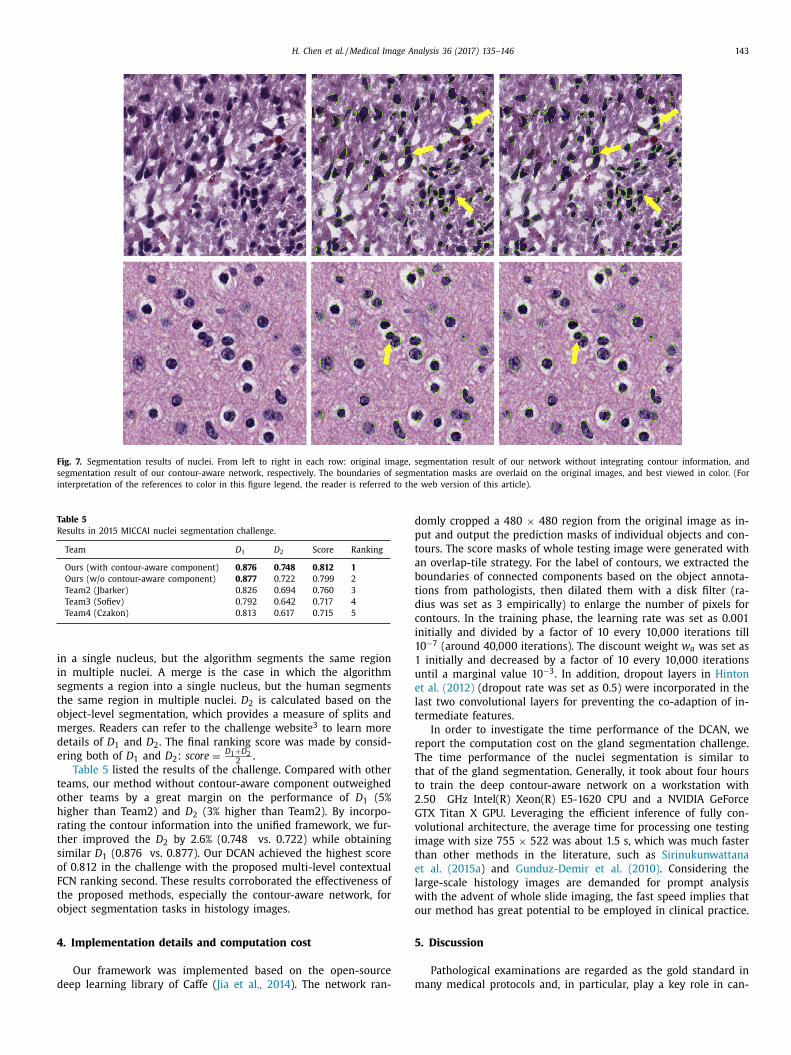
Fig. 7. Segmentation results of nuclei. From left to right in each row: original image, segmentation result of our network without integrating contour information, and
segmentation result of our contour-aware network, respectively. The boundaries of segmentation masks are overlaid on the original images, and best viewed in color. (For
interpretation of the references to color in this figure legend, the reader is referred to the web version of this article).
Table 5
Results in 2015 MICCAI nuclei segmentation challenge.
Team D 1 D 2 Score Ranking
Ours (with contour-aware component) 0 .876 0 .748 0 .812 1
Ours (w/o contour-aware component) 0 .877 0 .722 0 .799 2
Team2 (Jbarker) 0 .826 0 .694 0 .760 3
Team3 (Sofiev) 0 .792 0 .642 0 .717 4
Team4 (Czakon) 0 .813 0 .617 0 .715 5
i
i
s
t
o
m
d
e
t
o
h
r
t
s
o
F
t
o
4
d
d
p
t
a
b
t
d
c
i
1
1
u
e
l
t
r
T
t
t
2
G
v
i
t
e
l
w
o
5
m
n a single nucleus, but the algorithm segments the same region
n multiple nuclei. A merge is the case in which the algorithm
egments a region into a single nucleus, but the human segments
he same region in multiple nuclei. D 2 is calculated based on the
bject-level segmentation, which provides a measure of splits and
erges. Readers can refer to the challenge website 3 to learn more
etails of D 1 and D 2 . The final ranking score was made by consid-
ring both of D 1 and D 2 : score =
D 1 + D 2 2 .
Table 5 listed the results of the challenge. Compared with other
eams, our method without contour-aware component outweighed
ther teams by a great margin on the performance of D 1 (5%
igher than Team2) and D 2 (3% higher than Team2). By incorpo-
ating the contour information into the unified framework, we fur-
her improved the D 2 by 2.6% (0.748 vs. 0.722) while obtaining
imilar D 1 (0.876 vs. 0.877). Our DCAN achieved the highest score
f 0.812 in the challenge with the proposed multi-level contextual
CN ranking second. These results corroborated the effectiveness of
he proposed methods, especially the contour-aware network, for
bject segmentation tasks in histology images.
. Implementation details and computation cost
Our framework was implemented based on the open-source
eep learning library of Caffe ( Jia et al., 2014 ). The network ran-
omly cropped a 480 × 480 region from the original image as in-
ut and output the prediction masks of individual objects and con-
ours. The score masks of whole testing image were generated with
n overlap-tile strategy. For the label of contours, we extracted the
oundaries of connected components based on the object annota-
ions from pathologists, then dilated them with a disk filter (ra-
ius was set as 3 empirically) to enlarge the number of pixels for
ontours. In the training phase, the learning rate was set as 0.001
nitially and divided by a factor of 10 every 10,0 0 0 iterations till
0 −7 (around 40,0 0 0 iterations). The discount weight w a was set as
initially and decreased by a factor of 10 every 10,0 0 0 iterations
ntil a marginal value 10 −3 . In addition, dropout layers in Hinton
t al. (2012) (dropout rate was set as 0.5) were incorporated in the
ast two convolutional layers for preventing the co-adaption of in-
ermediate features.
In order to investigate the time performance of the DCAN, we
eport the computation cost on the gland segmentation challenge.
he time performance of the nuclei segmentation is similar to
hat of the gland segmentation. Generally, it took about four hours
o train the deep contour-aware network on a workstation with
.50 GHz Intel(R) Xeon(R) E5-1620 CPU and a NVIDIA GeForce
TX Titan X GPU. Leveraging the efficient inference of fully con-
olutional architecture, the average time for processing one testing
mage with size 755 × 522 was about 1.5 s, which was much faster
han other methods in the literature, such as Sirinukunwattana
t al. (2015a ) and Gunduz-Demir et al. (2010) . Considering the
arge-scale histology images are demanded for prompt analysis
ith the advent of whole slide imaging, the fast speed implies that
ur method has great potential to be employed in clinical practice.
. Discussion
Pathological examinations are regarded as the gold standard in
any medical protocols and, in particular, play a key role in can-

144 H. Chen et al. / Medical Image Analysis 36 (2017) 135–146
t
f
t
r
m
a
t
a
i
a
j
q
a
e
s
s
w
o
o
6
m
h
l
p
(
w
E
t
f
g
a
y
h
A
c
n
F
f
R
A
B
B
B
C
C
cer diagnosis process ( Gurcan et al., 2009 ). Aiming at improving
the efficiency and robustness of automated histopathological image
analysis, we present a novel deep contour-aware network. Exten-
sive experiments conducted on two challenging histopathological
object detection and segmentation tasks demonstrate the effective-
ness of the deep contour-aware network as well as the contribu-
tions of its components. It is worthwhile to note that the proposed
CNN-based method does not employ any prior on object shape
along with the cancer status of histopathological images. Therefore,
it is general enough to be applied to various objects and cancerous
tissues of different histologic grades. This is evidenced by the suc-
cesses of the proposed method on two different object segmenta-
tion tasks with both benign and malignance cases.
It is observed from the detection results on GlaS Challenge that
our method without the contour-aware component achieves a lit-
tle better detection performance than that with the contour-aware
component on malignant testing data. Through a careful study, we
find it probably arises from the fact that irregular structures in
some seriously degenerated malignant cases may reduce the effect
of contour information. For example, the interior structures with
dense nuclei as a result of high proliferation may lead to false
contours, and hence the method more likely fails in such cases.
Nevertheless, on the detection evaluation of overall test data as
well as benign data, the results of CUMedVision2 were much better
than those of CUMedVision1 , which validated the effectiveness of
introducing contour-aware branch. In addition, the CUMedVision2
achieved better performance than the CUMedVision1 in terms of all
metrics (including Dice index and Hausdorff distance) in the seg-
mentation task. These results clearly highlighted the superiority of
our unified framework by incorporating contour-aware branch. In
general, as the histological structures of glands are seriously de-
generated in some malignant cases, the segmentation task can be
much more challenging. We will further investigate this as part of
our future work with more histology images.
The detection and segmentation of clustered object instances
are crucially important and have a wide range of applica-
tions, such as gland segmentation for colon cancer diagno-
sis ( Sirinukunwattana et al., 2015a ), nuclei segmentation for can-
cer grading ( Irshad et al., 2014 ), and cell segmentation and track-
ing for understanding cellular mechanisms ( Meijering, 2012 ), etc.
Previous methods such as U-net in Ronneberger et al. (2015) and
SDS in Hariharan et al. (2014) focusing only on appearance infor-
mation may not perform well in the situation where many clus-
tered object instances exist. Unfortunately, this is a common phe-
nomenon in histology images. It is worth noting that contour de-
tection and object segmentation are two complementary tasks and
previous studies showed preliminarily promising results by explor-
ing semantic contours on PASCAL dataset ( Bertasius et al., 2015a;
Chen et al., 2015c ). In this paper, we propose a more efficient and
effective solution by harnessing the information of object appear-
ances and contours simultaneously. Furthermore, the efficiency is
greatly boosted with our unified framework within one forward
propagation (about 1.5 s), which was much faster than state-of-
the-art methods (for instance, 200+ s in Sirinukunwattana et al.,
2015a ).
The separation of clustered histological structures (e.g., nuclei)
in high-level density is a long-standing problem, which usually re-
quires to incorporate domain-specific shape prior. While different
methods have been developed to segment clustered or overlapping
nuclei with great improvements, this problem has not been com-
pletely solved yet. Some high-degree clustered nuclei can be fur-
ther separated with other advanced post-separating steps by in-
corporating specific shape prior of nuclei, such as maker-controlled
watershed algorithm in Cheng and Rajapakse (2009) and spatially
adaptive active physical model in Plissiti and Nikou (2012) . This
topic is out of scope of this paper since our main aim is to segment
ouching objects and provide initially good segmentation results
or high-degree clustered cases. As Al-Kofahi et al. (2010) indicated
hat the segmentation performance depends crucially on the accu-
acy and reliability of the initial seed points or shape markers, our
ethod can provide such good probability maps, which can serve
s ‘seeds’ or ‘markers’ in the subsequent algorithms for delineating
he spatial extent of each nucleus.
Another notoriously difficult issue in histopathological image
nalysis rests with the large variation of tissue appearance, which
s regarded as one of the major obstacles for developing a robust
utomated analysis tool ( Veta et al., 2015 ). The variation is sub-
ected to several factors, including different scanners for image ac-
uisition, different sectioning and staining operations, etc, which
re quite common when tissue samples are acquired from differ-
nt patients or at different time slots. Pre-processing normalization
teps ( Khan et al., 2014 ) can potentially address the inconsistent is-
ue of tissue appearance and further improve the performance.
Currently, our method has been evaluated on two applications
ith hundreds of histology images. In the future, we shall assess it
n more large-scale histopathological datasets acquired from vari-
us scanners under different conditions.
. Conclusions
We present a deep contour-aware network that integrates
ulti-level contextual features to accurately detect and segment
istological objects from histology images. We formulate this chal-
enging segmentation problem as a unified multi-task learning
rocess by harnessing the complementary appearance information
such as textures and colors) and contour information explicitly,
hich further boost the object instance segmentation performance.
xtensive experimental results on two challenging object segmen-
ation tasks from histology images demonstrate the superior per-
ormance of our method, surpassing state-of-the-art methods by a
reat margin. The proposed DCAN is inherently general and can be
pplied to other similar problems in histopathological image anal-
sis. Future investigations include evaluating our method on more
istology images and promoting its applications in clinical practice.
cknowledgments
This work is supported by Hong Kong Research Grants Coun-
il General Research Fund (Project no. GRF 14203115 and Project
o. CUHK 14202514) and a grant from the National Natural Science
oundation of China (Project no. 61233012 ). The authors also grate-
ully thank the challenge organizers for helping the evaluation.
eferences
l-Kofahi, Y. , Lassoued, W. , Lee, W. , Roysam, B. , 2010. Improved automatic detectionand segmentation of cell nuclei in histopathology images. IEEE Trans. Biomed.
Eng. 57 (4), 841–852 . Altunbay, D. , Cigir, C. , Sokmensuer, C. , Gunduz-Demir, C. , 2010. Color graphs for au-
tomated cancer diagnosis and grading. IEEE Trans. Biomed. Eng. 57 (3), 665–674 .artels, P. , Thompson, D. , Bibbo, M. , Weber, J. , 1992. Bayesian belief networks in
quantitative histopathology. Anal. Quant. Cytol. Histol./Int. Acad. Cytol. Am. Soc.
Cytol. 14 (6), 459–473 . ertasius, G. , Shi, J. , Torresani, L. , 2015a. High-for-low and low-for-high: efficient
boundary detection from deep object features and its applications to high-levelvision. In: Proceedings of the IEEE International Conference on Computer Vision,
pp. 504–512 . ertasius, G., Shi, J., Torresani, L., 2015. Semantic segmentation with boundary neu-
ral fields. CoRR . arXiv preprint arXiv: 1511.02674 hen, H. , Dou, Q. , Ni, D. , Cheng, J.-Z. , Qin, J. , Li, S. , Heng, P.-A. , 2015a. Automatic
fetal ultrasound standard plane detection using knowledge transferred recurrent
neural networks. In: Proceedings of Medical Image Computing and ComputerAssisted Intervention, MICCAI. Springer, pp. 507–514 .
hen, H. , Dou, Q. , Wang, X. , Qin, J. , Heng, P.A. , 2016a. Mitosis detection in breastcancer histology images via deep cascaded networks. In: Proceedings of thirti-
eth AAAI Conference on Artificial Intelligence .

H. Chen et al. / Medical Image Analysis 36 (2017) 135–146 145
C
C
C
C
C
C
C
C
C
C
D
D
D
D
D
D
E
E
F
F
F
F
G
G
G
G
H
H
H
I
I
I
J
J
J
K
K
K
L
L
L
M
N
N
N
P
R
R
R
R
S
S
S
S
S
hen, H. , Qi, X. , Cheng, J.-Z. , Heng, P.-A. , 2016b. Deep contextual networks for neu-ronal structure segmentation. In: Proceedings of thirtieth AAAI Conference on
Artificial Intelligence . hen, H. , Qi, X. , Yu, L. , Heng, P.-A. , 2016c. Dcan: deep contour-aware networks for
accurate gland segmentation. In: Proceedings of Computer Vision and PatternRecognition, CVPR .
hen, H. , Shen, C. , Qin, J. , Ni, D. , Shi, L. , Cheng, J.C. , Heng, P.-A. , 2015b. Automatic lo-calization and identification of vertebrae in spine CT via a joint learning model
with deep neural networks. In: Proceedings of Medical Image Computing and
Computer Assisted Intervention, MICCAI. Springer, pp. 515–522 . hen, H. , Zheng, Y. , Park, J.-H. , Heng, P.-A. , Zhou, S.K. , 2016d. Iterative multi-domain
regularized deep learning for anatomical structure detection and segmentationfrom ultrasound images. In: Proceedings of International Conference on Medical
Image Computing and Computer-Assisted Intervention. Springer, pp. 4 87–4 95 . hen, L.-C., Barron, J.T., Papandreou, G., Murphy, K., Yuille, A.L., 2015c. Semantic im-
age segmentation with task-specific edge detection using cnns and a discrimi-
natively trained domain transform. CoRR arXiv preprint arXiv: 1511.03328 . hen, L.-C. , Papandreou, G. , Kokkinos, I. , Murphy, K. , Yuille, A.L. , 2015d. Semantic
image segmentation with deep convolutional nets and fully connected CRFs. In:Proceedings of International Conference on Learning Representations, ICLR .
heng, J. , Rajapakse, J.C. , 2009. Segmentation of clustered nuclei with shape markersand marking function. IEEE Trans. Biomed. Eng. 56 (3), 741–748 .
iresan, D. , Giusti, A. , Gambardella, L.M. , Schmidhuber, J. , 2012. Deep neural net-
works segment neuronal membranes in electron microscopy images. In: Pro-ceedings of Neural Information Processing Systems, NIPS, pp. 2843–2851 .
ire ̧s an, D.C. , Giusti, A. , Gambardella, L.M. , Schmidhuber, J. , 2013. Mitosis detectionin breast cancer histology images with deep neural networks. In: Proceedings
of Medical Image Computing and Computer-Assisted Intervention, MICCAI 2013.Springer, pp. 411–418 .
ruz-Roa, A .A . , Ovalle, J.E.A . , Madabhushi, A . , Osorio, F.A .G. , 2013. A deep learn-
ing architecture for image representation, visual interpretability and automatedbasal-cell carcinoma cancer detection. In: Proceedings of Medical Image Com-
puting and Computer-Assisted Intervention, MICCAI 2013. Springer, pp. 403–410 .hungel, N. , Carneiro, G. , Bradley, A.P. , 2015. Deep learning and structured predic-
tion for the segmentation of mass in mammograms. In: Proceedings of Med-ical Image Computing and Computer Assisted Intervention, MICCAI. Springer,
pp. 605–612 .
iamond, J. , Anderson, N.H. , Bartels, P.H. , Montironi, R. , Hamilton, P.W. , 2004. Theuse of morphological characteristics and texture analysis in the identification of
tissue composition in prostatic neoplasia. Hum. Pathol. 35 (9), 1121–1131 . ou, Q. , Chen, H. , Jin, Y. , Yu, L. , Qin, J. , Heng, P.-A. , 2016a. 3d deeply supervised
network for automatic liver segmentation from ct volumes. In: Proceedings ofInternational Conference on Medical Image Computing and Computer-Assisted
Intervention. Springer, pp. 149–157 .
ou, Q. , Chen, H. , Lequan, Y. , Zhao, L. , Qin, J. , Defeng, W. , Vincent, M. , Shi, L. ,Heng, P.A. , 2016b. Automatic detection of cerebral microbleeds from MR im-
ages via 3D convolutional neural networks. IEEE Trans. Med. Imaging 35 (5),1182–1195 .
oyle, S. , Madabhushi, A. , Feldman, M. , Tomaszeweski, J. , 2006. A boosting cascadefor automated detection of prostate cancer from digitized histology. In: Proceed-
ings of Medical Image Computing and Computer Assisted Intervention, MICCAI.Springer, pp. 504–511 .
unne, B. , Going, J. , 2001. Scoring nuclear pleomorphism in breast cancer.
Histopathology 39 (3), 259–265 . lston, C.W. , Ellis, I.O. , et al. , 1991. Pathological prognostic factors in breast cancer. I.
The value of histological grade in breast cancer: experience from a large studywith long-term follow-up. Histopathology 19 (5), 403–410 .
veringham, M. , Van Gool, L. , Williams, C.K. , Winn, J. , Zisserman, A. , 2010. The pas-cal visual object classes (voc) challenge. Int. J. Comput. Vis. 88 (2), 303–338 .
akhrzadeh, A. , Sporndly-Nees, E. , Holm, L. , Hendriks, C.L.L. , 2012. Analyzing tubular
tissue in histopathological thin sections. In: Proceedings of 2012 InternationalConference on Digital Image Computing Techniques and Applications, DICTA.
IEEE, pp. 1–6 . arjam, R. , Soltanian-Zadeh, H. , Jafari-Khouzani, K. , Zoroofi, R.A. , 2007. An image
analysis approach for automatic malignancy determination of prostate patho-logical images. Cytometry Part B: Clin. Cytom. 72 (4), 227–240 .
leming, M. , Ravula, S. , Tatishchev, S.F. , Wang, H.L. , 2012. Colorectal carcinoma:
pathologic aspects. J. Gastrointest. Oncol. 3 (3), 153–173 . u, H. , Qiu, G. , Shu, J. , Ilyas, M. , 2014. A novel polar space random field model for
the detection of glandular structures. IEEE Trans. Med. Imaging 33 (3), 764–776 .leason, D.F. , 1992. Histologic grading of prostate cancer: a perspective. Hum.
Pathol. 23 (3), 273–279 . reenspan, H. , van Ginneken, B. , Summers, R.M. , 2016. Guest editorial deep learning
in medical imaging: overview and future promise of an exciting new technique.
IEEE Trans. Med. Imaging 35 (5), 1153–1159 . unduz-Demir, C. , Kandemir, M. , Tosun, A.B. , Sokmensuer, C. , 2010. Automatic seg-
mentation of colon glands using object-graphs. Med. Image Anal. 14 (1), 1–12 . urcan, M.N. , Boucheron, L.E. , Can, A. , Madabhushi, A. , Rajpoot, N.M. , Yener, B. ,
2009. Histopathological image analysis: a review. IEEE Rev. Biomed. Eng. 2,147–171 .
amilton, P. , Anderson, N. , Bartels, P. , Thompson, D. , 1994. Expert system support
using bayesian belief networks in the diagnosis of fine needle aspiration biopsyspecimens of the breast. J. Clin. Pathol. 47 (4), 329–336 .
ariharan, B. , Arbeláez, P. , Girshick, R. , Malik, J. , 2014. Simultaneous detection andsegmentation. In: Proceedings of European Conference on Computer Vision,
ECCV. Springer, pp. 297–312 .
inton, G.E., Srivastava, N., Krizhevsky, A., Sutskever, I., Salakhutdinov, R.R., 2012.Improving neural networks by preventing co-adaptation of feature detectors.
CoRR arXiv preprint arXiv: 1207.0580 . rshad, H. , Veillard, A. , Roux, L. , Racoceanu, D. , 2014. Methods for nuclei detection,
segmentation, and classification in digital histopathology: a reviewcurrent sta-tus and future potential. IEEE Rev. Biomed. Eng. 7, 97–114 .
rshad, H. , et al. , 2013. Automated mitosis detection in histopathology using mor-phological and multi-channel statistics features. J. Pathol. Inform. 4 (1), 10 .
sabella, N. , Lu, L. , Xiaosong, W. , Roth, H.R. , Nathan, L. , Jianbo, S. , Y., T. , Sum-
mers, R.M. , 2016. Automatic lymph node cluster segmentation using holistical-ly-nested networks and structured optimization. In: Proceedings of Medical Im-
age Computing and Computer-Assisted Intervention, MICCAI 2016 . acobs, J.G. , Panagiotaki, E. , Alexander, D.C. , 2014. Gleason grading of prostate tu-
mours with max-margin conditional random fields. In: Machine Learning inMedical Imaging. Springer, pp. 85–92 .
ia, Y. , Shelhamer, E. , Donahue, J. , Karayev, S. , Long, J. , Girshick, R. , Guadarrama, S. ,
Darrell, T. , 2014. Caffe: convolutional architecture for fast feature embedding.Proceedings of the 22nd ACM international conference on Multimedia. ACM,
pp. 675–678 . ung, C. , Kim, C. , 2010. Segmenting clustered nuclei using h-minima transfor-
m-based marker extraction and contour parameterization. IEEE Trans. Biomed.Eng. 57 (10), 2600–2604 .
ainz, P., Pfeiffer, M., Urschler, M., 2015. Semantic segmentation of colon glands
with deep convolutional neural networks and total variation segmentation.CoRR arXiv preprint arXiv: 1511.06919 .
han, A.M. , Rajpoot, N. , Treanor, D. , Magee, D. , 2014. A nonlinear mapping ap-proach to stain normalization in digital histopathology images using image-spe-
cific color deconvolution. IEEE Trans. Biomed. Eng. 61 (6), 1729–1738 . rizhevsky, A. , Sutskever, I. , Hinton, G.E. , 2012. Imagenet classification with deep
convolutional neural networks. In: Proceedings of Neural Information Processing
Systems, NIPS, pp. 1097–1105 . eCun, Y. , Bottou, L. , Bengio, Y. , Haffner, P. , 1998. Gradient-based learning applied to
document recognition. Proc. IEEE 86 (11), 2278–2324 . ee, C. , Xie, S. , Gallagher, P. , Zhang, Z. , Tu, Z. , 2015. Deeply-supervised nets. In: Pro-
ceedings of International Conference on Artificial Intelligence and Statistics, AIS-TATS .
ong, J. , Shelhamer, E. , Darrell, T. , 2015. Fully convolutional networks for seman-
tic segmentation. In: Proceedings of Computer Vision and Pattern Recognition,CVPR, pp. 3431–3440 .
eijering, E. , 2012. Cell segmentation: 50 years down the road [life sciences]. IEEESignal Process. Mag. 29 (5), 140–145 .
aik, S. , Doyle, S. , Agner, S. , Madabhushi, A. , Feldman, M. , Tomaszewski, J. , 2008.Automated gland and nuclei segmentation for grading of prostate and breast
cancer histopathology. In: Proceedings of 5th IEEE International Symposium on
Biomedical Imaging. IEEE, pp. 284–287 . guyen, K. , Jain, A.K. , Sabata, B. , et al. , 2011. Prostate cancer detection: fusion of
cytological and textural features. J. Pathol. Inform. 2 (2), 3 . guyen, K. , Sarkar, A . , Jain, A .K. , 2012. Structure and context in prostatic gland seg-
mentation and classification. In: Proceedings of Medical Image Computing andComputer Assisted Intervention, MICCAI. Springer, pp. 115–123 .
lissiti, M.E. , Nikou, C. , 2012. Overlapping cell nuclei segmentation using aspatially adaptive active physical model. IEEE Trans. Image Process. 21 (11),
4568–4580 .
onneberger, O. , Fischer, P. , Brox, T. , 2015. U-net: convolutional networks forbiomedical image segmentation. In: Proceedings of Medical Image Computing
and Computer Assisted Intervention, MICCAI. Springer, pp. 234–241 . oth, H.R. , Lu, L. , Farag, A. , Shin, H.-C. , Liu, J. , Turkbey, E.B. , Summers, R.M. , 2015.
Deeporgan: multi-level deep convolutional networks for automated pancreassegmentation. In: Proceedings of Medical Image Computing and Computer As-
sisted Intervention, MICCAI. Springer, pp. 556–564 .
oth, H.R. , Lu, L. , Farag, A. , Sohn, A. , Summers, R.M. , 2016. Spatial aggregation ofholistically-nested networks for automated pancreas segmentation. In: Proceed-
ings of Medical Image Computing and Computer-Assisted Intervention, MICCAI2016 .
oux, L. , Racoceanu, D. , Loménie, N. , Kulikova, M. , Irshad, H. , Klossa, J. , Capron, F. ,Genestie, C. , Le Naour, G. , Gurcan, M.N. , 2013. Mitosis detection in breast cancer
histological images an icpr 2012 contest. J. Pathol. Inform. 4, 1–4 .
abata, B. , Babenko, B. , Monroe, R. , Srinivas, C. , 2010. Automated analysis ofpin-4 stained prostate needle biopsies. In: Prostate Cancer Imaging. Springer,
pp. 89–100 . hin, H.-C. , Roth, H.R. , Gao, M. , Lu, L. , Xu, Z. , Nogues, I. , Yao, J. , Mollura, D. , Sum-
mers, R.M. , 2016. Deep convolutional neural networks for computer-aided de-tection: CNN architectures, dataset characteristics and transfer learning. IEEE
Trans. Med. Imaging 35 (5), 1285–1298 .
irinukunwattana, K., Pluim, J.P.W., Chen, H., Qi, X., Heng, P.-A., Guo, Y.B., Wang, L.Y.,Matuszewski, B.J., Bruni, E., Sanchez, U., and others, 2016a. Gland segmentation
in colon histology images: the GlaS challenge contest. arXiv preprint arXiv: 1603.00275 .
irinukunwattana, K. , Raza, S.E.A. , Tsang, Y.-W. , Snead, D.R.J. , Cree, I.A. , Rajpoot, N.M. ,2016b. Locality sensitive deep learning for detection and classification of nu-
clei in routine colon cancer histology images. IEEE Trans. Med. Imaging 35 (5),
1196–1206 . irinukunwattana, K. , Snead, D. , Rajpoot, N. , 2015a. A stochastic polygons model for
glandular structures in colon histology images. IEEE Trans. Med. Imaging 34(11), 2366–2378 .

146 H. Chen et al. / Medical Image Analysis 36 (2017) 135–146
W
W
Y
Z
Z
Sirinukunwattana, K. , Snead, D.R. , Rajpoot, N.M. , 2015b. A novel texture descriptorfor detection of glandular structures in colon histology images. SPIE Medical
Imaging. International Society for Optics and Photonics 94200S-94200S . Stierer, M. , Rosen, H. , Weber, R. , 1991. Nuclear pleomorphism, a strong prognostic
factor in axillary node-negative small invasive breast cancer. Breast Cancer Res.Treat. 20 (2), 109–116 .
Tabesh, A. , Teverovskiy, M. , Pang, H.-Y. , Kumar, V.P. , Verbel, D. , Kotsianti, A. , Saidi, O. ,2007. Multifeature prostate cancer diagnosis and gleason grading of histological
images. IEEE Trans. Med. Imaging 26 (10), 1366–1378 .
Tajbakhsh, N. , Shin, J.Y. , Gurudu, S.R. , Hurst, R.T. , Kendall, C.B. , Gotway, M.B. , Liang, J. ,2016. Convolutional neural networks for medical image analysis: full training or
fine tuning? IEEE Tran. Med. Imaging 35 (5), 1299–1312 . Veta, M. , Huisman, A. , Viergever, M.A. , van Diest, P.J. , Pluim, J.P. , 2011. Marker-con-
trolled watershed segmentation of nuclei in h&e stained breast cancer biopsyimages. In: Proceedings of 2011 IEEE International Symposium on Biomedical
Imaging: From Nano to Macro. IEEE, pp. 618–621 .
Veta, M. , Van Diest, P.J. , Willems, S.M. , Wang, H. , Madabhushi, A. , Cruz-Roa, A. , Gon-zalez, F. , Larsen, A.B. , Vestergaard, J.S. , Dahl, A.B. , et al. , 2015. Assessment of
algorithms for mitosis detection in breast cancer histopathology images. Med.Image Anal. 20 (1), 237–248 .
Weind, K.L. , Maier, C.F. , Rutt, B.K. , Moussa, M. , 1998. Invasive carcinomas and fi-broadenomas of the breast: comparison of microvessel distributions–implica-
tions for imaging modalities. Radiology 208 (2), 477–483 .
Wienert, S. , Heim, D. , Saeger, K. , Stenzinger, A. , Beil, M. , Hufnagl, P. , Dietel, M. ,Denkert, C. , Klauschen, F. , 2012. Detection and segmentation of cell nuclei in
virtual microscopy images: a minimum-model approach. Sci. Rep. 2, 503–510 .
illiams, D.R.G.H.R. , Hinton, G. , 1986. Learning representations by back-propagatingerrors. Nature 323, 533–536 .
U, H.-S. , Xu, R. , Harpaz, N. , Burstein, D. , Gil, J. , 2005. Segmentation of intestinalgland images with iterative region growing. J. Microsc. 220 (3), 190–204 .
Xie, S. , Tu, Z. , 2015. Holistically-nested edge detection. In: Proceedings of Interna-tional Conference on Computer Vision, ICCV, pp. 1395–1403 .
Xu, J. , Xiang, L. , Liu, Q. , Gilmore, H. , Wu, J. , Tang, J. , Madabhushi, A. , 2016. Stackedsparse autoencoder (SSAE) for nuclei detection on breast cancer histopathology
images. IEEE Trans. Med. Imaging 35 (1), 119–130 .
Xu, Y. , Li, Y. , Liu, M. , Wang, Y. , Lai, M. , Chang, E.I. , et al. , 2016. Gland instance seg-mentation by deep multichannel side supervision. In: Proceedings of Medical
Image Computing and Computer-Assisted Intervention, MICCAI 2016 . osinski, J. , Clune, J. , Bengio, Y. , Lipson, H. , 2014. How transferable are features in
deep neural networks? In: Proceedings of Neural Information Processing Sys-tems, NIPS, pp. 3320–3328 .
hang, Z. , Luo, P. , Loy, C.C. , Tang, X. , 2014. Facial landmark detection by deep mul-
ti-task learning. In: Proceedings of European Conference on Computer Vision,ECCV. Springer, pp. 94–108 .
heng, Y. , Liu, D. , Georgescu, B. , Nguyen, H. , Comaniciu, D. , 2015. 3d deep learningfor efficient and robust landmark detection in volumetric data. In: Proceedings
of Medical Image Computing and Computer-Assisted Intervention, MICCAI 2015.Springer, pp. 565–572 .