Embed Size (px)
Citation preview

Journal of Analytical Toxicology, Vol. 27, November/December 2003
Review I
Mechanisms Underlying Postmortem Redistribution of Drugs: A Review
Anne.Laure P~lissier-Alicot 1,*, Jean-Michel Gaulier 2, Pierre Champsaur 3, and Pierre Marquet 2 I Service de M~decine L~gale, Facult~ de M~decine, 13385 Marseille cedex 5, France; 2Service de Pharmacologie et Toxicologie, C H.U. Dupuytren, 87042 Limoges, France; and SLaboratoire d'Anatomie, Facult~ de M~decine, 13385 Marseille cedex 5, France
Abstract I
Postmortem drug concentrations do not necessarily reflect concentrations at the time of death, as drug levels may vary according to the sampling site and the interval between death and specimen collection. These site- and time-dependent variations are called "postmortem redistribution" (PMR). The underlying mechanisms are complex and of different types. Passive drug release from drug reservoirs such as the gastrointestinal tract, liver, lungs, and myocardium may occur immediately after death and, later on, cell autolysis and the putrefactive process participate in redistribution. There is evidence that basic lipophilic drugs with a large distribution volume are particularly susceptible to PMR. Nevertheless, this cannot explain the actual PMR of some nonbasic, nonlipophUic drugs. In addition, the persistence of drug metabolism immediately after death must be considered. Consequently, it is of great importance to analyze specimens from different sampling sites in order to detect potential PMR and avoid misinterpretation of results.
Introduction
In forensic toxicology, the severity or lethality of a given in- toxication is generally appreciated in the light of the blood con- centration of the toxic compound (or "xenobiotic') involved, for which reference values such as therapeutic, toxic, or lethal levels often exist. Although, in the living, blood concentrations may allow evaluation of the total amount administered (e.g., fol- lowing a single administration), taking into account the phar- macokinetic characteristics of the given molecule, this evalua- tion is generally not possible in the postmortem period. The main reason for this is that the concentrations obtained from postmortem samples do not necessarily reflect the blood con- centrations at the time of death due to variations in these con- centrations according to the sampling site and the interval be- tween death and sampling. These variations, gathered under the generic term of "postmortem redistribution" (PMR), some-
' Author Io whom correspondence should be addressed.
times render the interpretation of results difficult, in as much as they concern molecules frequently involved in forensic tox- icology such as opiates (1), amphetamines (2), cocaine (3), or tricyclic antidepressants (4).
This literature review shows that (1) certain organs are major sources of such PMR; (2) cell and tissue modifications during putrefaction are involved; and (3) the pharmacokinetic charac- teristics of the molecules are probably favoring factors, though knowledge is still limited as to the exact mechanisms involved.
Sources of Postmortem Redistribution
Many drugs are sequestered antemortem in organs qualified as "drug reservoirs" (Table I). After death, they are redistributed to the surrounding tissues. Hollow organs, such as different parts of the gastrointestinal tract, or viscera with a high con- centrating power, such as the liver and lungs (the myocardium also), can be classed as drug reservoirs. PMR from these organs can occur by two different mechanisms: diffusion through blood vessels and transparietal diffusion towards the surrounding organs.
Redistribution from the gastrointestinal tract Unabsorbed drugs in the stomach at the time of death can be
redistributed to mediastinal vessels and surrounding organs according to the two mentioned mechanisms (Figure 1). Through the vasculature, the molecules rapidly diffuse to the left cardiac chambers, aorta, right cardiac chambers, and infe- rior vena cava (5,6). This diffusion can begin within hours after death, as described for ethanol (5) and tricyclic antidepressants according to Hilberg et al. (4) and Pohland and Bernard (7), who reported increasing heart blood concentrations of amitriptyline and fluoxetine with in the first 2 h after death.
Passive diffusion from the gastric content into surrounding organs mainly concerns the lower lobe of the left lung, the left posterior margin of the liver, and to a lesser extent the caudate lobe and--when the corpse is in a supine position--the poste- rior part of the right lobe (8). The pericardial fluid and the
Reproduction (photocopying) of editorial content of this journal is prohibited without publisher's permission. 5 3 3 Downloaded from https://academic.oup.com/jat/article-abstract/27/8/533/872210by gueston 21 March 2018
Journal of Analytical Toxicology, Vol. 27, November/December 2003
myocardium are also affected by redistribution from the stomach, and it is likely that the close anatomical apposition of the fundus against the diaphragm is a determinant factor (5). Diffusion to these sites was unambiguously reported for ethanol (5), amitriptyline, methanol, and lithium (8).
Contamination of airways by regurgitation of drugs from the stomach can at last induce the redistribution of these drugs into the pulmonary vessels and then into cardiac blood (9). Such contamination could result from inhalation during agony or passive relaxation of the esophagogastric sphincter contem- porary to the advent of rigor mortis (9,10). This process may be facilitated by body handling and the supine position of the ca- daver (5,10). This contamination of airways is associated with an increase in the drug blood concentration. Pounder and Yone- mitsu (11) reported drug concentrations higher in blood from the aorta and superior vena cava than from the left and the right cardiac chambers, respectively, suggesting direct diffu- sion into these vessels rather than diffusion via the pulmonary and cardiac blood. This phenomenon was described for ethanol, paracetamol, and propoxyphene (11).
PMR concerns the whole upper gastrointestinal tract and not only the stomach (5). Moreover, it is influenced by physical fac- tors such as drug concentration in the gastrointestinal con- tent, the volume of this content, the temperature of the corpse, and time between death and sampling. As expected, it is slowed by refrigeration at 4~ and increases with the delay between death and autopsy (5). Finally, it is worth noting that pre-ex- isting pathological conditions may influence PMR, as suggested by Pounder and Smith (5), who described a much lower redis- tribution from the gastric content towards the pericardium and mediastinal blood vessels in a patient suffering from a large adenocarcinoma of the left lung basis than in an individual de- ceased because of myocardial infarction.
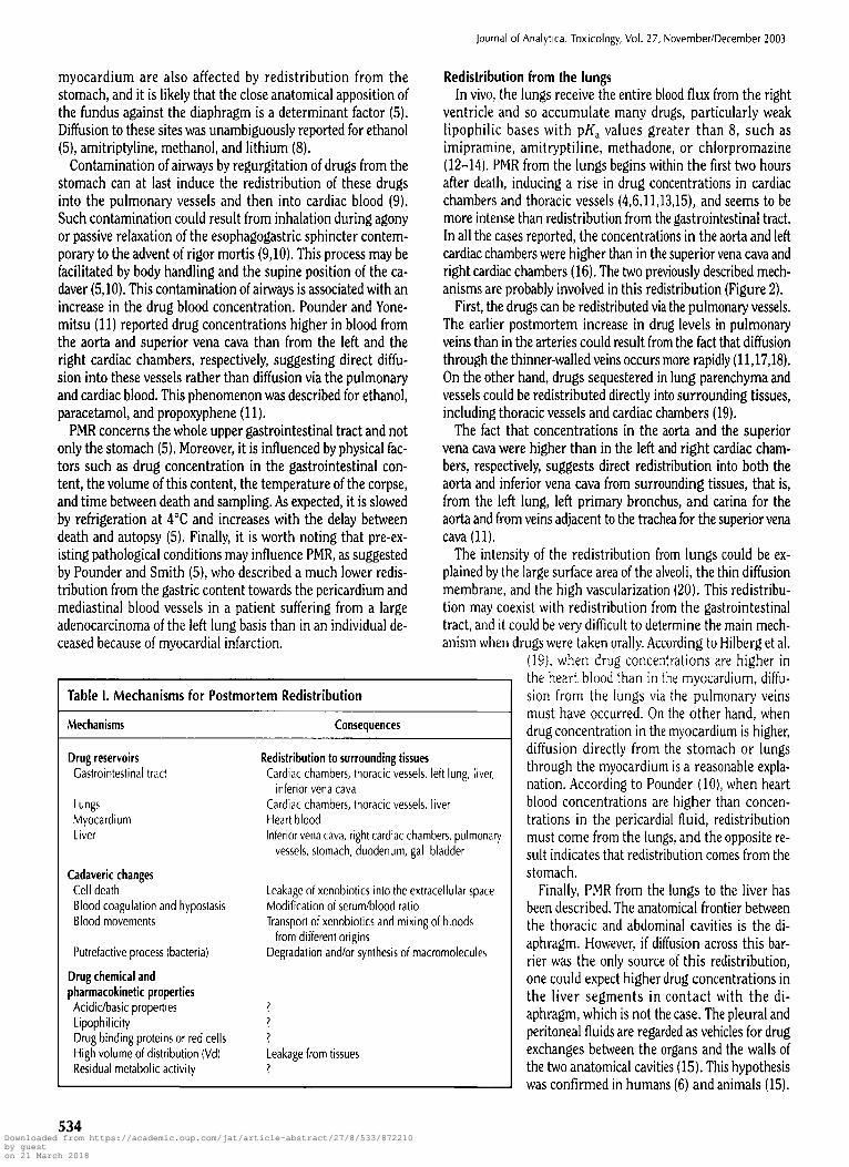
Table I. Mechanisms for Postmortem Redistribution
Mechanisms Consequences
Drug reservoirs Gastrointestinal tract
Lungs Myocardium Liver
Cadaveric changes Cell death Blood coagulation and hypostasis Blood movements
Putrefactive process (bacteria)
Drug chemical and pharmacokinetic properties
Acidic/basic properties Lipophilicity Drug binding proteins or red cells High volume of distribution (Vd) Residual metabolic activity
Redistribution to surrounding tissues Cardiac chambers, thoracic vessels, left lung, liver,
inferior vena cava Cardiac chambers, thoracic vessels, liver Heart blood Inferior vena cava, right cardiac chambers, pulmonaq/
vessels, stomach, duodenum, gall bladder
Leakage of xenobiotics into the extracellular space Modification of serum/blood ratio Transport of xenobiotics and mixing of bloods
from different origins Degradation and/or synthesis of macromolecules
? ? ? Leakage from tissues ?
Redistribution from the lungs In vivo, the lungs receive the entire blood flux from the right
ventricle and so accumulate many drugs, particularly weak lipophilic bases with pKa values greater than 8, such as imipramine, amitryptiline, methadone, or chlorpromazine (12-14). PMR from the lungs begins within the first two hours after death, inducing a rise in drug concentrations in cardiac chambers and thoracic vessels (4,6,11,13,15), and seems to be more intense than redistribution from the gastrointestinal tract. In all the cases reported, the concentrations in the aorta and left cardiac chambers were higher than in the superior vena cava and right cardiac chambers (16). The two previously described mech- anisms are probably involved in this redistribution (Figure 2).
First, the drugs can be redistributed via the pulmonary vessels. The earlier postmortem increase in drug levels in pulmonary veins than in the arteries could result from the fact that diffusion through the thinner-walled veins occurs more rapidly (11,17,18). On the other hand, drugs sequestered in lung parenchyma and vessels could be redistributed directly into surrounding tissues, including thoracic vessels and cardiac chambers (19).
The fact that concentrations in the aorta and the superior vena cava were higher than in the left and right cardiac cham- bers, respectively, suggests direct redistribution into both the aorta and inferior vena cava from surrounding tissues, that is, from the left lung, left primary bronchus, and carina for the aorta and from veins adjacent to the trachea for the superior vena cava (11).
The intensity of the redistribution from lungs could be ex- plained by the large surface area of the alveoli, the thin diffusion membrane, and the high vascularization (20). This redistribu- tion may coexist with redistribution from the gastrointestinal tract, and it could be very difficult to determine the main mech- anism when drugs were taken orally. According to Hilberg et al.
(19), when drug concentrations are higher in the heart blood than in the myocardium, diffu- sion from the lungs via the pulmonary veins must have occurred. On the other hand, when drug concentration in the myocardium is higher, diffusion directly from the stomach or lungs through the myocardium is a reasonable expla- nation. According to Pounder (10), when heart blood concentrations are higher than concen- trations in the pericardial fluid, redistribution must come from the lungs, and the opposite re- sult indicates that redistribution comes from the stomach.
Finally, PMR from the lungs to the liver has been described. The anatomical frontier between the thoracic and abdominal cavities is the di- aphragm. However, if diffusion across this bar- rier was the only source of this redistribution, one could expect higher drug concentrations in the liver segments in contact with the di- aphragm, which is not the case. The pleural and peritoneal fluids are regarded as vehicles for drug exchanges between the organs and the walls of the two anatomical cavities (15). This hypothesis was confirmed in humans (6) and animals (15).
534 Downloaded from https://academic.oup.com/jat/article-abstract/27/8/533/872210by gueston 21 March 2018

Journal of Analytical Toxicology, Vol. 27, November/December 2003
Redistribution from the liver PMR from the liver is complex, as it involves different mech-
anisms. Drugs sequestered in the liver at the time of death could be redistributed via the hepatic veins to the inferior vena cava and then into the right cardiac chambers and pulmonary vessels or to peripheral venous blood (20). One of the results of this redistribution is the decrease in drug concentrations in hepatic lobes, as described by Pohland and Bernhard (7) for flu- oxetine and norfluoxetine. Nevertheless, this process is not as intense and early as redistribution from the lungs.
Secondly, drugs could be redistributed directly into adjacent organs. The anatomical relationships of the human liver that are relevant to PMR are with the stomach, the pylorus, the proximal duodenum, and the gall bladder (20), but this is not as important as the redistribution via the hepatic vessels.
The liver is also the target of redistribution from the gas- trointestinal tract. The anatomical relationships between the di- gestive tract and liver are of great importance for the under- standing of these mechanisms. The greatest part of the inferior surface of the left liver lobe is in contact with the stomach, and the pylorus and proximal duodenum rest against the right lobe of the liver and the gall bladder. Finally, taking into account the close anatomical proximity between the liver and the stomach, the xenobiotics contained in the latter at the time of death can enter the hepatic parenchyma either by passive diffusion or through the portal vein (20). This diffusion phenomenon is very variable, with the left hepatic lobe, which is in close con- tact with the stomach, being more involved than the others. Ac- cordingly, Pounder and Davis (21) found much higher concen- trations of zopicione in the left lobe and in the gall bladder than in the right lobe. Fuke et al. (6) demonstrated, in a human ca- daver model, that after gastric instillation of 25 mL toluene and
I
Figure 1. Frontal view of thorax and abdomen through the left ven- tricle. The arrows show the direction of redistribution towards the main organs concerned (RL, right lung; LL, left lung; AO, aorta; RV, right ven- tricle; LV, left ventricle; ST, stomach; and LLL, left liver lobe).
isobutanol, the concentrations measured in the left lobe were markedly higher than those in the right. It is therefore partic- ularly difficult to correctly interpret the evolution of hepatic drug concentrations postmortem. Following Hilberg et al. (22), the molecular PKa could have an influence on these different mechanisms. It would be interesting to find such general rules, as redistribution from or towards the liver concerns a very large number of molecules, an exhaustive list of which is im- possible to establish.
Redistribution from the myocardium In the living, many cardiac drugs are concentrated in the
myocardium. One of the best examples is digoxin, with in vivo myocardic concentrations 30 times higher than that in heart blood. This is also the case for calcium channel blockers and quinidine. Rapidly, these drugs are redistributed into cardiac blood, in which concentrations rise dramatically. A moderate in- crease has also often been described in the subclavian venous blood, which cannot, for this reason, be considered as a pe- ripheral blood specimen (23). Such redistribution from the myocardium into heart blood has also been described for other drugs such as morphine (24,25); amphetamine (23,26); methamphetamine (13,26); propoxyphene and nor- propoxyphene (23); imipramine and desipramine (27); and amitriptyline, doxepin, maprotiline, and metoprolol (23). Be- cause the dramatic concentration increase observed in cardiac blood may result from redistribution from the stomach, lungs, or liver, proof of redistribution from the myocardium as the pri- mary mechanism responsible for this increase would be demon- strated by higher concentrations of the drug in both the left and right cardiac chambers (18).
Redistribution into body fat Conversely to the previously described mechanisms, some
very highly lipophilic drugs are concentrated in adipose tissues by simple physical dissolution in neutral fats. This distribution
Figure 2. Cross-section of thorax through the 8th thoracic vertebra (caudal view). The arrows show the direction of redistribution from the lungs into the organs concerned. Diffusion occurs via the pulmonary veins (LPV, left pulmonary vein and RPV, right pulmonary vein) and transparietally towards surrounding anatomic structures (LA, left atrium; LV, left ventricle; AO, aorta; LHL, left higher lobe of lungs; LLL, left lower lobe of lungs; RHL, right higher lobe of lungs; RLL, right lower lobe of lungs; RA, right atrium; and RV, right ventricle).
535 Downloaded from https://academic.oup.com/jat/article-abstract/27/8/533/872210by gueston 21 March 2018

occurs slowly, as the blood flow to adipose tissues is low and the equilibrium between blood and adipose tissue concentrations may not have been achieved at the time of death (20). In such a case, a continued distribution (rather than a real redistribu- tion) of these drugs from blood into adipose tissues, lowering the postmortem blood concentrations, can occur. This phenomenon occurs for anesthetics and volatile compounds (6,20).
Agonic and Cadaveric Changes
Cell death A prevalent hypothesis to explain the release of basic drugs
from solid tissues into the blood compartment within hours after death is that the decrease in blood pH would be responsible for a return of the basic molecules into this compartment, due to an increased concentration gradient in nonionized forms (Table I). Actually, it involves much more complex mechanisms, in as much as it is now clear that intracellular pH decreases be- fore plasma pH, which is responsible for an inverse gradient in nonionized forms for basic molecules. The molecular mecha- nisms responsible for cell death are complex. Hypoxia due to the toss of oxygen-carrying capacity of blood during the agonic processes is the first stage. It produces a loss of oxidative phos- phorylation capacity and halts the synthesis of adenosine triphosphate (ATP) by the aerobic pathway, inducing an in- creased rate of anaerobic glycolysis to provide the cell with some energy (28). Glycogen is, thus, rapidly depleted, and this anaerobic process results in the accumulation of lactic acid and inorganic phosphates that reduce the intracellular pH and, probably at this step, induces an increased accumulation of basic drugs into the ceils.
The second consequence of this loss of ATP production-- and consequently ATPase activity--is the failure of the energy- dependent sodium pump, which causes sodium to accumulate intracellularly with diffusion of potassium out of the cell. The net gain of solute is accompanied by an iso-osmotic gain of water resulting in cell swelling (28). The next phenomena to occur are detachment of ribosomes from the granular endo- plasmic reticulum, dissociation of polysomes into monosomes, vacuolization of the mitochondria, and massive calcium cell in- flux. Finally, intracellular acidification and changes in ionic composition lead to damage to the lysosomal membranes with leakage of their enzymes into the cytoplasm and activation of their acid hydrolysis (28). Activation of these enzymes leads to enzymatic digestion of cell components and membranes. There is then a widespread leakage of cellular enzymes and macro- molecules into the extracellular space. The basic lipophilic drugs, highly concentrated in the cells, are redistributed at this stage into the extracellular space and then into the blood com- partment. Neutral or acidic drugs are less affected. According to Langford and Pounder (29), the release of enzymes into the ex- tracellular space could be used as an indicator for PMR. They also demonstrated that there is a strong positive correlation be- tween individual amino acids (glycine, leucine, methionine, serine, and valine) and drug concentrations in pulmonary blood samples. These autolytic processes concern all cells and tis-
536
Journal of Analytical Toxicology, Vol. 27, November/December 2003
sues, but at different rates. The disintegration of physiological and anatomical barriers such as vascular walls leads to changes in drug concentrations. Skopp et al. (30) elaborated an in vitro experiment for studying postmortem vascular permeation. They demonstrated that in veins, sampled according to the selec- tion criteria (corpses stored at 4~ within 6 h after exitus and autopsy performed within 24--60 h after death), concentrations of morphine and its glucuronides in the extravascular space in- creased with time during the first 120 h postmortem, according to a sigmoidal curve. In addition, the molecules were absorbed in part by the vascular wall.
Furthermore, vascular permeation could be strongly influ- enced by the initial substrate concentration, the molecular structure (i.e., the size, shape, charge, and partitioning be- havior), and the orientation of solute flux. Finally, the corpse temperature appeared to have a large influence on this process: preservation at 4~ for the first two days tended to limit it, but it increased beyond 48 h postmortem, whatever the temperature of the corpse (30).
Blood coagulation and hypostasis Postmortem blood sediments and clots unevenly in the body.
The transformation is brought about by blood clotting followed by lysis (Table I). The two processes may occur simultaneously, and the effectiveness of the clot lysis will determine whether the blood is clotted or completely fluid, or partly clotted and partly fluid. This phenomenon varies from case to case (20). The clot generally entraps a large number of red blood cells, so sampling this clot for toxicological analysis may influence the concen- tration measured for any drug exhibiting unequal distribution between red blood cells and serum.
A few hours after death, hypostasis occurs by sedimentation of blood and plasma to the lower parts of the corpse. Hypostasis induces variations in the percentage of erythrocytes by volume, depending on the part of the body. Thomsen et al. (31) demon- strated that this percentage decreased by more than 50% during the first 9 h postmortem in blood samples taken from the cu- bital vein on the upper side of the arm. The position of the corpse was unfortunately not specified in this study.
Blood movements Movements of blood within the vessels occur early after death
and may be responsible for physical redistribution of drugs be- tween different vascular compartiments (Table I). The extent of these postmortem blood movements is influenced by pressure and fluidity changes (20). Rigor mortis, which occurs during the first 24 h postmortem, induces systolic ventricular contrac- tions with small movements of cardiac blood into the superior vena cava and the associated neck veins (32). With the increase in intra-abdominal pressure there is a blood reflux from the ab- dominal aorta into the thoracic aorta, from the inferior vena cava into the right atrium and superior vena cava, and from the left cardiac chambers into the pulmonary veins (20,32).
Later, at the onset of the putrefactive process and the dis- parition of rigor mortis, putrefactive gases distend the abdom- inal walls and the diaphragm, inducing a reflux of blood in pe- ripheral veins which become more apparent. This phenomenon, incorrectly called "postmortem circulation", is of weak ampli-
Downloaded from https://academic.oup.com/jat/article-abstract/27/8/533/872210by gueston 21 March 2018

Journal of Analytical Toxicology, Vol. 27, November/December 2003
tude. Furthermore, to our knowledge, the only experimental work designed to investigate this phenomenon dates back to 1961 (33) and should be brought up to date by studying the postmortem diffusion of autoradiographic blood flow tracers. Fi- nally, the only experimental work dedicated to this phenomenon in animals demonstrated that, in rabbits, these movements were related to the cadaver position and highly variable from case to case (34).
Putrefactive processes Putrefaction is highly variable depending on the ambient
conditions and the state of the corpse. As part of this process, degradation and/or synthesis of xenobiotics by bacteria are pos- sible, as previously described for ethanol (Table I).
Bacteria present in the gastrointestinal tract at the time of death are known to cross the gastrointestinal wall after death, enter the blood and lymph vessels, and then transmigrate throughout the body. This migration may occur within the first few hours postmortem, especially when ambient temper- ature is high. The bacteria most likely involved is this process are those originating from the gastrointestinal tract such as Bacillus spp., Pseudomonas ssp., Escherichia coli, Proteus mirabilis, Clostridium perfringens, Staphylococcus epider- midis, Streptococcus faecalis, and Bacteroides fragilis (35-37). These bacteria could produce and/or metabolize many com- pounds in the postmortem blood. The best example is ethanol synthesis. In the presence of glucidic substrates, such as glucose or ribose, and amino-acids from protein breakdown, bacteria and yeasts can produce ethanol (36).
According to O'Neal and Poklis (35), Escherichia coil and Candida albicans are the primary causes of postmortem ethanol synthesis, but there are 58 species of bacteria and 17 species of yeasts that can produce ethanol. Kupfer et al. (38) devel- oped a robust polymerase chain reaction (PCR) method for the rapid detection of Escherichia coil, Proteus vulgaris, and Candida albicans in human postmortem blood, in order to substantiate ethanol neo-formation when interpretation of results is difficult.
Because glucose is the primary substrate of postmortem ethanol production, the tissues with high glucose storage ca- pacity are the sites of greatest ethanol synthesis: mainly the liver, skeletal muscles, lungs, and myocardium (35). Urine seems to be a poor medium for microbial ethanol synthesis, un- less the deceased was diabetic (35,39). The brain would be con- cerned to a lesser extent: Davis et al. (40) reported that it was not until the third day postmortem that ethanol concentra- tion exceeded the 0.01% detection limit usual in forensic anal- ysis. As a result of microbial production, there is a wide vari- ability in ethanol concentration between the sampling sites (41). According to Jones et al. (42), mannitol administration just before death could favor postmortem ethanol synthesis. Other volatile compounds such as methanol, n-propanol, isopropanol, n-butanol, and sec-butanol (43) may also be produced in post- mortem blood during this process. The presence of these com- pounds can be used as a marker of postmortem ethanol syn- thesis (44,45). Taking into account these phenomena, most of the authors recommended sampling vitreous humor in order to distinguish between exogenous absorption and postmortem
synthesis. Vitreous humor is considered an ideal specimen for the interpretation of postmortem ethanol concentration be- cause (1) it contains no glucose or microorganisms, (2) it is pro- tected from putrefaction or trauma, and (3) the concentration of ethanol in this medium reflects (is not equal because of dif- ferences in water content) the antemortem blood concentra- tions. Otherwise, ethanol can be metabolized by oxidation to ac- etaldehyde and acetate. Some bacteria and yeasts with fermentative and/or oxidative metabolisms, such as Candida al- bicans or Serratia marcescens, are able to produce and/or me- tabolize ethanol depending on the chemical properties of the medium (46), whereas bacteria with strict oxidative metabolisms, such as Pseudomonas spp, can only metabolize ethanol (37). The chronological aspects of ethanol synthesis and degradation is under debate. Takayashu et ai. (47) studied the metabolism and postmortem changes of deuterium-labeled ethanol (ethanol-d6) in rabbits. They demonstrated that the ethanol-d6 concentrations decreased moderately in heart, liver, lungs, kidney, and femoral skeletal muscles during the first 90 rain after death, and nondeuterated ethanol and 1-propano[ concentrations showed marked increases depending on the de- gree of putrefaction of each organ or tissue, suggesting that ethanol precedes neo-formation. In contrast, according to Bouillerot and Laviano-Rousselin (37), ethanol degradation should occur after ethanol synthesis because of substrate de- pletion.
Postmortem degradation by bacteria does not only concern ethanol, some other compounds, such as benzodiazepines, could be metabolized by microorganisms during the putrefac- tive process. According to Robertson and Drummer (48), the ni- trobenzodiazepines clonazepam, nitrazepam, and flunitrazepam are metabolized to their respective 7-amino metabolite in the liver, lungs, myocardium, kidneys, and skeletal muscles by en- terobacteria (Escherichia coli, Bacillus spp, Proteus mirabilis, Clostridium perfringens, Staphyloccocus aureus, Staphyloc- cocus epiderrnidis, Streptoccocus fcecalis, and Bacteroides frag- ilis) containing an oxygen-sensitive nitroreductase enzyme able to reduce nitroaromatic compounds. As expected, this process is slowed down by keeping the corpses at + 4~ The hypothesis of cyanide degradation by bacteria during the putrefactive pro- cess has been evoked. Ballante et al. (49) described a significant decrease in cyanide concentration with time in postmortem blood and tissues (lungs, brain, liver, and kidneys) of rabbits killed with potassium cyanide. This may be due to the metabo- lization of cyanide to thiocyanates, but the bacteria involved are unknown.
Pharmacokinetics
The PMR process may be influenced by the pharmacokinetic behavior of drugs. First, it should be stated that in vivo, many drugs present marked differences in blood concentrations with respect to the sampling sites. Arterial concentrations are often higher than venous concentrations during the absorption phase, whatever the mode of administration, as has been de- scribed for ethanol, diazepam, or phenobarbital (23,50). During
537 Downloaded from https://academic.oup.com/jat/article-abstract/27/8/533/872210by gueston 21 March 2018

the elimination phase, the venous concentrations of drugs such as furosemide, procainamide, and propranolol are higher than the arterial concentrations. According to Chiou (50), drugs with a short half-life and/or a large apparent Vd may present more marked arteriovenous differences than drugs with a long half-life and/or a smaller Vd. The possibility that such arterio- venous differences at the time of death may influence PMR of the molecules concerned should be mentioned. This phe- nomenon was hypothesized by Elsirafy et al. (51) to explain differences in postmortem concentrations of diazinon between different sampling sites.
PMR of drugs depends not only on changes in cells and tissues during and after death, but also on the pharmacokinetic prop- erties of these drugs (Table I). Most studies have tried to link the intensity of PMR of a drug to its in vivo distribution, that is, its Vd. But the postmortem changes in pharmacokinetics are prob- ably more complex, and modifications may occur at each phar- macokinetic stage, that is, absorption, distribution, metabolism, and elimination.
Absorption There are many different processes by which a drug can cross
the lipidic membranes of cells. Most drugs cross membranes by passive diffusion, ruled by Fick's law, depending on the con- centration gradient of the molecule and the pH on both sides of the membrane; molecular size, lipid solubility, and the ioniza- tion state of the molecule; and on the pH on the both sides of the membrane.
Although no study has been conducted on the postmortem changes in drug absorption, it is more than likely that passive diffusion is affected by the postmortem changes of intracel- lular pH and by the loss of the membrane integrity. These changes may influence the absorption of drugs present in the gastrointestinal tract at the time of death, but pH changes along the digestive tract after death are unknown.
Filtration is another absorption process which concerns small, water-soluble molecules such as ethanol. These molecules cross the lipid membrane through proteic pores be- cause of an osmotic or oncotic gradient. Equilibration of water and electrolytes on both sides of the membrane and damage to the proteic pores early after death tend to accelerate the filtra- tion process at first and then stop it when equilibrium is reached.
In addition, some drugs, essentially weak organic acids and bases, cross the lipid membranes by active transport. Active transport requires energy input, often in the form of ATE as well as the presence of a carrier system to transfer the molecules across the membrane. The cessation of ATP production rapidly after death probably blocks this process.
Distribution After absorption, drugs reach the circulation in order to be
distributed throughout the body and reach their site(s) of ac- tion.
Blood transport ofxenobiotics. The first stage of distribution is the transport of xenobiotics in the bloodstream. In the cir- culating blood, drugs can be (1) dissolved in the plasma water, (2) bound to the plasma proteins, and/or (3) bound to the mem-
538
Journal of Analytical Toxicology, Vol. 27, November/December 2003
branes or contained in the cytoplasm of blood cells (mainly erythrocytes). Only water-soluble molecules can be completely or almost completely dissolved in plasma water. This is the case for ethanol, for example, which is dissolved in total body water and whose PMR is not influenced by changes in blood proteins or red cells.
Drug protein binding occurs in different ways. It may take the form of either weak, hydrophobic bonds between lipid soluble xenobiotics and the hydrophobic sites of albumin and/or lipoproteins, or stronger electrostatic bonds between ionized, weak organic acids (e.g., sulphonamides) and the cationic sites of albumin or between weak organic bases (e.g., [3-blockers, tricyclic antidepressants) and acid cq-glycoprotein. Irreversible, covalent bonds between plasma proteins and certain molecules (e.g., antimalarial drugs) are also possible (52). The total protein blood concentration decreases after death because of the break- down into amino acids and peptides resulting from acute anoxia (i.e., stopped synthesis) and from proteolysis by autoenzymes (53). This process would be accelerated at the beginning of pu- trefaction by proteolytic bacteria (54).
Oemichen et al. (55) reported the possibility of PMR of serum albumin from the intravascular space into the perivascular space. Even if no more recent studies have been devoted to the understanding of these postmortem changes, the rapid de- crease in protein binding is evidence. The major result of this process is an increase in the intravascular concentration of the free form of the drugs, which could theoretically influence their redistribution. No information could be found in the lit- erature about the potential postmortem changes in acid ~l-gly- coprotein and lipoproteins. However, as postmortem drug levels are generally analyzed in whole blood, modifications in drug binding have little consequence for the interpretation of the an- alytical results.
Some drugs are bound to the red cells in the circulating blood. The postmortem whole blood concentration of these drugs could be affected by the process of hypostasis, inducing variations in the apparent hematocrit (1). This phenomenon was hypothesized by Tomson et al. (56) to explain PMR of phenytoin and by Skopp et al. (57) for morphine glucuronides. This is, however, not the sole explanation for all the postmortem concentration variations observed (58).
Distribution to solid tissues and organs. Antemortem distri- bution of a given drug is a dynamic process depending on its physicochemical properties (e.g., molecular size, degree of ion- ization, and lipophilicity) and physiological properties such as the blood flow perfusing the organs and the affinity of this drug for the different tissues or organs.
Some lipophilic drugs are known to have a very wide distri- bution in tissues as a result of cellular uptake and accumulation. There are two probable mechanisms: binding to membrane phospholipids and accumulation in acidic compartments (14). Under physiological conditions, plasma pH is approximately 7.4, and the pH of cells is of the order of 5. Weak bases, mainly in their nonionized state in the plasma, permate cell mem- branes because of this pH gradient and accumulate extensively in lysosomes. This process of lysosomal trapping contributes to the uptake of the weak bases by different tissues such as the lungs, brain, heart, kidneys, and liver (12). The early pH changes
Downloaded from https://academic.oup.com/jat/article-abstract/27/8/533/872210by gueston 21 March 2018

Journal of Analytical Toxicology, Vol. 27, November~December 2003
may explain the relase of these weak bases into the vascular compartment.
The distribution of a drug in the body is characterized by its apparent Vd, which refers to the volume into which the total amount of the drug would have to be uniformly distributed to reach the concentration actually measured in plasma. The value obtained for the Vd may or may not have physiological signifi- cance. Some studies suggest that drugs with a large Vd are those that are most widely redistributed after death because of their accumulation in tissues (59). This is obviously the case for weak lipophilic bases. Consistently, PMR was extensively re- ported for the tricyclic antidepressants such as amitriptyline (4), nortriptyline (18), trimipramine (60), or dothiepine (61,62). Similar results were found for fiuoxetine (7), venlafaxine (63), methadone (64,65), amphetamine and methamphetamine (13), and even digoxine (66). According to Hilberg et al. (67), an ap- parent Vd of more than 3-4 L/kg is a good indication that the drug is liable to undergo PMR. But this hypothesis is not enough to explain the postmortem changes observed with many different types of molecules with different physicochemical properties. The case of the postmortem changes in blood levels of morphine and its glucuronides is of great interest. According to Sawyer and Forney (24), free and total morphine concentra- tions in rats increased significantly during the postmortem pe- riod, depending on the sampling site, with significant increases in cardiac blood, cardiac tissue, and liver, and on the post- mortem delay, with highest concentrations at 96 h postmortem. The swiftness of the observed redistribution did not support the hypothesis of bacterial hydrolysis of the glucuronides into mor- phine. However, the authors present no precise data on the po- tential source of such a redistribution. Moreover, these results were not completely confirmed in humans: according to Logan and Smirnov (26), who studied the PMR of morphine and its glucuronides in 32 deaths involving opiates, no evidence was found for changes in morphine levles with time at either cen- tral peripheral blood sites. However, they confirmed that mor- phine concentrations were higher in cardiac blood compared to peripheral blood at every sampling time, particularly when the ventricular blood concentration exceeded 0.3 mg/L. The fact that the corpses were stored at 4~ during the experiments and that blood samples were drawn from the left ventricle may only partly explain the discrepancy between these results and those of Sawyer and Forney (24). Finally, it should be kept in mind that the anatomical relationships between abdominal or- gans are different in humans and rats and that the latter have no gallbladder. Gerostamoulos et al. (68) confirmed, in 40 cases of heroin-related death, that there was no statistically significant difference in concentrations with respect to the postmortem in- terval, but observed a trend for higher concentrations in central than in peripheral blood. Nevertheless, other authors have also described variations in morphine and morphine glucuronides concentrations depending on the sampling sites (69,70). How- ever, the pharmacokinetic properties of morphine are quite dif- ferent from those of the molecules that typically undergo PMR. Morphine is not a weak base as described, but an amphoteric compound which becomes less lipophilic with decreased pH, which may explain its redistribution from lipophilic tissues after death (24). However, several other hypotheses have been
raised to explain this redistribution. According to Skopp et al. (1), the hypostasis process, resulting in plasma loss and hemo- concentration in the lower areas of the body, could play a part in the observed site-to-site variations (but this should also be the case for any drug or toxic compound dissolved in plasma, which is not likely). According to Carrupt et al. (71), morphine glucuronides can exist in two conformational forms, the folded one being more lipophilic than the unfolded one. The site-to- site variations in morphine glucuronide concentrations could be associated with this particularly. However, no single expla- nation currently prevails.
The PMR of acetaminophen is worth mentioning. Ac- etaminophen has a small apparent Vd of about I L/kg (72), low lipid solubility, and no known preferential tissue binding. Gomez et al. (73) administered 160 mg/kg of acetaminophen by oral gavage to rabbits and found site- and time-dependent changes in acetaminophen postmortem blood concentrations, with higher concentrations in cardiac blood compared to femoral blood at each sampling time, and an increase in cardiac blood concentrations over time. But the interpretation of these results must take into account the fact that the animals were sacrified only 20 rain after the gavage, so it is likely that the an- temortem distribution of acetaminophen was not complete at the time of death and that unabsorbed amounts of ac- etaminophen in the stomach could have diffused into the car- diac chambers. Such site-dependent variations were described in a case of lethal, acute acetaminophen intoxication (74), but were not found in a case of multiple-drug ingestion, indicating the possibility of a drug-drug interaction (27). The cause of ac- etaminophen redistribution remains unclear, and the possi- bility that the close absorbed may influence this process cannot be ruled out. As acetaminophen undergoes extensive hepatic metabolism, the hypothesis of redistribution from the liver might be explored in animal models, for example by adminis- tering paracetamol by a parenteral route and collecting liver samples at different times, in order to look for a potential de- crease in paracetamol concentration parallel to the increase in blood levels, as reported by Pohland and Bernard (7). The op- posite situation is exemplified by mirtazapine, a relatively new tetracyclic antidepressant with basic and lipophilic properties and an apparent Vd of 4.84 L/kg (75), which did not exhibit any significant difference between cardiac and femoral postmortem blood concentrations, but showed an increase in liver concen- trations (76,77). All these examples demonstrate that the post- mortem site- and time-dependent variations of drug concen- trations are not automatically associated with molecules with a high apparent Vd. Obviously, the absorption route must be taken into account, as diffusion from the gastrointestinal tract may concern all the drugs. Otherwise, drugs with a small ap- parent Vd can have a high affinity for some tissues from which they can be redistributed during the postmortem period (78). The liver and the lungs are obvious candidates, but probably also fatty tissues or skeletal muscles. Thus, paracetamol could be progressively redistributed from the liver parenchyma into the central blood vessels without affecting the peripheral ves- sels. It is worth noting that the brain, though highly vascular- ized, is tess subject to PMR than the organs mentioned: studies in rats (24,67) have shown that the brain concentrations in
539 Downloaded from https://academic.oup.com/jat/article-abstract/27/8/533/872210by gueston 21 March 2018

the postmortem period increase only slightly with respect to liver, lung, and kidney concentrations, even for molecules with high apparent Vd such as verapamil or nortriptyline (Vd = 5.1 L/kg and 13.7 L/kg in the rat, respectively) (67). The concen- tration variations between the different brain areas have been the subject of only a few studies, with contradictory results. Ac- cording to Moore et al. (79), ethanol concentrations in human cadavers are equal in the different sites of the gray matter and are slightly different from the white matter because of the dif- ference in water content. These results obtained with ethanol, a small hydrophilic molecule, have been confirmed by Kalasinsky et al. (80), who measured, in 14 corpses of metham- phetamine users, methamphetamine and amphetamine con- centrations in 15 different brain areas and did not find statisti- cally significant differences for these lipophilic alkaline molecules. On the contrary, Sawyer and Forney (24) found, in the rat, statistically higher free morphine concentrations in the posterior than in the anterior brain as soon as 24 h after eu- thanasia, as well as a subsequent significant increase with time in the posterior but not anterior brain. Though these results should be confirmed by further studies, they are in accordance with the hypothesis that the lipid solubility alone cannot explain all redistribution phenomena. On the other hand, the apparent protection of the brain from PMR is probably due to the low per- meability of the blood-brain barrier with regard to the other membranes and barriers of the body. The tight junction between the endothelial cells in brain capillaries and the sleeve of glial cells around these capillaries presents a formidable obstacle to the transfer of hydrophilic compounds (except the smaller ones such as ethanol). Additionally, the thickness of membranes to be crossed and the absence of proteins in the cerebrospinal fluid (CSF) and interstitial liquid considerably slow clown the transfer of large liposoluble molecules (77).
In conclusion, the PMR of a drug cannot be predicted only by its lipophilic properties and its apparent Vd. Other factors such as the absorption route, dose, or particular affinity of the drug for some tissues must be envisaged as well as the possibility of a residual metabolic activity in the first hours after death.
Metabolism The drug-metabolizing system may persist several hours after
death, inducing the breakdown of a drug and the synthesis of its metabolites. This phenomenon must be distinguished from the breakdown of drugs due to the putrefactive process. To our knowledge, all studies concerning this problem were devoted to the metabolism of cocaine. Apart from the fact that the post- mortem concentrations of cocaine and its metabolites present variations according to the time of sampling and sampling sites, the hypothesis of a postmortem, residual cocaine metabolism has been raised (81). In vivo, cocaine is rapidly converted into benzoylecgonine and ecgonine methyl ester in blood and the liver. In blood, cocaine is spontaneously con- verted into benzoylecgonine at physiological pH, and is me- tabolized to ecgonine methylester by plasma cholinesterase (82,83). In the liver, cocaine is metabolized by two nonspecific hepatic esterases: one produces benzoylecgonine and the other produces ecgonine methylester (84). Cocaine is broken down gradually after death (85). According to Hearn et al. (81), this
540
Journal of Analytical Toxicology, Vol. 27, November/December 2003
decrease in cocaine subclavian blood concentrations could not be explained by the spontaneous hydrolysis of cocaine to ben- zoylecgonine because hydrolysis is inhibited by the acidification of blood after death. The hypothesis of a residual esterase ac- tivity was then formulated (85). According to Isenschmid et al. (86), benzoylecgonine in postmortem blood samples arises as a result of in vivo cocaine hydrolysis and ecgonine methylester as the result of postmortem cocaine metabolism. McKinney et al. (87) confirmed this hypothesis by describing a gradual increase in postmortem ecgonine methylester femoral blood concen- trations in juvenile swine treated with cocaine hydrochloride at a dose of 10 mg/kg by IV bolus, then sacrificed. On the other hand, benzoylecgonine could be produced early after death by a residual activity of hepatic cocaine-methylesterase (3). Fi- nally, one should keep in mind that ecgonine methylester is normally found in the blood of living cocaine users (83). As far as cocaethylene is concerned, the results are more controversial. According to Logan et ai. (3), the femoral and ventricular co- caethylene blood concentrations decrease gradually after death in humans, but with no consistent pattern of direction or mag- nitude of change, whereas in rats, Moriya and Hashimoto (88) demonstrated that cocaine is still transformed into cocaethylene in the liver of alcohol-intoxicated rats during the first hour postmortem. Even if the persistence of a residual hepatic and/or plasmatic esterase activity after death has not been clearly demonstrated, most of the authors agree with this hypothesis.
The possibility of a postmortem, residual enzymatic activity was confirmed by Moriya and Hashimoto (89) in a previous study describing the postmortem metabolism of dichlorvos, an organophosphorus pesticide, by hepatic and plasmatic esterases. Yamazaki and Wakasugi (90) studied the postmortem changes in drug-metabolizing enzymes in rat liver microsomes. The results demonstrated that the activity of the liver enzymes did not stop immediately after death, but showed a progressive de- crease during the first 48 h postmortem. A residual enzymatic activity, variable with the nature of the enzymes involved, is likely during the first hours after death.
These results must be confirmed in humans, or rather in vitro using human microsomes. They underline the necessity of assaying the metabolites of the drugs studied in order to estab- lish the parent drug/metabolite ratio for the interpretation of the results.
Elimination As for absorption, to the best of our knowlegde no study has
been dedicated to the possible persistance of an elimination process after death.
In the nephron, the processes of glomerular filtration, tubular secretion and tubular reabsorption combine to produce urine and eliminate drugs (78). Glomerular filtration, depending on the afferent blood flow, probably stops at the time of death.
Tubular secretion is an active process depending on the pres- ence of ATP, which thus probably stops shortly after death. Conversely, tubular reabsorption is a passive process that could persist during the first postmortem hours. The acidification of plasma could modify tubular reabsorption and induce a leakage of weak acids.
Biliary excretion concerns glucuronides and polar drugs with
Downloaded from https://academic.oup.com/jat/article-abstract/27/8/533/872210by gueston 21 March 2018

Journal of Analytical Toxicology, Vol. 27, November/December 2003
a molecular weight greater than 500 and less than 1000 Da (91). These highly polar molecules are concentrated in bile by active transport processes similar to those involved in the se- cretion of similar compounds across the renal tubular cells into urine. These active processes probably stop with the inter- ruption of ATP synthesis. Similarly, the storage of primary bile in the gall bladder and its concentration by active water reab- sorption are interrupted, as is active emptying of the gall bladder into the second duodenum.
Practical consequences in forensic toxicology From a practical point of view, the respect of some precau-
tionary measures can limit misinterpretations. It is very im- portant in postmortem testing to be able to compare concen- trations in several blood and tissue samples, even if reference values for drug concentrations in tissues are often missing. Blood samples must be taken at central (cardiac) and peripheral sites. In the framework of postmortem drug redistribution studies, cardiac blood samples must be taken from the right and left cardiac chambers separately, in order to determine the PMR mechanism (92). Taking into account the intensity of re- distribution of certain drugs into the cardiac chambers, the estimation of the amount of drug present at the time of death from the cardiac blood concentrations must be avoided.
As for the peripheral blood sampling sites, all the authors rec- ommend collecting blood from the femoral vein. Femoral blood appears to be the specimen of choice for postmortem toxico- logical analysis as it is the least subject to PMR, which, in this case, can only come from local tissues such as muscles and fat (58). Accordingly, it was found that the femoral blood concen- centrations were less affected by the postmortem time delay than the concentrations in central blood (78). Femoral blood must ideally be sampled after cross-clamping the iliac vein and the inferior vena cava in order to avoid the risk of drawing blood from these vessels, but such collection is not always pos- sible under the usual forensic autopsy conditions (20). Even if femoral blood concentrations are more representative of the an- temortem blood concentrations than cardiac blood, they are fre- quently higher than the ante- or perimortem blood concentra- tions (22). More surprisingly, in a few cases, femoral blood concentrations were found to be higher than cardiac blood concentrations. This was observed in human cases where re- suscitation was attempted, probably causing a shift of cardiac blood into the peripheral vessels (22,23). The same results were found in a study of the PMR of amitriptyline (93) in an experi- mental pig model, where the animals were sacrified using 10 mL of potassium chloride. In such a case, death is caused by ventricular fibrillation where the heart stops in diastole, in- ducing a shift of cardiac blood, which could explain the higher postmortem concentrations found in femoral blood. Finally, as previously explained, subclavian venous blood should not be considered as peripheral blood (23), as its drug concentration variations follow those of heart blood.
Many authors previously reported that the left lung, the left kidney and the left lobe of the liver are more prone to PMR than the right lung, the right kidney, and the right lobe of the liver because of redistribution from the gastrointestinal tract. Ideally, the right lobe of the liver, the right kidney, and the right lung
should be sampled. Probably because the brain is not clearly af- fected by PMR, no precise recommendation concerning brain sampling is given in the literature.
Other biological matrices, less subject to PMR, have been proposed in order to avoid misinterpretations: vitreous humor, skeletal muscles, bone marrow, and CSF. Vitreous humor was the subject of many investigations and is of a great interest for forensic purposes. It contains no microorganisms or glucose, and it is also protected from putrefaction and trauma. For these reasons, it is considered a sample of choice for distinguishing exogenous from endogenous ethanol resulting from the pu- trefactive process (35,41,94--96). Blood ethanol concentration can be approximated from the vitreous humor concentration, taking into account that the vitreous humor/blood ethanol concentration ratio ranges from 0.9 to 1.38 (with a theoretical value of 1.27 at equilibrium) (41). Unfortunately, the concen- trations of other drugs during the postmortem period cannot al- ways be accurately estimated using vitreous humor. McKinney et al. (87) studied the PMR of cocaine in an animal model. They concluded that 8 h after death, the vitreous humor cocaine concentrations were significantly higher in all animals with respect to the concentration at the time of death and were sim- ilar to the femoral blood concentrations at the time of death. From our interpretation of the work published by Vorpahl et al. (97), there was no good quantitative correlation between vit- reous humor and blood concentrations of digoxin.
The skeletal muscle has been suggested as an alternative specimen for postmortem toxicology because it is present in large amounts and is affected by decomposition later than blood or viscera (98). In addition, muscle samples can be obtained from peripheral sites, far from drug reservoirs such as the stomach, liver, and lungs (98). Langford et al. (99) evaluated the homogeneity of different drugs' concentrations (temazepam, amitriptyline, paracetamol, propoxyphene) in samples obtained from different muscles such as the diaphragm, pectoralis major, sternomastoid, deltoid, biceps, triceps, and brachioradialis. They observed a large site-to-site variablility, with drug concentra- tions in the diaphragm invariably higher than in the other muscles. Furthermore, the variability of these concentrations between sites, excluding the diaphragm, was more pronounced for drugs with a large Vd. The muscle is therefore more inter- esting for qualitative analysis than for quantitative determina- tion of drugs (100). From our point of view, peripheral muscles (brachioradialis, sartorius) should only be sampled for qualita- tive screening, in cases where fluids and viscera are not available (burned cadavers, for example).
Bone marrow was also investigated as an alternative specimen (101). It is a lipid-rich matrix with a high degree of vascularity. Furthermore, its anatomical situation, encased in bones, re- duces the possibility of contamination from bacteria during the putrefactive process (102). In the absence of available data concerning the correlation between bone marrow and blood concentrations, bone marrow was used to perform qualitative analysis when no other sample was available. Winek et al. (103) demonstrated that in rabbits, bone marrow could predict blood concentrations of nortriptyline up to 24 h after death. On the contrary, in pigs, Hilberg et al. (93) found no correlation be- tween blood and bone marrow concentrations of amitriptyline
541 Downloaded from https://academic.oup.com/jat/article-abstract/27/8/533/872210by gueston 21 March 2018
nor did they find any correlation between sternal and femoral bone marrow or between early and late sternal bone marrow. Furthermore, the dehydration of bone marrow, which takes place about 96 h after death, reduces the amounts available. Here again, bone marrow cannot be recommended as an alter- native specimen to estimate the concentrations of drugs.
The CSF was also proposed as an alternative specimen, but some drugs do not diffuse into the CSF. Little is known about the evolution of drug concentrations in the CSF after death or about the possible correlation between CSF and blood drug concentrations. According to Logan and Smirnov (25), who studied the stability of morphine concentrations in CSF and their correlation with blood concentrations in 32 morphine- related death cases, CSF concentrations appeared to be stable with time. However, taking into account the large standard de- viation in the mean CSF-to-femoral or iliac blood ratios, the use of CSF concentrations for the prediction of peripheral blood concentration was not advised by the authors. Furthermore, other drugs, such as amitriptyline, could be redistributed into the CSF (93).
Finally, the hematic fluid found in the declive pleural spaces is the worst biological medium for the quantitation of drugs be- cause it is a mixture of blood and serous fluid from lungs and other thoracic organs, or even the stomach. Anyway, the lack of concentration data in the alternative specimens is another caveat of their utility in forensic investigations.
In this review, we have not considered the misinterpreta- tions related to errors in sample preparation and preservation, such as the absence of a preservative or inappropriate storage temperatures (i.e., too high), for example.
Conclusions and Perspectives
PMR of drugs may complicate the interpretation of the results in forensic toxicology. The competing processes of diffusion from drug reservoirs, cell lysis and putrefaction, and the par- ticular pharmacokinetic properties of certain drugs contribute to the differences in drug concentrations observed between sites and sampling intervals (3).
The most common problem is a difference in drug concen- tration between the different sampling sites. If these differ- ences are moderate and especially if all these site concentrations are in the same range--therapeutic, toxic, or fatal--the inter- pretation may not be an issue. However, interpretation is more difficult when these concentrations are very discordant. The pharmacokinetics of the drugs concerned must be taken into account, as well as, if possible, the autopsy findings, which in many cases give useful information. The position of the corpse and regurgitation of the gastric contents into the airways or thoracic trauma may, for example, explain differences in blood concentrations from different sampling sites.
These redistribution phenomena put into perspective the reliability of the databases of therapeutic/toxic/lethal blood levels, built from published data often reported with no mention of sampling sites, postmortem delay, or autopsy conditions. This is particularly important because a large number of toxic drugs are lipophilic weak bases with a large Vd, prone to PMR.
542
Journal of Analytical Toxicology, Vol. 27, November/December 2003
More surprisingly, there is little information on the metabolic activity in the first hours postmortem or on the real influence of drug physicochemical properties or pharmacokinetic pa- rameters on the redistribution phenomena. Further studies are thus warranted.
References
1. G. Skopp, R. Lutz, B. Gangmann, R. Mattern, and R. Aderjan. Postmortem distribution pattern of morphine and morphine glu- curonides in heroin overdose. Int. J. Legal Med. 109:118-124 (1996).
2. T. Nagata, K. Kimura, K. Hara, and K. Kudo. Methamphetamine and amphetamine concentrations in postmortem rabbit tissues. Forensic Sci. Int. 48:39-47 (1990).
3. B.K. Logan, D. Smirnow, and R.G. Gullberg. Lack of predictable site-dependent differences and time-dependent changes in post- mortem concentrations of cocaine, benzoylecgonine and co- caethylene in humans. J. Anal Toxicol. 20:23-31 (1997).
4. T. Hilberg, A. Bugge, K.M. Beylich, J. Ingum, A. Bj~rneboe, and J. Mgrland. An animal model of postmortem amitriptyline redis- tribution. J. Forensic Sci. 38:81-90 (1993).
5. D.J. Pounder and D.R.W. Smith. Postmortem diffusion of al- cohol from the stomach. Am. J. Forensic Med. Pathol. 16:89-96 (1995).
6. C. Fuke, C.L. Berry, and D.J. Pounder. Postmortem diffusion of in- gested and aspirated paint thinner. Forensic Sci. Int. 78:199-207 (1996).
7. R. Pohland and N.R. Bernhard. Postmortem serum and tissue re- distribution of fluoxetine and noffluoxetine in dogs following oral administration of fluoxetine hydrochloride (Prozac| J. Forensic Sci. 42:812-816 (1997).
8. D.J. Pounder, C. Fuke, D.E. Cox, D. Smith, and N. Kuroda. Post- mortem diffusion of drugs from gastric residue. Am. J. Forensic Med. Pathol. 17:1-7 (1996).
9. J.V. Marraccini, T. Carroll, S. Grant, S. Halleran, and J.A. Benz. Differences between multisite postmortem ethanol concentra- tions as related to agonal events. J. Forensic Sci. 35:1360-1366 (1990). D.J. Pounder. Postmortem diffusion of tracheal lidocaine into heart blood following intubation for cardiopulmonary resuscita- tion. J. Forensic Sci. 42:965-966 (1997). D.J. Pounder and K. Yonemitsu. Postmortem absorption of drugs and ethanol from aspirated vomitus--an experimental model. Forensic Sci. Int. 51:189-195 (1991). A.C. Maclntyre and D.J. Cutler. The potential role of lysosomes in tissue distribution of weak bases. Biopharm. Drug. Dispos. 9: 513-526 (1988). T. Miyazaki, T. Kojima, M. Yashiki, H. Wakamoto, Y. Iwasaki, and T. Taniguchi. Site dependence of methamphetamine concentra- tions in blood samples collected from cadavers of people who had been methamphetamine abusers. Am. J. Forensic Med. Pathol. 14:121-124 (1993). W.A. Daniel, M.H. Bickel, and U.E. Honegger. The contribution of lysosomal trapping in the uptake of desipramine and chloro- quine by different tissues. Pharmacol. Toxicol. 77:402-406 (1995). T. Hilberg, J. MorIand, and A. Bjorneboe. Postmortem release of amitriptyline from the lungs; a mechanism of postmortem drug redistribution. Forensic Sci. Int. 64:47-55 (1994). F. Moriya and Y. Hashimoto. Postmortem diffusion of tracheal li- docaine into heart blood following intubation for cardiopul- monary resuscitation. J. Forensic Sci. 42:296-299 (1997). D.J. Pounder, V. Owen, and C. Quigley. Postmortem changes in blood amitriptyline concentration. Am. J. Forensic Med. Pathol. 15:224-230 (1994).
10.
11.
12.
13.
14.
15.
16.
17.
Downloaded from https://academic.oup.com/jat/article-abstract/27/8/533/872210by gueston 21 March 2018

Journal of Analytical Toxicology, Vol. 27, November;December 2003
18. F. Moriya and Y. Hashimoto. Redistribution of basic drugs into cardiac blood from surrounding tissues during early-stages post- mortem. J. Forensic 5ci. 44:10-16 (1999).
19. T. Hilberg, A. Bugge, K.M. Beylich, J. Morland, and A. Bjorneboe. Diffusion as a mechanism of postmortem drug redistribution: an experimental study in rats. Int. J. Legal Med. 105:87-91 (1992).
20. D.J. Pounder. The nightmare of postmortem drug changes. In Legal Medicine 1993, C.H. Wecht, Ed. Butterworth Legal Pub- lishers, Salem, NH, 1994, pp 163-193.
21. D.J. Pounder and J.I. Davies. Zopiclone poisoning: tissue distri- bution and potential for postmortem diffusion. Forensic Sci. Int. 65:177-183 (1994).
22. T. Hilberg, S. Rogde, and J. M~rland. Postmortem drug redistri- bution-human cases related to results in experimental animals. J. Forensic Sci. 44:3-9 (1999).
23. R.W. Prouty and W.H. Anderson. The forensic science implica- tions of site and temporal influences on postmortem blood-drug concentrations. J. Forensic Sci. 35:243-270 (1990).
24. W.R. Sawyer and R.B. Forney. Postmortem disposition of mor- phine in rats. Forensic Sci. Int. 30:259-273 (1988).
25. B.K. Logan and D. Smirnow. Postmortem diffusion and redistri- bution of morphine in man. J. Forensic Sci. 41:37-46 (1996).
26. RE. Barnhart, J.R. Fogacci, and D.W. Reed. Methamphetamine-- a study of postmortem redistribution. J. Anal. ToxicoL 23:69-70 (1999).
27. G.R. Jones and D.J. Pounder. Site dependence of drug concen- trations in postmortem blood--a case study. J. Anal. Toxicol. 11:186-190 (1987).
28. R.S. Cotran, V. Kumar, and S.L. Robbins. Cellular injury and adaptation. In Robbin's Pathologic Basis of Disease, 4th ed. W.B. Saunders, Philadelphia, PA, 1989, pp 1-38.
29. A.M. Langford and D.J. Pounder. Possible markers for post- mortem drug redistribution. J. Forensic 5ci. 42:88-92 (1997).
30. G. Skopp, R. Lutz, L. P6tsch, B. GanBmann, K. Klinder, A. Schmidt, R. Aderjan, and R. Mattern. An in vitro experiment for postmortem vascular permeation. The passage of morphine and morphine glucuronides accross a vascular wall. J. Forensic Sci. 42:486-491 (1997).
31. H. Thomsen, H.J. Kaatsch, and B. Krisch. How and why does the platelet count in postmortem blood change the early postmortem interval? J. Forensic 5cL 101:185-194 [1999)
32. M. Durigon. Pratique M$dico-ldgafe. Masson, Paris, France, 1999, pp 39-49.
33. M. Fallani. Contributo allo studio della circolazione ematica post-mortale. Minerva Medicoleg. 81:108-115 (1961 ).
34. M. Gomez-Zapata, M. Alcaraz, and A. Luna. Studies on post- mortem circulation of the blood. Z Rechtsmed. 103:27-32 (1989).
35. C.L. O'Neal and A. Poklis. Postmortem production of ethanol and factors that influence interpretation: a critical review. Am. J. Forensic Med. Pathol. 17:8-20 (1996).
36. J.E.L. Corry. Possible sources of ethanol ante- and post-mortem: its relationship to the biochemistry and microbiology of decom- position. J. AppL Bacteriol. 44:1-56 (1978).
37. A. Bouillerot and C. Laviano-Rousselin. Dosage d'~thanol: les er- reurs pr~-analytiques. Alcools et Glycols. Journ@ th6matique de la Soci~t~ Francaise de Toxicologie Analytique. Paris, France, 8 d~cembre 1999.
38. D.M. Kupfer, A.K. Chaturvedi, D.V. Canfield, and B.A Roe. PCR- based identification of postmortem microbial contaminants--a preliminary study. J. Forensic Sci. 44:592-596 (1999).
39. W.D. Alexander. Postmortem urinary alcohol is unreliable in diabetes. Br. Med. J. 317:206 (1998).
40. G.L. Davis, R.L. Leffert, and N.W. Rantanen. Putrefactive ethanol sources in postmortem tissues of conventional and germ-free mice. Arch. Pathol. 94:71-74 (1972).
41. E.J. Briglia, J.H. Bidanset, and L.A. Dal Cortivo. The distribution of ethanol in postmortem blood specimens. J. Forensic Sci. 37: 991-998 (1992).
42. A.W. Jones, R. Andersson, J. Sakshaug, and J. Morland. Possible formation of ethanol in postmortem blood specimen after ante- mortem treatment with mannitol. J. Anal. ToxicoL 15:157-158 (1991}.
43. M. Deveaux. Alcool ~thylique. In Toxicologie et Pharmacologie M~dicoldgales, R Kintz, Ed. Elsevier, Paris, France, 1998, pp 111-126.
44. M.G. Gilliland and R.O. Bost. Alcohol in decomposed bodies: postmortem synthesis and distribution. J. Forensic Sci. 38: 1266-1274 (1993).
45. W. Grellner and R. Iffland. Assessment of postmortem blood alcohol concentrations by ethanol levels measured in fluids from putrefactive blisters. Forensic Sci. Int. 90:57-63 (1997).
46. C. Laviano. Production et consommation d'~thanol post-mortem dans deux liquides biologiques. Ann. Biol. Clin. 56:96-99 (1998).
47. T. Takayasu, T. Ohshima, N. Tanaka, H. Maeda, T. Kondo, J. Nishigami, and T. Nagano. Postmortem degradation of ad- ministered ethanol-d6 and production of endogenous ethanol: experimental studies using rats and rabbits. Forensic Sci. Int. 76:129-140 (1995).
48. M.D. Robertson and O.H. Drummer. Postmortem distribution and redistribution of nitrobenzodiazepines in man. J. Forensic Sci. 43:9-13 (1998).
49. B. Ballantyne, J.E. Bright, and R Williams. The post-mortem rate of transformation of cyanide. Forensic Sci. 3:71-76 (1974).
50. W.L. Chiou. The phenomenon and rationale of marked depen- dence of drug concentration on blood sampling site. Implications in pharmacokinetics, pharmacodynamics, toxicology and ther- apeutics (Part I). Clin. Pharmacokinet. 17" 175-199 (1989).
51. A.A. Elsirafy, A.A. Ghanem, A.E. Eid, and S.A. Eldakroory. Chronological study of diazinon in putrefied viscera of rats using GUMS, GC/EC, and TLC. Forensic 5cL Int. 109:147-157 (2000).
52. J.P. 1111ement and E. Lindenlaub. Protein Binding and Drug Trans- port. F.K. Schattauer Verlag, Stuttgart, Germany, 1986.
53. A.S. Konikova, A.A. Vinarskaya, V.I. Nikulin, A.V. Pogossova, and L.M. Petukhova. Protein degradation to low-molecular com- pouds after death and during reanimation. Virchows Arch. B. Cell. Pathol. 18:347-355 (1975).
54. W. Bonte, J. Bleifuss, and J. Volck. Experimental investigations in post-mortem protein degradation. Forensic 5ci. 7:9-22 (1976).
55. M. Oehmichen and M. Gencic. Postmortal diffusion of plasma albumin in rat brain. Z Rechtsmed. 84:I13-123 (1980).
56. T. Tomson, A.C. Sk61d, P. Holmgen, L. Nilsson, and B. Danielsson. Postmortem changes in blood concentrations of phenytoin and carbomazepine: an experimental study. Ther. Drug Monit. 20:309-312 (1998).
57. G. Skopp, L. P6tsch, B. Ganl~mann, R. Aderjan, and R. Mattern. A preliminary study on the distribution of morphine and its glucuronides in the subcompartiments of blood. J. AnaL ToxicoL 22:261-264 (1998).
58. D.S. Cook, R.A. Braithwaite, and K.A. Hale. Estimating ante- mortem drug concentrations from postmortem blood samples: the influence of postmortem redistribution. J. Clin. Pathol. 53: 282-285 (2000).
59. M.J. Ellenhom and D.G. Barceloux. Medical Toxicology, Diag- nosis and Treatment of Human Poisoning. Elsevier, New York, NY, 1988, pp 104--130.
60. A. Martin and D.J. Pounder. Post-mortem toxico-kinetics of tra- zodone. Forensic 5ci. Int. 56:201-207 (1992).
61. D.J. Pounder, A.K. Hartley, and P.J. Watmough. Postmortem re- distribution and degradation of dothiepin. Am. J. Forensic Med. Pathol. 15:231-235 (1994).
62. T. Keller, A. Schneider, and E. Tutsch-Bauer. Fatal intoxication due to dothiepin. Forensic 5ci. Int. 109:159-166 (2000).
63. P.D. Jaffe, H.P. Batziris, P. van der Hoeven, D. DeSilva, and I.M. Mclntyre. A study involving venlafaxine overdoses: comparison of fatal and therapeutic concentrations in postmortem speci- mens. J. Forensic 5ci. 44:193-196 (1999).
64. B. Levine, S.C. Wu, A. Dixon, and J.E. Smialek. Site dependence
543 Downloaded from https://academic.oup.com/jat/article-abstract/27/8/533/872210by gueston 21 March 2018

Journal of Analytical Toxicology, Vol. 27, November/December 2003
of postmortem blood methadone concentrations. Am. J. Forensic Med. Pathol. 16:97-100 (1995).
65. C.M. Milroy and A.R. Forrest. Methadone deaths: a toxicological analysis. J. Clin. Pathol. 53:277-281 (2000).
66. J.J. O'Sullivan, RT. McCarthy, and C. Wren. Differences in amio- darone, digoxin, flecaidine and sotalol concentrations between antemortem serum and femoral postmortem blood. Hum. Exp. Toxicol. 14:605-608 (1995).
67. T. Hilberg, A. Ripel, L. Slordal, A. Bjomeboe, and J. Morland. The extent of postmortem drug redistribution in a rat model. J. Forensic Sci. 44:956-962 (1999).
68. J. Gerostamoulos and O.H. Drummer. Postmortem redistribution of morphine and its metabolites. J. Forensic Sci. 45:843-845 (2OO0).
69. S. Felby, H. Christensen, and A. Lurid. Morphine concentrations in blood and organs in cases of fatal poisoning. J. Forensic Sci. 3:77-81 (1974).
70. S.C. Chan, E.M. Chan, and H.A. Kaliciak. Distribution of mor- phine in body fluids and tissues in fatal overdose. J. Forensic ScL 31:1487-1491 (1986).
71. R Carrupt, B. Testa, A. Bechalany, N. El Tayar, R Descas, and D. Perrisoud. Morpine-6-glucuronide and morphine-3-glu- curonide as molecular chameleons with unexpected lipophilicity. J. Med. Chem. 34:1272-1275 (1991).
72. A.C. Moffat, J.V. Jackson, M.S. Moss, and B. Widdop. Clarke's Isolation and Identification of Drugs, 2nd ed. Pharmaceutical Press, London, U.K., I986, pp 849-850.
73. H.F. Gomez, P. McKinney, S. Phillips, D.V. Roberts, J. Brent, and W.A. Watson. Postmortem acetaminophen pharmacoki- netics: an experimental study of site and time dependent con- centration changes. J. Forensic Sci. 40:980-982 (1995).
74. D.J. Gee. The morbid anatomist's role in drug detection. In Ciba Foundation Symposium No 26. The Poisoned Patient: the Role of the Laboratory. Associated Scientific, Amsterdam, the Nether- lands, 1974, pp 239-251.
75. C.J. Timmer, J.M. Ad Sitsen, and L.P. Delbressine. Clinical phar- macokinetics of mirtazapine. Drug Dispos. 38:461-474 (2000).
76. D.T. Anderson, K.L. Fritz, and J.J. Muto. Distribution of mirtaza- pine (Remeron | in thirteen postmortem cases. J. Anal. Toxicol. 23:544-548 (1999).
77. K.A. Moore, B. Levine, M.L. Smith, S. Saki, J. Schames, and J.E. Smialek. Tissue distribution of mirtazapine (Remeron~4 in post- mortem cases. J. Anal. ToxicoL 23:541-543 (1999).
78. R Marquet and G. Lach,~tre. Devenir des x~nobiotiques dans I'organisme. In Toxicologie et Pharmacologie M6dicol~gales, P. Kintz, Ed. Elsevier, Paris, France, 1998, pp 27-66.
79. K.A. Moore, G.W. Kunsman, B.S. Levine, M.M. Herman, J. Cer- venak, and T.M. Hyde. A comparison of ethanol concentrations in the occipital lobe and cerebellum. Forensic Sci. Int. 86: 127-134 (1997).
80. K.S. Kalasinsky, T.Z. Bosy, G.A. Schmunk, G. Reider, R.M. An- thony, Y. Furukawa, M. Guttman, and S.J. Kish. Regional distri- bution of methamphetamine in autopsied brain of chronic human methamphetamine users. Forensic Sci. Int. 116:163-169 (2001).
81. W.L. Hearn, E.E. Keran, H. Wei, and G. Hime. Site-dependent postmortem changes in blood cocaine concentrations. J. Forensic Sci. 36:673-684 (1991).
82. D. Stewart, T. Inaba, M. Lucassen, and M. Kalow. Cocaine metabolism: cocaine and norcocaine hydrolysis by liver and serum esterases. Clin. Pharm. Ther. 25:464-468 (1979).
83. D. Stewart, T. Inaba, B. Tang, and M. Kalow. Hydrolysis of cocaine in human plasma by cholinesterase. Life Sci. 20: 1557-1564 (1977).
84. R.A. Dean, J. Zhang, M.R. Brzezinski, and W.F. Boston. Tissue distribution of cocaine methyl esterase and ethyl transferase ac- tivities: correlation with carboxylesterase protein. J. PharmacoL Exp. Ther. 275:965-971 (1995).
85. F. Moriya and Y. Hashimoto. The effect of postmortem interval on the concentrations of cocaine and cocaethylene in blood and tis- sues: an experiment using rats. J. Forensic Sci. 41:129-133 (1996).
86. D.S. Isenschmid, B.S. Levine, and Y.H. Caplan. The role of ec- gonine methyl ester in the interpretation of cocaine concentra- tions in postmortem blood. J. Anal. ToxicoL 16:319-324 (1992).
87. P.E. McKinney, S. Phillips, H.F. Gomez, J. 8rent, M. Maclntyre, and W.A. Watson. Vitreous humor cocaine and metabolite con- centrations: do postmortem specimens reflect blood levels at the time of death? J. Forensic Sci. 40:102-107 (1995).
88. F. Moriya and Y. Hashimoto. Postmortem stability of cocaine and cocaethylene in blood and tissues of humans and rabbits. J. Forensic Sci. 41:612-616 (1996).
89. F. Moriya and Y. Hashimoto. Comparative studies on tissue dis- tributions of organophosphorus, carbamate and organochlorine pesticides in decedents intoxicated with these chemicals. J. Forensic Sci. 44:1131-1135 (1999).
90. M. Yamazaki and C. Wakasugi. Postmortem changes in drug- metabolizing enzymes of rat liver microsome. Forensic Sci. Int. 67:155-168 (1994).
91. R Lechat. Elimination des medicaments. In Pharmacologie M@li- cafe, 5th ed. R Lechat, E Calvo, R de Cr~moux, J.R Giroud, G. Lagier, P. Lechat, B. Rouveix, and S. Weber, Eds. Masson, Paris, France, 1990, pp 86-91.
92. E Moriya and Y. Hashimoto. Redistribution of methamphetamine in the early postmortem period. J. Anal ToxicoL 24:153-155 (2OOO).
93. T. Hilberg, A. Ripel, A.J. Smith, L. Slordal, J. Morland, and A. Bjorneboe. Postmortem amitriptyline pharmacokinetics in pigs after oral and intravenous routes of administration. J. Forensic Sci. 43:380-387 (1998).
94. D.J. Pounder, E. Adams, C. Fuke, and A. Langford. Site to site variability of postmortem drug concentrations in liver and lung. J. Forensic Sci. 41:927-932 (1996).
95. M. Deveaux and D. Gosset. Le vitrO: un milieu inconnu en tox- icologie m6dico-l~gale. Toxicorama 8:15-20 (1996).
96. I.V. Lima and A.F. Midio. Origin of blood ethanol in decomposed bodies. Forensic Sci. Int. 106:157-162 (1999).
97. T.E. Vorpahl and J.I. Coe. Correlation of antemortem and post- mortem digoxin levels. J. Forensic Sci. 23:329-234 (1978).
98. K.R. Williams and D.J. Pounder. Site-to-site variability of drug concentrations in skeletal muscle. Am. J. Forensic Med. Pathol. 18" 246-250 (1997).
99. A.M. Langford, K.K. Taylor, and D.J. Pounder. Drug concentration in selected skeletal muscles. J. Forensic Sci. 43:22-27 (1998).
100. J.C. Garriott. Skeletal muscle as an alternative specimen for al- cohol and drug analysis. J. Forensic Sci. 36:60-69 (1991).
101. T.T. Noguchi. Drug analysis in skeletonizing remains. J. Forensic Sci. 23:490-492 (1978).
102. C.L. Winek, S.E. Westwood, and W.W. Wahba. Plasma versus bone marrow desipramine: a comparative study. Forensic Sci. Int. 48' 49-57 (1990).
103. C.L. Winek, E.M. Morris, and W.W. Wahba. The use of bone marrow in the study of postmortem redistribution of nortriptyline. J. Anal. Toxicol. 17:93-98 (1993).
Manuscript received November 25, 2002; revision received May 22, 2003.
544 Downloaded from https://academic.oup.com/jat/article-abstract/27/8/533/872210by gueston 21 March 2018