Embed Size (px)
Citation preview

MICROBIOLOGICAL REVIEWS, June 1984, p. 95-1240146-0749/84/020095-30$02.00/0Copyright C) 1984, American Society for Microbiology
Mechanisms of Microbial Resistance and Detoxification ofMercury and Organomercury Compounds: Physiological,
Biochemical, and Genetic AnalysesJAYNE B. ROBINSON' 2* AND OLLI H. TUOVINEN'
Department of Microbiology, The Ohio State University,1 and Department of Biological Sciences, Battelle ColumbusLaboratories,2* Columbus, Ohio 43210
INTRODUCTION ....................................................................... 95
METHYLATION OF MERCURY BY MICROORGANISMS ..................................... 96Mechanism of Methylation of Mercury....................................................... 96Methylmercury Formation Under Anaerobic Conditions ........................................ 97Methylmercury Formation Under Aerobic Conditions .......................................... 97Effects of HgS on Methylation of Mercury .......... .......................................... 98
MICROBIAL RESISTANCE TO MERCURY AND ORGANOMERCURIALS....................... 98
Range of Resistance to Mercury and Organomercury Compounds................................ 99
Mechanism of Resistance to Mercury and Organomercury Compounds ........................... 99Detoxification of Hg2 ..................................................................... 99
Detoxification of Organomercury Compounds .............................................. 102INDUCIBILITY OF THE MERCURY AND ORGANOMERCURIAL DETOXIFICATION SYSTEMS 102Growth Inhibition Studies ................................................................. 103Induction of Mercuric Reductase and Organomercurial Lyase Detoxifying Enzymes .... ........... 103Range and Efficiency of Inducers........................................................... 104Coordinate Induction of Mercuric Reductase and Organomercurial Lyase Activity .... ............ 105
TRANSPOSABILITY OF mer GENES......................................................... 105THE mer OPERON....................................................................... 108
Mapping the mer Operon.................................................................. 108
Hypersensitivity and Hyperbinding Activity .................................................. 110
Regulation of the mer Operon.............................................................. 111
Gene Copy Number Effects................................................................ 111Products of the mer Operon............................................................... 112Analysis of the mer Operon in Staphylococcus aureus.......................................... 113
ENZYMOLOGY OF THE MERCURY AND ORGANOMERCURIAL DETOXIFICATIONSYSTEMS................................. 114
Mercuric Reductase Enzyme ................................. 114
Location of Mercuric Reductase Enzyme ................................. 114Properties of Mercuric Reductase Enzyme ................................. 114Antigenic Relationships Among Purified Mercuric Reductase Enzymes........................... 116
Cofactor Requirements of Mercuric Reductase............................................... 116Mechanism of Mercuric Reductase....................................................... 117
Relationship of Mercuric Reductase to Other Flavoenzymes ........ ............................ 118Organomercurial Lyase Enzyme....................................................... 119
CONCLUSIONS ........................................................ 120
LITERATURE CITED........................................................ 121
INTRODUCTION
The presence of heavy metals in the environment hasreceived a great deal of attention due to their highly toxicnature and translocation through the food chain. The prob-lem of mercury pollution came into focus after the discoveryof high levels of methylmercury in fish and shellfish inMinamata Bay, Japan, that resulted in 46 deaths (18). Thesource of mercury was found to be a fertilizer plant that usedmercury as a catalyst in the production of vinyl chloride, anddischarged mercury was shown to accumulate at variousstages of the food chain (75). In Sweden the use of phenyl-mercuric acetate (PMA) and methylmercury in fungicidalagents in seed dressings resulted in a significant decrease in
* Corresponding author.
the populations of seed-feeding birds (32). High levels ofmethylmercury have also been detected in fish from theGreat Lakes region of North America.Mercury and organomercurial compounds are highly tox-
ic. Methylmercury is 100 times more toxic than inorganic
mercury and has been found to be mutagenic under experi-mental conditions (26). The solubility of inorganic andorganic mercury compounds in lipids as well as their bindingto sulfhydryl groups of proteins in membranes and enzymes
(4) account for their cytotoxicity.Sources of mercury in the environment are both natural
and anthropogenic in origin. In nature, cinnabar (red HgS)and metacinnabar (black HgS) are the most importantmercury-containing ores. Livingstonite (HgSb4S7) and mer-
cury-containing sulfide minerals such as tetrahydrite(6Cu2S * Sb2S3) are also considered important sources (18).
95
Vol. 48, No. 2
Dow
nloa
ded
from
http
s://j
ourn
als.
asm
.org
/jour
nal/m
r on
19
Oct
ober
202
1 by
121
.153
.178
.231
.
96 ROBINSON AND TUOVINEN
Concentrations between 5 and 100 pug kg-' are common inrocks, and the level of mercury in the air above rocks andminerals high in mercury ranges from 1.6 to 16 ,ug liter-'(ppb) (26, 59).Understanding the chemistry of mercury is important in
understanding how mercury cycles through the environ-ment. Mercury is the only metal that occurs in liquid form inits elemental state at ordinary earth temperatures. Inorganicmercury exists in three valence states: (i) Hgo (metallicmercury); (ii) Hg2+ (mercuric mercury); and (iii) Hg+ (mer-curous mercury). These forms exist in equilibrium by chemi-cal dismutation as follows (65): Hg22 Hgo + Hg2 .Mercury is widely distributed in rocks, soils, air, and waterdue to its volatility, adsorption to surfaces, and ability toform complexes.
In surface waters Hg(OH)2 and HgCl2 are the mostcommon species, and reported levels in unpolluted watersare generally <0.1 p.g liter-' (21). The most common speciesof mercury in sediments is HgS due to the low redoxpotential (32). Most atmospheric mercury exists as Hg0 ormethylmercury, whereas much lower levels of dimethylmer-cury are reported. In unpolluted air the levels of mercury aregenerally 1 to 10 ppb and the distribution is highly variabledepending on levels of mercury in the soil, water, andmineral deposits in the area (51). Mercury released into theatmosphere due to natural degassing of the earth's crust isestimated at 2.5 x 10⁴ to 5.0 x 10⁵ tons year-1, whereas totallevels of mercury in the ocean are estimated at 2 x 10⁸ tons(2 x 10¹¹ kg) (119).Anthropogenic sources of mercury include those associat-
ed with its use in the chlor-alkali, paint, agriculture, pharma-ceutical, and paper and pulp industries as disinfectants,catalysts, and fungicidal agents. Consumption of >9 milliontons worldwide is estimated. Over 12,500 tons of mercuryper year are released into the environment from industrialmining activities (18). The burning of fossil fuels is believedto be a major source of mercury released into the environ-ment. Although the content of mercury in fuels is relativelylow (on the order of 180 ppb), over 3,000 tons of mercuryyear-l are released into the environment through the burn-ing of coal, and an additional 10,000 to 60,000 tons arereleased from crude oils (50). Therefore, human activitiesare estimated to account for 2 x 10⁴ to 7 x 10⁴ tons ofmercury year-' being released into the atmosphere andwater supply (103).Sewage treatment facilities constitute a widespread source
of both inorganic and organic mercury compounds (Hg0,Hg2+, methylmercuric chloride [MMC], and dimethylmer-cury) with values ranging from 0.5 to 105 ppb of Hg. Levelsof mercury were found to be highest close to the facility andto fall off rapidly within several miles (93). Similar concen-tration-distance dependency is found for airborne mercuryfallout from chlor-alkali plants in Sweden which releaselevels of 100 to 400 kg year-' (49).An increase in the deposition of mercury in the Greenland
ice sheet in recent years has been noted. The levels haveincreased from an average of 60 ± 17 ng kg of water-' before1952 to an average of 125 + 52 ng kg of water-' between1952 and 1965. This rise in total mercury content is taken asan indication of an increased input of mercury from humansources (119). Thus, it appears that although humans havecontributed to the levels of mercury in the environment, itremains a relatively small percentage of the total mercurypresent in the biosphere.The biological cycle of mercury in the environment has
received a great deal of study to determine the contributions
made by the activity of microorganisms. The roles of anumber of different bacteria in the transformations of mercu-ry have been documented (103). A positive correlationbetween the distribution of mercury compounds and that ofresistant microorganisms in metal-contaminated sedimentshas been reported (111). In addition, there is a strongpositive correlation between antibiotic resistance and heavy-metal resistance among both clinical and environmentalisolates (73, 91, 101, 103).Heavy-metal toxicity is influenced by a number of envi-
ronmental factors such as (i) binding to environmentalconstituents, (ii) pH, and (iii) ion interactions, which influ-ence the form and availability of mercury to microorganisms(20, 31).The reduction of Hg2+ to Hg0 by bacteria has been
reported. In one anomalous case the activity was alsoassociated with a culture filtrate (39). The methylation ofmercury by microorganisms from soil, sediments, and eventhe human intestinal tract has been reported (26, 47, 48, 122,124, 125). The methylmercury produced by microorganismsis believed to account for the elevated levels present inaquatic organisms (122).The reduction of Hg2+ to Hg0 and the decomposition of
organomercurial compounds by mercury-resistant bacteriahave been demonstrated among a wide range of bacterialgenera isolated from soil, sediments, and clinical sources(63, 103, 116). The detoxification of mercury and organomer-cury compounds involves an inducible plasmid-encodedenzyme system that is believed to be under positive regula-tion.With the widespread range of microorganisms and mercu-
ry compounds involved in the transformations outlinedabove, it is evident that they play an important role in thecycling of mercury in the environment. The purpose of thispaper is to examine the mercury detoxification systemspresent in microorganisms. A detailed survey of the organ-isms involved and ranges and patterns of resistance, as wellas the biochemical and genetic bases of detoxification, ispresented.
METHYLATION OF MERCURY BYMICROORGANISMS
The methylation of mercury has received a great deal ofattention since the discovery that methylmercury is presentat relatively high levels in aquatic organisms despite a lack ofinput of organomercury compounds into the aquatic environ-ment. The major form of mercury in fresh- and seawater isHg2+, whereas the predominant form in fish is methylmer-cury (32, 52, 120). Methylmercury and dimethylmercuryhave a high solubility in lipids and solvents and a highaffinity for the sulfhydryl groups on proteins. Methylmer-cury is a potent neurotoxin and may be accumulated in thefood chain, making it a potential health problem (4). Thetoxicity of methylmercury to microorganisms depends on itsresidence time and stability in the water system (8, 31, 84).Biological methylation of mercury by microorganisms isbelieved to play a role in the formation of methylmercury inaquatic organisms and sediments and may represent animportant link in the mercury cycle. Although methylmer-cury is more toxic than inorganic mercury, it is morevolatile, and therefore methylation may actually be a detoxi-fication mechanism.
Mechanism of Methylation of MercuryThree pathways involving the methylation of mercury
have received attention: (i) abiotic or photochemical methyl-
MICROBIOL. REV.
Dow
nloa
ded
from
http
s://j
ourn
als.
asm
.org
/jour
nal/m
r on
19
Oct
ober
202
1 by
121
.153
.178
.231
.
MECHANISMS OF MICROBIAL RESISTANCE OF MERCURY 97
ation of Hg2+; (ii) the methylation of Hg2+ in sediments bybacteria that excrete methylcobalamin which can act as amethyl donor; and (iii) the methylation of mercury bybacterial flora of aquatic organisms also perhaps utilizingmethylcobalamin (103).The photochemical methylation of mercury is believed to
account for as much as a 3% conversion of mercuric acetateday-' which is reported to be >2 orders of magnitudegreater than rates reported for microbial activities (103). Thisprocess is inhibited 99.9% if HgCl2 and acetic acid are usedin place of mercuric acetate, which may discount the role ofphotochemical methylation in seawater.The biological methylation of mercury has been demon-
strated under anaerobic conditions by bacteria in river andlake sediments and rotting fish, as well as by cell-freeextracts of methanogenic bacteria (47, 78, 124-127). Methyl-mercury formation has been reported to be affected by thegrowth stage of the microorganisms (84). Formation ofmethylmercury under aerobic conditions has also been dem-onstrated by soil and sediment organisms and even bybacteria isolated from the human intestinal tract (36, 80, 89,114). The conditions under which methylmercury formationby bacteria occurs, the organisms involved, and the mecha-nism of methylation are discussed below.The mechanism of methylation of mercury remains un-
clear but appears to involve the nonenzymatic transfer ofmethyl groups from methylcobalamin to Hg2+ (5, 17, 42, 86,122). Three major coenzymes are known to be involved inmethyl transfer: (i) N5-methyltetrahydrofolate derivatives;(ii) S-adenosylmethionine; and (iii) vitamin B12 (methylco-balamin). The last is believed to be responsible for methyl-ation of inorganic Hg2+ salts because they are the onlyagents capable of transferring carbanion methyl groups (5,17). The methylation reaction is believed to proceed viaelectrophilic attack of the mercuric ion on the carbanionspecies which is stabilized by the cobalt atom (17). Theoverall reaction proceeds as follows:
Hg2+ CH3BI; (CH3)Hg+ CH3BI; (CH3)2Hg
The first methylation reaction proceeds 6,000 times fasterthan the second (122).Methylcobalamin reacts rapidly with HgCl2 but at slower
rates with organic mercury compounds in aqueous solutions.The products of this reaction have been found to be hydrox-ycobalamin and methylmercury as detected by absorbancespectra and gas-liquid chromatography, respectively (5, 42).An increase in absorbance at 351 nm and a concomitantdecrease at 381 nm occur during the formation of hydroxyco-balamin from methylcobalamin.Enzymatic transfer of methyl groups to mercury has also
been proposed but has not been clearly demonstrated (124).Methylcobalamin is involved in the synthesis of methioninevia methylation of homocysteine in bacteria, making plausi-ble the possibility that it serves as a methyl donor tomercury. Although Hg2+ appears to be the most likely directprecursor of methylmercury, soil and aquatic microorga-nisms have been reported (62) that are capable of producingdimethylmercury from PMA.
Methylmercury Formation Under Anaerobic ConditionsBottom sediments from freshwater aquaria and putrescent
homogenates of fish have been shown to produce methyl-mercury from Hg2+ and dimethylmercury from methylmer-cury, respectively (47). Autoclaved sediments and blank
controls were shown to contain 40 ng of methylmercury g ofsediment-'. The experimental samples that were incubatedwith HgC12 contained 180 and 440 ppb of methylmercury at 5and 10 days, respectively. After 7 days approximately 125 ngof methylmercury had been formed g of sediment-' from theinitial 100 j±g of HgCl2 added g-1. Methylmercury could bequantitatively recovered from the controls. Dimethylmer-cury was formed from methylmercury by fish homogenatesduring 4 to 7 weeks of incubation under anaerobic conditionsand from Hg2+ within 4 days.The formation of mono- and dimethylmercury from Hg2+
under anaerobic conditions has been demonstrated in cellextracts of methanogenic bacteria isolated from canal mud inDelft, Holland (124). Methylcobalamin was present at sub-strate concentrations and the reaction was shown to requireATP and hydrogen as the source of electrons. The enzymatictransfer of methyl groups from Co2+ to Hg2+ was proposedas the mechanism of methylation of mercury. However,rapid methylation at higher Hg2+ levels suggested that themethyl transfer from Co2+ to Hg2+ may also proceed via anonenzymatic pathway. The constituents of the reactionmixture make it difficult to determine whether the reactionproceeds via an enzymatic or a nonenzymatic mechanism.Methylmercury is formed from HgCl2, HgI2, HgO,
Hg(NO3)2, Hg(SO4)2, and Hg(CH3COO)2 but not from HgSby the anaerobic bacterium Clostridium cochlearium. Theformation of methylmercury was confirmed by thin-layerchromatography and by the degradation of the product bythe Pseudomonas sp. K62 soil strain capable of degradingmethylmercury (125). The addition of 0.1% glucose andcysteine was found to enhance the formation of methylmer-cury. The role of cysteine may be to reduce the toxicity ofHg2+ by reducing it to Hg'. Methylcobalamin was producedby cell extracts of this strain as determined by the absor-bance spectrum. Exogenous vitamin B12 was shown tostimulate methylmercury formation. The formation of meth-ylmercury correlation with the formation of hydroxycobala-min measured as an increase in absorbance at 351 nm and aconcomitant decrease at 380 nm and led the investigators toconclude that methylcobalamin is responsible for the meth-ylation of mercury by these strains. A positive correlationbetween methylmercury formation and sporulation by Clos-tridium spp. was also observed (126).
Methylmercury Formation Under Aerobic ConditionsMethylmercury is also reportedly produced in aerobic
sediments and by pure cultures of aerobic microorganisms(36, 60, 84, 94, 114). A comparison of aerobic and anaerobicmethylation of HgCl2 in San Francisco Bay sediments indi-cated that methylmercury formation was faster and resultedin higher net levels under anaerobic conditions and insamples with the highest organic content (78). Autoclavedsediments and those receiving no HgCl2 did not producemethylmercury. The methylmercury formed under anaero-bic conditions was found to be stable due to the absence ofmethylmercury-degrading bacteria. However, 21 of 30 meth-ylmercury-degrading bacterial isolates from Lake St. Clairhave been found to degrade methylmercury under anaerobicconditions (94).Methylmercury has been shown to be formed from added
HgCl2 in lake sediments incubated under aerobic conditions.The organisms involved were identified as Pseudomonasspp. Methylmercury, identified by electron capture gaschromatography, increased from 0 to 0.31 ,ug g-' in sedi-ments during the first 50 days and appeared to be producedin cycles. Periods of methylmercury production were fol-
VOL. 48, 1984
Dow
nloa
ded
from
http
s://j
ourn
als.
asm
.org
/jour
nal/m
r on
19
Oct
ober
202
1 by
121
.153
.178
.231
.
98 ROBINSON AND TUOVINEN
lowed by a decrease in the amount of methylmercury and aconcomitant increase in Hgo, the product of microbial reduc-tion of Hg2+ (94).A mercury-resistant strain of Enterobacter aerogenes
isolated from river sediments was capable of methylatingmercury, but it was unable to reduce Hg2+ to metallicmercury (36). Aerobic growth conditions stimulated methyl-mercury production by this isolate, the process also appear-ing to be cyclic in nature. Methylmercury formation wasfound to decrease in the presence of L-cysteine in the culturefiltrates. When methylcobalamin was added to the cultures,methylmercury was formed in both the cell cultures and theuninoculated controls at 372 and 339 ng ml-1, respectively.Cultures of Pseudomonas fluorescens and Escherichia coliwere shown to both degrade methylmercuric chloride tomercuric chloride and methylate mercury to form methyl-mercury in Ottawa River water (84).Pure cultures ofPseudomonasfluorescens, Bacillus mega-
terium, Escherichia coli, and Enterobacter aerogenes havebeen found to methylate Hg2+ at higher levels under aerobicconditions, producing 240 to 865 ng of methylmercury liter-'from 20 mg of added HgCl2 liter-1. Addition of methylcobal-amin to Escherichia coli cultures had no significant effect,whereas no detectable levels of methylmercury were pro-duced by Enterobacter aerogenes in the absence of thiscompound (114). Fungal cultures of Aspergillus niger, Sco-pulariopsis brevicaulis, and Saccharomyces cerevisiae pro-duced methylmercury up to 240 mg g (dry weight) of cells-1after 28 days, results similar to those reported for Neurospo-ra crassa (60). None of the test strains were able to degrademethylmercury. Landner (60) suggested that methylation ofmercury may be an ancillary reaction in the synthesis ofmethionine.
Bacterial isolates from the human intestine have also beenshown to be capable of methylating mercury. Pure culturesof Escherichia coli, streptococci, staphylococci, lactobacilli,bacteroides, bifidobacteria, and yeasts were examined forthe ability to form methylmercury from HgCl2. Methylmer-cury was shown to be produced by a large percentage of thestreptococci, staphylococci, yeasts, and Escherichia coli(approximately 60%) isolates, whereas only a small percent-age of the obligate anaerobes, i.e., the bacteroides, bifido-bacteria, and lactobacilli, were able to form this compound.In addition, the anaerobic bacteria produced less methylmer-cury than the facultative anaerobes under similar conditions(89).
Effects of HgS on Methylation of Mercury
In natural environments hydrogen sulfide may be evolvedin anoxic sulfur-containing sediments. Mercuric sulfide isformed when divalent mercury ions and sulfide ions aresimultaneously present due to the extremely low solubility inwater (12, 19, 32). Therefore, the question of the availabilityof mercury for methylation in sediments is of interest.Methylmercury is formed from mercuric sulfide by aerobicorganic sediments but at much lower rates (100 to 1,000times slower) than those observed for HgCl2. No methylmer-cury was formed under anaerobic conditions, presumablybecause of the low redox potential. Under aerobic condi-tions sulfide is oxidized to sulfate, resulting in an increasedsolubility of Hg2' and hence a greater availability of theHg2+ for methylation (19). Methylmercury was not producedfrom HgS by cultures of Clostridium cochlearium or bychemical methylation with methylcobalamin under anaero-bic conditions in another study (18). Only during the forma-
tion of methane, when the total available HgS had beenexhausted, was the production of methylmercury detected.
In this context, it is also of interest to note that hydrogensulfide aids the volatilization of mercury (88). This has beendemonstrated in laboratQry studies with H2S and water-soluble CH3HgCl. An intermediate product [(CH3Hg)2S] isfirst formed which then decomposes to P-HgS and (CH3)2Hg(12).
Therefore, it is evident that microorganisms can methylatemercury under both aerobic and anaerobic conditions, thuscontributing to the mobilization of mercury from sedimentsand perhaps the accumulation of methylmercury in aquaticorganisms. Methylcobalamin is a known methyl donor and isproduced in many microorganisms. It may very well serve asthe source of methyl groups. However, whether the biologi-cal mechanism involves an enzymatic or a nonenzymaticmechanism remains unanswered. The rate of synthesis ofmethylmercury is dependent upon a number of variablesincluding the concentration and availability of Hg2+, compo-sition of the microbial population, pH, temperature, redoxpotential, and synergistic or antagonistic effects of chemicaland biological processes.
MICROBIAL RESISTANCE TO MERCURY ANDORGANOMERCURIALS
Mercury- and organomercurial-resistant bacteria werefirst isolated from mercury-contaminated soil in Japan (108,110). They have since been isolated from sediments of theNew York Bight heavily polluted for years by a variety ofdomestic and industrial wastes containing high concentra-tions of mercury and other heavy metals (111). Mercury-resistant isolates have also been obtained from the Chesa-peake Bay area, where high concentrations of mercury exist.These bacteria are capable of degrading petroleum in addi-tion to decomposing PMA and mercuric chloride (116).However, more attention has been paid to the frequency ofmercury resistance among clinical isolates (67-69, 77, 91,117, 118). Mercury-resistant enteric bacteria, staphylococci,and Pseudomonas spp. have all been isolated from clinicalsettings.
Bacterial resistance to mercury and organomercurials isdetermined by plasmids, which in many instances alsoencode resistance to other heavy metals and antibiotics (43,57, 77, 91, 95, 97, 103). The plasmid-determined nature ofresistance to mercury compounds was established by deter-mining the ability for cotransduction (with other plasmid-encoded determinants) and high frequency of conjugal trans-fer of the Hgr determinant. Isolation of covalently closedcircular DNA from the Hgr strains and its ability to trans-form Hgs recipients to the Hgr phenotype provided furtherevidence that the Hgr determinant is plasmid encoded, as didcuring Hgr strains with agents such as ethyl methanesulfon-ate (67-69, 77).The relationship between resistance to mercury and other
heavy metals and antibiotics in the hospital environment hasbeen explored in numerous studies. There appears to be astrong correlation between antibiotic resistance and resist-ance to mercury and several other metals (67-69, 77, 91). Inmost instances, the frequency of heavy-metal resistance isthe same as or higher than that of antibiotic resistance.The frequency of mercury resistance among clinical iso-
lates of the Hammersmith Hospital collection is about 25%.This represents a collection of 800 plasmids of enteric originthat have been transferred into a common host, Escherichiacoli K-12. A wide range of genera, including Proteus,Providentia, Salmonella, Shigella, Klebsiella, and Serratia,
MICROBIOL. REV.
Dow
nloa
ded
from
http
s://j
ourn
als.
asm
.org
/jour
nal/m
r on
19
Oct
ober
202
1 by
121
.153
.178
.231
.
MECHANISMS OF MICROBIAL RESISTANCE OF MERCURY 99
are represented (91). Of a total of 787 clinical isolates ofPseudomonas aeruginosa, 99.8% were found to be metalresistant, with 99.5% exhibiting multiple resistance. Thefrequency of mercury resistance among these isolates was75.1%. Only 53.2% of these metal-resistant isolates werealso multiply antibiotic resistant (68). These results suggestthat the frequency of resistance to metals is greater thanresistance to antibiotics and that most of the metal-resistantstrains are multiply resistant.An investigation of the frequency of drug and heavy-metal
resistance in clinical isolates of Escherichia coli, Klebsiellaspp., Pseudomonas aeruginosa, and Staphylococcus aureusrevealed that metal ion resistance occurred at frequenciesequal to or higher than resistance to antibiotics (69). Thefrequencies of mercury resistance were 57.3, 65.9, 75.1, and36.3% for the organisms listed above. The Hgr determinantwas transferred in bacterial matings 89.9% of the time andcould be cured at a high frequency by treatment withacriflavin or growth at 48°C (69). Among 338 isolates ofEscherichia coli from hospital patients, 58.6% were found tobe Hgr (67).The penicillinase plasmids of Staphylococcus aureus carry
determinants for resistance to mercury as well as arsenate,lead, cadmium, and bismuth ions all grouped in one region(57). A frequency of Hgr among hospital populations ofstaphylococci is reported to be 32%. There is a closeassociation of the Hgr phenotype with tetracycline resist-ance, a lack of mannitol fermentation, and coagulase-posi-tive phenotype (35). Staphylococci isolated from rural andurban populations of Iraq exposed and not exposed to heavymetals or antibiotics gave different results. Over 90% of theisolates from both rural and urban populations not known tobe exposed to either metals or antibiotics were resistant toone or more antibiotics. In contrast, resistance to metalsoccurred in only 39% of the isolates. Populations exposed tomethylmercury from grains coated with the compound ex-hibited no significant increase in the incidence of mercuryresistance. However, a higher incidence of Hgr isolates wasfound in the urban than in the rural populations (34). In allcases cited above, a clear distinction between mercury-susceptible and -resistant populations is demonstrated, withMIC levels of 10 to 50 ,ug ml-'.
It seems unlikely that metal-resistant microorganismswould arise merely by chance. The association of mercuryresistance with antibiotic resistance on R factors and thefrequency of occurrence in clinical situations raised ques-tions as to what the selective forces might be. The increasein frequency of mercury-resistant strains is believed to berelatively recent and concomitant with the increase in fre-quency of antibiotic resistance. Recently, it has been notedthat a reduction in the use of mercurial compounds asdiuretics and disinfectants has resulted in a 66% decrease infrequency of mercury-resistant Staphylococcus aureus iso-lates from hospitals in Tokyo and down to 2% of the isolatesin a St. Louis hospital (83, 92). The evolution of multiplyresistant metal and antibiotic strains may be the result of thesequential acquisition of individual transposons to formcomposite transposable units. In this case, the entire tran-sposon or simply one of the components may be lost (37).This would explain the loss of mercury resistance without aloss of resistance to antibiotics and the increase in heavy-metal-resistant bacteria concomitant with the increase inantibiotic resistance.The frequency of plasmids encoding resistance to metals
in natural settings has received little study. Untreated Bos-ton sewage, sewage from a hospital, and that from an
industrial plant that reprocesses used photographic film wereanalyzed for Ag-, Hg-, and tetracycline-resistant organisms(97). The frequency of metal-resistant isolates was found tobe much higher (virtually all isolates were Agr, Hgr, andTetr) than among a collection of standard plasmid-bearingstrains in a hospital bacteriology laboratory. Klebsiellapneumoniae comprised 85% of the multiply resistant citysewage isolates, whereas 80% of the film-processing isolateswere identified as Citrobacter freundii. In both city andhospital sewage sludge 25 to 35% of the antibiotic-resistantstrains were also resistant to Ag and Hg. In contrast, 8% ofthe metal-resistant isolates of the city sewage, 40 from thehospital sewage, and only 1% from the film-processingsludge were resistant to antibiotics. The transferability of theHgr plasmids among the city sewage, hospital sewage, andfilm-processing sludge isolates was found to be 28, 39, and69%, respectively. The frequency of metal ion-resistantisolates suggests that metal resistance is associated withresistance to antibiotics in nonclinical isolates and that thehigh silver concentration of the film-processing sludge pro-vides a strong selective force for metal-resistant bacteria.
Range of Resistance to Mercury and OrganomercuryCompounds
More detailed studies have provided information on therange of resistance of Hgr plasmid-bearing bacteria to mer-cury and organomercury compounds (Table 1). It appearsthat all Hgr bacteria confer resistance to Hg2+ and that allgram-negative Hgr bacteria also confer resistance to merbro-min and fluorescein mercuric acetate (FMA) (118).The Hgr plasmids of Escherichia coli and other enterics
fall into two classes of resistance: (i) "narrow-spectrum"resistance plasmids that are resistant to Hg2+, merbromin,and FMA; and (ii) "broad-spectrum" resistance plasmidswhich confer resistance to PMA and thimerosal in additionto Hg2+, merbromin, and FMA (117, 118). The broad-spectrum resistance plasmids have only been identified inthe A-C, L, and H2 incompatibility groups.The Hgr plasmids of Pseudomonas aeruginosa also fall
into narrow- and broad-spectrum classes. The narrow-spec-trum plasmids confer low-level resistance to p-hydroxymer-curibenzoate (pHMB) in addition to Hg2+, merbromin, andFMA. Broad-spectrum resistance plasmids differ from thoseof Escherichia coli by also conferring resistance to ethylmer-curic chloride (EMC), MMC, and pHMB (13, 82, 117, 118).The Pseudomonas aeruginosa plasmids conferring resist-ance to pHMB can be maintained in Escherichia coli but donot confer pHMB resistance (92). The Hgr plasmids ofStaphylococcus aureus are all considered to belong to thebroad-spectrum resistance class. These plasmids conferresistance to Hg2+, PMA, pHMB, and FMA, but are sensi-tive to EMC, MMC, merbromin, and thimerosal (117, 118).It is of interest that a catabolic plasmid (pWW17) in Pseudo-monas sp. strain MT14 soil isolate encodes ability to grow onphenyl acetate and resistance to mercuric chloride (81).
Mechanism of Resistance to Mercury and OrganomercuryCompounds
Detoxification of Hg2'. The mechanism of resistance tomercuric ions and organomercurials involves the eliminationof the metals from the growth medium. Two additionalmechanisms have been proposed: (i) the synthesis of thiolsthat bind the mercury compound, thereby reducing itstoxicity to the cell; and (ii) the existence of a permeability
VOL. 48, 1984
Dow
nloa
ded
from
http
s://j
ourn
als.
asm
.org
/jour
nal/m
r on
19
Oct
ober
202
1 by
121
.153
.178
.231
.

100 ROBINSON AND TUOVINEN
TABLE 1. Summary of plasmid resistance toward Hg2" and organomercurials and inducibility by these compounds (modified fromreference 118)1
Plasmid Hg2+ PMA Thimerosal EMC MMC pHMB Merbromin FMA
Escherichia colibNarrow spectrum R' S S S S S RBroad spectrum R' R' RND S S S R' R
Pseudomonas spp.Narrow spectrum R Sip SIP SND SND R R RNDBroad spectrum R' R' R' RND RND R' R' RND
Staphylococcus aureus R R'P S S S R' Si R'
a Boxed positions denote volatilization of Hg° from Hg2+ or volatilization of Hg° after hydrolysis of the organomercurial and subsequentreduction to Hgo. Unboxed resistances do not involve hydrolysis or volatilization. Narrow-spectrum plasmids confer the ability to volatilizemercury only from inorganic Hg2"; broad-spectrum plasmids confer the ability to volatilize mercury from both Hg2" and organomercurials.R, Resistance; S, sensitivity; i, ability of compound to induce volatilization of Hg0 from Hg2+ or various organomercurials; ip, poor inducer;ND, not determined.
b E. coli volatilizes Hg from EMC, MMC, and pHMB at such low levels that it does not confer resistance.
barrier that would limit access of the mercury to the cell(103).
In all cases studied to date, involving more than 100 Hgrorganisms, mercury has been shown to be converted to avolatile form which is eliminated from the growth medium(Fig. 1). The volatilization of mercury is the action of theinducible mercuric reductase enzyme. Assays of the mercu-ry-volatilizing activity routinely use washed cell suspensionsof Hg2+-induced cultures to which 203Hg-labeled substratehas been added. A closed system is used in which the vaporphase or liquid phase or both are analyzed to determine theloss of 203Hg from the growth medium or its appearance inthe vapor phase or both (100). The major difficulty inassessing the levels of 203Hg volatilized lies in the inability toquantitatively recover the radiolabeled metal. Losses of upto 25% of the radiolabeled mercury compounds have beenreported and are attributed to nonbiological factors such as
sorption to the surface of the vessel or to leaky seals (100).Numerous strains of Escherichia coli, Pseudomonas aeru-
ginosa, and Staphylococcus aureus, as well as Pseudomonasputida and Thiobacillusferrooxidans, have all been found tovolatilize mercury from added Hg2+ (7a, 11, 28-30, 38, 43,100, 102, 117, 118). Over 30 Hg(II)-resistant strains from theHammersmith Hospital collection were tested, and all werefound to volatilize added 203HgC12 when induced by priorexposure to Hg2+ (91). The distinction between sensitive andresistant strains is absolute with respect to conversion ofHg2+ to a volatile form. Sensitive plasmidless strains showno detectable loss of 203HgC12 or 203Hg-PMA, whereas Hg2+-resistant strains volatilize added HgCl2 and strains resistantto both Hg2+ and PMA are capable of volatilizing both (91).The broad-spectrum Hgr Pseudomonas sp. K62 strain, thePseudomonas putida strain harboring the mer plasmid, andPseudomonas aeruginosa strains harboring the narrow-spectrum resistance plasmids pMH1, pMG2, R26, R933,R93-1, and pVS1 all have been shown to confer resistance toHg2+ via the enzymatic reduction and subsequent volatiliza-tion of the added mercury (11, 28-30).
Several mercury-resistant Staphylococcus aureus strainsoriginally shown to be non-volatilizing exhibited an in-creased uptake and binding of Hg2+ believed to account forthe resistance phenotype. Subsequently this strain wasfound to be able to volatilize mercury (57). The discrepancyis believed to be due to poor induction conditions and the
bacteria have since been shown to volatilize mercury, albeitat lower rates than mercury-resistant volatilizing strains ofEscherichia coli and Pseudomonas spp. (100, 117). T. fer-rooxidans strain BA-4 has recently been found to convertadded 203Hg to a volatile form (80).
Purified enzyme preparations from Pseudomonas sp. K62,Escherichia coli J53-1(R831), and Staphylococcus aureusstrains have also been found to convert Hg2+ to a volatileform, as was the case for whole-cell suspensions of thesestrains (29, 30, 43, 55, 56, 109, 110, 117).The nature of the volatile mercury proved to be a critical
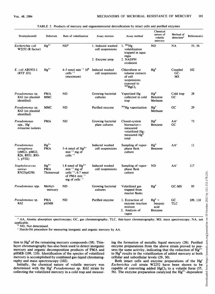
question, as both elemental mercury (Hg0) and organomer-cury compounds such as methyl- and dimethylmercury arevolatile. In most, but not all, cases, it has been demonstratedthat metallic mercury (Hgo) is the volatile end product ofmercury detoxification (Table 2), such that mercury resist-ance and volatilization of Hgo are essentially synonymous.Atomic absorption spectrophotometry is the most popularand widely used method of detecting mercury in biologicalmaterials; however, chemical speciation is not possible bythis method (18). The Hatch-Ott method is routinely used todetermine levels of metallic mercury in samples containingas little as 1 ppb. Total mercury can be determined by aprocedure involving acid hydrolysis, oxidation, and reduc-
FIG. 1. Detoxification of mercury by organomercurial lyase (1)and mercuric reductase (2) enzymes.
NADPH NADP+
RHge - - RH
Hg NADPH
MICROBIOL. REV.
Dow
nloa
ded
from
http
s://j
ourn
als.
asm
.org
/jour
nal/m
r on
19
Oct
ober
202
1 by
121
.153
.178
.231
.
MECHANISMS OF MICROBIAL RESISTANCE OF MERCURY 101
TABLE 2. Products of mercury and organomercurial detoxification by intact cells and purified enzymesChemical
Strain(plasmid) Substrate Rate of volatilization Assay mixture Assay method nature of detectiona Reference(s)mercury
Escherichia coli Hg2+ NDb 1. Induced washed 1. 203Hg ND NA 55, 56W2252 (R factor) cell suspensions
2. Enzyme prep
volatilizationtrapped in aquaregia
2. NADPHoxidation
E. coli AB1932-1(RTF JJ1)
Hg2+ 4-5 nmol min-1 108cells-1(maximum)
Induced washedcell suspensions
Chloroform ortoluene extractsof cellsuspensionsexposed to203HgC12
Pseudomonas sp.K62 (no plasmididentified)
Pseudomonas sp.K62 (no plasmididentified)
Pseudomonasspp., Hgrestuarine isolates
PMAMMC
MMC
PMA
ND Growing bacterialcultures
ND Purified enzyme
ND Growing bacterialplate cultures
Vaporized Hgcollected in coldtrap
203Hg vaporization
Closed-systembioreactor-measuredvolatilized Hg;measured Hg0total
Hg0BenzeneMethane
Cold trapGC
Hg0 GCMethane
Hg0 AAcBenzene GC
Pseudomonasaeruginosa(pMG1, pMG2,R26, R933, R93-1, pVS1)
Staphylococcusaureus
RN23(pI258)
Pseudomonas spp.
Hg2+PMA
Hg2+PMAThimerosal
Methyl-mercury
3-6 nmol of Hg2"min-' mg ofcells-
1.8 nmol of Hg2"min-' mg ofcells-'; 0.7 nmolof PMA min-'mg of cells-1
ND
Induced washedcell suspensions
Induced washedcell suspensions
Growing bacterialcultures
Sampling of vapor-phase flaskculture
Sampling of vapor-phase flaskculture
Volatilized gastrapped fromreactor flasks
Hg0Benzene
ND
Hgo
AAC
AA'
11
117
GC-MS 95
Pseudomonas sp.K62
PMApHMB
ND Purified enzyme 1. Extraction ofenzyme reactionmixture
2. Analysis ofvapor
Hg0 +benzoicacid
Benzene
GCTLC
109, 110
a AA, Atomic absorption spectroscopy; GC, gas chromatography; TLC, thin-layer chromatography; MS, mass §pectroscopy; NA, notapplicable.
b ND, Not determined.c Hatch-Ott procedure for measuring inorganic and organic mercury by AA.
tion to Hg0 of the remaining mercury compounds (38). Thin-layer chromatography has also been used to detect inorganicmercury and organic decomposition products of PMA andpHMB (109, 110). Identification of the species of volatilizedmercury is accomplished by combined gas-liquid chromatog-raphy and mass spectroscopy (102).
Initially, the chemical nature of volatile mercury wasdetermined with the Hgr Pseudomonas sp. K62 strain bycollecting the volatilized mercury in a cold trap and measur-
ing the formation of metallic liquid mercury (28). Purifiedenzyme preparations from the above strain proved to pos-sess the same activity, indicating that the reduction of Hg2+to Hg0 results in the volatilization of added mercury at bothcellular and subcellular levels (29, 30).Both intact cells and enzyme preparations of the Hgr
Escherichia coli strain W2252 have been shown to becapable of converting added HgCl2 to a volatile form (55,56). The enzyme preparation catalyzed the Hg2+-dependent
Hgo CoupledGC-MS
102
28
29
73
VOL. 48, 1984
Dow
nloa
ded
from
http
s://j
ourn
als.
asm
.org
/jour
nal/m
r on
19
Oct
ober
202
1 by
121
.153
.178
.231
.

102 ROBINSON AND TUOVINEN
oxidation of NADPH, which led these investigators toconclude that the Hg2" was concomitantly reduced to metal-lic mercury.
The most convincing evidence to date that metallic mercu-ry is the volatile form of mercury produced upon detoxifica-tion of Hg2+ comes from the combined gas chromatography-mass spectroscopy analysis of the volatilized mercury. Thevolatile form of mercury produced by narrow-spectrum HgrEscherichia coli strain AB1932-1 was found to be soluble inorganic solvents such as toluene, chloroform, benzene, andcyclohexane (102). Metallic mercury is soluble in organicsolvents at 10-5 M (65, 102). However, because organomer-curials such as methylmercury are also soluble in theseorganic solvents, the speciation of the volatile mercuryproved critical to rule out the possibility that methylmercurycould be the end product of detoxification.Toluene extracts of bacterial cultures exposed to 203HgC12
for 7 min at 37°C were analyzed by coupled gas-liquidchromatography-mass spectroscopy. These extracts showeda single peak containing mercury in the gas chromatographs,with a retention time characteristic of metallic mercury. Nohigher-molecular-weight compounds containing mercuryclusters were observed, such as the characteristic mercuryisotope clusters at 198 to 204 mle (Hg2+), 213 to 219 mle(CH3Hg+), and 228 to 234 mle (CH3HgCH3) (102).Thus, the volatilization of mercury by Hgr bacteria has
become synonymous with the production of metallic mercu-ry. This is somewhat misleading because, although themechanism of resistance to Hg2+ appears to be similaramong several bacterial genera, the speciation of the volatil-ized mercury from only a limited number of strains has beenconducted. However, this proposed mechanism is supportedby enzyme studies indicating the NADPH-dependent reduc-tion of Hg>. Since many bacteria are capable of convertingHg2 to other volatile forms of mercury including methyl-mercury, the possibility exists that under some conditionsthese strains may produce other volatile mercury com-pounds.
Detoxification of organomercury compounds. The detoxifi-cation of organomercurials is believed to result from thecleavage of the carbon-mercury linkage by the organomercu-rial lyase enzyme followed by the reduction of Hg2+ to Hg0by the mercuric reductase enzyme. Both of these enzymeshave been purified from Hgr bacteria and are discussed indetail in this paper. The resistance and volatilization patternsof broad-spectrum mercury-resistant bacteria are presentedin Table 1.The decomposition of organomercury compounds by mer-
cury-resistant bacteria was first detected in the Pseudomo-nas sp. K62 soil isolate. This strain was shown to catalyzethe degradatiorn of PMA to Hg0 and benzene by using 203Hg-or 14C-labeled PMA (28). Approximately 70% of the 203Hg or
l4C label was shown to disappear from the medium in 2 h.The products of the reaction could be separated by adsorp-tion to activated carbon and eluted with toluene, thusindicating that the carbon-mercury bond had been cleaved.The phenyl radical of PMA was shown to be converted tobenzene as identified by gas chromatography, whereas thevaporized mercury was found to be metallic mercury. Insimilar experiments, the products of methylmercury were
found to be methane and metallic mercury (28). Purifiedenzyme preparations from this strain were subsequentlyfound to be capable of degrading pHMB to Hg0 and benzoicacid as detected by thin-layer chromatography (109, 110).
Broad-spectrum mercury-resistance plasmids of Pseudo-monas aeruginosa encode resistance to PMA, MMC, EMC,
thimerosal, merbromin, FMA, and pHMB, but the bacteri-um is capable of degrading only the first four compounds(Table 1). In contrast, the broad-spectrum mercury-resist-ance plasmids of Escherichia coli encode volatilization ofmercury from PMA and thimerosal. The broad-spectrumstrains of Escherichia coli are sensitive to EMC, MMC, andpHMB, but they are able to volatilize mercury from thesecompounds at very low rates. Escherichia coli, like Pseudo-monas aeruginosa, is resistant to merbromin and FMA butdoes not decompose either. The mercury-resistant strains ofStaphylococcus aureus are capable of decomposing PMA,pHMB, and thimerosal while being sensitive to the latter(118). In all cases, the decomposition of the organomercurycompound was measured by the volatilization of mercury.The decomposition of PMA by Hgr estuarine isolates
exhibiting varying levels of resistance to the compoundrevealed Hg0 and benzene as products. Over 60% of theisolates were identified as Pseudomonas spp. Metallic andtotal mercury was measured by atomic absorption spec-trophotometry, and benzene was identified by gas chroma-tography as described above. The molar ratio of benzene toHgo varied from 4.4 up to 389 over a 48-h period among thestrains tested. In one case the ratio increased from 5.5 at 48 hto 105 at 143 h. This accumulation of benzene is attributed toa loss of Hgo observed in control experiments, presumablydue to its volatility and ability to adsorb to glass (73). Theseresults suggested again that the PMA was cleaved to produceHge and benzene.
Bacterial degradation of methylmercury has been demon-strated with both mixed and pure cultures from lake sedi-ments. Four isolates were able to degrade between 15 and55% of added (203Hg)CH3HgBr to the methane and Hg0 in 20h. After 170 h methylmercury was completely recoveredfrom the control flask (95). Methane was detected in thehead space of the reaction flask by flame ionization gas-liquid chromatography. All four pure cultures were identi-fied as Pseudomonas spp. and were very similar to thePseudomonas sp. K62 strain described previously (109).
Thus, it appears that bacterial resistance to organomer-cury compounds involves the degradation of methylmer-cury, ethylmercury, PMA, and pHMB to Hg0 and methane,ethane, benzene, and benzoic acid, respectively. The mech-anism in all instances appears to be the same; however, thedegradation products have not been speciated for all broad-spectrum mercury-resistant strains isolated. It is interestingthat purified enzyme preparations of the organomercuriallyase enzyme from the Pseudomonas sp. K62 strain candecompose pHMB, whereas intact cells of Pseudomonasaeruginosa cannot degrade pHMB although they are resist-ant to it. This may represent an alternate mechanism such asa permeability barrier, which may be clarified by uptakestudies with the pHMB-resistant Pseudomonas aeruginosastrain in coordination with enzyme studies.
INDUCIBILITY OF THE MERCURY ANDORGANOMERCURIAL DETOXIFICATION SYSTEMSThe mechanism of resistance to inorganic and organomer-
curials involves the inducible synthesis of the mercuricreductase and organomercurial lyase enzymes by subtoxiclevels of Hg2+ or various organomercurials. The induciblenature of mercury resistance systems has been well docu-mented, with all mercury resistance systems described forEscherichia coli, Staphylococcus aureus, and Pseudomonasspp. comprising over 100 Merr strains that possess inducibleenzyme systems (3, 28-30, 54-57, 90, 91, 100, 101, 104, 117,118).
MICROBIOL. REV.
Dow
nloa
ded
from
http
s://j
ourn
als.
asm
.org
/jour
nal/m
r on
19
Oct
ober
202
1 by
121
.153
.178
.231
.

MECHANISMS OF MICROBIAL RESISTANCE OF MERCURY 103
In all cases tested, the mercuric reductase or organomer-
curial lyase detoxifying enzymes or both are produced onlyin response to the presence of inorganic mercury or a varietyof organic mercury compounds, some of which are notthemselves substrates for degradation, thus acting as gratu-itous inducers. Only one case, involving a T. ferrooxidansstrain, has been reported to be constitutive for the produc-tion of the mercuric reductase enzyme, although initialreports indicated that it too was an inducible system (79, 80).This case bears further study, as it is reported that the strainlost the mercury-volatilizing ability after five passages with-out added Hg2+. This may indicate the loss of the mer genes
or it may represent a different regulatory mechanism, thuswarranting further study. Cross-induction of the mercury
resistance system has been demonstrated, and it appears
that induction by an organomercurial or Hg2+, to which thestrain is resistant, will induce resistance to other Hg com-
pounds. Gratuitous inducers also have been found, such as
merbromin and FMA, that are not themselves substrates forthe mercury-detoxifying enzymes yet are capable of induc-ing the system. However, evidence exists that indicates thatresistance to merbromin and FMA is also inducible, al-though these compounds are not volatilized (117, 118). Boththe mercuric reductase and the organomercurial lyase en-
zymes have been shown to be inducible, although not alwaysin a coordinate fashion.
Growth Inhibition StudiesThe original evidence for the inducible nature of the
mercury resistance system came from growth studies ofmercury-resistant and mercury-sensitive strains in the pres-
ence of inhibitory concentrations of Hg2+ or organomercur-ials. It has been observed that after a variable lag period,lasting from several minutes to several hours, growth of theresistant strains resumed at normal rates (101). The length ofthe lag period varied with the strain and the amount of themercurial compound added. The maximal concentration ofHgCl2 that a mer strain of Staphylococcus aureus was ableto tolerate on solid medium was 20 ,ug mlF-1. However, 10 ,ugml-1 in broth culture was found to be inhibitory to growth,giving rise to a rapid and steep decline in cell numbers duringthe first 8 h. Cell numbers started to increase after 12 h ofexposure, and by 30 h the cell density increased to the same
level as the control grown in the absencelof HgCl2 (57). Inanother case, using Escherichia coli Hgr strain W2252, a lagperiod of 5 h was observed before normal growth in thepresence of 0.02 mM HgC12, compared to a 1-h lag inmedium without added HgCl2 (44). When 0.02 mM HgCl2was added to cultures grown previously with and withoutHgCl2 during early growth (optical density = 0.05), therewas no detectable effect on the cells previously grown in thepresence of HgC12. The cultures previously grown in theabsence of HgCl2 demonstrated the same lag of 5 to 6 hbefore resuming normal growth (55, 56). In contrast, a
mercury-sensitive strain of Escherichia coli, W2252, grew
only after a 60-h lag period in medium containing 0.01 mMHgCl2, whereas immediate growth was observed in themedium without HgC12. In addition, it was noted that cells ofthe sensitive cultures appeared to lyse upon the addition of0.01 mM HgCl2 at an optical density of 0.5, whereas theresistant cells were not susceptible to lysis. The effects ofHgCl2 on the growth of other sensitive and resistant strainsincluding Escherichia coli and Enterobacter aerogenes were
similar (55, 56). Only the resistant strains grew with almostthe same numbers of colonies on plates with and without0.04 mM HgCl2. These results indicated that the resistant
population was homogeneous and that mercury resistancewas not being selected from a heterologous population.Previous growth in the presence of Hg2+ or an organomercu-rial compound at subinhibitory levels eliminates the lagphase observed with mercury-resistant strains.
Induction of Mercuric Reductase and OrganomercurialLyase Detoxifying Enzymes
Hgr and Hgs strains can be distinguished in that thesensitive strains are not capable of producing either one ofthe enzymes responsible for the detoxification of mercurycompounds. Because detoxification of Hg2+ or organomer-curials ultimately results in the volatilization of metallicmercury from the growth medium, the measurement ofenzyme levels or volatilization activity or both in response toadded 203Hg compounds may be used to confirm the induc-ible nature of the system (11, 22, 23, 28, 30, 90, 91, 102).The Pseudomonas sp. K62 strain is resistant to both
organic and inorganic mercurials. Prior exposure to PMA,MMC, or EMC at levels of 20, 10, and 5 mg liter- 1,respectively, was required for the bacteria to be able todecompose 14C-phenyl-labeled PMA (28). Later studiesdemonstrated that the formation of the enzymes dependedupon or were induced only when the organism was grown inthe presence of PMA, mercuric chloride, p-chloromercuricbenzoate (pCMB), merzonin, or metallic mercury. Further-more, the enzymes were not induced by HgS or other metalions; in fact, Cd2+ and Cu2+ were inhibitory (30). Therelationship between PMA and mercuric chloride concentra-tions and the formation of the mercuric reductase andorganomercurial lyase enzymes was studied in depth. Induc-tion constants were calculated for both PMA and mercuricchloride from Lineweaver-Burk plots in which 1/PMA or 1/mercuric chloride were plotted against 1/v (where v =velocity of decomposition of 14C-phenyl-labeled PMA, asdetermined by volatilization). The PMA-decomposing activi-ty reached a maximum when the organism was grown with 6x 10⁻⁵ M PMA or 7.4 x 10⁻⁵ M mercuric chloride, withinduction constants of 3.8 x 10⁻⁵ M for PMA and 4.5 x 10⁻⁵M for HgCI2. Similar results were reported with pCMB,merzonin, or metallic mercury as inducing agent (30). Addi-tional experiments on the effect of other metal ions onenzyme induction led to the conclusion that the induction isspecific for mercury. The ability to produce the enzyme waslost when the organism was subsequently grown withoutmercurials.
In a study (102) with various Hg(II)-resistant, plasmid-bearing strains of Escherichia coli, Pseudomonas aerugin-osa, and Staphylococcus aureus the volatilization activitywas only found to be induced by exposure to HgCl2 at 10-5M for not less than 3 h. Other mercurials were not tested inthis study. Only the resistant strains were found to volatilizemercury, and in all cases the rate of loss of 203Hg was muchhigher when cells had been exposed to 10-5 M HgCl2 beforedetermination of mercury-volatilizing activity (102). Theseresults are summarized in Table 3.
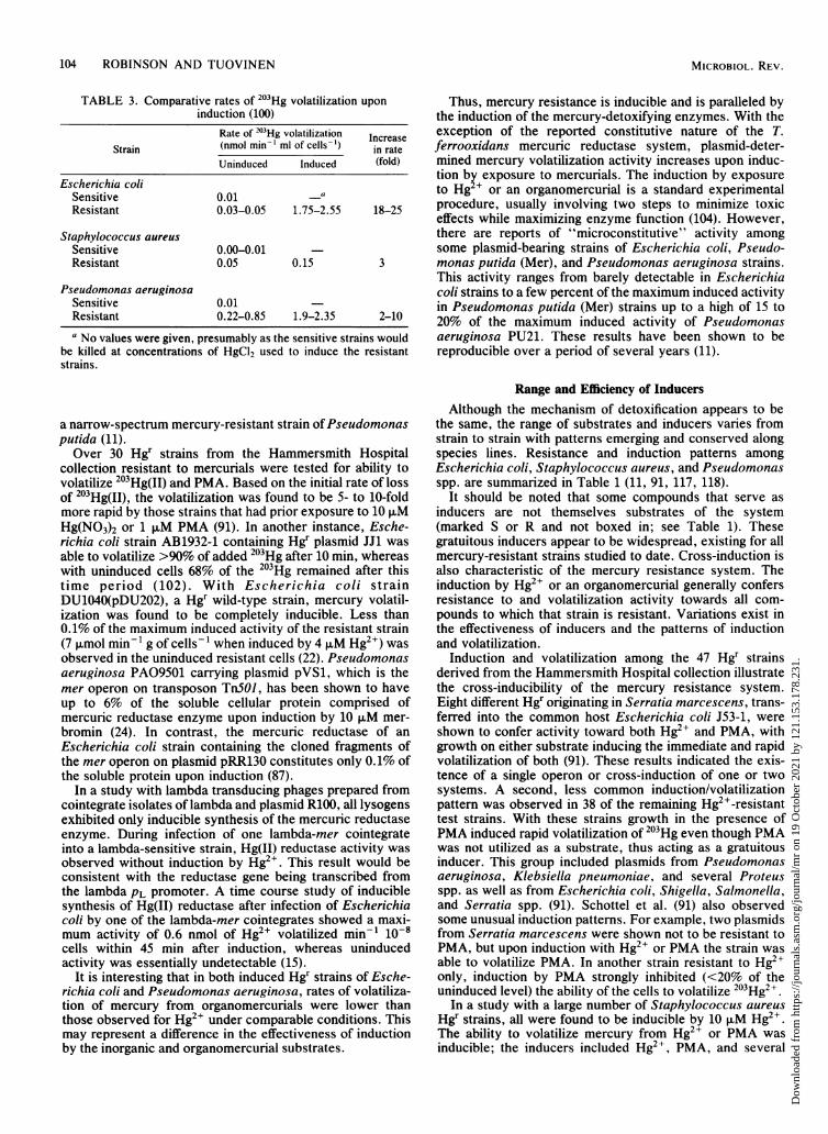
It is apparent that in all cases there is a significant increasein the rate of volatilization of 203Hg between uninduced andinduced resistant strains. The Staphylococcus aureus strainsdo not exhibit as marked a difference between sensitive andresistant cells or between uninduced and induced resistantstrains as seen with the Escherichia coli and Pseudomonasaeruginosa test strains (102). These results were reported tobe reproducible. Exposure to 10 ,uM Hg2+ was shown tocause a 10-fold increase in the rate of 203Hg volatilization by
VOL. 48, 1984
Dow
nloa
ded
from
http
s://j
ourn
als.
asm
.org
/jour
nal/m
r on
19
Oct
ober
202
1 by
121
.153
.178
.231
.
104 ROBINSON AND TUOVINEN
TABLE 3. Comparative rates of 203Hg volatilization uponinduction (100)
Rate of 203Hg volatilization IncreaseStrain (nmol min-1 ml of cells-') in rate
Uninduced Induced (fold)
Escherichia coliSensitive 0.01 aResistant 0.03-0.05 1.75-2.55 18-25
Staphylococcus aureusSensitive 0.00-0.01Resistant 0.05 0.15 3
Pseudomonas aeruginosaSensitive 0.01Resistant 0.22-0.85 1.9-2.35 2-10
a No values were given, presumably as the sensitive strains wouldbe killed at concentrations of HgCl2 used to induce the resistantstrains.
a narrow-spectrum mercury-resistant strain ofPseudomonasputida (11).Over 30 Hgr strains from the Hammersmith Hospital
collection resistant to mercurials were tested for ability tovolatilize 203Hg(II) and PMA. Based on the initial rate of lossof 203Hg(II), the volatilization was found to be 5- to 10-foldmore rapid by those strains that had prior exposure to 10 ,uMHg(NO3)2 or 1 ,uM PMA (91). In another instance, Esche-richia coli strain AB1932-1 containing Hgr plasmid JJ1 wasable to volatilize >90% of added 203Hg after 10 min, whereaswith uninduced cells 68% of the 203Hg remained after thistime period (102). With Escherichia coli strainDU1040(pDU202), a Hgr wild-type strain, mercury volatil-ization was found to be completely inducible. Less than0.1% of the maximum induced activity of the resistant strain(7 ,umol min- 1 g of cells- 1 when induced by 4 ,uM Hg2+) wasobserved in the uninduced resistant cells (22). Pseudomonasaeruginosa PA09501 carrying plasmid pVS1, which is themer operon on transposon TnSOJ, has been shown to haveup to 6% of the soluble cellular protein comprised ofmercuric reductase enzyme upon induction by 10 ,uM mer-bromin (24). In contrast, the mercuric reductase of anEscherichia coli strain containing the cloned fragments ofthe mer operon on plasmid pRR130 constitutes only 0.1% ofthe soluble protein upon induction (87).
In a study with lambda transducing phages prepared fromcointegrate isolates of lambda and plasmid R100, all lysogensexhibited only inducible synthesis of the mercuric reductaseenzyme. During infection of one lambda-mer cointegrateinto a lambda-sensitive strain, Hg(II) reductase activity wasobserved without induction by Hg2>. This result would beconsistent with the reductase gene being transcribed fromthe lambda PL promoter. A time course study of induciblesynthesis of Hg(II) reductase after infection of Escherichiacoli by one of the lambda-mer cointegrates showed a maxi-mum activity of 0.6 nmol of Hg2> volatilized min-1 10-8cells within 45 min after induction, whereas uninducedactivity was essentially undetectable (15).
It is interesting that in both induced Hgr strains of Esche-richia coli and Pseudomonas aeruginosa, rates of volatiliza-tion of mercury from organomercurials were lower thanthose observed for Hg2> under comparable conditions. Thismay represent a difference in the effectiveness of inductionby the inorganic and organomercurial substrates.
Thus, mercury resistance is inducible and is paralleled bythe induction of the mercury-detoxifying enzymes. With theexception of the reported constitutive nature of the T.ferrooxidans mercuric reductase system, plasmid-deter-mined mercury volatilization activity increases upon induc-tion by exposure to mercurials. The induction by exposureto Hg + or an organomercurial is a standard experimentalprocedure, usually involving two steps to minimize toxiceffects while maximizing enzyme function (104). However,there are reports of "microconstitutive" activity amongsome plasmid-bearing strains of Escherichia coli, Pseudo-monas putida (Mer), and Pseudomonas aeruginosa strains.This activity ranges from barely detectable in Escherichiacoli strains to a few percent of the maximum induced activityin Pseudomonas putida (Mer) strains up to a high of 15 to20% of the maximum induced activity of Pseudomonasaeruginosa PU21. These results have been shown to bereproducible over a period of several years (11).
Range and Efficiency of InducersAlthough the mechanism of detoxification appears to be
the same, the range of substrates and inducers varies fromstrain to strain with patterns emerging and conserved alongspecies lines. Resistance and induction patterns amongEscherichia coli, Staphylococcus aureus, and Pseudomonasspp. are summarized in Table 1 (11, 91, 117, 118).
It should be noted that some compounds that serve asinducers are not themselves substrates of the system(marked S or R and not boxed in; see Table 1). Thesegratuitous inducers appear to be widespread, existing for allmercury-resistant strains studied to date. Cross-induction isalso characteristic of the mercury resistance system. Theinduction by Hg2+ or an organomercurial generally confersresistance to and volatilization activity towards all com-pounds to which that strain is resistant. Variations exist inthe effectiveness of inducers and the patterns of inductionand volatilization.
Induction and volatilization among the 47 Hgr strainsderived from the Hammersmith Hospital collection illustratethe cross-inducibility of the mercury resistance system.Eight different Hgr originating in Serratia marcescens, trans-ferred into the common host Escherichia coli J53-1, wereshown to confer activity toward both Hg2' and PMA, withgrowth on either substrate inducing the immediate and rapidvolatilization of both (91). These results indicated the exis-tence of a single operon or cross-induction of one or twosystems. A second, less common induction/volatilizationpattern was observed in 38 of the remaining Hg2'-resistanttest strains. With these strains growth in the presence ofPMA induced rapid volatilization of 203Hg even though PMAwas not utilized as a substrate, thus acting as a gratuitousinducer. This group included plasmids from Pseudomonasaeruginosa, Klebsiella pneumoniae, and several Proteusspp. as well as from Escherichia coli, Shigella, Salmonella,and Serratia spp. (91). Schottel et al. (91) also observedsome unusual induction patterns. For example, two plasmidsfrom Serratia marcescens were shown not to be resistant toPMA, but upon induction with Hg2+ or PMA the strain wasable to volatilize PMA. In another strain resistant to Hg2Ionly, induction by PMA strongly inhibited (<20% of theuninduced level) the ability of the cells to volatilize 203Hg2+.
In a study with a large number of Staphylococcus aureusHgr strains, all were found to be inducible by 10 ,uM Hg2+.The ability to volatilize mercury from Hg2+ or PMA wasinducible; the inducers included Hg2+, PMA, and several
MICROBIOL. REV.
Dow
nloa
ded
from
http
s://j
ourn
als.
asm
.org
/jour
nal/m
r on
19
Oct
ober
202
1 by
121
.153
.178
.231
.
MECHANISMS OF MICROBIAL RESISTANCE OF MERCURY 105
organomercurials that do not serve as substrates (90, 91).Staphylococcus aureus RN23 harboring the broad-spectrummercury resistance plasmid PI258 was induced most effec-tively by Hg2+, as compared to PMA, pHMB, MMC, andmerbromin, based on measuring rate of 203Hg2+ volatiliza-tion. However, in other experiments merbromin was acomparable or better inducer, and although PMA is asubstrate it proved to be a poor inducer. MMC was found tobe neither a substrate nor an inducer of the system (117).Mutant variants of Staphylococcus aureus plasmids PI258and P1147 were induced to low levels of activity (10% of thewild type) with an optimum inducer level of 2.5 ,uM Hg2' ascompared to 10 ,uM for the wild type. This may indicate thatan uptake system is still intact while the mercuric reductaseis being produced at low levels. In this case, 10 ,uM Hg2+would now prove to be toxic. Therefore, only Hg2+, thimer-osal, and PMA are substrates for volatilization of 203Hg, withPMA as a poor inducer and pHMB and merbromin effectiveas gratuitous inducers.The same patterns of induction/volatilization do not exist
in Pseudomonas aeruginosa and Pseudomonas putida mer-cury-resistant strains. MMC and EMC are additional sub-strates for the volatilization of mercury by Pseudomonasspp. broad-spectrum resistance plasmids (Table 1). A low-level resistance to pHMB was observed but the cells areunable to hydrolyze this compound (11). Both PMA andthimerosal were found to be poor but significant inducers ofmercury volatilization activity, measured as 203Hg volatiliza-tion from 203Hg2+ by strains of Pseudomonas aeruginosaand Pseudomonas putida harboring narrow-spectrum resist-ance plasmids. The same compounds were much moreeffective inducers with the broad-spectrum organomercurialresistance plasmids, with volatilization rates comparable tothose observed with Hg2+ as the inducer. Narrow-spectrumplasmid-bearing strains were found to be preferentially in-duced by Hg2+, whereas merbromin and pHMB were effec-tive inducers of both narrow- and broad-spectrum resistantstrains of both Pseudomonas aeruginosa and Pseudomonasputida (11).
whereas under some conditions (not specified) thimerosaland merbromin induced PMA degradation preferentially(11). There were also differences observed between thestrains tested, with all inducers tested giving higher Hg2+-volatilizing activity in strain PU21, although the maximumvolatilization activities of PMA were higher with this strain.
Similar experiments were conducted with Staphylococcusaureus wild-type, mercury-resistant, plasmid-bearing strainRN23 and an Hgs mutant, RN987, exhibiting high lyaseactivity but low reductase activity (18% of the wild-typelevels). With strain RN23, induction of lyase and reductaseactivities approximately coordinate at the whole-cell level.However, the scatter observed was considered to be greaterthan could be attributed to experimental error, indicatingthat some induction conditions give rise to disproportionatelevels of the two activities, but not to the extent observedwith Pseudomonas sp. strains. With the mutant strainRN987, noncoordinate induction was observed. Merbrominwas shown to induce preferentially lyase activity, whereaspHMB preferentially induced reductase activity, as was alsoobserved with strain PU21 (118). These results suggest thatthe organomercurial lyase and mercuric reductase enzymesmay be transcribed from separate operator-promoter regionsthat are cross-induced under some conditions, but not al-ways in a coordinate fashion. In general, studies indicatethat the lyase activity is the limiting step in the detoxificationof organomercurial compounds. This may account for whatappears to be greater volatilization of mercury from Hg2+than from organomercurial substrates such as PMA, as wellas why some compounds appear to preferentially induceHg2+ volatilization activity. This does not explain preferen-tial induction ofPMA under some conditions as cited above.More detailed studies of coordinate induction are required,including a comparison of induction/volatilization patternsamong Hgr plasmids from different sources in a commonhost strain. This line of research would determine to whatextent, if any, the host cell background affects these pat-terns.
TRANSPOSABILITY OF mer GENESCoordinate Induction of Mercuric Reductase and
Organomercurial Lyase ActivitySeparate enzymes appear to be involved in the detoxifica-
tion of mercury and organomercurial compounds. The or-ganomercurial lyase enzyme hydrolyzes the organomercuri-al such as PMA to produce benzene and Hg2+, which isfollowed by the reduction of Hg2+ to Hgo by mercuricreductase. The question of coordinate induction of bothmercuric reductase and organomercurial lyase activities hasbeen addressed in studies with Staphylococcus aureus andseveral Pseudomonas sp. strains (11, 117). Coordinate in-duction of activity has been considered indicative of genesunder common regulatory control, constituting an operon.With the broad-spectrum mercury-resistant Pseudomonas
aeruginosa strains PU21 (FP2) and PU21 (R3108), organo-mercurial lyase activity (as percentage of the maximumPMA volatilization activity) was plotted against mercuricreductase activity (as percentage of Hg2+ volatilized), usingHg2+, PMA, thimerosal, pHMB, and merbromin as induc-ers. Both activities were found to be inducible by thesecompounds, but induction was not strictly coordinate. Therewas significant scatter from the line drawn at 450 represent-ing coordinate induction, with all experimental points fallingbelow the line for both strains. In particular, pHMB ap-peared to induce volatilization from Hg2+ preferentially,
The mercury resistance genes, as for other resistancegenes carried by plasmids, often occur on transposons (3,96). These transposable elements are specific DNA se-quences that can insert more or less randomly into otherDNA sequences in the absence of host cell-mediated recom-bination functions. It is generally accepted that these ele-ments play an important role in evolution (53). Many differ-ent transposable elements have been identified andcharacterized, and they are believed to be widespread innature. Plasmid-borne genes for resistance to ampicillin,chloramphenicol, kanamycin, streptomycin, and trimetho-prim have all been shown to exist on transposons singly or incombination (58). These elements have demonstrated theability to transpose from plasmid to plasmid and fromplasmid to chromosome, carrying genes for antibiotic resist-ance, as well as a variety of functions including fusion ofunrelated DNA molecules, deletions, inversions, excisions,and as functional transcriptional start and stop signals (53).Several such transposons associated with the acquisition ofmercuric resistance have been described (Table 4) (3-5, 7, 8,13, 27, 33, 96, 105, 107). The transposable element TnSOJ,associated with mercuric resistance, has been well charac-terized (3, 8, 96). TnSOJ has been described as a discretepiece ofDNA, 5.2 megadaltons (Mdal) in size, as determinedby intramolecular heteroduplex analysis of hybrid plasmid
VOL. 48, 1984
Dow
nloa
ded
from
http
s://j
ourn
als.
asm
.org
/jour
nal/m
r on
19
Oct
ober
202
1 by
121
.153
.178
.231
.

106 ROBINSON AND TUOVINEN
TABLE 4. Hgr transposons
Strain/plasmid Merf transposon (markers) Mol wt Terminal sequences (bp) Transpositional frequency Reference(s)(Hgr)
Pseudomonas TnS01 (Su, Hg) 5.2 x 10⁶ 38 2.9 x 10⁻³ (RR1-1) 3, 8, 96aeruginosa pVS1 5.9 x 10⁻³ (RPl)
(integrates in 5 sites)
P. putida (originally Tn1861 (same serotype 5.5 x 10⁶ NC" 1.5-2.0 x 10⁻¹ 27isolated from pVS1) as Tn501)
Pseudomonas sp. Tn502 (different >5.2 x 10⁶ NC NRb 92serotype fromTn501); TnSO3
Escherichia coli K-12 Tn2670 13.3 x 10⁶ 38 (30/38 homologous 3.0 x 10⁻³ to 37derivatives R100.1 Tn2671 (20 kbc) to Tn501) 6.0 x 10⁻⁴IncFII Tn21
Tn2603 20 kb 200 (inverted repeats)
E. coli K-12 R861 Not designated 8.5-11.2 x 10⁶ NC NDd (insertional 105inactivation of TCgene)
a NC, Not characterized or sequenced.b NR, Transposition frequency not reported/data not available for calculation.c kb, Kilobases.d ND, Not determined.
RP1-TnS01 carrying two copies of the putative transposonand by comparison of ColEl (4.2 Mdal) with hybrid plasmidpUB781(ColE1::Tn501), 9.4 Mdal (3). TnSOJ was originallydiscovered on the small nonconjugative Pseudomonas sp.plasmid pVS1 and can function in Escherichia coli (96). Intransduction studies with derivatives of Pseudomonas aeru-ginosa PAO or PAT strains, mercury-resistant (Hgr) trans-ductants were recovered from both Rec+ and Rec- recipi-ents at frequencies of 2.3 x 10⁻⁸ to 29 x 10-8 PFU-1. Thisindicates that inheritance of the mer genes present onplasmid pVS1 is not dependent on the host recombinationsystem (96). It has been demonstrated that TnSOJ is able totranslocate into RP1, as well as to other broad-host-rangeconjugative plasmids. This occurs in either orientation atmany sites in both Escherichia coli and Pseudomonas sp.host strains, as determined by restriction endonucleaseanalysis of RP1::Tn501 recombinant plasmids, and is notdependent on host-mediated recombination mechanisms (3).Furthermore, TnSOJ insertion into a number of RP1 sites hasbeen demonstrated to lead to the loss of RP1-borne resist-ance determinants of RP1::TnS5O recombinants, using Hgrtransconjugants generated from Pseudomonas sp.-Esche-richia coli matings. The loss of the Tra+ phenotype occurredin 1 to 8% of the transconjugants, whereas 1 to 4% of theTnSOI insertions resulted in the Cb5 or Kms RP1 phenotype.All RP1::TnSOJ hybrids, except Cbs/Amps mutants, showeda constant increase in size, indicative of integration of TnSOJ(5.2 x 10⁶). Mutational events leading to Amps strains mayhave occurred by a deletion in TnA (Ampr gene) as the resultof TnSOJ transposition, a well-documented recombinationalphenomenon associated with transposons as stated above.The recombinant plasmids pUB404 and pUB399 apparent-
ly carry two copies of TnSOJ as inverted repeats, demon-strated by intramolecular heteroduplex experiments (3).Double insertions of TnA and Tn801 have also been report-ed, with the two copies inverted with respect to one another(3), perhaps necessary for molecular stability avoiding intra-molecular recombination. TnSOJ has been shown to exist
integrated into the chromosome of Escherichia coli strainUB5203, appearing to be quite stable with no detectable(<10-9) auxotrophic mutations (96).The terminal nucleotide sequences of the mercury resist-
ance transposon TnSOJ have recently been determined withpUB781 (CoIEl::TnS5O hybrid) which was previously de-scribed by Bennett and co-workers (3). The terminal se-quences were found to be inverted repeat sequences of 38nucleotide base pairs (bp) at either end. The transposon wasalso found to be flanked by a 5-bp direct repeat, presumablygenerated in recipient DNA upon insertion (87). The invert-ed repeats of TnSOJ are similar to those found in Tn3 andothers, suggesting a functional similarity in the transposi-tional process. They may have a common evolutionaryorigin, as the inverted repeated sequences are required fortransposition, presumably to act as recognition sites forspecific nucleases.A Pseudomonas putida mercury resistance determinant
has been shown to be a 5.2-Mdal transposon, designatedTnJ861 (27). Tn1861 is described as a discrete element thatcan transpose from the chromosome of Pseudomonas putidato plasmids, then to the Escherichia coli chromosome, andback again to other plasmids as deduced from varioustransformation studies with RP4 and RSF1010 plasmids (27).These investigators failed to isolate plasmid DNA from amercury-resistant Pseudomonas putida strain, even afterlong-term cultivation in the presence of 160 ,ug of Hg2+ mI-1.The level of mercury used was much higher than the 12- to15-,ug ml-' levels routinely used by others. Transfer of theMerr phenotype was not detectable (<10-9 cell-) in theseplasmidless Merr Pseudomonas putida strains, whereas ad-ditional mutations causing phenotypes such as Ade-, Trp-,and Thr- were detected at a frequency of 10-2 to 10-3 cell- 1
generation-1. Spontaneous reversion of Ade- to Ade+ phe-notype in Ade- mutants ofPseudomonas putida strain AC77(AC1000, Met-/Ade-) was about 10-8 (27).
Transposition of Mer resistance from chromosome tobroad-host-range plasmids RP4 and RSF1010, and thereby
MICROBIOL. REV.
Dow
nloa
ded
from
http
s://j
ourn
als.
asm
.org
/jour
nal/m
r on
19
Oct
ober
202
1 by
121
.153
.178
.231
.
MECHANISMS OF MICROBIAL RESISTANCE OF MERCURY 107
transmission to other strains, was studied by Friello andChakrabarty (27). Broad-host-range plasmids RP4 andRSF1010 were introduced into Pseudomonas putida strainAC100, subsequently isolated, and transformed into Esche-richia coli and other Pseudomonas putida strains. With RP4,15 to 20% of the transformants were both Cbr (an RP4marker) and Hgr, i.e., a transposition frequency of 1.5 x10⁻¹ to 2.0 x 10⁻¹, exhibiting a 1 to 2% loss of Hgr whengrown in the absence of Hg2 . In contrast, the acquisition ofmercury resistance by Escherichia coli and Pseudomonasputida transformants, using RSF1010, was 2 to 5%, i.e., atransposition frequency of 2 x 10⁻² to 5.0 x 10⁻² and a 50 to70% loss of Hgr when grown in the absence of Hg2+ for 20generations. This may indicate there is some site preferencefor TnJ861 in RP4 or instability in RSF1010. TnJ861 transpo-sition and mutation induction were found to be independentof host recombination functions, as indicated by performingexperiments in RecA- hosts. Analysis of RP4::TnJ861 hy-brids by restriction endonuclease digestion and electronmicroscopic examination showed Tn1861 to be 5.5 (± 0.2)Mdal in 90% of the molecules examined. The remaining 10%of the hybrids showed a 11.5-Mdal increase, indicating theacquisition of two Tn1861 molecules (24). TnJ861 and TnSOJare similar in several respects. Both were originally isolatedfrom the nonconjugative plasmid pVS1 in Pseudomonasaeruginosa. They are similar in molecular weight (5.2 x 10⁶versus 5.5 x 10⁶), and both have demonstrated the ability totranspose from one plasmid to another or to the hostchromosome independent of host recombination systems (3,27, 96). In addition, they appear to be immunologicallysimilar. A crude enzyme preparation of the mercuric reduc-tase from Pseudomonas putida AC77 was immunologicallysimilar to a large number of mercuric reductases, includingthose specified by Escherichia coli plasmid R100, Pseudo-monas fluorescens B69, and from Tn501 (92). However,whereas both transposons have three EcoRI restriction sitesand a single HindIII site, TnS01 has a unique SalGI site andboth appear as multiple copies on hybrid plasmids studied.The most striking dissimilarity between the two transposonsis the genetic instability of both Escherichia coli and Pseudo-monas putida host strains harboring TnJ861, resulting inmultiple auxotrophic mutants which have not been observedwith Tn5O0. However, Friello and Chakrabarty (27) did notspecify background auxotrophic mutation frequencies instrains not harboring Tn1861, nor did they investigate thepossibility of the role of other transposons in such mutation-al events.The nucleotide sequences at the ends of TnJ861 have not
been determined. Further work in this area is warranted todetermine the extent of similarity between Tn1861 andTn501 which has been sequenced (8). TnJ861 may possess amodified transposase or resolvase gene that may account forthe observed differences between it and TnSOJ, including theincreased frequency of mutations.A number of other transposons have been shown to carry
genes for mercury resistance (Hg2+). Some well-character-ized examples are presented in Table 1, and these have beendescribed in detail previously (13, 37, 107). In studies ofdeletion mutants, Tn2J has been shown to possess genesdesignated tnpA and tnpR which encode a transposase andresolvase, respectively (13). These genes are necessary fortransposition to occur via cointegrate formation (tnpA) andresolution of the cointegrates (tnpR). The products of thetnpA genes of Tn2J and TnSOJ have been shown to compli-ment one another as indicated by transposition complemen-tation assays with tnpA mutants (13). The terminal inverted
repeat sequences of Tn2J and TnSOJ are homologous in 30 of38 bp, as expected, because the product of the tnpA gene isknown to act on the ends of these transposable elements.The resolvase activity of both TnSOJ and Tn2J is well belowthat of Tn3, making secondary rearrangements a greaterpossibility, such as an increased mutation frequency orincorporation of new genes.The transposition of mer genes from the IncH plasmid
R826 to IncN plasmid R45 in an Escherichia coli recAbackground has been investigated (105). With a recA- donorand a recA- (R-) recipient, at temperatures restrictive to thetransfer of R826, 50% of the transconjugants had all of themarkers associated with R45 plus mercury resistance. Fourof these Merf transconjugants were studied further. Onlytwo (RAS305 and RAS324) were stable with regard to themercury phenotype; the other two were considerably lessstable and lost mercury resistance if Hg> was not presentfor selection. One of the stable derivatives, RAS324, had lostTcr, suggesting that insertion of mer genes had inactivatedthis gene. Inactivation of insertion was also discovered withTnSOJ and Tn1861. Although a recA--independent transpo-sition of sequences, including the mer resistance genes,appears to occur, a consistent amount of DNA was nottransferred in all cases. Sequences ranging from 8.5 to 11.2Mdal in size were found in the Merr transconjugants, asdetermined by restriction digests. These acquired sequenceswere also found to be larger than those of several well-characterized mercury resistance transposons or operons(e.g., 6.2 Mdal in R100 and 5.2 Mdal for Tn5OJ). In addition,no inverted repeat sequences were observed in heteroduplexstudies of the Merf transconjugants, whereas there was someevidence for tandem repeats of mer genes in one transconju-gant. These observations are problematic and argue againstthe existence of a unique mer transposon in the strainsstudied.Examples of transposons in the gram-positive organism
Staphylococcus aureus have been demonstrated, such asTn.SS, a 5.2-kilobase transposon carrying erythromycinresistance genes (76). No mercury resistance transposon hasbeen demonstrated in any of the broad-spectrum Merrstrains of Staphylococccus aureus.The existence of transposons encoding mercury resistance
genes has a number of implications with regard to theevolution and dissemination of mercury resistance in bothclinical and natural environments. Further studies involvingmore stringent selections for transposition of mercury resist-ance genes and heteroduplex analysis between R45 deriva-tives and the cloned segments of other mer transposons arewarranted. In a recent study, the molecular interrelation-ships were examined among a family of transposons encod-ing Hg resistance and resistance to multiple antibiotics (108).Physical and functional mapping indicated that the multiple-antibiotic-resistant transposons Tn21, Tn2603, Tn2607, andTn4 have evolved from an ancestral mercury transposon.A new Hg resistance transposon designated Tn26131 was
detected on the naturally occurring plasmid pCS229 and isbelieved to be this ancestral transposon (108). Tn2613 wassimilar to TnSOJ except for a difference in the right-hand end.Tn2613 appears to have acquired streptomycin and sulfon-amide resistance genes, resulting in Tn2608, with the struc-tural features of Tn2613 conserved. This suggests a closeevolutionary relationship between Tn2613 and Tn2608. Theother multiple-antibiotic-resistance transposons Tn2l,Tn2603, Tn2607, and Tn4 were shown to have sequencessimilar to those of Tn2613 and Tn2608. It is conceivable thatthe transposons have evolved by the sequential acquisition
VOL. 48, 1984
Dow
nloa
ded
from
http
s://j
ourn
als.
asm
.org
/jour
nal/m
r on
19
Oct
ober
202
1 by
121
.153
.178
.231
.
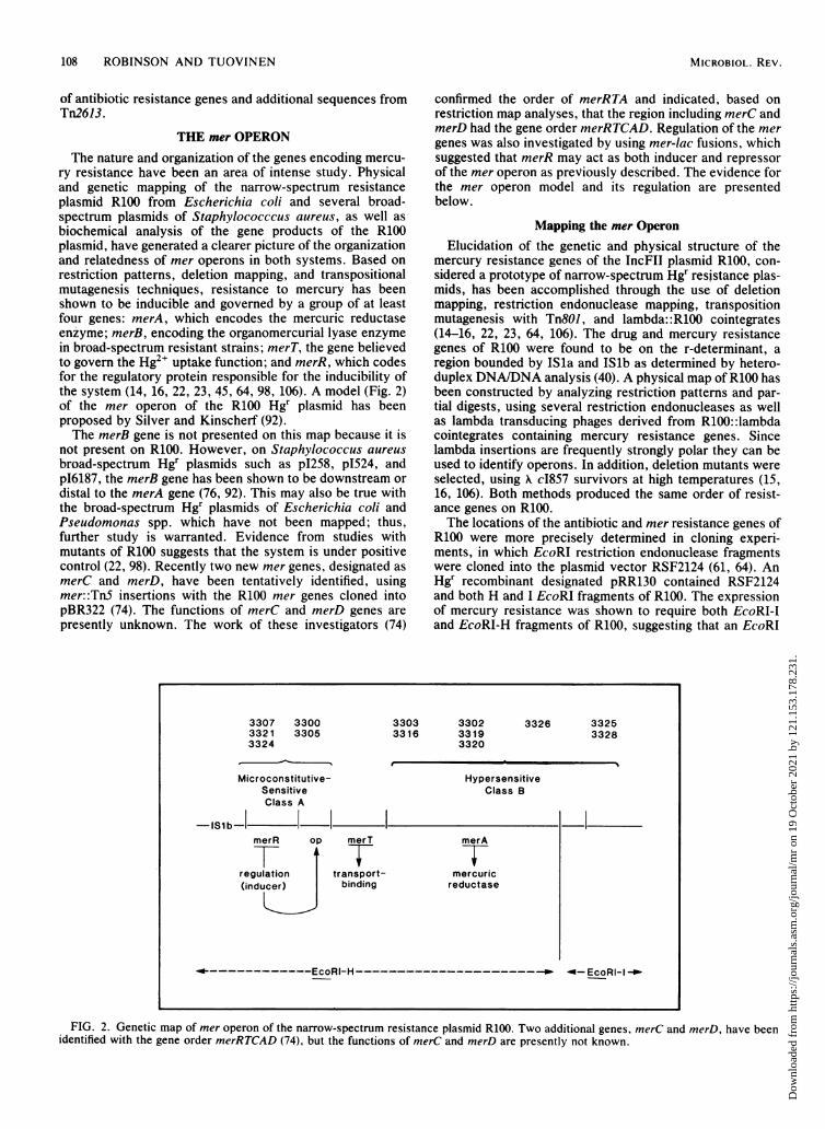
108 ROBINSON AND TUOVINEN
of antibiotic resistance genes and additional sequences fromTn2613.
THE mer OPERONThe nature and organization of the genes encoding mercu-
ry resistance have been an area of intense study. Physicaland genetic mapping of the narrow-spectrum resistanceplasmid R100 from Escherichia coli and several broad-spectrum plasmids of Staphylococccus aureus, as well asbiochemical analysis of the gene products of the R100plasmid, have generated a clearer picture of the organizationand relatedness of mer operons in both systems. Based onrestriction patterns, deletion mapping, and transpositionalmutagenesis techniques, resistance to mercury has beenshown to be inducible and governed by a group of at leastfour genes: merA, which encodes the mercuric reductaseenzyme; merB, encoding the organomercurial lyase enzymein broad-spectrum resistant strains; merT, the gene believedto govern the Hg2+ uptake function; and merR, which codesfor the regulatory protein responsible for the inducibility ofthe system (14, 16, 22, 23, 45, 64, 98, 106). A model (Fig. 2)of the mer operon of the R100 Hgr plasmid has beenproposed by Silver and Kinscherf (92).The merB gene is not presented on this map because it is
not present on R100. However, on Staphylococcus aureusbroad-spectrum Hgr plasmids such as pI258, p1524, andp16187, the merB gene has been shown to be downstream ordistal to the merA gene (76, 92). This may also be true withthe broad-spectrum Hgr plasmids of Escherichia coli andPseudomonas spp. which have not been mapped; thus,further study is warranted. Evidence from studies withmutants of R100 suggests that the system is under positivecontrol (22, 98). Recently two new mer genes, designated asmerC and merD, have been tentatively identified, usingmer::Tn5 insertions with the R100 mer genes cloned intopBR322 (74). The functions of merC and merD genes arepresently unknown. The work of these investigators (74)
330733213324
33003305
confirmed the order of merRTA and indicated, based onrestriction map analyses, that the region including merC andmerD had the gene order merRTCAD. Regulation of the mergenes was also investigated by using mer-lac fusions, whichsuggested that merR may act as both inducer and repressorof the mer operon as previously described. The evidence forthe mer operon model and its regulation are presentedbelow.
Mapping the mer OperonElucidation of the genetic and physical structure of the
mercury resistance genes of the IncFII plasmid R100, con-sidered a prototype of narrow-spectrum Hgr resistance plas-mids, has been accomplished through the use of deletionmapping, restriction endonuclease mapping, transpositionmutagenesis with Tn801, and lambda: :R100 cointegrates(14-16, 22, 23, 64, 106). The drug and mercury resistancegenes of R100 were found to be on the r-determinant, aregion bounded by ISla and ISlb as determined by hetero-duplex DNA/DNA analysis (40). A physical map of R100 hasbeen constructed by analyzing restriction patterns and par-tial digests, using several restriction endonucleases as wellas lambda transducing phages derived from R100::lambdacointegrates containing mercury resistance genes. Sincelambda insertions are frequently strongly polar they can beused to identify operons. In addition, deletion mutants wereselected, using X c1857 survivors at high temperatures (15,16, 106). Both methods produced the same order of resist-ance genes on R100.The locations of the antibiotic and mer resistance genes of
R100 were more precisely determined in cloning experi-ments, in which EcoRI restriction endonuclease fragmentswere cloned into the plasmid vector RSF2124 (61, 64). AnHgr recombinant designated pRR130 contained RSF2124and both H and I EcoRI fragments of R100. The expressionof mercury resistance was shown to require both EcoRI-Iand EcoRI-H fragments of R100, suggesting that an EcoRI
3303 33023316 3319
3320
Microconstitutive-SensitiveClass A
merR op merT
regulation transport-(inducer) binding
3326 33253328
HypersensitiveClass B
merA
mercuricreductase
-- EcoRl-I -_
FIG. 2. Genetic map of mer operon of the narrow-spectrum resistance plasmid R100. Two additional genes, merC and merD, have beenidentified with the gene order merRTCAD (74), but the functions of merC and merD are presently not known.
MICROBIOL. REV.
Dow
nloa
ded
from
http
s://j
ourn
als.
asm
.org
/jour
nal/m
r on
19
Oct
ober
202
1 by
121
.153
.178
.231
.
MECHANISMS OF MICROBIAL RESISTANCE OF MERCURY 109
cleavage site lies within the mer operon. This was deter-mined by examining Hgr transformants containing EcoRIfragment H or I. The transformants containing H or I onlydid not confer mercury resistance, regardless of their orien-tation. Additional studies were performed to confirm thatboth fragments were required to confer the Hgr phenotypeand to rule out the possibility that the orientation of eitherfragment alone could confer resistance. Plasmid DNA frompRR132 (containing PSF2124 plus fragment I) and pRR134(containing RSF2124 plus fragment H), as well as a mixtureof both, was treated with EcoRI, ligated, and used intransformations to select for Hgr (64). Results indicated thatonly a mixture of the DNA from both plasmids yielded Hgrtransformants. Similar conclusions were reached withR100.1 and R6-5 plasmids by other investigators (10, 61).The organization of the genes on the mercury resistance
region of the mer operon of R100 was determined primarilyby examining point and deletion mutants constructed bytranspositional mutagenesis and subsequent complementa-tion and recombinant analysis (22, 23, 71, 106). Preliminarystudies indicated that Tn8O1 insertions into R100 resulted inmercuric ion sensitivity at a relatively high frequency, thusleading to its use as a mutagenic probe for biochemical andgenetic analyses of the mer operon (23). A series of 23transposon Tn801-induced Hgs mutants of pDU202 (a tetra-cycline-sensitive derivative of R100.0 which is a mutant ofR100 depressed for conjugal transfer) were studied. Basical-ly two classes of mutants were obtained upon testing forsensitivity to HgCl2, merbromin, and FMA: (i) sensitivemutants which were shown to be as sensitive to the mercurycompounds as were the plasmidless strains; and (ii) hyper-sensitive mutants which were four- to sevenfold more sensi-tive than the plasmidless strains. The hypersensitive mutantswere also hypersensitive to the organomercurials merbrominand FMA, and they were shown to be inducible such thatuninduced cells were as sensitive as the plasmidless strainsand upon induction became hypersensitive. These mutantswere assayed for mercuric reductase enzyme, using bothwhole cells and cell-free extracts. In 18 sensitive and hyper-sensitive mutants of the 23 examined, there was no detect-able enzyme at the whole-cell level (at a resolution limit of<0.1% of the wild-type activity). The other five sensitivestrains, designated pDU3300, pDU3305, pDU3307,pDU3321, and pDU3324, exhibited a low level of activitythat was expressed constitutively. No in vitro enzymeactivity was found in the sensitive or hypersensitive mu-
tants, whereas the microconstitutive-sensitive mutants hadlow levels of Hg(II) reductase, consistent with their pheno-type. Thus, no cryptic mutants were observed, i.e., low-activity mutants that had high cell-free levels of Hg(II)reductase enzyme (23). In studies with the recombinants ofRSF2124 containing R100 EcoRI-H and -I fragments, it hasbeen demonstrated that pRR130 (RSF2124 plus both EcoRIfragments) exhibited wild-type resistance levels, whereasstrains containing pRR132 (RSF2124 plus the EcoRI-I frag-ment) were as sensitive to Hg2+, merbromin, and FMA asthe plasmidless strains. However, the strains containingpRR134 (RSF2124 plus EcoRI-H fragment) were hypersensi-tive to Hg2+ and the organomercurials with the phenotypeindistinguishable from that of the hypersensitive Tn8O1mutants (71).The organization and the nature of the genes of the R100
mer operon were determined by using the Tn8O1 insertionmutants described above. Complementation tests revealedthat the majority of the mutants fell into two groups,designated A and B, based on their ability to form mercury-resistant colonies at high frequencies with other mutants. Athird class of mutants did not complement mutants in eitherclass A or B and were shown to have extensive deletions.The mutants were divided into four major groups as follows:(i) a group of microconstitutive mutants belonging to class Awhich are able to complement class B mutants to full Hg2+resistance; (ii) 2 microconstitutive-sensitive mutants in classB and therefore capable of complementing class A mutantsto full Hg2+ resistance; (iii) 13 hypersensitive mutants whichproduced no Hg(II) reductase under any circumstances andwere capable of complementing class A mutants; and (iv) a
group of sensitive mutants which produced no Hg(II) reduc-tase enzyme and were unable to complement either group Aor B mutants (21). The properties of these mutants aresummarized in Table 5.
Tn8OJ insertions were assigned to either EcoRI-I orEcoRI-H fragments based on the mutants and pRR132 orpRR134 recombinant plasmid. The data from the comple-mentation and recombination tests (combined with restric-tion digest analysis using EcoRI and BamHI; a BamHI site islocated asymmetrically in Tn801) provided evidence neededto order the genes on the operon. The Tn8O1 insertion sitesand the order of the mer genes are presented in Fig. 2. Sevensensitive and hypersensitive mutants failed to recombinewith either EcoRI-I or EcoRI-H fragments, as they carrieddeletions that covered both fragments. The hypersensitive
TABLE 5. pDU202::Tn8O0 mutants (23)
GroupMutantComplementation PhenotypeRecombination with:
Group Mutant Complgrent Phenotype Enzyme activity pRR132 pRR134
1 3307 A Sensitive Microconstitutive - +33213324
2 3300 B Sensitive Microconstitutive - +3305
3 3303 3302 B Hypersensitive None +" +3312 33193320 33263325 3328
4 Noncomplementing Sensitive Nonea Some mutants recombined with pRR132, some with pRR134, and some with both.
VOL. 48, 1984
Dow
nloa
ded
from
http
s://j
ourn
als.
asm
.org
/jour
nal/m
r on
19
Oct
ober
202
1 by
121
.153
.178
.231
.
110 ROBINSON AND TUOVINEN
deletion mutants are not believed to extend into the merTgene or the O/P regions, as otherwise they would exhibit asensitive or microconstitutive phenotype. All microconstitu-tive mutants were found to have TnSOI inserted into theEcoRI-H fragment, indicating that merT, merR, and at leastpart of merA reside on this fragment. The hypersensitivemutants were shown to have Tn8O1 inserted in both EcoRI-Iand EcoRI-H fragments, indicating that the mercuric reduc-tase gene spans both fragments.The ability of the class B hypersensitive and sensitive
mutants as well as that of the cloned EcoRI-H fragment tocomplement class A mutants suggests the existence of atransacting regulatory element encoded by the EcoRI-Hfragment that is nonfunctional in the class A mutants. Thefailure of the two class B sensitive-microconstitutive mu-tants to be complemented by the EcoRI-H cloned fragmentor other class B hypersensitive mutants suggests the exis-tence of a cis-acting regulatory site (23). That the mercuricreductase function can be altered without altering the merTtransport function has led to the hypothesis that the order ofgenes on the mer operon is O/P-merT-merA and that tran-scription proceeds from the EcoRI-H to the EcoRI-I frag-ment. This proposed order is corroborated by the studiesconducted on the products of the mer operon (15, 45).To overcome the incompatibility problem associated with
using mutants of the same Inc group, Tnl-generated mutantsof Inc plasmid R702 originally isolated from Proteus mirabi-lis was used in crosses with the Tn8OJ::pDU202 mutantsdiscussed above, with crosses being done in both directionsto detect any host effects. The Tnl insertion mutants of R702were found to be hypersensitive, have no detectable Hg(II)reductase, and exhibit inducible uptake of Hg2+, indicatingthat the insert is in the merA gene (98). This was confirmedby complementation and recombination tests with theknown point and deletion mutants of pDU202 describedabove. The R702 TnJ insertion mutants were unable tocomplement merA pDU202 mutants, but they were able tocomplement class A mutants. These results suggest that theR702 Hgs mutants have a functional merR gene that can acton the O/P region of the pDU202 IncFIl plasmid. Host cellbackground had no apparent effect.
In a recent study, a 260-bp fragment of the EcoRI-Hfragment of R100, believed to be the promoter of the meroperon, was cloned into the promoter-cloning vehiclepBRH4 (7). This vehicle expresses tetracycline resistanceonly when the deleted tet promoter is replaced by a sequencethat can serve as a promoter. Recombinant plasmid pFB4containing the 260 bp of R100 was shown to direct expres-sion of tetracycline resistance only when there is a secondplasmid in the strain that carries the wild-type merR gene,encoding the regulatory protein, and tetracycline resistancewas directly proportional to the concentration of Hg2+present. When microconstitutive merR mutants were usedinstead of the wild-type Hgr plasmid, low levels of tetracy-cline were observed (7). These observations show that this260-bp cloned fragment responds to merR and merA mutantsin the same manner as the mer operon does. Further studiesare warranted in the area of promoter identification.
Hypersensitivity and Hyperbinding ActivityHypersensitivity to mercurials is believed to be the result
of a functional mercury uptake system, encoded by the merTgene located on the EcoRI-H fragment in the absence of afunctional mercuric reductase coded by both EcoRI-H and -Ifragments (22, 23). The uptake of binding of 203Hg2+ byinduced and uninduced hypersensitive and sensitive Tn8O1
insertion mutants and strains containing the cloned EcoRI-Hand -I fragments was examined to test this model (23, 71).
Binding activity was measured in Hg2+-induced and unin-duced cells, which were grown in the presence of 203Hg2+ atlevels that distinguished between sensitive and hypersensi-tive strains. Samples were periodically removed, filtered onmembrane filters, washed, and counted by liquid scintilla-tion spectroscopy. J3y this method, the Tn801-generatedhypersensitive mutants were shown to bind three to fivetimes more 203Hg2+ when induced compared with inducedsensitive mutants or sensitive plasmidless strains. In con-trast, the microconstitutive mutants bound no more 203Hg2+when induced than when they were not, the same behaviorexhibited by wild-type Hgr strains (23). This indicates thatthe hyperbinding activity expressed by the hypersensitivemutants is inducible in nature, and thus it is governed by aninducible transport system. In the case of the wild-type andmicroconstitutive strains it is presumed that the transportsystem and mercuric reductase are intact so that mercury isreduced and volatilized as fast as it is taken up. Induction ofhyperbinding activity in hypersensitive cells carryingpRR134 was found to be approximately coordinate with theinduction of mercury volatilization activity of the wild-typeresistant cells carrying pRR130 (71).
Similar experiments were conducted with 203Hg2+-in-duced and uninduced strains carrying pRR130, pRR132, andpRR134 recombinant plasmids. At low levels (2 ,uM) ofHg2+, the induced hypersensitive strains (pRR134) exhibitedgreater binding of mercury than the induced sensitive straincontaining pRR132, with the total 203Hg2+ content constant.The resistant strain harboring pRR130 was able to volatilizeadded Hg2+ and it bound less mercury than the sensitivestrain (71). However, when the Hg2+ concentration wasincreased to 10 to 20 ,uM, a level that caused rapid inhibitionof metabolism in both sensitive and hypersensitive strains,the sensitive strains bound increasingly more 203Hg2+, withboth strains showing saturation at approximately the samelevel. The hypersensitive cells bound the 203Hg2+ rapidly,reaching a maximum of 15 ,umol g of cells-' within 2 min,after which the binding leveled off, whereas the sensitivecells reached a maximum of 5 pumol g of cells1 only after 60min. This suggests a very specific binding mechanism.Factors affecting Hg2+ binding have also been investigat-
ed. Hypersensitive strains such as pRR134 confer hypersen-sitivity to merbromin and FMA. Although it has beendemonstrated that neither one binds to the cells to anyappreciable extent, they inhibit binding of Hg2+ by inducedhypersensitive cells at 700 and 25 ,uM, respectively (71). Inaddition, thiol reagents such as cysteine and P-mercaptoeth-anol (100 ,uM each) have been shown to have essentially noeffect on Hg2+ binding, whereas dithiothreitol has a some-what inhibitory effect. The reversibility and energy depen-dence of Hg2+ binding by sensitive and hypersensitive cellshave also been tested. Binding of 203Hg2+ by both sensitive(pRR132) and hypersensitive (pRR134) strains was inhibitedby incubation at 40°C, and whereas carbonyl cyanide m-chlorophenylhydrazone (100 to 200 ,uM) inhibited initialbinding by induced hypersensitive cells, NaCN (1 mM) hadno effect (71). Cells treated with agents such as toluene orsodium deoxycholate that increase permeability were foundto bind more 203Hg2+.The bound 203Hg2+ was found to be precipitated by cold
trichloroacetic acid, but under conditions that dissolve cellu-lar protein the bound 203Hg2+ was solubilized (71). Thedistribution of the bound 203Hg2+ was determined by analyz-ing French press-disrupted cells subjected to high-speed
MICROBIOL. REV.
Dow
nloa
ded
from
http
s://j
ourn
als.
asm
.org
/jour
nal/m
r on
19
Oct
ober
202
1 by
121
.153
.178
.231
.
MECHANISMS OF MICROBIAL RESISTANCE OF MERCURY 111
centrifugation. The distribution was found to be the same forboth sensitive and hypersensitive cells, with most of the203Hg2+ (140,000 x g) in the pellet containing both mem-brane material and ribosomes.Pseudomonasfluorescens B69 contains a 34-Mdal plasmid
and is resistant to mercury. Curing of this plasmid resulted ina hypersensitive phenotype, B69A, from which a sensitivederivative, B69AR1, could be isolated by using Hg2+ as theselective agent (1). Neither phenotype B69A nor B69AR1was able to volatilize Hg2+. Transconjugants of the hyper-sensitive and sensitive strains carrying the 34-Mdal plasmidwere able to volatilize Hg2+, indicating that the 34-Mdalplasmid encoded the Hg(II) reductase in these strains.However, the transconjugant of the sensitive strain exhibit-ed only 25% of the activity that was detected in B69 and inthe transconjugant of the hypersensitive B69A. The sensitivestrain and its transconjugant contained more mercury in thecell material than did the transconjugant of the hypersensi-tive strain. Such differences may reflect alterations in cellwall permeability and the mercury transport system and areyet to be explained in further work.
Regulation of the mer OperonThe mer operon described above is believed to be under
positive regulation. This hypothesis rests on several lines ofevidence such as: (i) isolation of temperature-sensitive in-duction mutants; and (ii) the isolation of microconstitutivesensitive mutants (71, 92). The system may be under simplepositive regulation such that merR of the microconstitutivemutants is altered in such a way that it acts as an activator(albeit a poor one) regardless of the presence of Hg2+. Undersimple positive regulation the presence of the wild-typemerR gene would not affect the phenotype. However, if thesystem is under positive/negative regulation such as thatseen in the arabinose operon, the microconstitutive mutantswould have to be modified in both the repressor and activa-tor functions. In this instance, the presence of the wild-typemerR would repress the system in the absence of Hg2+. Thishypothesis was examined by forming stable heteroplasmidstrains, using R702 merA mutants and pDU202 microconsti-tutive merR mutants, and assaying the effect of wild-typemerR (98). Strains carrying both the wild type and themicroconstitutive-sensitive mutant had the same uninducedHg-volatilizing activity as that of a strain harboring themicroconstitutive mutant plasmid alone. Therefore, the mi-croconstitutive mutant plasmid appears not to be switchedoff in the presence of wild-type merA allele, suggesting thatthe operon is under simple positive regulation.
Recent studies have shown that the mer operon is subjectto a 2.5-fold decrease in expression when glucose is addedwith the inducer (99). Two types of experiments wereconducted: (i) short-term or "transient repression" experi-ments using a strain with wild-type crp and cya alleles; and(ii) experiments using cya and crp mutants. During transientrepression experiments, the addition of glucose and HgCl2resulted in a 2.0- to 2.7-fold repression in Hg(II) reductaseactivity depending on growth stage at the time of inductioil.The addition of cyclic AMP at the same time as the glucosecaused a significant increase in activity. Permanent catabo-lite repression of the operon was observed in mutantslacking either the crp or cya protein, resulting in a 1.6- to 1.9-fold decrease in expression. The cya mutation could beovercome by the addition of cyclic AMP, but had no effecton the crp mutant (99). Similar effects were observed withcell-free extracts of Hg(II) reductase from wild type, crp,and cya mutants, indicating that the catabolite repression
effect seen in intact cells is not simply due to an alteration inHg2+ uptake.Under conditions of transient catabolite repression, glu-
cose did not appear to have a negative effect; indeed, it had apositive one on the induction of Hg2+ uptake in a hypersensi-tive mutant strain. Whereas cyclic AMP had a slight negativeeffect, the combination of cyclic AMP and glucose was thesame as that of glucose alone. These results were confirmedin crp and cya mutants and are essentially the opposite ofthose demonstrated for the induction of the Hg(II) reductaseactivity. Since Hg(II) reductase is a flavin adenine dinu-cleotide (FAD)-containing enzyme and appeared to be undercatabolite repression while the Hg2+ uptake system was not,it seems possible that a decrease in cellular FAD levels couldaccount for the observed results. However, flavin levels (permilligram of cell protein) remained constant under bothconditions.
Gene Copy Number Effects
The gene copy number effects in the mer operon ofplasmid R100(NR1) have recently been examined. ThreeR100 variants were analyzed in an Escherichia coli K-12strain: (i) wild-type R100; (ii) pRR12, a round of replicationmutant containing three to four times as many plasmidcopies as wild-type R100; and (iii) pRR130, a high-copy-number plasmid containing five to eight times as manycopies per cell as the wild-type R100. Proteus mirabilisstrain 4S38 harboring R100 wild-type plasmid and Proteusmirabilis 4538(R11) "transitioned" to a high r-determinantgene copy number of exposure to chloramphenicol were alsoexamined. Resistance levels to Hg2+ and the Hg2+ volatiliza-tion rates from intact cells and cell-free extracts weredetermined.There were no detectable differences in the levels of
resistance to Hg2+ among the Escherichia coli copy numbervariants. The Proteus mirabilis 4~S38 cells were more sensi-tive than the Escherichia coli cells, but when transitioned,resistance levels increased to those observed for Escherichiacoli. The volatilization rates of mercury from Hg2+ bywashed intact cells were found to parallel the levels of Hg2+resistance by all strains; however, high levels of Hg2+resulted in substrate inhibition. Substrate inhibition was notobserved for the cell-free extracts of these strains up to 50,uM Hg2+ (70).
Cell-free preparations demonstrated typical Michaelis-Menten kinetics with a Km value of 8 to 10 ,uM Hg2+ andVmax values that increased with increasing copy number.Extracts from both low- and high-copy-number strains ofProteus mirabilis had lower activity compared with intactcells of this bacterium or extracts of Escherichia coli. Incontrast, cell-free extracts of Escherichia coli copy numbervariants exhibited the effect of increasing copy number,whereas the intact cell did not.The results suggest that the Proteus mirabilis cell uptake
function is to concentrate the Hg2+, thereby resulting inhigher whole-cell volatilization rates; i.e., the uptake pro-tein/mercuric reductase ratio is low. In the Escherichia coliR100 wild-type strain the whole-cell and cell-free volatiliza-tion rates were essentially equivalent, whereas the rates ofpRR12 and pRR130 variants exhibited higher cell-free vola-tilization rates. These results suggest that the increasedenzyme levels in the latter cells are effectively cryptic andthe uptake is the limiting step.
This represents additional support for the existence of atransport protein and demonstrates the existence of host cell
VOL. 48, 1984
Dow
nloa
ded
from
http
s://j
ourn
als.
asm
.org
/jour
nal/m
r on
19
Oct
ober
202
1 by
121
.153
.178
.231
.
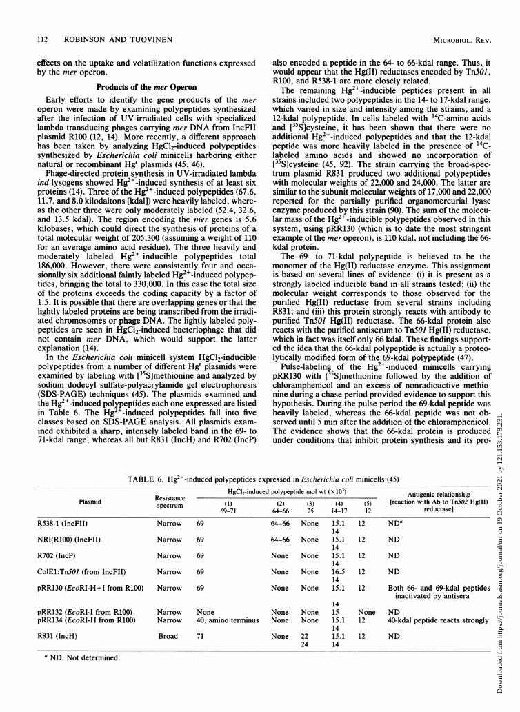
112 ROBINSON AND TUOVINEN
effects on the uptake and volatilization functions expressedby the mer operon.
Products of the mer OperonEarly efforts to identify the gene products of the mer
operon were made by examining polypeptides synthesizedafter the infection of UV-irradiated cells with specializedlambda transducing phages carrying mer DNA from IncFllplasmid R100 (12, 14). More recently, a different approachhas been taken by analyzing HgCl2-induced polypeptidessynthesized by Escherichia coli minicells harboring eithernatural or recombinant Hgr plasmids (45, 46).
Phage-directed protein synthesis in UV-irradiated lambdaind lysogens showed Hg2+-induced synthesis of at least sixproteins (14). Three of the Hg2'-induced polypeptides (67.6,11.7, and 8.0 kilodaltons [kdal]) were heavily labeled, where-as the other three were only moderately labeled (52.4, 32.6,and 13.5 kdal). The region encoding the mer genes is 5.6kilobases, which could direct the synthesis of proteins of atotal molecular weight of 205,300 (assuming a weight of 110for an average amino acid residue). The three heavily andmoderately labeled Hg2+-inducible polypeptides total186,000. However, there were consistently four and occa-sionally six additional faintly labeled Hg2+-induced polypep-tides, bringing the total to 330,000. In this case the total sizeof the proteins exceeds the coding capacity by a factor of1.5. It is possible that there are overlapping genes or that thelightly labeled proteins are being transcribed from the irradi-ated chromosomes or phage DNA. The lightly labeled poly-peptides are seen in HgCl2-induced bacteriophage that didnot contain mer DNA, which would support the latterexplanation (14).
In the Escherichia coli minicell system HgCl2-induciblepolypeptides from a number of different Hgr plasmids wereexamined by labeling with [35S]methionine and analyzed bysodium dodecyl sulfate-polyacrylamide gel electrophoresis(SDS-PAGE) techniques (45). The plasmids examined andthe Hg2+-induced polypeptides each one expressed are listedin Table 6. The Hg +-induced polypeptides fall into fiveclasses based on SDS-PAGE analysis. All plasmids exam-ined exhibited a sharp, intensely labeled band in the 69- to71-kdal range, whereas all but R831 (IncH) and R702 (IncP)
also encoded a peptide in the 64- to 66-kdal range. Thus, itwould appear that the Hg(II) reductases encoded by TnSOJ,R100, and R538-1 are more closely related.The remaining Hg2+-inducible peptides present in all
strains included two polypeptides in the 14- to 17-kdal range,which varied in size and intensity among the strains, and a12-kdal polypeptide. In cells labeled with 14C-amino acidsand [35S]cysteine, it has been shown that there were noadditional Hg2+-induced polypeptides and that the 12-kdalpeptide was more heavily labeled in the presence of 14C-labeled amino acids and showed no incorporation of[35S]cysteine (45, 92). The strain carrying the broad-spec-trum plasmid R831 produced two additional polypeptideswith molecular weights of 22,000 and 24,000. The latter aresimilar to the subunit molecular weights of 17,000 and 22,000reported for the partially purified organomercurial lyaseenzyme produced by this strain (90). The sum of the molecu-lar mass of the Hg2+-inducible polypeptides observed in thissystem, using pRR130 (which is to date the most stringentexample of the mer operon), is 110 kdal, not including the 66-kdal protein.The 69- to 71-kdal polypeptide is believed to be the
monomer of the Hg(II) reductase enzyme. This assignmentis based on several lines of evidence: (i) it is present as a
strongly labeled inducible band in all strains tested; (ii) themolecular weight corresponds to those observed for thepurified Hg(II) reductase from several strains includingR831; and (iii) this protein strongly reacts with antibody topurified TnSOJ Hg(II) reductase. The 66-kdal protein alsoreacts with the purified antiserum to TnSOJ Hg(II) reductase,which in fact was itself only 66 kdal. These findings support-ed the idea that the 66-kdal polypeptide is actually a proteo-lytically modified form of the 69-kdal polypeptide (47).
Pulse-labeling of the Hg2+-induced minicells carryingpRR130 with [35S]methionine followed by the addition ofchloramphenicol and an excess of nonradioactive methio-nine during a chase period provided evidence to support thishypothesis. During the pulse period the 69-kdal peptide washeavily labeled, whereas the 66-kdal peptide was not ob-served until 5 min after the addition of the chloramphenicol.The evidence shows that the 66-kdal protein is producedunder conditions that inhibit protein synthesis and its pro-
TABLE 6. Hg2+-induced polypeptides expressed in Escherichia coli minicells (45)
Resistance HgCl2-induced polypeptide mol wt (x103) Antigenic relationshipPlasmid spectrum (1) (2) (3) (4) (5) [reaction with Ab to Tn5O2 Hg(II)69-71 64-66 25 14-17 12 reductasel
R538-1 (IncFII) Narrow 69 64-66 None 15.1 12 NDa14
NRI(R100) (IncFII) Narrow 69 64-66 None 15.1 12 ND14
R702 (IncP) Narrow 69 None None 15.1 12 ND14
ColE1:Tn5OI (from IncFII) Narrow 69 None None 16.5 12 ND14
pRR130 (EcoRI-H+I from R100) Narrow 69 None None 15.1 12 Both 66- and 69-kdal peptidesinactivated by antisera
14pRR132 (EcoRI-I from R100) Narrow None None None 15 None NDpRR134 (EcoRI-H from R100) Narrow 40, amino terminus None None 15.1 12 40-kdal peptide reacts strongly
14R831 (IncH) Broad 71 None 22 15.1 12 ND
24 14
a ND, Not determined.
MICROBIOL. REV.
Dow
nloa
ded
from
http
s://j
ourn
als.
asm
.org
/jour
nal/m
r on
19
Oct
ober
202
1 by
121
.153
.178
.231
.
MECHANISMS OF MICROBIAL RESISTANCE OF MERCURY
duction is accompanied by a concomitant decrease in the 69-kdal peptide, whereas the sum of the 69- and 66-kdal proteinsis produced from the 69-kdal protein by in vivo modificationand simply due to nonspecific proteolysis (42). Degradedforms of the Hg(II) reductase of Tn.5OI and plasmid R831have been described (24, 90). Similar studies have indicatedthat the 12-kdal peptide is a modified form of a 13-kdalpeptide formed during the pulse period.The polypeptides encoded by the EcoRI-I and EcoRI-H
cloned fragments were examined (Table 6). Plasmid pRR134carrying the EcoRI-H fragment was shown to encode thethree smallest peptides (15.1, 14, and 12 kdal) as well as a 40-kdal protein believed to be the amino terminus of the Hg(II)reductase because it reacted strongly with the antiserumproduced against purified Hg(II) reductase of Tn.OJ. Incontrast, pRR132 produced a single mer-specific protein of15 kdal which did not react with the TnSOJ Hg(II) reductaseantiserum. An 81-kdal fusion protein was observed whichwas produced constitutively and did not react with theantiserum but is believed to contain the 29-kdal carboxyterminus of Hg(II) reductase (45). The lack of antigeniccross-reactivity of the three smallest peptides with theantiserum to purified Hg(II) reductase and their associationwith the EcoRI-H fragment indicated that they may havesome uptake or regulatory role.The sum of the molecular weights of the Hg2'-induced
polypeptides in the minicell system is 110 x 103, excludingthe 66 x 103-molecular-weight peptide, thus requiring aminimum of 3,000 bp of DNA (45). The EcoRI-H fragmentfrom the ISJb element to the EcoRI-I junction is 2,700 bp(23). If the 40-kdal amino terminus of the Hg(II) reductase isencoded by the EcoRI-I fragment, this leaves 1,610 bp toencode the remaining 41.1 kdal of the polypeptides observedin the minicell system. This leaves 490 bp unaccounted for,which could possibly encode a polypeptide that has not yetbeen detected, such as a regulatory protein that may bepresent in very small quantities.The minicell Hg2'-induced polypeptides correspond to
some extent to those observed with the lambda-mer system.The heavily labeled 68-kdal protein of the lambda-mersystem is believed to correspond to the 69- to 71-kdalproteins observed in the minicell system, whereas either ofthe smaller lambda-mer peptides (11.5 or 8.5 kdal) maycorrespond to the 12-kdal polypeptide from the minicellsystem. Only the lightly labeled 13.5-kdal peptide of thelambda-mer system corresponds to either the 15.1- or the 14-kdal peptide observed in the Escherichia coli minicell system(12, 42). Alternatively, it may be that the 8.5-kdal peptidecorresponds to the 12-kdal minicell protein, whereas the11.5- and 13-kdal proteins represent the 14- and 15.1-kdalminicell proteins, owing to poor resolution in the lower-molecular-weight range.
In studies of labeling Hg2+-induced minicells in the pres-ence of ethanol, which has been shown to inhibit theprocessing of outer membrane and periplasmic proteins inEscherichia coli, it was found that only the 66- and 12-kdalMer proteins disappeared, whereas the 69-, 15.1-, and 14-kdal Mer polypeptides were not affected (46). The cellularlocation of the Hg2+-induced polypeptides in Escherichiacoli minicells was determined by SDS-PAGE analysis ofsoluble and cell membrane fractions as prepared by Frenchpress disruption of the cells and sucrose density centrifuga-tion techniques (44). The membrane fraction was found tocontain both the 15.1- and 14-kdal Mer proteins, whereas the69-, 66-, and 12-kdal proteins were in the soluble fraction.During sucrose density centrifugation the 15.1- and 14-kdal
proteins were found in the light and intermediate densityfractions, along with the 36-kdal tet-induced inner membraneprotein, indicating that they are located in the inner mem-brane. These findings were confirmed by the solubilizationof both the 15.1- and 14-kdal proteins by Sarcosyl, whichspecifically solubilizes the inner membranes of Escherichiacoli. Whereas the 66-kdal protein is predominantly in thesoluble fraction, a small but consistent amount of the proteinwas found to be associated with the inner membrane fractionof the sucrose density gradients. As neither the 69- nor 12-kdal protein was associated with this fraction, the 66-kdalprotein is not believed to be due to contamination of theenvelope fraction by soluble proteins (46).
Thus, it appears that the 15.1- and 14-kdal peptides arecomponents of the inner membranes, but unlike many mem-brane and periplasmic enzymes they do not appear to bederived from larger peptides.The 12-kdal protein, associatedpredominantly with the soluble fraction, appears to beprocessed from a 13-kdal mer-specific peptide which isinhibited in the presence of ethanol (46). These results areconsistent with the hypothesis that the 12-kdal protein islocated in the periplasmic space rather than in the cytosol. Amodel based on the data presented above is shown in Fig. 3.It may be that the 15.1- and 14-kdal proteins participate inHg2+ uptake, whereas the 12-kdal proteins may act as aHg2+ binding protein, bringing the Hg2+ in contact with theHg(II) reductase enzyme. It is likely then that all of the 15.1-and the 14-kdal proteins are products of the merT gene, asmay also be the case for the 12-kdal protein. Although amembrane-bound Hg(II) reductase has never been clearlydemonstrated, the loose association of the 66-kdal degrada-tion products with the membrane fraction mat' serve apurpose in allowing closer contact with the Hg + as it isbrought into the cell. Such a closely coordinated systemwould have obvious advantages in terms of efficiency andreduced toxicity to the cell. The regulatory protein whichwould be expected to be present at very low levels may notyet have been detected and warrants further investigation.
Analysis of the mer Operon in Staphylococcus aureusAll mercury-resistant strains of Staphylococcus aureus
have been shown to express broad-spectrum resistance(117). The Staphylococcus aureus Hgr plasmids pI258,pII147, and p1524 have been studied most thoroughly, and allare believed to have the genes for resistance to both Hg2+and PMA in close proximity (66, 76). The mapping of theseplasmids was accomplished by using a combination of dele-tion mapping, restriction analysis, heteroduplex DNA/DNAmapping, cloning of restriction fragments, and transposoninsertion techniques in the same manner as for Escherichiacoli plasmid R100.
Restriction digests of p1524 with EcoRI generated sixfragments and revealed that both fragments C and E arerequired to confer resistance to both Hg2+ and PMA. TheEcoRI cleavage appears to be in the merB gene encoding theorganomercurial lyase enzyme such that resistance to PMAand volatilization of mercury from PMA required bothfragments cloned together (61). Transpositional mutagenesiswith plasmid p1258 revealed that the regulatory genes andthe Hg(II) reductase gene are encoded by the proximalfragment, whereas the merB gene is either cleaved by EcoRIor exists totally on the distal fragment. The Tn inserts ofp1258 inactivated either Hg2+ and PMA resistance both orPMA resistance alone (76). In another study, Hgs mutants ofStaphylococcus aureus were able to lose either reductase orlyase alone, indicating separate genetic loci for the merA and
VOL. 48, 1984 113
Dow
nloa
ded
from
http
s://j
ourn
als.
asm
.org
/jour
nal/m
r on
19
Oct
ober
202
1 by
121
.153
.178
.231
.
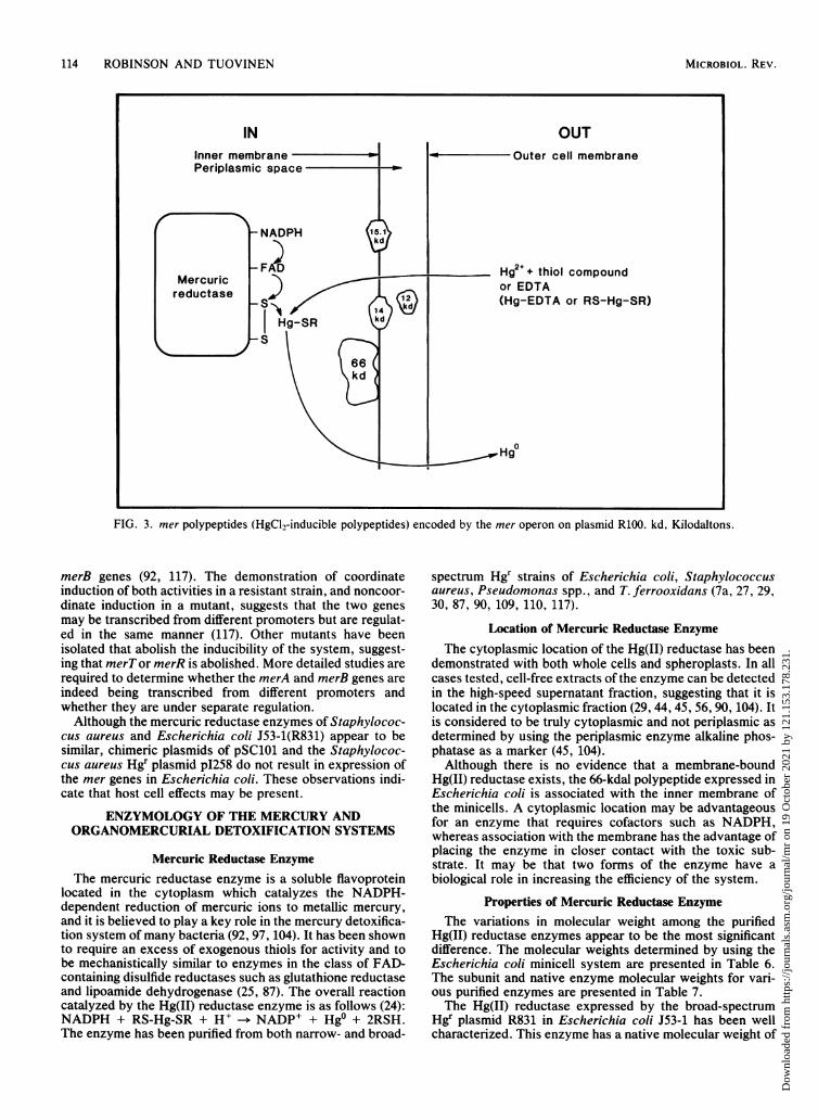
114 ROBINSON AND TUOVINEN
INInner membrane -Periplasmic space
Mercuricreductase
OUT-Outer cell membrane
Hgd + thiol compoundor EDTA(Hg-EDTA or RS-Hg-SR)
FIG. 3. mer polypeptides (HgClI-inducible polypeptides) encoded by the mer operon on plasmid R100. kd, Kilodaltons.
merB genes (92, 117). The demonstration of coordinateinduction of both activities in a resistant strain, and noncoor-dinate induction in a mutant, suggests that the two genesmay be transcribed from different promoters but are regulat-ed in the same manner (117). Other mutants have beenisolated that abolish the inducibility of the system, suggest-ing that merT or merR is abolished. More detailed studies arerequired to determine whether the merA and merB genes areindeed being transcribed from different promoters andwhether they are under separate regulation.Although the mercuric reductase enzymes of Staphylococ-
cus aureus and Escherichia coli J53-1(R831) appear to besimilar, chimeric plasmids of pSC101 and the Staphylococ-cus aureus Hgr plasmid pI258 do not result in expression ofthe mer genes in Escherichia coli. These observations indi-cate that host cell effects may be present.
ENZYMOLOGY OF THE MERCURY ANDORGANOMERCURIAL DETOXIFICATION SYSTEMS
Mercuric Reductase EnzymeThe mercuric reductase enzyme is a soluble flavoprotein
located in the cytoplasm which catalyzes the NADPH-dependent reduction of mercuric ions to metallic mercury,and it is believed to play a key role in the mercury detoxifica-tion system of many bacteria (92, 97, 104). It has been shownto require an excess of exogenous thiols for activity and tobe mechanistically similar to enzymes in the class of FAD-containing disulfide reductases such as glutathione reductaseand lipoamide dehydrogenase (25, 87). The overall reactioncatalyzed by the Hg(II) reductase enzyme is as follows (24):NADPH + RS-Hg-SR + H+ -- NADP+ + Hgo + 2RSH.The enzyme has been purified from both narrow- and broad-
spectrum Hgr strains of Escherichia coli, Staphylococcusaureus, Pseudomonas spp., and T. ferrooxidans (7a, 27, 29,30, 87, 90, 109, 110, 117).
Location of Mercuric Reductase EnzymeThe cytoplasmic location of the Hg(II) reductase has been
demonstrated with both whole cells and spheroplasts. In allcases tested, cell-free extracts of the enzyme can be detectedin the high-speed supernatant fraction, suggesting that it islocated in the cytoplasmic fraction (29, 44, 45, 56, 90, 104). Itis considered to be truly cytoplasmic and not periplasmic asdetermined by using the periplasmic enzyme alkaline phos-phatase as a marker (45, 104).Although there is no evidence that a membrane-bound
Hg(II) reductase exists, the 66-kdal polypeptide expressed inEscherichia coli is associated with the inner membrane ofthe minicells. A cytoplasmic location may be advantageousfor an enzyme that requires cofactors such as NADPH,whereas association with the membrane has the advantage ofplacing the enzyme in closer contact with the toxic sub-strate. It may be that two forms of the enzyme have abiological role in increasing the efficiency of the system.
Properties of Mercuric Reductase EnzymeThe variations in molecular weight among the purified
Hg(II) reductase enzymes appear to be the most significantdifference. The molecular weights determined by using theEscherichia coli minicell system are presented in Table 6.The subunit and native enzyme molecular weights for vari-ous purified enzymes are presented in Table 7.The Hg(II) reductase expressed by the broad-spectrum
Hgr plasmid R831 in Escherichia coli J53-1 has been wellcharacterized. This enzyme has a native molecular weight of
MICROBIOL. REV.
Dow
nloa
ded
from
http
s://j
ourn
als.
asm
.org
/jour
nal/m
r on
19
Oct
ober
202
1 by
121
.153
.178
.231
.
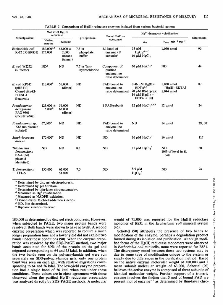
MECHANISMS OF MICROBIAL RESISTANCE OF MERCURY
TABLE 7. Comparison of Hg(II) reductase enzymes isolated from various bacteria,l genera
Mol wt of Hg(II) Hg2+-dependent volatilizationreductase Bound FAD as __________________
Strain(plasmid) pH optimum Reference(s)Subunit coenzyme Km Vmax (min-' mg-')
enzymeEscherichia coli 180(y000a,b 63,000 ± 7.5 in 3.12/mol of 13 ,uM 1,050 nmol 90
K-12 J531(R831) 175,000 2,000 phosphate enzyme (1/ HgC12def(mean) buffer subunit)C 16 ,uM HgC12(trimer)
E. coli W2252 ND5 ND 7.7 in Tris- Component of 20 p.M HgCl2e ND 44(R factor) hydrochloride purified
enzyme; noratio determined
E. coli KP245 110,000b 56,000 ND FAD bound to 0.46 ,uM Hg(II)- 1,038 nmol 87(pRR130) (dimer) enzyme; no EDTAfh [Hg(II)-EDTA]Cloned EcoRI- ratio determined 70 ,uM RS-Hg-SR 1,044 nmolH and -I 14 ,uM Hg(II) +fragments EDTA + SH
Pseudomonas 123,000 ± 56,000 ND 1 FAD/subunit 12 ,uM HgCl2deh 12 pmol 24aeruginosa 5,000b 62,OQOPAO 9501 (dimer)(pVS1/Tn5O2)
Pseudomonas sp. 67,000b ND ND FAD bound to ND 14 p.mol 29, 30K62 (no plasmid enzyme; noisolated) ratio determined
Staphylococcus 170,000b NJD ND ND 10 ,uM HgClif 16 ,umol 117aureus
Thiobacillus ND ND 8.1 ND 15 ,uM HgCl{ ND 80ferrooxidans 10% of level in E.BA-4 (no) coliplasmididentified)
T. ferrooxidans 130,000 62,000 7.5 ND 8.9 ,uM ND 7aTFI-29 54,000 HgCl2f
a Determined by disc gel electrophoresis.b Determined by gel filtration.c Determined by thin-layer chromatography.d Measured as Hg0 volatilization.e Measured as NADPH oxidation.f Demonstrates Michaelis-Menten kinetics.9 ND, Not determined.h Biphasic kinetics observed.
180,000 as determined by disc gel electrophoresis. However,when subjected to PAGE, two major protein bands wereresolved. Both bands were shown to have activity. A secondenzyme preparation which was reported to require a muchlonger preparation time and a lower yield did not exhibit twobands under these conditions (90). When the enzyme prepa-ration was resolved by the SD$-PAGE method, two majorbands accounted for 80% of the protein on the gel andmigrated corresponding to 64 and 55 kdal. In addition, whenthe two bands seen on the polyacrylamide gel were runseparately on SDS-polyacrylamide gels, only one proteinband was seen on each gel, with relative migrations corre-sponding to 64 and 56 kdal. The low-yield enzyme prepara-tion had a single band of 56 kdal when run under theseconditions. These values are in close agreement with thoseobserved when the purified Hg(II) reductase preparationwas analyzed directly by SDS-PAGE methods. A molecular
weight of 71,000 was reported for the Hg(II) reductasemonomer of R831 in the Escherichia coli minicell system(45).
Schottel (90) attributes the presence of two bands tomodification of the enzyme, perhaps a degradation productformed during its isolation and purification. Although modi-fied forms of the Hg(II) reductase monomers were observedin Escherichia coli minicells, none were reported for R831.The discrepancy noted between these two systems may bedue to some type of modification unique to the system orsimply due to differences in the purification method. Basedon the native enzyme molecular weight of 180,000 and amean subunit molecular weight of 63,000, Schottel (90)believes the active enzyme is composed of three subunits ofidentical molecular weight. Further support of a trimericenzyme involves the finding that 3 mol of bound FAD arepresent mol of enzyme-' as determined by thin-layer chro-
115VOL. 48, 1984
Dow
nloa
ded
from
http
s://j
ourn
als.
asm
.org
/jour
nal/m
r on
19
Oct
ober
202
1 by
121
.153
.178
.231
.
116 ROBINSON AND TUOVINEN
matography, using the supernatant of boiled purified enzymepreparation (86). This represents the only trimeric Hg(II)reductase. The Hg(II) reductase from a Staphylococcusaureus strain containing a penicillinase plasmid has a report-ed molecular weight of 180,000, like R831, but a subunitconfiguration is not reported (117).The purified Hg(II) reductase from Escherichia coli KP245
containing the cloned mercury resistance genes from plas-mid R100(NR1) is a dimer with a molecular weight of 110,000+ 10,000 and subunits of 56 kdal as determined by gelfiltration on a Sephadex-200 column and SDS-gel electro-phoresis (81). This is in contrast to the 69-kdal monomer anda 54-kdal peptide seen occasionally in the Escherichia coliminicell system (42). It is also of interest to note that, again,a modified 66-kdal protein was present in the minicellpreparations, whereas J. W. Williams (personal communica-tion) has only detected the 56-kdal monomer. This contradic-tion was recently resolved (Williams, personal communica-tion) by the demonstration that the 66-kdal protein in theminicell system was the same as the 56-kdal protein whentested by the same method (45).
Recently, the Hg(II) reductase from Tn5OJ has beenshown to be a dimer with a molecular weight of 123,000 ±
5,000, with subunits of 56,000 and 62,000 as determined bygel filtration on a Sephadex 0-150 column (24). The twobands of 56 and 62 kdal seen on SDS-polyacrylamide gelscomprised 95% of the protein on the gel. At the time ofisolation, 80 to 85% of the protein was in the higher-molecular-weight form, whereas upon storage up to 90% wasfound in the lower-molecular-weight band. There was nosignificant change in the activity of the preparation despitethe shift in molecular weight. Although an in vivo modifiedform of the TnSOJ Hg(II) reductase was detected in theEscherichia coli minicells, it is unclear whether this is thecase here.The 67,000 molecular weight observed for Pseudomonas
sp. K62 Hg(II) reductase is much lower than those discussedabove. It may be that this represents the monomer molecularweight, in which case it agrees closely with the otherenzymes listed in Table 7. For the T. ferrooxidans Hg(II)reductase a molecular weight of 130,000 has been deter-mined (7a); this protein has been separated to 62- and 54-kdalsubunits by SDS-PAGE.
Antigenic Relationships Among Purified Mercuric ReductaseEnzymes
A recent comparative study was conducted with antiser-um prepared to both TnSO- and R831-purified Hg(II) reduc-tase enzymes. All gram-negative bacterial Hg(II) reductasestested were inactivated by the antiserum prepared againstthe R831 enzymes (92). These enzymes included thoseexpressed by various enteric organisms and those fromPseudomonas and Proteus spp. (91). In contrast, antisera topurified TnS01 Hg(II) reductase separated the gram-negativebacterial enzymes into three serotypes based on spur forma-tion in double-diffusion precipitation tests. Neither one ofthe antisera cross-reacted with the Hg(II) reductase ofStaphylococcus aureus. These results indicate that the gram-negative and gram-positive Hg(II) reductase enzymes exam-ined thus far are immunologically distinct. Interestingly, theHg(II) reductase purified from T. ferrooxidans was notinactivated by either of the antisera, but was nonspecificallyinhibited by both (92).The thermostability of the purified Hg(II) reductase en-
zymes reinforces this immunological distinction between
gram-negative and gram-positive sources. All of the Hg(II)reductase enzymes purified from gram-negative bacteriahave been found to retain activity upon incubation at 80°Cfor 10 min. In contrast, the Hg(II) reductase of Staphylococ-cus aureus is completely inactivated at 55°C (92). The Hg(II)reductase enzyme purified from T. ferrooxidans is reportedto be at least as heat resistant as that of Escherichia coli Hgrplasmid R100, retaining activity after 10 min at 80°C (80).
Cofactor Requirements of Mercuric ReductaseAll Hg(II) reductase enzymes have been shown to contain
bound FAD as coenzyme (24, 29, 30, 43, 87, 90). In mostinstances this was determined by examining the supernatantof the boiled, purified enzyme, using thin-layer chromatogra-phy. The number of moles ofFAD per subunit of the enzymewas determined fluorometrically, with an extinction coeffi-cient of FAD in mercuric reductase of 11.3 mM cm-1 at 458nm (24). Values of 1 FAD per subunit of the Hg(II) enzymehave been reported (Table 8) (24, 87, 90). The absorptionspectra of the purified enzymes of pRR130 and Tn5OJ havemaxima at both 370 and 460 nm, identifying them as flavo-proteins (24, 87). A listing of all cofactor requirements ispresented in Table 8.A requirement for NADP(H) as a cofactor in the reduction
of Hg2> to Hgo has been demonstrated for all purified Hg(II)reductase enzymes studied to date (11, 24, 29, 30, 43, 55, 87,90, 104). NADPH serves as the electron donor in thereduction of Hg . Some studies have demonstrated thatNADH can substitute for NADPH as the electron donor invarious Escherichia coli and Pseudomonas sp. strains aswell as in T. ferrooxidans and Yersinia enterocolitica, but itwas much less effective than NADPH (6, 29, 30, 43, 80).The stoichiometry of the NADPH-dependent reduction of
Hg2+ has been shown to be 1:1 (NADPH:Hg2+) for allexcept Escherichia coli W2252, which was found to be 2:1(Table 8) (44). The degree of coupling of the system has beenexamined by measuring NADPH oxidation and Hg2> reduc-tion in parallel. The ratio was 1.3 mol of NADPH consumed1.0 mol of Hg2> reduced-1, with <0.3% of the total electronflow going to 02 as measured with an 02 electrode (24).An additional cofactor requirement shared by all Hg(II)
reductase enzymes is that of thiol reagents for activity.However, the nature of the thiol as well as the levelsrequired for maximum activity appear to vary among theenzymes examined (Table 8). One consistent finding is theneed for an excess of the preferred thiol reagent. This ispresumably to insure the reduced state of the enzyme activesite and also that Hg be present as RS-Hg2+ or RS-Hg-SR(92, 97). Although an excess of thiol reagent is required foractivity, inhibition may occur. For example, the Hg(II)reductase purified from T. ferrooxidans exhibited optimalactivity at a 250:1 ratio of 3-mercaptoethanol, whereas at2,500:1 there was a 50% inhibition (80).The role of EDTA has proven to be less clear. In many
cases EDTA has not been included in the reaction mixture(see Table 8). EDTA was found to be required for maximumenzyme activity of the Hg(II) reductase partially purifiedfrom T. ferrooxidans. In the absence of EDTA, there was a95% reduction in activity from that observed at the optimalconcentration of 5 mM EDTA (13). The same optimal levelof EDTA was observed for Escherichia coli C600(pDU1003)when tested under the same conditions. A highly purifiedHg(II) reductase from Escherichia coli Kp245(pRR130) wasshown to require EDTA or thiol reagents. No activity wasobserved if both were omitted, and the kinetics of thereaction were dependent on which reagents were present (87).
MICROBIOL. REV.
Dow
nloa
ded
from
http
s://j
ourn
als.
asm
.org
/jour
nal/m
r on
19
Oct
ober
202
1 by
121
.153
.178
.231
.
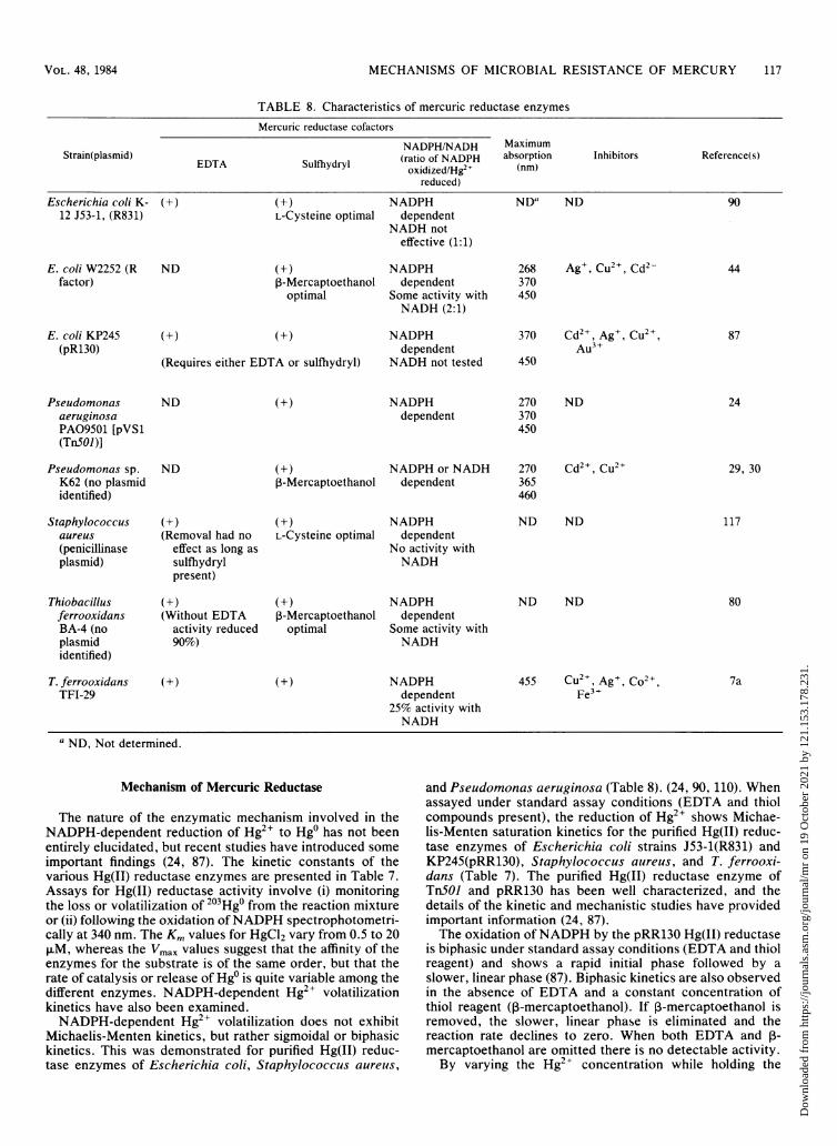
MECHANISMS OF MICROBIAL RESISTANCE OF MERCURY 117
TABLE 8. Characteristics of mercuric reductase enzymes
Mercuric reductase cofactors
NADPH/NADH MaximumStrain(plasmid) (ratio of NADPH absorption Inhibitors Reference(s)
EDTA Sulfhydryl oxidized/Hg" (nm)reduced)
Escherichia coli K- (+) (+) NADPH ND" ND 9012 J53-1, (R831) L-Cysteine optimal dependent
NADH noteffective (1:1)
E. coli W2252 (R ND (+) NADPH 268 Ag+, Cu2+, Cd2- 44factor) ,B-Mercaptoethanol dependent 370
optimal Some activity with 450NADH (2:1)
E. coli KP245 (+) (+) NADPH 370 Cd2, Ag+, Cu2 , 87(pR130) dependent Au3+
(Requires either EDTA or sulfhydryl) NADH not tested 450
Pseudomonas ND (+) NADPH 270 ND 24aeruginosa dependent 370PA09501 [pVS1 450(Tn5OI )]
Pseudomonas sp. ND (+) NADPH or NADH 270 Cd2+, Cu2+ 29, 30K62 (no plasmid P-Mercaptoethanol dependent 365identified) 460
Staphylococcus (+) (+) NADPH ND ND 117aureus (Removal had no L-Cysteine optimal dependent(penicillinase effect as long as No activity withplasmid) sulfhydryl NADH
present)
Thiobacillus (+) (+) NADPH ND ND 80ferrooxidans (Without EDTA 3-Mercaptoethanol dependentBA-4 (no activity reduced optimal Some activity withplasmid 90%) NADHidentified)
T. ferrooxidans (+) (+) NADPH 455 Cu2+, Age, Co2+ 7aTFI-29 dependent Fe'+
25% activity withNADH
a ND, Not determined.
Mechanism of Mercuric Reductase
The nature of the enzymatic mechanism involved in theNADPH-dependent reduction of Hg2+ to Hgo has not beenentirely elucidated, but recent studies have introduced someimportant findings (24, 87). The kinetic constants of thevarious Hg(II) reductase enzymes are presented in Table 7.Assays for Hg(II) reductase activity involve (i) monitoringthe loss or volatilization of 203Hg0 from the reaction mixtureor (ii) following the oxidation ofNADPH spectrophotometri-cally at 340 nm. The Km values for HgCl2 vary from 0.5 to 201±M, whereas the Vmax values suggest that the affinity of theenzymes for the substrate is of the same order, but that therate of catalysis or release of Hgo is quite variable among thedifferent enzymes. NADPH-dependent Hg2+ volatilizationkinetics have also been examined.NADPH-dependent Hg2> volatilization does not exhibit
Michaelis-Menten kinetics, but rather sigmoidal or biphasickinetics. This was demonstrated for purified Hg(II) reduc-tase enzymes of Escherichia coli, Staphylococcus aureus,
and Pseudomonas aeruginosa (Table 8). (24, 90, 110). Whenassayed under standard assay conditions (EDTA and thiolcompounds present), the reduction of Hg2> shows Michae-lis-Menten saturation kinetics for the purified Hg(II) reduc-tase enzymes of Escherichia coli strains J53-1(R831) andKP245(pRR130), Staphylococcus aureus, and T. ferrooxi-dans (Table 7). The purified Hg(II) reductase enzyme ofTnSOJ and pRR130 has been well characterized, and thedetails of the kinetic and mechanistic studies have providedimportant information (24, 87).The oxidation ofNADPH by the pRR130 Hg(II) reductase
is biphasic under standard assay conditions (EDTA and thiolreagent) and shows a rapid initial phase followed by aslower, linear phase (87). Biphasic kinetics are also observedin the absence of EDTA and a constant concentration ofthiol reagent (,-mercaptoethanol). If ,B-mercaptoethanol isremoved, the slower, linear phase is eliminated and thereaction rate declines to zero. When both EDTA and ,B-mercaptoethanol are omitted there is no detectable activity.By varying the Hg2> concentration while holding the
VOL. 48, 1984
Dow
nloa
ded
from
http
s://j
ourn
als.
asm
.org
/jour
nal/m
r on
19
Oct
ober
202
1 by
121
.153
.178
.231
.

118 ROBINSON AND TUOVINEN
EDTA levels constant in the absence of ,B-mercaptoethanol,there is a time-dependent decrease in the reaction rate whichvaries as a function of the Hg2+ concentration. At high levelsof Hg2+, the reaction ceases after only a few minutes. Thissuggests the formation of an enzyme-inhibitory complex.The same results were obtained when the enzyme waspreincubated with Hg2+ and EDTA and the reaction wasinitiated with NADPH. Substantial substrate inhibition byHg2+ in the presence of EDTA is observed. No detectablereaction rate is observed until the ratio of EDTA/Hg2+reaches 1, after which it rises rapidly, remaining unchangedover a 10-fold increase in the ratio (87). Thus it appears thatit is the EDTA-Hg2+ complex that acts as the substrate in theabsence of thiol compounds.
Thiol compounds exert a markedly different effect. In theabsence of EDTA and a constant ,B-mercaptoethanol con-
centration, the reaction is biphasic. When preincubated with,B-mercaptoethanol, the initial phase is eliminated, indicatingthat the enzymne exists in two separate forms which are
converted from one to the other by the thiol compound. Amechanism which accounts for the observed behavior hasbeen proposed by Rinderle et al. (87) (Fig. 4). The Km valuesfor the first and seconid phases are 70 and 53 ,uM HgCl2,respectively, with Vmax values of 1,044 and 152,umol min-'mg of protein-'. It appears that, although the affinity of theenzyme for the substrate is not altered greatly in the secondphase, the rate of catalysis or release of product is muchslower. The Vmax values are similar for assays run with
FIG. 4. Kinetic mechanism of mercuric reductase from Esche-richia coli (87).
either ,3-mercaptoethanol/Hg2+ or EDTA/Hg2+ but the Kmvalues indicate that the Hg(II)-EDTA complex has moreaffinity for the enzyme than the Hg(II)-}-mercaptoethanolcomplex. However, incubation with thiols reactivates theinhibited EDTA-Hg(II) enzyme complex, presumably bycompeting as ligands for the enzyme-bound Hg2+.
Relationship of Mercuric Reductase to Other FlavoenzymesSpectroscopic and thiol titration data identify the TnSOJ
and pRR130 Hg(II) reductase enzymes as belonging to thepyridine-nucleotide disulfide reductase class of proteins.Upon addition of reducing equivalents (NADPH or dithio-nite) the spectrum of Hg(II) reductase is similar to the two-electron-reduced (EH2) form of glutathione reductase andlipoamide dehydrogenase (24, 87). When glutathione reduc-tase and lipoamide dehydrogenase are reduced by twoelectrons, the EH7 species contains oxidized FAD and areduced disulfide, but when reduced by four electrons EH4is formed, which contains reduced flavin and reduced disul-fide as indicated by spectra and oxidation-reduction poten-tial couplings (24, 87).When the Hg(II) reductase enzyme was reduced by two
electron equivalents per FAD, a species was formed withabsorbance at 540 nm, and two more electrons were requiredfor complete flavin reduction. This indicates that four elec-trons are required to reduce FAD bound to the enzyme,suggesting a second two-electron acceptor on the active site.Thiol titrations with 5,5'-dithiobis(2-nitrobenzoate) indicatethe presence of an oxidation-reduction active disulfide onboth enzymes (24, 87). The species characterized by long-wavelength absorbance generated upon the addition of re-ducing equivalents bears a strong resemblance to the chargetransfer complex between reduced thiols and FAD on bothglutathione reductase and lipoamide dehydrogenase. Theseenzymes are known to possess an additional electron accep-tor, an oxidation-reduction active disulfide (a cysteine resi-due) (121). In addition, the oxidation-reduction potentials ofE/EH2 and EH2/EH4 complexes of Hg(II) reductase andlipoamide dehydrogenase are very similar (24). No substratecross-reactivity between TnSOJ Hg(II) reductase and pigheart lipoamide dehydrogenase, yeast glutathione reductase,or thioredoxin reductase has been reported (24). However,5,5'-dithiobis(2-nitrobenzoate) is reportedly a substrate forthe pRR130 Hg(II) reductase, with a Km of 3 mM and a Vmaxof about 1% of that observed for Hg2+ (81).These results have evolutionary implications. The amino
acid sequence of the active site of the pRR130 Hg(II)reductase has been determined and found to be very similarto that of lipoamide dehydrogenase (25). Homologous aminoacid sequences have also been found in TnSOJ mercuricreductase and human erythrocyte glutathione reductase (9).It is interesting to note that a mercury-detoxifying enzymeencoded on a plasmid may be similar to enzymes encoded bythe chromosome that are essential to the cell. It is not toodifficult to imagine that selective pressure, i.e., the presenceof toxic mercurials, could result in a slightly modifiedenzyme capable of reducing Hg2+ to elemental mercurywhich is effectively removed from the microenvironment ofthe cell.
Further studies are required to determine, whether theenzyme cycles between EH/EH2, with FAD acting as media-tor in the electron flow from NADPH to disulfide, or fromEH2IEH4 with electrons flowing to FAD creating FADH2,which is the actual reductant, and then on to Hg2+. In thelatter case the disulfide-dithiol site may act as a substrate-binding site, accessible only after it has been reduced.
ARS-Hg-SR
k A. -EA k3 oE+P
RSH >k5 k6
E1 k7A EAk8
BHg-EDTA
E klA -A k3 i+EA--"E+ Pi k2
k5A k6
EA*,-k7--EA**
MICROBIOL. REV.
Dow
nloa
ded
from
http
s://j
ourn
als.
asm
.org
/jour
nal/m
r on
19
Oct
ober
202
1 by
121
.153
.178
.231
.
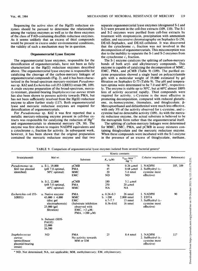
MECHANISMS OF MICROBIAL RESISTANCE OF MERCURY 119
Sequencing the active sites of the Hg(II) reductase en-zymes should be pursued to determine the relationshipsamong the various enzymes as well as to the three enzymesof the class of FAD-containing disulfide reductase enzymes.As it seems unlikely that an excess of thiol compoundswould be present in excess under environmental conditions,the validity of such a mechanism may be in question.
Organomercurial Lyase Enzyme
The organomercurial lyase enzymes, responsible for thedetoxification of organomercurials, have not been as fullycharacterized as the Hg(II) reductase enzymes describedabove. The organomercurial lyase enzyme is responsible forcatalyzing the cleavage of the carbon-mercury linkages oforganomercurial compounds (Fig. 2), and it has been charac-terized in the broad-spectrum mercury-resistant Pseudomo-nas sp. K62 and Escherichia coli J53-1(R831) strain (90, 109).A crude enzyme preparation of the broad-spectrum, mercu-
ry-resistant, plasmid-bearing Staphylococccus aureus strainhas also been shown to possess activity towards PMA, buthas not been sufficiently resolved from the Hg(II) reductaseenzyme to allow further study (117). Both organomercuriallyase and mercuric reductase enzymes are required fordetoxification of organomercurials.For Pseudomonas sp. K62 it was first reported that a
metallic mercury-releasing enzyme present in cell-free ex-tracts was responsible for catalyzing the reduction of Hg2+and organomercurials to elemental mercury (29, 30). Theenzyme was first shown to require NADPH equivalents anda cytochrome cl fraction for activity. In subsequent work,however, it has been shown that the original preparationcontained the mercuric reductase enzyme and that two
separate organomercurial lyase enzymes (designated S-1 andS-2) were present in the cell-free extracts (109, 110). The S-1and S-2 enzymes were purified from cell-free extracts bytreatment with streptomycin, precipitation with ammoniumsulfate, and successive chromatography on Sephadex G-150,DEAE-Sephadex, and DEAE-cellulose. It was discoveredthat the cytochrome cl fraction was not involved in thedecomposition of organomercurials. This misconception wasdue to the inability to separate the S-1 and S-2 enzymes fromthe cytochrome cl fraction.The S-1 enzyme catalyzes the splitting of carbon-mercury
bonds of both aryl- and alkylmercury compounds. Thisenzyme is capable of catalyzing the decomposition of MMC,EMC, PMA, and pCMB (Table 9) (109). The purified en-
zyme preparation showed a single band on polyacrylamidegels with a molecular weight of 19,000 estimated by gelfiltration on Sephadex G-75 (Table 9). The pH and tempera-ture optima were determined to be 7.0 and 40°C, respective-ly. The enzyme is stable up to 50°C, but at 60°C almost 100%loss of activity occurred rapidly. Thiol compounds wererequired for activity. L-Cysteine is the most effective inpromoting decomposition, compared with reduced glutathi-one, DL-homocysteine, thiomalate, and thioglycolate. ,B-Mercaptoethanol and dithiothreitol were much less effective,with only 3% of the activity observed for L-cysteine, and L-cysteine had no detectable activity (109). As with the mercu-ric reductase enzyme, the actual substrate is believed to bethe mercaptide form rather than the organomercurial itself.The splitting of carbon-mercury linkages were determined
for MMC, EMC, PMA, and pCMB in assay mixtures con-taining thioglycolate and the mercuric reductase enzyme.When these compounds were incubated with the S-1 enzymein the presence of an excess of thioglycolate, methane,
TABLE 9. Comparison of organomercurial lyase enzymes isolated from several bacterial genera'Kinetic constants
Strain(plasmid) Mol wt Substrate K, (M) Vmax (min' Cofactor requirements Reference(s)SubstrateK, (~LM) mg 1)
Pseudomonas sp. a. S-1, 19,000 pCMB 78 0.26 ,mol 1. NADPH 105, 109K62 (no plasmid (pH 7.0 optimal, PMA 45 0.68 ,umol 2. Sulfhydryl (L-identified) 50°C optimal) MMC 20 5.4 nmol cysteine most
EMC ND ND effective)
b. S-2, 22,000 pCMB 180 3.1 p.mol(pH 5.0 optimal, PMA 250 20 ,umol40°C optimal) MMC NA NA
Escherichia coli J53- a. Native enzyme PMA a. 0.24-0.3 26 nmol 1. NADPH 901(R831) 43,000 + 4,000 MMC b. >200 2,800 nmol 2. EDTA
(disc gel EMC 6.7-7.7 13 nmol 3. Sulfhydryl (L-electrophoresis) (Substrate inhibition 0.56-0.61 16 nmol cysteine most
25,000 (gel observed with effective)filtration) EMC, >2 ,M;
PMA, >200 ,uM)
b. Subunit (SDS-PAGE)22,00016,500
Staphylococcus ND PMA 25 8.4 nmol 1. NADPH 117aureus No activity towards 2. Sulfhydryl (L-(penicillinase MM or EM cysteine mostplasmid-bearing effective)strain)a ND, Not determined; NA, not applicable; MM, methylmercury; EM, ethylmercury.
VOL. 48, 1984
Dow
nloa
ded
from
http
s://j
ourn
als.
asm
.org
/jour
nal/m
r on
19
Oct
ober
202
1 by
121
.153
.178
.231
.
120 ROBINSON AND TUOVINEN
ethane, benzene, and benzoic acid were produced, respec-tively (109). Benzoic acid was detected by thin-layer chro-matography, whereas the other products were identified bygas-liquid chromatography methods. Although 100% ofPMA was decomposed in 2 h, <50% EMC and about 25%MMC were decomposed during 3 h of incubation. Thesubstrates and their kinetic constants are presented in Table9. Kinetic data indicate that the enzyme has greater affinityfor MMC than for PMA, but a slower rate of decomposition.No activity was observed in the absence of the mercuricreductase enzyme.The molecular weight of the S-2 enzyme is 20,000 as
determined by gel filtration on a Sephadex G-75 column, andthe enzyme was found to migrate as a single band with amolecular weight of 17,000 on a 10% SDS-polyacrylamidegel. The enzyme was found to have pH and temperatureoptima of 5.0 and 40°C (completely inactivated at 55°C),respectively (Table 9), indicating that the enzyme is moreacidic and more heat labile than the S-1 enzyme (110).Both PMA and pCMB can serve as substrates for this
enzyme, whereas MMC and EMC cannot, suggesting thatthe S-2 enzyme is involved in the splitting of carbon-mercurylinkages of arylmercury compounds. In the presence of anexcess of thioglycolate, benzene and benzoic acid wereproduced from PMA and pCMB, respectively. When 14C-labeled PMA was used as the substrate, [14C]benzene wasproduced and decomposition could be followed by the lossof radioactivity from the reaction mixture (110). Michaelis-Menten kinetics were observed with PMA and pCMB assubstrates. The Km and Vmax values are listed in Table 9. TheS-2 enzyme appears to have higher affinity for both sub-strates than does S-1, but lower rates of decomposition.The S-2 enzyme also requires an excess of thiols for
maximum activity and is inactivated by reagents which reactwith sulfhydryl groups such as monoiodoacetic acid, mono-iodoacetamide, PMA, pCMB, MMC, and HgCl2 but not byEDTA, cyanide, and azide (110). The lack of inhibition byEDTA, cyanide, and azide suggests that EDTA does notform an S-2-EDTA complex, as it does with Hg2+, and is notcoupled to energy transduction and respiration.Another organomercurial lyase enzyme has been purified
from the broad-spectrum plasmid-bearing Escherichia coliJ53-1(R831) (24). This enzyme was purified in a mannersimilar to the S-1 and S-2 enzymes and was monitored for thelyase activity by using MMC and PMA throughout thepurification to detect the presence of more than one lyase.Enzyme activity is dependent on the presence of EDTA anda sulfhydryl compound. L-Cysteine is the most effectivesulfhydryl compound whereas dithiothreitol is the leasteffective. Enzyme activities of 50 and 25% were obtainedwith thioglycolate and P-mercaptoethanol, respectively. Thesame order of effectiveness of sulfhydryl compounds is notseen with the Hg(II) reductase from the same strain (90).Lyase activity was measured by following the loss of Hg2+
from a reaction mixture containing phosphate buffer, EDTA,Mg-acetate, L-cysteine, bovine serum albumin, and 203Hg-labeled substrate. NADPH and either purified Hg(II) reduc-tase or SnC12 were added to reduce Hg2+ formed by lyaseaction. A need for high concentrations of protein wassuggested by a 65 to 80% loss of activity when bovine serumalbumin is eliminated. This was not observed with either S-1or S-2 lyase. In addition, when Hg(II) reductase or NADPHwas omitted from the reaction mixture containing SnCl2, theactivity decreased by approximately 85%. This suggests thatHg(II) reductase stimulates the lyase activity and that thelyase reaction is the rate-limiting step (90).
The molecular weight of the R8331 lyase is 25,000 ± 1,000as determined by gel filtration on a Sephadex G-200 columnand 39,000 and 47,000 in other experiments using disc gelelectrophoresis (90). This discrepancy was also observed forthe Hg(II) reductase of the same strain and may simplyrepresent a poor correlation between the two techniques.Two protein bands in equimolar amounts were seen on SDS-polyacrylamide gels, with molecular weights of 22,000 and16,500. The enzyme may be a monomer of 25 kdal with a 16-kdal degradation product. It may also be a dimer based on amolecular weight of 39 x 103 with dissimilar subunits of22 x 103 and 16 x 103. Further characterization is neededto resolve this discrepancy. In Escherichia coli minicellscontaining R831, two polypeptides of 22 and 24 kdal wereproduced (45). These peptides were present only in R831,which was the only broad-spectrum Hgr plasmid studied,and are similar in molecular weight to those reported for thesubunits of the partially purified lyase from R831 describedabove. The activity of these polypeptides towards organo-mercurials was not determined.The substrates and kinetic constants for the R831 lyase
enzyme are listed in Table 9. The organomercurial lyaseenzyme of R831 can decompose both aryl- and alkylmercurycompounds. Although Schottel (90) was not able to detect orseparate two organomercurial lyases from this strain, kineticanalysis showed two Km and two Vmax values for PMA,suggesting the possibility of two enzymes. Further charac-terization of this enzyme is required to determine whethertwo separate enzymes do indeed exist, or perhaps there aretwo active sites, or a modulation or modification of theenzymes may result in differential activity.The differences noted in the kinetic properties of the lyase
enzymes of Escherichia coli and Pseudomonas spp. may bedue in part to host strain effects. These effects could bedetermined by using a common host strain and by compari-son of the products in the Escherichia coli minicell systemdescribed previously. Since the Hg(II) reductase of Staphy-lococcus aureus appears to be quite different from the gram-negative Hg(II) reductase enzymes, it would be of interest todetermine the relationship of its lyase enzyme to those fromgram-negative sources. More detailed mechanistic studiesare warranted. The location of the lyase enzyme has not yetbeen determined. Although it appears to be in the solublefraction, it may be periplasmic or have a limited associationwith the membrane.
CONCLUSIONSThe relationship among the mer operons of different
bacterial species needs to be examined in even greater detail.Characteristic patterns of resistance to mercury compoundsamong different genera have emerged. Only A-C, L, and Hincompatibility groups of enteric plasmids specify broad-spectrum resistance capabilities, whereas all Staphylococ-cus aureus plasmids studied to date fall into this class. Thesepatterns may suggest a limitation in transfer among strains,maintenance instability, or additional host cell backgroundeffects. Examples of host cell effects that have been docu-mented include: (i) the lack of expression of Staphylococcusaureus mer genes in Escherichia coli cells, using chimericplasmids of pSC101 and the Staphylococcus aureus merregion; (ii) the inability of Escherichia coli cells to expressresistance to pHMB from a broad-spectrum resistance plas-mid from Pseudomonas aeruginosa that expresses resist-ance to this compound; and (iii) the variations in expressionof mer operon of R100 in Escherichia coli and Proteusmirabilis in studies on gene copy number effects. Variations
MICROBIOL. REV.
Dow
nloa
ded
from
http
s://j
ourn
als.
asm
.org
/jour
nal/m
r on
19
Oct
ober
202
1 by
121
.153
.178
.231
.
MECHANISMS OF MICROBIAL RESISTANCE OF MERCURY 121
in resistance range and levels may be attributed to the hostbackground effects in a number of instances, suggesting thatfurther efforts in identifying such effects on regulation andexpression of mer operons in these systems are warranted.The mercuric reductase enzymes from gram-negative and
gram-positive bacteria differ in a number of aspects aspresented above. Examination of the Staphylococcus aureusmercury-induced polypeptides and their location within thecell, using the Escherichia coli minicell system for directcomparison with gram-negative strains, would provide valu-able information on the extent of such differences. It isespecially interesting to note that in gram-negative systemsthere is a polypeptide believed to be located in the periplas-mic space that may act as the mercury-binding protein. Theexistence and location of an analogous protein in Staphylo-coccus aureus cells would be useful in understanding themechanism of mercury uptake and how it may differ amongmercury-resistant isolates. Sequencing of the mer region ofStaphylococcus aureus should also be undertaken to estab-lish the homology with mer genes from other sources.The relationship of the mercuric reductase enzymes en-
coded by TnSOJ and pRR130 to the glutathione reductaseand lipoamide dehydrogenase enzymes might provide a linkin the evolution of mercury resistance systems. The use ofX-ray crystallography in addition to amino acid and DNAsequencing would provide useful information on the degreeof homology among these enzymes.The specificity of thiol requirements and the effects of
these compounds on the mercuric reductase enzymes alsoneed clarification. The differences may reflect heterogeneityamong the enzymes or differences in the affinity of thecompound for the substrate, and its ability to exchange ontothe active site, thus promoting its reduction.
Identification of the merT uptake protein is of majorimportance. The 12-kdal protein shown in the Escherichiacoli minicells to be in the periplasmic space has no cysteineresidues which may be necessary for a protein that bindsmercury to prevent its inactivation. This may also explainthe preference for L-cysteine as sulfhydryl compound, as itwould allow for the exchange of mercury from the carrierprotein to the active site on the enzyme. The isolation ofmerT mutants and examination of their products in theminicell system would be one approach to identifying theproduct(s) of this gene. The uptake function appears to be ahighly specific and high-affinity process as indicated bybinding studies. This may be a rate-limiting step in thedetoxification of both mercuric ions and organomercurycompounds, as observed with the high-copy-number vari-ants. Studying the effects of various ionophores may beuseful in establishing the energetics of the uptake system.The location of the organomercurial lyase enzymes has
not yet been established. Difficulties in purifying and resolv-ing this enzyme have slowed down research in this area. Theisolation of two different lyase enzymes from the Pseudomo-nas sp. K62 strain and a single lyase enzyme from Escherich-ia coli with what appears to have different substrate specific-ities needs clarification. There is some evidence that theorganomercurial lyase enzyme is transcribed from a differentpromoter than the mercuric reductase enzyme but is undercontrol of the mer operon. This is indicated by the lack ofcoordinate induction, preferential induction by merbromin,and isolation of mutants defective in mercuric reductaseactivity that retain the lyase activity. Establishing the num-ber and position of promoters on the mer operon region ofbroad-spectum plasmids will aid in resolving this point.Detailed studies of coordinate induction of the mercuric
reductase and lyase activities of different plasmids in acommon host strain would indicate whether this is a com-mon occurrence.One of the most important questions that remains is the
mechanism of resistance to organomercurials such as mer-bromin and FMA, which does not include degradation. It isnot readily apparent why the carbon-mercury bond in thesecompounds would not be catalyzed by the lyase enzymewhile the others are. This may be explained simply as theresult of the lower toxicity of these compounds or that of amodified uptake system. However, these compounds areknown to inhibit the hyperbinding of mercuric ions byhypersensitive mutants but are not themselves bound to thecell. It may be that these compounds actually bind themercuric ions, making them unavailable, or they may bindloosely to the uptake protein specified by the mer operonand will result in induction but not uptake.The methylation of mercury by microorganisms may rep-
resent a detoxification mechanism which may or may not beenzymatic in nature, a question that must be clarified.Studying methylation in Escherichia coli, which does notproduce methylcobalamin, and mutants in other strains thatdo produce methylcobalamin may provide answers to thisquestion. The balance of methylation and demethylation ofmercury should be explored. It may be that under conditionsin which methylation is optimum (pH 4.5 and no thiolrequirement) fewer organisms that degrade methylmercury(pH 7.0 and presence of thiol compounds) or a reduced ratemay be observed. Bacteria that possess well-characterizedmercury reduction and organomercurial degradation sys-tems should be assayed for the ability to methylate mercuryunder conditions known to be optimum for methylation. Thedifferences in optimal conditions for these two activities maymake them mutually exclusive if they exist in the sameorganism or may result in microenvironments in situ inwhich one activity predominates.
ACKNOWLEDGMENTSWe thank J. W. Williams for providing unpublished information
for the preparation of this review. We are grateful to Lisa A. Linderfor typing the manuscript.
LITERATURE CITED1. Barkay, T., and R. R. Colwell. 1983. Altered mercury transport
in a mutant of Pseudomonas fluorescens B69. J. Gen. Micro-biol. 129:2945-2950.
2. Benes, P., E. T. Gjessing, and E. Steinnes. 1976. Interactionsbetween humus and trace elements in fresh water. Water Res.10:711-716.
3. Bennett, P. M., J. Grinsted, C. L. Choi, and M. H. Richmond.1978. Characterization of Tn501, a transposon determiningresistance to mercuric ions. Mol. Gen. Genet. 159:101-106.
4. Berlin, M. 1979. Mercury, p. 503-530. In L. Friberg, G. F.Nordberg, and V. B. Vouk (ed.), Handbook on the toxicologyof metals. Elsevier/Holland Biomedical Press, Amsterdam.
5. Bertilsson, L., and H. Y. Neujahr. 1971. Methylation of mercu-ry compounds by methylcobalamin. Biochemistry 10:2805-2808.
6. Blaghen, M., M.-C. Lett, and D. J.-M. Vidon. 1983. Mercuricreductase activity in a mercury-resistant strain of Yersiniaenterocolitica. FEMS Microbiol. Lett. 19:93-96.
7. Bohlander, F. A., A. 0. Summers, and R. B. Meagher. 1981.Cloning a promoter that puts the expression of tetracyclineresistance under the control of the regulatory elements of themer operon. Gene 15:395-403.
7a.Booth, J. E., and J. W. Williams. 1984. The isolation of amercuric ion-reducing flavoprotein from Thiobacillus ferrooxi-dans. J. Gen. Microbiol. 130:725-730.
8. Brown, N. L., C.-L. Choi, J. Grinsted, M. H. Richmond, and
VOL. 48, 1984
Dow
nloa
ded
from
http
s://j
ourn
als.
asm
.org
/jour
nal/m
r on
19
Oct
ober
202
1 by
121
.153
.178
.231
.
122 ROBINSON AND TUOVINEN
P. R. Whitehead. 1980. Nucleotide sequences at the ends of themercury resistance transposon, TnS01. Nucleic Acids Res.8:1933-1945.
9. Brown, N. L., S. J. Ford, R. D. Pridmore, and D. C. Fritzinger.1983. Nucleotide sequence of a gene from the Pseudomonastransposon TnS01 encoding mercuric reductase. Biochemistry22:4089-4095.
10. Cabello, F., K. Timmis, and S. N. Cohen. 1978. Cloning ofHindlIl and EcoRI fragments of R6-5 plasmid by insertionalinactivation, p. 42-44. In D. Schiessinger (ed.), Microbiolo-gy-1978. American Society for Microbiology, Washington,D.C.
11. Clark, D. L., A. A. Weiss, and S. Silver. 1977. Mercury andorganomercurial resistances determined by plasmids in Pseu-domonas. J. Bacteriol. 132:186-196.
12. Craig, P. J., and P. D. Bartlett. 1978. The role of hydrogensulphide in environmental transport of mercury. Nature (Lon-don) 275:635-637.
13. De La Cruz, F., and J. Grinsted. 1982. Genetic and molecularcharacterization of Tn2I, a multiple resistance transposonfrom R100.1. J. Bacteriol. 151:222-228.
14. Dempsey, W. B., and S. A. McIntire. 1979. Lambda transduc-ing phages derived from a FinO- R100::A cointegrate plasmid:proteins encoded by the R100 replication/incompatibility re-gion and the antibiotic resistance determinant. Mol. Gen.Genet. 176:319-334.
15. Dempsey, W. B., S. A. McIntire, N. Willetts, J. Schottel, T. G.Kinsherf, S. Silver, and W. A. Shannon, Jr. 1978. Properties oflambda transducing bacteriophages carrying R100 plasmidDNA: mercury resistance genes. J. Bacteriol. 136:1084-1093.
16. Dempsey, W. B., and N. S. Willetts. 1976. Plasmid co-integratesof prophage lambda and R factor R100. J. Bacteriol. 126:166-176.
17. DeSimone, R. E., M. W. Penley, L. Charbonneau, S. G. Smith,J. M. Wood, H. A. 0. Hill, J. M. Pratt, S. Ridsdale, and R. J. P.Williams. 1973. The kinetics and mechanism of cobalamin-dependent methyl and ethyl transfer to mercuric ion. Biochim.Biophys. Acta 304:851-863.
18. D'Itri, F. M. 1972. The environmental mercury problem. CRCPress, Inc., Boca Raton, Fla.
19. Fagerstrom, T., and A. Jernelov. 1971. Formation of methylmercury from pure mercuric sulphide in aerobic organic sedi-ment. Water Res. 5:121-122.
20. Fagerstrom, T., and A Jernelov. 1972, Some aspects of thequantitative ecology of mercury. Water Res. 6:1193-1202.
21. Faust, S. D., and 0. M. Aly. 1981. Chemistry of natural waters.Ann Arbor Science Publishers, Inc., Ann Arbor, Mich.
22. Foster, T. J., and H. Nakahara. 1979. Deletions in the r-determinant mer region of plasmid R100-1 selected for loss ofmercury hypersensitivity. J. Bacteriol. 140:301-305.
23. Foster, T. J., H. Nakahara, A. A. Weiss, and S. Silver. 1979.Transposon A-generated mutations in the mercuric resistancegenes of plasmid R100-1. J. Bacteriol. 140:167-181.
24. Fox, B., and C. T. Walsh. 1982. Mercuric reductase: purifica-tion and characterization of a transposon-encoded flavoproteincontaining an oxidation-reduction-active disulfide. J. Biol.Chem. 257:2498-2503.
25. Fox, B. S., and C. T. Walsh. 1983. Mercuric reductase:homology to glutathione reductase and lipoamide dehydroge-nase. lodoacetamide alkylation and sequence of the active sitepeptide. Biochemistry 22:4082-4088.
26. Friberg, L., and J. Vostal (ed.). 1972. Mercury in the environ-ment. CRC Press, Inc., Boca Raton, Fla.
27. Friello, D. A., and A. M. Chakrabarty. 1980. Transposablemercury resistance in Pseudomonas putida, p. 249-260. In C.Stuttard and K. R. Rozee (ed.), Plasmids and transposons:environmental effects and maintenance mechanisms. Academ-ic Press, Inc., New York.
28. Furukawa, K., T. Suzuki, and K. Tonomura. 1969. Decomposi-tion of organic mercurial compounds by mercury-resistantbacteria. Agric. Biol. Chem. 33:128-130.
29. Furukawa, K., and K. Tonomura. 1972. Metallic mercury-releasing enzyme in mercury-resistant Pseudomonas. Agric.
Biol. Chem. 36:217-226.30. Furukawa, K., and K. Tonomura. 1972. Induction of metallic
mercury-releasing enzyme in mercury-resistant Pseudomonas.Agric. Biol. Chem. 36:2441-2448.
31. Gadd, G. M., and A. J. Griffiths. 1978. Microorganisms andheavy metal toxicity. Microb. Ecol. 4:303-317.
32. Gavis, J., and J. F. Ferguson. 1972. The cycling of mercurythrough the environment. Water Res. 6:989-1008.
33. Grinsted, J., P. M. Bennett, S. Higginson, and M. H. Rich-mond. 1978. Regional preference of insertion of TnS01 andTn802 into RP1 and its derivatives. Mol. Gen. Genet. 166:313-320.
34. Groves, P. J., H. Short, A. J. Thewaini, and F. E. Young. 1975.Epidemiology of antibiotic and heavy metal resistance inbacteria: resistance patterns in staphylococci isolated frompopulations in Iraq exposed and not exposed to heavy metalsor antibiotics. Antimicrob. Agents Chemother. 7:622-628.
35. Groves, D. J., and F. E. Young. 1975. Epidemiology ofantibiotic and heavy metal resistance in bacteria: resistancepatterns in staphylococci isolated from populations not knownto be exposed to heavy metals. Antimicrob. Agents Che-mother. 7:614-621.
36. Hamdy, M. K., and 0. R. Noyes. 1975. Formation of methylmercury by bacteria. Appl. Microbiol. 30:424-432.
37. Hanni, C., J. Meyer, S. lida, and W. Arber. 1982. Occurrenceand properties of composite transposon Tn2672: evolution ofmultiple drug resistance transposons. J. Bacteriol. 150:1266-1273.
38. Hatch, W. R., and W. L. Ott. 1968. Determination of sub-microgram quantities of mercury by atomic absorption spec-trophotometry. Anal. Chem. 40:2085-2087.
39. Holm, H. W., and M. F. Cox. 1975. Transformation of elemen-tal mercury by bacteria. Appl. Microbiol. 29:491-494.
40. Hu, S., E. Ohtsubo, N. Davidson, and H. Saedler. 1975.Electron microscope heteroduplex studies of sequence rela-tions among bacterial plasmids: identification and mapping ofthe insertion sequences IS1 and IS2 in F and R plasmids. J.Bacteriol. 122:764-775.
41. Iida, S., C. Hanni, C. Echarti, and W. Arber. 1981. Is the IS1-flanked r-determinant of the R plasmid NR1 a transposon? J.Gen. Microbiol. 126:413-425.
42. Imura, N., E. Sukegawa, S. Pan, K. Nagao, J. Kim, T. Kwan,and T. Ukita. 1971. Chemical methylation of inorganic mercuryand methylcobalamin, a vitamin B12 analog. Science 172:1248-1249.
43. Izaki, K. 1977. Enzymatic reduction of mercurous ions inEscherichia coli bearing R factor. J. Bacteriol. 131:696-698.
44. Izaki, K., Y. Tashiro, and T. Funaba. 1974. Mechanism ofmercuric chloride resistance in microorganisms. III. Purifica-tion and properties of a mercuric ion reducing enzyme fromEscherichia coli bearing R factor. J. Biochem. (Tokyo) 75:591-599.
45. Jackson, W. J., and A. 0. Summers. 1982. Polypeptidesencoded by the mer operon. J. Bacteriol. 149:479-487.
46. Jackson, W. J., and A. 0. Summers. 1982. Biochemical charac-terization of HgCl2-induced polypeptides encoded by the meroperon of plasmid R100. J. Bacteriol. 151:962-970.
47. Jensen, S., and A. Jernelov. 1969. Biological methylation ofmercury in aquatic organisms. Nature (London) 223:753-754.
48. Jernelov, A., and A. L. Martin. 1975. Ecological implicationsof metal metabolism by microorganisms. Annu. Rev. Micro-biol. 29:61-77.
49. Jernelov, A., and T. Wallin. 1973. Air-borne mercury fall-outon snow around five Swedish chlor-alkali plants. Atmos.Environ. 7:209-214.
50. Joensuu, 0. I. 1971. Fossil fuels as a source of mercurypollution. Science 172:1027-1028.
51. Johnson, D. L., and R. S. Braman. 1974. Distribution ofatmospheric mercury species near ground. Environ. Sci. Tech-nol. 8:1003-1009.
52. Kamps, L. R., R. Carr, and H. Miller. 1972. Total mercury-monomethylmercury content of several species of fish. Bull.Environ. Contam. Toxicol. 8:273-279.
MICROBIOL. REV.
Dow
nloa
ded
from
http
s://j
ourn
als.
asm
.org
/jour
nal/m
r on
19
Oct
ober
202
1 by
121
.153
.178
.231
.
MECHANISMS OF MICROBIAL RESISTANCE OF MERCURY 123
53. Kleckner, N. 1981. Transposable elements in prokaryotes.Annu. Rev. Genet. 15:341-404.
54. Kleckner, N. 1977. Translocatable elements in procaryotes.Cell 11:11-23.
55. Komura, I., and K. Izaki. 1971. Mechanism of mercuricchloride resistance in microorganisms. I. Vaporization of a
mercury compound from mercuric chloride by multiple drugresistance strains of Escherichia coli. J. Biochem. (Tokyo)70:885-893.
56. Komura, I., T. Funaba, and K. Izaki. 1971. Mechanism ofmercuric chloride resistance in microorganisms. II. NADPH-dependent reduction of mercuric chloride and vaporization ofmercury from mercuric chloride by a multiple drug resistantstrain of Escherichia coli. J. Biochem. (Tokyo) 70:895-901.
57. Kondo, I., T. Ishikawa, and H. Nakahara. 1974. Mercury andcadmium resistances mediated by the penicillinase plasmid inStaphylococcus aureus. J. Bacteriol. 117:1-7.
58. Kopecko, D. J., J. Brevet, and S. N. Cohen. 1976. Involvementof multiple translocating DNA segments and recombinationalhotspots in the structural evolution of bacterial plasmids. J.Mol. Biol. 108:333-360.
59. Kothny, E. L. (ed.). 1973. Trace elements in the environment.American Chemical Society, Washington, D.C.
60. Landner, L. 1971. Biochemical model for the biological meth-ylation of mercury suggested from methylation studies in vivowith Neurospora crassa. Nature (London) 230:452-454.
61. Lane, D., and M. Chandler. 1977. Mapping of the drug resist-ance genes carried by the r-determinant of the R100.1 plasmid.Mol. Gen. Genet. 157:17-23.
62. Matsumura, F., Y. Gotoh, and G. M. Boush. 1971. Phenylmer-curic acetate: metabolic conversion by microorganisms. Sci-ence 173:49-51.
63. Meissner, P. S., and J. 0. Falkinham III. 1984. Plasmid-encoded mercuric reductase in Mycobacterium scrofulaceum.J. Bacteriol. 157:669-672.
64. Miki, T., A. M. Easton, and R. H. Rownd. 1978. Mapping of theresistance genes of the R plasmid NRI. Mol. Gen. Genet.158:217-224.
65. Moser, H. C., and A. F. Voight. 1957. Dismutation of themercurous dimer in dilute solutions. J. Am. Chem. Soc.79:1837-1839.
66. Murphy, E., and R. P. Novick. 1979. Physical mapping ofStaphylococcus aureus penicillinase plasmid p1524: character-ization of an invertible regioti. Mol. Gen. Genet. 175:19-30.
67. Nakahara, H., T. Ishikawa, Y. Sarai, I. Kondo, H. Kozukue,and S. Mitsuhashi. 1977. Mercury resistance and R plasmids inEscherichia coli isolated from clinical lesions in Japan. Antimi-crob. Agents Chemother. 11:999-1003.
68. Nakahara, H., T. Ishikawa, Y. Sarai, I. Kondo, H. Kozukue,and S. Silver. 1977. Linkage of mercury, cadmium, and arse-
nate and drug resistance in clinical isolates of Pseudomonasaeruginosa. Appl. Environ. Microbiol. 33:975-976.
69. Nakahara, H., T. Ishikawa, Y. Sarai, I. Kondo, and S. Mitsuha-shi. 1977. Frequency of heavy-metal resistance in bacteriafrom inpatients in Japan. Nature (London) 266:165-167.
70. Nakahara, H., T. G. Kinscherf, S. Silver, T. Miki, A. M.Easton, and R. H. Rownd. 1979. Gene copy number effects inthe mer operon of plasmid NR1. J. Bacteriol. 138:284-287.
71. Nakahara, H., S. Silver, T. Miki, and R. H. Rownd. 1979.Hypersensitivity to Hg2" and hyperbinding activity associatedwith cloned fragments of the mercurial resistance operon ofplasmid NR1. J. Bacteriol. 140:161-166.
72. Neet, K. E., and G. R. Ainslie, Jr. 1980. Hysteretic enzymes.
Methods Enzymol. 64:192-226.73. Nelson, J. D., W. Blair, F. E. Brinkman, R. R. Colwell, and
W. 0. Iverson. 1973. Biodegradation of phenylmercuric ace-
tate by mercury-resistant bacteria. Appl. Microbiol. 26:321-326.
74. Ni' Bhriain, N. N., S. Silver, and T. J. Foster. 1983. TnSinsertion mutations in the mercuric ion resistance genes de-rived from plasmid R100. J. Bacteriol. 155:690-703.
75. Nishimura, H., and M. Kumagai. 1983. Mercury pollution offishes in Minamata Bay and surrounding water: analysis of
pathway of mercury. Water Air Soil Pollut. 20:401-411.76. Novick, R. P., I. Edelman, M. D. Schwesinger, A. D. Gruss,
E. C. Swanson, and P. A. Pattee. 1979. Genetic translocation inStaphylococcus aureus. Proc. Natl. Acad. Sci. U.S.A. 76:400-404.
77. Novick, R. P., and C. Roth. 1968. Plasmid-linked resistance toinorganic salts in Staphylococcus aureus. J. Bacteriol.95:1335-1342.
78. Olson, B. H., and R. C. Cooper. 1976. Comparison of aerobicand anaerobic methylation of mercuric chloride by San Fran-cisco Bay sediments. Water Res. 10:113-116.
79. Olson, G. J., W. P. Iverson, and F. E. Brinkman. 1981.Volatilization of mercury by Thiobacillus ferrooxidans. Curr.Microbiol. 5:115-118.
80. Olson, G. J., F. D. Porter, J. Rubinstein, and S. Silver. 1982.Mercuric reductase enzyme from a mercury-volatilizing strainof Thiobacillus ferrooxidans. J. Bacteriol. 151:1230-1236.
81. Pickup, R. W., R. J. Lewis, and P. A. Williams. 1983. Pseudo-monas sp. MT14, a soil isolate which contains two largecatabolic plasmids, one a TOL plasmid and one coding forphenylacetate catabolism and mercury resistance. J. Gen.Microbiol. 129:153-158.
82. Pinney, R. J. 1978. Survival of plasmid-containing strains ofEscherichia coli, Pseudomonas aeruginosa and Staphylococ-cus aureus in phenylmercuric nitrate and thiomersal. J. Pharm.Pharmacol. 30:228-232.
83. Porter, F. D., S. Silver, C. Ong, and H. Nakahara. 1982.Selection for mercurial resistance in hospital settings. Antimi-crob. Agents Chemother. 22:852-858.
84. Ramamoorthy, S., T. C. Cheng, and D. J. Kushner. 1982.Effect of microbial life stages on the fate of methylmercury innatural waters. Bull. Environ. Contam. Toxicol. 29:167-173.
85. Ramamoorthy, S., and D. J. Kushner. 1975. Binding of mercu-ric and other heavy metals ions by microbial growth media.Microb. Ecol. 2:162-176.
86. Ridley, W. P., L. J. Dizikes, and J. W. Wood. 1977. Biomethy-lation of toxic elements in the environment. Science 197:329-332.
87. Rinderle, S. J., J. E. Booth, and J. W. Williams. 1982. Mercuricreductase from R-plasmid NR1: characterization and mecha-nistic study. Biochemistry 22:869-876.
88. Rowland, I. R., M. J. Davies, and P. Grasso. 1977. Volatiliza-tion of methylmercuric chloride by hydrogen sulphide. Nature(London) 265:718-719.
89. Rowland, I. R., P. Grasso, and M. J. Davies. 1975. Themethylation of mercuric chloride by human intestinal bacteria.Experientia 31:1064-1065.
90. Schottel, J. L. 1978. The mercuric and organomercurial detoxi-fying enzymes from a plasmid-bearing strain of Escherichiacoli. J. Biol. Chem. 253:4341-4349.
91. Schottel, J., A. Mandal, D. Clark, S. Silver, and R. W. Hedges.1974. Volatilisation of mercury and organomercurials deter-mined by inducible R-factor systems in enteric bacteria. Na-ture (London) 251:335-337.
92. Silvet, S., and T. G. Kinscherf. 1982. Genetic and biochemicalbasis for microbial transformations and detoxification of mer-cury and mercurial compounds, p. 85-103. In A. M. Chakra-barty (ed.), Biodegradation and detoxification of environmen-tal pollutants. CRC Press, Inc., Boca Raton, Fla.
93. Soldano, B. A., P. Bien, and P. Kwan. 1975. Air-borne organo-mercury and elemental mercury emissions with emphasis oncentral sewage facilities. Atmos. Environ. 9:941-944.
94. Spangler, W. J., J. L. Spigarelli, J. M. Rose, R. S. Flippin, andH. H. Miller. 1973. Degradation of methylmercury by bacteriaisolated from environmental samples. Appl. Microbiol.25:488-493.
95. Spangler, W. J., J. L. Spigarelli, J. M. Rose, and H. H. Miller.1973. Methylmercury: bacterial degradation in lake sediments.Science 180:192-193.
96. Stanisich, V. A., P. M. Bennett, and M. H. Richmond. 1977.Characterization of a translocation unit encoding resistance tomercuric ions that occurs on a nonconjugative plasmid inPseudomonas aeruginosa. J. Bacteriol. 129:1227-1233.
VOL. 48, 1984
Dow
nloa
ded
from
http
s://j
ourn
als.
asm
.org
/jour
nal/m
r on
19
Oct
ober
202
1 by
121
.153
.178
.231
.

124 ROBINSON AND TUOVINEN
97. Summers, A. O., G. A. Jacoby, M. N. Swartz, G. McHugh, andL. Sutton. 1978. Metal cation and oxyanion resistances inplasmids of gram-negative bacteria, p. 128-131. In D. Schles-singer (ed.), Microbiology-1978. American Society for Micro-biology, Washington, D.C.
98. Summers, A. O., and L. Kight-Olliff. 1980. Tnl generatedmutants in the mercuric ion reductase of the Inc P plasmid,R702. Mol. Gen. Genet. 180:91-97.
99. Summers, A. O., L. Kight-Olliff, and C. Slater. 1982. Effect ofcatabolite repression on the mer operon. J. Bacteriol. 149:191-197.
100. Summers, A. O., and E. Lewis. 1973. Volatilization of mercuricchloride by mercury-resistant plasmid-bearing strains of Esch-erichia coli, Staphylococcus aureus, and Pseudomonas aeru-ginosa. J. Bacteriol. 113:1070-1072.
101. Summers, A. O., J. Schottel, D. Clark, and S. Silver. 1974.Plasmid-borne Hg(II) and organomercurial resistance, p. 219-226. In D. Schlessinger (ed.), Microbiology-1974. AmericanSociety for Microbiology, Washington, D.C.
102. Summers, A. O., and S. Silver. 1972. Mercury resistance in aplasmid-bearing strain of Escherichia coli. J. Bacteriol.112:1228-1236.
103. Summers, A. O., and S. Silver. 1978. Microbial transformationsof metals. Annu. Rev. Microbiol. 32:637-672.
104. Summers, A. O., and L. I. Sugarman. 1974. Cell-freemercury(II)-reducing activity in a plasmid-bearing strain ofEscherichia coli. J. Bacteriol. 119:242-249.
105. Summers, A. O., R. B. Weiss, and G. A. Jacoby. 1980.Transposition of mercury resistance from a transferable Rplasmid of Escherichia coli. Plasmid 3:35-47.
106. Tanaka, M., J. H. Cramer, and R. H. Rownd. 1976. EcoRIrestriction endonuclease map of the composite R plasmid NR1.J. Bacteriol. 127:619-636.
107. Tanaka, M., H. Harafuji, and T. Yamamoto. 1982. A gene andits product required for transposition of resistance transposonTn2603. J. Bacteriol. 151:723-728.
108. Tanaka, M., T. Yamamoto, and T. Sawai. 1983. Evolution ofcomplex resistance transposons from an ancestral mercurytransposon. J. Bacteriol. 153:1432-1438.
109. Tezuka, T., and K. Tonomura. 1976. Purification and proper-ties of an enzyme catalyzing the splitting of carbon-mercurylinkages from mercury-resistant Pseudomonas K-62 strain. J.Biochem. (Tokyo) 80:79-87.
110. Tezuka, T., and K. Tonomura. 1978. Purification and proper-ties of a second enzyme catalyzing the splitting of carbon-mercury linkages from mercury-resistant Pseudomonas K-62.J. Bacteriol. 135:138-143.
111. Timoney, J. F., J. Port, J. Giles, and J. Spanier. 1978. Heavy-metal and antibiotic resistance in the bacterial flora of sedi-ments of New York Bight. Appl. Environ. Microbiol. 36:465-472.
112. Tonomura, K., and F. Kanzaki. 1969. The reductive decompo-
sition of organic mercurials by cell-free extracts of a mercury-resistant pseudomonad. Biochim. Biophys. Acta 184:227-229.
113. Tonomura, K., T. Nakagami, F. Futai, and K. Maeda. 1968.Studies on the action of mercury-resistant microorganisms onmercurials. I. The isolation of mercury-resistant bacterium andthe binding of mercurials to the cells. J. Ferment. Technol.46:506-512.
114. Vonk, J. W., and A. K. Sijpesteijn. 1973. Studies on themethylation of mercuric chloride by pure cultures of bacteriaand fungi. Antonie van Leeuwenhoek J. Microbiol. Serol.39:505-513.
115. Vouk, V. B. 1979. General chemistry of metals, p. 15-31. In L.Friberg, G. F. Nordberg, and V. B. Vouk (ed.), Handbook onthe toxicology of metals. Elsevier/North-Holland BiomedicalPress, Amsterdam.
116. Walker, J. D., and R. R. Colwell. 1974. Mercury-resistantbacteria and petroleum degradation. Appl. Microbiol. 27:285-287.
117. Weiss, A. A., S. D. Murphy, and S. Silver. 1977. Mercury andorganomercurial resistances determined by plasmids in Staph-ylococcus aureus. J. Bacteriol. 132:197-208.
118. Weiss, A. A., J. L. Schottel, D. L. Clark, R. G. Beller, and S.Silver. 1978. Mercury and organomercurial resistance withenteric, staphylococcal, and pseudomonad plasmids, p. 121-124. In D. Schlessinger (ed.), Microbiology-1978. AmericanSociety for Microbiology, Washington, D.C.
119. Weiss, H. V., M. Koide, and E. D. Goldberg. 1971. Mercury ina Greenland ice sheet: evidence of recent input by man.Science 174:692-694.
120. Westoo, B. 1973. Methylmercury as percentage of total mercu-ry in flesh and viscera of salmon and sea trout of various ages.Science 181:567-568.
121. Williams, J. R., and J. F. Morrison. 1979. The kinetics ofreversible tight-binding inhibition. Methods Enzymol. 63:437-467.
122. Wood, J. M. 1974. Biological cycles for toxic elements in theenvironment. Science 183:1049-1052.
123. Wood, J. M., A. Cheh, L. J. Dizikes, W. P. Ridley, S. Rakow,and J. R. Lakowicz. 1978. Mechanisms for the biomethylationof metals and metalloids. Fed. Proc. 37:16-21.
124. Wood, J. M., F. S. Kennedy, and C. G. Rosen. 1968. Synthesisof methyl-mercury compounds by extract of a methanogenicbacterium. Nature (London) 220:173-174.
125. Yamada, M., and K. Tonomura. 1972. Formations of methyl-mercury compounds from inorganic mercury by Clostridiumcochlearium. J. Ferment. Technol. 50:159-166.
126. Yamada, M., and K. Tonomura. 1972. Further study offorma-tion of methylmercury from inorganic mercury by Clostridiumcochlearium T-2. J. Ferment. Technol. 50:893-900.
127. Yamada, M., and K. Tonomura. 1972. Microbial methylationof mercury in hydrogen sulfide-evolving environments. J.Ferment. Technol. 50:901-909.
MICROBIOL. REV.
Dow
nloa
ded
from
http
s://j
ourn
als.
asm
.org
/jour
nal/m
r on
19
Oct
ober
202
1 by
121
.153
.178
.231
.