Embed Size (px)
Citation preview

/ . Embryol. exp. Morph. 92,11-32 (1986) 11
Printed in Great Britain © The Company of Biologists Limited 1986
Mechanism of polar body formation in the mouse
oocyte: an interaction between the chromosomes, the
cytoskeleton and the plasma membrane*
BERNARD MARO1'2, MARTIN H. JOHNSON1,MICHELLE WEBB1 AND GIN FLACH1
1 Department of Anatomy, University of Cambridge, Downing Street, CambridgeCB2 3DY, UK2Department de Biologie Cellulaire, Centre de Genetique Moleculaire du C.N.R.S.,91190 Gifsur Yvette, France^
SUMMARYThe influence of mouse oocyte chromosomes on their immediate environment has been
investigated following their dispersal by dissolution of the metaphase spindle with nocodazole.Small clusters of chromosomes become redistributed around the egg cortex in a microfilament-dependent process. Each cluster has the capacity, on removal from nocodazole, to organize aspindle that rotates to yield a polar body. In this process of spindle formation, the chromosomeclusters are able both to promote tubulin polymerization in their vicinity and to recruitmicrotubule-organizing centres (MTOCs) which organize the polymerized tubulin into spindles.In addition each oocyte chromosome cluster, as well as the non-dispersed sperm-derived haploidgroup of chromosomes, induces a focal accumulation of subcortical actin (corresponding to afilamentous area devoid of organelles) and a loss of surface Concanavalin A binding activity(corresponding to a loss of surface microvilli) in the overlying cortex. This induction ceases withthe formation of pronuclei whether or not the pronuclei migrate centrally. Pronuclear formationis sensitive to the action of nocodazole for up to 2-4 h postinsemination, and pronuclearmigration is totally sensitive to the drug. If pronuclei are blocked in a peripheral location bynocodazole they are associated with an elevation in Con A binding activity of the overlyingmembrane which corresponds to an area of the surface rich in blebby microvilli.
INTRODUCTION
The first few hours after fertilization of the mouse oocyte are characterized bymajor changes in cytoskeletal organization involving both microfilaments (Maro,Johnson, Flach & Pickering, 1984) and microtubules (Maro, Howlett & Webb,1985; Schatten, Simerly & Schatten, 1985). These changes are associated with thereestablishment of a diploid state, and include sperm incorporation, ejection ofthe second polar body and formation and migration of pronuclei. The resumption
* Part of this work has been presented at the European Developmental Biology Congress,1984 (Maro, B., Johnson, M. H., Flach, G. & Pickering, S. J., 1984a).
t Address for correspondence and reprint requests.
Key words: polar body, chromosome, microtubule-organizing centres, microtubule, micro-filament, plasma membrane, oocyte, mouse.

12 B. MARO, M. H. JOHNSON, M. WEBB AND G. FLACH
of meiosis results in the formation of two very different cells each possessing thesame maternal DNA content: a very large cell, the egg, and the polar body whichis about 1 % the size of the egg. This unequal division retains the bulk of theconstituents synthesized during oogenesis in the egg for use subsequently duringearly embryogenesis. Very little is known about the mechanisms involved in polarbody formation. We reported previously on the close association between thecytocortex and both the meiotic female and newly introduced male chromosomes,and demonstrated that in both cases the overlying membrane lacked microvilli (asassessed by a reduction in Concanavalin A binding and by electron microscopicanalysis; Eager, Johnson & Thurley, 1976; Johnson, Eager, Muggleton-Harris &Graves, 1975; Shalgi, Phillips & Kraicer, 1978) and was associated with asubcortical focus of stable polymerized actin (Maro et al. 1984; Longo & Chen,1985; Karasiewicz & Soltynska, 1985; Van Blerkom & Bell, 1986; see alsoFig. 1E-H). We suggested that the chromosomes, or some factor closely associ-ated with them, might have induced these modifications of the cell cortex. We nowpresent evidence that supports this proposal and suggests that when pronucleiform, the influence of the chromatin on the overlying cytocortex is lost. We alsodemonstrate two other types of interaction between the chromosomes and thecytoskeleton, namely their ability to promote tubulin polymerization in theirvicinity and their ability to recruit cytoplasmic MTOCs in order to form a spindle.Taken together these three properties of the chromosomes provide a basis forexplaining how polar body extrusion occurs.
MATERIALS AND METHODS
Recovery and fertilization ofoocytesA sperm suspension was prepared from the cauda epididymides of male HC-CFLP mice
(Hacking and Churchill). Two epididymides were immersed in a 0-5 ml drop of Whittingham'smedium (Whittingham, 1971) containing SOmgrnl"1 bovine serum albumin (BSA, Sigma) underliquid paraffin (BDH), which had been equilibrated overnight at 37°C in 5% CO2 in air. Thesperm were released into suspension and left to capacitate for 1-5 h at 37°C.
Oocytes were recovered from 3- to 5-week-old (C57B1.10xCBA) ¥± or MF1 mice aftersuperovulation with 5i.u. pregnant mares' serum (PMS, Intervet) followed 48 h later by 5i.u.human chorionic gonadotrophin (hCG, Intervet). The females were killed 12-5 h post hCG andthe ovulated oocytes released from the oviducts into preequilibrated drops of Whittingham'smedium containing SOmgrnl"1 BSA. At 13-5 h post hCG, the sperm suspension was mixed anddiluted 1:9 in the drops containing the oocytes, giving a final sperm concentration of
Eggs that were cultured for more than 4 h were removed from the spermatozoa and washedtwo or three times in preequilibrated medium 16 (Whittingham & Wales, 1969) containing4mgml~1 BSA. The eggs were then cultured further in this medium until harvesting.Unfertilized oocytes were treated in the same way, simply omitting addition of the spermsuspension.
Harvesting of eggs and oocytes for analysisOocytes and eggs were freed of their cumulus cells by brief exposure to 0-1 M-hyaluronidase
(Sigma), and all fertilized eggs and oocytes were freed from their zonae pellucidae by brief

Polar body formation in mouse oocytes 13
exposure to acid Tyrode's solution (Nicolson, Yanagimachi & Yanagimachi, 1975), followed bya rinse in medium 2 (M2; Fulton & Whittingham, 1978) containing 4mg ml"1 BSA.
Staining of unfixed cells with Concanavalin AZona-free cells were incubated for 5min at room temperature in FITC-Concanavalin A
(FITC-Con A, 700//g ml"1; Miles) or tetramethylrhodamine-labelled succinyl Concanavalin A(TRMTC-S-Con A, 500 ,ug ml"1; Polysciences) and rinsed through three changes of M2+BSA.
Cell fixation and immunocytological stainingCells were then placed in specially designed chambers as described previously (Maro et al.
1984) except that the chambers were coated first with a solution of 0-1 mgrnl"1 Concanavalin A(Con A) or, when the cells had been first stained with FITC-Con A or TRMTC-S-Con A, witha 1/20 dilution of Gibco stock PHA.
The cells were then fixed in one of two ways:(i) For tubulin, actin and lamin staining with either mouse anti-tubulin monoclonal antibody
(Amersham), affinity-purified rabbit anti-actin polyclonal antibodies (Gounon & Karsenti,1981), or human anti-lamin serum (McKeown, Tuffanelli, Fukuyama & Kirschner, 1983) cellswere fixed for 30-45 min at 37°C with 3-7 % formaldehyde (BDH) in phosphate-buffered saline(PBS), washed in PBS then extracted for 10min in 0-25 % Triton X-100 (Sigma) and washed inPBS.
(ii) For MTOC staining with a human anti-pericentriolar material (PCM) serum (Calarco-Gillam et al. 1983) cells were extracted for 10 min in HPEM buffer (10 mM-EGTA, 1 mM-MgCl2,60mM-PIPES, 25mM-HEPES, pH 6-9; Schliwa, Euteneuer, Bulinsky & Izant, 1981) containing0-25 % Triton X-100, washed in HPEM buffer and fixed for 30 min with 3-7 % formaldehyde inHPEM buffer.
Immunocytological staining was performed as described in Maro et al. (1984). In order to staincondensed chromosomes, fixed cells were incubated in Hoechst dye 33258 (5 jug ml"1 in PBS) for20minat20°C.
PhotomicroscopyThe coverslips were removed from the chambers and samples were mounted in 'Citifluor'
(City University, London) to reduce fading of the reagents and viewed on a Leitz Ortholux IImicroscope with filter sets N2 for rhodamine-labelled reagents, L2 for fluorescein-labelledreagents and A for Hoechst dye. Photographs were taken on Kodak Tri-X film using a LeitzVario-Orthomat photographic system.
The three-dimensional structure of the cell is preserved in the whole mount, but as the size ofthe cells is large (70jum in diameter), it is impossible to photograph the whole cell in the samefocal plane. Therefore, in most figures, optical sections showing only one plane through the cellare shown in sharp focus; in some figures optical sections at different focal planes are illustrated.
Scanning electron microscopyScanning electron microscopy (SEM) was used to examine the surface of the specimens. The
procedure used was essentially that of Johnson & Ziomek (1982) as modified in Maro &Pickering (1984). Eggs were examined in a Jeol ISM-35CF microscope under 20 kV.
Transmission electron microscopyOocytes and eggs were fixed in 3 % glutaraldehyde in 0-1 M-cacodylate buffer pH7-3 for 30 to
60 min at room temperature and washed twice in the same buffer. They were then postfixed,embedded and sectioned as described in Fleming, Warren, Chisolm & Johnson (1984). Sectionswere viewed in a Philips 600 electron microscope at 80 kV.

14 B. MARO, M. H. JOHNSON, M. WEBB AND G. FLACH
DrugsA stock solution of 1 nig ml"1 cytochalasin D (CCD, Sigma) in dimethylsulphoxide, a stock
solution of lOmM-taxol (Lot T-4-112, N.I.H., Bethesda, USA) in dimethylsulphoxide and astock solution of 10 mM-nocodazole (Aldrich) in dimethylsulphoxide stored at — 20 °C were usedin these experiments.
RESULTS
Dispersal of oocyte-derived chromosomes by spindle disruption
Oocytes were placed in medium either with or without the microtubule inhibitornocodazole (Hoebeke, Van Nigen & De Brabander, 1976), inseminated withspermatozoa 1 h later and then cultured for up to 8 h before staining the chromatinwith Hoechst dye 33258. Spermatozoa fertilized oocytes readily, whether or notnocodazole was present, and the sperm chromatin could be distinguished fromthat of the oocyte by its sharply defined boundary, by its association with theremnants of the sperm tail and, in controls, with the fertilization cone. In controleggs, spindle rotation and polar body extrusion occurred within 2h (Fig. 1A-D),leaving discrete haploid masses of sperm and oocyte chromosomes within the egg(Fig. 1E,F). In contrast, oocytes incubated in nocodazole underwent spindledissolution, as assessed both by differential interference contrast microscopy andstaining with an anti-tubulin antibody (compare Fig. II with 1A) and in most eggsthe oocyte-derived chromosomes were dispersed among two to six discrete clumpsof variable size located around the egg cortex (Figs IN, 2). Polar body extrusioncould not of course occur in any egg treated continuously with nocodazole(Fig. 1M) and the formation of the fertilization cone at the site of sperm entry wasalso prevented.
Fig. 1. (A,B) Unfertilized oocyte at second meiotic metaphase double stained fortubulin (A) and chromatin (B).(C,D) Egg 2h postinsemination double stained for tubulin (C) and chromatin (D).Note spindle rotation and separation of the two haploid oocyte chromosome masses.(E,H) Egg 2 h postinsemination triple stained for chromatin (F), actin (G) and Con A(H). Note sperm entry point (arrow) and spindle (arrowhead) in the differentialinterference contrast (DIC) picture (E) which are associated with cortical actin (G) andloss in Con A binding (H).
(I) Unfertilized oocyte treated for 3h with 10 jUM-nocodazole and stained for tubulin.Note absence of spindle, compare with Fig. 1A.(J-L)Unfertilized oocyte treated for 3 h with 10 jUM-nocodazole and triple stained forchromatin (J), actin (K) and Con A (L). Note chromosome clusters dispersed(arrowheads) around the cortex and associated with cortical actin (K) and loss of ConA binding (L).(M-P) Egg treated with 10 ̂ M-nocodazole for lh prior to insemination and 6hpostinsemination and then triple stained for chromatin (N), actin (O) and Con A (P).Note sperm entry point (small arrow) and degenerating first polar body (large arrow)which binds Con A strongly (P). Oocyte chromosome clusters are dispersed(arrowheads) round the cortex and associated with cortical actin (O) and loss of Con Abinding (P) as is the sperm-derived chromatin (N, arrow, slightly off the plane offocus). X320.

Polar body formation in mouse oocytes 15
The process of chromosome dispersal appears to depend upon a functionalmicrofilament system since the presence of cytochalasin D (CCD) in addition tonocodazole over the first hour postinsemination blocked dispersal completely(Fig. 2). However, the dispersal of the chromosomes does not depend uponfertilization, since unfertilized oocytes incubated for up to 8 h in nocodazole alsoshowed evidence of chromosome dispersal (Fig. 1J, 2), although the dispersal
1A B D
H
K
N O
16 B. MARO, M. H. JOHNSON, M. WEBB AND G. FLACH
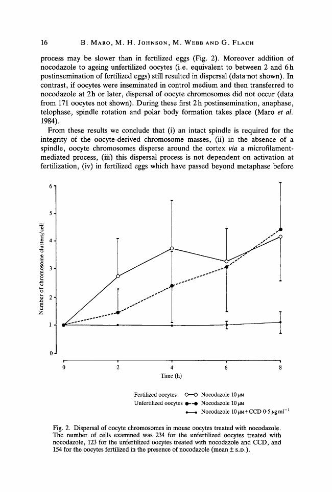
process may be slower than in fertilized eggs (Fig. 2). Moreover addition ofnocodazole to ageing unfertilized oocytes (i.e. equivalent to between 2 and 6hpostinsemination of fertilized eggs) still resulted in dispersal (data not shown). Incontrast, if oocytes were inseminated in control medium and then transferred tonocodazole at 2h or later, dispersal of oocyte chromosomes did not occur (datafrom 171 oocytes not shown). During these first 2h postinsemination, anaphase,telophase, spindle rotation and polar body formation takes place (Maro et al.1984).
From these results we conclude that (i) an intact spindle is required for theintegrity of the oocyte-derived chromosome masses, (ii) in the absence of aspindle, oocyte chromosomes disperse around the cortex via a microfilament-mediated process, (iii) this dispersal process is not dependent on activation atfertilization, (iv) in fertilized eggs which have passed beyond metaphase before
5-
4-
2-
4Time (h)
Fertilized oocytes O—O Nocodazole 10 JUM
Unfertilized oocytes • — • Nocodazole 10 JUM
•—• Nocodazole 10 /XM+CCD 0-5 ng ml"1
Fig. 2. Dispersal of oocyte chromosomes in mouse oocytes treated with nocodazole.The number of cells examined was 234 for the unfertilized oocytes treated withnocodazole, 123 for the unfertilized oocytes treated with nocodazole and CCD, and154 for the oocytes fertilized in the presence of nocodazole (mean ± S.D.).

Polar body formation in mouse oocytes 17
addition of nocodazole, the chromosomes do not disperse. We next examined thecapacity of dispersed chromosomes to affect the cytocortical organization of theegg-
Cytocortical changes in nocodazole-treated eggs
In the continuing presence of nocodazole, a focus of bright subcortical actinstaining and an area lacking Con A binding sites developed adjacent to the clumpof sperm chromosomes and at almost all sites of dispersed oocyte chromosomes inboth fertilized and unfertilized oocytes (Table 1; Fig. 1J-P). The area of cortexaffected at each site was often extensive, and coalescence of adjacent areas oftenoccurred (compare Fig. IK and 1O). Only when very small clusters of oocytechromosomes were examined was this association not detected. These foci of actinand reduced Con A binding persisted in the presence of nocodazole, but were lostin control eggs when pronuclei formed and migrated centrally.
When nocodazole-treated eggs were examined on the scanning electronmicroscope (SEM) they were found to have either an increase in the size ofthe area devoid of microvilli or the presence of more than one of these areas(Fig. 3A-C; Table 2). Using transmission electron microscopy, an electron-densearea was observed in the cytocortex overlying the chromosomes and extending in awide area around them (Fig. 3E). This electron-dense area was seen at highermagnification to be rich in filamentous structures (Fig. 3F). Very few organelleswere found in this area of the cytocortex while many were observed on thecytoplasmic side of the chromosomes (Fig. 3E) or in other regions of the cellcortex remote from the chromatin (Fig. 3D). No microvilli were observed in thearea of the cell surface adjacent to the chromosomes (Fig. 3E) while many of themwere observed in other regions of the cell surface (Fig. 3D), confirming the SEMdata.
From these results we conclude that chromosome clusters induce in their vicinity(i) a disappearance of Con A binding sites and of microvilli at the cell surface, (ii)an actin-rich filamentous area in the cell cortex between the chromosomes andthe plasma membrane. We next examined whether each clump of dispersedchromosomes could also engage in polar body formation.
Multiple polar body formation in nocodazole-treated eggs after removal of the drug
Eggs were fertilized in vitro in the presence of nocodazole, then removed fromthe drug after 2h or later and cultured in control medium to 8h before analysis.Despite removal from the drug, the dispersal of the oocyte-derived chromosomesnonetheless persisted (Table 3). Moreover, many eggs showed formation ofmultiple spindles that were fully or partially rotated, or of multiple polar bodies,each associated with a cluster of oocyte-derived chromosomes but never withsperm-derived chromosomes (Fig. 4A-E; Table 3). The proportion of eggs withpolar bodies formed or forming was higher when drug removal occurred at 2 or 4 hthan at 6h. These polar bodies were forming in the cortical area rich in

18 B. MARO, M. H. JOHNSON, M. WEBB AND G. FLACH
Table 1. Cortical changes induced by oocyte-derived chromosome clusters innocodazole-treated oocytes
Incubationconditions (37 °C)
Control2h4h6h8h
Nocodazole IOJUM2h4h6h8h
Numberof cells
examined
10322795
35204059
% ()f oocytes witngiven number of oocyte
chromosome clusters
1
30100100100
25574
2
70
17252018
>2
58707378
% of oocytes whereoocyte chromosomes
are associatedwith an area:
D 'Ai
Con Abinding
sites
10078182
971009897
Rich incortical
actin
1005611
66100100100
microfilaments and devoid of microvilli as do normal polar bodies (Fig. 4F-K).Not all chromosome clusters were associated with spindles, but most were.
From these results we conclude that (i) the clumps of dispersed oocytechromosomes have the capacity to induce the formation of a spindle that canrotate and yield a polar body, (ii) the sperm-derived chromosomes lack thiscapacity, and (iii) the capacity to form spindles and polar bodies persists innocodazole-treated fertilized oocytes well beyond the 2h period within whichthese processes are completed normally in control fertilized oocytes.
In order to form a polar body, both microtubules and micro tubule-organizingcentres (MTOCs) are required to organize a spindle. Therefore, we next examinedwhether these were present in the vicinity of dispersed chromosomes.
Chromosomes and microtubule-organizing centres (MTOCs)In the mouse oocyte, the MTOCs do not have associated centrioles (Szollosi,
Calarco & Donahue, 1972; Calarco-Gillam et al. 1983), instead multiple foci of
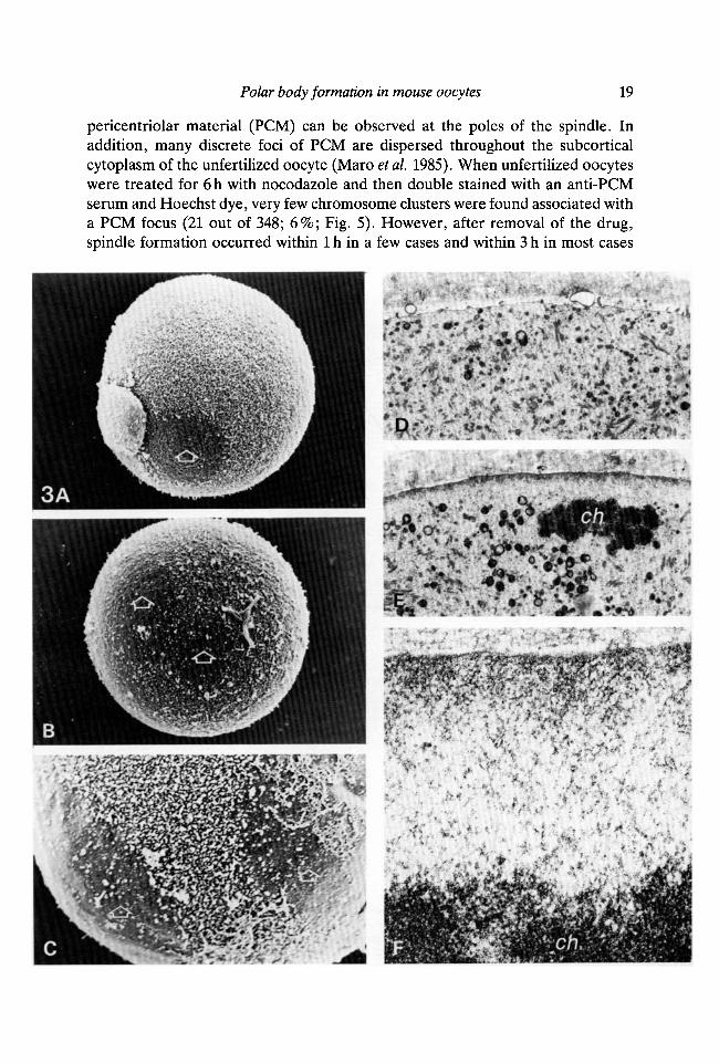
Fig. 3. (A-C) Scanning electron microscopy of mouse oocytes.(A) Control oocyte, 20h post hCG. Note area free of microvilli (open arrow)
adjacent to the first polar body, reflecting the presence of the underlying secondmeiotic spindle. X1200.
(B) Oocyte treated with 10 jUM-nocodazole for 6h. Note two areas free of microvilli(open arrows). X1200.
(C) Oocyte treated with 10 /iM-nocodazole for 6h. Note two areas free of microvilli(open arrows) and the boundary of microvilli between these two areas. X2400.(D-F) Transmission electron microscopy of an oocyte treated with 10 ̂ M-nocodazolefor 6h. ch, chromosomes. Note filamentous area devoid of organelles between thechromosomes and the plasma membrane and extending beyond the immediateconfines of the chromosomes (E,F). In contrast organelles can be found in the vicinityof the plasma membrane in region of the cell remote from the chromosomes (D).X6500 (D,E), X52000 (F).
Polar body formation in mouse oocytes 19
pericentriolar material (PCM) can be observed at the poles of the spindle. Inaddition, many discrete foci of PCM are dispersed throughout the subcorticalcytoplasm of the unfertilized oocyte (Maro etai. 1985). When unfertilized oocyteswere treated for 6h with nocodazole and then double stained with an anti-PCMserum and Hoechst dye, very few chromosome clusters were found associated witha PCM focus (21 out of 348; 6%; Fig. 5). However, after removal of the drug,spindle formation occurred within 1 h in a few cases and within 3 h in most cases
3A

20 B. MARO, M. H. JOHNSON, M. WEBB AND G. FLACH
and PCM could be observed at the spindle poles (Fig. 6). We must note that thedispersion of the PCM foci seems also to be a microfilament-dependent processsince 83 % of the polar PCM foci remained associated with the chromosomes inoocytes treated for 6h with both nocodazole and cytochalasin D (data from 21oocytes not shown).
From these results we conclude that (i) in the absence of microtubules, PCMfoci are not associated with chromosome clusters, (ii) in the presence ofmicrotubules, oocyte chromosome clusters are able to recruit the PCM which isnecessary for spindle formation.
Table 2. Effect of 10 \m-nocodazolefor 6hon distribution of microvilli in unfertilizedoocytes as judged by scanning electron microscopy
Nocodazole-Patterns of microvilli Control cells treated cells
distribution % (n = 43) % (n = 65)Uniform microvillous 26 20
Microvillous free patches1 small patch1 large patch2 patches3 patches
5123
2523213
No microvilli detected 8
* Note that the number of patches will underestimate the number of separate chromosomeclusters since (a) the influence of chromatin on the cytocortex is quite extensive leading tocoalescence of adjacent microvillous-free areas, and (b) it is not possible to examine the wholeof the oocyte surface under the SEM.
Table 3. Multiple polar body formation in eggs treated with 10 nu-nocodazole afterremoval of the drug. Analysis was carried out after 8h culture at 37 °C
Periodof
presenceof thedrug
Control
0-2 h0-4h0-6h0-8h
Numberof cells
examined
95
31322359
1
100
13544
Chromosomeclusters
2
29
1418
3
295
1113
%
>3
29907165
of eggs with given number of:
Spindlesor polar bodies*
1 2 3 >3
100
35 49 105 31 37 17
14 15 14 5
Pronuclei(including male)f
1
4
13
2
96
39
3
25
>3
2011
* Groups do not add up to 100 % because of the absence of formation of polar bodies in someeggs.
t Groups do not add up to 100 % because of the absence of formation of pronuclei in someeggs. When eggs are kept in nocodazole for the 8 h period, pronuclei do not form.

Polar body formation in mouse oocytes 21
B
I
H
K
Fig. 4. Eggs treated with 10 jUM-nocodazole for lh prior to insemination and for 4hpostinsemination, washed, and cultured in control medium for a further 4h.(A) Multiple polar body extrusion in living eggs. Arrowheads point at polar bodies.(B,C) Egg double stained for tubulin (B) and chromatin (C). Note three metaphasespindles. The sperm-derived chromosomes (arrow) are not associated with a spindle.(D,E) Egg double stained for tubulin (D) and chromatin (E). Note three rotatinganaphase spindles. Arrows point to the sperm head which is not associated with aspindle.(F-K) Egg double stained for actin (G,J) and Con A (H,K) and photographed at twodifferent focal planes (F-H and I-K). Note two rotated spindles and polar bodiesforming (open arrows). Another forming polar body can be seen out of focus (lowerright). Large arrow points at the first polar body heavily stained with Con A. x225 (A),X320 (B-K).

22 B. MARO, M. H. JOHNSON, M. WEBB AND G. FLACH
T
5 A1rB I
Fig. 5. (A,B) Oocyte treated with 10/iM-nocodazole for 6h double stained for PCM(A) and chromatin (B). Note that PCM foci are not associated with the twochromosome clusters (arrowheads) which can be observed in this plane of focus (firstpolar body stains for chromatin only, lower right). X400.
Chromosomes and microtubules
It has been shown that in oocytes arrested at metaphase II of meiosis, thedispersed cytoplasmic PCM foci do not nucleate microtubules, the onlymicrotubules found being those forming the spindle (Wassarman & Fujiwara,1978; Maro et al 1985; Schatten et al. 1985; Fig. 1A). In order to test whether theoocyte chromosomes themselves promote tubulin polymerization in their vicinity
8
1001
75-
50
25-
•—• Total number of spindlesO—O Normal spindles• — • Abnormal spindles
Time (h)
Fig. 6. Spindle reformation in nocodazole-treated oocytes (10/iM for 4h) cultured forvarious periods of time in control medium after removal of the drug. In this experimenta spindle is defined as a chromosome cluster located on a microtubule bundle. Spindlesare categorized as normal if PCM can be detected unambiguously at both poles.Abnormal spindles are those in which we could not identify a PCM focus at both poles.21 cells were examined at 1 h, 122 at 2h and 34 at 3 h.

Polar body formation in mouse oocytes 23
7A B
Fig. 7. (A-D) Oocytes treated with 10 jUM-nocodazole for 6h, washed and im-mediately incubated in 6^M-taxol for 5min at 37°C, fixed and double stained fortubulin (A,C) and chromatin (B,D). Note microtubule bundles associated withchromosome clusters (arrowheads) and the small asters nucleated by PCM foci. x400.
as has been shown in frog oocytes (Karsenti, Newport, Hubble & Kirschner,1984), we used taxol to decrease the critical concentration for tubulinpolymerization (Schiff, Fant & Horwitz, 1979). Unfertilized oocytes, in whichchromosomes and PCM had been dispersed by nocodazole, were incubated in thepresence of taxol for 5min and then immediately fixed so that no associationbetween PCM foci and chromosomes could form and stained with an anti-tubulinantibody. Large bundles of microtubules were observed around most of thechromosome clusters (380 out of 460; 83 %; Fig. 7), whilst the separate small asterswere nucleated by the cytoplasmic PCM foci (Maro et al. 1985). From this result weconclude that chromosome clusters favour microtubule polymerization in theirvicinity.
Formation and migration ofpronuclei
In no egg treated continuously with nocodazole did pronuclei form, as assessedboth by differential interference contrast microscopy and staining with antibodiesto nuclear lamins (Table 4; Fig. 8A-F). Moreover, in nocodazole-treated eggs,central migration and aggregation of sperm- and oocyte-derived chromosomeclusters failed to occur. When nocodazole was removed 2h after insemination,

24 B. MARO, M. H. JOHNSON, M. WEBB AND G. FLACH
B
H
K
\
uV
0 P

Polar body formation in mouse oocytes 25
multiple pronuclei formed in many eggs, each corresponding to a dispersedchromosome cluster, but no migration of these pronuclei occurred (Table 4) evenwhen eggs were incubated for 12 h, well beyond the normal time of migration.These results are consistent with the observations that in vivo colchicine induces anincrease in egg ploidy (Edwards, 1958). When nocodazole was removed at timeslater than 4h, pronuclear formation did not occur at 8h (Table 4). Conversely, ifeggs were placed in nocodazole at 2 h or later postinsemination, the incidence ofpronuclear formation was unaffected (Table 4; Fig. 8D-L) but migration wassuppressed completely (Table 4; Fig. 8D-I). The drug did not reverse migrationwhen it had already occurred (Table 4; Fig. 8J-L).
Having established the conditions required to disperse chromosomes and tocontrol both their incorporation into pronuclei and their centrally directedmigration, we examined next the cytocortical changes under these variousconditions. When nocodazole was washed out at 2 h, pronuclei formed and corticalchanges were lost (Table 4; Fig. 8M-P). This loss occurred despite the failure ofpronuclei to migrate centrally. In contrast when removal of nocodazole wasdelayed beyond 2h pronuclei did not form and the cytocortical changes adjacentto the chromosomes persisted (Fig. 4F-K; Table 4). These results suggested thatonly chromosomes not located in a nucleus could induce the focal response.Confirmation of this impression came from the analysis of eggs that were firsttransferred to nocodazole at 2 h postinsemination or later. In most of these eggspronuclei formed but did not migrate, and in most, foci of reduced Con A bindingand elevated focal actin were also absent. Indeed, where migration had not
Fig. 8. (A-C) Fertilized egg 6h postinsemination stained for nuclear lamins (B,C).Note that a very weak lamin staining is seen around the chromosomes extruded in thesecond polar body (C; as visualized by overexposure of Fig. 6B) probably cor-responding to a reduced quantity of available lamin molecules in the polar body ratherthan to a selective mechanism to avoid normal nuclear envelope formation in the polarbody.(D-F) Egg transferred to medium containing 10 /iM-nocodazole 2 h postinseminationand cultured for a further 6h before staining for nuclear lamins (E) and Con A (F).Note that pronuclei form but do not migrate towards the centre of the egg and that themembrane overlying the pronuclei is enriched in Con A binding sites.(G-I) Egg transferred to medium containing 10 jUM-nocodazole 4 h postinseminationand cultured for a further 4h before staining for actin (H) and Con A (I). Note non-migrated pronuclei with enriched Con A binding in the adjacent plasma membrane (I)whole cortical actin is evenly distributed (H). Arrow points at the dead first polar bodywhich stains heavily with Con A.(J-L) Egg transferred to medium containing 10 jUM-nocodazole 6h postinseminationand cultured for a further 2h before staining for nuclear lamins (K) and Con A (L).Note that pronuclei form and migrate towards the centre of the egg and that Con Abinds evenly to the cell surface.(M-P) Egg cultured in medium containing 10 jUM-nocodazole for lh prior to in-semination and for 2h postinsemination, at which time nocodazole was removed andthe egg cultured for a further 6h in control medium, stained for actin (O) and Con A(P). Note multiple non-migrated pronuclei (six are visible in M and N) with an evendistribution of cortical actin (O) and Con A binding (P). Arrow points at the dead firstpolar body which stains heavily with Con A. x320.

26 B. MARO, M. H. JOHNSON, M. WEBB AND G. FLACH
Table 4. Effect of pronuclear formation and migration on cortical changes in fertilizedeggs
Incubation in presence of:
Controlmedium
0-2 h0-4 h0-6 h0-8 h
0-2 h0-4 h0-6 h
Noco-dazoleIOJUM
0-2 h0-4 h0-6 h0-8 h2-8 h4-8 h6-8 h
Controlmedium
2-8 h4-8 h6-8 h
Cellnumber
1032279531322759456066
% of eggs withpronuclei:
Formed Migrated
82100 40100 779711
100100100 32
% of eggs where chromosomesare associated with an area:Rich inactin
1005611
1691
100100
Con Anegative
10078182
1691
10097111610
Con Apositive
826547
occurred, an elevated Con A binding was associated with the underlying pronucleiin most cases (Table 4; Fig. 8D-I). This elevated binding was not accompanied byany obvious inequity in the subcortical distribution of actin (Fig. 8G-I).
Eggs in which elevated Con A binding had occurred were also examined usingscanning and transmission electron microscopy. One or two areas of blebbymicrovilli were observed in most of these eggs under the scanning electronmicroscope (Fig. 9A). This result was confirmed by observation of thin sectionsunder the transmission electron microscope where large microvilli and blebs wereobserved in the portion of the cell cortex overlying the pronuclei (Fig. 9B,C).
From these results we conclude that (i) only chromosomes not located in anucleus are able to induce the cortical changes, (ii) the reestablishment of a normalmicrovillous cortex over the pronuclei is sensitive to nocodazole and maytherefore require microtubules, (iii) microtubules are required for the migration ofpronuclei, (iv) nocodazole prevents the appearance of pronuclei possibly by adirect effect, but more probably indirectly (see Discussion).
DISCUSSION
In this paper we have demonstrated three types of interaction between thechromosomes and the cytoskeleton. First, chromosomes which are not containedin a nucleus are associated, with an extensive subcortical focus of microfilamentsand the loss of overlying microvilli. Second, condensed meiotic chromosomespromote microtubule growth in their vicinity. Third, when condensed meioticchromosomes surrounded by microtubules associate with pericentriolar material,a spindle forms. Together, these three interactions go some way towardsexplaining the mechanism of polar body formation.
Previous work has shown that two domains exist in the egg cortex, a smalldomain covering the spindle area rich in microfilaments and devoid of microvilli

Polar body formation in mouse oocytes 27
'^^iMji**^
Fig. 9. (A) Scanning electron microscopy of an egg which was transferred to mediumcontaining 10 jUM-nocodazole 2 h postinsemination and cultured for a further 6 h. Noteprotrusion of membranes and microvilli (open arrow). X1260.(B,C) Transmission electron microscopy of an egg which was transferred to mediumcontaining 10/iM-nocodazole 2 h postinsemination and cultured for a further 6 h. Noteprotrusion of membranes and microvilli in the portion of the plasma membrane facingthe pronucleus (defined by arrowheads in B); C is an enlargement of this area andreveals no obvious structural links between the pronucleus and the overlyingmembrane, pn, pronucleus. X4725 (B), x 10800 (C).Note that for SEM it is essential to remove the zonae pellucidae and thus themembrane protrusions appear more marked than when compressed under the zonaand viewed only in thin section.
and a large one covering the rest of the cell surface rich in microvilli and containingless microfilaments (Eager et ah 1976; Johnson et al. 1975; Maro et al. 1984; Longo& Chen, 1985; Karasiewicz & Soltynska, 1985; Van Blerkom & Bell, 1986).Spermatozoa do not usually fertilize the egg in the area devoid of microvilli(Johnson et al. 1975). When the sperm head decondenses with loss of its nuclearmembrane (Stefanini, Oura & Zamboni, 1969) a cortical domain similar to thatexisting near the meiotic chromosomes is induced (Shalgi et al. 1978; Maro et al.1984). This latter observation added to the fact that after nocodazole-induced

28 B. MARO, M. H. JOHNSON, M. WEBB AND G. FLACH
dispersion of the oocyte chromosomes, each cluster of chromosomes is able toinduce the formation of one of these domains despite the absence of associatedmicrotubules (see also Longo & Chen, 1985), suggested that this cortical domainwas induced by chromatin rather than by spindle microtubules. Similarly, althoughlinks between MTOCs and the nuclear envelope have been documented in manysystems (Bornens, 1977; Maro & Bornens, 1980; Kallenback & Mazia, 1982;Tassin, Maro & Bornens, 1985), including mouse oocytes (Calarco, Donahue &Szollosi, 1972; Maro et al. 1985) we were unable to detect any role for them inmediating chromosome-induced changes in the cell cortex, since an antigen linkedto the pericentriolar material (Calarco-Gillam etal. 1983; Maro etal. 1985) was notassociated with the chromosomes in nocodazole-treated oocytes. Only 1-3 h afterremoval of nocodazole did pericentriolar material and chromosomes cometogether to form functional spindles.
This effect of chromatin on the cell cortex explains the existence of thefertilization cone which develops after sperm entry into the egg. The spermnuclear envelope breaks down because of the meiotic environment persisting inthe oocyte during the first 30min after fertilization, the metaphase-anaphasetransition occurring only 20 to 30 min after the fusion between the oocyte and thesperm head (Sato & Blandau, 1979). The presence of the non-enveloped malechromatin then induces a changed cortical domain. In addition, previousobservations suggested that the cleavage furrow of meiosis II (and thus polar bodyformation) could only take place in one of these domains, rich in microfilamentsand devoid of microvilli (Eager et al. 1976; Johnson et al. 1975; Maro et al. 1984).Our present work has confirmed these previous suggestions: when chromosomesare dispersed by nocodazole prior to fertilization and when the drug is removedlater, multiple polar bodies form in many of the cortical domains associated withthe chromosomes. We do not at present understand what, if anything, controls thedirection of rotation of the spindle, and thereby determines which haploid set ofchromosomes are discarded in the polar body.
However, polar body formation requires not merely a modified cortical domain,but also an organized spindle. The oocyte chromatin, but not sperm chromatin,has two properties that allow elaboration of spindles. First, we have shown thatcondensed chromosomes promote tubulin polymerization, thereby allowingmicrotubule formation. Thus, most of the chromosome clusters observed afternocodazole-induced dispersion are not linked to associated MTOCs (as defined bythe anti-PCM serum) but nonetheless promote the formation of large microtubulebundles after taxol treatment (this may be more readily apparent because of theincreased concentration of free tubulin which has been induced by nocodazoleprior to taxol addition). Moreover, we know that inactive PCM foci exist in thecytoplasm of the normal metaphase II arrested oocyte, but they do not nucleatemicrotubules unless the critical concentration for tubulin polymerization is re-duced experimentally (Maro etal. 1985), whereas microtubules are clearly presentin the vicinity of the chromosomes, again suggesting that chromatin promotespolymerization. Second, we have shown here that in the presence of microtubules,

Polar body formation in mouse oocytes 29
oocyte meiotic chromosomes can recruit PCM foci and, in conjunction with thepromoting effect of meiotic chromosomes on tubulin polymerization, a spindle isorganized.
We do not know how chromosomes influence either tubulin or actinpolymerization in their vicinity. The effects of chromosomes on actin and the cellcortex seem to be unique to chromosomes in the meiotic oocyte and newlyfertilized interphase mouse egg, as we have not observed such an effect in othermouse cells. However an analogous situation has been described in the Drosophilablastoderm (Warn, Magrath & Webb, 1984). It is of interest that a featurecommon to both these systems is the peripheral location of the chromosomes, andthis may prove to be the most important determinant of whether the effect isobserved. In contrast, the effects on tubulin polymerization and MTOCrecruitment appear to be a general property of chromosomes in the meiotic ormitotic state. Thus, a decrease in the apparent critical concentration for tubulinpolymerization in the vicinity of the chromosomes has also been observed inXenopus oocytes (Karsenti et al 1984). In this latter case, centrosomes injectedinto the cytoplasm of a metaphase arrested egg were not able to nucleatemicrotubules unless located close to the chromosomes. However, followingactivation at fertilization in both Xenopus and the mouse, all of the MTOCsdispersed throughout the egg were able to nucleate, suggesting that a drop in thecytoplasmic critical concentration for tubulin polymerization occurs during inter-phase (Karsenti et al 1984; Maro et al. 1985; Schatten et al 1985). Moreover, adependence of the total length of spindle microtubules on the number of chromo-somes present in the spindle has been demonstrated in grasshopper spermatocytes(Nicklas & Gordon, 1985).
The effect on tubulin polymerization of the transition from meiosis or mitosis tointerphase may reflect a fall in the level of maturation promoting factor (MPF),the activity of which oscillates during the cell cycle (Masui & Markert, 1971;Wasserman & Smith, 1978; Gerhart, Wu & Kirschner, 1984). In the egg, MPF isstabilized prior to fertilization by the activity of cytostatic factor (CSF). CSF isfound only in unfertilized eggs and has been shown to cause metaphase arrest uponinjection into cleaving eggs (Masui, Meyerhof & Miller, 1980). MPF inducesnuclear membrane breakdown and chromosome condensation when injected intoeggs arrested at the end of S phase (Miake-Lye, Newport & Kirschner, 1983),hence loss of the sperm nuclear membrane after fertilization. This factor mightalso regulate, directly or indirectly, microtubule assembly into spindles in mitoticcells. Both CSF and MPF activities fall rapidly after fertilization as evidenced bythe incapacity of the egg to condense chromosomes or break down nuclear mem-branes within 30-60 min after activation (Czotowska, Modlinski & Tarkowski,1984; Howlett & Maro, unpublished observation) and there is evidence that theirloss is dependent on a transient Ca2+ influx at fertilization that destroys CSF anddestabilizes MPF (Newport & Kirschner, 1984). However, it is clear from ourexperiments here that when fertilization occurs in the presence of nocodazole,MPF activity (and perhaps CSF activity) must persist for at least 4 to 6h. Thus

30 B. MARO, M. H. JOHNSON, M. WEBB AND G. FLACH
removal of nocodazole at any point during this period is associated with rapidspindle formation in the vicinity of the condensed chromosomes. Since there isevidence that MPF drives microtubular and nuclear membrane changes during thecell cycle, rather than being dependent upon them (Miake-Lye et al. 1983), itseems likely that the maintained level of MPF (and perhaps CSF) in the presenceof nocodazole may be secondary to an effect of the drug on the normal mechanismby which these activities are destroyed or maintained.
Although the precise mechanisms by which the chromosomes are able to inducechanges in the organization of both microfilaments and microtubules are unclear,it is nevertheless possible to propose a sequence for the various events leading topolar body formation. First it seems that at the end of maturation, just beforegerminal vesicle break down, the chromosomes gain the capacity to modify theorganization of actin in their vicinity (Van Blerkom & Bell, 1986). Second, whenthe germinal vesicle breaks down at entry into metaphase, the MTOC material(PCM) which was associated previously with the nuclear envelope, becomesdispersed in several foci within the cytoplasm (Calarco et al. 1972; Maro et al.1985). At the same time a spindle is formed because of the effect of thechromosomes on tubulin polymerization during M-phase (Karsenti etal. 1984; ourdata) in conjunction with the recruitment of nearby MTOCs. Third, the spindlemoves towards the periphery of the cell in a microfilament-dependent process(Longo & Chen, 1985), probably related to the effect of chromatin on themicrofilament network (Van Blerkom & Bell, 1986). Finally, when the spindlereaches the cell periphery, the chromosomes induce the formation of an actin-rich,microvillous-free domain of the cortex (Maro et al. 1984; Longo & Chen, 1985;Van Blerkom & Bell, 1986). After fertilization (or activation), meiosis resumesand, after the metaphase-anaphase transition, the cleavage furrow forms only inthis microfilament-rich domain of the cortex (Maro et al. 1984). The rotation of thespindle follows (possibly as a consequence of the limited area available for thedevelopment of the furrow), the cleavage furrow is completed, and the polar bodyforms (Maro et al. 1984). Having defined the various steps of the sequence, andsome of the underlying mechanisms, it remains to determine the nature of theirmolecular interrelationships in more detail.
We wish to acknowledge the technical assistance of Susan Pickering and Ian Edgar. We thankEric Karsenti and Pierre Gounon for the gift of the anti-actin antibodies, Jean-Claude Courvalinand Frank McKeown for the gift of the anti-lamin sera, Tim Mitchinson and Marc Kirschner forthe gift of the anti-PCM serum, Eric Karsenti and Tim Hunt for helpful discussions. This workwas supported by grants from the Medical Research Council of Great Britain and the CancerResearch Campaign to M. H. Johnson and from the Fondation pour la Recherche Me'dicale toB. Maro. B.M. is an EMBO fellow.
REFERENCESBORNENS, M. (1977). Is the centriole bound to the nuclear membrane? Nature, Lond. 270,80-82.CALARCO, P. G., DONAHUE, R. P. & SZOLLOSI, D. (1972). Germinal vesicle breakdown in the
mouse oocyte. J. CellSci. 10, 369-385.
Polar body formation in mouse oocytes 31
CALARCO-GILLAM, P. D., SIEBERT, M. C , HUBBLE, R., MITCHISON, T. & KIRSCHNER, M. (1983).Centrosome development in early mouse embryos as defined by an autoantibody againstpericentriolar material. Cell 35, 621-629.
CZOLOWSKA, R., MODLINSKI, J. A. &TARKOWSKI, A. K. (1984). Behaviour of thymocytes nuclei innon-activated and activated mouse oocytes. /. Cell Sci. 69,19-34.
EAGER, D., JOHNSON, M. H. & THURLEY, K. W. (1976). Ultrastructural studies on the surfacemembrane of the mouse egg. /. Cell Sci. 22, 345-353.
EDWARDS, R. G. (1958). Colchicine-induced heteroploidy in the mouse. /. exp. Zool. 137,317-362.
FLEMING, T. P., WARREN, P. D., CHISOLM, J. C. & JOHNSON, M. H. (1984). Trophectodermalprocesses regulate the expression of totipotency within the inner cell mass of the mouseexpanding blastocyst. /. Embryol. exp. Morph. 84, 63-90.
FULTON, B. P. & WHITTINGHAM, D. G. (1978). Activation of mammalian oocytes by intracellularinjection of calcium. Nature, Lond. 273, 149-151.
GERHART, J., WU, M. & KIRSCHNER, M. (1984). Cell-cycle dynamics of an M-phase-specificcytoplasmic factor in Xenopus laevis oocytes. /. Cell Biol. 98, 1247-1255.
GOUNON, P. & KARSENTI, E. (1981). Involvement of contractile proteins in the changes inconsistency of oocyte nucleoplasm in the newt Pleurodeles waltii. J. Cell Biol. 88, 410-421.
HOEBEKE, J., VAN NIGEN, G. & DE BRABANDER, M. (1976). Interaction of Oncodazole (R17934),a new anti-tumoral drug, with rat brain tubulin. Biochem. Biophys. Res. Commun. 69,319-342.
JOHNSON, M. H., EAGER, D., MUGGLETON-HARRIS, A. L. & GRAVES, H. M. (1975). Mosaicism inthe organisation of concanavalin A receptors on surface membrane of mouse eggs. Nature,Lond. 257, 321-322.
JOHNSON, M. H. & ZIOMEK, C. A. (1982). Cell subpopulations in the late morula and earlyblastocyst of the mouse. Devi Biol. 91, 431-439.
KALLENBACK, R. & MAZIA, D. (1982). Origin and maturation of centrioles in association with thenuclear envelope in hypertonic stressed sea urchin eggs. Eur. J. Cell Biol. 28, 68-76.
KARASIEWICZ, J. & SOLTYNSKA, M. S. (1985). Ultrastructural evidence for the presence of actinfilaments in mouse eggs at fertilization. Wilhelm Roux Arch devl Biol. 194, 369-372.
KARSENTI, E., NEWPORT, J., HUBBLE, R. & KIRSCHNER, M. (1984). Interconversion of metaphaseand interphase micro tubule arrays as studied by the injection of centrosomes and nuclei intoXenopus eggs. /. Cell Biol. 98, 1730-1745.
LONGO, F. J. & CHEN, D. Y. (1985). Development of cortical polarity in mouse eggs:involvement of the meiotic apparatus. Devi Biol. 107, 382-394.
MARO, B. & BORNENS, M. (1980). The centriole-nucleus association: the effect of cytochalasin Band nocodazole. Biol. Cell 39, 287-290.
MARO, B., HOWLETT, S. K. & WEBB, M. (1985). Non-spindle MTOCs in metaphase II-arrestedmouse oocytes. /. Cell Biol. 101, 1665-1672.
MARO, B., JOHNSON, M. H., FLACH, G. & PICKERING, S. J. (1984a). /. Embryol. exp. Morph. 82Supplement, 70.
MARO, B., JOHNSON, M. H., PICKERING, S. J. & FLACH, G. (19846). Changes in the actindistribution during fertilisation of the mouse egg. /. Embryol. exp. Morph. 81, 211-237.
MARO, B. & PICKERING, S. J. (1984). Microtubules influence compaction in preimplantationmouse embryos. /. Embryol. exp. Morph. 84, 217-232.
MASUI, Y. & MARKERT, C. L. (1971). Cytoplasmic control of nuclear behavior during meioticmaturation of frog oocytes. /. exp. Zool. 117, 129-146.
MASUI, Y., MEYERHOF, P. G. & MILLER, M. A. (1980). Cytostatic factor and chromosomebehaviour in early development. In The Cell Surface. New York: Academic Press.
MCKEOWN, F. D., TUFFANELLI, D. L., FUKUYAMA, K. & KIRSCHNER, M. (1983). Autoimmuneresponse against conserved determinants of nuclear envelope proteins in a patient with linearsclerodermia. Proc. natn. Acad. Sci., U.S.A. 80, 4374-4378.
MIAKE-LYE, R., NEWPORT, J. W. & KIRSCHNER, M. W. (1983). Maturation promoting factorinduces nuclear envelope breakdown in cycloheximide-arrested embryos of Xenopus laevis.J. Cell Biol. 97,81-91.
NEWPORT, J. W. & KIRSCHNER, M. W. (1984). Regulation of the cell cycle during early Xenopusdevelopment. Cell 37, 731-742.
32 B . M A R O , M. H. J O H N S O N , M. W E B B AND G. F L A C H
NICKLAS, R. B. & GORDON, G. W. (1985). The total length of spindle microtubules depends onthe number of chromosomes present. /. Cell Biol. 100, 1-7.
NICOLSON, G. L., YANAGIMACHI, R. & YANAGIMACHI, H. (1975). Ultrastructural localisation oflectin binding sites on the zonae pellucidae and plasma membranes of mammalian eggs. /. CellBiol. 66, 263-274.
SATO, K. & BLANDAU, R. J. (1979). Second meiotic division and polar body formation in mouseeggs fertilized in vitro. Gamete Res. 2, 283-293.
SCHATTEN, G., SIMERLY, C. & SCHATTEN, H. (1985). Microtubule configuration during fertilization,mitosis and early development in the mouse. Proc natn. Acad. Sci., U.S.A. 82, 4152-4156.
SCHIFF, P. B., FANT, J. & HORWTTZ, S. B. (1979). Promotion of microtubule assembly in vitro bytaxol. Nature, Lond. 277, 665-667.
SCHLIWA, M., EUTENEUER, U., BULINSKY, J. C. & IZANT, J. G. (1981). Calcium lability ofcytoplasmic microtubules and its modulation by microtubule associated proteins. Proc. natn.Acad. Sci., U.S.A. 78,1037-1041.
SHALGI, R., PHILLIPS, D. M. & KRAICER, P. F. (1978). Observations on the incorporation cone inthe rat. Gamete Res. 1, 27-37.
STEFANINI, M., OURA, C. & ZAMBONI, L. (1969). Ultrastructure of fertilization in the mouse:penetration of sperm into the ovum. J. Submicr. Cytol. 1,1-23.
SZOLLOSI, D., CALLARCO, P. & DONAHUE, R. P. (1972). Absence of centrioles in the first andsecond meiotic spindles of mouse oocytes. J. Cell Sci. 11, 521-541.
TASSIN, A. M., MARO, B. & BORNENS, M. (1985). Fate of MTOCs during myogenesis in vitro./. Cell Biol. 100, 35-47.
VAN BLERKOM, J. & BELL, H. (1986). Regulation of development in the fully grown mouse oocyte:chromosome-mediated temporal and spatial differentiation of the cytoplasm and plasmamembrane. /. Embryol. exp. Morph. 93 (in press).
WARN, R. M., MAGRATH, R. & WEBB, S. (1984). Distribution of F-actin during cleavage of theDrosophila syncytial blastoderm. /. Cell Biol. 98,156-162.
WASSARMAN, P. M. & FUJIWARA, K. (1978). Immunofluorescent anti-tubulin staining of spindlesduring meiotic maturation of mouse oocytes in vitro. J. Cell Sci. 29,171-188.
WASSERMAN, W. J. & SMITH, L. D. (1978). The cyclic behavior of a cytoplasmic factor controllingnuclear membrane breakdown. J. Cell Biol. 78, R15-R22.
WHITTINGHAM. D. G. (1971). Culture of mouse ova. /. Reprod. Fert. 14 Supplement, 7-21.WfflnTNGHAM, D. G. & WALES, R. G. (1969). Storage of two-cell mouse embryos in vitro. Austr.
J. biol. Sci. 22, 1065-1068.
(Accepted 22 October 1985)