Embed Size (px)
Citation preview

The EMBO Journal Vol.16 No.21 pp.6559–6573, 1997
Mechanism of open complex and dual incisionformation by human nucleotide excision repairfactors
Elizabeth Evans, Jonathan G.Moggs,Jae R.Hwang1, Jean-Marc Egly1 andRichard D.Wood
Imperial Cancer Research Fund, Clare Hall Laboratories,South Mimms, Hertfordshire EN6 3LD, UK and 1Institut de Genetiqueet de Biologie Moleculaire et Cellulaire, BP 163, F-67404,Illkirch Cedex, Universite Louis Pasteur, Strasbourg, France
During nucleotide excision repair in human cells, adamaged DNA strand is cleaved by two endonucleases,XPG on the 3� side of the lesion and ERCC1-XPF onthe 5� side. These structure-specific enzymes act atjunctions between duplex and single-stranded DNA.ATP-dependent formation of an open DNA structureof ~25 nt around the adduct precedes this dual incision.We investigated the mechanism of open complex form-ation and find that mutations in XPB or XPD, theDNA helicase subunits of the transcription and repairfactor TFIIH, can completely prevent opening anddual incision in cell-free extracts. A deficiency in XPCprotein also prevents opening. The absence of RPA,XPA or XPG activities leads to an intermediate levelof strand separation. In contrast, XPF or ERCC1-defective extracts open normally and generate a 3�incision, but fail to form the 5� incision. This samerepair defect was observed in extracts from humanxeroderma pigmentosum cells with an alteration in theC-terminal domain of XPB, suggesting that XPB hasan additional role in facilitating 5� incision by ERCC1-XPF nuclease. These data support a mechanism inwhich TFIIH-associated helicase activity and XPCprotein catalyze initial formation of the key openintermediate, with full extension to the cleavage sitespromoted by the other core nucleotide excision repairfactors. Opening is followed by dual incision, with the3� cleavage made first.Keywords: cisplatin/DNA repair/nucleases/TFIIH/xeroderma pigmentosum
Introduction
The dual incision reaction during human nucleotideexcision repair (NER) involves cleavage 3� and 5� to alesion in the damaged DNA strand by the XPG proteinand ERCC1-XPF protein complex respectively. Theseare structure-specific endonucleases, cleaving at junctionsbetween duplex and single-stranded DNA with a specificpolarity, resulting in excision of 24–32-mer oligonucleo-tides containing the damage (Mu et al., 1996; Sijberset al., 1996). Reconstitution of the incision reaction usingpurified mammalian repair proteins demonstrated that theproteins required include XPA, RPA, XPC-hHR23B, theTFIIH complex (including XPB and XPD) and the XPG
© Oxford University Press 6559
and ERCC1-XPF nucleases (Aboussekhra et al., 1995;Moggs et al., 1996; Mu et al., 1996). Biochemical studiesof these proteins have given clues to their roles in thereaction, but the mechanistic steps leading to the formationof 3� and 5� incisions are unclear. For instance, XPA, RPAand XPC-hHR23B have been implicated as candidatedamage recognition factors. Alternatively, these proteinsmay be involved in stabilizing single-stranded inter-mediates of the reaction, or displacing the damagedoligonucleotide.
TFIIH is a multisubunit factor that is required for basaltranscription initiation at RNA polymerase II promoters,as well as in NER as part of the core incision machinery,even during repair of non-transcribed DNA (Svejstrupet al., 1996). The importance of TFIIH during NER ishighlighted by the fact that mutations in components ofTFIIH can give rise to three inherited human disorders,xeroderma pigmentosum (XP), Cockayne syndrome (CS)and trichothiodystrophy (TTD), with NER defects ofdifferent types (Hoeijmakers et al., 1996). XP-B patientsare very rare with two cases exhibiting combined XP andCS, while another has TTD symptoms. Patients withinthe XP-D complementation group are more frequent andexhibit either XP alone, or XP combined with CS or TTD.A distinct complementation group, TTD-A, is associatedwith an unidentified defect in TFIIH (Vermeulen et al.,1994b).
The isolated XPB and XPD subunits of TFIIH are 3�–5� and 5�–3� DNA helicases respectively (Schaeffer et al.,1993, 1994; Sung et al., 1993; Ma et al., 1994a), but inboth transcription and NER they act only as part of TFIIH.In transcription, ATP-dependent TFIIH helicase activityis used to catalyze limited opening of a 10–20 bp regionaround the promoter which enables priming of the nascentRNA on the template strand (Holstege et al., 1996).During repair, the DNA helicase activities of XPB andXPD have been postulated to catalyze a similar localunwinding around the site of a DNA lesion (Weeda et al.,1990). TFIIH may also have a role in damage recognition,as suggested by the finding that the helicase activity ofthe XPD homolog in Saccharomyces cerevisiae (Rad3) isinhibited by bulky DNA lesions (Naegeli et al., 1993).
TFIIH-mediated opening could generate the junctionsbetween duplex and single-stranded DNA which arecleaved by the XPG and ERCC1-XPF nucleases.
Direct evidence for the formation of an unwound openDNA intermediate prior to incision was recently obtainedby chemical footprinting around the site of a singlecisplatin-DNA adduct (Evans et al., 1997). This reactionrequired XPA protein and was completely dependenton the presence of ATP. We have now carried out acomprehensive study to determine which core NER factorsparticipate in the formation of this key open intermediateduring the sequence of events that leads to dual incision

E.Evans et al.
formation and removal of DNA damage. Of particularimportance, we asked whether the TFIIH-associated DNAhelicases are involved in this step. TFIIH was indeedfound to be involved in opening, in a role that is likelyto be similar to its function in initiation of RNA polymeraseII transcription. We also uncovered the biochemical defectassociated with a human mutation in XPB, which causesrepair to be trapped at an unusual intermediate stage whereopening and 3� incision can occur, but 5� incision doesnot take place.
Results
Dissection of open complex formation by the core
NER factors
The experimental approach makes use of a 1,3-intrastrandd(GpTpG)-cisplatin crosslink located at a unique site inclosed-circular duplex DNA. Mammalian cell extractsform incisions on each side of the adduct and release itin a 24 to 32-mer oligonucleotide segment. The reactionis monitored using a probe that simultaneously detectsthis dual incision, as well as any uncoupled 3� or 5�incisions that form (Figure 1A). During incubation of theadducted DNA with repair-proficient HeLa cell extract,dual incisions were detectable after 4 min and increasedfor ~15 min (Figure 1B and C). Uncoupled 3� incisionswere detectable throughout the time course but fewuncoupled 5� incisions were observed (Figure 1B andC). The coordinate appearance of 3� incisions and dualincisions indicates that the 3� and 5� incisions are nearlysynchronous during the normal course of a reaction.
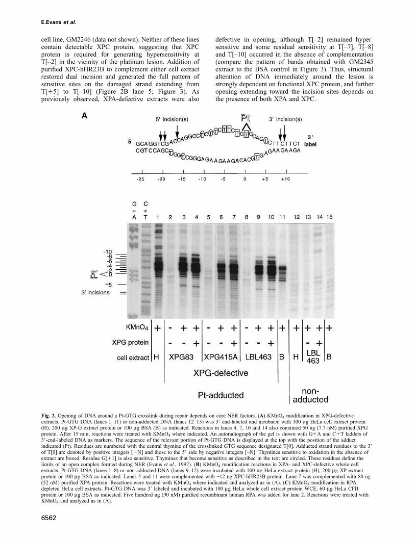
To monitor the formation of an open DNA intermediatearound the lesion prior to strand scission, potassiumpermanganate footprinting is used. Linearized duplex DNAis terminally labelled with 32P and incubated with cellextracts. Permanganate oxidizes T residues in regions withsingle-stranded character and results in sensitivity topiperidine cleavage. Analysis on a denaturing polyacryl-amide gel yields a pattern of sensitive T residues on theadducted and complementary non-adducted strands. Thisdefines a region of opening produced during the courseof a repair reaction, summarized at the top of Figure 2A(Evans et al., 1997). The 1,3-d(GpTpG)-cisplatin lesionitself causes some disruption of the duplex DNA structure,with an induced bend on the order of 25–30°, helicalunwinding of about 23°, local denaturation that includesthe central T residue and distortion over 4–5 base pairs(Anin and Leng, 1990; Bellon et al., 1991; van Garderenand van Houte, 1994). This causes an intrinsic permangan-ate sensitivity of residues T[0], T[–2], T[–4], T[–5] andG[�1] which can be observed in the absence of cellextract (Figure 2A, lane 11).
In order to dissect the roles of the core NER factors inopen complex formation, extracts defective in each of thefactors have now been tested for their ability to promotethe sensitivity of T residues surrounding the adduct tooxidation by permanganate. The adducted-strand residuesT[�5], T[–2], T[–7], T[–8] and T[–10] are particularlyinformative in this regard, as they show the most con-spicuous increases in sensitivity during DNA repair(Figure 2A).
6560
XPG endonuclease and open complex formation
Permanganate sensitivity indicating opening around thecisplatin adduct was detected with an extract from XPG-defective XPG83 cells (Evans et al., 1997). The causativemutation in this line is an amino acid change at a conservedresidue in the presumed active site of XPG (Nouspikeland Clarkson, 1994). This suggested that the nucleaseactivity of XPG is not needed for opening up of the DNAprior to incision. In order to test the generality of thisobservation, we analyzed opening in cell extracts fromother independent XP-G defective cell lines (Figure 2Aand Figure 3) and all four tested gave similar results. InFigure 2A, DNA containing the cisplatin adduct (lanes 1–11), or non-adducted (lanes 12–15) was radiolabeled atthe 3� end and reactions treated with KMnO4 as indicated.On adducted DNA, each mutant extract displayed a patternof KMnO4-sensitive bands that included the sites T[0],T[–2], T[–4] and T[–5] which show intrinsic sensitivity(Figure 2A, lane 11). Active opening by the extracts alsotook place, as indicated by the hypersensitivity of T[–2],T[–7], T[–8] and T[–10] towards the 5� side of the lesion,and sensitivity of T[�5] towards the 3� side (lanes 3, 6and 9). These extracts catalyze little or no 3� uncoupledincisions or dual incisions (not shown). In each case, thesensitivity of 5� T residues was slightly enhanced whenXPG protein was included in the reaction to restore dualincision formation (Figure 2A, compare lanes 3 and 4, 6and 7, 9 and 10). This is presented graphically in Figure3 for the severely defective XP-G cell line AG08802(Moriwaki et al., 1996). Although the exact pattern variessomewhat between different mutant lines, it appears thatsignificant opening takes place in the absence of XPGnuclease activity, but that addition of active XPG proteinmodulates the pre-incised DNA structure on the 5� sideof damage. A small amount of full-length mutant XPGexists in some of these cells, leaving open thepossibility that the mutant XPG may participate structur-ally but non-catalytically in formation of pre-incisioncomplexes.
XPC and XPA participate in open complex
formation
The XPC protein normally associates with hHR23B proteinin human cells and is required for the repair of someDNA lesions in vitro, including major UV photoproducts.The repair of certain other lesions does not require XPCin vitro (Mu and Sancar, 1997), nor is this factor neededduring transcription-coupled repair of UV lesions in vivo(van Hoffen et al., 1995). These observations have sug-gested that XPC may have a role in changing the DNAstructure around lesions at an early stage of NER. SinceXPC is needed for dual incision of the cisplatin adductin vitro (Moggs et al., 1996), we asked whether XPCparticipated in the opening reaction.
Figure 2B compares the pattern of permanganate-sensit-ive T residues formed by extracts from a repair deficientXP group C cell line (GM2249) and an XPA-defectiveline (GM2345; see Evans et al., 1997). On damaged DNA,the pattern of KMnO4-sensitive bands with both extracts(Figure 2B, lanes 4 and 6) included three sites (T[0],T[–4], T[–5]) that show sensitivity in the absence ofextract (lane 8). The XP-C extract gave no detectablesensitivity of the flanking residues T[�5], T[–7], T[–8]

TFIIH-mediated open complex during DNA repair
Fig. 1. The 3� and 5� incisions are tightly coupled (A) Schematic of hybridization assay to detect uncoupled and dual incisions. The products of dualincisions are detected as 24 to 32-mer platinated oligonucleotides. Uncoupled incisions at the 8th and 9th phosphodiester bonds 3� to the lesion aredetected as platinated 48 and 49-mers. Incubation in extracts can lead to 3� to 5� degradation of these fragments up to the Pt crosslink, resulting in aband migrating at ~42 nt. Non-incised and fully repaired DNAs are detected as 84-mers. Uncoupled 5� incisions would result in bands of 61, 64 and65 nt. (B) Time course of incision formation. Pt-GTG DNA was incubated with HeLa extract protein for the times indicated, and the DNA analyzedas in panel A. A phosphorimage of the gel is shown. The sizes of DNA fragments corresponding to uncoupled and dual incisions are indicated at theleft. Lane M contains a 5� phosphorylated 24-mer containing a single 1,3 intrastrand d(GpTpG)-cisplatin crosslink. (C) Quantification of B. Valuesfor the each incision product are displayed as a percentage of the total signal in each lane.
and T[–10], indicating a strong defect in opening aroundthe lesion. Interestingly, T[–2], which is only weaklysensitive in the absence of extract, also did not becomehypersensitive in the presence of GM2249 extract. This
6561
is in contrast to extracts from repair proficient cells, orcells from most other XP groups (compare Figure 2Blanes 4, 6, 8 and the densitometric traces in Figure 3).This same pattern was observed in another XPC-defective
E.Evans et al.
cell line, GM2246 (data not shown). Neither of these linescontain detectable XPC protein, suggesting that XPCprotein is required for generating hypersensitivity atT[–2] in the vicinity of the platinum lesion. Addition ofpurified XPC-hHR23B to complement either cell extractrestored dual incision and generated the full pattern ofsensitive sites on the damaged strand extending fromT[�5] to T[–10] (Figure 2B lane 5; Figure 3). Aspreviously observed, XPA-defective extracts were also
Fig. 2. Opening of DNA around a Pt-GTG crosslink during repair depends on core NER factors. (A) KMnO4 modification in XPG-defectiveextracts. Pt-GTG DNA (lanes 1–11) or non-adducted DNA (lanes 12–15) was 3� end-labeled and incubated with 100 μg HeLa cell extract protein(H), 200 μg XP-G extract protein or 100 μg BSA (B) as indicated. Reactions in lanes 4, 7, 10 and 14 also contained 50 ng (7.7 nM) purified XPGprotein. After 15 min, reactions were treated with KMnO4 where indicated. An autoradiograph of the gel is shown with G�A and C�T ladders of3�-end-labeled DNA as markers. The sequence of the relevant portion of Pt-GTG DNA is displayed at the top with the position of the adductindicated (Pt). Residues are numbered with the central thymine of the crosslinked GTG sequence designated T[0]. Adducted strand residues to the 3�of T[0] are denoted by positive integers [�N] and those to the 5� side by negative integers [–N]. Thymines sensitive to oxidation in the absence ofextract are boxed. Residue G[�1] is also sensitive. Thymines that become sensitive as described in the text are circled. These residues define thelimits of an open complex formed during NER (Evans et al., 1997). (B) KMnO4 modification reactions in XPA- and XPC-defective whole cellextracts. Pt-GTG DNA (lanes 1–8) or non-adducted DNA (lanes 9–12) were incubated with 100 μg HeLa extract protein (H), 200 μg XP extractprotein or 100 μg BSA as indicated. Lanes 5 and 11 were complemented with ~12 ng XPC-hHR23B protein. Lane 7 was complemented with 80 ng(52 nM) purified XPA protein. Reactions were treated with KMnO4 where indicated and analyzed as in (A). (C) KMnO4 modification in RPAdepleted HeLa cell extracts. Pt-GTG DNA was 3� labeled and incubated with 100 μg HeLa whole cell extract protein WCE, 60 μg HeLa CFIIprotein or 100 μg BSA as indicated. Five hundred ng (90 nM) purified recombinant human RPA was added for lane 2. Reactions were treated withKMnO4 and analyzed as in (A).
6562
defective in opening, although T[–2] remained hyper-sensitive and some residual sensitivity at T[–7], T[–8]and T[–10] occurred in the absence of complementation(compare the pattern of bands obtained with GM2345extract to the BSA control in Figure 3). Thus, structuralalteration of DNA immediately around the lesion isstrongly dependent on functional XPC protein, and furtheropening extending toward the incision sites depends onthe presence of both XPA and XPC.

TFIIH-mediated open complex during DNA repair
6563

E.Evans et al.
Fig. 3. Graphic representation of the pattern of permanganate sensitivity in repair-proficient and deficient extracts. Phosphorimages of KMnO4
footprinting experiments were traced using NIH Image software. For comparison, normalized traces from two related reaction conditions aresuperimposed, with one trace displayed as a solid line and the other as a shaded area.
Full open complex formation depends on RPA
protein
RPA is required for dual incision formation and has beenimplicated, together with XPA, in preferential binding to
6564
DNA damage. To explore the participation of RPA in opencomplex formation, KMnO4 sensitivity was monitored ina HeLa extract that had been fractionated to deplete it ofRPA and PCNA proteins (Shivji et al., 1992). The resulting

TFIIH-mediated open complex during DNA repair
extract (designated CFII) showed only barely detectabledual incision of the platinum adduct, and repair could berestored by adding purified RPA (data not shown). Additionof HeLa CFII caused a marked sensitivity of T[–2] tooxidation and a modest sensitivity of T residues [–7],[–8] and [–10] flanking the lesion to the 5� side (Figure2C, compare lane 1 with 4). Adding neutralizing levelsof an antibody against the p34 subunit of RPA to the CFIIdid not reduce the sensitivity of these residues (data notshown). Complementation of the HeLa CFII with RPAsignificantly enhanced the sensitivity of T residues [–7],[–8] and [–10], indicating a role for RPA in formation ofthe full open complex (Figure 2C, lane 2). A strong bandalso appeared at the position of the 3� incision. Dualincision intermediates accumulate in this case because theabsence of the DNA polymerase cofactor PCNA from thereaction mixture prevents repair synthesis.
Mutations that inactivate the 5� endonuclease stillallow full opening
Extracts from ERCC1 and XPF-defective rodent cells aredefective in the 5� cleavage reaction, but can still form 3�incisions (Sijbers et al., 1996). We tested their ability toopen the DNA, which is presumed to be a prerequisitefor 3� incision formation by XPG. Adducted DNA wasincubated with extracts of XPF-defective human(GM8437, Figures 4A and 3) or rodent cells (UV41, datanot shown), or ERCC1-defective cell extract (CHO 43–3B, Figure 4A) and treated with KMnO4 as indicated. Witheach mutant extract, a complete pattern of permanganatesensitivities appeared on the adducted strand T residues[–10], [–8], [–7], [–2] and [�5], as well as a bandcorresponding to the position of 3� incisions (Figure 4A,lanes 2 and 4). This pattern was indistinguishable fromthat seen in the corresponding complemented reaction(Figure 4A, lanes 3 and 5) in which dual incision formationis restored (Figure 4B and data not shown). Like the XPF-defective rodent lines, human XP-F extracts were defectivein 5� and dual incision formation but formed uncoupled3� incisions (Figure 4B, lane 1). Complementing withERCC1-XPF proteins resulted in 5� cleavage and gaveproducts of dual incision (Figure 4B, lanes 2 and 3). Thus,extracts specifically defective in 5� nuclease activity canstill open the DNA normally.
ATP-hydrolysis and open complex formation
Open complex formation was inhibited when ATP wasomitted from the reaction (Evans et al., 1997), suggestingthat it is needed for opening up of the duplex DNA, mostlikely as a cofactor for one or both of the DNA helicasesubunits of TFIIH. We tested the effect of substitutingnon-hydrolyzable analogs during a reaction with HeLaextract (Figure 5). Adducted DNA was incubated withextract in the absence or presence of ATP, ATPγS orAMP-PNP, and treated with KMnO4 after 15 min. T[–2]became distinctly more sensitive to oxidation even withoutadded nucleotide (lane 1) or in the presence an analog(lanes 3 and 4), compared with the reaction without extract(lane 5). A modest sensitivity of T residues on the 5� sideof the lesion was seen in the presence of ATPγS or AMP-PNP, but this sensitivity was much less than that seen inthe presence of ATP (lane 2). This suggests that mostopening requires ATP hydrolysis.
6565
TFIIH subunits XPB and XPD are needed for open
DNA complex formation
The requirement for ATP hydrolysis for full DNA openingaround the lesion strongly implicates the DNA-dependentATPase/DNA helicase activities of the XPB and/or XPDsubunits of TFIIH in this reaction. To test this directly,extracts were used from two XP-D lines (GM2485 andGM2253) and three XP-B lines (GM2252, XPCS1BA andCHO 27-1). The XP-D cell extracts and two of the XP-Bextracts (CHO 27-1, XPCS1BA) were completely defect-ive in opening the DNA surrounding the platinum adduct(Figure 6A, lanes 5, 7, 9 and Table I). This defect wascorrected by the addition of TFIIH (lanes 6, 8 and10). Uniquely, GM2252 (XP-B) cell extract reproduciblyexhibited a complete pattern of thymine hypersensitivityon the adducted strand (lane 3), similar to that seenwith HeLa cell extract (lane 2). Furthermore, fragmentscorresponding to 3� incisions were also produced byGM2252 extract (lane 3). This pattern of permanganatesensitivity was unaltered by adding functional TFIIH(lane 4).
One XPB mutant is specifically defective in 5�incision formation
The ability of the TFIIH-defective cell extracts to form3� and 5� incisions was examined. Both XP-D and two ofthe XP-B cell extracts (XPCS1BA and CHO 27–1) werecompletely defective in both 3� and 5� cleavage reactions(Figure 6B, lanes 1, 3, 5 and 7; Table I). Dual incisionactivity was restored by adding TFIIH (Figure 6B, lanes2, 4, 6 and 8). We also tested the single cell line in theTTD-A group (TTD1BR) which has been proposed toharbor an unidentified mutation in TFIIH (Vermeulenet al., 1994b). TTD1BR cell extract could form lowlevels of dual incisions (Figure 6B, lane 11) which werestimulated ~3-fold upon addition of TFIIH (lane 12).
In marked contrast to the cases above, the XPB-defective cell extract GM2252 was specifically defectivein 5� incision formation. The extract produced bandsmigrating in the position of uncoupled 3� incisions, butno uncoupled 5� incisions and only a trace amount of dualincision products (Figure 6B, lane 9). Complementingwith TFIIH restored 5� incision function, leading to dualincisions (lane 10). To check that the 3� uncoupledincisions were authentic, a primer extension assay (Moggset al., 1996) was used and confirmed that the positions ofthe uncoupled 3� incisions in GM2252 cell extract werepredominantly at the 9th phosphodiester bond 3� to thecisplatin-DNA lesion (data not shown). Furthermore,neutralizing anti-XPG antiserum inhibited the formationof uncoupled 3� incisions by GM2252 extract, and thisinhibition was reversed by adding excess XPG protein(not shown). The uncoupled 3� incisions depended onATP hydrolysis (Figure 6C). In repair-proficient extracts,neither dual nor uncoupled 3� incisions occurred in theabsence of ATP, nor did they form in XPB-defectiveGM2252 or XPF-defective GM8437 extracts. ATPγS didnot substitute for ATP, indicating that ATP hydrolysisrather than just binding is necessary for 3� cleavage ofthe damaged DNA strand (Figure 6C).
These results show that the XPB mutation in GM2252cells does not interfere with the opening of the DNA orcapacity of XPG to form a 3� incision, yet despite the

E.Evans et al.
Fig. 4. Opening and dual incision in extracts defective in the 5� nuclease. (A) KMnO4 modification in ERCC1– and XPF-defective cell extracts.Pt-GTG DNA (lanes 1–6) or non-adducted DNA (lanes 7–10) were incubated with 100 μg HeLa cell extract protein (H), 200 μg ERCC1– orXPF-defective extract protein, or 100 μg BSA as indicated. Lanes 3, 5 and 9 were complemented by adding ~40 ng (5.1 nM) purified recombinantERCC1-XPF complex. Reactions were treated with KMnO4 and analyzed as described in Figure 2A. (B) Incision in an XPF-defective cell extract.Pt-GTG DNA was incubated with GM8437 extract. The reactions for lanes 2 and 3 contained 20 ng (2.6 nM) and 40 ng (5.1 nM) purifiedERCC1-XPF respectively. The DNA was analyzed as indicated in Figure 1A.
presence of functional ERCC1-XPF, 5� incisions fail toform. Thus the opening and incision defect in GM2252cell extract is identical to that observed in ERCC1– andXPF-defective cell extracts, suggesting that XPB maynormally play a role in promoting 5� incision formation.
Discussion
TFIIH functions in open complex formation during
DNA repair
Human nucleotide excision repair of DNA proceedsthrough an open intermediate, which is thought to providethe junctions between double- and single-stranded DNAthat are cleaved by the structure-specific nucleases XPGand ERCC1-XPF (Evans et al., 1997). Although theoverall NER reaction is ATP-dependent, neither of thesenucleases requires ATP for cleavage. The experimentsreported here show that the two DNA helicase subunitsof TFIIH are crucial components of the protein machinerythat forms this open intermediate. The reaction requires
6566
ATP hydrolysis and results in opening across a region of~25 bp region between the two incision sites.
Mutations in either the XPB or XPD proteins candisable NER, but do both DNA helicase activities functionduring the NER reaction? Studies of the XPB and XPDhomologs in S.cerevisiae, Rad25 (Ssl2) and Rad3 respect-ively, suggest that they do. Many mutations in both Rad3and Rad25 result in UV sensitivity. Rad25 helicase activityis, moreover, essential for viability as shown by thelethality of a K392R change in the conserved Walker Anucleotide-binding motif of Rad25 (Park et al., 1992).Consistent with this, inactivation of human XPB helicaseby the equivalent K346R substitution yields a proteinthat inhibits both repair and transcription in a dominantnegative manner (van Vuuren et al., 1994). In contrast,the comparable Rad3 K48R helicase mutant is viable butUV sensitive (Sung et al., 1988), and TFIIH containing amutant Rad3 K48E helicase is active in transcription(Feaver et al., 1993). These results suggest that Rad3/XPD helicase activity is not required for transcription, but

TFIIH-mediated open complex during DNA repair
Fig. 5. Full opening depends on ATP hydrolysis. (A) The effect of ATP analogs on permanganate sensitivity during repair. Pt-GTG DNA incubatedwith 100 μg HeLa cell extract protein (lanes 1–4) or 100 μg BSA (lane 5) in repair buffer lacking ATP, dNTPs and the ATP regenerating system.ATP or the non-hydrolyzable analogs ATPγS or AMP-PNP were added to 2 mM. Each reaction was treated with KMnO4 and analyzed as describedin Figure 2A. (B) Graphic representation of (A).
does function during damage removal. Interestingly, theRad3 K48R strain can carry out limited incision of UVdamaged DNA, and a low level of nicking of UV-irradiated DNA without damage release was observed ina reconstituted incision reaction when Rad3 was replacedwith a K48R mutant protein (Sung et al., 1988, 1996). Itis possible that uncoupled incisions are formed in thismutant, implying that Rad3/XPD helicase might performa catalytic function after the formation of one or bothincisions. Deletion of the RAD3 gene is lethal, indicatingthat the protein does have an essential role, probably as astructural component of TFIIH.
These results suggest that both TFIIH helicase activitiesdo function during NER, and our results implicate one orboth of them in DNA opening, since functional XPBand XPD, as well as ATP hydrolysis, are necessary forformation of a normal open intermediate. Because thehelicase activities associated with the mutant proteins inthe opening-defective XP-B and XP-D cell extracts arenot yet known, the precise catalytic and/or structural rolesof the subunits in each step of the reaction remain to bedissected.
Pinpointing the biochemical repair defect in cells
from an individual with the disorders XP and CS
Although the XPB and XPD subunits of TFIIH arerequired for open complex formation during NER, they
6567
may also function during subsequent steps. Our resultswith extracts from GM2252 mutant cells (patient XP11BE)suggest a further role for XPB in the formation of 5�incisions. The C to A transversion mutation in XP11BEcells causes a splicing defect that changes the C-terminal41 amino acids of the XPB polypeptide (Weeda et al.,1990). This alteration of the C-terminus does not inhibitopen complex formation or 3� incisions but preventsformation of 5� incisions (Figure 6). Mutation of theC-terminus of XPB does not disrupt the conserved helicasemotifs in the primary sequence (Figure 7A). A truncationof the C-terminus of the yeast homolog Rad25 is alsooutside these motifs and leads to impaired repair but nottranscription (Guzder et al., 1994). Moreover, biochemicalcharacterisation of TFIIH purified from XP11BE cells andof recombinant XPB with an identical mutation showsthat the mutant XPB protein retains appreciable 3�–5�DNA helicase activity, ~40% of normal (Hwang et al.,1996). The reduced DNA helicase activity may resultfrom a conformational change in XPB caused by themutated C-terminus. However, the helicase activityremaining in XP11BE cells appears to be sufficient toform a full open complex of ~25 nt and uncoupled 3�incisions.
The occurrence of inactivating mutations in theC-termini of XPB and Rad25 has led to the suggestionthat this domain may interact with other NER factors

E.Evans et al.
(Park et al., 1992; Ma et al., 1994b; Sweder and Hanawalt,1994; van Vuuren et al., 1994; Hwang et al., 1996). Thealteration of XPB protein in XP11BE cells does not appearto influence targeting or cleavage by the 3� endonucleaseXPG, since normally positioned 3� incisions are stillformed in XP11BE cell extract. Consistent with this,although XPG is known to interact with several TFIIHsubunits including XPB, XPG does not interact with theC-terminus of XPB (Iyer et al., 1996). The very similarrepair defects in XP11BE, XP-F and ERCC1-defectivecells specifically suggest that the C-terminus of XPBinteracts with an NER factor involved in the formation of5� incisions. XPB might recruit ERCC1-XPF to the 5�incision site or may interact with another NER factorwhich facilitates the 5� cleavage reaction. We attemptedto overcome the defect in XP11BE cell extract by addinga large excess of purified ERCC1-XPF and RPA proteins,but were unable to induce 5� incision (data not shown).
In contrast to XP11BE, the mutation in XPCS1BA cells
Fig. 6. The role of TFIIH in open complex formation and dual incision. (A) The effect of mutations in XPB and XPD on DNA opening duringrepair. Pt-GTG DNA was incubated with 100 μg HeLa cell extract protein, 200 μg protein from XP-B or XP-D extracts or BSA as indicated.Reactions in lanes 4, 6, 8 and 10 were complemented by adding TFIIH. Reactions in lanes 2–11 were treated with KMnO4 and analyzed as in Figure2A. (B) The effect of mutations in TFIIH on 3� and 5� incision formation. Pt-GTG DNA was incubated with 200 μg protein from extracts of XP-B,XP-D extracts or TTD-A defective TTD1BR. Purified TFIIH was included for lanes 2, 4, 6, 8, 10 and 12. DNA was purified and analyzed asindicated in Figure 1A. (C) The effect of ATP analogs on incision formation. Pt-GTG DNA was incubated in repair buffer lacking ATP, dNTPs andthe ATP regenerating system, with 100 μg repair proficient SW48 cell extract protein (lane 7–9), 200 μg extract protein from GM2252 (lanes 1 and4), GM8437 (lanes 2 and 5) or a mixture of the two (lanes 3 and 6). Reactions were supplemented with 2 mM ATP or 2 mM ATPγS as indicated.DNA was purified and analyzed as shown in Figure 1A. Degradation of 3� uncoupled incision fragments, resulting in a band at ~42 nt, isparticularly pronounced in the absence of dNTPs.
6568
is an F99S amino acid substitution (Vermeulen et al.,1994a) close to or within the domain of XPB whichinteracts with XPG and XPD (Figure 7A; Iyer et al.,1996). Consistent with either type of disruption, weobserved neither open complex formation nor incisionwith XPCS1BA extract.
Other factors facilitating open complex formation
In addition to TFIIH, other NER proteins participate inreaction steps preceding and coincident with open complexformation. One important question is whether the nucleasesthemselves participate in creation of the open complex.
ERCC1-XPF and XPG nucleases are considered in turn.We tested cell extracts that were defective in either of thetwo subunits of the 5� incision nuclease, and each wasable to open the DNA to the same extent as repairproficient extracts. Each mutant extract could make 3�incisions, but could not catalyze 5� incisions, showingthat the nuclease activity of ERCC1-XPF is not necessary

TFIIH-mediated open complex during DNA repair
for opening. But is the presence of ERCC1-XPF necessaryas a structural component? CHO 43-3B cells produce agreatly reduced amount of ERCC1 protein containing asingle amino acid change and a small but detectableamount of XPF protein. In GM8437 cells, only a minuteamount of mutated XPF protein is detectable by immuno-blotting, but a small amount of ERCC1 is readily detectable(Biggerstaff et al., 1993; Yagi et al., 1997). Taken together,the data leave open the possibility that ERCC1 mighthave a structural role in open complex formation, even ifits catalytic function is inactivated. Indeed, ERCC1 mayaid the damage recognition step (Nagai et al., 1995).
6569
Further delineation of any role for ERCC1-XPF in theopening reaction requires experiments with defined mutantforms of ERCC1-XPF nuclease in a reconstituted system.
Since opening was also observed using XPG83 cellextract (Evans et al., 1997), it appears that neither endo-nuclease activity is required to initiate the opening reaction.However, the presence of nuclease-defective XPG proteinin a reconstituted NER reaction can permit uncoupled 5�incisions to be made by ERCC1-XPF, suggesting thatXPG protein may play a structural role in the repaircomplex distinct from its nuclease activity (Wakasugiet al., 1997). Similarly, inactivation of the 3� nuclease in

E.Evans et al.
Table I. Repair defects in extracts from mutant cell lines
Cell line Mutated NER Open complex Uncoupled Uncoupled Dual(patient derivation) protein formation 3� incision 5� incision incision
HeLa – ��� � � ���HeLa CFII RPA depleted � n.d. n.d. –SW48 – n.d. � � ���GM2345 (XP20S) XPA � – – –GM2252A (XP11BE) XPB ��� � – –XPCS1BA XPB – – – –CHO 27-1 XPB – – – –GM2246 (XP1BE) XPC – – – –GM2249 (XP8BE) XPC – – – –GM2485 (XP7BE) XPD – – – –GM2253 (XP17BE) XPD – – – –CHO UV41 XPF ��� � – –GM8437 (XP2YO) XPF ��� � – –CHO 43-3B ERCC1 ��� � – –LBL463 (XP3BR) XPG �� – – –XPG83 (XP125LO) XPG �� – – –AG08802 (XP20BE) XPG � – – –XPG415A (XP2BI) XPG �� – – –TTD1BR (TTD–A) n.d. � – �
n.d., not determined.
HeLa cell extract by anti-XPG antibody permits correctly-positioned 5� incisions (J.G.Moggs, unpublished data).Consistent with these observations, we found that variousXPG-defective extracts induced permanganate sensitivityeven when 3� incision activity was missing, but that thispattern was consistently enhanced by adding functionalXPG protein to the reaction. This effect was most pro-nounced in extracts of cells from the severely affectedpatient XP20BE (Moriwaki et al., 1996), which do notcatalyze detectable dual incisions in vitro and lack detect-able full-length XPG protein. Adding XPG to these extractsreproducibly enhanced the sensitivity of the 5� residues(Figure 3) which may represent increased turnover ofrepair complexes by allowing dual incision, or mayrepresent a structural modification of the open pre-incisioncomplex induced by the binding of XPG.
XPC protein also plays an essential role in promotingopen complex formation. Both XPC-defective extractstested were completely defective in opening and displayedthe same inability to induce hypersensitivity of the T[–2]site. This hypersensitivity has been observed in repair-proficient extracts even when repair is inhibited by theomission of ATP from the reaction, as well as in mostother incision-defective extracts including those fromXP-A, XP-G, XP-F and ERCC1 mutant cell lines, andHeLa extracts depleted of RPA. This suggests that XPChas a role in modulating DNA structure immediatelysurrounding a lesion and may perform an initial step thatallows the further action of other factors to promoteopening and dual incision. Such a role may not be requiredin the context of a stalled RNA polymerase complexduring transcription-coupled repair (van Hoffen et al.,1995; Mu and Sancar, 1997).
XPA binds to damaged DNA in vitro (Jones and Wood,1993), interacts with TFIIH (Park et al., 1995), RPA (Heet al., 1995; Li et al., 1995) and ERCC1 (Li et al., 1994;Nagai et al., 1995), and may help recruit these repairfactors to the site of a lesion prior to or during opencomplex formation. RPA also binds preferentially to dam-
6570
aged DNA in vitro (Clugston et al., 1992) and this bindingis increased synergistically in the presence of XPA (Heet al., 1995). RPA is able to unwind duplex DNA to someextent (Georgaki et al., 1992) and binds in modes of 8–10 and 30 nt (Blackwell et al., 1996). We found thatdepletion of RPA from a HeLa extract inhibited the NERincision reaction and reduced permanganate sensitivityaround the Pt adduct, but that T residues toward the 5�incision remained somewhat sensitive. This pattern ofsensitivity in the absence or presence of repair appearedsimilar to that observed in GM2345 XP-A cell extracts(see Figure 3). GM2345 cells harbor no detectable XPAprotein and catalyze no NER. These results are consistentwith the close cooperation of RPA and XPA duringthe reaction. The remaining permanganate sensitivity inextracts depleted of either factor suggests that other NERcomponents may be involved in the initial steps of damagerecognition.
Mechanism of opening and incision during
mammalian NER
A survey of the incision activity in cells defective in coreNER proteins reveals deficiencies in either the 5� incisionor both cleavages together but not in the 3� incisionreaction only (Table I). This suggests a sequential cleavagemechanism during NER with 3� incisions being made first(O’Donovan et al., 1994; Aboussekhra and Wood, 1995;Mu et al., 1996). Although some uncoupled incisionsoccur in repair-proficient cell extracts the majority ofincision events around the Pt-GTG adduct are tightlycoupled, leading to excision of the characteristic 24 to32-mer oligonucleotides (Figure 1). Some uncoupled 3�incisions were found to precede dual incision during repairof a single pyrimidine dimer in a reconstituted system(Mu et al., 1996). Uncoupling may be more readilyobserved during repair of thymine dimers due to theirmuch slower repair relative to 1,3-intrastrand d(GpTpG)-cisplatin crosslinks. Based on their location and theirinhibition by neutralizing antibodies, it is clear that 3�

TFIIH-mediated open complex during DNA repair
Fig. 7. (A) Domain structure and positions of relevant mutations in theXPB protein subunit of TFIIH. (B) Model of open complex formationleading to dual incision around the cisplatin crosslink during humannucleotide excision repair.
6571
uncoupled incisions are mediated by XPG at the correctposition, but they may not be normal intermediates leadingto dual incision. Indeed, if 5� and 3� incisions are usuallytightly coupled, uncoupled incisions may represent anabortive reaction product. This view is supported by thefact that complementation of ERCC1, XP-F or GM2252extracts defective in 5� cleavage restores dual incisionformation, but does not reduce the number of uncoupled3� incisions detected (Figures 4B and 6B).
The complete insensitivity of residues flanking the Pt-GTG crosslink in XPC and most TFIIH-defective extractssuggests that these two factors play a fundamental role ininitiating the opening that leads to dual incision formation.One possible model (Figure 7B) is that TFIIH-associatedDNA helicases initially catalyze limited opening betweenthe lesion and the 3� incision site followed by binding ofRPA to an 8–10 nt open intermediate, facilitating furtherATP-driven opening by a combination of factors whichfinally targets nuclease cleavage. This interpretation sug-gests that opening catalyzed by TFIIH during repair maybe mechanistically related to the limited strand openingobserved during RNA polymerase II transcription initi-ation, where the formation of a ~10 bp bubble around thepromoter depends on TFIIH and ATP hydrolysis (Jiangand Gralla, 1995; Dvir et al., 1996; Holstege et al., 1996).Finally, following the promotion of an open complexduring NER, it appears that TFIIH has a further role inevents leading to the 5� incision. Our data suggest thatthe C-terminal domain of XPB might be involved inrecruitment of the XPF-ERCC1 endonuclease to the 5�incision site or may interact with another NER factorwhich facilitates the 5� cleavage reaction.
Materials and methods
Human cell extracts and purified repair proteinsWhole cell extracts were prepared as described previously (Wood et al.,1995) from the cell lines listed in Table I. The presence of the XP11BEmutation in GM2252 lymphoblastoid cells was confirmed by G.Weeda(Erasmus University, Rotterdam). Fractionation of HeLa whole cellextract on phosphocellulose to obtain CFII was as described (Shivjiet al., 1992). Preparation of purified XPA, XPC-hHR23B, XPG, RPAand TFIIH (Heparin-5PW fraction) proteins was as described(Aboussekhra et al., 1995; Hwang et al., 1996; Evans et al., 1997). Theexpression in Escherichia coli and purification of recombinant XPF-ERCC1 protein (R.Ariza, M.Biggerstaff and R.D.W.) will be presentedelsewhere.
DNA containing a 1,3-intrastrand d(GpTpG)-cisplatincrosslinkCovalently closed circular DNA containing a single 1,3-intrastrandd(GpTpG)-cisplatin crosslink (Pt-GTG) or lacking damage (Con-GTG)was produced as described, using M13mp18GTGx bacteriophage DNA(Moggs et al., 1996). For chemical footprinting of repair reactions,closed circular DNA was cleaved at the AvaII site 140 bp away fromthe platinum lesion and radiolabeled uniquely at the 3�-end of theadducted strand prior to incubation with cell extract.
In vitro repairReaction mixtures (50 μl) contained 200 ng of the platinated or controlDNA substrate and whole cell extract protein (100 μg unless otherwiseindicated in a buffer containing 45 mM HEPES-KOH (pH 7.8), 70 mMKCl, 7.5 mM MgCl2, 0.9 mM DTT, 0.4 mM EDTA, 2 mM ATP, 20 μMeach of dATP, dCTP, dGTP and TTP, 22 mM phosphocreatine (di-Trissalt), 2.5 μg creatine phosphokinase, 3.4% glycerol and 18 μg bovineserum albumin (Wood et al., 1995). Some experiments used repairbuffer without dNTPs, ATP or an ATP-regenerating system and weresupplemented with ATP, ATP-γ-S or AMP-PNP (Fluka) as indicated.Cell extract was pre-incubated with buffer at 30°C for 5 min, DNA

E.Evans et al.
substrate was added and reactions incubated at 30°C for 30 min oras indicated.
Analysis of DNA repair reaction intermediatesIncisions 3� and 5� to the lesion were detected by Southern hybridizationas described previously (Moggs et al., 1996; Sijbers et al., 1996). Briefly,purified reaction products were cleaved with HindIII and XhoI, separatedin denaturing 12% polyacrylamide gels, transferred to charged nylonmembrane and hybridized with a 32P-labelled oligonucleotide probecomplementary to the DNA sequence surrounding the cisplatin-DNAadduct. Chemical footprinting of single-stranded DNA was performedas described (Evans et al., 1997). After 15 min of repair, 6 mM KMnO4
was added to reactions for 1 min to oxidize unpaired (primarilythymine) bases, followed by the quenching of oxidation with 1 M2-mercaptoethanol. Oxidized residues were cleaved with 1 M piperidineand DNA was separated on denaturing 6% polyacrylamide gels. Maxam-Gilbert sequencing ladders of 3� end-labeled, non-adducted M13 DNAwere prepared as markers. Gels were fixed, dried and visualized byautoradioradiography or the phosphorimager. In some cases, parallelreactions were done using unlabeled, AvaII-cleaved Pt-GTG DNA andthe products analyzed for dual incision formation to confirm thatcomplementation of repair proceeded as expected on the linear template.
Acknowledgements
We thank M.Biggerstaff and M.K.K.Shivji, for advice, assistance andunpublished experiments, and J.F.X.Diffley, J.Q.Svejstrup and D.Gunzfor comments on the manuscript. We are grateful to W.Vermeulen forTTD1BR cell extract, G.Weeda for confirming the identity of GM2252and N.Jaspers for XPCS1BA lymphoblasts. The ICRF Cell ServicesDepartment provided invaluable assistance in mammalian cell culture.
References
Aboussekhra,A. and Wood,R.D. (1995) Detection of nucleotide excisionrepair incisions in human fibroblasts by immunostaining for PCNA.Exp. Cell Res., 221, 326–332.
Aboussekhra,A. et al. (1995) Mammalian DNA nucleotide excision repairreconstituted with purified protein components. Cell, 80, 859–868.
Anin,M.-F. and Leng,M. (1990) Distortions induced in double-strandedoligonucleotides by the binding of cis- or trans-diammine-dichloroplatinum(II) to the d(GTG) sequence. Nucleic Acids Res., 18,4395–4400.
Bellon,S.F., Coleman,J.H. and Lippard,S.J. (1991) DNA unwindingproduced by site-specific intrastrand crosslinks of the antitumor drugcis-diamminedichloroplatinum(II). Biochemistry, 30, 8026–8035.
Biggerstaff,M., Szymkowski,D.E. and Wood,R.D. (1993) Co-correctionof the ERCC1, ERCC4 and xeroderma pigmentosum group F DNArepair defects in vitro. EMBO J., 12, 3685–3692.
Blackwell,L.J., Borowiec,J.A. and Masrangelo,I.A. (1996) Single-stranded-DNA binding alters human replication protein A structureand facilitates interaction with DNA-dependent protein kinase. Mol.Cell. Biol., 16, 4798–4807.
Clugston,C.K., McLaughlin,K., Kenny,M.K. and Brown,R. (1992)Binding of human single-stranded-DNA binding-protein to DNAdamaged by the anticancer drug cis-diamminedichloroplatinum(II).Cancer Res., 52, 6375–6379.
Dvir,A., Garrett,K.P., Chalut,C., Egly,J.-M., Conaway,J.W. andConaway,R.C. (1996) A role for ATP and TFIIH in activation ofthe RNA polymerase II preinitiation complex prior to transcriptioninitiation. J. Biol. Chem., 271, 7245–7248.
Evans,E., Fellows,J., Coffer,A. and Wood,R.D. (1997) Open complexformation around a lesion during nucleotide excision repair providesa structure for cleavage by human XPG protein. EMBO J., 16, 625–638.
Feaver,W.J., Svejstrup,J.Q., Bardwell,L., Bardwell,A.J., Buratowski,S.,Gulyas,K.D., Donahue,T.F., Friedberg,E.C. and Kornberg,R.D. (1993)Dual roles of a multiprotein complex from Saccharomyces cerevisiaein transcription and DNA-repair. Cell, 75, 1379–1387.
Georgaki,A., Strack,B., Podust,V. and Hubscher,U. (1992) DNAunwinding activity of replication protein A. FEBS Lett., 308, 240–244.
Guzder,S.N., Sung,P., Bailly,V., Prakash,L. and Prakash,S. (1994) Rad25is a DNA helicase required for DNA repair and RNA-polymerase-IItranscription. Nature, 369, 578–581.
He,Z., Henricksen,L.A., Wold,M.S. and Ingles,C.J. (1995) RPAinvolvement in the damage-recognition and incision steps of nucleotideexcision repair. Nature, 374, 566–569.
6572
Hoeijmakers,J.H.J., Egly,J.M. and Vermeulen,W. (1996) TFIIH – a keycomponent in multiple DNA transactions. Curr. Opin. Genet. Dev., 6,26–33.
Holstege,F.C.P., van der Vliet,P.C. and Timmers,H.T.M. (1996) Openingof an RNA polymerase II promoter occurs in two distinct steps andrequires the basal transcription factors IIE and IIH. EMBO J., 15,1666–1677.
Hwang,J.R., Moncollin,V., Vermeulen,W., Seroz,T., van Vuuren,H.,Hoeijmakers,J.H.J. and Egly,J.M. (1996) A 3�-5� XPB helicase defectin repair/transcription factor TFIIH of xeroderma pigmentosum group-B affects both DNA-repair and transcription. J. Biol. Chem., 271,15898–15904.
Iyer,N., Reagan,M.S., Wu,K.J., Canagarajah,B. and Friedberg,E.C.(1996) Interactions involving the human RNA-polymerase-IItranscription/nucleotide excision-repair complex TFIIH, the nucleotideexcision repair protein XPG and Cockayne syndrome group-B (CSB)protein. Biochemistry, 35, 2157–2167.
Jiang,Y. and Gralla,J.D. (1995) Nucleotide requirement for activatedRNA polymerase II open complex formation in vitro. J. Biol. Chem.,270, 1277–1281.
Jones,C.J. and Wood,R.D. (1993) Preferential binding of the xerodermapigmentosum group A complementing protein to damaged DNA.Biochemistry, 32, 12096–12104.
Li,L., Elledge,S.J., Peterson,C.A., Bales,E.S. and Legerski,R.J. (1994)Specific association between the human DNA repair proteins XPAand ERCC1. Proc. Natl Acad. Sci. USA, 91, 5012–5016.
Li,L., Lu,X.Y., Peterson,C.A. and Legerski,R.J. (1995) An interactionbetween the DNA repair factor XPA and replication protein-A appearsessential for nucleotide excision repair. Mol. Cell. Biol., 15, 5396–5402.
Ma,L.B., Siemssen,E.D., Noteborn,M.H.M. and van der Eb,A.J. (1994a)The xeroderma-pigmentosum group-B protein ERCC3 produced inthe baculovirus system exhibits DNA helicase activity. Nucleic AcidsRes., 22, 4095–4102.
Ma,L.B., Westbroek,A., Jochemsen,A.G., Weeda,G., Bosch,A.,Bootsma,D., Hoeijmakers,J.H.J. and van der Eb,A.J. (1994b)Mutational analysis of ERCC3, which is involved in DNA-repair andtranscription initiation – identification of domains essential for theDNA-repair function. Mol. Cell. Biol., 14, 4126–4134.
Moggs,J.G., Yarema,K.J., Essigmann,J.M. and Wood,R.D. (1996)Analysis of incision sites produced by human cell extracts andpurified proteins during nucleotide excision repair of a 1,3-intrastrandd(GpTpG)-cisplatin adduct. J. Biol. Chem., 271, 7177–7186.
Moriwaki,S.I. et al. (1996) DNA repair and ultraviolet mutagenesis incells from a new patient with xeroderma pigmentosum group-G andCockayne syndrome resemble xeroderma pigmentosum cells. J. Invest.Dermatol., 107, 647–653.
Mu,D. and Sancar,A. (1997) Model for XPC-independent transcription-coupled repair of pyrimidine dimers in humans. J. Biol. Chem., 272,7570–7573.
Mu,D., Hsu,D.S. and Sancar,A. (1996) Reaction mechanism of humanDNA-repair excision nuclease. J. Biol. Chem., 271, 8285–8294.
Naegeli,H., Bardwell,L. and Friedberg,E.C. (1993) Inhibition of Rad3DNA helicase activity by DNA adducts and abasic sites—implicationsfor the role of a DNA helicase in damage-specific incision of DNA.Biochemistry, 32, 613–621.
Nagai,A. et al. (1995) Enhancement of damage-specific DNA bindingof XPA by interaction with the ERCC1 DNA repair protein. Biochem.Biophys. Res. Commun., 211, 960–966.
Nouspikel,T. and Clarkson,S.G. (1994) Mutations that disable the DNArepair gene XPG in a xeroderma pigmentosum group G patient. Hum.Molec. Genet., 3, 963–967.
O’Donovan,A., Davies,A.A., Moggs,J.G., West,S.C. and Wood,R.D.(1994) XPG endonuclease makes the 3� incision in human DNAnucleotide excision repair. Nature, 371, 432–435.
Park,C.H., Mu,D., Reardon,J.T. and Sancar,A. (1995) The generaltranscription-repair factor TFIIH is recruited to the excision-repaircomplex by the XPA protein independent of the TFIIE transcriptionfactor. J. Biol. Chem., 270, 4896–4902.
Park,E., Guzder,S.N., Koken,M.H.M., Jaspers-Dekker,I., Weeda,G.,Hoeijmakers,J.H.J., Prakash,S. and Prakash,L. (1992) Rad25 (Ssl2),the yeast homolog of the human xeroderma pigmentosum group-BDNA-repair gene, is essential for viability. Proc. Natl Acad. Sci. USA,89, 11416–11420.
Schaeffer,L., Roy,R., Humbert,S., Moncollin,V., Vermeulen,W.,Hoeijmakers,J.H.J., Chambon,P. and Egly,J.M. (1993) DNA repairhelicase: A component of BTF2 (TFIIH) basic transcription factor.Science, 260, 58–63.
TFIIH-mediated open complex during DNA repair
Schaeffer,L., Moncollin,V., Roy,R., Staub,A., Mezzina,M., Sarasin,A.,Weeda,G., Hoeijmakers,J.H.J. and Egly,J.M. (1994) The ERCC2/DNArepair protein is associated with the class-II BTF2/TFIIH transcriptionfactor. EMBO J., 13, 2388–2392.
Shivji,M.K.K., Kenny,M.K. and Wood,R.D. (1992) Proliferating cellnuclear antigen is required for DNA excision repair. Cell, 69, 367–374.
Sijbers,A.M. et al. (1996) Xeroderma pigmentosum group F caused bya defect in a structure-specific DNA repair endonuclease. Cell, 86,811–822.
Sung,P., Higgins,L., Prakash,L. and Prakash,S. (1988) Mutation of lysine-48 to arginine in the yeast RAD3 protein abolishes its ATPase andDNA helicase activities but not the ability to bind ATP. EMBO J., 7,3263–3269.
Sung,P., Bailly,V., Weber,C., Thompson,L.H., Prakash,L. and Prakash,S.(1993) Human xeroderma pigmentosum group D gene encodes a DNAhelicase. Nature, 365, 852–855.
Sung,P., Guzder,S.N., Prakash,L. and Prakash,S. (1996) Reconstitutionof TFIIH and requirement of its DNA helicase subunits, Rad3 andRad25, in the incision step of nucleotide excision-repair. J. Biol.Chem., 271, 10821–10826.
Svejstrup,J., Vichi,P. and Egly,J.-M. (1996) The multiple roles oftranscription/repair factor TFIIH. Trends Biochem. Sci., 21, 346–350.
Sweder,K.S. and Hanawalt,P.C. (1994) The COOH terminus of suppressorof stem-loop (Ssl2/Rad25) in yeast is essential for overall genomicexcision-repair and transcription-coupled repair. J. Biol. Chem., 269,1852–1857.
van Garderen,C.J. and van Houte,L.P.A. (1994) The solution structure ofa DNA duplex containing the cis-Pt(NH3)2[d(-GTG-)-N7(G), N7(G)]adduct, as determined with high-field NMR and molecular mechanics/dynamics. Eur. J. Biochem., 225, 1169–1179.
van Hoffen,A., Venema,J., Meschini,R., van Zeeland,A.A. andMullenders,L.H.F. (1995) Transcription-coupled repair removes bothcyclobutane pyrimidine dimers and 6-4-photoproducts with equalefficiency and in a sequential way from transcribed DNA in xerodermapigmentosum group-C fibroblasts. EMBO J., 14, 360–367.
van Vuuren,A.J. et al. (1994) Correction of xeroderma pigmentosumrepair defect by basal transcription factor BTF2 (TFIIH). EMBO J.,13, 1645–1653.
Vermeulen,W. et al. (1994a) Clinical heterogeneity within xeroderma-pigmentosum associated with mutations in the DNA-repair andtranscription gene ERCC3. Am. J. Hum. Genet., 54, 191–200.
Vermeulen,W. et al. (1994b) Three unusual repair deficiencies associatedwith transcription factor BTF2(TFIIH): evidence for the existence ofa transcription syndrome. Cold Spring Harb. Symp. Quant. Biol., 59,317–329.
Wakasugi,M., Reardon,J.T. and Sancar,A. (1997) The non-catalyticfunction of XPG protein during dual incision in human nucleotideexcision repair. J. Biol. Chem., 272, 16030–16034.
Weeda,G., van Ham,R.C.A., Vermeulen,W., Bootsma,D., van der Eb,A.J.and Hoeijmakers,J.H.J. (1990) A presumed DNA helicase encoded bythe excision repair gene ERCC-3 is involved in the human repairdisorders xeroderma pigmentosum and Cockayne’s Syndrome. Cell,62, 777–791.
Wood,R.D., Biggerstaff,M. and Shivji,M.K.K. (1995) Detection andmeasurement of nucleotide excision repair synthesis by mammaliancell extracts in vitro. Methods, 7, 163–175.
Yagi,T., Wood,R.D. and Takebe,H. (1997) A low content of ERCC1 anda 120 kDa protein is a frequent feature of group F xerodermapigmentosum fibroblast cells. Mutagenesis, 12, 41–44.
Received on July 11, 1997; revised on August 14, 1997
6573