Embed Size (px)
Citation preview

Gao et al. AMB Expr (2017) 7:23 DOI 10.1186/s13568-017-0323-3
ORIGINAL ARTICLE
Mechanism of anti-Vibrio activity of marine probiotic strain Bacillus pumilus H2, and characterization of the active substanceXi‑Yan Gao1,2, Ying Liu1, Li‑Li Miao1, Er‑Wei Li3, Ting‑Ting Hou1 and Zhi‑Pei Liu1*
Abstract
Vibriosis is a major epizootic disease that impacts free‑living and farmed fish species worldwide. Use of probiotics is a promising approach for prevention of Vibrio infections in aquaculture. A probiotic anti‑Vibrio strain, Bacillus pumilus H2, was characterized, and the mechanism of its effect was investigated. All 29 Vibrio strains tested were growth‑inhibited by H2. The anti‑Vibrio substance present in cell‑free supernatant of H2 was purified and characterized by reversed‑phase HPLC. Minimum inhibitory concentrations of the purified substance, determined in liquid media for various Vibrio strains, ranged from 0.5 to 64 µg/ml. Addition of the purified substance to Vibrio vulnificus culture inhib‑ited cell growth (estimated by OD600). Confocal microscopy and scanning electron microscopy analyses showed that surface structure of V. vulnificus cells was damaged by the purified substance, as reflected by presence of membrane holes, disappearance of cellular contents, and formation of cell cavities. The major mechanism of this anti‑Vibrio activ‑ity appeared to involve disruption of cell membranes, and consequent cell lysis. The purified anti‑Vibrio substance was shown to be structurally identical to amicoumacin A by MS and NMR analysis. Our findings indicate that B. pumilus H2 has strong potential for prevention or treatment of fish vibriosis in the aquaculture industry.
Keywords: Anti‑Vibrio, Bacillus pumilus H2, Mechanism, Amicoumacin A, Vibriosis control
© The Author(s) 2017. This article is distributed under the terms of the Creative Commons Attribution 4.0 International License (http://creativecommons.org/licenses/by/4.0/), which permits unrestricted use, distribution, and reproduction in any medium, provided you give appropriate credit to the original author(s) and the source, provide a link to the Creative Commons license, and indicate if changes were made.
IntroductionDuring the course of aquaculture development, major production problems have been caused by a number of bacterial diseases (Paillard et al. 2004; Stentiford et al. 2012; Toranzo et al. 2005). These disease-related prob-lems are the largest single cause of economic losses in aquaculture (Stentiford et al. 2012; Zhou et al.2009). A small number of opportunistic bacterial pathogens are responsible for the majority of such losses worldwide (Austin and Austin 2007). The Gram-negative genus Vibrio is one of the most important groups of bacte-rial pathogens, and a major source of mortality (Col-well and Griems 1984; Egidius 1987; Li and Woo 2003). Vibrio species are widespread and ubiquitous in aquatic environments worldwide, occupy a variety of habitats in
marine, freshwater, and estuarine ecosystems, and are frequently found in aquaculture facilities (Heidelberg et al. 2002; Tall et al. 2013; Thompson et al. 2004).
Vibriosis, a collective Vibrio infection (Egidius 1987), is a widespread epizootic disease that affects most free-living and farmed fish species worldwide, and is currently the major limiting factor in development of intensive mariculture industry (Egidius 1987). In association with the rapid expansion of intensive mariculture and con-sequent deterioration of culture conditions, a steadily increasing number of Vibrio species are recognized as pathogens in vibriosis outbreaks (Austin and Zhang 2006; Cui et al. 2014; Hou et al. 2016). A limited number of antibiotics have been successfully applied, and resistance to these antibiotics may reduce the success of treatment programs (Al-Othrubi et al. 2014; Elmahdi et al. 2016).
The term “probiotic” was introduced by Parker in 1974, referring to “organisms and substances that have a beneficial effect on the host animal by contributing to its intestinal microbial balance” (Parker 1974). Many
Open Access
*Correspondence: [email protected] 1 State Key Laboratory of Microbial Resources, Institute of Microbiology, Chinese Academy of Sciences, No. 1 West Beichen Road, Chaoyang District, Beijing 100101, People’s Republic of ChinaFull list of author information is available at the end of the article

Page 2 of 10Gao et al. AMB Expr (2017) 7:23
groups have investigated the benefits of using probiotic strains in aquaculture (Balcázar et al. 2006; Desriac et al. 2010; Moriarty 1998; Newaj-Fyzul et al. 2014; Verschuere et al. 2000). Species and strains of Bacillus, a genus of Gram-positive, rod-shaped bacteria, exert antagonistic or inhibitory activities against a variety of bacterial and fungal pathogens, and have been utilized frequently as probiotics for treatment and/or prevention of infectious processes in many plants and animals (Mongkolthanaruk 2012; Mondol et al. 2013; Patel et al. 2009; Wulff et al. 2002).
In previous study of our lab, probiotic effect of Bacil-lus pumilus H2 to juvenile shrimp was carried out in aquaculture tanks (Fu et al. 2009). Juvenile shrimp were exposed to B. pumilus H2 at 0 (as control), 103 and 104 CFU/ml for 14 days before a challenge with Vibrio natrie-gens at 104 CFU/ml for 1 day infection. The final mor-tality of the shrimp group treated with 104 CFU/ml B. pumilus H2 was only 12.5%, much lower than the group treated with 103 CFU/ml B. pumilus H2 (28.3%) and the control group (30.8%, P < 0.05); and the average weight and length of the shrimp group treated with 104 CFU/ml B. pumilus H2 were also higher than those of the control group (Fu et al. 2009). And results showed that H2 might have good application prospects and significance.
In the present study, we: (1) further screened Bacillus strains that displayed sufficient anti-Vibrio activity to be considered as biocontrol agents, (2) measured in vitro antagonistic activity of probiotic strain B. pumilus H2 against Vibrio species, and (3) extracted and purified antimicrobial compounds from H2, and made prelimi-nary studies of their inhibitory mechanisms. The major of anti-Vibrio mechanism of H2 appeared to be disruption of the cell membrane, and the active anti-Vibrio com-pound was structurally identified as amicoumacin A. Our
findings indicate that H2 has strong potential application in prevention or control of fish vibriosis.
Materials and methodsBacterial strains and culture conditionsBacterial strains used in this study included 29 Vibrio species, four Bacillus species, and two Aeromonas spe-cies (Table 1). All strains were confirmed by sequencing of their 16S rRNA gene. All Vibrio and Aeromonas spe-cies were used as target strains (indicator strains). Strains were recovered from a lyophilized ampoule or frozen stocks for 36 h aerobic incubation in liquid LB medium before use, and they were grown in LB medium or on LB plates at 30 °C under aerobic conditions.
Preparation of cell suspension of indicator strainsIndicator strains were inoculated in LB broth, incubated 24 h at 30 °C with shaking (150 rpm), and optical density at 600 nm (OD600) was determined. Cell suspensions of indicator strains were obtained by adjusting OD600 to 0.8 using sterile LB broth.
Preparation of cell‑free supernatant (CFS) of Bacillus strainsBacillus strains were inoculated and incubated as above, and cells were removed by centrifugation (8000×g) for 10 min at 4 °C. Supernatants were passed through sterile syringe filters to obtain CFS.
Screening and characterization of anti‑Vibrio strainsTwo approaches were used for screening of Bacillus strains having anti-Vibrio activity: (1) A given Bacillus strain was inoculated as a spot (diameter ~2–3 mm) on the surface of a LB agar plate spread with cell suspension of a given indicator strain. Cells were incubated 48 h at 30 °C, and antagonistic activity was evaluated based on
Table 1 Bacterial strains used in this study
Genus Species and strain Source(s) Date of collection
Vibrio V. vulnificus CZ‑A2, V. diazotrophic CZ‑G1, V. ponticus CZ‑L7, V. neptu-nius CZ‑D1, V. rotiferianus CZ‑F1, V. sinaloensis PE7, V. communis J7, V. azureus D3, V. scophthalmi E3, V. chagasii T3, V. campbellii AF5, Vibrio ponticus B8
Biofilters, fish ponds of marine aqua‑culture recirculating system
Collected by our lab in 2011
V. algoinfesta QBST8, V. alfacsensis QBST3, V. alginolyticus LM3‑1, V. sinaloensis QBSM3, V. cyclitrophicus DFWB3, V. fortis QBLM3, V. owensii QBST1, V. ponticus W6‑3, V. harveyi LM2, V. rotiferianus W5‑3
Skin, liver, and spleen of diseased marine aquaculture animals
Collected by our lab in 2013
V. alginolyticus CGMCC 1.1607, V. parahaemolyticus CGMCC 1.2164, V. fischeri CGMCC 1.1613
China General Microbiological Cul‑ture Collection Center (CGMCC)
Bought from CGMCC in 2013
Bacillus B. pumilus H2 (CGMCC No. 1004), B. safensis H2‑2 (CGMCC No. 1006) Marine sediment Collected by our lab in 2005
B. velezensis V4 (CGMCC No. 10149) Marine aquaculture pond Collected by our lab in 2011
B. methylotrophicus L7 Stored in lab Collected by our lab in 2011
Aeromonas A. hydrophila CGMCC 1.0927 CGMCC Bought from CGMCC in 2013
A. salmonicida E11I4 Diseased marine fish Collected by our lab in 2014

Page 3 of 10Gao et al. AMB Expr (2017) 7:23
the presence of a growth inhibition zone around the spot. (2) 10 µl CFS was dropped onto a 6-mm paper disk on an agar plate spread with cell suspension of a given indica-tor strain, and incubated 24 h at 30 °C. Anti-Vibrio activ-ity was assessed as diameter (mm) of the inhibition zone between the disk and the bacterial lawn.
Aeromonas salmonicida E11I4 and A. hydrophila CGMCC 1.0927 were tested as reference strains
Extraction and purification of antimicrobial compoundsStrain H2 was inoculated on three 200 ml LB broth for 24 h using shaking flasks (150 r/min) at 30 °C. 600 ml CFS in total was lyophilized (Heto PowerDry PL6000, Thermo Scientific, USA), lyophilized material was extracted with methanol, and dried methanol extract was dissolved in 20 mM Tris–HCl (pH 7.0) and applied to a solid-phase extraction (SPE) column (Bond Elut C18, Varian, USA) to remove impurities. Fractions (each 10 ml) were eluted from the SPE column by acetoni-trile concentration gradient (0, 10, 20, 30, 40% acetoni-trile in H2O), and anti-Vibrio activity of each fraction was tested using V. vulnificus as indicator. Active frac-tions were pooled, lyophilized, and further purified by reversed-phase high performance liquid chromatog-raphy (RP-HPLC). A C18 semi-preparative column (Zorbax SB-C18, 5 µm, 9.4 × 150 mm, Agilent) was developed with gradient 20% acetonitrile/0.1% trifluoro-acetic acid (TFA) in H2O to 40% acetonitrile/0.1% TFA in H2O, from 5 to 42 min, at flow rate 2 ml/min. Anti-Vibrio activity of each collected peak was assessed using V. vulnificus as indicator. The active peak was identified at 16 min, which corresponds to acetonitrile concentra-tion 34%. Purified anti-Vibrio substance was obtained by lyophilization of this fraction.
Determination of minimum inhibitory concentration (MIC)MICs of purified antimicrobial substances from various Vibrio strains were determined by broth microdilution assays in 96-well microwell plates. 100 µl of twofold serial dilutions of purified anti-Vibrio substance was mixed with an equal volume of 1:100-diluted overnight Vibrio cultures in sterile LB. Negative control wells (without purified anti-Vibrio substance) and wells containing only LB were included in the assay. Plates were incubated 24 h at 30 °C. Concentration of colony-forming units (CFUs) in the bacterial inoculum was ~105 CFU/ml. MIC was defined as the lowest concentration of antimicrobial sub-stance that completely inhibited bacterial growth.
Confocal microscopyVibrio vulnificus cells grown for 24 h were incubated 24 h with anti-Vibrio substance (final concentration 0.5, 5, or 10 µg/ml), or with PBS alone. SYTOX green (SG)
(Molecular Probes, Invitrogen, USA) was then added (final concentration 0.8 µM), and samples were incubated 15 min in the dark. Cells were washed, resuspended in phosphate buffer saline (PBS), prepared as confocal slides, and visualized by confocal microscopy. Fluores-cence was photographed with a fluorescence microscope (model CTR 5000, Leica, Germany), with filters set at excitation wavelength 488 nm/emission wavelength 538 nm, for SG detection.
Scanning electron microscopy (SEM)Vibrio vulnificus cells grown for two days in LB were incubated 24 h with anti-Vibrio substance (0.5 µg/ml), with sterile PBS (pH 7.2) as control. Cells were resus-pended in 2.5% (v:v) glutaraldehyde solution in 0.1 M PBS, and fixed for 24 h. The glutaraldehyde was removed, and 1% osmium tetroxide solution (pH 7.2) was added. After 1.5 h, cells were washed three times with PBS. Cells were then (1) dehydrated by cold ethanol concentration gradient (10, 30, 50, 70, 90%; 10 min each), and (2) dehy-drated twice in 100% ethanol at 10 min intervals. For SEM assay, cells were washed with 50, 70, 90, and 100% isoamyl acetate (each 3 min), critical point dried, coated with gold/palladium, and observed and photographed with a scanning electron microscope (model S-3400N, Hitachi Instruments, Japan).
Structure determination of antimicrobial compoundFor mass spectrometer (MS) analysis, purified anti-Vibrio substance was dissolved in 30% acetonitrile in H2O and injected into an Orbitrap Fusion mass spectrometer (Thermo-Fisher, USA). For nuclear magnetic resonance (NMR) analysis, purified anti-Vibrio substance (5 mg) was dissolved in 200 μl dimethyl sulfoxide (DMSO), and samples were pipetted into a DMSO-matched NMR tube (Shigemi Co., Japan) for NMR analysis (model Avance III, 500 MHz, Bruker, USA).
ResultsScreening of probiotic Bacillus strainsFour Bacillus strains (B. velezensis V4, B. methylotrophi-cus L7, B. pumilus H2, B. safensis H2-2) were screened for anti-Vibrio activity. Each of the four strains had growth-inhibiting effects on various Vibrio strains. B. pumilus H2 had the broadest anti-Vibrio activity spec-trum; it inhibited all 29 Vibrio strains tested to varying degrees (Additional file 1: Table S1). When CFS of H2 was tested, the diameter of its inhibition zone for the Vibrio strains ranged from 7 to 18 mm. When superna-tant was concentrated, the inhibition zone diameters became significantly larger (17 to 25 mm). We therefore selected H2 as the probiotic strain used in further studies of anti-Vibrio activity.

Page 4 of 10Gao et al. AMB Expr (2017) 7:23
Growth curve and anti‑Vibrio activity of B. pumilus H2H2 accumulated anti-Vibrio substance in culture broth before 24 h. As incubation continued, anti-Vibrio activity declined, and was essentially gone after 3–4 days (Fig. 1).
Effects of enzymes, heat, pH, and chemicals on anti‑Vibrio activityWe performed a series of stability assays to gather informa-tion on chemical structure of the anti-Vibrio substance, and reference data for its practical application as a probiotic.
The results (Additional file 1: Table S2) indicated that the anti-Vibrio substance presented in the CFS was very thermal stable, there was 69.73% activity remained after being treated at 121 °C for 15 min, in comparison with the control (−20 °C, 60 min). The anti- Vibrio sub-stance also performed quite well in resistance to enzyme digestion, since none of the enzymes tested (proteinase K, trypsin, chymotrypsin, lysozyme) caused complete disappearance of activity. The results also showed that organic solvents only slightly affected the anti-Vibrio substance, most of the activity (80–90% relative activity) was remained after being treated with addition of equal volume of organic solvents at 37 °C for 1 h. Activity was maintained over a wide range of pH values, from 2 to 10. UV irradiation had little effect on activity, there was only a 12% reduction even after exposure to UV at a distance of 25 cm for 5 h (Additional file 1: Table S2).
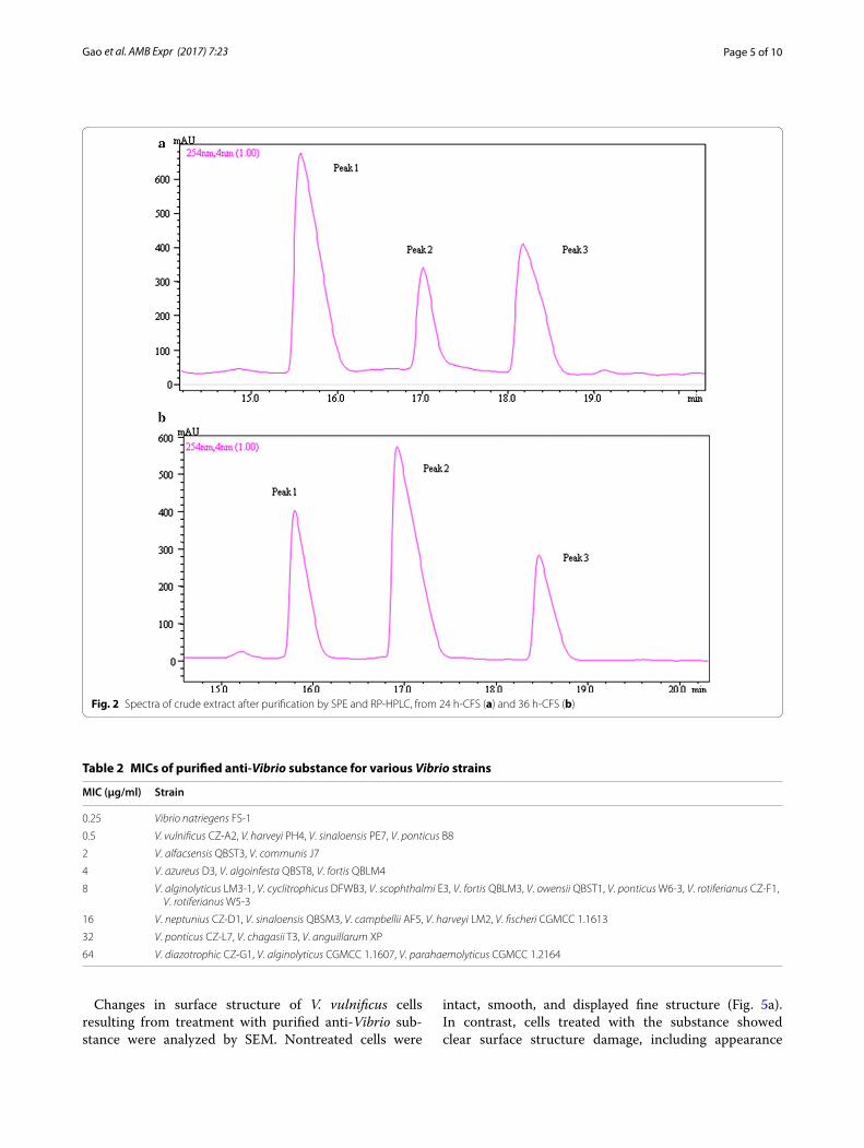
Extraction and purification of anti‑Vibrio substanceThe anti-Vibrio substance present in B. pumilus H2 CFS was purified by SPE and RP-HPLC. The HPLC spectrum showed three peaks (Fig. 2). In anti-Vibrio activity assays using V. vulnificus CZ-A2 as target organism, activity was strong for peak 1 and very weak for peak 2, suggesting that the peak 1 substance was the major anti-Vibrio com-pound. In CFS cultured for 24 h, peak 1 was dominant, whereas in CFS cultured for 36 h peak 2 was dominant
one and peak 1 declined greatly. This observation is con-sistent with the activity curve associated with H2 growth (Fig. 1).
A total of 20 mg purified anti-Vibrio substance was obtained by semi-preparative RP-HPLC and used for subsequent experiments.
MICs of purified anti‑Vibrio substance for various Vibrio strainsMICs of purified anti-Vibrio substance for various Vibrio strains, determined in liquid media, ranged from 0.25 to 64 μg/ml (Table 2). The purified substance showed high inhibitory activity against V. natriegens FS-1, V. vulnificus CZ-A2, V. harveyi PH4, V. sinaloensis PE7, and V. ponti-cus B8, but less activity against V. diazotrophicus CZ-G1, V. alginolyticus CGMCC 1.1607, and V. parahaemolyti-cus CGMCC 1.2164.
Effect of purified anti‑Vibrio substance on growth of V. vulnificusVibrio vulnificus was selected as a model target strain for experiments on the mode of interaction of purified anti-Vibrio substance with Vibrio strains. V. vulnificus was inoculated into LB broth (100 ml) to OD600 ~0.25, and purified anti-Vibrio substance was added (control = ster-ile distilled water). Growth was monitored by measuring OD600 of the culture at predetermined intervals. Growth of the target strain was clearly inhibited by addition of anti-Vibrio substance; i.e., OD600 did not increase as it did in control culture (Fig. 3). OD600 declined continuously after 8 h incubation, suggesting that cell lysis was occur-ring. In the control group, the target strain showed expo-nential growth immediately after inoculation (Fig. 3).
Effect of purified anti‑Vibrio substance on cell surface structure of V. vulnificusMembrane integrity of V. vulnificus (target strain) fol-lowing treatment with purified anti-Vibrio substance was evaluated by confocal microscopy and an assay based on uptake of the fluorescent dye SG. SG, a high-affinity nucleic acid stain, is often used to assess integ-rity of plasma membranes, because it easily penetrates cells with compromised membranes, but does not pass through membranes of non-compromised cells.
Nontreated V. vulnificus cells (control) showed no appreciable fluorescent signal (Fig. 4). Treatment with 0.5 μg/ml purified substance resulted in detection of only a very weak fluorescent signal. Intensity of the fluores-cent signal increased steadily as substance concentration increased. At substance concentration 10 µg/ml, the fluo-rescent signal was distinct, clear, and strong, indicating that the cell membrane was completely permeabilized.Fig. 1 Growth curve and anti‑Vibrio activity of B. pumilus strain H2
Page 5 of 10Gao et al. AMB Expr (2017) 7:23
Changes in surface structure of V. vulnificus cells resulting from treatment with purified anti-Vibrio sub-stance were analyzed by SEM. Nontreated cells were
intact, smooth, and displayed fine structure (Fig. 5a). In contrast, cells treated with the substance showed clear surface structure damage, including appearance
Fig. 2 Spectra of crude extract after purification by SPE and RP‑HPLC, from 24 h‑CFS (a) and 36 h‑CFS (b)
Table 2 MICs of purified anti-Vibrio substance for various Vibrio strains
MIC (µg/ml) Strain
0.25 Vibrio natriegens FS‑1
0.5 V. vulnificus CZ‑A2, V. harveyi PH4, V. sinaloensis PE7, V. ponticus B8
2 V. alfacsensis QBST3, V. communis J7
4 V. azureus D3, V. algoinfesta QBST8, V. fortis QBLM4
8 V. alginolyticus LM3‑1, V. cyclitrophicus DFWB3, V. scophthalmi E3, V. fortis QBLM3, V. owensii QBST1, V. ponticus W6‑3, V. rotiferianus CZ‑F1, V. rotiferianus W5‑3
16 V. neptunius CZ‑D1, V. sinaloensis QBSM3, V. campbellii AF5, V. harveyi LM2, V. fischeri CGMCC 1.1613
32 V. ponticus CZ‑L7, V. chagasii T3, V. anguillarum XP
64 V. diazotrophic CZ‑G1, V. alginolyticus CGMCC 1.1607, V. parahaemolyticus CGMCC 1.2164

Page 6 of 10Gao et al. AMB Expr (2017) 7:23
of membrane holes, disappearance of cellular contents, and formation of cell cavities (Fig. 5b–d). The size of these cavities (292 × 732 nm in Fig. 5c; 361 × 559 nm in Fig. 5d) indicated that cell lysis had occurred.
Structural characterization of purified anti‑Vibrio substanceThe molecular mass of the purified anti-Vibrio sub-stance (peak 1 in Fig. 2) was 423.2076 Da, as determined by Orbitrap Fusion MS. The MS analysis also revealed a plausible chemical formula (C20H30N3O7) for the sub-stance (Fig. 6). Chemical shifts and coupling constants were assigned to protons in the molecule through NMR analysis. The 13C-NMR spectra of the purified anti-Vibrio substance (peak 1 in Fig. 2) showed twenty signals at 21.7 (q), 23.6 (q), 25.2 (d), 30.0 (t), 32.3 (t), 39.1 (t), 50.2 (d), 51.2 (d), 71.2 (d), 73.2 (d), 82.1 (d), 108.6 (s), 170.5 (s), 173.8 (s) and 175.1 (s) ppm, respectively. Comparisons of molecular mass, chemical formula, and NMR data with those of compounds previously described in the lit-erature indicated that the purified anti-Vibrio substance is identical to amicoumacin A (Itoh et al. 1981). Two structurally related substances (peaks 2 and 3 in Fig. 2) were also characterized. Based on MS analysis, peak 2
Fig. 3 Effect of purified anti‑Vibrio substance on growth of V. vulnifi-cus
Fig. 4 Confocal microscopic images of V. vulnificus cells treated with purified anti‑Vibrio substance at 0 μg/ml (a), 0.5 μg/ml (b), 5 μg/ml (c), and 10 μg/ml (d)

Page 7 of 10Gao et al. AMB Expr (2017) 7:23
((M + H)+ ion at m/z 425 Da) was identified as amicou-macin B (C20H28N2O8), and peak 3 ((M + H)+ ion at m/z 407 Da) was identified as amicoumacin C (C20H26N2O7) (data not shown).
DiscussionAs the aquaculture industry expands worldwide, and the variety of fish species involved increases, many unknown fish-pathogenic Vibrio species are reported (Li and Woo 2003; Thompson et al. 2004; Austin and Zhang 2006; Cui et al. 2014). In the past, vibriosis was controlled (pre-vented or treated) almost exclusively through applica-tion of antibiotics or chemotherapeutic agents, either as feed additives or in immersion baths. However, extensive use of this approach over time has resulted in increased resistance of pathogenic Vibrio strains to the commonly used antibiotics: ampicillin, amikacin, kanamycin, peni-cillin G, streptomycin, and tetracycline (Austin and Aus-tin 2007; Li et al. 1999; Elmahdi et al. 2016).
In present study, we screened Bacillus strains for anti-Vibrio activity. Bacillus strains are good candidates as biological control agents for prevention or treatment of plant and animal infections for several reasons (Wulff et al. 2002; Mongkolthanaruk 2012; Mondol et al. 2013).
(1) They produce antibiotics having well-documented antagonistic activity against a variety of fungal and bac-terial pathogens. (2) They form spores that can be easily formulated, and have high viability in comparison with vegetative cells. (3) The robustness of the spores enables them to cross the gastric barrier. A certain proportion of spores is thus able to germinate in and colonize (albeit briefly) the intestinal tract (Mazza 1994). (4) Bacillus spe-cies are abundant in a wide variety of environments and habitats.
Bacillus pumilus strain H2 has notable anti-Vibrio effects. It inhibited 29 Vibrio strains to varying degrees (Table 2). No anti-Vibrio probiotic has been previously reported to inhibit such a large number of Vibrio strains. V. vulnificus CZ-A2, V. natriegens FS-1, V. harveyi PH4, V. sinaloensis PE7, V. ponticus B8, V. alfacsensis QBST3, and V. communis J7 were highly sensitive to purified anti-Vibrio substance. V. anguillarum is the most well-studied and widespread fish-pathogenic Vibrio species, and is responsible for the majority of fish loss in aquacul-ture worldwide (Austin and Austin 2007). We measured MIC of purified anti-Vibrio substance from H2 against V. anguillarum XP as 32 µg/ml, indicating its potential application for control of this major pathogen.
Fig. 5 SEM images of V. vulnificus CZ‑A2 cells treated with purified anti‑Vibrio substance at 0 μg/ml (a, 20,000×; control) and 0.5 μg/ml (b, 8000×; c, 35000×; d, 30,000×), showing formation of membrane holes

Page 8 of 10Gao et al. AMB Expr (2017) 7:23
We selected V. vulnificus CZ-A2 as the target strain for screening of Bacillus strains having anti-Vibrio activity, and for follow-up studies of the mechanism of H2 activ-ity, because CZ-A2 was highly sensitive to the substance present in H2 CFS. V. vulnificus is a widespread marine bacterium categorized into three biotypes. Strains of V. vulnificus include one of the most widely occurring fish pathogens, and another strain that can cause wound infections in humans, resulting in high mortality among susceptible individuals (Efimov et al. 2013; Ziolo et al. 2014).
The anti-Vibrio substance produced by B. pumilus H2 was found to be structurally identical to amicoumacin A, which was described in 1981 (Itoh et al. 1981). Ami-coumacin A and related compounds display inhibitory activity against numerous pathogenic bacteria, including Helicobacter pylori and methicillin-resistant Staphylo-coccus aureus (MRSA) (Pinchuk et al. 2001; Lama et al. 2012). Anti-inflammatory and antitumor effects of ami-coumacin A have also been reported (Itoh et al. 1981), but no study to date has addressed its effects against pathogenic Vibrio that cause economic losses in aquacul-ture. Amicoumacin B was isolated from B. pumilus and reported to display gastroprotective activity, but weak antibacterial and weak antiulcer activity (Shimojima et al. 1984; Han et al. 2013), consistent with the weak
anti-Vibrio activity that we observed. Activity of amicou-macin C has not been studied. Although application of H2 preparation was used and showed no toxic to juvenile shrimp, further tests on toxic of purified Amicoumacin A to one or more farmed species need to be conducted in the future.
The mode of action of amicoumacin A remains unclear. Lama et al. reported amicoumacin A-induced alteration of transcription of genes that regulate various cellular processes, including cell envelope turnover, cross-mem-brane transport, virulence, metabolism, and general stress. The gene most highly induced by amicoumacin A was lrgA, which encodes an antiholin-like product (LrgA) that appears in cells undergoing collapse of Δψ, and modulates murein hydrolase activity (Lama et al. 2012). Taken together, the findings of Lama et al. sug-gest that amicoumacin A provokes perturbation of the cell membrane and consequent energy dissipation (Lama et al. 2012). Polikanov et al. proposed that amicoumacin A is a potent inhibitor of protein synthesis, but without direct experimental evidence (Polikanov et al. 2014). Our observations of reduced cell density (Fig. 3), formation of membrane holes, disappearance of cellular contents, and formation of cell cavities (Figs. 4, 5) indicates that the major mechanism of amicoumacin A activity against
2014-20-3-334 #18567 RT: 37.74 AV: 1 NL: 1.27E9F: FTMS + p NSI d Full ms2 [email protected] [50.00-450.00]
50 100 150 200 250 300 350 400 450m/z
Rel
ative
Abu
ndan
ce
0
10
20
30
40
50
60
70
80
90
100424.2076
250.1436 407.1805390.1551232.1339 285.2815140.473273.1797
Fig. 6 MS analysis of purified anti‑Vibrio substance from HPLC peak 1 in Fig. 2

Page 9 of 10Gao et al. AMB Expr (2017) 7:23
pathogens involves disruption of cell membranes, and consequent cell lysis.
Bacillus pumilus H2 was isolated from marine sedi-ment, and therefore has priority and inherent advan-tages for use in marine aquaculture as a biocontrol agent or probiotic. Under the generally accepted definition of the term “probiotic”, we can consider two applica-tion approaches. In the first approach, H2 fermentation broth would be added, in proportion, directly to the aquaculture pond. Live H2 cells would then produce amicoumacin A continuously. In simulated gastroen-teric environments designed to test spore robustness, H2 spores were able to cross the gastric barrier (data not shown), and may therefore have the ability to germinate and colonize in the intestinal tract. Probiotic effects in aquaculture are not limited to the intestinal tract, but may also improve the health of the host by inhibiting pathogens and improving water quality through modifi-cation of microbial community composition in the water and sediment (Perez-Sanchez et al. 2013).
In the second approach, extracted amicoumacin A would be added to aquaculture feeds. Amicoumacin A is potentially suitable for this purpose because it is heat stable, pH stable, UV stable, and not sensitive to vari-ous enzymes and organic solvents. On the other hand, a major disadvantage of the second approach is that ami-coumacin A is difficult to extract and purify. The first approach appears more feasible.
In conclusion, probiotic B. pumilus strain H2 demon-strated notable antagonistic activity against 29 Vibrio strains tested. This activity was attributable to production of amicoumacin A, which has been reported previously to inhibit methicillin-resistant Staphylococcus aureus and Helicobacter pylori, and to display anti-inflammatory and antitumor effects. The major mechanism of amicoumacin A activity against pathogens involves disruption of cell membranes, and consequent cell lysis.
AbbreviationsCFS: cell‑free supernatant; SPE: solid‑phase extraction; RP‑HPLC: reversed‑phase high performance liquid chromatography; MIC: minimum inhibitory concentration; CFUs: colony‑forming units; SG: SYTOX green; PBS: phosphate buffer saline; DMSO: dimethyl sulfoxide; MS: mass spectrometer; NMR: nuclear magnetic resonance; SEM: scanning electron microscope; MRSA: methicillin‑Resistant Staphylococcus aureus.
Authors’ contributionsXYG performed the experiments, analyzed the data as well as results and wrote the manuscript. EWL assisted NMR data analysis. TTH isolated vibrio strains used in this work. ZPL, YL and LLM conceived this study. ZPL supervised all the experiments and revised the manuscript. All authors read and approved the final manuscript.
Additional file
Additional file 1. Additional tables.
Author details1 State Key Laboratory of Microbial Resources, Institute of Microbiology, Chinese Academy of Sciences, No. 1 West Beichen Road, Chaoyang District, Beijing 100101, People’s Republic of China. 2 University of Chinese Academy of Sciences, Beijing 100049, People’s Republic of China. 3 State Key Labora‑tory of Mycology, Institute of Microbiology, Chinese Academy of Sciences, Beijing 100101, People’s Republic of China.
AcknowledgementsThe authors are grateful to Dr. S. Anderson for English editing of the manuscript.
Competing interestsThe authors declare that they have no competing interests.
FundingThis study was supported by the Innovation Project of Shandong Prov‑ince, China (2014ZZCX06204) and CAS Knowledge Innovation Project (KZCX2‑EW‑Q212).
Received: 9 November 2016 Accepted: 2 January 2017
ReferencesAl‑Othrubi SMY, Kqueen CY, Mirhosseini H, Hadi YA, Radu S (2014) Antibiotic
resistance of Vibrio parahaemolyticus isolated from cockles and shrimp sea food marketed in Selangor, Malaysia. Clin Microbiol 3:1–7
Austin B, Austin DA (2007) Bacterial fish pathogens, 4th edn. Ellis Horwood Ltd., Chichester
Austin B, Zhang XH (2006) Vibrio harveyi: a significant pathogen of marine vertebrates and invertebrates. Lett Appl Microbiol 43:119–124
Balcázar JL, Blas I, Ruiz‑Zarzuela I, Cunningham D, Vendrell D, Múzquiz JL (2006) The role of probiotics in aquaculture. Vet Microbiol 114:173–186
Colwell RR, Griems DJ (1984) Vibrio diseases of marine fish populations. Helgol Mar Res 37:265–287
Cui J, Fan XT, Liu WZ, Li HY, Zhou YC, Wang SF, Xie ZY (2014) Isolation and identification of vibriosis pathogens of marine cultured fishes in southern China. Nat Sci J HaiNan Univ 32:244–251
Desriac F, Defer D, Bourgougnon N, Brillet B, Chevalier PL, Fleury Y (2010) Bacteriocin as weapons in the marine animal‑associated bacteria warfare: inventory and potential applications as an aquaculture probiotic. Mar Drugs 8:1153–1177
Efimov V, Danin‑Poleg Y, Raz N, Elgavish S, Linetsky A, Kashi Y (2013) Insight into the evolution of Vibrio vulnificus biotype 3′s genome. Front Microbiol 4:393
Egidius E (1987) Vibriosis: pathogenicity and pathology: a review. Aquaculture 67:15–28
Elmahdi S, DaSilva LV, Parveen S (2016) Antibiotic resistance of Vibrio para-haemolyticus and Vibrio vulnificus in various countries: a review. Food Microbiol 57:128–134
Fu SZ, Song BB, Liu Y, Liu ZP (2009) Screening of probiotic strains and their protection against vibriosis in juvenile shrimp (Litopenaeus vannamei). China Environ Sci 29:867–872
Han XY, Liu SW, Wang FF, Liu JM, Chen C, Hu XX, Jiang ZK, You XF, Shang GD, Zhang YB, Sun CH (2013) Research progress of amicoumacin group antibiotic. World Notes Antibiot 34:106–114
Heidelberg JF, Heidelberg KB, Colwell RR (2002) Bacteria of the gamma‑subclass proteobacteria associated with zooplankton in Chesapeake Bay. Appl Environ Microbiol 68:5498–5507
Hou TT, Zhong ZP, Liu Y, Liu ZP (2016) Bacterial community characterization of rearing water of marine recirculating aquaculture systems for yellow grouper (Epinephelus awoara). Acta Microbiol Sin 56:253–263
Itoh J, Omoto S, Shomura T, Nishizawa N, Miyado S, Yuda Y, Shibata U, Inouye S (1981) Amicoumacin‑A, a new antibiotic with strong anti‑inflammatory and antiulcer activity. J Antibiot 34:611–613
Lama A, Pane‑Farre J, Chon T, Wiersma AM, Sit CS, Vederas JC, Hecker M, Nakano MM (2012) Response of Methicillin‑resisitant Staphylococcus aureus to Amicoumacin A. PLoS ONE 7:e34037

Page 10 of 10Gao et al. AMB Expr (2017) 7:23
Li J, Woo NYS (2003) Pathogenicity of vibriosis in fish: an overview. J. Ocean Univ China 2:117–128
Li J, Yie J, Foo RWT, Ling JML, Xu H, Woo NYS (1999) Antibiotic resistance and plasmid profiles of vibrio isolates from cultured silver sea bream, Sparus sarba. Mar Pollut Bull 39:245–249
Mazza P (1994) The use of Bacillus subtilis as an antidiarrhoeal microorganism. Boll Chim Farm 133:3–18
Mondol MAM, Shin HJ, Islam MT (2013) Diversity of secondary metabolites from marine Bacillus species: chemistry and biological activity. Mar Drugs 11:2846–2872
Mongkolthanaruk W (2012) Classification of Bacillus beneficial substances related to plants, humans and animals. J Microbiol Biotechnol 22:1597–1604
Moriarty DJW (1998) Control of luminous Vibrio species in penaeid. Aquacul‑ture 164:351–358
Newaj‑Fyzul A, Al‑Harbi AH, Austin B (2014) Review: developments in the use of probiotics for disease control in aquaculture. Aquaculture 431:1–11
Paillard C, Roux FL, Borrego JJ (2004) Bacterial disease in marine bivalves, a review of recent studies: trends and evolution. Aquat Living Resour 17:477–498
Parker RB (1974) Probiotics, the other half of the antibiotic story. Anim Nutr Health 29:4–8
Patel AK, Deshattiwar MK, Chaudhari BL, Chincholkar SB (2009) Production, purification and chemical characterization of the catecholate sidero‑phore from potent probiotic strains of Bacillus spp. Bioresour Technol 100:368–373
Perez‑Sanchez T, Ruiz‑Zarzuela I, Blas I, Balcazar JL (2013) Probiotics in aquacul‑ture: a current assessment. Rev in Aquac 5:1–14
Pinchuk IV, Bressollier P, Verneuil B, Fenet B, Sorokulova IB, MéGraud F, Urdaci MC (2001) In vitro anti‑Helicobacter pylori activity of the probiotic strain Bacillus subtilis 3 is due to secretion of antibiotics. Antimicrob Agents Chemother 45:3156–3161
Polikanov YS, Osterman IA, Szal T, Tashlitsky VN, Serebryakova MV, Kusochek P, Bulkley D, Malanicheva IA, Efimenko TA, Efremenkova OV, Konevega AL, Shaw KJ, Bogdanov AA, Rodnina MV, Dontsova OA, Mankin AS, Steitz TA, Sergiev PV (2014) Amicoumacin A inhibits translation by stabilizing mRNA interaction with the ribosome. Mol Cell 56:531–540
Shimojima Y, Hayashi H, Ooka T, Shibukawa M (1984) Studies on AI‑77s, micro‑bial products with gastroprotective activity. Structures and the chemical nature of AI‑77s. Tetrahedron 40:2519–2527
Stentiford GD, Neil DM, Peeler EJ, Shields JD, Small HJ, Flegel TW, Vlak JM, Jones B, Morado F, Moss S, Lotz J, Bartholomay L, Behringer DC, Hauton C, Light‑ner DV (2012) Disease will limit future food supply from the global crusta‑cean fishery and aquaculture sectors. J Invertebr Pathol 110:141–157
Tall A, Hervio‑Heath D, Teillon A, Boisset‑Helbert C, Delesmont R, Bodilis J, Touron‑Bodilis A (2013) Diversity of Vibrio spp. isolated at ambient envi‑ronmental temperature in the Eastern English Channel as determined by pyrH sequencing. J Appl Microbiol 114:1713–1724
Thompson JR, Randa MA, Marcelino LA, Tomita‑Mitchell A, Lim E, Polz MF (2004) Diversity and dynamics of a north Atlantic coastal Vibrio commu‑nity. Appl Environ Microbiol 70:4103–4110
Toranzo E, Magariños B, Romalde JL (2005) A review of the main bacterial fish diseases in mariculture systems. Aquaculture 246:37–61
Verschuere L, Rombaut G, Sorgeloos P, Verstraete W (2000) Probiotic bacteria as biological control agents in aquaculture. Microbiol Mol Biol Rev 64:655–671
Wulff EG, Mguni CM, Mansfeld‑Giese K, Fels J, Lübeck M, Hockenhull J (2002) Biochemical and molecular characterization of Bacillus velezensis, B. subtilis and B. pumilus isolates with distinct antagonistic potential against Xanthomonas campestris pv. campestris. Plant Pathol 51:574–584
Zhou Q, Li K, Jun X, Bo L (2009) Role and functions of beneficial microorgan‑isms in sustainable aquaculture. Bioresour Technol 100:3780–3786
Ziolo KJ, Jeong HG, Kwak JS, Yang S, Lavker RM, Satchell KJF (2014) Vibrio vulnificus biotype 3 multifunctional autoprocessing RTX toxin is an adenylate cyclase toxin essential for virulence in mice. Infect Immun 82:2148–2157