Embed Size (px)
Citation preview

Mechanical Ventilation in
the Brain-Injured Patient
Jeffrey M Singh, MD FRCPC MSc
Critical Care and Neurocritical Care
Toronto Western Hospital
and
Interdepartmental Division of Critical Care
University of Toronto

Outline
• Epidemiology of lung injury after brain injury
• Why is there reluctance in providing lung-protective ventilation in brain injury?
• Ventilation parameters to pay attention to:
– PCO2
– PEEP
• Pragmatic approach to hypoxemic respiratory failure / ARDS in the brain-injured patient


Etiology of Respiratory Failure
• Aspiration
• Pneumonia
• Pulmonary
Contusions
• ARDS
• Neurogenic
Pulmonary
Edema
• TRALI
This list is common to many
MICU / SICU patients…
But are brain-injured patients
really different?

ALI / ARDS is Common…
• In polytrauma with traumatic brain injury
But Also:
• Isolated traumatic brain injury 1,2
• Subarachnoid hemorrhage3
ALI rates as high as 30% in severe brain injury
ALI/ARDS risk associated with severity of injury
Associated with worse outcomes
1 - Bratton SL et al. Neurosurgery 1997. 3 - Kahn JM et al. Crit Care Med 2006. 2 - Holland MC et al. J Trauma 2003.

ARDS & Ventilation
We are sometimes
reluctant to implement
this knowledge in
brain-injured patients
WHY?

Conflicting Paradigms
Historical “brain-directed”
strategy
• Optimize oxygen
delivery
• Control of PCO2
(higher VT and VE)
• Minimize potential
effects of PEEP
Lung protective ventilation
• Avoid overdistention
(Volutrauma)
• Open the lung
• Avoid cyclical collapse
(Atelectrauma)

What Really Matters to the Brain
• Avoid hypoxemia
• Protect cerebral perfusion
– Avoid hypotension
– Avoid high intracranial
pressure
– Avoid inadvertent hypocapnia
ICPMAPCPP
Two Parameters To Pay Attention To:
• PCO2
• Positive End Expiratory
Pressure (PEEP)

CO2 : Concern Over Hypercapnia
• Concern that hypercapnia may worsen:
– Hyperemia
– ICP cerebral herniation
This is a concern in patients with
very low intracranial compliance
(little compensatory reserve)

Intracranial Compliance

Low Intracranial Compliance
Increase in intracranial blood
from vasodilation / decreased
venous drainage is critical
Parenchyma
1300cc
Intravascular
Blood
150cc
CSF
50cc

CO2 : Too Little of a Good Thing!
• Hypocapnia-related reduction in CBF
causes:
– Metabolic crisis 1,2
– Increases ischemic brain volume1
• Early, prophylactic hyperventilation in
traumatic brain injury associated with
worse outcomes 3
1 - Coles JP et al. Crit Care Med. 2007. 3 - Muizelaar et al. J Trauma. 1991.
2 - Carrera E et al. J Neurol Neurosurg Psychiatry. 2010

CO2 : The Bottom Line
• Eucapnia
• Hypocapnia only if ICP emergency
• No ICP? Be wary of PCO2 if signs of
low intracranial compliance
– CT:
• Sulcal effacement
• Effaced basal cisterns
• Small ventricles
• Hydrocephalus

Two Parameters To Pay Attention To:
• PCO2
• Positive End Expiratory
Pressure (PEEP)

PEEP: Concern Over ICP
• High levels of PEEP may be bad in brain-injured patients:
1. Decreased venous drainage
2. Transmission of intrathoracic pressure
Increased ICP
2. Deceased Cardiac Output
Decreased CBF

PEEP & ICP: Complex Relationship
ICP Pulmonary Compliance
Intracranial Compliance
Venous Drainage
Head Elevation
Starling Resistors

PEEP, Compliance and ICP
• Respiratory System Compliance (Crs)
– Hemodynamic and hydrostatic impact of
PEEP is attenuated in patients with low Crs
Patients in whom we want to use PEEP can
often tolerate it
Caricato A, et al. J Trauma 2005.

Why PEEP May Not Affect ICP…
• Cerebral circulation is a Starling Resistor
• Flow usually dependent on
MAP-ICP
Hence CPP = MAP-ICP
• PEEP-related increases in CVP only relevant if CVP > ICP
MAP CVP
Brain
Pressure
ICP
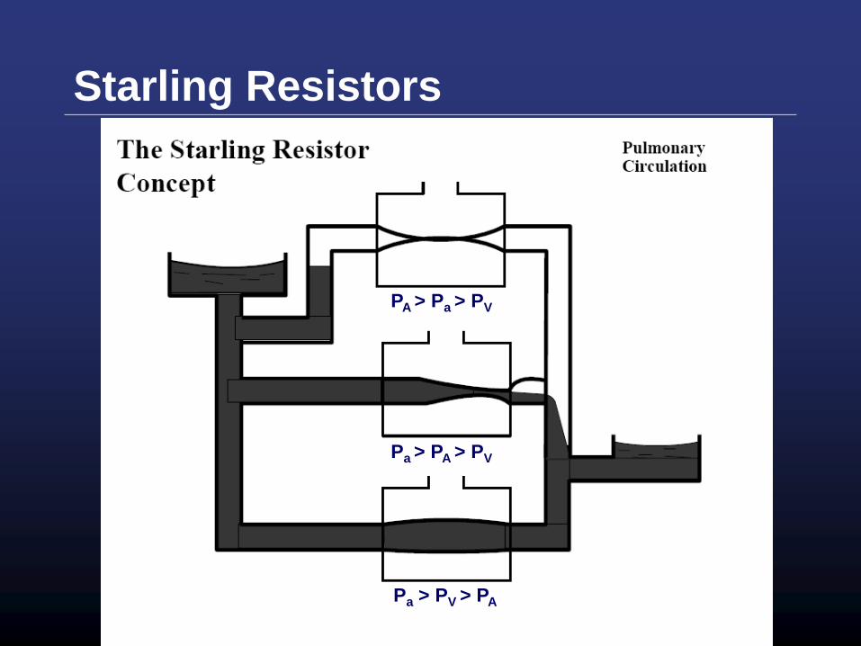
Starling Resistors
PA > Pa > PV
Pa > PA > PV
Pa > PV > PA

Head of Bed Elevation
MAP CVP
Brain Pressure
ICP is
relatively
constant
MAP is
relatively
constant
Diminished effect of
increased intrathoracic
pressure due to
decreased transmission
of RAP to head
1 -Durward et al. J Neurosurg. 1983

PEEP is Usually Well-Tolerated
…in Patients Who Need It
•Moderate levels of PEEP associated with little change cerebral perfusion
– As long as MAP maintained
•High PEEP may ICP reserve
•HOB at 30-45°
•Don’t forget about PCO2 !
McGuire et al. Crit Care Med. 1997. Muench E, et al. Crit Care Med. 2005. Georgiadis et al. Stroke. 2001. Mascia L et al. Intensive Care Med. 2005.

Bottom Line: PEEP
• Can use PEEP as indicated, but:
– Ensure no change in MAP
• Adequate intravascular volume
– Keep head elevated
– Follow ICP
– Consider maintaining PEEP < ICP

Putting It All Into Practice:

Lung Protection and Brain Injury
• Lung-protective ventilation
saves lives
• High tidal volumes predict
ALI/ARDS in TBI patients 1
“Can we afford to relax control of PCO2
To achieve low tidal volumes?”
1 - Mascia L. et al. Crit Care Med 2007

Not-so Permissive Hypercapnea
• Many large ARDS ventilation studies
excluded patients with increased ICP
• But…PCO2 levels in these studies were
quite average, normal (at least for first
72 hrs)

Comparison of CO2 and pH in ARDS
Studies
ALVEOLI: ARDSNet, NEJM, 2004
LOVS: Meade et al. JAMA. 2008
ARMA: ARDSNet, NEJM, 2004

ARDS and Brain Injury
1. Start with what we know are best practices (low VT, Pplat)
2. Monitor PCO2 and ICP
3. Carefully consider anything to improve CO2 clearance and minimize pressures – Dead space in circuit
– Synchrony
– Draining effusions or ascites
You may have to decide which is more important to you – VT or PCO2

Rational Approach to ALI / ARDS in Brain Injury
Normal ICP Higher ICP
Start with…
Normocapnia
Low VT (6 ml/kg)
Limit Pplat
Normocapnia
Low VT (6 ml/kg)
Limit Pplat
If respiratory acidosis
(CO2 rising)…
Moderate Hypercarbia
Follow ICP
Remove excess dead
space
Sedation / Ensure
synchrony
Drain large effusions /
ascites
Consider increasing VT
Normocarbia
Follow ICP
Remove excess dead
space
Sedation / Ensure
synchrony
Drain large effusions /
ascites
Then…
Alternative modes,
prioritizing lung protection
(but following ICP)
Alternative modes,
prioritizing CO2
elimination

Summary
• Acute Lung Injury is common in patients with
brain injury
• Be vigilant about PCO2 – hypocapnia can be
harmful!
• PEEP appears safe in patients who need it
• Protect the lungs, but prioritize CO2 control
and cerebral perfusion


Extra Slides

Mechanical Ventilation In Brain Injury
• Brain injury may be main indication for
mechanical ventilation in up to 20% of cases
• Major contributor to prolongation of
mechanical ventilation in over a third of
patients
• Associated with 3-fold risk of dying or
unfavourable outcome
Esteban A et al. Am J Respir Crit Care Med. 2000. Kelly BJ et al. Chest. 1993 Holland MC et al. J Trauma. 2003 Pelosi P et al. Crit Care Med. 2011

Cerebral Perfusion
• Adult brain is only 2% of body weight
– 15 % of the resting cardiac output
– 20 % of the total body oxygen
consumption
• Blood flow regulated by three primary
factors 1. Metabolic stimuli
2. Chemical stimuli
3. Perfusion pressure

Cerebral Perfusion Pressure
• Cerebral Perfusion Pressure
Mechanical Ventilation may affect MAP
and ICP through multiple mechanisms
ICPMAPCPP

3 Key Concepts
1. Intracranial Pressure
2. CO2 and the Brain
3. Intracranial Compliance
(ICP compensatory reserve)

Key Concept 1: ICP
Monro-Kellie Doctrine
Skull has fixed volume
1. Brain parenchyma (80%)
2. CSF (10%)
3. Intravascular blood
(10%)
Parenchyma
1200cc
Blood
150cc
CSF
150cc
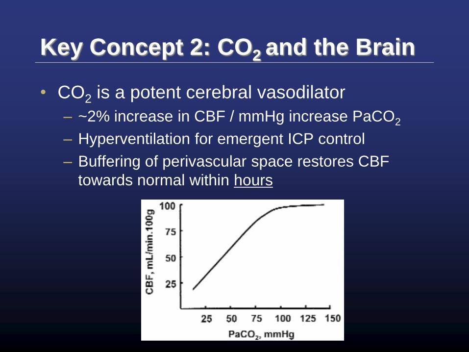
Key Concept 2: CO2 and the Brain
• CO2 is a potent cerebral vasodilator
– ~2% increase in CBF / mmHg increase PaCO2
– Hyperventilation for emergent ICP control
– Buffering of perivascular space restores CBF
towards normal within hours
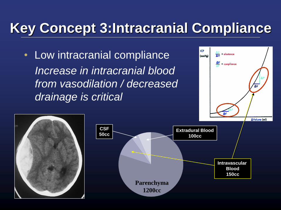
Key Concept 3:Intracranial Compliance
• Low intracranial compliance
Increase in intracranial blood
from vasodilation / decreased
drainage is critical
Parenchyma
1200cc
Intravascular
Blood
150cc
Extradural Blood
100cc
CSF
50cc
High Frequency Oscillation
• Existing case series suggest safe:
– Most patients had little / no ICP increase
attributable to HFO 1,2
– Volume loading important to avoid drops in MAP /
CPP on initiation 1
– Infrequent, but clinically important rapid changes
in PCO2 on initiation
1 - David M et al. Acta Anaesthesiol Scand. 2005. 2 - Bennett SS et al. Neurocrit Care. 2007.
Careful monitoring of PCO2 at start
Initial use of aggressive CO2 settings
Prone Positioning
• 16 Patients with SAH
– Improved brain tissue oxygenation
• 2° improved arterial oxygenation
– BUT:
• Can’t maintain head elevation
• Increased transmission of intrathoracic
pressure
increase in ICP and reduction in CPP
with prone position
Reinprecht A et al. Crit Care Med. 2003.

Low-Tidal Volume Ventilation
• Only 30% of SAH patients with ALI
received low VT ventilation
– Patients who did had
• Higher applied PEEP
• Similar arterial pH
• Similar PCO2
• Worse oxygenation
Kahn JM et al. Crit Care Med. 2006.

VT in Brain-Injured Patients?
• Mascia et al.
– Retrospective review: 86 pts with severe
brain injuries who developed ALI/ARDS
within 8 days
– Most ventilated to PaCO2 ~ 35 mmHg
– Baseline Vt (ml/kgPBW) associated with
5.4x odds of development of subsequent
ALI/ARDS
1 - Mascia L. et al. Crit Care Med 2007