Embed Size (px)
Citation preview
ORIGINALRESEARCH
Mechanical Characterization of Thromboemboli inAcute Ischemic Stroke and Laboratory EmbolusAnalogs
J.Y. ChuehA.K. Wakhloo
G.H. HendricksC.F. Silva
J.P. WeaverM.J. Gounis
BACKGROUND AND PURPOSE: Mechanical behavior of the thromboembolus is one of the key factorsthat determine the efficacy of thrombectomy devices for revascularization in AIS. We characterized themechanical properties and composition of thromboemboli from clinical cases and compared them withcommonly used EAs.
MATERIALS AND METHODS: Thromboemboli were obtained from patients with AIS by using aspirationdevices and from carotid atherosclerotic plaques harvested during endarterectomy. In the laboratory,common EAs were created by varying blood donor species (human, porcine, and bovine), thrombinconcentration, and presence of barium sulfate. Stiffness and elasticity of the specimens were mea-sured with DMA. Scanning electron microscopy and histology were used to investigate the ultrastruc-ture and composition of all specimens.
RESULTS: Red thromboemboli from patients composed mainly of fibrin and erythrocytes were muchsofter than the calcified and cholesterol-rich material. Of the EAs created in the laboratory, those madefrom bovine blood presented the highest stiffness that was independent of thrombin concentration.Addition of thrombin increased the stiffness and elasticity of human and porcine EAs (P � .05). Thepresence of barium sulfate significantly reduced the elasticity of all EAs (P � .05).
CONCLUSIONS: Endovascular device testing and development requires realistic EAs. The stiffness andelasticity of the cerebral thromboemboli analyzed in this study were closely matched by recalcifiedporcine EAs and thrombin-induced human EAs. Stiffness of the thrombus extracted from carotidendarterectomy specimens was similar with that of the thrombin-induced bovine and porcine EAs.
ABBREVIATIONS: ACD � anticoagulant citrate dextrose; AIS � acute ischemic stroke; CEA �carotid endarterectomy; DMA � dynamic mechanical analyzer; Eintitial strain-end strain � secantmodulus in a given strain range (initial strain to end strain); EA� embolus analog; EDS � energydispersive x-ray spectroscopy; H&E � hematoxylin-eosin; ICA � internal carotid artery; MCA �middle cerebral artery; MSB � Martius scarlet blue; NIHU � National Institutes of Health unit;S-S � stress-strain; SEM � scanning electron microscopy
With growing experience of penumbral imaging for pa-tient selection and newer, more effective mechanical re-
vascularization devices, alternative endovascular therapy forAIS has become a viable treatment option.1 There are pres-ently 2 devices available in the United States for thrombec-tomy in AIS, and many other technologies are under evalua-tion. These devices produce recanalization in 50%– 82% of
cases.2,3 Currently, it is poorly understood what variables pre-dict successful recanalization. Biomechanical characterizationof the thromboembolus is helpful in device development.
Preclinical evaluation of thrombectomy devices mea-sures efficacy and safety in animal or in vitro vascular oc-clusion models.4-7 Common end points of these studies arethe number of thrombectomy attempts, the amount of EAsremoved from the occlusion site, and the risk of distal em-bolic shower.5,6,8-10 All of these metrics are significantlyaffected by the mechanical properties of the EAs, an oftenoverlooked component of the testing system. In the litera-ture, there are many protocols to manufacture EAs, andcommon variables include the donor species, concentra-tion of thrombin, and radiopaque additive such as bariumsulfate.6,9-13 Variation of hematologic values among differ-ent donors along with the pH and ionic strength of theclotting environment result in different structure and com-position of the EAs.14-18 This leads to the hypothesis thatcommonly used EAs have different mechanical properties.Two recent studies reported the composition of emboli re-trieved from the stroke patients,19,20 and substantiated thestructural and compositional variations between the in vivoemboli and in vitro EAs.21 However, the mechanical prop-erties of EAs and how they relate to thromboemboli frompatients has not been studied.
The goal of this study is to compare the mechanical prop-
Received September 21, 2010; accepted after revision November 18.
From the Department of Radiology (J.Y.C., A.K.W., C.F.S., M.J.G.), New England Center forStroke Research, University of Massachusetts Medical School, Worcester, Massachusetts;Department of Cell Biology (G.H.H.), University of Massachusetts Medical School, Worces-ter, Massachusetts; and Department of Surgery (J.P.W.), Division of Neurosurgery Univer-sity of Massachusetts Medical School, Worcester, Massachusetts.
This work was supported by grant 1R21EB007767 from the National Institutes of HealthNational Institute of Biomedical Imaging and Bioengineering. The contents are solely theresponsibility of the authors and do not necessarily represent the official views of theNational Institutes of Health.
Previously presented as posters at: 48th Annual Meeting of the American Society ofNeuroradiology, May 15–20, 2010, Boston, Massachusetts; Summer Bioengineering Con-ference, June 16 –19, 2010, Naples, Florida; and 7th Annual Meeting of the Society forNeurointerventional Surgery, July 26 –29, 2010, Carlsbad, California.
Please address correspondence to Matthew J. Gounis, PhD, Asst Professor, Department ofRadiology, Director, New England Center for Stroke Research, University of Massachusetts,55 Lake Ave N, SA-107R, Worcester, MA 01655; e-mail: [email protected]
Indicates open access to non-subscribers at www.ajnr.org
Indicates article with supplemental on-line table.
DOI 10.3174/ajnr.A2485
INTERVEN
TION
AL
ORIGINAL
RESEARCH
AJNR Am J Neuroradiol 32:1237– 44 � Aug 2011 � www.ajnr.org 1237
erties (stiffness and elasticity) and composition of thrombo-emboli from patients with EAs made in the laboratory.
Materials and Methods
Collection of Thrombi from PatientsAll human specimens were collected with approval from our Institu-
tional Review Board. Nine thromboemboli were obtained from the
ICA or MCA of 4 stroke patients by aspiration and were prepared for
material characterization. Because debris from atherosclerotic
plaques and atrial fibrillation are well documented as important
sources of emboli to cause AIS,22,23 26 atherosclerotic plaques ob-
tained during CEA and 1 atrial appendage were collected. Materials
loosely attached to the luminal surface of the plaques and red thrombi
found under the fibrous cap were considered potential sources of
cerebral emboli and therefore harvested. After careful examination
under the microscope, 13 thromboemboli in total were extracted
from 7 asymptomatic and 1 symptomatic plaque (8 patients). Clinical
data of the patients and details of thromboemboli are provided in the
On-line Table, where the tested specimens were numbered by the
procedure type (C, carotid endarterectomy; A, aspiration in acute
ischemic stroke) followed by an integer denoting the order in which
the embolus was obtained.
Preparation of EAsThe effects of thrombin and barium sulfate on the stiffness and elas-
ticity of the bovine, porcine, and human EAs were studied. The donor
species, concentration of thrombin (0, 2.5, and 5 NIHU/mL blood),
and presence of barium sulfate (0 and 1 g/10 mL blood) were selected
based on those commonly used for EA preparation in previous work
(Fig 1).5,6,9-12
Without the presence of thrombin, spontaneous coagulation was
initiated by mixing bovine, porcine, or human whole blood/ACD
mixture (10:1) and calcium chloride (97 mmol/L) at a 5:1 ratio. Based
on the species divergence of structure and function in fibrinogen18
and specificity of the thrombin-fibrinogen interaction,17 bovine
thrombin (T-7513; Sigma, St. Louis, Missouri) was used to initiate
fibrin formation in bovine and porcine EAs, whereas human throm-
bin (T-6884; Sigma) was used in human blood samples. Thrombin-
induced clotting was conducted by simultaneously injecting whole
blood/ACD mixture (10:1) and calcium chloride (97 mmol/L)/
thrombin solution (1 NIHU thrombin/4 �L solution) into silicone
tubing (6.35 mm inner diameter) to give blood mixtures with final
thrombin concentration of 2.5 and 5 NIHU/mL blood. Before injec-
tion, silicone tubing was rinsed with 70% alcohol followed by 0.9%
saline. The radiopaque EA was created by adding 1 g of barium sulfate
into the 10-mL blood mixture that had a thrombin concentration of
2.5 NIHU/mL blood.12 All EAs were aged in saline at room tempera-
ture for 1 day before characterization.
Mechanical CharacterizationDMA (Q800; TA Instruments, New Castle, Delaware), which has a
force resolution of 0.00001 N and a strain resolution of 1 nm, was used
to explore the stiffness and elasticity of the thromboemboli and EAs.
Stiffness is a material characteristic that describes the resistance to
deformation under an applied load. Elasticity, in contrast, is the abil-
ity of a material to return to its original shape after being deformed.
All mechanical examinations were conducted by using a submersion
compression clamp within saline at 37°C. The EAs were cut to have a
height of 2 mm, and a caliper having a resolution of 0.1 mm was used
to measure the diameter of the EA. The specimens retrieved from
patients had an irregular shape and were carefully trimmed under a
microscope to have a height between 1 and 2 mm. To prevent test
material (thromboemboli or EAs) from slipping, 220-grit sandpaper
was adhered to the compression disk.
In the controlled force mode, the test materials (a minimal sample
size of n � 5 for each group) were first subjected to a preload force of
0.0001 N, followed by a compression force ramp from 0.0001 to 15 N
at a rate of 0.5 N/min. The deformation of the test material caused by
the compression stress was presented by the engineering S-S curve.
Onset point, which is the intersection of the initial tangent line with
the final tangent line, was recorded to determine the strain at which a
change in the slope of the S-S curve occurred. Stress is defined as force
over surface area, and strain refers to deformation (percent change in
sample height). To quantitatively describe the deformation of the test
material under the compression force that simulates the large strain
induced by the thrombectomy devices during treatment, stress vari-
ation over a range of strain (the slope of the S-S curve) was calculated.
The resulting Eintitial strain-end strain is an indication of material stiff-
ness. Area under the S-S curve, which represents the energy required
to deform the material, also was calculated.
In the stress-relaxation mode, the test materials were subjected to
an initial strain of 60% for 5 minutes, followed by a recovery period of
15 minutes. The strain recovery (percentage), a measure of material
elasticity, was acquired.
Histologic AssessmentThromboemboli and EAs were fixed in a 10% buffered formalin so-
lution for 48 hours. Specimens were then embedded in paraffin wax
and cut into 5-�m sections. Sections were dewaxed and hydrated with
distilled water in preparation for a modified version of the MSB
method for staining fibrin, collagen, and erythrocytes.24 Alternating
sections were stained with H&E.
SEMThromboemboli and EAs were fixed with 2.5% glutaraldehyde and
dehydrated in a series of ethanol concentrations up to 100%. To ob-
serve the interior of the specimens, they were frozen in liquid nitrogen
Fig 1. Preparation of EAs. Three variables of in vitro clotting include species, thrombin concentration, and addition of barium sulfate.
1238 Chueh � AJNR 32 � Aug 2011 � www.ajnr.org

and fractured. Samples were critical-point-dried, mounted, and sput-
ter-coated with iridium for SEM observation. EDS was performed on
2 samples.
Statistical AnalysisData were presented as mean � standard error of the mean. The
unpaired t test was performed to compare the means of the noncalci-
fied plaque materials and cerebral emboli retrieved from the AIS pa-
tients. One-way analysis of variance followed by a Dunnett or Tukey
posttest was used to determine significance between patients’ throm-
boemboli (control) and EAs or between the EAs, respectively. Statis-
tical significance was set at P � .05. Statistical analysis was performed
by using Prism software (GraphPad Software, San Diego, California).
Results
Composition and Structure of Patients’ ThromboemboliFrom September 2009 to May 2010, 26 atheroscleroticplaques, 1 atrial appendage, and 9 cerebral thromboemboliwere collected from 31 patients. Thirteen thrombi were har-vested from 8 plaques collected at CEA under a microscope;no thromboemboli were found in the atrial appendage. Of the13 specimens obtained from the CEA, 7 where adherent to thelumen of the vessel, and the remaining specimens were foundunder the fibrous cap. In total, 22 clinical specimens from 12patients (mean age, 70 years) with an average thickness of1.43 � 0.11 mm and surface area of 6.22 � 0.97 mm2 werecharacterized (On-line Table).
Figure 2 illustrates 2 representative patients who under-went thromboemboli extraction by aspiration or surgical re-moval. Based on composition and mechanical properties, wefound that thromboemboli harvested from the patients can beclassified into 3 categories: 1) calcified, 2) aged, and 3) redthromboemboli. The calcified thromboemboli (specimennumbers C2 and C7, On-line Table) had large amounts ofcalcium-phosphate apatite (Fig 3A). Aged thromboembolihaving a dark reddish brown color and loosely attached to thevessel lumen had highly compact structures that consisted ofold fibrin, adhesive proteins, connective tissue, and other cells,such as erythrocytes and platelets (Fig 3B, -C). In contrast, redthromboemboli (Fig 3D, -F) were composed of an aggregationof erythrocytes (yellow in Fig 3D) trapped in the fibrin net-work and leukocytes dispersed throughout the thromboem-bolus (Fig 3E). At higher magnification, a red thromboem-bolus from an AIS patient showed that the cellularcomponents were arranged in a specific lamellar pattern (Fig3F).
Biomechanics of Patient ThromboemboliThe S-S curves of the calcified thromboemboli showed anearly onset point (42.9 � 8.0%) and small strain under a 15-Nload (the highest force experienced by the sample), comparedwith the aged and red thromboemboli (Fig 4). High stress wasrequired to cause 45% strain in the calcified thromboembolicompared with other specimens. Aged thromboemboli col-lected during CEA had a later onset point (85.4 � 0.9%) andlarger strain under a 15-N force (Fig 4). The stiffness of theaged thromboemboli (E0%– 45% � 0.17 � 0.039 MPa) was be-
tween that of the calcified (E0%– 45% � 0.63 � 0.38 MPa) andred (E0%– 45% � 0.026 � 0.0026 MPa) samples. The redthromboemboli had the latest onset point (91.29 � 0.82%)and largest strain upon loading with 15 N.
The average E0%–75% and E75%–95% values of the thrombo-emboli retrieved from the AIS patients were 0.04 � 0.01 and0.43 � 0.06 MPa, respectively, whereas those of the noncalci-fied thromboemboli obtained from the CEA plaques were0.11 � 0.037 and 1.60 � 0.50 MPa, respectively. It should benoted that large variations in E0%–75% and E75%–95% were seenin the CEA thromboemboli (Fig 5A) reflecting the inhomoge-neity of the structure and composition of these materials.Stress relaxation tests, only performed in the red thromboem-boli of AIS patients, revealed the average strain recovery of32.9 � 2.3% (Fig 5B). It was not possible to perform the stressrelaxation measurements in the calcified material due to thehigh force required to reach 60% strain that could havedamaged the equipment. Furthermore, the strain recoveryof the aged thromboemboli was immeasurable due tofragmentation.
Composition, Structure, and Biomechanics of EAsA layer of attenuated fibrin mesh was formed on the outersurface of the in vitro EA against the silicone tubing (Fig 6A,-C), and the EAs were mainly composed of homogeneouslydispersed erythrocytes with several interspersed fibrin bands(Fig 6B, -D). The average onset point of these erythrocyte-richEAs analyzed under DMA-controlled force mode (n � 65) was89.1 � 0.6%.
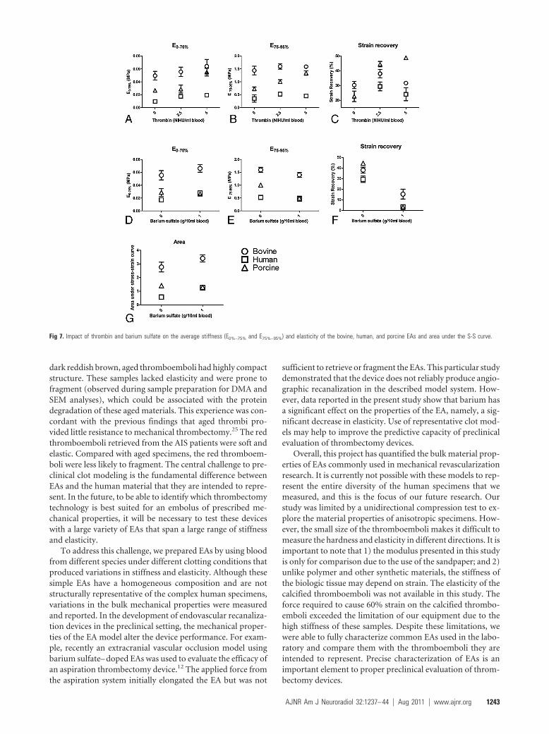
Overall, bovine EAs showed the highest stiffness followedby porcine and human EAs (Fig 7). Without addition ofthrombin, E0%–75% and E75%–95% of bovine EAs were statisti-cally higher than those of porcine and human EAs (P � .05).However, there was no significant difference in stiffness be-tween EAs made from porcine and human blood (Fig 7A, -B).Increases in stiffness and elasticity of human and porcine EAswere found when thrombin was present at a concentration of5 NIHU/mL blood (P � .05) (Fig 7A–C). Elasticity of thethrombin-induced porcine EAs was higher than that of thethrombin-induced human EAs (P � .05) (Fig 7C). Comparedwith the counterparts of the bovine and human EAs, throm-bin-induced porcine EAs retracted the most (data not shown).Addition of thrombin resulted in increases in E0%–75% andelasticity of human EAs (P � .05) (Fig 7A–C). In the absence ofthrombin and barium sulfate, the bovine EAs had strain recov-ery of 30.3 � 2.5%, whereas strain recovery in human blood-derived EAs was immeasurable due to their fragile nature. LessEA shrinkage and a significant decrease in elasticity (P � .05)were found in all barium sulfate-containing EAs (Fig 7F). Dueto the immiscibility between barium sulfate and blood, bar-ium sulfate precipitation at the bottom of EAs was observed.However, the accelerated clotting process induced by throm-bin improved the homogeneity of barium sulfate dispersion.The structure of the barium sulfate-impregnated EAs is shownin Fig 6E, -F. The area under the S-S curve from initial to 95%strain increased with the addition of barium sulfate, indicatingthe EAs became tougher (Fig 7G).
A series of comparisons between the different types of EAsand emboli obtained from patients showed recalcified porcineEAs and thrombin-induced (5 NIHU/mL blood) human EAs
AJNR Am J Neuroradiol 32:1237– 44 � Aug 2011 � www.ajnr.org 1239

are similar to cerebral emboli retrieved from the AIS patientsin terms of stiffness and elasticity (P � .05). The thrombin-induced bovine EAs (2.5 and 5 NIHU/mL blood) and throm-bin-induced porcine EAs (5 NIHU/mL blood) best representthe plaque material collected at CEA.
DiscussionEmbolus characteristics in terms of mechanical properties,composition, and structure are important in the endovasculartreatment of AIS. Recently, manual elongation tests were per-formed by using forceps to stretch EAs in an effort to under-stand their mechanics.4 In this present work, stiffness and elas-ticity of the in vitro EAs were quantitatively measured and
compared with the clinical thromboemboli that they are in-tended to represent.
From the human specimens, we were able to ascertain alarge variation of bulk mechanical properties associated withheterogeneous composition. Some of this variability was in-herent to the sample population, which included both emboliretrieved from patients in the acute phase of the stroke andmaterial from CEA. Most CEA specimens were attached to thelumen of the plaque and represent potential stroke-inducingemboli. The other CEA specimens were located immediatelyunder the fibrous cap, and though it is possible that thesematerials could lead to an artery-to-artery embolism afterplaque rupture, we assume that this is an unlikely event. The
Fig 2. A 65-year-old man (patient 5) presents with an AIS due to MCA occlusion and associated right ICA origin segmental stenosis (�90%) with intraluminal clot (A, arrow). The clot(B) is removed by using the aspiration device for characterization. A 53-year-old man (patient 11) with transient ischemic attacks has a clot located at the carotid bifurcation (C). After CEA,red thrombus (D) is collected for mechanical and structural analyses.
1240 Chueh � AJNR 32 � Aug 2011 � www.ajnr.org

Fig 4. The engineering S-S curves show that at the end of the test, a 15-N force causes a 45.7% strain to a highly calcified embolus (black solid line) and a 64.4% strain to a partiallycalcified embolus (black dashed line). The same compression force results in a higher strain (�80%) on the other emboli.
Fig 3. Morphologic features and composition of a variety of emboli from patients. A, Calcified embolus calcium-phosphate apatite is detected by the EDS scan (inset). B, An aged embolushas a compact structure with fissures that were occupied by the cholesterol crystals (arrows) and fibrin at the edge of the specimen (star). The SEM findings are related to the MSB result(�20; bar � 100 �m) shown in C. D, MSB results show that a red embolus is mainly composed of fibrin and erythrocytes (old fibrin in blue, erythrocytes in yellow, and erythrocyte-fibrinmixture in red) (�2, bar � 1 mm). E, Photomicrograph of a red embolus retrieved from the stroke patient (H&E �10, bar � 200 �m) reveals that the leukocytes are distributed throughoutthe embolus. F, At �10 magnification, erythrocytes and fibrin strands are arranged in a layer-by-layer manner in the red embolus (MSB, bar � 200 �m).
AJNR Am J Neuroradiol 32:1237– 44 � Aug 2011 � www.ajnr.org 1241

Fig 6. A, A layer of fibrin is formed on the surface of the thrombin-induced bovine EA against the silicone tubing (star). MSB staining, bar � 10 �m. B, EA has a homogeneous structure,and is mainly composed of erythrocytes (shown in yellow) with fibrin clumps dispersed in it (arrows, bar � 10 �m). The corresponding SEM findings are presented in C (�5000) and D(�1000), respectively. E, Secondary electron image of the bovine EA with barium sulfate (�20 000). F, Mixed secondary and backscattered electron image of barium sulfate agglomerates(�20 000).
Fig 5. A, Comparisons of thromboemboli from CEA and AIS in terms of E0%–75% and E75%–95%. Large variations are observed in the CEA groups. B, Strain recovery and length changeof one of the thromboemboli during the stress relaxation test. The specimen has an initial length of 1.5 mm and was compressed to 60% strain for 5 minutes. Increases in length andstrain recovery are seen after the load is removed.
1242 Chueh � AJNR 32 � Aug 2011 � www.ajnr.org
dark reddish brown, aged thromboemboli had highly compactstructure. These samples lacked elasticity and were prone tofragment (observed during sample preparation for DMA andSEM analyses), which could be associated with the proteindegradation of these aged materials. This experience was con-cordant with the previous findings that aged thrombi pro-vided little resistance to mechanical thrombectomy.25 The redthromboemboli retrieved from the AIS patients were soft andelastic. Compared with aged specimens, the red thromboem-boli were less likely to fragment. The central challenge to pre-clinical clot modeling is the fundamental difference betweenEAs and the human material that they are intended to repre-sent. In the future, to be able to identify which thrombectomytechnology is best suited for an embolus of prescribed me-chanical properties, it will be necessary to test these deviceswith a large variety of EAs that span a large range of stiffnessand elasticity.
To address this challenge, we prepared EAs by using bloodfrom different species under different clotting conditions thatproduced variations in stiffness and elasticity. Although thesesimple EAs have a homogeneous composition and are notstructurally representative of the complex human specimens,variations in the bulk mechanical properties were measuredand reported. In the development of endovascular recanaliza-tion devices in the preclinical setting, the mechanical proper-ties of the EA model alter the device performance. For exam-ple, recently an extracranial vascular occlusion model usingbarium sulfate– doped EAs was used to evaluate the efficacy ofan aspiration thrombectomy device.12 The applied force fromthe aspiration system initially elongated the EA but was not
sufficient to retrieve or fragment the EAs. This particular studydemonstrated that the device does not reliably produce angio-graphic recanalization in the described model system. How-ever, data reported in the present study show that barium hasa significant effect on the properties of the EA, namely, a sig-nificant decrease in elasticity. Use of representative clot mod-els may help to improve the predictive capacity of preclinicalevaluation of thrombectomy devices.
Overall, this project has quantified the bulk material prop-erties of EAs commonly used in mechanical revascularizationresearch. It is currently not possible with these models to rep-resent the entire diversity of the human specimens that wemeasured, and this is the focus of our future research. Ourstudy was limited by a unidirectional compression test to ex-plore the material properties of anisotropic specimens. How-ever, the small size of the thromboemboli makes it difficult tomeasure the hardness and elasticity in different directions. It isimportant to note that 1) the modulus presented in this studyis only for comparison due to the use of the sandpaper; and 2)unlike polymer and other synthetic materials, the stiffness ofthe biologic tissue may depend on strain. The elasticity of thecalcified thromboemboli was not available in this study. Theforce required to cause 60% strain on the calcified thrombo-emboli exceeded the limitation of our equipment due to thehigh stiffness of these samples. Despite these limitations, wewere able to fully characterize common EAs used in the labo-ratory and compare them with the thromboemboli they areintended to represent. Precise characterization of EAs is animportant element to proper preclinical evaluation of throm-bectomy devices.
Fig 7. Impact of thrombin and barium sulfate on the average stiffness (E0%–75% and E75%–95%) and elasticity of the bovine, human, and porcine EAs and area under the S-S curve.
AJNR Am J Neuroradiol 32:1237– 44 � Aug 2011 � www.ajnr.org 1243
ConclusionsDifferences in structure and composition were observed be-tween EAs and human thromboemboli. Recalcified porcineEAs and thrombin-induced human EAs (5 NIHU/mL blood)were similar to cerebral thromboemboli retrieved from pa-tients with AIS in terms of stiffness and elasticity. There was nosignificant variation between the stiffness of the material col-lected at CEA and thrombin-induced bovine (2.5 and 5NIHU/mL blood) or porcine (5 NIHU/mL blood) EAs. Addi-tion of barium sulfate for radio-opacity in the EAs dramati-cally reduces the elasticity. Fully characterized EAs withknown mechanical properties are now available for preclinicalevaluation of thrombectomy devices.
AcknowledgmentsWe are grateful to Andres Schanzer, MD, and Mohammad H.Eslami, MD, for providing atherosclerotic plaques for charac-terization and to Stanley Tam, MD, for providing the atrialappendage. We also thank Eileen A. Duhamel, NP, and AnnaThors, NP, for dedicated assistance with this study.
Disclosures: A.K.W., Research Support (including provision of equipment or materials):Philips Med, Details: Research support in form of equipment lease and support of researchstaff and research projects, Consultant: Covidien, Codman JNJ, Boston Scientific, Soteira.Details: Consulting agreement, payment on hourly basis, Ownership Interest: Surpass Med.,Details: Major stockholder; J.P.W., Consultant: Boston Biomedical Associates, Details:Serve on a safety review board for clinical trial of an implantable spine device; M.J.G.,Research Support (including provision of equipment or materials): NIH, TSI, BostonScientific, Micrus Endovascular, Codman Neurovascular, Philips Healthcare, ConcentricMedical, NIT, Details: NIH-salary support, research support; Industry noted above: Researchsupport on a fee-for-service basis, Consultant: Soteira Inc, Codman Neurovascular, MicrusEndovascular, Details: Engineering consultation on a fee-for-service basis; Other FinancialRelationships: Hologic, Details: Spouse is employed at Hologic.
References1. Donnan GA, Baron JC, Ma H, et al. Penumbral selection of patients for trials of
acute stroke therapy. Lancet Neurol 2009;8:261– 692. Smith WS, Sung G, Saver J, et al. Mechanical thrombectomy for acute ischemic
stroke: final results of the Multi MERCI trial. Stroke 2008;39:1205–123. The Penumbra pivotal stroke trial: safety and effectiveness of a new genera-
tion of mechanical devices for clot removal in intracranial large vessel occlu-sive disease. Stroke 2009;40:2761– 68
4. Kan I, Yuki I, Murayama Y, et al. A novel method of thrombus preparation for
use in a swine model for evaluation of thrombectomy devices. AJNR Am JNeuroradiol 2010;31:1741– 43
5. Asakura F, Yilmaz H, Abdo G, et al. Preclinical testing of a new clot-retrievingwire device using polyvinyl alcohol hydrogel vascular models. Neuroradiology2007;49:243–51
6. Liebig T, Reinartz J, Hannes R, et al. Comparative in vitro study of five mechan-ical embolectomy systems: effectiveness of clot removal and risk of distal em-bolization. Neuroradiology 2008;50:43–52
7. Brekenfeld C, Schroth G, El-Koussy M, et al. Mechanical thromboembolec-tomy for acute ischemic stroke: comparison of the catch thrombectomy de-vice and the Merci Retriever in vivo. Stroke 2008;39:1213–19
8. Muller-Hulsbeck S, Grimm J, Leidt J, et al. Comparison of in vitro effectivenessof mechanical thrombectomy devices. J Vasc Interv Radiol 2001;12:1185–91
9. Salazar GM, Faintuch S, Gladstone SR, et al. In vitro analysis of downstreamparticulates with mechanical thrombectomy devices: comparison of 20-kHzsonothrombolytic and rotating dispersion wire systems. J Vasc Interv Radiol2009;20:634 –39
10. Krueger K, Deissler P, Coburger S, et al. How thrombus model impacts the invitro study of interventional thrombectomy procedures. Invest Radiol2004;39:641– 48
11. Gralla J, Schroth G, Remonda L, et al. A dedicated animal model for mechanicalthrombectomy in acute stroke. AJNR Am J Neuroradiol 2006;27:1357– 61
12. Gralla J, Schroth G, Remonda L, et al. Mechanical thrombectomy for acuteischemic stroke: thrombus-device interaction, efficiency, and complicationsin vivo. Stroke 2006;37:3019 –24
13. Hong AS, Chae JS, Dubin SB, et al. Ultrasonic clot disruption: an in vitro study.Am Heart J 1990;120:418 –22
14. Gersh KC, Nagaswami C, Weisel JW. Fibrin network structure and clot me-chanical properties are altered by incorporation of erythrocytes. Thromb Hae-most 2009;102:1169 –75
15. Di Stasio E, Nagaswami C, Weisel JW, et al. Cl� regulates the structure of thefibrin clot. Biophys J 1998;75:1973–79
16. Ryan EA, Mockros LF, Weisel JW, et al. Structural origins of fibrin clot rheol-ogy. Biophys J 1999;77:2813–26
17. Stormorken H. Species differences of clotting factors in ox, dog, horse, andman; thrombin and fibrinogen. Acta Physiol Scand 1957;40:167– 81
18. Gentry PA. Comparative aspects of blood coagulation. Vet J 2004;168:238 –51
19. Marder VJ, Chute DJ, Starkman S, et al. Analysis of thrombi retrieved fromcerebral arteries of patients with acute ischemic stroke. Stroke 2006;7:2086 –93
20. Almekhlafi MA, Hu WY, Hill MD, et al. Calcification and endothelialization ofthrombi in acute stroke. Ann Neurol 2008;64:344 – 48
21. French JE. The structure of natural and experimental thrombi. Ann R Coll SurgEngl 1965;36:191–200
22. Wechsler LR. Ulceration and carotid artery disease. Stroke 1988;19:650 –5323. Wolf PA, Abbott RD, Kannel WB. Atrial fibrillation as an independent risk
factor for stroke: the Framingham Study. Stroke 1991;22:983– 8824. Lendrum AC, Fraser DS, Slidders W, et al. Studies on the character and staining
of fibrin. J Clin Pathol 1962;15:401–1325. Grimm J, Jahnke T, Muhle C, et al. Influence of thrombus age on the mechan-
ical thrombectomy efficacy of the Amplatz thrombectomy device in vitro.Cardiovasc Intervent Radiol 2003;26:265– 68
1244 Chueh � AJNR 32 � Aug 2011 � www.ajnr.org