Embed Size (px)
Citation preview

Measuring Esophageal Distention byHigh-Frequency Intraluminal Ultrasound Probe
Ravinder K. Mittal, MD
This investigation used a high-frequency intraluminalultrasound (HFIUS) probe to study changes in esoph-ageal muscle thickness as a marker of muscle con-traction. The aim of this study was to determinewhether this technique could accurately measuredistention of the esophagus during swallows of liquidbolus and during gastroesophageal reflux (GER) innormal subjects. Despite some limitations, HFIUSappears to be a valid technique for both of thesemeasurements in normal subjects. Future studies inpatients with symptomatic GER disease (GERD)should be able to determine whether typical symp-toms such as heartburn, as well as atypical symp-toms such as chest pain, are related to the GER-related distention of the esophagus. Am J Med. 2003;115(3A):130S–136S. © 2003 by Excerpta Medica, Inc.
Distention of the esophagus is a reproducible stim-ulus that can induce heartburn and chest pain inthe laboratory setting in humans.1– 4 This is par-
ticularly relevant when the patient presents with the com-plaint of chest pain, as the typical cardiac workup can beboth extensive and expensive. Distention of the esopha-gus may also occur during spontaneous gastroesophagealreflux (GER), and may play a role in the development ofesophageal symptoms, both typical and atypical. Com-monly used techniques to study esophageal function donot measure esophageal distention. The most recently de-veloped of these techniques— esophageal impedancemeasurement— can reveal the nature of reflux (i.e., liq-uid or air, acid, or neutral reflux) during spontaneousepisodes of GER but not the extent of distention of theesophagus during GER events.5–7
This investigation used the high-frequency intralumi-nal ultrasound (HFIUS) probe to study changes in esoph-ageal muscle thickness as a marker of muscle contrac-tion.8 –10 The aim of this study was to determine whetherHFIUS could accurately measure the cross-sectional area(CSA) of a tube in vitro, as well as distention of the esoph-agus during swallows of liquid bolus and GER in normalsubjects. This type of study may have applicability in thedifferential diagnosis of atypical symptoms of GER dis-ease (GERD).
METHODS
Use of HFIUS Technique to Measure CSA inIn Vitro StudiesWe constructed tubes of 2 different and known diametersfrom agarose gel. These tubes were filled with water, andthe HFIUS probe was placed inside the tubes for mea-surement of the luminal CSA. The ultrasound probe wasfirst placed vertically in the center and then put at anoff-center position in the tube (diameter � 15.6 mm).We also imaged the tube with the ultrasound probeplaced at a 15° angle from the side of the tube (Figure 1).
Data analysis. The ultrasound images were digitized ona personal computer equipped with a high-definitionvideo card (DC30 Pro; Pinnacle, Silicon Valley, CA) andanalyzed using a commercially available image analysissoftware package (SigmaScan Pro; Jandel Scientific, SanRafael, CA). Images were displayed on a 17-inch, high-resolution monitor with a pixel size of 640 � 480, whichcorresponds to an image magnification of approximately�12 (10 pixels � 1 mm). We selected 8 ultrasound im-
From the Division of Gastroenterology, San Diego VA Medical Center,University of California–San Diego, San Diego, California, USA.
Requests for reprint should be addressed to Ravinder K. Mittal, MD,Division of Gastroenterology 111D, San Diego VA Medical Center,University of California–San Diego, San Diego, California 92161.
130S © 2003 by Excerpta Medica, Inc. 0002-9343/03/$22.00All rights reserved. doi:10.1016/S0002-9343(03)00211-0
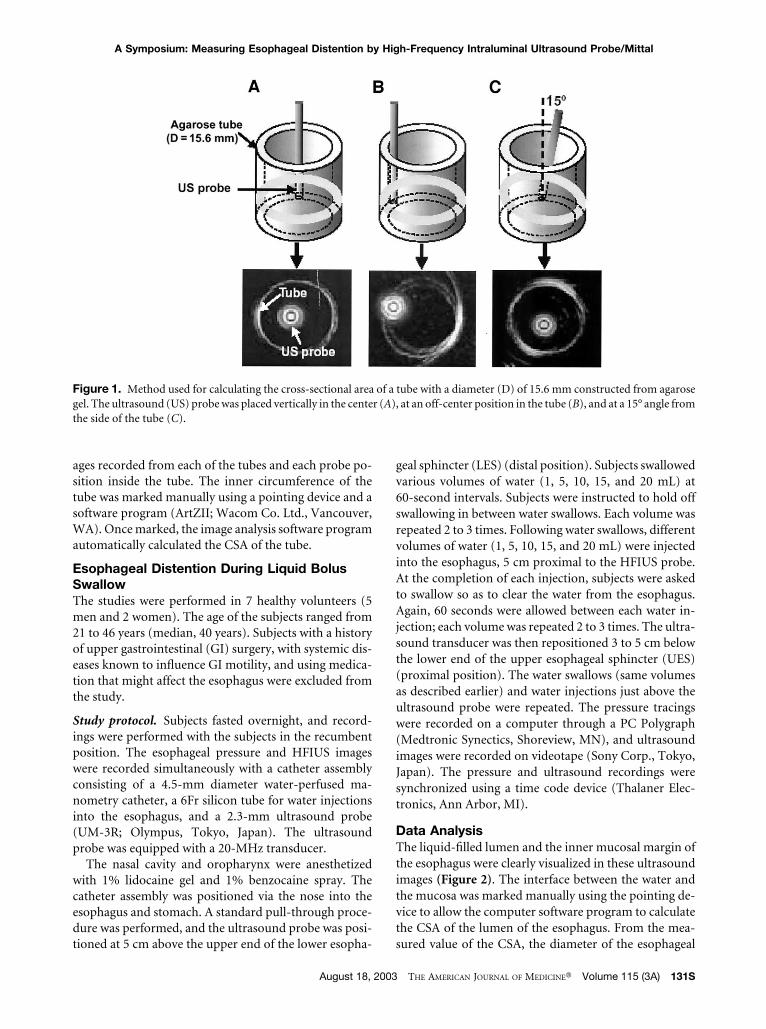
ages recorded from each of the tubes and each probe po-sition inside the tube. The inner circumference of thetube was marked manually using a pointing device and asoftware program (ArtZII; Wacom Co. Ltd., Vancouver,WA). Once marked, the image analysis software programautomatically calculated the CSA of the tube.
Esophageal Distention During Liquid BolusSwallowThe studies were performed in 7 healthy volunteers (5men and 2 women). The age of the subjects ranged from21 to 46 years (median, 40 years). Subjects with a historyof upper gastrointestinal (GI) surgery, with systemic dis-eases known to influence GI motility, and using medica-tion that might affect the esophagus were excluded fromthe study.
Study protocol. Subjects fasted overnight, and record-ings were performed with the subjects in the recumbentposition. The esophageal pressure and HFIUS imageswere recorded simultaneously with a catheter assemblyconsisting of a 4.5-mm diameter water-perfused ma-nometry catheter, a 6Fr silicon tube for water injectionsinto the esophagus, and a 2.3-mm ultrasound probe(UM-3R; Olympus, Tokyo, Japan). The ultrasoundprobe was equipped with a 20-MHz transducer.
The nasal cavity and oropharynx were anesthetizedwith 1% lidocaine gel and 1% benzocaine spray. Thecatheter assembly was positioned via the nose into theesophagus and stomach. A standard pull-through proce-dure was performed, and the ultrasound probe was posi-tioned at 5 cm above the upper end of the lower esopha-
geal sphincter (LES) (distal position). Subjects swallowedvarious volumes of water (1, 5, 10, 15, and 20 mL) at60-second intervals. Subjects were instructed to hold offswallowing in between water swallows. Each volume wasrepeated 2 to 3 times. Following water swallows, differentvolumes of water (1, 5, 10, 15, and 20 mL) were injectedinto the esophagus, 5 cm proximal to the HFIUS probe.At the completion of each injection, subjects were askedto swallow so as to clear the water from the esophagus.Again, 60 seconds were allowed between each water in-jection; each volume was repeated 2 to 3 times. The ultra-sound transducer was then repositioned 3 to 5 cm belowthe lower end of the upper esophageal sphincter (UES)(proximal position). The water swallows (same volumesas described earlier) and water injections just above theultrasound probe were repeated. The pressure tracingswere recorded on a computer through a PC Polygraph(Medtronic Synectics, Shoreview, MN), and ultrasoundimages were recorded on videotape (Sony Corp., Tokyo,Japan). The pressure and ultrasound recordings weresynchronized using a time code device (Thalaner Elec-tronics, Ann Arbor, MI).
Data AnalysisThe liquid-filled lumen and the inner mucosal margin ofthe esophagus were clearly visualized in these ultrasoundimages (Figure 2). The interface between the water andthe mucosa was marked manually using the pointing de-vice to allow the computer software program to calculatethe CSA of the lumen of the esophagus. From the mea-sured value of the CSA, the diameter of the esophageal
Figure 1. Method used for calculating the cross-sectional area of a tube with a diameter (D) of 15.6 mm constructed from agarosegel. The ultrasound (US) probe was placed vertically in the center (A), at an off-center position in the tube (B), and at a 15° angle fromthe side of the tube (C).
A Symposium: Measuring Esophageal Distention by High-Frequency Intraluminal Ultrasound Probe/Mittal
August 18, 2003 THE AMERICAN JOURNAL OF MEDICINE� Volume 115 (3A) 131S

lumen was calculated based on the equation,diameter � 2 � �CSA/�.
Analysis of Esophageal Contents andDimension During Transient LES RelaxationTwelve healthy volunteers, 6 women and 6 men, rangingin age from 18 to 53 years (median, 27.5 years), wereevaluated to determine the distention of the esophaguscaused by physiologic GER. None of these volunteers hadany upper GI symptoms or history of abdominal surgery.
Study protocol. The LES, esophageal, and gastric pres-sures, esophageal pH, and HFIUS images were recordedsimultaneously. The catheter assembly consisted of asphinctometer catheter (Sentron, Roden, Netherlands), apH probe (Synectics Medical, Stockholm, Sweden), andan ultrasound probe with the 20-MHz UM-3R trans-ducer. The sphinctometer was calibrated at 0 (atmo-spheric pressure) and 50 mm Hg at room temperature.Following a standard pull-through technique, the cathe-ter assembly was placed in such a fashion that the sphinc-tometer straddled the LES. The pH sensor was positionedat 5 cm above the LES, the esophageal pressure transduc-ers were placed at 5, 10, and 15 cm above the LES, and theultrasound transducer was positioned at 5 cm above theLES. At the beginning of each recording period, 3 to 5 wetswallows of 5 mL of water at room temperature were per-formed. Recordings were then performed for 1 hour inthe fasting state and for 2 additional hours after the inges-tion of a standardized meal, consisting of a sandwich anda soft drink (1,000 kcal, 40 g carbohydrates, 40 g protein,and 20 g fat). All recordings were performed with thesubjects in the left recumbent position during the study(mean observation time, 3 hours and 22 minutes).
Data analysis. The LES pressure record was screened fortransient LES relaxations (TLESRs). The ultrasound im-ages during TLESRs were visually analyzed to determinethe nature of the refluxed material (predominantly liq-uid, predominantly air, or a mixture of liquid and air).The ultrasound image, corresponding to the maximal di-mension of the esophageal lumen during TLESR, was se-lected for the CSA measurement and the estimation of thediameter.
Statistical analysis. The statistical differences were esti-mated using analysis of variance on ranks or linear regres-sion for in vitro and in vivo validation studies. The Mann-Whitney rank sum test was used for analysis of esopha-geal distention during swallows and TLESRs.
RESULTS
Validation of the HFIUS Probe In VitroThe CSA of the agarose tube (diameter � 15.6 mm) wasmeasured from the HFIUS probe images, with the ultra-sound probe placed at the center of the tube, off-center,
and at a 15° oblique angle. The mean percentage differ-ence between the ultrasound-measured CSA and the ac-tual CSA was 3.3% for the central location (range, 2.1% to4.7%), 3.2% for the off-center location (range, 0% to6.5%), and 2.4% for the 15° oblique angle location(range, 1.7% to 3.7%).
CSA of the Esophagus In Vivo DuringSwallowsThe CSAs noted during 1, 5, 10, 15, and 20 mL bolusswallows in the distal esophagus (5 cm above the LES)and proximal esophagus are depicted graphically in Fig-ure 3, and the counterpart ultrasound images are shownin Figure 4. There was a linear increase in the CSA of theesophagus, with an increase in bolus volumes up to 15 mL(R � 0.75, P �0.05). The esophageal CSAs between 15and 20 mL swallows did not differ.
The relation between the volume of swallowed bolusand the cross-sectional analysis of the proximal esopha-gus was also linear (R � 0.78, P �0.05). The median CSAduring 15-mL water swallows in the distal esophagus wasgreater than that of the proximal esophagus (P �0.05).The maximal estimated diameters during water swallowsat the distal and proximal esophagus were 22 and 21 mm,respectively.
Esophageal Distention by Injection of Waterinto the EsophagusThe esophageal CSA during intraesophageal water injec-tion increased in a linear fashion with an increase of theinjected water volume (R � 0.76, P � 0.05). The CSA ofthe proximal esophagus during 1, 5, 10, 15, and 20 mL ofwater injection was 65 mm2 (range, 34 to 90 mm2), 76mm2 (range, 49 to 153 mm2), 129 mm2 (range, 58 to 185mm2), 170 mm2 (range, 69 to 272 mm2), and 202 mm2
(range, 71 to 307 mm2), respectively (R � 0.68, P �0.05).
Nature of Esophageal Contents and CSA ofthe Esophagus During Swallows and TLESREpisodesA total of 46 5-mL water swallows and 62 episodes ofTLESR were analyzed in the 12 normal subjects. Acidreflux was detected during 29 of 62 TLESR episodes (de-fined as a decrease in intraesophageal pH to �4, lasting�5 seconds). The following patterns of esophageal lumi-nal contents were noted: (1) no luminal opening; (2) lu-minal opening predominantly with liquid only; (3) lumi-nal opening predominantly with liquid, followed by air;(4) luminal opening predominantly with air, followed byliquid; and (5) luminal opening predominantly with aironly.
During water swallows, the passage of liquid followedby air was the most frequent pattern (67%). There wereno episodes of failed esophageal luminal opening. Themost frequent pattern during TLESR was the passage ofair only (58%). The frequency of other patterns was as
A Symposium: Measuring Esophageal Distention by High-Frequency Intraluminal Ultrasound Probe/Mittal
132S August 18, 2003 THE AMERICAN JOURNAL OF MEDICINE� Volume 115 (3A)

follows: air followed by liquid (21%), failed luminalopening (15%), and liquid followed by air (7%). The pat-terns of the esophageal contents during positive pH de-crease and negative pH decrease episodes did not differ.During 17 of the 29 acid reflux–positive episodes (59%),no liquid material was seen in the esophagus. The CSA ofthe esophagus could be measured during 43 of the 46
water swallows, compared with only 17 of the 62 TLESRepisodes. In the remainder, either there was no openingof the esophageal lumen, or the presence of too much airin the esophagus precluded the measurement of CSA.The median CSA of the esophagus during these 17 refluxepisodes was 44 mm2 (range, 24 to 187 mm2). In the samesubjects, the median CSA of the esophagus during 5-mL
Figure 2. (A–C) Ultrasound images depicting the method used for calculating the cross-sectional area (CSA) of the esophagusduring swallows. The interface between the water and the mucosa was marked manually using a pointing device, which allowed thecomputer software program (ArtZII; Wacom Co. Ltd., Vancouver, WA) to calculate the CSA of the lumen of the esophagus using theequation D � 2 · �CSA/�.
Figure 3. The calculated cross-sectional area of the esophagus in normal subjects during 1, 5, 10, 15, and 20 mL bolus swallows inthe distal esophagus (5 cm above the lower esophageal sphincter) and proximal esophagus. * Nonsignificant difference between 15mL and 20 mL.
A Symposium: Measuring Esophageal Distention by High-Frequency Intraluminal Ultrasound Probe/Mittal
August 18, 2003 THE AMERICAN JOURNAL OF MEDICINE� Volume 115 (3A) 133S

water swallows was 53 mm2 (range, 25 to 158 mm2).There was no difference between the CSA during 5-mLswallows and liquid GER episodes (P � NS). Further-more, no difference was detected in the CSA of the esoph-agus during acid reflux–positive and acid reflux–negativeGER (44 vs. 44 mm2, P � NS).
DISCUSSION
Our in vitro studies show that the HFIUS probe can mea-sure the CSA, and thus the diameter of a tube, fairly ac-curately. In vivo measurements of the esophagus duringwater swallows reveal that adequate images of the esoph-agus are obtained as the liquid bolus traverses throughthe esophagus. The CSA of the esophagus increases in alinear fashion as the volume of swallowed bolus increases,with a peak esophageal diameter of 22 mm at 15-mL wa-ter swallows. The CSA of the distal esophagus was largerthan that of the proximal esophagus. Interestingly, theCSA and the diameter of the esophagus during spontane-ous liquid GER in healthy, asymptomatic subjects issmall, comparable to a 5-mL swallow. Finally, the natureof the esophageal contents—that is, air, liquid, acidic, and
neutral reflux— can be detected by the technique of si-multaneous pressure, and by pH and ultrasound imagerecordings.
In Vitro ValidationFor in vitro studies, we tested the tubes of different CSAsthat are relevant to the in vivo dimensions of the humanesophagus. Our data show that the ultrasound probe canmeasure the CSA of tubes up to 50 mm in diameter fairlyaccurately. We also tested other possibilities, that is,movement of the ultrasound transducer from the centerto an off-center position and the possibility that the ul-trasound transducer may not be perpendicular to the wallof the esophagus. The angle between the probe and theesophageal wall depends on the diameter and the lengthof the esophagus. Based on geometric principles, themaximal angle in a 20 cm-long tube with a diameter of 1.0cm is 2.86° (tan�1 [1.0 cm/20.0 cm]). The same angle in atube with a lumen measuring 5 cm in diameter will be 14°.Our data show that the off-center position and probeangles of �15° do not affect the accuracy of CSAmeasurement.
Figure 4. The patterns of esophageal luminal contents during swallows and transient lower esophageal sphincter relaxation in 5subjects. (A) No luminal opening; (B) opening predominantly with liquid only; (C) opening predominantly with liquid followed byair; (D) opening predominantly with air followed by liquid; and (E) opening predominantly with air only.
A Symposium: Measuring Esophageal Distention by High-Frequency Intraluminal Ultrasound Probe/Mittal
134S August 18, 2003 THE AMERICAN JOURNAL OF MEDICINE� Volume 115 (3A)

In Vivo Measurements of the EsophagusThe CSA of the proximal and distal esophagus, using a10-mL liquid bolus, have been reported using the tech-nique of an ultrafast computed tomography (CT) scan.11
It is noteworthy that the measurements reported in thatstudy are similar to our findings. The ultrafast CT scan-ning, unlike HFIUS imaging, cannot be used for long-term recordings. Our in vivo studies show that the esoph-ageal CSA increases as the volume of ingested bolus in-creases. The maximal CSA was recorded with swallowedand injected boluses of 15 mL of water. From these CSAmeasurements, we estimated that the maximal diameterof the distal esophagus during physiologic swallows is�23 mm. Several studies report that the proximal esoph-agus is less compliant than the distal esophagus,12,13 andwe found the same: that is, at higher ingested volumes,the CSA of the proximal esophagus is lower than the dis-tal esophagus.
Patterns of Liquid and Air Contents in theEsophagus During SwallowsThe air residing in the oropharynx is ingested along withother oral contents during a swallow. Ultrafast CT scanstudies show that a normal subject ingests approximately10 to 15 mL of air with each swallow.11 Our study con-firms that air is ingested along with the liquid duringwater swallows. The difference between CT studies andour ultrasound studies, however, is that we noticed liquidtraversing ahead of the air during the majority of swal-lows. On the other hand, CT studies revealed the transitof air through the esophagus first, followed by liquid. Thereason for the difference in the 2 types of studies may berelated to the manometry technique used in our studies,which requires infusion of water into the esophagus. Theinfused water would be expected to move ahead of the airentering into the esophagus from the pharynx. The slightdifference in the position of the subject may also make adifference, because in the upright position, air, which islighter than water, would be expected to rise and movebehind the liquid bolus.
Patterns of Gas and Liquid RefluxThe patterns of gas and liquid reflux during TLESR epi-sodes have been reported in several recent studies usingthe technique of impedance measurements.5,6 The “aironly” was the predominant pattern (58%) of reflux in ourstudy. Impedance studies show similar findings, eventhough the incidence of air-only reflux was 18%.5 Thephysical posture of the subject seems to be a major deter-minant of the nature of the material that refluxes into theesophagus. We studied our subjects in the left lateral po-sition, a position in which reflux of air occurs more fre-quently than does liquid reflux. On the other hand, refluxof liquid dominates in the right lateral position.14,15
Therefore, to determine esophageal distention by ultra-sound imaging, the right lateral position of the subject
may be ideal because liquid reflux in this position willallow adequate ultrasound images for distentionmeasurements.
We detected a large number of reflux episodes in whichthe pH probe registered acid reflux but no liquid was seenin the esophagus on the HFIUS images. Similar observa-tions have been made with the impedance measurementtechnique.5 We suspect that tiny droplets of acid withgas-predominant reflux (mist of acid) may be registeredas a major reflux in the pH recordings, and may not showany liquid reflux on the HFIUS images. The low incidenceof liquid flow during GER in our study compared withthe impedance studies may be related to a higher positionof the ultrasound transducer (5 cm above the LES) com-pared with the impedance electrode position (3 cm abovethe LES) in those studies.5,6
Significance of Esophageal DistentionMeasurementUnlike the impedance technique, the HFIUS imagingtechnique can detect the esophageal CSA during liquidGER episodes. It seems that the incidence of liquid refluxduring spontaneous episodes of GER in normal subjectsis infrequent. Even when liquid reflux occurs, its volumeis extremely small. In fact, there was no opening of theesophageal lumen seen during several reflux episodes.During those episodes that did show the esophageal lu-men opening with liquid-predominant reflux, the CSA ofthe esophagus was comparable to a 5-mL swallow. Thelimitation of our study is that we performed our record-ings in the left lateral position. It is possible that if we hadrecorded our subjects in the right lateral position, wemight have obtained different results. It remains to beseen whether patients with reflux disease have larger dis-tention of the esophagus during symptomatic GERepisodes.
It must be recognized that the HFIUS technique hasseveral limitations. First, it is unable to differentiate be-tween the ingested versus the refluxed esophageal con-tents because we recorded ultrasound images at only 1site in the esophagus. To overcome this difficulty, we se-lected TLESRs and studied the nature of esophageal con-tents during these periods. Because TLESR is the majormechanism of GER in normal subjects, the esophagealcontents during these periods can only be related toGER.16 –18 Second, the HFIUS technique cannot measurethe esophageal CSA during air-predominant reflux epi-sodes. Finally, esophageal distention can only be mea-sured at 1 site in the esophagus using 1 transducer.
SUMMARY
Despite the noted limitations, HFIUS is a valid techniquefor measuring the CSA of the esophagus and esophagealdistention in vivo during liquid swallows and liquid re-flux episodes. The maximal diameter of esophagus under
A Symposium: Measuring Esophageal Distention by High-Frequency Intraluminal Ultrasound Probe/Mittal
August 18, 2003 THE AMERICAN JOURNAL OF MEDICINE� Volume 115 (3A) 135S

physiologic swallows is approximately 22 mm. The max-imal diameter during physiologic liquid GER is approxi-mately 4 to 5 mm. Future studies in patients with symp-tomatic GERD should be able to address whether heart-burn and esophageal pain are related to the GER-relateddistention of the esophagus.
REFERENCES1. Barish CF, Castell DO, Richter JE. Graded esophageal bal-
loon distention: a new provocative test for noncardiac chestpain. Dig Dis Sci. 1986;31:1292–1298.
2. De Caestecker JS, Pryde A, Heading RC. Site and mecha-nism of pain perception with oesophageal balloon disten-tion and intravenous edrophonium in patients with oesoph-ageal chest pain. Gut. 1992;33:580–58.
3. Richter JE, Barish CF, Castell DO. Abnormal sensory per-ception in patients with esophageal chest pain. Gastroen-terology. 1986;91:845–852.
4. Takeda T, Liu J, Gui A, Kassab G, Mittal RK. The relation-ship between esophageal sensation and biomechanicalproperties of the wall of the esophagus [abstract]. Gastro-enterology. 2001;120(suppl 1):A631.
5. Sifrim D, Silny J, Holloway R, Janssens JJ. Patterns of gasand liquid reflux during transient lower oesophagealsphincter relaxation: a study using intraluminal electricalimpedance. Gut. 1999;44:47–54.
6. Sifrim D, Holloway R, Silny J, et al. Acid, non-acid, and gasreflux in patients with gastroesophageal reflux disease dur-ing ambulatory 24-hour pH-impedance recordings. Gastro-enterology. 2001;120:1588–1598.
7. Vela MF, Camacho-Lobato L, Srinivasan R, Tutuian R, KatzPO, Castell DO. Simultaneous intraesophageal impedanceand pH measurement of acid and non-acid gastroesopha-geal reflux: effect of omeprazole. Gastroenterology. 2001;120:1599–1606.
8. Balaban DH, Yamamoto Y, Liu J, et al. Sustained esopha-geal contraction: a marker of esophageal chest pain iden-
tified by intraluminal ultrasonography. Gastroenterology.1999;116:29–37.
9. Pehlivanov N, Liu J, Kassab GS, Puckett JL, Mittal RK.Relationship between esophageal muscle thickness andintraluminal pressure: an ultrasonographic study. Am JPhysiol. 2001;280:G1093–G1098.
10. Yamamoto Y, Liu J, Smith TK, Mittal RK. Distention-relatedresponses in circular and longitudinal muscle of the humanesophagus: an ultrasonographic study. Am J Physiol. 1998;275:G805–G811.
11. Pouderoux P, Ergun GA, Lin S, Kahrilas PJ. Esophagealbolus transit imaged by ultra-fast computerized tomogra-phy. Gastroenterology. 1996;110:1422–1428.
12. Mayrand S, Diamant NE. Measurement of human esopha-geal tone in-vivo. Gastroenterology. 1993;105:1411–1420.
13. Mayrand S, Tremblay L, Diamant NE. In-vivo measurementof feline esophageal tone. Am J Physiol. 1994;267:G914–G921.
14. Shay SS, Conwell DL, Mehindru V, Hertz B. The effect ofposture on gastroesophageal reflux event frequency andcomposition during fasting. Am J Gastroenterol. 1996;91:54–60.
15. van Herwaarden MA, Katzka DA, Smout AJ, Samsom M,Gideon M, Castell DO. Effect of different recumbent posi-tions on postprandial gastroesophageal reflux in normalsubjects. Am J Gastroenterol. 2000;95:2731–2376.
16. Kahrilas PJ, Shi G, Manka M, Joehl RJ. Increased fre-quency of transient lower esophageal sphincter relaxationinduced by gastric distention in reflux patients with hiatalhernia. Gastroenterology. 2000;118:688–695.
17. Mittal RK, Holloway RH, Penagini R, Blackshaw LA, Dent J.Transient lower esophageal sphincter relaxation. Gastroen-terology. 1995;109:601–610.
18. Pehlivanov N, Liu J, Arora T, Yamamoto Y, Mittal RK. Loweresophageal sphincter monitoring with sphinctometer: in-vitro and in-vivo studies. Am J Physiol. 1999;277:G577–G584.
A Symposium: Measuring Esophageal Distention by High-Frequency Intraluminal Ultrasound Probe/Mittal
136S August 18, 2003 THE AMERICAN JOURNAL OF MEDICINE� Volume 115 (3A)