Embed Size (px)
Citation preview
Benefits• Sensitive fluorometric
tryptophan detection for protein analysis
• Automated identification of peak shifts with spectral scanning
• Quick assay optimization with the Spectral Optimization Wizard
Measure intrinsic tryptophan fluorescence on the SpectraMax iD3 microplate reader
IntroductionThe intrinsic fluorescence of proteins is due to the aromatic amino acids tryptophan, tyrosine, and phenylalanine. Tryptophan, which excites maximally around 270-280 nm and has an emission peak near 350 nm in water, dominates the emission of proteins and is the most sensitive to solvent polarity and the local environment. Exposure of tryptophan residues to water, which occurs when a protein is denatured, leads to a shift to longer emission wavelengths. This shift in peak emission can be used to monitor protein unfolding.
Here, we demonstrate performance of the SpectraMax® iD3 Multi-Mode Microplate Reader for assays measuring intrinsic tryptophan fluorescence. High sensitivity is demonstrated with a tryptophan standard curve, and a shift in peak emission is shown using a lysozyme denaturation assay. Lysozyme contains two tryptophan residues that fluoresce when excited with UV light. When lysozyme is denatured, tryptophan’s emission peak shifts about 6-10 nm. This peak shift can be measured by performing fluorescence spectral scans on native and denatured lysozyme.
The SpectraMax iD3 reader offers the sensitivity and wavelength scanning capability needed to perform intrinsic tryptophan fluorescence measurements. Additionally, SoftMax® Pro Software automatically identifies the optimal excitation and emission wavelengths via the Spectral Optimization Wizard. The shift in emission peak is also automatically calculated for streamlined analysis.
APPLICATION NOTE
Materials• SpectraMax iD3 Multi-Mode
Microplate Reader (Molecular Devices cat. #ID3-STD)
• L-Tryptophan (Sigma cat. #T-0254)
• Lysozyme (Sigma cat. #L6876)
• Guanidine hydrochloride (Sigma cat. #G7294)
• Clear-walled, UV-clear 96-well
microplate (Greiner cat. #655801)
• Phosphate buffered saline (PBS), pH 7.4
Methods A two-fold dilution series of tryptophan starting at 100 µM was prepared and pipetted in triplicate into a UV-clear, 96-well microplate at 200 µL/well. Six wells of PBS were used as blanks for calculating the lower limit of detection (LLD). SoftMax Pro Software’s Spectral Optimization Wizard was used to
Hoang Ha | Applications Scientist | Molecular Devices
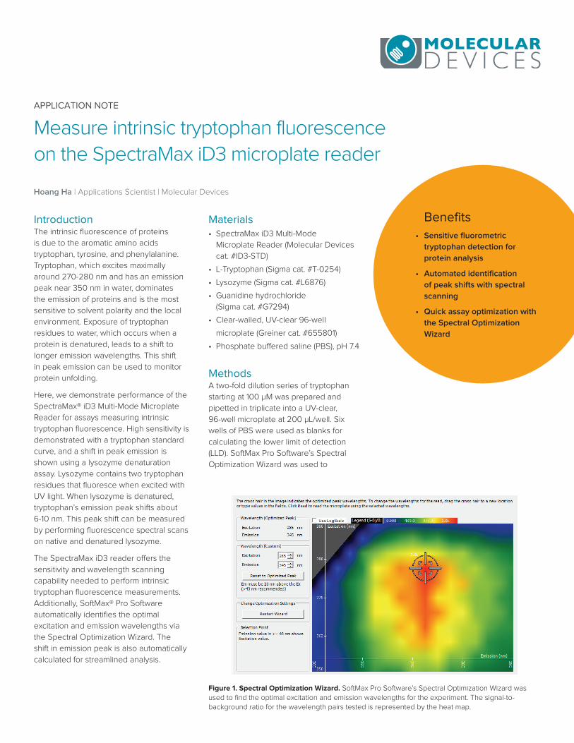
Figure 1. Spectral Optimization Wizard. SoftMax Pro Software’s Spectral Optimization Wizard was used to find the optimal excitation and emission wavelengths for the experiment. The signal-to-background ratio for the wavelength pairs tested is represented by the heat map.
determine the optimal excitation and emission wavelengths for this assay, and the tryptophan dilution series was detected using this wavelength combination.
Guanidine hydrochloride at a final concentration of 5 M was used to denature a 10 mg/mL solution of lysozyme. Spectral scans were performed on native and denatured lysozyme samples using the SpectraMax iD3 reader with excitation at 270 nm and emission ranging from 300 nm to 450 nm. Emission peaks were automatically identified using a preconfigured protocol in SoftMax Pro Software.
ResultsExcitation and emission wavelengths were optimized using SoftMax Pro’s Spectral Optimization Wizard (Figure 1). This feature identified the wavelength combination with the best signal-to-noise ratio as excitation at 285 nm and emission at 345 nm.
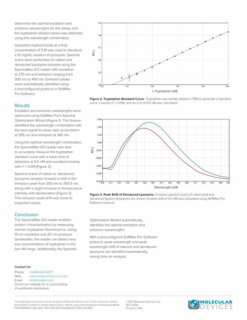
Using the optimal wavelength combination, the SpectraMax iD3 reader was able to accurately measure the tryptophan standard curve with a lower limit of detection at 9.5 nM and excellent linearity with r2 > 0.99 (Figure 2).
Spectral scans of native vs. denatured lysozyme samples showed a shift in the emission peak from 350 nm to 355.5 nm, along with a slight increase in fluorescence intensity with denaturation (Figure 3). This emission peak shift was close to expected values.
ConclusionThe SpectraMax iD3 reader enables protein characterization by measuring intrinsic tryptophan fluorescence. Using 15 nm excitation and 25 nm emission bandwidths, the reader can detect very low concentrations of tryptophan in the low nM range. Additionally, the Spectral
Contact Us
Phone: +1-800-635-5577Web: www.moleculardevices.comEmail: [email protected] our website for a current listing of worldwide distributors.
The trademarks used herein are the property of Molecular Devices, LLC or their respective owners. Specifications subject to change without notice. Patents: www.moleculardevices.com/productpatents FOR RESEARCH USE ONLY. NOT FOR USE IN DIAGNOSTIC PROCEDURES.
©2017 Molecular Devices, LLC 8/17 2123APrinted in USA
Figure 2. Tryptophan Standard Curve. Tryptophan was serially diluted in PBS to generate a standard curve. Linearity (r2 > 0.99), and an LLD of 9.5 nM was calculated.
Figure 3. Peak Shift of Denatured Lysozyme. Emission spectral scans of native (red) and denatured (green) lysozymes are shown. A peak shift of 5.5 nM was calculated using SoftMax Pro Software protocol.
L-Tryptophan (nM)R
FU
Wavelength (nM)
RF
U
Optimization Wizard automatically identifies the optimal excitation and emission wavelengths.
With a preconfigured SoftMax Pro Software protocol, peak wavelength and peak wavelength shift of natured and denatured lysozyme are identified automatically, saving time on analysis.