Embed Size (px)
Citation preview

MCL Injuries: When and How to Repair
Scott D. Mair, MDProfessor and Team Physician: Orthopaedic Surgery
University of Kentucky School of Medicine

Disclosure • Institution: Research/Education
–Smith-Nephew Endoscopy

Isolated MCL Injury
Usually heal – non-operative treatment– Grade I and II– Grade III mid-substance and femoral side
Indelicato– Classic paper, CORR 1990– Treatment Phases –
• Orthosis at 30o, partial weight bearing with crutches• ROM 30-90o, isokinetic exercises, full weight bearing• Removal of brace, full ROM, gradual exercise progression
– Uniformly good results if ACL intact

Our Operative Indications
• Combined injury & Grade III MCL/POL– Opens at 0 and 30 degrees– Increased ER/IR– Positive scope drive through sign
• Isolated Grade III MCL/POL w/ avulsion off tibia: Stener Lesion– Opens at 0 and 30 degrees– Increased ER– Positive scope drive through sign
• Chronic MCL/POL deficiency– Anatomic surgery– Evaluate for malalignment– LaPrade Technique for reconstruction
MCL

Anatomy• Superficial MCL
– 11 cm long; 1.5 cm wide– Insertions:
• Medial femoral epicondyle• 6 cm distal to joint line• Inserts underneath pes tendons
– Biomechanics• Mobile• Posterior: Tight in full extension• Anterior: Tight in both flexion & extension• Most lax @ 30o flexion
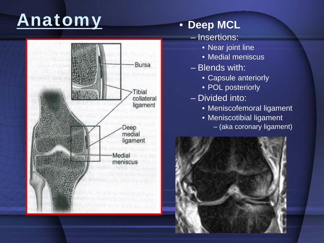
Anatomy • Deep MCL– Insertions:
• Near joint line• Medial meniscus
– Blends with:• Capsule anteriorly• POL posteriorly
– Divided into:• Meniscofemoral ligament• Meniscotibial ligament
– (aka coronary ligament)

Anatomy • Posterior Oblique Ligament– Insertions:
• Adductor tubercle• Tibial joint line• Posteromedial capsule• Semimembranosus
– Loose in flexion– Internal rotation restraint

8
• ACL strain increased significantly after isolated MCL injury• MCL injury can place ACL at risk
• Partial and complete MCL tears significantly increased the load on the anterior cruciate ligament.
Battaglia et al. AJSM Vol 37, No 2, 2009
Combined ACL/MCL injuries

Combined Ligament InjuryOperative Indications
• Combined Injury– 2 schools of thought:
• Non-op treatment of MCL for 4-6 wks, and then reconstruct ACL and/or PCL
• Acute treatment of MCL andreconstruct ACL and/or PCL
– Recommend surgery for MCL when:• Gross opening at 0o
• Avulsed off tibia• When treated non-op, failure of MCL to
heal within 1 month
• Medial Hockey-stick Incision• Layered approach

20yr elite gymnast


Pivot Shift

Valgus Stress


Operative Technique (ACUTE) • Deep MCL
– Repair with suture anchors– Meniscal repair as indicated– Tie sutures w/ knee in full extension
• Superficial MCL– Anchors or Spiked washer– Tie sutures w/ knee at 30o
– Careful not to overtighten…flexion contracture– Check knee w/ gentle ROM to ensure not
“captured”• Cruciate Ligaments
– Reconstruct first– Place tibial fixation only after MCL repair
complete
Minimum of 3 “spot welds” with suture anchors
Suture Anchors or Spiked washer


Isolated MCL Injury
• Operative Treatment?

Isolated MCL -Operative Indications
• Femoral Avulsions– More Common– Heal without surgery– Higher % of stiffness
• Especially w/ surgery
• Tibial Avulsions– Slower healing– Decrease % of stiffness– Synovial fluid
• inhibition of healing– Meniscal instability– Stener lesion
• MCL flipped over pes
TibialAvulsion

MRI
MCL flipped into knee
Tibial Avulsion

Chronic MCL Injury
• Surgical Indications

Operative Technique (CHRONIC)
• MCL Reconstruction– Free graft
• Autograft ST/G• Achilles Tendon Allograft
– Semitendinosus in-situ• Use open-ended tendon
stripper• Maintain tibial insertion• Attach to:
– Semimembranosus– Medial Epicondyle– Back to Pes insertion
• Reconstructs MCL & POL
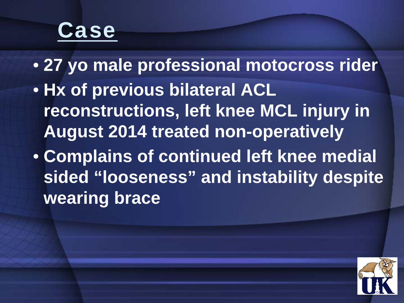
Case• 27 yo male professional motocross rider• Hx of previous bilateral ACL
reconstructions, left knee MCL injury in August 2014 treated non-operatively
• Complains of continued left knee medial sided “looseness” and instability despite wearing brace

Case• Physical Exam
–Full AROM/PROM–(-) Lachman’s, Anterior Drawer, Pivot Shift–(-) Posterior Drawer, Dial test–Grade 3 opening medial joint line with valgus
stress at 0° and 30°

Case

Case

Case
ACL Intact

Case
Medial Side “Drive through sign”

Case• Medial Sided Posterior Oblique
Ligament Reconstruction with Semi-T autograft

Case

CaseImproved “drive-through” sign after medial sided repair

Conclusions
• Majority of MCL injuries heal without surgery
• MCL injuries increase load of ACL
• Consider Acute MCL Repair– Combined injury with Grade III opening MCL– Acute isolated Grade III injuries off tibial side
• Chronic Injury - Reconstruction– Functional MCL laxity with symptoms after adequate
healing time– Normal alignment

Thank You