Embed Size (px)
Citation preview
M.Ch Neurosurgery 5years program
Department of Neurosurgery
2
CURRICULUM DEVELOPMENT AND CREDIT-BASED EVALUATION: RECOMMENDATIONS BY BOARD OF STUDIES FOR
NEUROSURGERY
MCh NEUROSURGERY ( 5yrs) PROGRAMME
TABLE OF CONTENTS
1. AIMS AND OBEJECTIVES
2. SYLLABUS (Theoretical knowledge to be acquired with reference
books/journals)
3. CURRICULUM(Practical /clinical /laboratory experience to be imparted)
4. DEPARTMENTAL ACADEMIC PROGRAM
5. EVALUATION OF THE RESIDENT
6. MENTORING/MONITORING/COUNSELLING OF RESIDENT
7. RESIDENT FEEDBACK
8. DEPARTMENT POST GRADUATE COMMITTEE
3
AIMS AND OBJECTIVES The neurosurgery curriculum is designed with the aim that a candidate should
have acquired sufficient knowledge, skills, aptitude and attitudes to be able to function
as an independent clinician/consultant and a teacher acquainted with research
methodology.
At the end of his training each resident
• Should be well acquainted with the current literature on relevant aspects of the
basic, investigative, clinical and operative neurosciences.
• Should have acquired performance skills and ability to interpret relevant clinical
investigations.
• Should be able to diagnose, plan investigations and treat common conditions in the
specialty by relevant current therapeutic methods.
• Should have learned indications and performance skills of common neurosurgical
operations
• Should be acquainted with allied and general clinical disciplines to ensure
appropriate and timely referral.
• Should be capable of imparting basic neurosurgical training.
• Should be able to identify, frame and carry out research proposals in the relevant
specialty.
• Develop essential skills in conducting medical research, and to get them presented
in scientific forums and published in peer-reviewed journals.
• Develop into an effective communicator to the patients, their family, colleagues and
students
Eligibility : 1. Should be an Indian National 2. Age below 35 years on the date of application 3. Should not have more than 2 attempts to pass any examinations, and the total number of
attempts overall should not be more than 2 (TWO) 4. Should have completed one year of training in General Surgery in a hospital recognized by
the Medical Council of India for M.S (General Surgery) training program or should have completed one year of training in General Surgery in a hospital as a student of the Diplomate of National Board of Medical Examinations program. The one year period of General Surgery training should be completed by time the candidate appears for his/her entrance examinations for MCh program
4
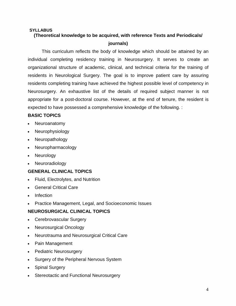
SYLLABUS (Theoretical knowledge to be acquired, with reference Texts and Periodicals/
journals) This curriculum reflects the body of knowledge which should be attained by an
individual completing residency training in Neurosurgery. It serves to create an
organizational structure of academic, clinical, and technical criteria for the training of
residents in Neurological Surgery. The goal is to improve patient care by assuring
residents completing training have achieved the highest possible level of competency in
Neurosurgery. An exhaustive list of the details of required subject manner is not
appropriate for a post-doctoral course. However, at the end of tenure, the resident is
expected to have possessed a comprehensive knowledge of the following. :
BASIC TOPICS
• Neuroanatomy
• Neurophysiology
• Neuropathology
• Neuropharmacology
• Neurology
• Neuroradiology
GENERAL CLINICAL TOPICS
• Fluid, Electrolytes, and Nutrition
• General Critical Care
• Infection
• Practice Management, Legal, and Socioeconomic Issues
NEUROSURGICAL CLINICAL TOPICS
• Cerebrovascular Surgery
• Neurosurgical Oncology
• Neurotrauma and Neurosurgical Critical Care
• Pain Management
• Pediatric Neurosurgery
• Surgery of the Peripheral Nervous System
• Spinal Surgery
• Stereotactic and Functional Neurosurgery
5
An exhaustive syllabus is provided as ANNEXURE 1
REFERENCE TEXT BOOKS SUBJECT S.NO TITLE AUTHOR
PUBLISHER Clinical Neurology/Neurosurgery
1. 2.
Localization in clinical neurology DeJong’s The Neurological Examination
Paul W. Brazis, Joseph C. Masdeu, José Biller William Wesley Campbell, Russell N. DeJong, Armin F. Haerer
Lippincott Williams & Wilkins, 2011 Lippincott Williams & Wilkins, 2005
Neurosurgery
1. 2. 3. 4. 5. 6.
Handbook of neurosurgery Youman's Neurological Surgery Neurosurgery Text Book of Neurosurgery. Textbook of neurological surgery: principles and practices Practical handbook of Neurosurgery .
Mark S. Greenberg, Nicolas Arredondo H. Richard Winn Robert H.Wilkins, Setti S. Rengachary Ravi Ramamurthi, PN Tandon H. Hunt Batjer, Christopher M. Loftus Marc Sindou
Thieme, 2005 Elsevier 2010 McGraw-Hill, 1996 BI Churchill Livingstone; 2011 Lippincott Williams & Wilkins, 2003 Elsevier 2010
6
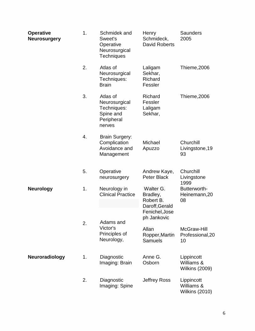
Operative Neurosurgery
1. 2. 3. 4. 5.
0BSchmidek and Sweet's Operative Neurosurgical Techniques 1BAtlas of Neurosurgical Techniques: Brain 2BAtlas of Neurosurgical Techniques: Spine and Peripheral nerves 3BBrain Surgery: Complication Avoidance and Management Operative neurosurgery
Henry Schmideck, David Roberts Laligam Sekhar, Richard Fessler Richard Fessler Laligam Sekhar, Michael Apuzzo Andrew Kaye, Peter Black
Saunders 2005 Thieme,2006 Thieme,2006 Churchill Livingstone,1993 Churchill Livingstone 1999
Neurology
1. 2.
Neurology in Clinical Practice
4BAdams and Victor's Principles of Neurology,
Walter G. Bradley, Robert B. Daroff,Gerald Fenichel,Joseph Jankovic Allan Ropper,Martin Samuels
Butterworth-Heinemann,2008 McGraw-Hill Professional,2010
Neuroradiology
1. 2.
5BDiagnostic Imaging: Brain 6BDiagnostic Imaging: Spine
Anne G. Osborn Jeffrey Ross
Lippincott Williams & Wilkins (2009) Lippincott Williams & Wilkins (2010)
7
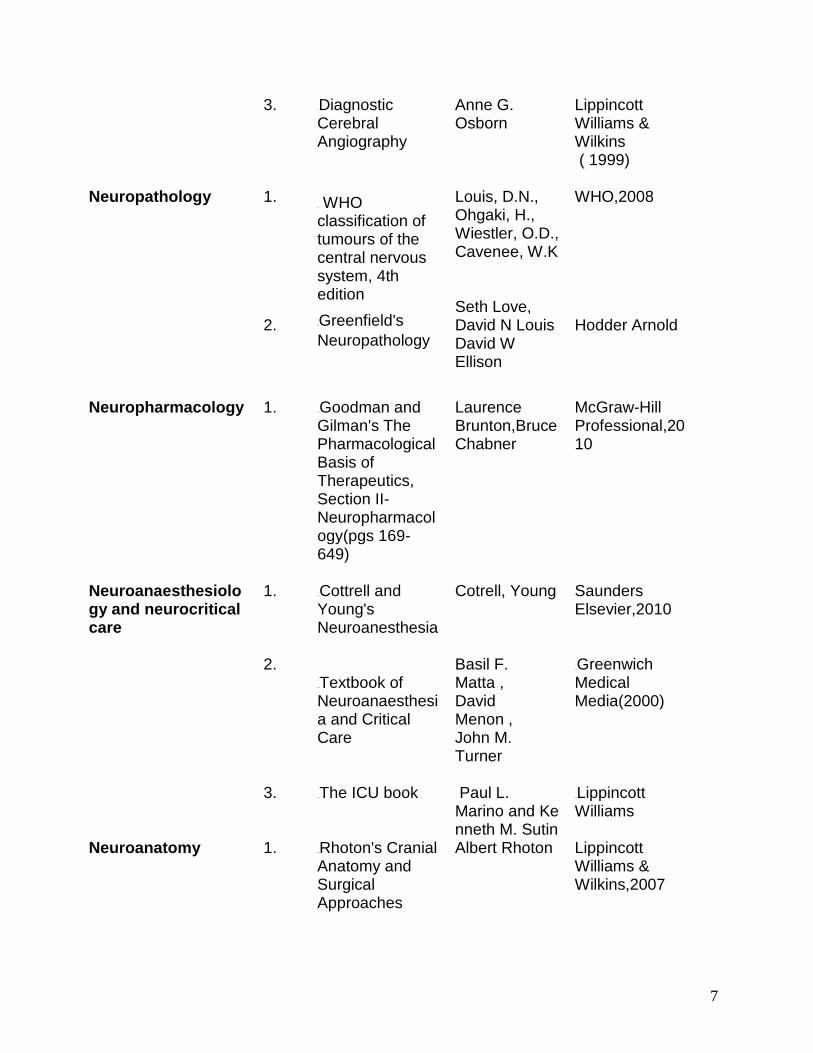
3. 7BDiagnostic Cerebral Angiography
Anne G. Osborn
Lippincott Williams & Wilkins ( 1999)
Neuropathology
1. 2.
8B WHO classification of tumours of the central nervous system, 4th edition 9BGreenfield's Neuropathology
Louis, D.N., Ohgaki, H., Wiestler, O.D., Cavenee, W.K Seth Love, David N Louis David W Ellison
WHO,2008 Hodder Arnold
Neuropharmacology
1. 10BGoodman and Gilman's The Pharmacological Basis of Therapeutics, Section II- Neuropharmacology(pgs 169-649)
Laurence Brunton,Bruce Chabner
McGraw-Hill Professional,2010
Neuroanaesthesiology and neurocritical care
1. 2. 3.
11BCottrell and Young's Neuroanesthesia 12BTextbook of Neuroanaesthesia and Critical Care 13BThe ICU book
Cotrell, Young Basil F. Matta , David Menon , John M. Turner Paul L. Marino and Kenneth M. Sutin
Saunders Elsevier,2010 14BGreenwich Medical Media(2000) 15BLippincott Williams
Neuroanatomy
1.
16BRhoton's Cranial Anatomy and Surgical Approaches
Albert Rhoton
Lippincott Williams & Wilkins,2007
8
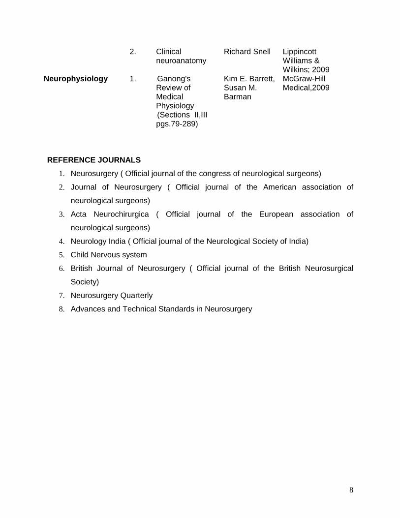
2. Clinical neuroanatomy
Richard Snell Lippincott Williams & Wilkins; 2009
Neurophysiology
1. 17BGanong's Review of Medical Physiology 18B(Sections II,III pgs.79-289)
Kim E. Barrett, Susan M. Barman
McGraw-Hill Medical,2009
REFERENCE JOURNALS
1. Neurosurgery ( Official journal of the congress of neurological surgeons)
2. Journal of Neurosurgery ( Official journal of the American association of
neurological surgeons)
3. Acta Neurochirurgica ( Official journal of the European association of
neurological surgeons)
4. Neurology India ( Official journal of the Neurological Society of India)
5. Child Nervous system
6. British Journal of Neurosurgery ( Official journal of the British Neurosurgical
Society)
7. Neurosurgery Quarterly
8. Advances and Technical Standards in Neurosurgery
9
CURRICULUM (Practical /clinical /laboratory experience to be imparted)
COMPONENTS OF THE CURRICULUM
The five year program consists of fundamental clinical evaluation, neurosurgical
training and research to allow for acquisition of graduated experience in all aspects of
neurological surgery and develop the following skills A. Clinical and theoretical skills: knowledge based on texts/journals/ departmental
academic activities. Clinical skills include the ability to take discerning history, perform
relevant clinical examination, decide the appropriate investigations and derive the
management plan.
B. Surgical and procedural skills: The candidate should be able to perform basic
neurosurgical procedures independently, and should have a firm grasp on many others.
To assure this, each resident is expected to assist and independently perform a
minimum number of procedures
C. Communication skills: The candidate is expected to develop into an effective
communicator to the patients, their family, colleagues and students.
D. Research aptitudes: The curriculum is intended to provide essential skills in
conducting medical research, and to get them presented in scientific forums and
published in peer-reviewed journals. An essential training in bio statistics will also be a
part of the curriculum.
10
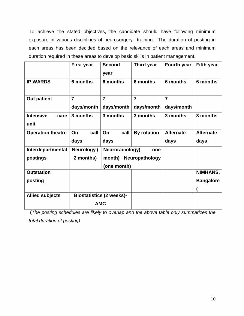
To achieve the stated objectives, the candidate should have following minimum
exposure in various disciplines of neurosurgery training. The duration of posting in
each areas has been decided based on the relevance of each areas and minimum
duration required in these areas to develop basic skills in patient management.
First year Second year
Third year Fourth year Fifth year
IP WARDS 6 months 6 months 6 months 6 months 6 months
Out patient 7 days/month
7 days/month
7 days/month
7 days/month
Intensive care unit
3 months 3 months 3 months 3 months 3 months
Operation theatre On call days
On call days
By rotation Alternate days
Alternate days
Interdepartmental postings
Neurology ( 2 months)
Neuroradiology( one month) Neuropathology (one month)
Outstation posting
NIMHANS, Bangalore (
Allied subjects Biostatistics (2 weeks)- AMC
(The posting schedules are likely to overlap and the above table only summarizes the
total duration of posting)
11
IP WARDS 1. Independent ward duties will be performed only after 1 month of joining the MCh
course. Until then, the new resident would be attached with his senior colleagues.
2. Each resident will be assigned a ward; each ward will be under the care of two
residents – one junior and one senior.
New admissions
3. The junior resident is responsible for getting investigations done and receiving
reports regarding the various biochemical, endocrine, hematological and radiological
tests that may be required for patient management. Report should be collected and
duty entered in the patient file in the investigation report section.
4. The day after a routine admission the entire case sheet should be written completely
with the work up plan. The case sheet will be cross checked and findings clarified by
the consultant on rounds. The senior resident in charge of the ward is responsible for
the same . He should discuss with the consultant regarding any the management plan
for each patient and carry out the same with the help of the junior resident.
5. Any seriously ill patient admitted through priority basis should be informed to the
consultant on duty and the head of the department.
6. Any urgent bedside procedures or maneuvers such as placement of cervical
traction, EVD, LP, shunt aspiration etc has to be performed in a timely manner .
7. Ward duty Residents should periodically monitor all patients and enter the progress
in the progress charts in file.
8. All attempts are to be made such that any patient admitted on a routine basis from
the OPD and waiting in the ward for many days, does not get cancelled from the next
days OT list due to investigation not being performed/not adequate/result not available
or abnormal lab parameters.
9. The resident in charge of the ward should daily ( during visiting hours ) communicate
with the relatives of the patient and keep them informed about the management plans
and progress of the patient.
Discharges
10. Patients should be informed at least one day in advance about the plan for
discharge. Discharge summary and all discharge related documents should be ready
12
before 11 am on the day of discharge. Residents should ensure that the patients for
discharge leave the ward before 12 noon to facilitate early admission of new patients
Ward Rounds
11. The junior and senior resident in charge of the ward should take independent ward
rounds well before the consultant rounds. They should discuss all patient related
problems with the consultant on rounds
OUT PATIENTS DEPARTMENT 1. Two residents will be assigned OPD duty on a given day. The junior resident among
the two will assist the consultant in charge of new cases and admissions. The senior
resident will assist the consultant in charge of review patients.
2. No resident should leave the OPD until all the new cases have been worked up,
dates given for all patients, and all necessary formalities completed.
3. Residents are expected to give dates for admission ( in consultation with the OPD
consultant) , fill up of investigation forms, medical certificates etc with a careful
explanation of the procedure to the patients and attendants. In case of any doubt or
lack of clarity in instruction, they should always contact the concerned faculty.
4. Patients in the OPD who require wound-care procedures, drainage of collections,
LPs, stitch removal, sinus exploration, dressings etc. will be referred to the
neurosurgery ward where the same will be attended to by the resident on ward duty.
INTENSIVE CARE UNIT (ICU) 1. Independent ICU duties will be performed only after 3 month of joining the MCh
course. Till then the new resident would be attached with his senior colleagues.
2. It is the primary response of the ICU Senior Resident to
• continuously monitor all patients in the ICU and enter progress in the charts in the
case sheets.Timely performance of investigation such as CT, ABG Electrolyte levels,
Chest radiographs etc as decided or as per urgency and need.
• Timely therapy and procedures for various situations arising in these patients should
be given.
• In case of any deterioration in the condition of any patient, the faculty on call as well
as the patient’s attendants should be informed.
13
• Report of ICU patients should be given in a polite and concise yet comprehensive
manner at least once every day (twice – in case of critically ill patients).
• Take over the ICU duty has to be performed by 7.30 a.m in the morning and both the
residents (resident on previous night duty as well as the resident on duty on the same
day) should be present for the morning rounds.
OPERATING ROOMS Residents will assume responsibilities in the theatre on a graded basis and under
constant supervision. First year residents will be generally allowed to observe or scrub
in as a second assistant. During their second year and final year residents will be
gradually allowed to t o perform surgeries once the seniors are confident of their ability.
To assure this, each resident is expected to assist and independently perform a
minimum number of procedures which is listed in ANNEXURE 2. The resident assigned
to assist/perform a surgery should write a detailed preoperative assessment and should
present the same in the preoperative session. Post surgery he should write a detailed
operative note with relevant diagrams. He should enter the same on the OT register. He
should also ensure that all specimens are neatly labelled and despatched to the
respective laboratories
DUTY SCHEDULE There shall be two residents on duty after routine working hours and on holidays.- One
resident on duty and the other resident “on call”. In case of any emergency the HOD
may insist on more number of doctors to stay back on duty.
DUTY RESIDENT The duty doctor will be responsible for all the ICU and ward patients after routine duty
hours.
In addition his duty consists of
1. Taking late night and early morning rounds of all the inpatients along with duty
consultant
2. Inform the Head of the department about all problems in the ICU and ward daily
evening
3. Inform the faculty on call / operating surgeon in case of any problem
14
4. Perform urgent therapeutic intervention e.g twist drill, ventric tap, tracheostomy or
such emergency procedure, which should be done promptly without any delay
First on Call duty The duty consists of:
• To respond all the casualty pages immediately.
• Evaluating and informing the consultant on duty about the patients seen in the
casualty.
• For patients who are referred to other Hospitals, to make sure that this takes place in
a timely manner.
• To inform the duty resident anytime a patient is admitted to the Ward /ICU
• To inform duty neuroanesthetist, and OT Nursing Staff each time any patient is
admitted for emergency Surgery.
• Assist the duty consultant for all emergency surgery procedures
• To arrange for all urgent blood investigations, CT, Cardiology or other consultation
etc or whatever is required before admission and operation.
• To make sure that the patient promptly reaches the OT, ICU or ward from the
Casualty as the case may be.
• In case of any ward hazard the resident should inform the duty consultant and Head
of the department ..
• To attend to all routine and urgent consultations from other Departments.
Call schedules
• Call Schedules are the responsibility of the Senior Resident. The call schedule ,
however needs to be approved by the Head of the Department and a copy of the same
needs to be distributed to the all the concerned departments
Duty Hours
• Duty hours are defined as all clinical and academic activities related to the residency
program, ie, patient care (both inpatient and outpatient), administrative duties related to
patient care, the provision for transfer of patient care, time spent in-house during call
activities, and scheduled academic activities such as conferences. Duty hours do not
include reading and preparation time spent away from the duty site.
15
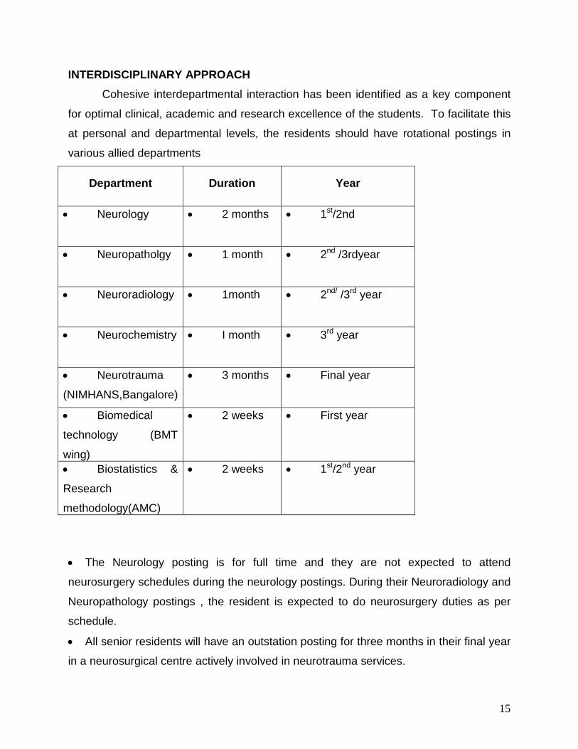
INTERDISCIPLINARY APPROACH Cohesive interdepartmental interaction has been identified as a key component
for optimal clinical, academic and research excellence of the students. To facilitate this
at personal and departmental levels, the residents should have rotational postings in
various allied departments
Department Duration Year
• Neurology • 2 months • 1st/2nd
• Neuropatholgy • 1 month • 2nd /3rdyear
• Neuroradiology • 1month • 2nd/ /3rd year
• Neurochemistry • I month • 3rd year
• Neurotrauma
(NIMHANS,Bangalore)
• 3 months • Final year
• Biomedical
technology (BMT
wing)
• 2 weeks • First year
• Biostatistics &
Research
methodology(AMC)
• 2 weeks • 1st/2nd year
• The Neurology posting is for full time and they are not expected to attend
neurosurgery schedules during the neurology postings. During their Neuroradiology and
Neuropathology postings , the resident is expected to do neurosurgery duties as per
schedule.
• All senior residents will have an outstation posting for three months in their final year
in a neurosurgical centre actively involved in neurotrauma services.
16
COMPETENCE EXPECTED AT END OF TRAINING Junior Resident (First 30 months)
• Develop a knowledge base in basic and clinical neurosurgery,
• Develop a thorough understanding of basic neurological examinations
• Become knowledgeable in the physiology of pre and post operative care of
neurosurgical patients.
• On-call as required by the rotations in compliance with regulations.
• Become comfortable with minor neurosurgical procedures, specifically lumbar
puncture, external ventricular drain placement, and burr hole placement,placement of
head-frames for stereotactic radiosurgery procedures.
• Formulate a research project.
• Knowledge of the biomechanics of the spine, cranial base anatomy, management of
all tumors encountered in neurosurgical practice.
• Successfully complete rotations in neurology, neuropathology, neuroradiology.
• Show evidence of learning and undertake progressively responsible patient
management
Senior Resident (Second 30 months)
• Become comfortable with basic cranial and spine surgery in both adult and pediatric
patients, assuming a more active role in the OR
• Further develop knowledge base in clinical neurosurgery.
• Augment skills and become proficient in the management of complex neurosurgical
conditions
• Become comfortable with most neurosurgical cases and perform surgery with
moderate supervision
• Be involved in basic science neurolab and complete an anatomical project in skull
base lab and a spine project in the biomechanics lab (preferable)
• Develop knowledge base concerning more complex neurosurgical procedures,
focusing attention on subspecialties.
17
• Learn fundamentals of head trauma, spinal cord injury and critical care
management.
• Become proficient in basic post-operative neurosurgical management.
• Formulate evidence-based treatment plans
• Successfully complete publication of research project and all publication
requirements.
18
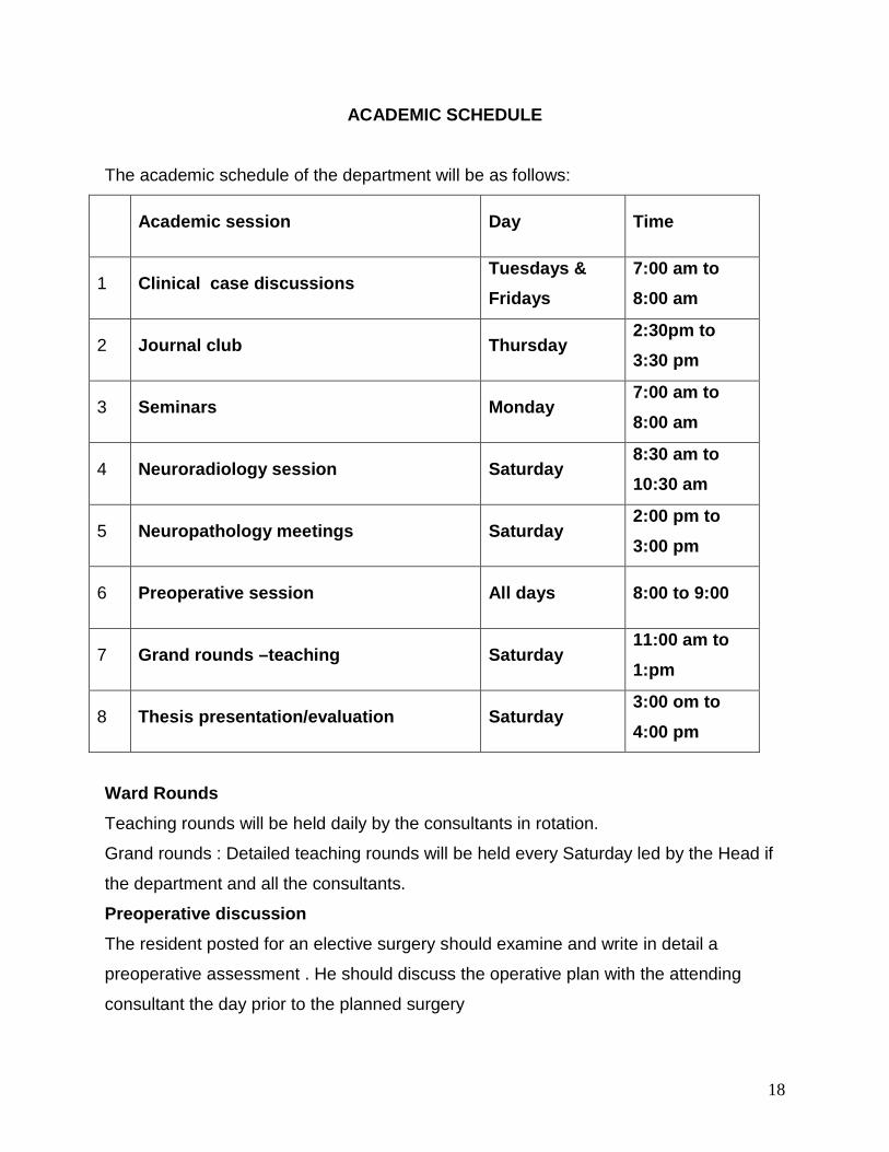
ACADEMIC SCHEDULE
The academic schedule of the department will be as follows:
Academic session Day Time
1 Clinical case discussions Tuesdays & Fridays
7:00 am to 8:00 am
2 Journal club Thursday 2:30pm to 3:30 pm
3 Seminars Monday 7:00 am to 8:00 am
4 Neuroradiology session Saturday 8:30 am to 10:30 am
5 Neuropathology meetings Saturday 2:00 pm to 3:00 pm
6 Preoperative session All days 8:00 to 9:00
7 Grand rounds –teaching Saturday 11:00 am to 1:pm
8 Thesis presentation/evaluation Saturday 3:00 om to 4:00 pm
Ward Rounds Teaching rounds will be held daily by the consultants in rotation.
Grand rounds : Detailed teaching rounds will be held every Saturday led by the Head if
the department and all the consultants.
Preoperative discussion The resident posted for an elective surgery should examine and write in detail a
preoperative assessment . He should discuss the operative plan with the attending
consultant the day prior to the planned surgery
19
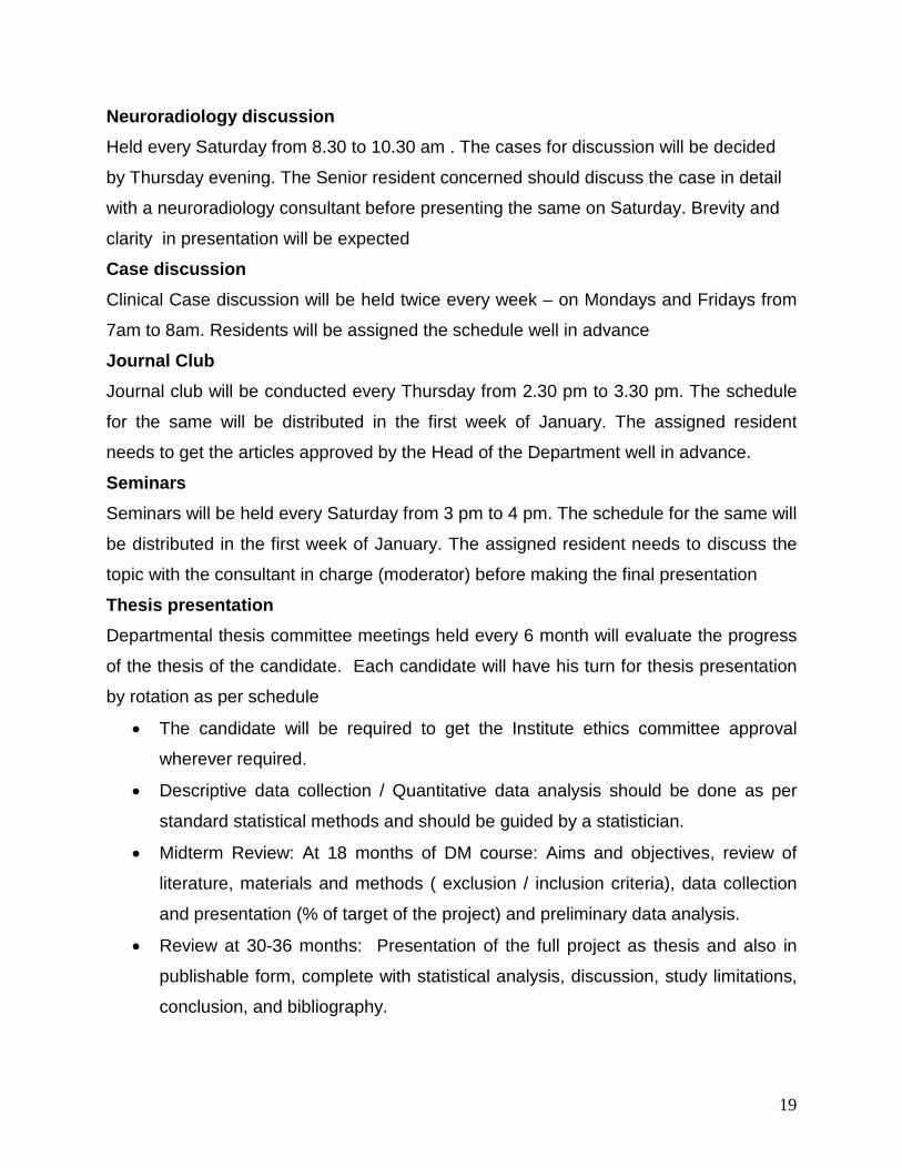
Neuroradiology discussion Held every Saturday from 8.30 to 10.30 am . The cases for discussion will be decided
by Thursday evening. The Senior resident concerned should discuss the case in detail
with a neuroradiology consultant before presenting the same on Saturday. Brevity and
clarity in presentation will be expected
Case discussion Clinical Case discussion will be held twice every week – on Mondays and Fridays from
7am to 8am. Residents will be assigned the schedule well in advance
Journal Club Journal club will be conducted every Thursday from 2.30 pm to 3.30 pm. The schedule
for the same will be distributed in the first week of January. The assigned resident
needs to get the articles approved by the Head of the Department well in advance.
Seminars Seminars will be held every Saturday from 3 pm to 4 pm. The schedule for the same will
be distributed in the first week of January. The assigned resident needs to discuss the
topic with the consultant in charge (moderator) before making the final presentation
Thesis presentation Departmental thesis committee meetings held every 6 month will evaluate the progress
of the thesis of the candidate. Each candidate will have his turn for thesis presentation
by rotation as per schedule
• The candidate will be required to get the Institute ethics committee approval
wherever required.
• Descriptive data collection / Quantitative data analysis should be done as per
standard statistical methods and should be guided by a statistician.
• Midterm Review: At 18 months of DM course: Aims and objectives, review of
literature, materials and methods ( exclusion / inclusion criteria), data collection
and presentation (% of target of the project) and preliminary data analysis.
• Review at 30-36 months: Presentation of the full project as thesis and also in
publishable form, complete with statistical analysis, discussion, study limitations,
conclusion, and bibliography.

20
• Between 40- 48 months, the project should be sent for publication to peer
reviewed journals.
• Presentation of the project work as scientific presentation at national level and at
state level- mandatory.
21
RESEARCH AND PUBLICATIONS
Thesis: The candidate should be involved in two research project, one of which which should
preferably be prospective.
• Each candidate will be allotted a mentor/ guide by the Head of department in
consultation with the faculty members in the initial 3months.
• The areas of project work should be decided in discussion with these mentors,
and the research project should be presented in the departmental research meeting at
the end of 3 months of joining the training period
• The candidate should get his thesis approved by the technical advisory
committee and the institute ethics committee before commencing his work
• Departmental thesis committee meetings held every 3 month will evaluate the
progress of the thesis of the candidate. Each candidate will have his turn for thesis
presentation by rotation
• The completed research work should be presented at completion of six months
before completion of residency.
• The research projects should have been published or publishable in peer
reviewed journals at this point of training period.
• Additional credits will be awarded to residents who involve in peer reviewed
projects funded by institute/external funding agencies
Paper publications and presentations
• The residents should have at least two clinical paper submitted in a peer-
reviewed journal indexed in “Index Medicus” prior to appearing the final examination.
• Credits will be given for these papers as per the guidelines mentioned. Added
credits will be given for publications in addition to the mandatory ones.
• At least one abstract presentation should be made at national level scientific
meeting.
Outstanding research Residents who engage in and excel in research activities in addition to the above
stipulated ones shall earn credits for outstanding performance
22
EVALUATION OF THE RESIDENT This includes three essential components:
• the documentation of the academic activities in a structured format,
• a credit-based evaluation of the academic performance,
• internal examination conducted at periodic intervals periodic review and appraisal of
the resident based on the evaluation. In
A: Comprehensive documentation by E-portfolio, logbook etc. The residents are expected to document their academic activities in a personal log book
(clinical dossier,academic diary) under the corresponding subheadings. This should be
countersigned by the Head of the department or the supervising consultant at periodic
intervals. This document will be the basis of credit-based evaluation,. This Dossier will
also serve as the platform to prepare the ‘E-portfolio’ for the residents at the end of their
tenure in the institute. The E-portfolio would reflect academic, clinical and research
experience of the concerned resident in the department.
The clinical dossier/academic diary shall have the following sections
• Operative log book
• Clinical case discussions
• Seminars presented
• Neuroradiology presentations
• Journal club presentations
• Papers published
• Papers presented
23
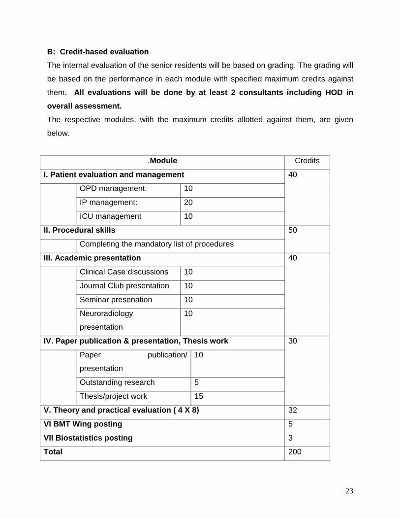
B: Credit-based evaluation The internal evaluation of the senior residents will be based on grading. The grading will
be based on the performance in each module with specified maximum credits against
them. All evaluations will be done by at least 2 consultants including HOD in overall assessment. The respective modules, with the maximum credits allotted against them, are given
below.
.Module Credits
I. Patient evaluation and management 40
OPD management: 10
IP management: 20
ICU management 10
II. Procedural skills 50
Completing the mandatory list of procedures
III. Academic presentation 40
Clinical Case discussions 10
Journal Club presentation 10
Seminar presenation 10
Neuroradiology
presentation
10
IV. Paper publication & presentation, Thesis work 30
Paper publication/
presentation
10
Outstanding research 5
Thesis/project work 15
V. Theory and practical evaluation ( 4 X 8) 32
VI BMT Wing posting 5
VII Biostatistics posting 3
Total 200
24
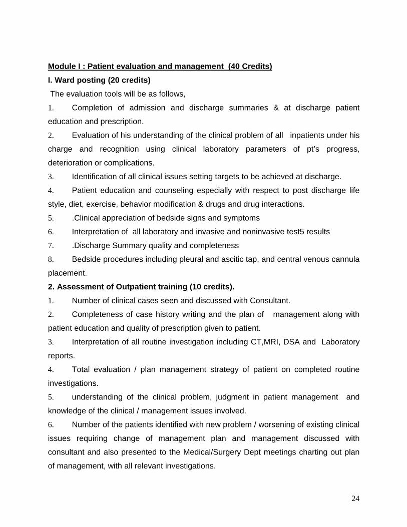
Module I : Patient evaluation and management (40 Credits) I. Ward posting (20 credits) The evaluation tools will be as follows,
1. Completion of admission and discharge summaries & at discharge patient
education and prescription.
2. Evaluation of his understanding of the clinical problem of all inpatients under his
charge and recognition using clinical laboratory parameters of pt’s progress,
deterioration or complications.
3. Identification of all clinical issues setting targets to be achieved at discharge.
4. Patient education and counseling especially with respect to post discharge life
style, diet, exercise, behavior modification & drugs and drug interactions.
5. .Clinical appreciation of bedside signs and symptoms
6. Interpretation of all laboratory and invasive and noninvasive test5 results
7. .Discharge Summary quality and completeness
8. Bedside procedures including pleural and ascitic tap, and central venous cannula
placement.
2. Assessment of Outpatient training (10 credits). 1. Number of clinical cases seen and discussed with Consultant.
2. Completeness of case history writing and the plan of management along with
patient education and quality of prescription given to patient.
3. Interpretation of all routine investigation including CT,MRI, DSA and Laboratory
reports.
4. Total evaluation / plan management strategy of patient on completed routine
investigations.
5. understanding of the clinical problem, judgment in patient management and
knowledge of the clinical / management issues involved.
6. Number of the patients identified with new problem / worsening of existing clinical
issues requiring change of management plan and management discussed with
consultant and also presented to the Medical/Surgery Dept meetings charting out plan
of management, with all relevant investigations.
25
7. Identification of critically ill patients and channeling their acute management.
8. Inter-departmental consultations
3. ICU and Emergency (10 Credits) A) This includes evaluation of patient management in the ICCU (newly admitted.
transferred from OT, wards, transferred after intervention procedure, etc) and charting
out plan of management and carrying out the same.
ICU training will include all emergency procedures like external ventricular drainage,
tracheostomy, traction,central venous cannulation, endo tracheal intubation, ventilator
management and blood gases interpretation, CPR, CPR protocol.
Module II: Procedural skills (50 credits) 1. Completion of the mandatory numbers of each procedures
2. Documentation : both preoperative work up and post operative follow up
3. Maintaining an operative logbook
Module III : Academic Presentation (40 credits) 1. Journal Review [10 credits] ( Minimum 10 in 5 yrs; 1 hr each) Purpose of journal presentation it to instill qualities of enquiry and analysis of scientific
medical articles and to evaluate its relevance and impact in understanding pathobiology
of disease or in clinical management. The resident can select recent articles of clinical
relevance, or consult the faculty to help select scientific articles with original research
content for presentation. The presentation should reflect the resident’s understanding of
the problem under discussion and the outcome and analysis of the results with regard to
various aspects of disease state and the clinical relevance.3-4 articles with brief
exposition of the highlights of the study and its clinical relevance and the take home
message. The senior resident should submit a short report of the articles presented in
print with a copy for the dept. and one for the individual, .highlighting the aim,
methodology, patient recruitment criteria, results, discussion and implications for clinical
practice. Each resident will have to present a minimum of ten journal clubs during his
entire residency program. The oral presentation and the write up will be equally
weighted. Each resident will be required to present around 12 journal clubs during his
residency. The best ten presentation will be used for deciding the credits
26
2. Seminar (3/year) 45 min [10 credits] It is intended to encourage extensive literature review on the topic and present the
highlights of the topic under review in a succinct manner with clear take home
messages, but at the same time the extensive literature search elevates the presenter
as an authority on the topic. The topic should be prepared as a review article with
complete bibliography in a publishable format, along with the topic presentation. The
presentation and the write up are equally weighted. Each resident will be required to
present around 12 seminars during his residency. The best ten presentation will be
used for deciding the credits
3. Neuroradiology meetings (2nd year &3rd year: 20-25 presentations, 15 min each) {10 credits} 1. Brief clinical summary
2. Discussion of imaging findings
3. Interpretation of images with differential diagnosis
4. Short synopsis when needed.
4. Bedside Clinical Presentation (1st year 3/year, 2nd year:5/year, 3rd year: 6-8/year) {10 credits} The following parameters will be assesed 1. History taking, presentation and analysis of history.
2. Physical findings, presentation and discussion with differential diagnosis.
3. Investigation-CT, MRI ,DSA and laboratory investigations.
4. Final Diagnosis: Physiological abnormalities/anatomical defects / etiology/
functional class / associated conditions/ complications
5. Further evaluation / Laboratory / Invasive investigations and plan of patient
management including surgical approach and complication avoidance.
Module IV: Paper publication and presentation (10 credits) in national /
international conference , outstanding research (5 credits) & Evaluation of Thesis
(15 credits). (Total 30 credits)
27
Publication of papers in journals and presentation of papers in conferences will also
earn credits based on the assessment. The total credits will be limited to 10 for
publications and for paper presentation
Thesis work (15 credits) Credits will be allotted based on the following criteria
1. Timely presentation of Mid-term evaluation and progress of the research project.
2. Prospective / Retrospective Study
3. Ethical Committee clearance / Institute funding obtained
4. Contribution of candidate in the study
5. Overall impact of the project in adding to our knowledgebase, and patient
management..
6. Publication of the research work in an indexed journal and presentation of the
project work in scientific meeting
7. Review at 48 months: Presentation of the full project as thesis and also in
publishable form, complete with statistical analysis, discussion, study limitations,
conclusion, and bibliography.
8. Overall impact of the project in adding to our knowledgebase, and patient
management. Between 48-52 months, the project should have been sent for publication
to preferably peer reviewed journals (before appearing for Part 2 examination).
9.
Research (5 credits) Residents who engage in and excel in research activities in addition to the above
stipulated ones shall earn credits for outstanding performance
Module V. Internal Examination : Theory (40; 4 X 8 credits) There will be 4 internal theory examinations, each having one theory paper of 100
marks during the 5-year course. These examination will have theory papers only, and
the answer papers will be evaluated by the faculty members of the department. The
results will be conveyed to the residents as a part of the regular appraisal.
No. Schedule Topics
1 12 months NeuroAnatomy, Neuroc Physiology, , Neurochemistry, Clinical
28
neurology.
2 24 months Genetics, NeuroPathology, Pharmacology, Basic principles in
neurosurgery, Fluid and electrolyte balance, critical care
3 42 months Operative neurosurgery, neuroradiology, critical care neurosurgery
4 54 months Recent advances ,complication avoidance, technical notes
Split up of Allocation of marks: Grand total: 1000 SPLIT UP 1.Internal assessment 200 (50% mandatory for appearing for Part II examination) 2. Part I: Theory Examination (paper 1& 2) 200
( At the end of 2 years) Paper 1: 100 Paper 2: 100
( a minimum of 50% in each paper is required for a pass)
3. Part II Theory Examination ( Paper 1 & 2): 200 ( At the end of 5years) Paper 1: 100 Paper 2: 100 ( a minimum of 50% in each paper is required for a pass)
4.Part II: Practical Examination (at the end of 5 years) 400
29
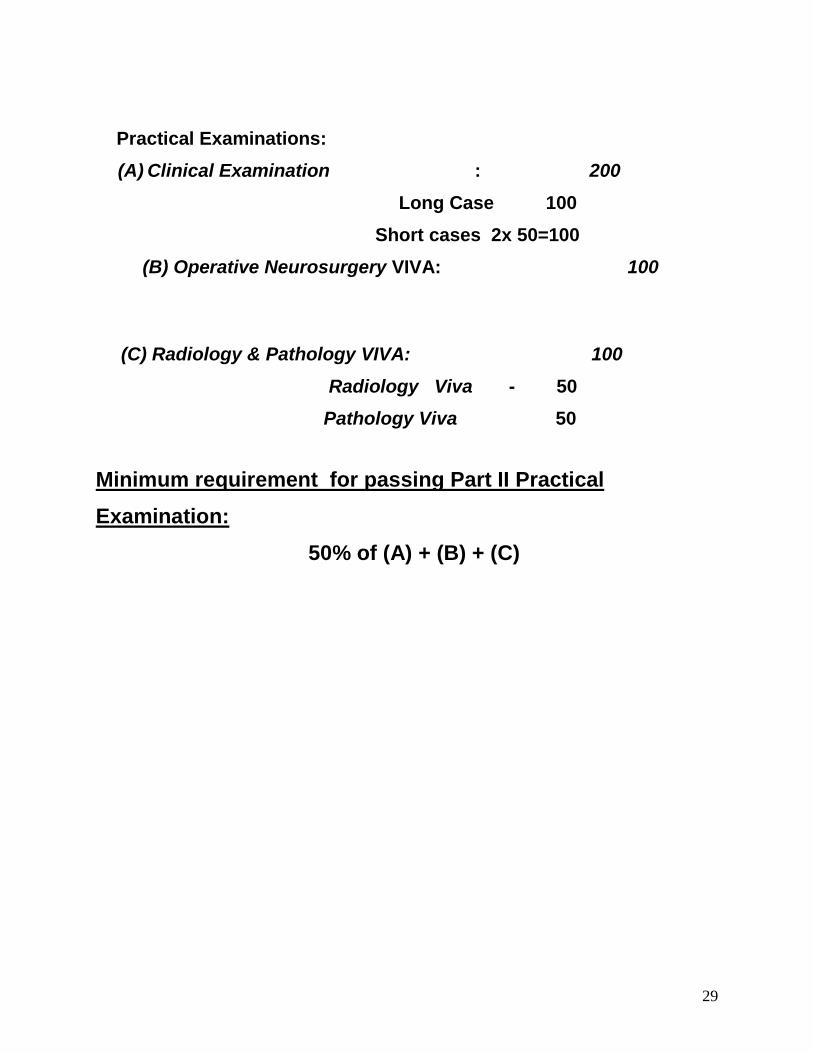
Practical Examinations: (A) Clinical Examination : 200 Long Case 100 Short cases 2x 50=100 (B) Operative Neurosurgery VIVA: 100
(C) Radiology & Pathology VIVA: 100
Radiology Viva - 50 Pathology Viva 50
Minimum requirement for passing Part II Practical Examination: 50% of (A) + (B) + (C)
30
Personal Development plan/ Periodic review and appraisal
Mid-term appraisal and appraisal report card will be introduced. The primary aim of this
periodic (6-monthly) appraisal is to help the resident to identify his academic deficits if any, and
to help the residents to improve on those aspects. The correction strategies to be adopted by
the resident will be discussed during the appraisal and it will be reviewed in the next appraisal
meeting. A copy of the periodic appraisal card signed by the programme-in-charge and the
resident will be handed over to the resident, and a copy of the same will be kept in the
departmental clinical dossier. The department will also try to identify and facilitate the specific
academic interest of the residents during these periodic appraisals. The residents will be
encouraged to communicate their special interest to the head of the department and every
possible step will be taken to facilitate/offer special training and research opportunity in those
areas.

31
RESIDENT’S FEEDBACK Residents’ feedback about the academic curriculum is an integral component of the
programme. The feedback form will be given to the resident during the periodic
appraisal. This will be kept as a document in the clinical dossier and all possible steps
will be taken to improve the academic programme based on the suggestions, if they are
found appropriate by the department. The confidentiality of the resident’s feedback will
be maintained by the head, and only the anonymous suggestions will be presented
before the department.
32
DEPARTMENT POST GRADUATE PROGRAM COMMITTEE
Academic Program Committee
Academic Program Committee will over see the implementation of the curriculum
including the academic activities including research projects of the Senior Residents
and the continuous evaluation process of the Senior Residents over the three years.
The committee will consist of
1. Chairman of Academic Program (Head of Department)
2. Program-in-Charge (Senior Faculty from the department)
3. Program Coordinator (Associate / Assistant Professor: One for each batch of
residents)

33
Annexure 1
SYLLABUS INTRODUCTION
This curriculum reflects the body of knowledge which should be attained by an individual completing residency training in Neurological Surgery. It serves to create an organizational structure of academic, clinical, and technical criteria for the training of residents in Neurological Surgery. The goal is to improve patient care by assuring residents completing training have achieved the highest possible level of competency in Neurological Surgery.
The curriculum is constructed in such a manner that the educational experience is divided into two levels - Junior and Senior. The resident should display competency in each level before progressing to the next. Those individuals who do not stay on track will be promptly identified in an objective manner, thereby enabling more timely remedial attention or dismissal. The programs is structured to allow residents to act independently at various tasks commensurate with their skills and the specific medical situation.
PART 1
NEUROANATOMY
UNIT OBJECTIVES
Demonstrate knowledge of anatomy that is pertinent to the diagnosis of diseases of the nervous system and the practice of neurological surgery.
1. Review the embryological development of the brain, cerebellum, brain stem, glial elements, spinal cord, conus medullaris, cauda equina, sympathetic and parasympathetic systems and the peripheral nervous system.
2. Discuss the embryologic development of the skull, craniovertebral junction, and spine.
3. Describe and differentiate the different types of neurons. 4. Discuss the microanatomy of the neuron including the:
a. cell body b. dendritic process c. axonal process
5. Diagram and describe the microanatomy of the synapse. 6. List the microglial elements and review their microanatomy:
a. astrocytes b. oligodendrocytes
34
c. microglia d. ependyma e. choroid epithelium
7. Diagram and describe in detail the carotid and vertebral arteries and their branches which provide blood supply to the face, scalp, skull, meninges, brain, brain stem, cerebellum, and rostral spinal cord.
8. Discuss in detail the arterial blood supply to the spinal cord. Include in the discussion the spinal and radicular arteries and the concept of watershed ischemia.
9. Identify and review the venous drainage of the central nervous system. 10. List and identify the bones of the skull. 11. Describe each of the sutures of the skull. 12. Identify each named foramen of the skull and list its contents. 13. Describe the anatomy of the meninges including the:
a. dura mater b. arachnoid mater c. pia mater
14. Describe the anatomy of the dura including the falx cerebri and tentorium. 15. Review the layers of the scalp and discuss its innervation. 16. Diagram the cerebral ventricles. 17. Discuss the major arachnoid cisterns. 18. Review the anatomy of the arachnoid villi. 19. Discuss the anatomic correlates pertinent to the production, flow, and
reabsorption of cerebrospinal fluid. 20. Identify and describe the gross anatomy of the spine including:
a. atlas b. axis c. subaxial cervical vertebrae d. thoracic vertebrae e. lumbar vertebrae f. sacrum g. coccyx h. intervertebral disc complex i. supporting ligaments of the spine
21. List the muscles related to the skull and spine. 22. Describe the gross anatomy of the neck. 23. Discuss the anatomical basis for the blood-brain barrier in detail.
Central Nervous System
24. Describe the gross anatomy of the brain, brain stem, cerebellum, cranial nerves, and spinal cord in detail.
25. Describe the anatomy of the cerebral cortex in detail including: a. cortical layers b. sensory areas c. motor areas
35
d. prefrontal cortex e. fiber tracts f. calcarine cortex
26. Describe the anatomy of the olfactory pathways, hippocampal formation and amygdala in detail including:
a. rhinencephalon b. olfactory pathways c. anterior commissure d. hippocampal formation (including cytoarchitecture) e. amygdala f. limbic system
27. Describe the anatomy of the corpus striatum in detail including: a. striatum b. globus pallidus c. claustrum d. subthalamic region e. striatal afferent and efferent connections f. pallidal afferent and efferent connections g. pallidofugal fiber systems
28. Describe the anatomy of the hypothalmus and pituitary in detail including: a. cytoarchitecture of the hypothalmus b. afferent and efferent connections of the hypothalmus c. supraoptic nuclei and tracts d. hypophysial portal system e. anatomy of the pituitary stalk f. anterior and posterior pituitary g. cellular organization of the anterior pituitary h. hormonally active cells of the hypothalmus and pituitary
29. Describe the anatomy of the diencephalon in detail including: a. midbrain-dienencephalon junction b. caudal diencephalon c. epithalamus d. thalamus (including nuclei) e. thalamic radiations f. internal capsule g. visual pathways
30. Describe the anatomy of the cerebellum in detail including: a. cerebellar cortex including organization b. deep cerebellar nuclei c. cerebellar connections d. cerebellar peduncles
31. Describe the anatomy of the mesencephalon in detail including: a. superior colliculus b. inferior colliculus c. pretectal region d. posterior commissure
36
e. mesencephalic nuclei f. oculomotor nerve g. tegmentum h. mesencephalic reticular formation i. substantia nigra j. crus cerebri k. ascending and descending tracts
32. Describe the anatomy of the pons in detail including: a. vestibulocochlear nerve b. facial nerve c. abducens nerve d. trigeminal nerve e. ascending and descending tracts
33. Describe the anatomy of the medulla in detail including: a. olivary nucleus b. medullary reticular formation c. cranial nerves of the medulla d. ascending and descending tracts
34. Review the location and connections of each cranial nerve nuclei. Trace the course of each cranial nerve from nucleus to end organ termination.
35. Describe the external topography and landmarks of the fourth ventricle. 36. Describe the anatomy of the spinal cord in detail including:
a. nuclei and cell groups b. cytoacrchitectural lamination (Rexed laminae) c. somatic and visceral efferent neurons d. posterior horn neurons e. descending tracts f. ascending tracts g. upper and lower motor neurons h. somatotopic organization
Autonomic Nervous System
37. Distinguish pre- and postganglionic neurons. 38. Describe the sympathetic nervous system. 39. Describe the parasympathitic nervous system. 40. Review the visceral afferent fibers. 41. Describe the structure of the autonomic ganglia. 42. Discuss the central autonomic pathways.
Peripheral Nervous System
43. Differentiate between segmental and peripheral innervation. 44. Diagram the anatomy of the spinal nerve root. 45. Diagram and dDiscuss the cervical, brachial, and lumbosacral plexi.

37
46. Outline the anatomy of the major peripheral nerves of the upper and lower extremity including:
a. axillary b. suprascapular c. median d. ulnar e. radial f. long thoracic g. musculocutaneous h. lateral femoral cutaneous i. femoral j. obturator k. sciatic l. saphenous m. peroneal n. tibial
47. Describe the microanatomy of the peripheral nerves in detail. 48. Explain the difference between myelinated and unmyelinated nerves. 49. Review the anatomy of the Schwann cell. 50. List the peripheral afferent receptors and describe the anatomy of each. 51. Segregate peripheral neurons by size and explain the rationale for such a
classification scheme.
Muscle
52. Explain the concept of the motor unit. 53. Describe the anatomy of the motor end plate. 54. Describe the microscopic anatomy of striated and smooth muscle. 55. Discuss the subcellular components of muscle. 56. Discuss the clinical presentation in anatomical terms of syndromes of the
brain and its coverings including: a. epidural hematoma b. acute subdural hematoma c. chronic subdural hematoma d. subgaleal hematoma e. injury to innervation of the scalp
57. Discuss the syndromes produced by mass lesions affecting the cranial nerves including:
a. suprasellar lesions b. lesion of jugular foramen c. lesion of internal auditory canal d. lesions or distortion at the incisura
58. Review the expected effects of stroke or mass lesion at different locations within the brain stem and cerebellum.
59. List the expected effects of destructive lesions in the basal ganglia and cerebellum.
38
60. Describe the expected effects of ischemic or destructive lesions of the white matter tracts of the cerebrum.
61. Discuss the expected effect of destructive lesions of specific regions of the cerebral cortex.
62. Review the clinical presentation of strokes in the distribution of the supratentorial cerebral blood vessels.
63. Discuss the relationship of the spinal nerves to the vertebral level of exit. 64. Diagram the structures comprising the boundaries of the spinal neural
foramina. 65. Discuss the clinical manifestation of injury for each of the major peripheral
nerves. 66. Describe the anatomy and presentation of common entrapment
syndromes of peripheral nerves including: a. thoracic outlet syndrome b. carpal tunnel syndrome c. ulnar nerve entrapment syndrome at wrist and elbow d. anterior interosseous syndrome e. posterior interosseous syndrome f. meralgia paresthetica g. peroneal nerve palsy h. tarsal tunnel syndrome
67. Describe the surgical exposure of common peripheral nerve entrapments including:
a. carpal tunnel b. ulnar nerve at elbow c. ulnar nerve at wrist d. lateral femoral cutaneous nerve e. peroneal nerve
68. Discuss the clinical presentation and neurological deficits associated with common lesions of and injuries to the spinal cord and nerve roots.
69. Identify at the time of surgery: a. occipital artery b. superficial temporal artery c. frontalis muscle d. pterion e. inion f. asterion g. coronal suture h. sagittal suture i. middle meningeal artery j. sagittal sinus k. transverse sinus l. foramen rotundum m. foramen ovale n. foramen spinosum o. superior orbital fissure
39
p. jugular foramen q. internal auditory canal r. superior sagittal sinus s. sigmoid sinus t. incisura u. each cranial nerve v. each named cerebral artery and vein w. components of the brain stem x. named structures on the floor of the fourth ventricle y. Foramina of Magendie and Luschka z. cerebral peduncles aa. components of the cerebellum bb. cerebellar tonsils cc. brachium cerebelli dd. vermis ee. major supratentorial gyri ff. supratentorial lobes gg. sylvian fissure hh. central sulcus
70. Identify at the time of surgery structures visible in the lateral ventricles including:
a. Foramen of Monro b. fornix c. caudate d. thalamus e. choroidal fissure f. named veins g. glomus of the choroid plexus h. hippocampus
71. Identify the parts of the vertebral column, spinal cord, and nerve roots at the time of surgery including:
a. spinous process b. lamina c. superior facet d. inferior facet e. pedicle f. pars interarticularis g. uncovertebral joint h. neural foramen and nerve root i. nerve root ganglion j. disc space k. vertebral artery l. dorsal column and lateral column of spinal cord m. intradural afferent and efferent rootlets
40
NEUROPHYSIOLOGY
UNIT OBJECTIVES
Demonstrate knowledge of physiology that is pertinent to the understanding of neurological disease.
1. Review the basic biology of the nerves including: a. synthesis and movement of proteins in the nerve b. membrane potential and membrane properties c. ion channels d. generation and conduction of an action potential
2. Discuss synaptic transmission including: a. types of synaptic transmission b. transmitter release c. nerve-muscle transmission d. chemical messengers e. direct gated receptors f. second messenger linked receptors
3. Describe the physiology of the sensory systems including: a. sensory receptor physiology b. anatomy of somatic sensory system c. coding of modality specific sensory information d. pain and analgesia e. cortical integration of sensory perception f. visual system
1. processing of information in the retina 2. processing of vision in the central visual pathways 3. columnar units of visual cortex 4. processing in the geniculate nucleus 5. visual perception of motion and form.
g. auditory system. Within this description review the processing of hearing in the cochlea and the central auditory pathways.
h. olfaction and taste 4. Discuss the physiology of the motor system including:
a. mechanisms of muscle contraction b. muscle receptors, spinal reflexes c. spinal reflexes concerned with position d. brain stem reflexes controlling motion e. vestibular nuclei control of movement and posture f. red nucleus control of movement g. cortical control of movement h. cerebellar control of movement
1. regional and cellular organization of the cerebellum 2. functional divisions of the cerebellum
41
3. the role of the cerebellum in planning movement i. basal ganglia
1. the anatomy of basal ganglia pathways 2. neural transmitters in the circuits within the basal ganglia
j. thalamus 5. Describe the attributes of the autonomic nervous system including both
the sympathetic and parasympathetic systems. 6. Review the physiological basis of arousal and emotion. Include within this
review the: a. noradrenergic systems b. limbic system. Include within this review the physiologic basis for
emotion and memory c. sleeping and sleep states d. reticular activating system
7. Describe the higher cortical functions including: a. anatomy of language b. function of association cortex
8. Describe the physiological basis for cerebrospinal fluid production and reabsorption.
9. Review the physiological control of the cerebral vasculature. 10. Discuss, in detail, the physiology of the hypothalamus and pituitary,
particularly as related to endocrinology
42
NEUROPATHOLOGY
UNIT OBJECTIVES
Demonstrate knowledge of neuropathology that is pertinent to the diagnosis of diseases of the nervous system and practice of neurological surgery.
General Neuropathology
1. Describe the techniques available for examination of surgical specimens from central nervous system, peripheral nervous system, skeletal muscle, pineal and pituitary.
2. Review the use of standard chromatic, histochemical and selected immunohistochemical stains employed in the evaluation of surgical specimens from the central nervous system, peripheral nervous system, skeletal muscle, pineal and pituitary.
3. List the techniques available for morphological examination of cerebrospinal fluid and the abnormalities observed in cerebrospinal fluid from patients with meningeal carcinomatosis, meningeal lymphomatosis, pyogenic meningitis and aseptic meningitis.
Central Nervous System
4. Describe the gross and histopathological features and, when applicable, the genetic basis of the following congenital and perinatal disorders:
a. encephaloceles and cranial meningoceles b. myelomeningoceles and meningoceles c. hydromyelia d. diastematomyelia and diplomyelia e. syringomyelia and syringobulbia f. Chiari I malformation g. Chiari II malformation h. Dandy-Walker malformation i. arachnoid cysts j. porencephaly k. aqueductal stenosis l. subependymal germinal matrix hemorrhages m. posthemorrhagic hydrocephalus n. periventricular leukomalacia (white matter infarcts)
5. Describe the gross and histopathological features and characteristics of the causative agents of the following infectious diseases:
a. cranial and spinal epidural abscesses b. cranial and spinal subdural abscesses c. pyogenic bacterial meningitis and ventriculitis d. brain abscesses
43
e. tuberculous meningitis and tuberculomas f. central nervous system sarcoidosis g. central nervous system cryptococcosis h. central nervous system mucormycosis i. central nervous system toxoplasmosis j. central nervous system cysticercosis k. Herpes simplex encephalitis l. central nervous system HIV infections m. central nervous system cytomegalovirus infection
6. Describe the gross and histopathological features of the following vascular lesions:
a. acute, subacute, and remote infarcts b. border zone and watershed infarcts c. manifestations of embolic infarcts including those secondary to
atheromatous embolization and embolization from extracoporeal pumps
d. vasculitis including temporal arteritis, primary central nervous system vasculitis, granulomatous angiitis, and Wegener's granulomatosis
e. moyamoya f. hypertensive intracerebral hemorrhages g. lobar intracerebral hemorrhages h. amyloid angiopathy i. malformations including arteriovenous malformations, cavernous
angiomas, venous angioma and capillary telangiectases j. Vein of Galen "aneurysms" k. saccular aneurysms l. infectious ("mycotic") aneurysms m. giant aneurysms n. traumatic and dissecting aneurysms o. venous and dural sinus occlusive disease p. vascular malformations of the spinal cord q. spinal cord infarcts
7. Describe the gross and histopathological features of the following traumatic lesions:
a. skull fractures b. entrance and exit gunshot wounds of the skull c. gunshot wounds of the brain including internal ricochet d. epidural hematomas e. acute subdural hematomas f. chronic subdural hematomas g. recent and remote cerebral contusions h. traumatic intraparenchymal hemorrhages i. diffuse axonal injury j. traumatic cranial nerve injuries k. spinal cord injuries
44
l. cerebral herniation syndromes m. fat embolization n. central nervous system trauma in infancy o. central nervous system radiation injuries p. manifestations of prior surgical intervention
8. Describe the gross and histopathological features and, when applicable, the metabolic basis for the following intoxications and deficiency states:
a. hypoxic-anoxic encephalopathy b. carbon monoxide intoxication c. ethanol intoxication d. alcoholic cerebellar degeneration e. central pontine myelinolysis f. CNS complications of diagnostic agents including contrast material g. CNS complications of antimicrobial therapy h. CNS complications of antineoplastic therapy i. CNS complications of "street drugs" j. Wernicke's encephalopathy and thiamine deficiency k. Subacute combined degeneration and B12 deficiency
9. Describe the gross and histopathological features of the following demyelinating diseases:
a. multiple sclerosis b. progressive multifocal leukoencephalopathy c. HIV vacuolar myelopathy d. postinfectious encephalomyelitis
10. Describe the gross and histopathological features and the metabolic basis for the following leukodystrophies:
a. adrenoleukodystrophy and adrenomyeloneuropathy b. Krabbe's disease c. metachromatic leukodystrophy
11. Describe the gross and histopathological features and, when applicable, the genetic basis for the following dementias and degenerations:
a. Alzheimer's disease including familial forms b. vascular dementia including Binswanger's disease and cerebral
autosomal dominant arteriopathy (CADASIL) c. Pick's disease d. other fronto-temporal dementias e. Creutzfeldt-Jacob disease and other prion diseases f. Parkinson's disease g. diffuse Lewy body disease h. Huntington's disease i. amyotrophic lateral sclerosis j. paraneoplastic degenerative diseases
12. Describe the gross and histopathological features and, when applicable, the biochemical and genetic basis for the following metabolic diseases:
a. Wilson's disease b. Tay Sachs disease and other GM-2 gangliosidoses
45
c. neuronal ceroid-lipofuscinoses d. hepatic encephalopathy e. Reye's syndrome
13. stopathological features and, when applicable, the grading criteria for the following central nervous system neoplasms:
a. diffuse fibrillary astrocytomas b. gemistocytic astrocytomas c. anaplastic astrocytomas d. glioblastoma multiforme including giant cell glioblastoma and
gliosarcomas e. pilocytic astrocytomas including cerebellar, diencephalic, dorsal
exophytic pontine, and cerebral pilocytic astrocytomas f. subependymal giant cell astrocytomas g. pleomorphic xanthoastrocytoma h. oligodendrogliomas including anaplastic oligodendrogliomas and
mixed oligoastrocytomas i. ependymomas including myxopapillary ependymomas j. subependymomas k. choroid plexus tumors l. colloid cysts m. gliomatosis cerebri n. gangliocytomas and gangliogliomas o. dysembryoplastic neuroepithelial neoplasms p. central neurocytomas q. medulloblastomas r. atypical teratoid/rhabdoid tumors s. primitive neuroectodermal tumors and cerebral neuroblastomas t. olfactory neuroblastoma u. spinal paragangliomas v. meningiomas including meningothelial (syncytial) fibrous,
transitional, psammomatous, angiomatous, and papillary meningiomas
w. anaplastic and malignant meningiomas x. meningeal hemangiopericytomas y. other meningeal mesenchymal tumors z. meningeal melanomatosis and melanomas aa. hemangioblastomas bb. lipomas cc. primary central nervous system lymphomas dd. metastatic carcinomas including leptomeningeal carcinomatosis ee. teratomas ff. dermoids and epidermoids gg. schwannomas including acoustic neurinomas or vestibular
schwannomas, schwannomas of other cranial nerves, and spinal root schwannomas
46
14. Describe the gross and histopathological features and the genetic basis for the following tumor syndromes:
a. Neurofibromatosis type 1 b. Neurofibromatosis type 2 c. von Hippel-Lindau syndrome d. Tuberous sclerosis e. Cowden syndrome f. Turcot syndrome
Peripheral Nervous System
15. Describe the gross and histopathological features and, when applicable, the genetic and biochemical basis for the following disorders of peripheral nerves:
a. compressive and traumatic neuropathies b. leprosy c. diabetic and uremic neuropathy d. Charcot-Marie-Tooth disease e. Guillain-Barre syndrome f. sympathetic dystrophy
16. Describe the gross and histopathological features of the following neoplastic and tumorous disorders of peripheral nerves:
a. peripheral schwannoma b. neurofibromas c. malignant peripheral nerve sheath tumors d. spinal root and peripheral nerve root cysts
Pituitary and Pineal
17. Describe the gross and histopathological features of the following pituitary conditions:
a. pituitary adenomas including null cell adenomas, growth hormone secreting adenomas, prolactin secreting adenomas, ACTH secreting adenomas, and oncocytomas
b. craniopharyngiomas including adamantinomatous and squamopapillary craniopharyngiomas
c. Rathke pouch (cleft) cysts d. pituitary involvement by metastatic neoplasms e. lymphocytic hypophysitis f. pituitary infarcts including pituitary "apoplexy" g. pituitary lesions resulting from closed head trauma h. empty sella syndromes
18. Describe the gross and histopathological features of the following lesions of the pineal:
a. germinomas b. teratomas and embryonal carcinomas
47
c. pineoblastomas and pineocytomas d. metastatic carcinoma
Skull and Spine (including intervertebral discs)
19. Describe the gross and histopathological features of the following disorders of the skull:
a. dermoids and epidermoids b. hemangiomas c. osteomas d. chordomas e. solitary and multifocal eosinophilic granuloma f. Paget's disease including secondary osteosarcoma g. metastatic carcinomas h. plasmacytoma including myeloma
20. Describe the gross and histopathological features of the following disorders of the spine and intervertebral discs:
a. herniated intervertebral discs b. pyrophosphate disease including involvement of ligamentum flavum c. tumoral calcinosis d. hemangiomas e. chordomas f. eosinophilic granulomas g. metastatic carcinomas including epidural metastases h. plasmacytoma including myeloma i. lymphomas j. primary bone tumors k. spinal osteomyelitis including tuberculous and fungal spinal
osteomyelitis
Eye and Orbit
21. Describe the gross and histopathological features of the following ocular lesions:
a. retinoblastomas b. ocular melanomas
22. Describe the gross and histopathological features of the following orbital lesions:
a. optic nerve gliomas b. optic nerve meningiomas c. orbital lymphomas and pseudotumors d. orbital metastases
Miscellaneous
48
23. List the gross and histopathological features found in temporal lobectomy and cerebral hemispherectomy specimens removed during epilepsy surgery.
24. Review the gross, histopathological, and cytopathological features that can be observed in shunt revision specimens.
25. Describe the gross, histopathological, and cytopathological features that can be observed with indwelling pump and intrathecal catheter specimens.
26. Cite the techniques for examination of foreign objects removed from the nervous system and the need for documentation of chain of custody when of potential legal significance.
27. Describe the histopathological features of myotonic dystrophy and central core myopathy and list the potential implications of these diseases with regard to adverse anesthetic reactions including development of malignant hyperthermia.
49
NEUROPHARMACOLOGY
UNIT OBJECTIVES Demonstrate knowledge of pharmacology that is pertinent to the treatment of neurological disorders and diseases which affect the nervous system.
KNOWLEDGE OBJECTIVES:
1. Review basic cellular neurotransmission. In the course of this review discuss:
a. the synapse b. membrane potentials c. ion pumps d. ion channels e. transmitter secretion f. transmitter identification
2. Define and discuss receptors and receptor pharmacodynamics including: a. receptor classification b. receptor identification c. dose response curves d. agonists and antagonists e. receptor modulation
3. Discuss the neurotransmitter acetylcholine in detail. Include within the context of the discussion:
a. cholinergic receptor classification b. functional aspects of cholinergic receptors c. synthesis, storage, and release of acetylcholine
4. Discuss the catecholamine neurotransmitters (norepinephrine and dopamine) in detail. Include within the context of the discussion:
a. biosynthesis of catecholamines b. storage and release of catecholamines c. anatomy of catecholamine receptors d. adrenergic receptors e. dopaminergic receptors
5. Discuss the neurotransmitter serotonin in detail. Include within the context of the discussion:
a. anatomy of serotonin receptors b. biosynthesis, storage and release of serotonin c. sub-types of serotonin receptors
6. Discuss the neurotransmitter glutamate in detail. Include within the context of the discussion
a. biosynthesis, storage and release of glutamate b. ionotropic glutamate receptors
1. NMDA receptors and subunits 2. non-NMDA receptors and subunits

50
c. metabotropic glutamate receptors 1. Group I metabotropic receptors and subunits 2. Group II metabotropic receptors and subunits 3. Group III metabotropic receptors and subunits
d. role in neurological disorders 7. Discuss the neurotransmitters GABA and glycine in detail.
a. synthesis, uptake, and release b. physiology and pharmacology c. clinically relevent agonists and antagonists of GABA and glycine
receptors 8. Discuss the peptide neurotransmitters. 9. Describe the pharmacology of each of the drugs used to treat neurological
disorders
FLUIDS, ELECTROLYTES, AND NUTRITION
UNIT OBJECTIVES Demonstrate an understanding of normal and pathologic fluid and electrolyte homeostasis. Demonstrate an ability to maintain normal electrolyte balance. Demonstrate an understanding of the basics of nutritional management in neurosurgical patients.
KNOWLEDGE OBJECTIVES:
1. Discuss the normal distribution of intracellular and extracellular fluid and electrolytes including:
a. sodium and water distribution and metabolism b. clinical assessment of water and sodium balance and the concept
of osmolality c. normal maintenance requirements d. management of pathologic conditions such as diabetes insipidus
and the syndrome of inappropriate antidiuretic hormone secretion e. cerebral salt wasting
2. Review the potential implications of diuresis and fluid restriction on water and electrolyte balance.
3. Briefly review the potential clinical implications of calcium, phosphorous, and magnesium excesses and deficiencies and the treatment of same.
4. Review the criteria for nutritional assessment including: a. history of significant weight loss b. hypoalbuminemia c. impaired immune response including diminished total lymphocyte
count and anergy d. physical signs of malnutrition
5. Briefly describe the metabolic responses to starvation and stress.

51
6. Describe and contrast the indications, contraindications, complications, and benefits of enteral and parenteral nutrition.
7. Analyze the implications of specific nutritional deficiencies as they relate to neurological and neurosurgical diseases.
8. Briefly review swallowing disorders. 9. Describe the common changes of metabolism and nutritional requirements
of trauma patients and their evaluation. 10. Demonstrate an ability to manage the fluid and electrolyte requirements of
neonatal, pediatric, and adult neurosurgical patients. 11. Demonstrate the ability to place central venous catheters. 12. Demonstrate the ability to place enteral feeding tubes. 13. Demonstrate an ability to prescribe appropriate parenteral and enteral
nutrition. 14. Recognize and treat the complications of parenteral and enteral feeding
including: a. line sepsis b. glucose intolerance c. diarrhea d. dehydration
15. Recognize swallowing disorders and manage same.
52
PART 2 NEUROLOGY
UNIT OBJECTIVES Demonstrate an understanding of the neurologic examination, diagnostic neurologic testing, neurologic diseases and their treatment.
KNOWLEDGE OBJECTIVES:
1. Discuss electroencephalography. Recognize normal and abnormal EEG patterns. Identify specific epileptic conditions by EEG findings.
2. Describe the principles of sensory evoked potential testing (SEPs). Discuss how SEPs may be useful diagnostically.
3. List the indications for using intraoperative SEP monitoring and describe in detail how the procedure may be performed.
4. Describe the principles of visual evoked potential testing (VEPs). Discuss how VEPs may be useful diagnostically.
5. Describe the principles of motor evoked potential testing (MEPs). Discuss how MEPs may be useful diagnostically.
6. List the indications for using intraoperative MEP monitoring and describe in detail how the procedure may be performed.
7. Discuss electromyographic (EMG) testing in detail. Describe how the testing is performed and review the diagnostic capabilities of EMG testing. Describe the EMG changes associated with neuromuscular pathology.
8. List the indications for using intraoperative EMG testing and describe in detail how the procedure may be performed.
9. Discuss nerve conduction velocity (NCV) testing in detail. Describe how the testing is performed and review its diagnostic capabilities. List the transmission velocities of the major nerves. Describe NCV changes observed in neuropathy.
10. Define delirium and dementia. List the differential diagnoses for each. 11. Define and discuss coma and altered states of conciousness. 12. Describe the evaluation of a patient with syncope. 13. Describe the etiology and pathogenesis of cerebrovascular disease. 14. Review the clinical presentation and discuss the radiographic evaluation,
clinical evaluation, and management of the following: a. transient ischemic attacks b. cerebral infarction c. cerebral and cerebellar hemorrhage d. subarachnoid hemorrhage e. venous infarction
15. Identify the primary causes of stroke in the pediatric population. 16. Comprehensively discuss the etiology, clinical presentation, diagnostic
evaluation, and management of cerebral vasculitis.
53
17. Differentiate between basal occlusive disease with and without telangiectasia. Review the prognosis and treatment options for each.
18. Describe the acute and chronic effects of ionizing radiation on the central nervous system.
19. Review the diagnosis and management of pseudotumor cerebri. 20. Discuss the diagnosis and management of normal pressure
hydrocephalus. 21. Discuss the management of hyperosmolar hyperglycemic nonketotic
diabetic coma. 22. Review the neurological manifestations of altitude sickness. 23. List the neurological manifestations of decompression sickness. 24. Describe autism. 25. Revivew the general topic of chromosomal abnormalities as they may
relate to the central nervous system including etiology, inheritance patterns, penetrance, and laboratory diagnosis.
26. List the major syndromes characterized by obesity and hypogonadism, including Prader-Willi syndrome.
27. Discuss agenesis of the corpus callosum. 28. Discuss anencephaly, microencephaly, and megalencephaly. 29. List the major disorders of amino acid and purine metabolism. Discuss the
neurological manifestations of each. 30. Review each of the major storage diseases including:
a. GM1-Gangliosidoses b. GM2-Gangliosidoses c. Fabry disease d. Gaucher disease e. Niemann-Pick disease f. Farber disease g. Wolman disease h. Refsum disease i. Cerebrotendinous Xanthomatosis j. Neuronal ceroid lipofuscinoses
31. Review each of the major leukodystrophies including: a. Krabbe leukodystrophy b. metachromatic leukodystrophy c. X-linked leukodystrophies with and without adrenal involvement.
32. Review each of the major mucopolysaccharidoses including: a. Hurler syndrome (MPS IH) b. Hunter syndrome (MPS II) c. Sanfilippo syndrome (MPS III) d. Morquio syndrome (MPS IV) e. Maroteaux-Lamy syndrome (MPS VI)
33. Review the disorders of carbohydrate metabolism including: a. glycogen storage diseases b. Lafora disease and other polyglucosan storage diseases
34. Discuss hyperammonemia as it relates to neurological dysfunction.
54
35. Discuss adrenoleukodystrophy as it relates to neurological dysfunction including Reye's syndrome.
36. Review the major syndromes of dysfunctional copper metabolism including:
a. hepatolenticular degeneration (Wilson disease) b. trichopoliodystrophy (Menkes' syndrome)
37. Review the pathogensis, clinical presentation, diagnosis, and treatment of acute intermittent porphyria. List drugs to avoid in patients with porphyria (i.e., sulfa drugs, etc.).
38. Review the pathogensis, clinical presentation, diagnosis, and treatment of abetalipoproteinemia.
39. orders associated with xeroderma pigmentosum. 40. List the major cerebral degenerative disorders of childhood including:
a. progressive sclerosing poliodystrophy b. spongy degeneration c. infantile neuraxonal dystrophy d. Hallervorden-Spatz disease e. Pelizaeus-Merzbacher disease f. Alexander disease g. Cockayne syndrome h. peroxisomal diseases i. Leigh disease
41. Review in detail the major neurocutaneous disorders including: a. neurofibromatosis, Type 1 and Type 2 b. encephalotrigeminal angiomatosis c. incontinentia pigmenti d. tuberous sclerosis
42. Discuss Leber Herditary Optic Atrophy. 43. Review the salient features of progressive external ophthalmoplegia. 44. Define peripheral neuropathy, polyneuropathy, mononeuropathy,
mononeuropathy multiplex, and neuritis. 45. Review the major inherited neuropathies including:
a. peroneal muscle atrophy b. Dejerine-Sottas disease c. Refsum disease d. hereditary sensory neuropathy e. porphyric neuropathy
46. Discuss the etiology, clinical presentation, diagnosis, treatment, and prognosis of Guillain-Barre syndrome.
47. List the major acquired neuropathies other than Guillain-Barre syndrome including:
a. chronic demyelinating polyneuritis b. acute and chronic idiopathic sensory neuropathy c. acute pandysautonomia d. tick paralysis e. brachial neuropathy (neuralgic amyotrophy)
55
f. radiation neuropathy g. cold neuropathy h. cryoglobin neuropathy i. diabetic neuropathy j. hypothyroid neuropathy k. acromegalic neuropathy l. vasculitic neuropathy m. uremic neuropathy n. hepatic neuropathy o. infectious neuropathies
i. leprosy ii. acquired immunodeficiency virus
iii. Lyme iv. herpes zoster
p. sarcoid neuropathy q. paraneoplastic neuropathy r. amyloid neuropathy s. polyneuropathy associated with plasma cell dyscrasia t. polyneuropathy associated with dietary deficiencies u. neuropathy induced by metals
i. arsenic ii. lead
iii. mercury iv. thallium
v. drug-induced neuropathy w. neuropathy produced by aliphatic chemicals
48. Discuss the major hereditary ataxias including: a. Friedreich ataxia b. Levy-Roussy syndrome c. hereditary cerebellar ataxia
49. Review the major noninherited forms of cerebellar ataxia including: a. acute cerebellar ataxia in children b. ataxia telangiectasia c. Marinesco-Sjögren syndrome d. Ramsay-Hunt syndrome e. Joseph disease
50. Discuss the pathophysiology, clinical presentation, treatment, and prognosis of Alzheimer's disease, Pick disease, and diffuse Lewy body disease.
51. Define hemichorea and hemiballismus. 52. Review the pathophysiology, clinical presentation, treatment, and
prognosis of Syndenham chorea, Huntington's disease, and senile chorea. 53. Define myoclonus. 54. Review Tourette's syndrome. 55. Review the major general and focal dystonic conditions. 56. Define benign essential tremor.
56
57. Discuss the pahtophysiology, clinical presentation, diagnosis, treatments and prognosis of Parkinsonism in detail.
58. Define progressive supranuclear palsy. 59. Review the pathophysiology, clinical presentation, diagnosis, and
treatment of tardive dyskinesia. 60. Discuss hereditary spastic paraplegia. 61. List the major generalized and focal forms of spinal muscular atrophy
including: a. Wernig-Hoffmann disease b. Kugelberg-Welander syndrome c. benign focal amyotrophy
62. Describe the pathophysiology and neurological manifestations of poliomyelitis.
63. Review the pathophysiology, clinical presentation, diagnosis, treatment, and prognosis of amyotrophic lateral sclerosis.
64. Review the pathophysiology, clinical presentation, diagnosis, treatment, and prognosis of subacute combined degeneration of the spinal cord.
65. Review the pathophysiology, clinical presentation, diagnosis, treatment, and prognosis of juvenile and adult myasthenia gravis.
66. Review the pathophysiology, clinical presentation, diagnosis, treatment, and prognosis of botulism.
67. Review the common muscular dystrophies including: a. Duchenne muscular dystrophy b. fascioscapulohumeral muscular dystrophy c. myotonic muscular dystrophy d. myotonia congenita e. congenital muscular dystrophy
68. Review the major periodic paralysis syndromes including: a. familial periodic paralysis b. hypokalemic periodic paralysis c. hyperkalemic periodic paralysis d. paramyotonia congenita
69. Discuss polymyositis. 70. Review the epidemiology, pathophysiology, clinical presentation,
diagnosis, treatment, and prognosis of multiple sclerosis. 71. Define Marchiafava-Bignami disease. 72. Review central pontine myelinolysis in detail. 73. Discuss multiple system atrophy. 74. Review the pathophysiology, clinical presentation, diagnosis, treatment,
and prognosis of migraine headaches. 75. Discuss the diagnosis and management of non-migrainous headache
syndromes. 76. Review the pathophysiology, clinical presentation, diagnosis, treatment,
and prognosis of the common epileptic disorders in detail. 77. Define status epilepticus and discuss the medical treatment of same.
57
78. Describe the neurological implications of the common collagen-vascular diseases.
79. Describe the neurological implications of alcoholism. 80. Discuss the neurological aspects of pregnancy. 81. Review malignant hyperthermia.
58
NEURORADIOLOGY
UNIT OBJECTIVES Demonstrate an understanding of neuroradiological imaging and interventions as they specifically relate to neurosurgical patients.
KNOWLEDGE OBJECTIVES:
1. Describe the precautions which should be taken when performing radiologic examinations.
2. Identify the normal anatomical structures of the skull on antero-posterior, lateral, Towne, and submental vertex radiographs.
3. List the indications for carotid and cerebral angiography. 4. Review the potential complications to intravenous contrast agents and
discuss the management of same. 5. Identify the major arteries and veins of the neck and brain on angiograms. 6. Describe the concepts of computerized tomographic (CT) scanning. 7. dentify the normal anatomical structures of the scalp, skull, dura, brain,
and cranial vasculature on CT scans. 8. Describe the concepts of magnetic resonance (MR) scanning. Review the
various imaging sequences which may be obtained. 9. Identify the normal anatomical structures of the scalp, skull, dura, brain,
and cranial vasculature on MR scans. 10. Recognize common traumatic injuries which may be detected by skull
radiographs including: a. linear skull fractures b. depressed skull fractures c. pneumocephalus d. foreign bodies
11. Recognize common pathologic conditions which may be detected by skull radiographs including:
a. neoplasms b. fibrous dysplasia c. congenital bone diseases d. metabolic bone disorders e. infections
12. Recognize common traumatic injuries which may be detected by head CT including:
a. skull fractures b. pneumocephalus c. intracranial hematomas
i. epidural ii. acute subdural
iii. chronic subdural iv. intraparenchymal
59
v. intraventricular d. cerebral contusions e. subarachnoid hemorrhage f. foreign bodies
13. Recognize common pathologic conditions which may be detected by head CT including:
a. ischemic infarction b. venous infarction c. hydrocephalus d. cysts e. tumors f. cerebral edema g. infections h. congenital abnormalities i. infections
14. Recognize common traumatic injuries which may be detected by head MR scans including:
a. pneumocephalus b. intracranial hematomas
i. epidural ii. acute subdural
iii. chronic subdural iv. intraparenchymal v. intraventricular
c. cerebral contusions d. diffuse axonal injury
15. Recognize common pathologic conditions which may be detected by head MR scans including:
a. ischemic infarction b. venous infarction c. hydrocephalus d. cysts e. tumors f. cerebral edema g. vascular occlusions h. infections i. congenital abnormalities
16. Identify the normal anatomical structures of the craniovertebral junction on plain radiographs.
17. Review the radiographic diagnoses of platybasia and cranial settling. 18. Describe the plain radiographic findings of common traumatic injuries to
the craniovertebral junction including: a. occipital condyle fractures b. atlanto-occipital dislocation c. Jefferson fractures d. posterior atlas fractures
60
e. dens fractures f. axis body fractures g. hangman's fracture h. atlas and axis facet fractures i. atlanto-axial rotatory dislocation
19. Distinguish between orthotropic and dystropic os odontoideum. 20. Describe the common congenital abnormalities of the craniovertebral
junction. 21. Recognize common spinal congenital abnormalities on plain radiographs. 22. Recognize common spinal traumatic injuries which may be detected by
plain radiographs including: a. vertebral body fractures b. facet fractures and dislocations c. posterior element fractures d. transverse process fractures e. vertebral subluxation/dislocation
23. Recognize common spinal degenerative conditions which may be detected by plain radiographs.
24. Discuss the indications for CT and MR scanning of the spine in the setting of trauma.
25. Describe the CT scan appearance of each of the traumatic spinal lesions previously listed.
26. Describe the MR scan appearance of: a. spinal ligament injury b. traumatic disc herniation c. spinal cord contusion d. spinal epidural hematoma
27. Recognize common spinal degenerative conditions which may be detected by MR including:
a. disc degeneration b. disc herniation c. degenerative spinal stenosis d. facet hypertrophy e. osteophyte formation f. foraminal stenosis g. degenerative spondylolisthesis h. degenerative scoliosis i. ossification of the posterior longitudinal ligament
28. Identify spinal and spinal cord tumors on CT and MR scans. 29. Discuss the indications for spinal myelography. 30. Review the indications for spinal angiography. 31. Discuss the use of both the radiographic contrast and radionuclide
shuntogram in evaluating neurosurgical patients. 32. Identify the common carotid and vertebral circulation congenital variants
on angiograms. 33. Recognize intracranial aneurysms on angiograms.
61
34. Identify and characterize intracranial vascular malformations on angiograms. Recognize:
a. arteriovenous malformations b. venous angiomas c. arteriovenous fistula d. feeding vessels e. draining veins f. associated aneurysms g. degree of shunting
35. Discuss the angiographic evaluation of carotid and vertebral disease. 36. Review the role of MR angiography and venography in the evaluation of
cerebrovascular disease, neoplasms, and trauma. 37. Describe the radiological evaluation of CNS vasculitis. 38. Describe the radiological evaluation of spinal vascular malformations. 39. Discuss the role of myelography in the evaluation of neurosurgical
patients. 40. Discuss the radiological evaluation of suspected CNS and spinal infection. 41. Review MR neurography. 42. Describe the appearance of peripheral nerve tumors on MR scans. 43. Revie the role of radionuclide scans in the evaluation of patients with
suspected cranial and spinal disease. 44. Discuss the use of intraperative radiographs and fluoroscopy. 45. List the indications for CT- and MR-guided biopsies. 46. Describe the concepts of ultrasonography. 47. Review the findings of normal and abnormal neonatal cranial ultrasound. 48. Review the findings of normal and abnormal carotid ultrasounds. 49. Discuss the use of transcranial doppler ultrasonography in the
management of patients with subarachnoid hemorrhage, trauma, and occlusive vascular disease.
50. Review the indications for interventional endovascular therapies for: a. aneurysms b. vasospasm c. cranial vascular malformations d. spinal vascular malformations e. tumor embolization f. carotid and vertebral stenosis g. carotid and vertebral dissection
51. Describe the indications and techniques of endovascular trial occlusions. 52. Review the role of quantitative cerebral blood flow studies in the
management of neurosurgical patients. 53. Describe the concepts of positron emission tomography. Review the
indications for obtaining such scans. 54. Describe the concepts of functional MR imaging. Review the indications
for obtaining such scans. 55. Describe the concepts of MR spectroscopy. Review the indications for
obtaining such evaluations in neurosurgical patients.
62
56. Discuss the indications and technique of discography. Describe the procedure.
57. Discuss the indications for percutaneous vertebroplasty. Describe the procedure.
PERFORMANCE OBJECTIVES:
58. Order appropriate radiological evaluations in a timely fashion. 59. Complete radiological requisitions properly. 60. Demonstrate the ability to accurately interpret the radiographic studies of
trauma patients. 61. Demonstrate the ability to accurately interpret carotid and vertebral
angiograms. 62. Demonstrate the ability to accurately interpret spinal angiograms. 63. Demonstrate the ability to accurately interpret spinal myelograms and
post-myelogram CT scans. 64. Demonstrate the ability to accurately interpret cranial and spinal CT and
MR scans of nontraumatic lesions. 65. Demonstrate the ability to accurately interpret radiological examinations of
neurosurgical patients. 66. Demonstrate the ability to use intraoperative ultrasonography
63
GENERAL CRITICAL CARE UNIT OBJECTIVES Demonstrate an ability to triage neurosurgical patients to and from a critical care setting. Demonstrate a knowledge of and the ability to manage neurosurgical patients in the critical care setting.
1. Define the adult and pediatric patient which would be best served in a critical care setting; include both medical and neurosurgical issues within the context of this discussion.
2. Review general medical issues pertinent to the management of neurosurgical patients in a critical care setting including:
a. prophylaxis of gastrointestinal hemorrhage b. prophylaxis of pulmonary morbidity c. prophylaxis, diagnosis, and treatment of venous thrombosis and
pulmonary embolism d. skin care e. eye care f. physical therapy to maintain strength and joint range of motion g. universal precautions h. workup and treatment of sepsis
3. Describe the indications and pharmacokinetics for medications commonly used in the management of critically ill neurosurgical patients including:
a. vasoactive drugs b. ionotropic drugs c. bronchodilators d. diuretics e. antiarrhythmics f. antihypertensives g. antimicrobials h. anticonvulsants
4. Describe the clinical presentation, evaluation, and treatment of infections which commonly occur in critical care neurosurgical patients.
5. Review the indications for intubation including: a. loss of patient airway b. respiratory insufficiency c. inability to protect airway
6. Discuss commonly used pulmonary values including: a. measured pulmonary functions
i. rate ii. minute ventilation
iii. spontaneous tidal volume iv. forced vital capacity (FVC) v. functional residual capacity (FRC)
vi. maximum ventilatory volume (MVV) b. ventilator modes and settings
i. pressure versus volume ventilation
64
ii. continuous positive airway pressure (CPAP) iii. intermittent positive airway pressure (IPAP) iv. pressure support v. assist control
vi. intermittent mandatory ventilation (IMV) vii. positive end expiratory pressure (PEEP)
viii. rate ix. tidal volume
7. Review the indications for weaning patients from ventilatory support. Describe the methods by which this is accomplished and the general pulmonary parameters a patient must demonstrate prior to extubation.
8. Discuss the medications used to improve pulmonary function. 9. Briefly review the following cardiac function parameters:
a. preload b. afterload c. contractility
10. Review the indications for implementing the following monitoring devices. Briefly describe how the information obtained is utilized to optimize patient management:
a. arterial catheters b. central venous catheters c. Swan-Ganz catheters d. pulse oximetry e. electrocardiographic monitoring f. end-tidal CO2 monitors
11. List the signs of acute myocardial ischemia and briefly discuss the emergent treatment of this condition.
12. Review the impact of renal insufficiency as it pertains to the management of neurosurgical patients.
13. Briefly discuss the diagnosis and management of acute renal insufficiency. 14. Describe the diagnosis and management of an ileus. List the differential
diagnosis for an ileus. 15. review the diagnosis and management principles of the following
endocrine disorders: a. hypo/hyperthyroidism b. hypo/hyperparathyroidism c. adrenal cortical excess and deficiency d. diabetes mellitus e. diabetes insipidus
16. Review the medical and legal definitions of brain death. 17. Discuss moral and ethical issues pertaining to critically ill neurosurgical
patients including: a. patient or family requests to withhold or withdraw treatment b. organ donation.
18. Summarize the physiology of hydrogen ion production and excretion. 19. Briefly discuss acute and chronic buffering systems.
65
20. Discuss metabolic acidosis and alkalosis. 21. Discuss respiratory acidosis and alkalosis. 22. Review the effects of acid-base disturbances on the central nervous
system and intracranial pressure. 23. Obtain ACLS and ATLS certification. 24. Demonstrate the ability to perform an initial evaluation and management
of critically ill neurosurgical patients. 25. Perform the following procedures:
a. orotracheal intubation b. nasogastric intubation c. bladder intubation
26. Serve on a trauma team. 27. Demonstrate an ability to manage neurosurgical patients in a critical care
setting. 28. Diagnose and treat acid-base abnormalities in neurosurgical patients. 29. Demonstrate an understanding of the management of complex acid-base
disturbances in the critical care setting. 30. Oversee and direct the junior and middle level resident management of
critically ill neurosurgical patients
66
INFECTION
OBJECTIVES Demonstrate an understanding of the factors related to the acquisition, diagnosis, and treatment of infections as they pertain to neurosurgical patients. Describe the typical presentation and treatment of common neurosurgical infections. Review the methods used to minimize infectious complications in neurosurgical patients. Demonstrate an understanding of the techniques to minimize the risk of spread of viral infections, including hepatitis and human immunodeficiency virus (HIV).
1. List the common organisms responsible for meningitis in an age related fashion.
2. List the common CNS infections and describe the populations which are most at risk for each.
3. List the common opportunistic CNS infections and describe the populations which are most at risk for each.
4. Describe in detail the clinical and pathological symptoms and findings associated with CNS infections.
5. Discuss the radiological evaluation of patients with suspected and known CNS infections.
6. Review the indications for alerting individuals at risk for infections based on exposure to a patient with a known CNS infectious process.
7. Review each major class of antimicrobial drugs: a. describe the potential of resistance to each drug b. list the potential complications of each agent c. review the serological monitoring of each antimicrobial agent
including the need for monitoring renal, hepatic, and hemopoietic function
d. indicate which drugs will traverse the blood-brain barrier and which will not
e. demonstrate a knowledge of the pharmacokinetics of each antimicrobial agent
f. describe the potential complications of each antimicrobial drug and explain how to monitor for and detect same
g. review the rationale for monitoring drug levels and list the therapeutic levels of antimicrobials commonly used to treat neurosurgical infections
8. Discuss the advantages and disadvantages of treatment of CNS infections with corticosteroids.
9. Review the role of anticonvulsant therapy in the management of CNS infections.
10. List the universal precautions for prevention of infection as they pertain to health care workers in general and neurosurgeons in particular.
11. Discuss the role of hand washing as the most important method of preventing infection.
67
12. Describe the role of the clinical epidemiologist in tracking infectious disease incidence and potential sources of infection within the hospital and community setting.
13. Review the mode of transmission, diagnosis, and treatment of non-CNS infections which may commonly arise in neurosurgical patients such as:
a. respiratory infections b. urinary tract infections c. wound infections
14. Review the prevention, diagnosis and management of sepsis. 15. List the common sources of a postoperative fever. 16. Describe the workup for a febrile patient. 17. Discuss the use of prophylactic antibiotics. 18. Review the symptoms, clinical evaluation and management of patients
with shunt infections. 19. Discuss prion disease and precautions to be taken when it is suspected. 20. Demonstrate the ability to use universal precautions. 21. Demonstrate the ability to use sterile technique. 22. Appropriately diagnose and treat non-CNS infections in neurosurgical
patients. 23. Appropriately diagnose and treat CNS infections in neurosurgical patients.
CEREBROVASCULAR SURGERY
OBJECTIVES
Demonstrate an understanding of the anatomy, physiology, pathophysiology and presentation of cerebrovascular diseases, including ischemic and hemorrhagic stroke, and other diseases and malformations of intracranial, extracranial, and spinal vasculature. Demonstrate the ability to formulate and implement a diagnostic and treatment plan for cerebrovascular diseases, including medical and surgical management.
1. Describe the anatomy of the extracranial and intracranial vessels, including the carotid, vertebral, and spinal arteries.
2. Describe the location of key perforating arteries involving the anterior and posterior circulation, their target distribution, and the consequence of occlusion or injury.
3. Review the anatomy of the venous circulation as it pertains to the central nervous system.
4. Identify the classic syndromes of vessel occlusion of the following: a. internal carotid artery b. middle cerebral artery c. anterior cerebral artery d. recurrent artery of Heubner e. anterior choroidal artery f. vertebral artery
68
g. posterior inferior cerebellar artery (PICA) h. lower and upper basilar trunk
5. Identify the classic brain stem ischemic syndromes. 6. Explain the concepts of cerebral blood flow, cerebral autoregulation
(hemodynamic and metabolic), ischemic thresholds, intracranial pressure, and cerebral perfusion pressure. Describe the impact of intracranial hypertension with and without mass lesion on cerebral blood flow.
7. Recognize the common causes of brain ischemic states including: a. cardiac embolism b. embolism from proximal vasculature c. large vessel occlusion d. intracranial conducting vessel occlusion e. small vessel disease
8. Associate computed tomography (CT) and magnetic resonance (MR) evidence of ischemic injury with likely anatomic substrate.
9. Describe the epidemiology, physiology, and underlying pathophysiology of ischemic brain injury, including concepts of critical therapeutic window.
10. Recognize the common causes of intracranial and intraspinal hemorrhage including:
a. aneurysmal disease b. vascular malformations c. hypertension d. vasculopathies e. degenerative diseases f. hemorrhagic arterial infarction g. venous infarction.
11. Relate typical imaging characteristics of central nervous system hemorrhagic lesions to probable causes.
12. Categorize common causes of intracranial hemorrhage, subarachnoid hemorrhage, and ischemic stroke.
13. Explain the principles of fluid and electrolyte resuscitation and maintenance, respiratory physiology, cardiac physiology, and nutritional physiology, as applied to the neurological patient following ischemic or hemorrhagic stroke. Integrate this knowledge with the specific issues of the perioperative period.
14. Recognize the need for laboratory evaluation for systemic illness. 15. List the appropriate diagnostic neuro-imaging studies utilized to evaluate
ischemic and hemorrhagic stroke. 16. Recognize the typical clinical course of patients with ischemic and
hemorrhagic stroke, including peak risk intervals for edema, vasospasm, re-bleeding, etc.
17. Identify the periods of high vulnerability to systemic complications of cerebrovascular illness, including deep venous thrombosis, pulmonary embolism, bacterial pneumonia, aspiration, congestive heart failure, etc.
18. Explain the principles of augmentation of cerebral blood flow during cerebral vasospasm.
69
19. Discuss the principles and indications for medical, endovascular, and surgical interventions for ischemic and hemorrhagic stroke.
20. Relate the principles of timing of medical, endovascular, and surgical intervention in these same disease states.
21. Explain the principles, indications for, and complications of barbiturate coma.
22. Recognize the principles and interpretation of normal and common abnormal findings on skull, chest, and abdominal x-rays in the Critical Care Unit.
23. Describe the fundamentals of CT scanning, including the typical appearance of acute, subacute, and chronic blood, calcification, ventricular anatomy, and mass effect.
24. Describe the typical CT appearance of hemorrhagic and ischemic stroke. Provide a detailed explanation for the typical delay between the onset of stroke and appearance of confirmatory CT findings.
25. Explain the fundamentals of MR imaging. Distinguish between normal and abnormal findings within the realm of cerebrovascular disease. Recognize the classic MR appearance of:
a. arteriovenous malformations b. venous angiomas c. cavernous malformations d. aneurysms
26. List the indications for non-invasive vascular imaging, including ultrasound, magnetic resonance angiography (MRA), and CT angiography. Recite the limitations of non-invasive studies.
27. Describe the practical application of commonly employed non-invasive studies, such as transcranial Doppler, in the setting of cerebral vasospasm.
28. List the indications for catheter angiography. Interpret the findings of angiography in ischemic and hemorrhagic cerebrovascular conditions. Identify the key segments of the internal carotid artery including the upper cervical, petrous, cavernous, and supraclinoid components.
29. Recite the principles of localizing focal intracranial and spinal vascular pathology by the use of traditional topographic measurements and the application of stereotactic guidance.
30. Describe the surgical anatomy and the principles of exposure of the cervical carotid artery.
31. Describe the principles of pterional craniotomy, including scalp and bony anatomy, as well as the anatomy of the sphenoid ridge.
32. Explain the principles of cerebrovascular surgery detailed in the previous objectives to medical students and allied health personnel during conferences.
33. Recognize controversies regarding the basic neuroscience knowledge concepts mastered during junior residency.
34. Explain the principles of ischemic neuronal protection and salvage.
70
35. Review the principles of guideline development and outcome assessment related to the basic knowledge objectives achieved during junior residency.
36. Display an understanding of the principles of hypothesis development and testing, and statistical analysis as applied to clinical research trials, as well as the critique of scientific manuscripts.
37. Recognize areas of controversy related to management protocols in cerebrovascular patients achieved during junior residency.
38. Demonstrate a sophisticated understanding of current literature related to basic neuroscience knowledge objectives acquired as a junior and middle resident. Define scientific hypotheses in relationship to controversies and evolving knowledge regarding these same objectives and demonstrate the ability to interpret and adapt new knowledge to evolving patient-care paradigms.
39. Demonstrate a mature fundamental knowledge in clinical and teaching conferences, specialty conferences, and in publications and scientific presentations.
40. Understand the guidelines, protocols, and literature controversies regarding the diagnostic imaging modalities available in cerebrovascular disease.
41. Perform a comprehensive neurological history and clinical examination. 42. Perform a comprehensive systemic evaluation. 43. Adapt comprehensive evaluation to specific pertinent positives and
negatives with regard to ischemic and hemorrhagic stroke. 44. Demonstrate an understanding of urgency and the ability to prioritize
during emergent aspects of hemorrhagic and ischemic disease states. 45. Demonstrate the ability to manage cardiac and pulmonary complications
following cerebrovascular illness and therapy, and review the need for specialty and subspecialty consultations.
46. Apply the principles of perioperative care following common endovascular and surgical procedures directed at cerebrovascular disease.
47. Demonstrate the ability to be vigilant in the clinical detection of subtle neurological change during the acute and subacute phases of illness.
48. Demonstrate the ability to place an arterial catheter, central venous catheter, and pulmonary artery catheter. Perform placement of a ventricular catheter via a burr hole or twist-drill craniostomy.
49. Perform lumbar puncture and cerebrospinal fluid (CSF) reservoir tapping. 50. Define the proper placement of a craniotomy flap in the planned surgical
evacuation of hematoma. This should be performed using both topographical as well as stereotactic-assisted navigation techniques.
51. Assist in the opening, exposure, and closure of cervical carotid procedures.
52. Assist during pterional craniotomy for vascular disease. 53. Assist in the performance of intracranial hematoma evacuation. 54. Demonstrate the ability to keep accurate and timely records. 55. Perform pterional craniotomy for vascular disease.
71
56. Demonstrate the ability to make independent management decisions regarding ischemic and hemorrhagic stroke states.
57. Supervise care delivered by PGY1 and junior resident physicians for cerebrovascular patients.
58. Demonstrate efficient prioritization skills for clinical assessment of multiple simultaneous problems in the same or different patients. Display a clear sense of prioritization regarding timing and urgency of medical and surgical intervention for ischemic and hemorrhagic stroke states. Recognize the impact of systemic conditions on prioritization and timing issues.
59. Correctly interpret and respond to changes in patient status related to systemic and neurological parameters.
60. Implement patient-care protocols regarding perioperative management. 61. Display independence in making decisions regarding the critical care of
cerebrovascular patients. Recognize the need for reporting to senior resident and attending staff such decisions.
62. Demonstrate the ability to obtain appropriate medical and surgical consultation.
63. Display skills in prioritization of diagnostic interventions, including the choice and sequence of studies in the setting of ischemic and hemorrhagic states.
64. Interpret invasive and noninvasive diagnostic imaging studies in relationship to cerebrovascular disease.
65. Formulate preliminary and surgical planning. 66. Perform frameless navigation procedures. 67. Perform routine and complicated twist drill or burr-hole procedures for the
drainage of the ventricular system or intracranial hematomas. 68. Peform exposure of the cervical carotid artery for endarterectomy or
proximal arterial control. 69. Observe and assist in the performance of plaque removal and arterial
closure during carotid endarterectomy. 70. Practice microsurgical techniques in the laboratory setting. 71. Demonstrate a mature understanding of the planning and performance of
pterional craniotomy for intracranial vascular pathology. Perform pterional craniotomy with initiation of microsurgical clinical skills. Observe the microsurgical dissection of the Sylvian fissure and basal cisterns for vascular pathology.
72. Perform the surgical approach to vascular structures via a craniotomy other than pterional.
73. Supervise and assist junior residents in burr-hole and twist-drill procedures for ventricular access or intracranial pressure monitoring.
74. Realize an increasingly progressive teaching responsibility to medical students, interns, and junior residents in the various educational objectives of the cerebrovascular curriculum.
72
75. Supervise the junior residents in the technical performance of cerebrovascular procedures, as well as critical-care catheter procedures commensurate with their level of expertise.
76. Organize clinical and teaching rounds and conferences, as well as the presentation of cases.
77. Prepare topic reviews in lecture and manuscript formats, including literature summaries and reference compilations.
78. Review fundamental concepts of cerebrovascular disease during conferences and clinical rounds with the house staff and medical student.
79. Demonstrate a mature clinical judgment related to the spectrum of problems encountered in hemorrhagic and ischemic stroke states.
80. Formulate independent plans for patient assessment and management, including prioritization in cerebrovascular disease while maintaining a clear reporting relationship with faculty.
81. Supervise house staff and medical student team in daily patient assessment and care.
82. Identify the indications and controversies of endovascular catheter procedures, perioperative management, and follow-up. Implement and supervise patient care protocols related to these procedures.
83. Display a mature and detailed understanding of indications, principles, and interpretation of the full spectrum of neurodiagnostic armamentarium. Formulate independent management plans based on sophisticated interpretation of diagnostic studies for concise presentation to faculty.
84. Apply evolving technology and new methods to patient protocols and the education of house staff and medical students.
85. Demonstrate a mature understanding of surgical strategies and approaches to common and unusual vascular disease.
86. Apply the principles of intraoperative anesthetic management, proximal and distal control, temporary arterial occlusion, brain protective strategies, and intraoperative localization as applied to vascular disease.
87. Complete the planning, positioning, and execution of pterional craniotomy for common vascular disease.
88. Perform microsurgical dissection of the Sylvian fissure and exposure of the basal cisterns for vascular disease.
89. Perform microsurgical exposure and clipping of intracranial aneurysm. 90. Complete the planning, positioning, and execution of non-pterional
craniotomy for intracranial vascular disease. 91. Assist in the microsurgical management of highly complex
cerebrovascular disease. 92. Plan and execute the craniotomy for the evacuation of intracranial
hematomas. 93. Supervise other house staff in meeting their surgical objectives. 94. Describe the exposure and treatment of intraspinal vascular lesions.
Assist in such operations. 95. Oversee all aspects of patient care, identification of appropriate cases for
database analysis, morbidity, mortality, conferences, and discussions.
73
Supervise medical students and house staff in every aspect of patient care.
96. Report appropriate patient care issues to responsible faculty members in a timely fashion.
97. Organize and administer teaching conferences. 98. Display mature participation in specialty conferences. 99. Assign responsibilities to junior residents and residents, with the aim of
fulfilling their respective educational objectives.
74
NEUROSURGICAL ONCOLOGY
OBJECTIVES
Demonstrate an understanding of the anatomy, physiology, pathophysiology, and presentation of tumor-related diseases of the cranium. Demonstrate the ability to formulate and implement a diagnostic and treatment plan for tumor-related diseases of the cranium that are amenable to surgical intervention.
1. Summarize the epidemiology, incidence, and risk factors for intracranial neoplasms.
2. Summarize the tenets of tumor biology including genetic factors and biochemical processes associated with invasion. Describe the natural history of intracranial neoplasms.
3. List a differential diagnosis of lesions requiring biopsy and describe their pathophysiology.
4. List the various types of bone tumors involving the calvarium. 5. Describe and differentiate:
a. astrocytomas, including the accepted World Health Organization (WHO) grading scheme
b. gliomas other than astrocytomas c. metastatic tumors, including location and common origins d. infectious, granulomatous, and cystic lesions that may present in a
tumor-like manner 6. Define the cell or origin of meningioma, its common intracranial locations,
and the expected presentation for each location. 7. Define the embryological origin of arachnoid cysts and their natural
history; list the etiologies of other cystic lesions of the brain, including tumoral and infectious.
8. Describe the anatomic location, cell of origin, clinical presentation, age at presentation, and natural history of common intrinsic posterior fossa neoplasms, including cerebellar astrocytoma, medulloblastoma, and ependymoma.
9. Describe the anatomy of the posterior fossa and the relation of the cranial nerves to the brain stem and skull.
10. Illustrate the relationship of the facial, vestibular, and cochlear components of the acoustic nerve at the internal auditory meatus.
11. Describe the various tumors that may arise in the cerebellopontine angle (CPA).
12. Describe the management of a patient with a brain abscess, including the role of stereotactic drainage or open drainage.
13. Explain the medical workup of a patient with a diagnosed brain abscess. 14. Specify the follow-up and evaluation of the patient with a brain abscess
following surgical treatment.

75
15. Describe the embryological origin of craniopharyngioma. List the common locations of the tumor.
16. Describe the common presentations of pituitary tumors, the cell of origin, and endocrinopathies associated with:
a. null cell adenomas b. somatotrophic adenomas c. prolactinomas d. corticotrophic secreting adenomas e. thyrotrophic-secreting adenoma
17. Define the medical management of the secreting pituitary tumors. Explain the role of surgery in each of the tumors above.
18. Describe the etiology of fibrous dysplasia, its presentation and general management. List the indications for surgery for benign tumors of bone at the base of the skull, and potential adjuvant therapy.
19. List the tumors that may be routinely approached through a transtemporal route.
20. Describe the indications for use of lumbar spinal drainage in skull base surgery, and its implementation. List all complications associated with continuous lumbar spinal drainage.
21. Illustrate the general principles of stereotaxis and the underlying localization techniques used in the presently used frame-based and frameless systems.
22. Describe appropriate postoperative management with drainage of brain abscess or cyst.
23. Describe the appropriate surgical management and postoperative treatment of bony skull lesions.
24. Describe the role of surgery in arachnoid cysts, infectious cysts, and tumor-related cystic lesions. Describe the adjuvant treatment of parasitic cysts.
25. Explain the rationale and indications for various skull base approaches to the anterior, middle and posterior cranial fossae. Identify the important anatomical landmarks for each approach. Illustrate the general principles used in prophylaxis of CSF leaks employed in skull base surgery.
26. Describe the neurosurgical management for the following tumors involving the anterior cranial fossa:
a. meningioma b. fibrous dysplasia c. esthesioneuroblastoma d. osteoma of the frontal sinus e. chondroma, chordoma f. mucocele g. bony metastasis
27. Explain the use of the balloon occlusion test of the carotid artery, its indication for use in skull base tumor surgery, how it is performed, and how the information gained influences surgical management.
76
28. Explain the surgical advantage of transposing the facial nerve during a transtemporal skull base approach.
29. Describe the transcondylar approach, the relationship of the lower cranial nerves, and the exposure gained over a routine suboccipital craniectomy.
30. llustrate the transpetrosal approach and the relationship of the transverse and sigmoid sinuses with skull bony landmarks such as the asterion, mastoid and inion.
31. Describe the intradural course of the trochlear nerve, trigeminal nerve through Meckel's cave and the abducens nerve and Dorello's canal.
32. Describe the surgical management of the frontal sinus which has been exposed during craniotomy for anterior skull base surgery. Illustrate the development and use of a frontal vascularized pericranial flap and explain its indication. Similarly, illustrate the use of a myocutaneous flap of the temporalis muscle and list the locations for application.
33. Describe the general methods employed for embolization of tumors of the head and neck, and the indications for such procedures.
34. Compare and contrast the methods for stereotactic radiation, including particle beam, gamma ray or linear accelerator, and the indications for each technique.
35. Describe the indications for transcranial orbitotomy and list the lesions which require this approach.
36. Discuss the surgical management and postoperative treatment of astrocytomas, gliomas other than astrocytomas, metastatic brain tumors, infectious granulomas, and cystic lesions presenting in a tumor-like manner. Review the role of radiotherapy, chemotherapy, and other adjunctive treatments of these neoplasms.
37. Describe the role of surgery for intracranial meningioma, and the relation between the surgical option and location of tumor. Discuss adjuvant treatments of meningioma and their efficacy.
38. Discuss the surgical treatment of common intrinsic posterior fossa neoplasms, including cerebellar astrocytoma, medulloblastoma, and ependymoma including the role of ventricular drainage, and surveillance imaging. Present adjuvant treatment options and outcomes for the various posterior fossa intrinsic tumors.
39. Address the surgical goals of treatment, complications of surgical treatment, and adjuvant therapy for posterior fossa meningioma.
40. List and illustrate the various approaches for removal of a vestibular schwannoma, and the rationale and indication for each approach.
41. Describe the role of stereotactic radiosurgery and microsurgery in the management of vestibular schwannoma.
42. List the various approaches to the midline clivus and review the indications for each approach. Outline the surgical and medical management of tumors of the clivus and midline skull base.
43. Explain the management goal for a patient with craniopharyngioma, and the risks of surgical treatment and conservative treatment. Describe the various surgical approaches used to resect craniopharyngiomas and the

77
options for adjuvant treatment, including radiotherapy and chemotherapy (systemic and local).
44. Illustrate the transnasal-transphenoidal approach and its indications. Define the options for treatment of recurrent pituitary tumors of all types (including medical management). Describe the risks of the approach and the management of the complication of CSF leak.
45. Illustrate the various skull base approaches to the anterior, middle and posterior cranial fossae in detail, explaining the key anatomical landmarks and strict indications for the approach. List the complications relevant to each approach and the management of each complication.
46. List a differential diagnosis of orbital tumors, their usual location within the orbit, medical and surgical management of the tumor and the approach used to remove the tumor if indicated.
47. List the various tumors and their location in which an orbitocranial approach may be indicated for their removal.
48. Compare and contrast the exposure offered by the pre-and postauricular infratemporal approach, and the indications for each approach.
49. Illustrate transposition of the facial nerve during a transtemporal skull base approach.
50. Describe the location of meningiomas intracranially which are amenable to preoperative embolization.
51. Perform a complete history and physical examination on patients with intracranial neoplasms.
52. Review appropriate radiographic studies with a radiologist and formulate a differential diagnosis for patients with intracranial neoplasms.
53. Prepare patients for cranial tumor surgery. 54. Understand the positioning of patients for craniotomy and craniectomy. 55. Assist in the opening and closing of craniotomies and craniectomies for
neoplasms. 56. Place lumbar drains. 57. Demonstrate the ability to open and close scalp incisions. 58. Perform ventriculostomies. 59. Demonstrate proper postoperative wound care. 60. Independently determine a differential diagnosis based on the patient's
history, physical examination, and radiographic studies. 61. Position patients for craniotomy and craniectomy. 62. Perform the opening and closing of craniotomies and craniectomies. 63. Assist in the resection of intracranial neoplasms. 64. Resect skull lesions. 65. Operatively treat supra- and infratentorial brain abscess. 66. Demonstrate the ability to manage postoperative complications including
but not limited to: a. brain edema b. meningitis c. cranial flap infection d. postoperative seizures
78
67. Assess the need for appropriate pre-, intra-, and postoperative monitoring. 68. Obtain proper nonneurosurgical consultation in tumor patients. 69. Identify patients requiring rehabilitation services. 70. Utilize appropriate support agencies and groups for patients with
intracranial neoplasms. 71. Demonstrate the capability to function independently in all phases of
management of patients with intracranial neoplasms. 72. Perform resection of supra- and infratentorial intra-axial and extra-axial
neoplasms. 73. Perform resection of pituitary lesions. 74. Perform or serve as first assistant for skull base procedures. 75. Oversee the pre- and postoperative management of patients with
intracranial neoplasms. 76. Assume teaching responsibilities for junior residents as assigned. 77. Assume responsibility for managing the pyschosocial aspects of
intracranial neoplasms
79
NEUROTRAUMA AND NEUROSURGICAL CRITICAL CARE
OBJECTIVES
Demonstrate an understanding of the anatomy, physiology, pathophysiology, and presentation of traumatic injuries of the brain, spinal cord, and peripheral nervous system, including their supporting structures. Demonstrate the ability to formulate and implement appropriate diagnostic and treatment plans for traumatic injuries to the nervous system, including both surgical and nonsurgical management.
1. Describe the systematic assessment of polytrauma patients. 2. Rank management priorities in polytrauma patients appropriately. 3. Discuss principles of resuscitation of polytrauma patients including
appropriate fluid resuscitation, and explain the anticipated effects of shock and resuscitation on fluid shifts and on electrolyte balance.
4. Name an initial choice for intravenous fluids for a newly admitted Intensive Care Unit (ICU) patients with the following diagnoses and explain changes in that choice based upon specific changes in the patient's diagnosis, clinical condition, electrolyte and volume status:
a. head injury b. stroke c. tumor d. infection e. hydrocephalic
5. Propose appropriate initial ventilator settings for patients with different types of common neurosurgical conditions and explain changes in that choice based upon specific changes in the patient's metabolic or pulmonary status.
6. List the mechanisms of action and potential complications of commonly used pressors and hypotensive agents.
7. Discuss indications, pharmacologic mechanism, duration of action, and effect on the neurologic examination for sedative, paralytic, and analgesic agents commonly used in the ICU.
8. Explain the indications, advantages, and risks for various hemodynamic monitoring tools (e.g., pulmonary artery catheters, indwelling arterial lines) used in critically ill patients.
9. Discuss the pathophysiology and management of coagulopathy after head injury.
10. Describe basic principles of nutritional management in neurosurgical critical care.
11. Explain the treatment of posttraumatic seizures. 12. Outline basic principles of ICU management of patients with spinal cord
injury. 13. name the major structures supplied by the major vessels of the brain and
spinal cord.
80
14. Discuss the evaluation, treatment, and prognosis of subarachnoid hemorrhage, both traumatic and spontaneous.
15. Explain the pathophysiology and treatment of cerebral vasospasm. 16. Formulate a diagnostic and treatment plan for patients with cerebral
ischemia. 17. Explain the evaluation and management of birth-related intracranial
hemorrhage, spinal cord injury, and brachial plexus injury. 18. Describe a systematic approach to the examination of the peripheral
nervous system. 19. Describe the basic principles of management of peripheral nerve injuries. 20. List principles of rehabilitation of different types of neurosurgical patients. 21. Define brain death and discuss methods of making such a diagnosis. 22. Describe the pathophysiology of electrical injuries to the nervous system
and review treatment of same. 23. Describe the pathophysiology of intracranial hypertension and explain a
plan for its management, including arguments for and against various treatments.
24. Discuss management priorities in polytrauma patients with severe neurological and systemic trauma.
25. Perform and document pertinent history, physical findings, and radiologic findings in a polytrauma patient.
26. Differentiate central from peripheral nervous system injuries. 27. Insert intravascular monitoring devices for use in the hemodynamic
management of critically ill patients, including central venous lines, pulmonary artery catheters, and arterial catheters.
28. Insert intracranial pressure monitoring devices, including ventriculostomy catheters and electronic (fiberoptic or miniaturized strain gauge) devices.
29. Perform twist-drill or burr-hole drainage of subdural fluid collections. 30. Decide appropriately which patients require emergency craniotomy and
other procedures. 31. Position patients appropriately for procedures/surgery and begin
emergency procedures if more experienced neurosurgeons have not yet arrived.
32. Assist with opening and closure of craniotomies. 33. Perform elective tracheotomies and be able to perform emergency
tracheotomies. 34. Be able to intubate patients in both emergency and elective situations. 35. Perform the following surgical procedures in uncomplicated cases:
a. craniotomy for subdural and/or epidural hematoma b. craniotomy for penetrating head injury c. craniotomy for intracerebral hematoma or contusion d. craniotomy for depressed skull fracture e. decompressive craniectomy f. repair/cranialization of frontal sinus fracture g. craniotomy/craniectomy for posterior fossa epidural, subdural, or
intracerebral hematoma
81
h. simple cranioplasty 36. Manage traumatic skull base fractures with CSF leak. 37. Manage infections associated with open CNS injuries. 38. Perform the above procedures (listed under #1 for "A Middle Level" in
complicated cases. 39. Reconstruct complex cranial defects, with assistance from other
specialties as indicated. 40. Reconstruct traumatic skull base defects, with assistance from other
specialties as indicated. 41. Explore and repair peripheral nerve injuries. 42. Supervise and teach junior and middle level residents with cases
appropriate for their level. 43. Lead the critical care team in the treatment of patients with neurological
injuries, either in isolation or in polytrauma patients.
82
SPINAL SURGERY
OBJECTIVES
Demonstrate an understanding of the anatomy, physiology, pathophysiology, and presentation of disorders of the spine, its connecting ligaments, the spinal cord, the cauda equina, and the spinal roots. Demonstrate the ability to formulate and implement a diagnostic and treatment plan for diseases of the spine, its connecting ligaments, the spinal cord, the cauda equina, and the spinal roots that are amenable to surgical intervention.
1. Review the anatomy of the craniocervical junction, cervical, thoracic, and lumbar spine, sacrum, and pelvis.
2. Interpret plain and dynamic radiographs, bone scans, myelograms, computerized tomographic (CT) scans, and magnetic resonance (MR) scans of patients with spinal disorders.
3. Review the signs, symptoms, and pathophysiology of common syndromes of degenerative spinal disorders: radiculopathy, myelopathy, instability, and neurogenic claudication.
4. Identify the common syndromes of spinal cord injury, including complete transverse injury, anterior cord injury, Brown-Sequard injury, central cord injury, cruciate paralysis, syringomyelia, conus syndrome, and sacral sparing. Describe the pathophysiology of spinal cord injury.
5. Describe the cauda equina syndrome. 6. Recite the differential diagnosis of cervical, thoracic, and lumbar pain. 7. Discuss the indications for cervical, thoracic, and lumbar discectomy. 8. Identify non-surgical spinal cord syndromes including amyotrophic lateral
sclerosis, demyelinating conditions, and combined systems disease. 9. Review the initial management of spine and spinal cord injured patients
including immobilization, traction, reduction, appropriate radiographic studies, and medical management.
10. Classify fractures, dislocations, and ligament injuries of the craniocervical region, subaxial cervical spine, thoracic, thoracolumbar junction, lumbar, and sacral spine. Describe the mechanism of injury and classify the injuries as stable or unstable. Review the indications for surgical management.
11. Discuss briefly the concept of grading schemes for spinal cord injury and myelopathy.
12. Review the biomechanics of the craniocervical junction, cervical spine, and thoracolumbar and lumbar spine.
13. Review the biomechanics of common internal spinal fixators. 14. Review the definition of spinal instability based upon the principles of
Punjabi and White and other authors. 15. Recognize the radiographic signs of degenerative neoplastic, traumatic,
and congenital spinal instability.

83
16. Review the indications for, and uses, and relative effectiveness of common spinal orthoses. Discuss the degree of segmental and regional immobilization these orthoses provide.
17. Review the indications for, and physiology of, intraoperative spinal cord monitoring. Describe the technical aspects of intraoperative spinal cord monitoring.
18. Compare and contrast indications for anterior and posterior approaches to the cervical spine for the treatment of herniated cervical discs, spondylosis, and instability.
19. Discuss the role of corpectomy in the management of cervical disorders. 20. Compare and contrast the indications for anterior cervical discectomy with
and without anterior interbody fusion. 21. Discuss the indications and techniques for anterior and posterior cervical
spinal internal fixators. 22. Explain the biology of bone healing and options for bone grafting in spinal
surgery. 23. Review the diagnosis and management of primary spinal tumors, spinal
cord tumors, and spinal metastatic disease including indications for dorsal decompression, ventral decompression, and radiotherapy.
24. Discuss the management principles for gunshot and other penetrating wounds to the spine.
25. Review the signs, symptoms, and management options in the treatment of the adult tethered cord syndrome and syringomyelia.
26. Review management principles for spontaneous and postoperative spinal infections.
27. Review the management principles for intraoperative and postoperative cerebrospinal fluid leaks.
28. Discuss the surgical management of intradural congenital, neoplastic, and vascular lesions.
29. Describe indications for the use of angiography and endovascular procedures in the management of spinal disorders.
30. Discuss the management of cervical degenerative disease secondary to rheumatoid arthritis. Describe factors which make it different from the management of non-rheumatoid disease.
31. Compare and contrast the treatment options for cervical spondylotic myelopathy and ossification of the posterior longitudinal ligament, including multilevel anterior cervical corpectomy and fusion, laminectomy, laminectomy and fusion, laminoplasty, and nonoperative therapies.
32. Discuss the indications for posterior cervical spinal internal fixators. 33. Compare and contrast the transthoracic, transpedicular, costotransverse,
and lateral extracavitary approaches to a herniated thoracic disc, thoracic tumor, or thoracic spinal injury.
34. Discuss the indications for lumbar fusion for congenital disorders, iatrogenic disease, and degenerative disease, ranking indications from least to most controversial.
84
35. Compare and contrast the indications for anterior or posterior lumbar interbody fusion and intertransverse fusion for lumbar disease.
36. Discuss internal fixation options for posterior lumbar interbody fusion and intertransverse fusion.
37. Summarize the most common types of spinal tumors in the following categories:
a. intradural/intramedullary b. intradural/extramedullary c. extradural/extramedullary.
38. Discuss nonoperative and operative treatment options for fractures and dislocations affecting the atlas and axis.
39. Compare and contrast the indications for nonoperative treatment, anterior approaches, and posterior operative approaches for the treatment of fractures and dislocations of the subaxial cervical spine.
40. Describe the indications for anterior, posterior, and posterolateral procedures in the management of thoracolumbar tumor, trauma, or infection.
41. Compare and contrast the indications for anterior and posterior spinal fixators in the management of thoracolumbar tumor, trauma, or infection.
42. Discuss reconstruction options for vertebral body defects after corpectomy for tumor, trauma, or infection.
43. Perform a complete history and physical examination on patients with spinal disorders.
44. Interpret plain x-rays, dynamic x-rays, myelograms, CT scans and MR scans of patients with spinal disorders.
45. Prepare patients for spinal surgery, including proper positioning, protection to pressure points, and placement of indicated arterial and central venous catheters, indwelling urinary catheters and anti-embolism devices.
46. Perform lumbar punctures and placement of lumbar drains. 47. Demonstrate the ability to place and manage cranial traction devices for
reduction and immobilization of the unstable cervical spine. 48. Demonstrate the ability to place and manage a halo vest, including
indications for placement and criteria for removal. 49. Demonstrate the ability to properly place the Mayfield head holder and
other headrests. 50. Demonstrate the ability to harvest autologous bone graft from the
calvarium, rib, fibula, and anterior or posterior iliac crest. 51. Perform dorsal exposure of the spinous processes, laminae, and facets of
the cervical, thoracic, and lumbar spine. 52. Demonstrate the ability to close dorsal, ventral, and lateral spinal
incisions. 53. Demonstrate proper postoperative wound care. 54. Demonstrate appropriate postoperative management of patients who have
undergone spinal procedures. 55. Demonstrate the ability to perform, with supervision, a lumbar
decompressive laminectomy for spinal stenosis.
85
56. Demonstrate the ability to excise, with supervision, a herniated lumbar disc.
57. Demonstrate the appropiate use of the operating microscope. 58. Demonstrate the ability to prepare structural allografts for use in spinal
surgery. 59. Determine the need for postoperative inpatient or outpatient rehabilitation
in patients with spinal disorders. 60. Demonstrate the ability to perform a ventral exposure of the cervical spine
followed by anterior cervical discectomy. 61. Demonstrate the ability to perform an anterior cervical interbody
arthrodesis. 62. Demonstrate the ability to place anterior cervical instrumentation. 63. Demonstrate the ability to perform posterior cervical decompressive
laminectomy. 64. Demonstrate the ability to perform posterior cervical foraminotomy with or
without discectomy. 65. Demonstrate the ability to perform medial and lateral approaches to a far
lateral lumbar disc herniation. 66. Demonstrate appropriate surgical technique in the management of
recurrent lumbar disc herniations and recurrent lumbar stenosis. 67. Demonstrate the ability to perform posterior lumbar arthrodesis with or
without the use of interbody instrumentation. 68. Demonstrate exposure of the cervical lateral masses, thoracic and lumbar
transverse processes, and the sacral ala. 69. Demonstrate the ability to perform posterior/intertransverse arthrodesis in
the cervical, thoracic and lumbar regions. 70. Demonstrate the ability to perform a laminectomy with or without
transpedicular decompression for tumor, infection, or trauma. 71. Demonstrate techniques for spinous process arthrodesis of the subaxial
cervical spine for fracture or dislocation. 72. Demonstrate the ability to manage postoperative complications of spinal
surgery including: a. hematoma b. infection c. spinal fluid leak d. new neurologic deficit
73. Demonstrate the ability to perform a tethered cord release. 74. Demonstrate the ability to function independently in all phases of
management of patients with spinal disorders. 75. Demonstrate the ability to perform occipital-cervical arthrodesis. 76. Demonstrate the ability to properly place sublaminar wires, lateral mass
screws, lower cervical/upper thoracic pedicle screws, C2 pars interarticularis screws, and C1-2 transarticular screws for the management of cervical spine disorders.
77. Demonstrate the ability to perform, with assistance if necessary, transoral odontoidectomy.
86
78. Demonstrate common techniques for performing C1-2 arthrodesis. 79. Demonstrate the ability to perform anterior cervical corpectomy followed
by arthrodesis. 80. Demonstrate the ability to perform, with assistance if necessary,
transthoracic, thoracoabdominal, retroperitoneal, and transabdominal approaches to the thoracic and lumbar spine.
81. Demonstrate the ability to perform costotransverse and lateral extracavitary approaches to the thoracolumbar spine.
82. Demonstrate the ability to excise a herniated thoracic disc by use of the above-mentioned approaches.
83. Demonstrate the ability to perform vertebral corpectomy of the thoracolumbar spine for tumor, infection, or trauma, utilizing the above-mentioned approaches.
84. Demonstrate the ability to perform anterior arthrodesis of the thoracolumbar spine.
85. Demonstrate the proper placement of transpedicular screws in the thoracic and lumbar spine.
86. Demonstrate the proper placement of laminar, transverse process, and pedicle hooks in the thoracic and lumbar spine.
87. Demonstrate the ability to resect intradural spinal neoplasms. 88. Demonstrate the ability to perform methylmethacrylate vertebroplasty. 89. Demonstrate techniques of open reduction of fractures and dislocations of
the cervical, thoracic, and lumbar spine. 90. Demonstrate the ability to surgically manage arachnoid cysts and spinal
cord syrinx. 91. Demonstrate the ability to perform intradural procedures for congenital,
neoplastic, and vascular lesions.
87
PEDIATRIC NEUROSURGERY
OBJECTIVES
Demonstrate an understanding of the anatomy, physiology, pathophysiology, and presentation of diseases in children which a neurosurgeon may be called upon to diagnose and treat. Demonstrate the ability to formulate and implement a diagnostic and treatment plan for these diseases.
Myelomeningocele and its Variants, Meningocele, Encephalocele, Chiari Malformations, Occult Spinal Dysraphism, Split Cord Anomalies, Segmentation Anomalies, Craniofacial Syndromes and Phakomatosis
1. Review the embryology of the central nervous system (CNS) and its supporting structures.
2. List the abnormalities a neurosurgeon may treat which are congenital/developmental in nature and classify them with respect to their embryology defect.
3. Describe the incidence, epidemiology and inheritance patterns. 4. State other disorders associated with this set of diseases. 5. Describe the anatomic and pathophysiologic parameters which distinguish
amongst these diseases. 6. Develop a diagnostic treatment plan along with prognostication of
outcome with optimal management. 7. List disorders which may be referred for neurosurgical care but do not
require surgery. 8. Display current knowledge of the molecular basis for these diseases
where known. 9. Describe the expected outcome if treatment is not undertaken.
Hydrocephalus and Other Disorders of CSF Circulation
10. Describe the normal physiology of CSF. 11. Delineate the different etiologies of hydrocephalus and their relative
incidence. 12. Explain how to differentiate between CSF collections which require
treatment and those which do not. 13. Indicate the various treatment options for the management of
hydrocephalus. 14. Distinguish between treatment options for hydrocephalus with normal CSF
and contaminated (e.g. infection, blood) CSF. 15. List the complications associated with each treatment option for
hydrocephalus and the diagnosis and treatment of same. 16. Differentiate between low-pressure and high-pressure hydrocephalus.
88
17. Describe the presentation and diagnostic approach to a patient with suspected shunt malfunction.
18. Define how the diagnosis of hydrocephalus is made. 19. List nonsurgical diseases which may be mistaken for hydrocephalus but
require treatment different than surgery. 20. Review the causes of cerebral atrophy.
Neoplasia
21. Delineate the differences between pediatric and adult tumors. 22. List the common tumor types occurring in children and their typical
location. 23. Describe the changing tumor type and location based upon age. 24. Identify lesions which require biopsy as part of the treatment/diagnostic
plan. 25. Describe the typical presentations of tumors. 26. Describe appropriate evaluation for patients suspected of having a tumor. 27. Classify tumor types as to degree of malignancy, role of surgical vs.
nonsurgical therapy, and outcomes of optimal treatment. 28. Discuss the possible complications associated with specific tumor types. 29. Describe the pertinent anatomy for surgical treatment of midline or
hemispheric cerebellar tumors and hemispheric cerebral tumors. 30. Discuss appropriate preoperative management of patients with tumors. 31. Compare the role of biopsy, subtotal resection and total resection in the
management of tumors. 32. List possible complications of the treatment options, their diagnostic
evaluation and treatment.
Infection
33. Describe the presentations of a shunt infection. 34. List the indications for ventricular lumbar and subarachnoid CSF sampling. 35. List the common organisms seen in shunt infections. 36. Describe treatment plans for shunt infection. 37. List risk factors and risks of shunt infection and the proper diagnostic
protocol to establish the presence of a shunt infection. 38. Describe common presentations of intracranial and intraspinal
suppuration. 39. List host risk factors which are associated with CNS infections. 40. Describe appropriate diagnostic protocol to establish the presence of CNS
infection. 41. Discuss the timeliness and utility of surgical therapy for the treatment of
CNS infection both shunt-related and non-shunt-related.
Other
89
42. Delineate the various types of spasticity and movement disorders seen in children.
43. List seizure types. 44. Describe surgical lesions which may be related to seizures. 45. Describe surgical and non-surgical treatment options regarding the
alleviation of spasticity in children. 46. Discuss the pathophysiology of craniosynostosis.
Cerebrovascular
47. Delineate the possible causes of an atraumatic intracerebral or subarachnoid hemorrhage.
48. Delineate the possible causes of cerebral infarction/ischemia. 49. Discuss the common locations of arteriovenous shunts and their
presentation, evaluation, and treatment (includes dural AVM). 50. Discuss the embryology of the cerebral and spinal vasculature and its
possible role in vascular anomalies in children. 51. Describe the common locations and types of aneurysms seen in children
and how they differ from those seen in adults. 52. List the possible presentations of Vein of Galen aneurysms, their
diagnosis and management. 53. List the possible causes of aneurysms in children which are not congenital
in nature. 54. Describe the pathophysiology, treatment, and outcome of intraventricular
hemorrhage in the neonate.
Trauma
55. List the appropriate diagnostic tests to evaluate a children who has sustained multisystem trauma.
56. Describe the Glasgow Coma Scale and its use. 57. List the salient historical and exam features which lead one to the
diagnosis of non-accidental trauma. 58. Discuss the management of the cervical spine in a child who is comatose. 59. Describe the anatomy of the child's spine which causes the epidemiology
of spinal cord injury to differ from adults. 60. Describe the common injuries seen as a result of birth trauma and discuss
their diagnosis and management. 61. Describe the use of antibiotics and anticonvulsants in CNS trauma. 62. Review the evaluation and management of a child who has sustained a
head injury with loss of consciousness but is now awake. 63. Discuss the management of depressed skull fractures, both open and
closed. 64. Describe the diagnosis and management of spinal column injury. 65. Discuss the diagnosis and management of spinal cord injury without
radiologic abnormality (SCIWORA).
90
66. Describe the intracranial pressure (ICP) compliance curve and discuss its utility in the management of head injury.
67. List the parameters needed to decide on letting an athlete who has sustained a CNS injury return to activity.
68. Discuss the concept of "brain death", its diagnosis and role in organ donation.
69. Discuss the importance and interplay between ICP and cerebral perfusion pressure (CPP) in the management of head and spinal cord injury.
70. Define the concept of "secondary injury". 71. Discuss the role of invasive monitoring in all its forms in closed head injury
(CHI).
Myelomeningocele and its Variants, Meningocele, Encephalocele, Chiari Malformations, Occult Spinal Dysraphism, Split Cord Anomalies, Segmentation Anomalies, Craniofacial Syndromes and Phakomatosis
72. Enumerate the indications for surgery, surgical options and expected outcomes for each disease entity.
73. Explain the indications for and utility of intraoperative monitoring. 74. Describe appropriate timing of intervention and its rationale. 75. Describe the pathophysiology and presentation of the tethered cord
syndrome.
Hydrocephalus and Other Disorders of CSF Circulation
76. Describe normal ICP dynamics and their relation to establishing a differential diagnosis of CSF flow disturbance.
77. Define "slit ventricle system" and how it is diagnosed and treated. 78. Define "brain compliance" and relate how that can affect ventricular size. 79. List indications for and describe technique of accessing a shunt for CSF
samples. 80. List disease states which are commonly associated with hydrocephalus.
Neoplasia
81. Discuss the differential diagnosis and evaluation of tumors located in the following areas:
a. suprasellar b. pineal region c. intraventricular
82. Discuss the treatment/diagnostic options for tumors in each location listed in #1 including surgical approaches.
83. Describe the appropriate evaluation and treatment of patients with neoplastic processes associated with:
a. neurofibromatosis b. tuberous sclerosis
91
c. von Hippel Lindau 84. Discuss the appropriate use of skull base approaches for specific tumor
locations. 85. List tumors which will require adjunctive therapy and describe those
therapies and potential complications thereof. 86. Discuss the global management of tumoral hydrocephalus. 87. Cite the long-term outcome and complications for treatment of the
common cerebellar and supratentorial hemispheric tumors.
Infection
88. Compare the differing patterns of infection as seen in immune-compromised patients to those with a functioning immune system.
89. Discuss the sequelae of CNS infection, both shunt-related and non-shunt-related.
90. List all acceptable treatment options for CNS infection with the pros and cons of each plan.
91. Demonstrate an understanding of the different etiologies for subdural and epidural empyema and brain abscess and differing treatments thereof.
92. Provide a complete differential diagnosis in regard to infectious disease for ring enhancing brain lesions.
93. Discuss the role of osteomyelitis in CNS infection. 94. Differentiate radiographically between infection and tumor of bone.
Other
95. Discuss variance in the surgical management of tumoral vs non-tumoral seizure foci.
96. Discuss surgical options, indications and outcome for non-lesional approaches (e.g., callosotomy).
97. Discuss various surgical options for the management of spasticity. 98. Discuss preoperative evaluation and planning for seizure treatment. 99. Discuss preoperative evaluation and planning for treatment of spasticity
and postoperative management.
Cerebrovascular
100. Describe the nomenclature for congenital vascular anomalies and what, if any, role inheritance plays.
101. Describe the pathology, risk factors, diagnosis and treatment of moyamoya in children.
102. List the phakomatoses which have vascular anomalies associated with them and their treatment.
Trauma
92
103. Discuss the role of apoptosis in brain and spinal cord injury. 104. Compare the utility of epidural, subdural, parenchymal, and
intraventricular ICP monitoring. 105. Differentiate between retinal hemorrhages and Terson's syndrome. 106. Describe the role of electrophysiological monitoring in the
management and prognostication of the CNS injured patient. 107. Discuss the evidence for and role of steroid therapy in CNS injury. 108. Discuss the prognosis and management of penetrating injuries to
the brain and spine. 109. Discuss the management of CSF leaks after head injury. 110. Describe the diagnosis and treatment of a traumatic leptomeningeal
cyst.
Myelomeningocele and its Variants, Meningocele, Encephalocele, Chiari Malformations, Occult Spinal Dysraphism, Split Cord Anomalies, Segmentation Anomalies, Craniofacial Syndromes and Phakomatosis
111. Differentiate between the use of rigid and non-rigid skeletal fixation in the appropriate surgical setting for this group of disorders.
112. Explain the rationale for surgical treatment of a symptomatic disease.
Hydrocephalus and Other Disorders of CSF Circulation
113. Discuss the utility of expansion craniotomy in the treatment of hydrocephalus.
114. Differentiate between ventriculomegaly, compensated hydrocephalus, and pseudotumor cerebri.
115. Describe the pertinent anatomy of the ventricular system and prepontine cisterns.
116. Describe the role of venous outflow obstruction in hydrocephalus.
Neoplasia
117. Describe the pertinent surgical anatomy for approaches to tumors in the following locations:
a. suprasellar b. pineal region c. intraventricular
118. Discuss the role of endoscopic third ventriculostomy in management of tumoral hydrocephalus.
119. Cite the long-term outcome and complications of all treatment options for tumors arising in the following locations:
a. suprasellar b. pineal region c. intraventricular
93
120. Discuss the utility of preoperative embolization and/or chemotherapy in the surgical management of specific tumors.
121. Discuss the role of stereotactic radiosurgery in the management of selected tumors.
122. Describe the presentations of hypothalamic hamartomas and the role of surgery in management.
123. Describe options for CNS monitoring during surgical therapy and their efficacy.
124. Discuss options for treatment and expected outcomes for recurrent tumors.
Infection
125. Describe in detail the differential diagnosis, evolution and treatment options of an immune-compromised patient with a ring enhancing brain lesion.
126. List the important aspects of the patient's history which may lead one to entertain the diagnosis of CNS infection, both shunt-related and non-shunt-related.
127. List diagnostic tools, other than CSF culture, which are utilized to diagnose a shunt infection.
Cerebrovascular
128. List the locations for traumatic vascular lesions and their risk factors, diagnosis, and treatment.
129. Discuss management options and controversies in the treatment of vascular disease in children.
Trauma
130. Discuss the potential complications and evaluation of comatose patients with skull base fractures.
131. Discuss the utility of lumbar drains and expansion craniectomy and the removal of frontal or temporal lobe in the management of refractory elevated ICP.
132. Describe the approaches to the management of traumatic ICH and its supporting data, both surgical and non-surgical.
133. List the vascular and endocrine complications seen after head injury.
134. Discuss the long-term management of a child who has sustained CNS trauma including rehabilitation and neuro-cognitive issues.
135. Discuss the management of peripheral nerve injuries in a child. 136. Perform complete history, physical examination and assessment on
newborns, infants, and children.
94
137. Interpret results of the physical examination, laboratory and radiological studies to arrive at a differential diagnosis.
138. Perform subdural, intraventricular and lumbar punctures in children. 139. Perform a shunt tap. 140. Perform a twist drill or burr hole for subdural, parenchymal, or
ventricular access or as part of a craniotomy. 141. Perform a craniotomy or craniectomy for evacuation of subdural or
epidural lesion. 142. Perform a craniectomy as part of skull biopsy. 143. Perform craniotomy for elevation of depressed skull fracture. 144. Place a ventriculoperitoneal, jugular, or pleural shunt. 145. Revise a ventriculoperitoneal, jugular, or pleural shunt. 146. Perform a cranioplasty with artificial material or homologous
material. 147. Perform a laminectomy in a patient with normal spinal anatomy. 148. Position a patient for intracranial or intraspinal surgery. 149. Demonstrate an ability to open and close cranial and spinal wounds
to include dural opening and repair. 150. Complete a sagittal synostectomy. 151. Close an open spinal or cranial neural tube defect. 152. Repair an intracranial encephalocele. 153. Perform the opening for a complex craniofacial repair. 154. Perform the exposure for supratentorial and infratentorial lesions
(excluding pineal, suprasellar and intraventricular locations). 155. Perform the exposure for spinal exploration in a patient with
abnormal spinal anatomy or reoperation. 156. Evacuate an intraparenchymal hematoma. 157. Accomplish endoscopic third ventriculostomy in uncomplicated
settings. 158. Apply and utilize frameless or framed stereotactic modalities for
lesion location and shunt placement. 159. Accomplish repair of a Chiari malformation. 160. Accomplish an uncomplicated detethering procedure. 161. Perform a cranial vault expansion. 162. Perform placement of baclofen type pumps. 163. Perform spinal fusion without instrumentation. 164. Apply a vagal nerve stimulator. 165. Perform exposure for suprasellar, pineal and intraventricular lesion
(including orbito-frontal, transcallosal and supracerebellar). 166. Remove uncomplicated posterior fossa and supratentorial lesions. 167. Repair complex tethered cords (e.g. lipomyelomeningocele,
retethering, and diastematomyelia). 168. Accomplish exposure for intradural spinal neoplasms. 169. Utilize an endoscope to communicate trapped CSF spaces. 170. Remove intracranial vascular malformation less than 3 cm in size
and in non-eloquent brain.
95
171. Perform placement of grids for seizure monitoring. 172. Perform rhizotomy for spasticity. 173. Perform temporal lobectomy in an uncomplicated patient. 174. Perform stereotactic biopsy of supratentorial lesion. 175. Perform spinal fusion utilizing instrumentation. 176. Accomplish endoscopic third ventriculostomy in uncomplicated
settings. 177. Assist with complex craniofacial surgery. 178. Assist with a vascular procedure for moyamoya disease.
96
STEREOTACTIC AND FUNCTIONAL NEUROSURGERY
OBJECTIVES
Define neurosurgical stereotactic procedures and recognize their proper application. Describe the appropriate anatomy, physiology, and presentation of patients that are candidates for stereotactic procedures.
1. Discuss the considerations of stereotactic frame placement in regard to target localization and purpose of procedure (biopsy, craniotomy, functional, radiosurgery).
2. Describe the direct and indirect basal ganglion-thalamocortical motor pathways.
3. Define and distinguish each of the following entities: a. tremor b. rigidity c. dystonia d. chorea e. athetosis
4. Describe the pathophysiology of Parkinson's disease and cerebellar tremor.
5. Explain the primary symptoms treated by ventro-lateral (VL) thalamotomy pallidotomy.
6. Discuss the advantages and disadvantages of stereotactic biopsy compared to open biopsy procedures.
7. Discuss the differential diagnosis of a newly discovered ring-enhancing intracranial mass.
8. Discuss the differential diagnosis of a newly discovered non-enhancing intracranial mass.
9. Define different seizure types (partial, partial-complex, generalized, etc). 10. Define medically intractable epilepsy. 11. Describe the anatomy of the mesial temporal lobe. 12. Define brachytherapy. 13. Define conventional care for patients with high-grade gliomas. 14. Review the limitations of conventional care for patients with high-grade
gliomas. 15. Describe the anatomy of the trigeminal nuclei, root, ganglion and divisions. 16. Define typical trigeminal neuralgia, atypical trigeminal neuralgia, and
trigeminal neuropathy. 17. Explain the potential causes for trigeminal neuralgia. 18. Define stereotactic radiosurgery. 19. Explain the differences between radiosurgery and radiation therapy. 20. List the potential indications for radiosurgery. 21. List the reported complications of radiosurgery.

97
22. Compare advantages and disadvantages of frame-based or frameless stereotactic craniotomies to non-stereotactic craniotomies.
23. Describe factors guiding the choice of neuroimaging (CT, MRI, angiography) for stereotactic procedures.
24. Explain the rationale for various MRI sequences used for tumor localization and functional procedures.
25. Discuss the benefits and limitations of frame-based stereotactic procedures.
26. Discuss patient selection for VL thalamotomy and pallidotomy. 27. Discuss the advantages and disadvantages of ablative procedures. 28. List the potential complications of VL thalamotomy, pallidotomy, and
bilateral thalamotomies or pallidotomies. 29. Discuss technical considerations to minimize the potential for an
intracranial hemorrhage after a stereotactic biopsy. 30. Discuss technical considerations to minimize the potential for a non-
diagnostic stereotactic biopsy. 31. Describe the appropriate trajectories to biopsy a lesion in the pineal
region, midbrain, pons, and medulla. 32. Compare the advantages and disadvantages of radiosurgery and surgical
resection for tumors and vascular malformations. 33. Identify the microelectrode recordings of the thalamus and globus pallidus. 34. Identify the primary indications for medial thalamotomy and cingulotomy. 35. Describe the evaluation of a patient with medically intractable epilepsy. 36. Discuss the indications for placement of depth electrodes. 37. Describe the surgical treatment of epilepsy in detail. 38. Discuss the theoretical advantages of brachytherapy over external beam
radiation therapy. 39. Describe the most common complications of brachytherapy and their
treatment. 40. Explain the effect of patient selection on the reported results of
brachytherapy for high-grade gliomas. 41. Describe the methods used to localize and percutaneously penetrate the
foramen ovale. 42. List the potential advantages and disadvantages for the following
trigeminal rhizotomy procedures: a. glycerol b. radiofrequency c. balloon compression
43. Discuss the dose-volume relationships for radiation-related complications after radiosurgery.
44. Discuss potential sources of inaccuracy for stereotactic procedures. 45. Discuss advantages and disadvantages of deep brain stimulation
compared to ablative techniques. 46. Perform simple radiosurgery dose-planning. 47. Perform complex radiosurgery dose-planning. 48. Perform stereotactic craniotomies.

98
99
SURGERY OF THE PERIPHERAL NERVOUS SYSTEM
OBJECTIVES
Demonstrate an understanding of the anatomy, physiology, pathophysiology, and presentation of peripheral nerve diseases. Demonstrate the ability to formulate and implement a diagnostic and treatment plan for diseases of the peripheral nerves that are amenable to surgical intervention.
1. Define the peripheral nervous system versus the central nervous system. 2. Discuss the major structural elements of a peripheral nerve:
a. epineurium b. perineurium c. endoneurium d. axon e. fascicle f. Schwann cell g. connective tissue h. motor end plate i. sensory receptor
3. Discuss the blood supply of the peripheral nerves. 4. Discuss the blood-nerve barrier. 5. Define axonal transport and differentiate fast from slow. 6. Describe an action potential including the flow of ions. 7. Describe the various nerve fibers in terms of size. 8. Discuss the significance of fiber size in terms of function ( e.g., c-fiber -
nociceptive). 9. Discuss the various forms of action potential propagation. 10. Discuss the pathophysiological response to various injuries by a nerve:
a. compression b. ischemia c. metabolic d. concussive e. stretch
11. Define and discuss apoptosis. 12. Define Wallerian degeneration. 13. Discuss nerve regeneration:
a. sprouting b. nerve growth factors c. rate of growth d. remyelination
14. Define neuroma: a. axonal tangle b. mechano-sensitivity c. neuroma-in-continuity
100
15. Define and discuss the pathophysiology and clinical significance of the Tinel's sign.
16. Describe the symptoms and signs of typical nerve injuries: a. entrapment syndromes b. stretch injuries c. laceration injuries d. concussive injuries e. injection injuries
17. Distinguish upper versus lower motor neuron symptoms and signs in nerve injury:
a. anatomical definition b. degree of atrophy c. distribution of weakness d. reflex changes e. potential for recovery
18. Describe the classification of nerve injury: a. Seddon classification b. Sunderland classification
19. List the major peripheral nerves of body. Describe the motor and sensory innervation of each.
20. Draw the major components of the brachial plexus. 21. Describe the rating scales for motor power. 22. Describe the various sensory modalities and how to examine each. 23. Describe the symptoms and signs of common nerve entrapments:
a. carpal tunnel b. ulnar entrapment at the elbow c. lateral femoral cutaneous nerve d. peroneal at fibular head
24. Define EMG and NCV. 25. Describe the changes in EMG and NCV in nerve entrapment. 26. Describe the nonoperative and operative treatment of entrapment
syndromes. 27. Define:
a. coaptation b. neurorrhaphy c. neurotization d. nerve transfer
28. Define the autonomic nervous system: a. differentiate sympathetic and parasympathetic b. discuss anatomic distribution c. identify the various neurotransmitters d. discuss Horner's syndrome
29. Compare and contrast a peripheral nerve to a cranial nerve: a. histology b. response to injury c. root entry zone
101
30. Describe nerve regeneration in terms of: a. specificity b. pruning of sprouts c. end to side sprouting
31. Draw the complete brachial plexus. 32. Discuss the lumbar plexus. 33. Discuss stretch injury, missile injury and avulsion injury:
a. definition b. typical etiology c. physical findings d. electrical findings e. nonoperative management f. indications for surgery g. intraoperative findings h. potential for recovery
34. Describe the anatomical location of the common entrapment sites. List the various bands and arcades that produce entrapment.
35. Provide a differential diagnosis for common entrapment syndromes: a. differentiate radiculopathies from entrapments b. discuss repetitive strain disorder
36. Discuss uncommon entrapment neuropathies: a. Guyon's canal b. suprascapular entrapment c. radial tunnel/PIN d. median nerve in forearm/AIN e. tarsal tunnel (anterior and posterior) f. pyriformis syndrome
37. Explain the use of EMG/NCV in the management of peripheral nerve disorders:
a. physiology b. typical findings in neuropathy c. typical findings in nerve injury d. typical findings in nerve regeneration
38. Discuss the common metabolic/inherited neuropathies. 39. Discuss burn and electrical injury effects on nerves. 40. Classify peripheral nerve tumors. 41. Discuss the pathophysiology of NF1 and NF2. 42. Discuss the timing of peripheral nerve surgery:
a. laceration injury b. blunt injury c. missile injury d. iatrogenic injury e. surgical injury f. injection injury
43. Discuss outcome priorities in brachial plexus surgery: a. motor versus sensory
102
b. functional outcome- elbow flexion, shoulder abduction, etc. 44. Discuss tension at the nerve repair site. 45. Discuss nerve repair techniques:
a. direct coaptation b. nerve graft c. nerve transfer d. donor (graft) nerves e. epineurial repair f. fascicular repair
46. Describe intra-operative nerve evaluation: a. visual b. palpation c. internal neurolysis d. nerve conduction e. biopsy
47. Discuss with the aid of diagrams the anatomy of the peripheral nervous system:
a. common sites of entrapments b. the brachial and lumbar plexus c. innervation of the bladder
48. Discuss the use of nerve grafting: a. types of fixation (suture/glue) b. types of grafts (nerve, vein, artificial) c. end to side
49. Discuss entrapment syndromes: a. thoracic outlet b. double crush syndrome c. repetitive strain
50. Discuss ulnar nerve decompression: a. in situ decompression b. transposition (subcutaneous/intramuscular/submuscular) c. medial epicondylectomy
51. Differentiate brachial plexus injury from brachial plexitis. 52. Formulate a management plan for:
a. birth brachial plexus injury b. acute nerve injury (stretch/compression/laceration/injection) c. chronic nerve injury d. failed nerve decompression e. painful nerve/neuroma
53. Describe the management of nerve tumors: a. imaging techniques, including MR neurography b. indications for surgery in NF1 c. operative and adjuvant treatment for malignant peripheral nerve
sheath tumors d. use of monitoring during tumor surgery e. fascicular dissection
103
54. Describe adjuvant therapies in nerve injury: a. muscle and tendon transfers b. prosthesis c. joint fusion
55. Obtain a history and perform a motor and sensory examination of the peripheral nervous system.
56. Based on history and physical, anatomically localize the lesion. 57. Obtain appropriate ancillary tests:
a. EMG/NCV b. metabolic screens c. imaging studies
58. Formulate a differential diagnosis for common entrapments. 59. Position and prep for common entrapment releases. 60. Perform a diagnostic nerve and muscle biopsy. 61. Obtain sural nerve for grafting. 62. Perform pre- and postoperative care of the patient with a peripheral nerve
injury. 63. Evaluate a child with birth palsy. 64. Position a patient for nerve surgery:
a. all entrapment sites b. brachial plexus surgery
65. Perform a neurolysis/decompression. 66. Expose the brachial plexus. 67. Manage the pain associated with nerve injury:
a. use of medications b. use of rehabilitation c. use of stimulation
68. Perform a consultation concerning a nerve injury. 69. Discuss the risks versus benefits of a surgical repair of a given nerve
injury. 70. Determine the parameters confirming anticipated nerve regeneration:
a. anticipated advancing Tinel's sign b. order of muscle re-innervation
71. Perform a nerve decompression: a. carpal tunnel b. ulnar nerve at elbow c. peroneal nerve
72. Perform a nerve repair: a. neurolysis b. internal neurolysis c. intraoperative nerve conductions d. placement and suture of nerve graft
73. Excise a nerve sheath tumor. 74. Expose a brachial plexus injury:
a. determine possible repairs including nerve transfers b. expose the spinal accessory nerve

104
c. expose the intercostal nerves
105
PAIN MANAGEMENT
OBJECTIVES
Illustrate an understanding of the anatomical and physiological substrates of pain and pain disorders. Demonstrate an ability to formulate and execute diagnostic and therapeutic plans for management of pain and disorders giving rise to pain.
1. Describe the anatomy and physiology of nociception within the peripheral and central nervous system.
2. Differentiate the basic categories of pain syndromes: a. acute b. chronic c. nociceptive d. neuropathic (including complex regional pain syndromes) e. myofascial f. cancer-related g. postoperative
3. Explain the concept of pain as a biopsychosocial disorder. 4. Discuss the role of rehabilitation in pain management. 5. Describe methods for assessing pain in pediatric patients. 6. Discuss ethical standards in pain management and research. 7. Discuss methods of assessing outcomes of pain treatment and describe
common assessment tools. 8. Describe a typical history of a patient with trigeminal neuralgia, trigeminal
neuropathic pain, and atypical facial pain. 9. Diagram the anatomy of the following: trigeminal nerve divisions
(ophthalmic, maxillary and mandibular nerves), foramen ovale, Meckel's cave, trigeminal (gasserian) ganglion, cistern of Meckel's cave, retrogasserian root, descending tract and nuclei, nervus intermedius, glossopharyngeal nerve.
10. Illustrate the appropriate medical management of patients with trigeminal neuralgia, trigeminal neuropathic pain, and atypical facial pain.
11. Discuss the potential complications of percutaneous procedures for trigeminal neuralgia.
12. Describe the brain stem anatomy and physiology of the spinothalamic and trigeminothalamic systems.
13. Describe the anatomy of the primary sensory cortex (S1), Rolandic fissure, and the relationship of S1 to the primary motor cortex.
14. Describe the functional anatomy of the following thalamic nuclei: ventral posterolateral (VPL), ventralis caudalis externus (Vce), ventral posteromedial (VPM), and ventralis caudalis internus (Vci). Review the functional anatomy of the medial thalamic nuclei (e.g., n. parafascicularis).
15. Identify the primary indications for spinal cord stimulation, peripheral nerve stimulation, and intraspinal (epidural, intrathecal) drug infusion therapy.
106
16. Diagram the spine anatomy pertinent to SCS and intraspinal drug administration, including the spinous process/interspinal ligament/spinous process complex, ligamentum flavum and dorsal epidural space. Review the different degrees of angulation of the spinous processes at various spine levels in the cervical and thoracic area.
17. Diagram the spinal cord anatomy pertinent to spinal ablative procedures for pain management.
18. Recognize complications arising from implantation of pulse generators/receivers and infusion pumps.
19. Describe the anatomy of the major peripheral nerves, brachial plexus, and lumbosacral plexus.
20. Describe the anatomy of the sympathetic nervous system and explain its role in pain.
21. List the common mechanisms of peripheral nerve injury and describe the changes which occur in an injured nerve at both the microscopic and macroscopic level. Explain the theories of pain generation in peripheral nerve injury.
22. Describe the pharmacology of local anesthetic agents (e.g., lidocaine, procaine, tetracaine, bupivacaine) and the use of epinephrine with local anesthetic agents.
23. Discuss the indications for peripheral neural blockade. Explain the principles of blocking procedures including the techniques and expected outcomes. Cite the complications of peripheral neural blockade (including anaphylaxis, neural injury, intravascular or intrathecal administration). List the alternatives to temporary blockade including neurolytic blocks, ablative neurosurgical procedures, augmentative neurosurgical procedures, alternative traditional pain management procedures, and alternative medicine approaches.
24. Review the indications for radiofrequency facet rhizolysis. 25. Discuss the anatomy and biomechanics of the facet complex with
emphasis on bone, cartilage, fibrous capsule, synovial fluid, and innervation of this structure.
26. Name and differentiate the major classes of medications that are used commonly for pain treatment (opioids, non-steroidals and acetaminophen, antidepressants, anticonvulsants).
27. Review the psychosocial issues that may influence a pain disorder and describe the role of behavioral interventions in pain management.
28. Explain the rationale for multidisciplinary management of pain disorders. 29. Contrast impairment and disability. 30. Explain the basis of chemical, balloon compression, and radiofrequency
neurolysis in the treatment of trigeminal neuralgia. 31. Relate subcortical and brain stem sites that appear to be involved in the
modulation of nociception to targets for deep brain stimulation (DBS) for pain control.
32. Explain how central neurostimulation (cortical, subcortical) is thought to produce analgesia.
107
33. Explain the role of ablative brain and brain stem procedures, (e.g., cingulotomy, mesencephalic tractotomy, trigeminal tractotomy) in the management of chronic benign pain and cancer pain.
34. Discuss the possible complications of subcortical and brain stem ablative procedures for deafferentation pain.
35. List the primary indications for the following spinal ablative lesions: dorsal root entry zone lesion, open and percutaneous anterolateral cordotomy, myelotomy.
36. Discuss spinal cord stimulation (SCS), including types of stimulation systems and electrodes available, basic techniques of insertion of percutaneous and plate electrodes, the rationale and goals of intraoperative SCS testing (paresthesia coverage of painful area, avoidance of undesirable stimulation), the rationale and techniques for trialing SCS, and advantages and disadvantages of different sites of implantation of SCS pulse generator/receiver.
37. Explain the key aspects of intraspinal drug administration, including the pharmacology of intraspinal drugs, the various types of infusion systems available, the rationale for trialing intraspinal drug infusions, basic techniques for insertion of intrathecal and epidural catheters, and the proper location for infusion pump implantation.
38. Discuss the role of neuroectomy and neurolysis for pain control in nerve injury and compare alternative techniques for pain control.
39. Describe the anatomy of the dorsal root ganglion, the bony anatomy of the nerve root foramen and the location of the ganglion within that foramen. Discuss indications for ganglionectomy and describe long-term outcome from ganglionectomy with emphasis on pain recurrence and deafferentation.
40. Describe the indications for peripheral nerve stimulation and contrast to spinal cord stimulation.
41. Describe indications for ablative peripheral neurolysis. Review the pharmacology and histopathologic effects of neurolytic agents (e.g., phenol, glycerine/glycerol, chlorcreosol, absolute alcohol, ammonium chloride/sulfate).
42. Discuss basic principles of ablative neurolytic procedures in terms of technique, expected outcomes, and complications including neural injury, injury to surrounding soft tissue, inadvertent intravascular or intrathecal administration. Describe the alternatives to neurolysis, including temporary anesthetic blocks, ablative neurosurgical procedures, augmentative procedures, alternative traditional pain management procedures, and alternative medicine approaches.
43. Describe the principles of radiofrequency lesioning. Include in the discussion the following topics:
a. probe b. thermocouple and thermistor c. time d. intensity of heat
108
e. isotherm fields 44. Discuss basic principles of radiofrequency facet rhizolysis and list the
equipment utilized, technique employed, expected outcomes, and complications (including damage to other nerve root branches, potential for spinal instability, inadvertent damage to radicular artery, CSF leak, and spinal cord injury).
45. Compare the alternatives to radiofrequency lesioning: a. local anesthetic facet blocks b. epidural injections c. neurolytic facet blocks d. ablative neurosurgical procedures e. augmentative neurosurgical procedures f. alternative traditional pain management procedures g. alternative medicine approaches h. surgical intervention such as instrumentation and fusion
46. Distinguish the indications for surgical and non-surgical treatment of pain. 47. Construct a management strategy relating to application of percutaneous
trigeminal neurolytic procedures, retrogasserian rhizotomy, and microvascular decompression in the care of patients with trigeminal neuralgia.
48. Describe and contrast the approaches to the cerebellopontine angle for microvascular decompression or rhizotomy of the trigeminal and glossopharyngeal nerves.
49. Identify the various target spine levels for spinal cord stimulation according to the pain topography (simple and complex).
50. Identify the various intraspinal structures based on their responses to mechanical and electrical stimulation (dura mater, lateral canal wall, dorsal columns, dorsal roots, ventral roots, motor neurons).
51. Compare the different methods of intraspinal drug administration (epidural, intrathecal, tunneled catheter, implanted infusion system).
52. Describe the techniques for trialing intraspinal drugs. 53. Compare the pharmacodynamics of different drugs delivered intrathecally
(e.g., hydrophilic vs. lipophilic). 54. Describe the possible complications of spinal cord stimulation electrode or
spinal catheter insertion and their evaluation and treatment: a. paralysis b. nerve root damage c. electrode or catheter migration d. electrode or catheter breakage e. epidural hematoma f. cerebrospinal fluid leak
55. Describe the common drug side effects associated with intraspinal analgesic administration.
56. Describe the correct placement of lesions for DREZ, cordotomy, and myelotomy, including lesion depth and structures affected.

109
57. Discuss the possible neurological sequellae of spinal ablative procedures with both correct and incorrect lesion placement, with anatomical correlates.
58. Describe the role of DREZ lesioning in the overall management of the patient with deafferentation pain.
59. Describe the techniques for exposure of the major peripheral nerves. 60. Demonstrate knowledge of basic principles of nerve grafting, including
regeneration, graft length considerations, and use of allograft donor nerves.
61. Describe the role and outcomes of ganglionectomy in the management of various pain syndromes, contrasting it with augmentative techniques.
62. Discuss in detail the surgical technique of ganglionectomy. 63. Describe percutaneous methods of gangliolysis. 64. Explain the effects of blocking agents at the membrane and synaptic cleft,
and the biochemistry and histology of neurotoxicity. 65. Explain the histologic effects of neurolytic agents at the membrane level
and display a comprehensive level of understanding with regard to toxicity. 66. Describe the histologic effects of radiofrequency lesioning. 67. discuss in detail the evaluation and management of a patient selected for
radiofrequency lesioning of the facets. 68. Discuss the alternatives to radiofrequency lesioning, with particular
emphasis on the potential surgical remedies including decompression, instrumentation, and fusion.
69. Obtain a pertinent history and perform an appropriate physical examination for a patient with a primary complaint of pain.
70. Formulate and implement treatment plans for simple pain syndromes (e.g., acute postoperative pain, acute low back pain).
71. Evaluate and diagnose a patient with trigeminal neuralgia, trigeminal neuropathic pain, and atypical facial pain.
72. Assist with radiofrequency, glycerol or balloon compression neurolysis of the trigeminal nerve in patients with trigeminal neuralgia.
73. Assist with surgical exploration of the trigeminal nerve, nervus intermedius, or glossopharyngeal nerve for MVD or rhizotomy.
74. Illustrate appropriate patient selection for spinal ablative or augmentative procedures for pain management.
75. Locate the spinal epidural space and place a percutaneous spinal cord stimulation electrode with supervision.
76. Assist with implantation of a plate electrode for spinal cord stimulation. 77. Insert with supervision a spinal catheter for drug administration. 78. Implant with supervision a spinal cord stimulation system pulse
generator/receiver and extension wire. 79. Implant with supervision an intraspinal drug infusion pump. 80. Assist with spinal ablative procedure for pain management (cordotomy,
myelotomy, DREZ).
110
81. For peripheral nerve repair, neurectomy, and neurolysis perform, record, and report complete patient evaluation and assessment, including comprehensive neuromuscular examination of affected nerve distribution.
82. Evaluate electrodiagnostic studies pertaining to peripheral nerve injury. 83. Recognize and treat the potential complications of peripheral nerve repair,
neurectomy, and neurolysis including hematoma formation, infection, and local wound problems.
84. Assist in surgical treatment of peripheral nerves. 85. Assist with implantation of a peripheral nerve stimulation system. 86. Perform, record, and report complete patient evaluation and assessment
for dorsal root ganglionectomy. 87. Recognize and treat the potential complications of dorsal root
ganglionectomy including cerebrospinal fluid leak, infection, and local wound problems.
88. Assist in foraminotomy and exposure of dorsal root ganglion. 89. Assess patients for appriateness of local anesthetic block(s). 90. Perform simple superficial blocks with supervision and assist in
complicated procedures. Following such procedures: a. assess outcome of nerve block b. recognize and treat complications c. record and monitor effects of block over a specified time interval d. assess need for repeat blocks
91. Assess patient for appropriateness of ablative neurolysis. Perform simple superficial neurolysis with supervision and assist in complicated procedures. Following ablative neurolysis:
a. assess outcome of procedure b. recognize and treat complications c. record and monitor effects of neurolysis over a specified time
interval d. assess need for repeat procedures
92. Formulate and implement an appropriate treatment program for complicated pain syndromes (e.g., chronic back pain, "failed back surgery syndrome").
93. Assess the need for multidisciplinary management of pain disorders. 94. Demonstrate appropriate management of psychosocial factors
complicating a pain disorder. 95. Employ the Hartel technique to perform radiofrequency, glycerol or balloon
compression neurolysis of the trigeminal nerve in patients with trigeminal neuralgia.
96. Implant a plate electrode. 97. Demonstrate appropriate methods for trialing spinal cord stimulation and
intraspinal drug administration systems. 98. Implant a peripheral nerve stimulation system. 99. Assess patient for appropriateness of radiofrequency facet blocks.
Perform radiofrequency facet blocks with supervision. Following the performance of such procedures:
111
a. assess outcome of facet blocks b. recognize and treat complications c. record and monitor effects of facet blocks over a specified time
interval d. assess need for repeat facet blocks
100. Diagnose and formulate appropriate treatment plans for sympathetically-maintained pain.
101. Diagnose and formulate an appropriate treatment plan for a patient with occipital neuralgia.
102. Recognize and execute intelligent treatment choices for different pain syndromes including nociceptive, neuropathic, and cancer pain.
103. Demonstrate appropriate use of each of the major classes of medications in common use for treating pain.
104. Demonstrate appropriate selection of patients for surgical treatment of pain disorders.
105. Perform microvascular decompression and rhizotomy of the trigeminal nerve and glossopharyngeal nerves.
106. Assist a junior resident in performing a percutaneous ablative procedure for trigeminal neuralgia.
107. Formulate and implement an appropriate treatment plan for management of pain using spinal ablative and augmentative techniques according to pain etiology, pain topography, and status of spinal column (e.g. previous surgery at implant level, scoliosis, stenosis, etc.).
108. Select and implant an appropriate SCS system, recognizing how to modify electrode insertion technique and location based upon intraoperative responses.
109. Implant a plate electrode in a patient with previous spinal surgery at the same level.
110. Demonstrate proficiency with maintenance and programming of spinal drug administration systems and spinal cord stimulation systems.
111. Recognize and evaluate malfunctions of SCS and intraspinal drug administration systems.
112. Perform surgical revision of SCS and intraspinal drug administration systems.
113. Demonstrate proficiency in identification and lesioning of the dorsal root entry zone, even in cases of nerve root avulsion.
114. Demonstrate proficiency in performing myelotomy and cordotomy. 115. Expose major peripheral nerves and perform closure of extremity
incision for peripheral neurectomy/neurolysis. 116. Demonstrate proficiency in neurolysis and nerve grafting
techniques. 117. Plan and execute surgical approaches to injuries of the major
peripheral nerves. 118. Plan a peripheral nerve reconstruction including exposure,
preparation of donor site, and nerve graft. 119. Demonstrate proficiency in technique of ganglion resection.
112
120. Incorporate ganglionectomy as one part of an integrated approach to the patient with intractable pain.
121. Display appropriate patient selection for local anesthetic blocks. 122. Perform simple superficial blocks with minimal supervision. Relative
to these blocks perform the following: a. assess outcome of block b. recognize and treat complications c. maintain detailed records of effects of block and follow-up d. assess need for repeat blocks
123. Provide information regarding alternatives for failed nerve block. 124. Perform complicated nerve block procedures with direct
supervision. Recognize and treat the complications of these procedures. 125. Display appropriate patient selection for ablative peripheral
neurolysis. 126. Perform simple neurolytic procedures with minimal supervision.
Relative to these procedures perform the following: a. assess outcome of the procedure b. recognize and treat complications c. maintain detailed records of effects of neurolysis and follow-up d. assess need for repeat neurolysis
127. Provide information regarding alternatives for failed neurolysis. 128. Perform complicated neurolytic procedures with direct supervision. 129. Display appropriate patient selection for radiofrequency facet
rhizolysis. 130. Perform simple facet blocks with minimal supervision. Relative to
these procedures perform the following: a. assess outcome of the procedure b. recognize and treat complications c. maintain detailed records of effects of facet blocks and follow-up d. assess need for repeat facet blocks
131. Provide information regarding alternatives for failed facet blocks. 132. Perform complicated facet blocks with direct supervision. 133. Perform sympathectomy.
113
RESEARCH
The resident should review and understand all principles of the scientific method.
The resident should master the area of scientific inquiry involved in his research. This knowledge should be adequate to serve as the basis for future academic activity.
The resident should teach the basic principles of his research area to other residents and faculty
The resident should master the principles of scientific manuscript preparation during this rotation.
The resident should continue to participate, when possible, in the departmental conference schedule.
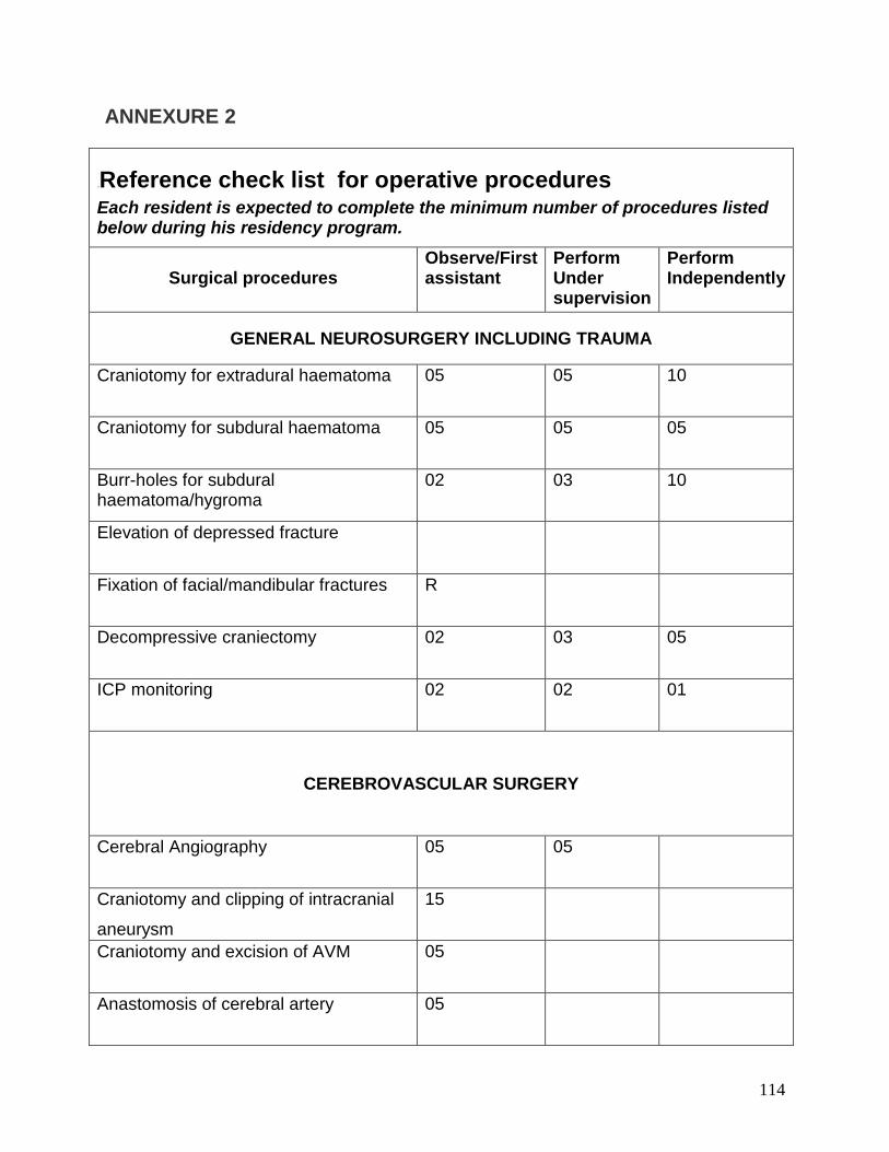
114
ANNEXURE 2
19BReference check list for operative procedures Each resident is expected to complete the minimum number of procedures listed below during his residency program.
Surgical procedures Observe/First assistant
Perform Under supervision
Perform Independently
GENERAL NEUROSURGERY INCLUDING TRAUMA
Craniotomy for extradural haematoma 05 05 10
Craniotomy for subdural haematoma 05 05 05
Burr-holes for subdural haematoma/hygroma
02 03 10
Elevation of depressed fracture
Fixation of facial/mandibular fractures R
Decompressive craniectomy 02 03 05
ICP monitoring 02 02 01
CEREBROVASCULAR SURGERY
Cerebral Angiography 05 05
Craniotomy and clipping of intracranial
aneurysm
15
Craniotomy and excision of AVM 05
Anastomosis of cerebral artery 05
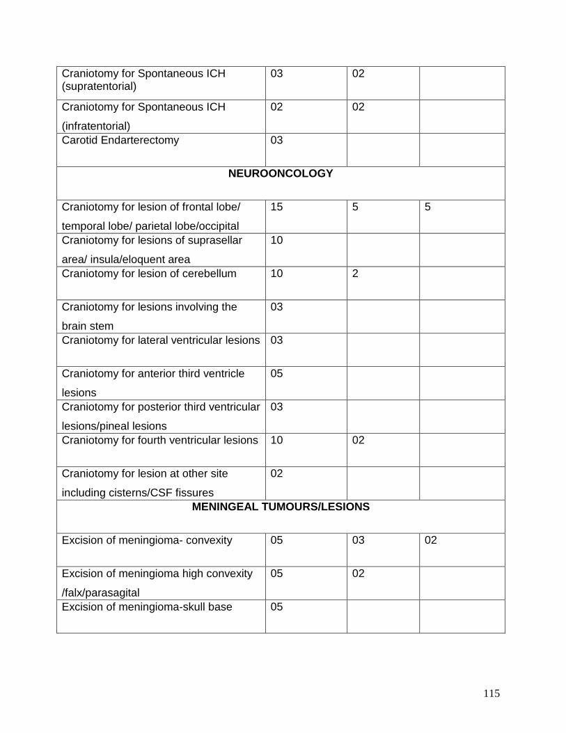
115
Craniotomy for Spontaneous ICH (supratentorial)
03 02
Craniotomy for Spontaneous ICH
(infratentorial)
02 02
Carotid Endarterectomy 03
NEUROONCOLOGY
Craniotomy for lesion of frontal lobe/
temporal lobe/ parietal lobe/occipital
15 5 5
Craniotomy for lesions of suprasellar
area/ insula/eloquent area
10
Craniotomy for lesion of cerebellum 10 2
Craniotomy for lesions involving the
brain stem
03
Craniotomy for lateral ventricular lesions 03
Craniotomy for anterior third ventricle
lesions
05
Craniotomy for posterior third ventricular
lesions/pineal lesions
03
Craniotomy for fourth ventricular lesions 10 02
Craniotomy for lesion at other site
including cisterns/CSF fissures
02
MENINGEAL TUMOURS/LESIONS
Excision of meningioma- convexity 05 03 02
Excision of meningioma high convexity
/falx/parasagital
05 02
Excision of meningioma-skull base 05
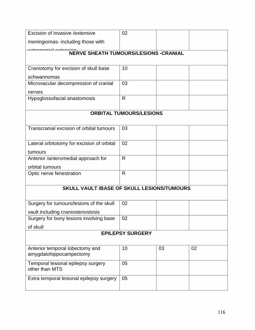
116
Excision of invasive /extensive
meningiomas- including those with
extracranial extension
02
NERVE SHEATH TUMOURS/LESIONS -CRANIAL
Craniotomy for excision of skull base
schwannomas
10
Microvacular decompression of cranial
nerves
03
Hypoglossofacial anastomosis R
ORBITAL TUMOURS/LESIONS
Transcranial excision of orbital tumours 03
Lateral orbitotomy for excision of orbital
tumours
02
Anterior /anteromedial approach for
orbital tumours
R
Optic nerve fenestration R
SKULL VAULT /BASE OF SKULL LESIONS/TUMOURS
Surgery for tumours/lesions of the skull
vault including craniostenostosis
02
Surgery for bony lesions involving base
of skull
02
EPILEPSY SURGERY
Anterior temporal lobectomy and amygdalohippocampectomy
10 03 02
Temporal lesional epilepsy surgery other than MTS
05
Extra temporal lesional epilepsy surgery 05
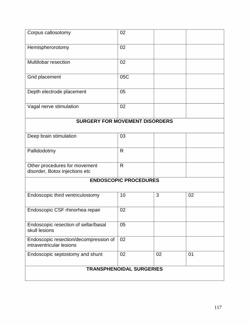
117
Corpus callosotomy 02
Hemispherorotomy 02
Multilobar resection 02
Grid placement 05C
Depth electrode placement 05
Vagal nerve stimulation 02
SURGERY FOR MOVEMENT DISORDERS
Deep brain stimulation 03
Pallidodotmy R
Other procedures for movement disorder, Botox injections etc
R
ENDOSCOPIC PROCEDURES
Endoscopic third ventriculostomy 10 3 02
Endoscopic CSF rhinorhea repair 02
Endoscopic resection of sellar/basal skull lesions
05
Endoscopic resection/decompression of intraventricular lesions
02
Endoscopic septostomy and shunt 02 02 01
TRANSPHENOIDAL SURGERIES
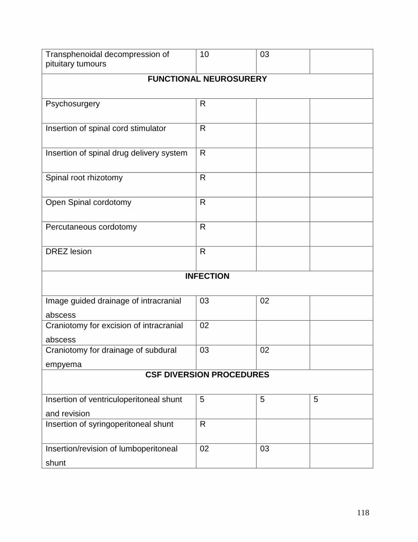
118
Transphenoidal decompression of pituitary tumours
10 03
FUNCTIONAL NEUROSURERY
Psychosurgery R
Insertion of spinal cord stimulator R
Insertion of spinal drug delivery system R
Spinal root rhizotomy R
Open Spinal cordotomy R
Percutaneous cordotomy R
DREZ lesion R
INFECTION
Image guided drainage of intracranial
abscess
03 02
Craniotomy for excision of intracranial
abscess
02
Craniotomy for drainage of subdural
empyema
03 02
CSF DIVERSION PROCEDURES
Insertion of ventriculoperitoneal shunt
and revision
5 5 5
Insertion of syringoperitoneal shunt R
Insertion/revision of lumboperitoneal
shunt
02 03
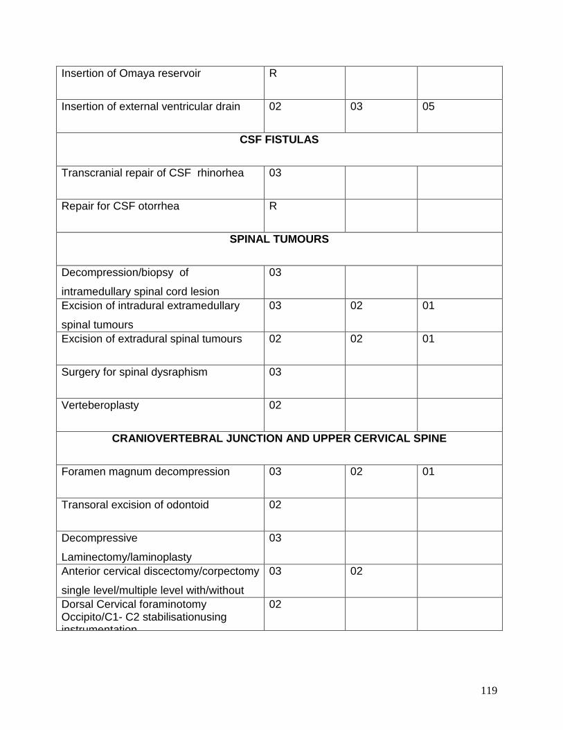
119
Insertion of Omaya reservoir R
Insertion of external ventricular drain 02 03 05
CSF FISTULAS
Transcranial repair of CSF rhinorhea 03
Repair for CSF otorrhea R
SPINAL TUMOURS
Decompression/biopsy of
intramedullary spinal cord lesion
03
Excision of intradural extramedullary
spinal tumours
03 02 01
Excision of extradural spinal tumours 02 02 01
Surgery for spinal dysraphism 03
Verteberoplasty 02
CRANIOVERTEBRAL JUNCTION AND UPPER CERVICAL SPINE
Foramen magnum decompression 03 02 01
Transoral excision of odontoid 02
Decompressive
Laminectomy/laminoplasty
03
Anterior cervical discectomy/corpectomy
single level/multiple level with/without
03 02
Dorsal Cervical foraminotomy Occipito/C1- C2 stabilisationusing instrumentation
02
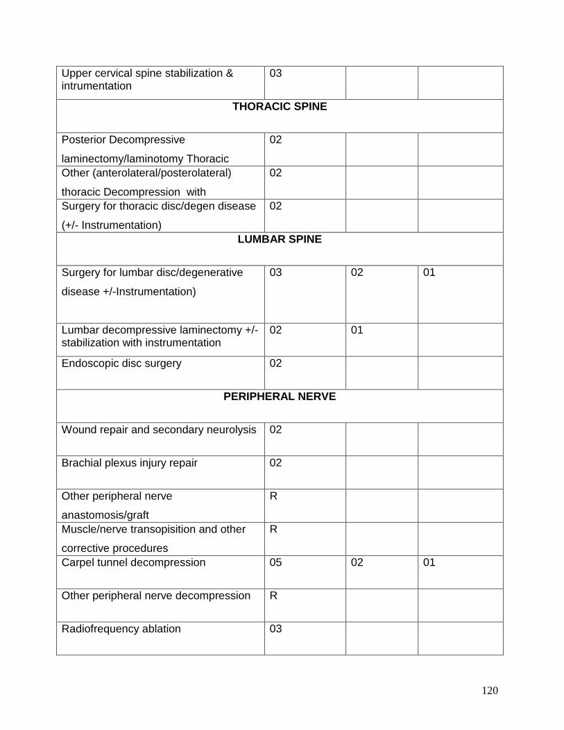
120
Upper cervical spine stabilization & intrumentation
03
THORACIC SPINE
Posterior Decompressive
laminectomy/laminotomy Thoracic
02
Other (anterolateral/posterolateral)
thoracic Decompression with
02
Surgery for thoracic disc/degen disease
(+/- Instrumentation)
02
LUMBAR SPINE
Surgery for lumbar disc/degenerative
disease +/-Instrumentation)
03 02 01
Lumbar decompressive laminectomy +/- stabilization with instrumentation
02 01
Endoscopic disc surgery 02
PERIPHERAL NERVE
Wound repair and secondary neurolysis 02
Brachial plexus injury repair 02
Other peripheral nerve
anastomosis/graft
R
Muscle/nerve transopisition and other
corrective procedures
R
Carpel tunnel decompression 05 02 01
Other peripheral nerve decompression R
Radiofrequency ablation 03
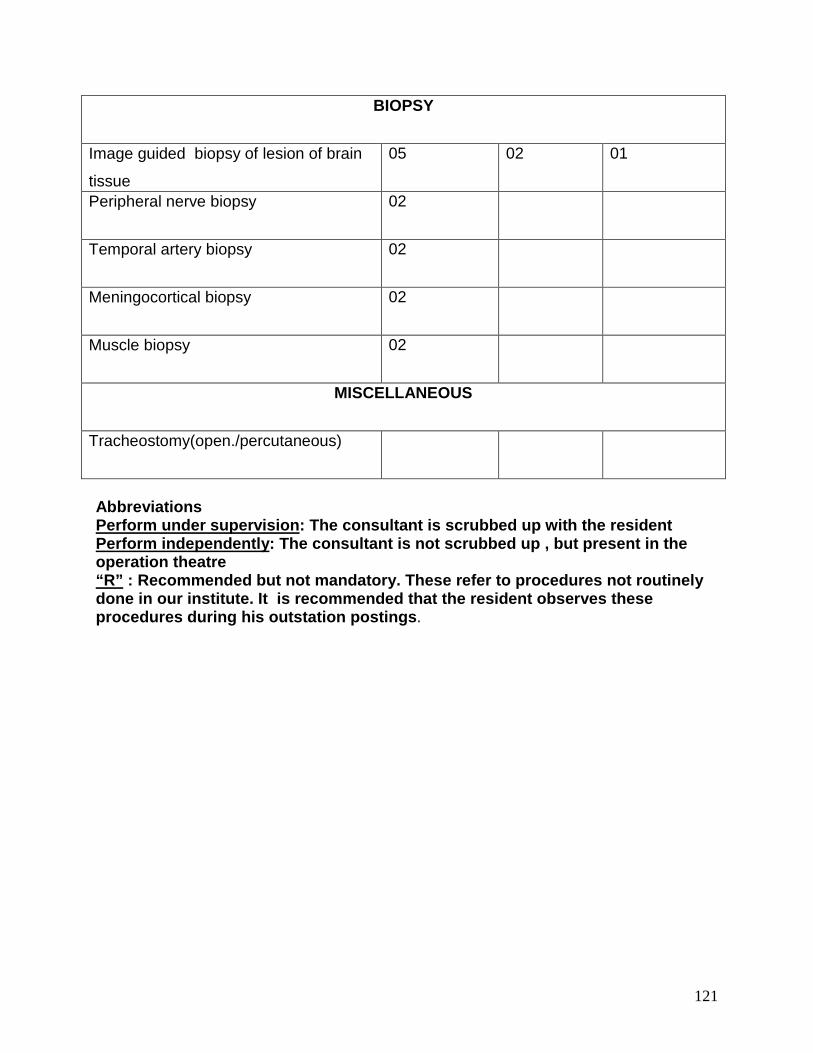
121
BIOPSY
Image guided biopsy of lesion of brain
tissue
05 02 01
Peripheral nerve biopsy 02
Temporal artery biopsy 02
Meningocortical biopsy 02
Muscle biopsy 02
MISCELLANEOUS
Tracheostomy(open./percutaneous)
Abbreviations Perform under supervision: The consultant is scrubbed up with the resident Perform independently: The consultant is not scrubbed up , but present in the operation theatre “R” : Recommended but not mandatory. These refer to procedures not routinely done in our institute. It is recommended that the resident observes these procedures during his outstation postings.
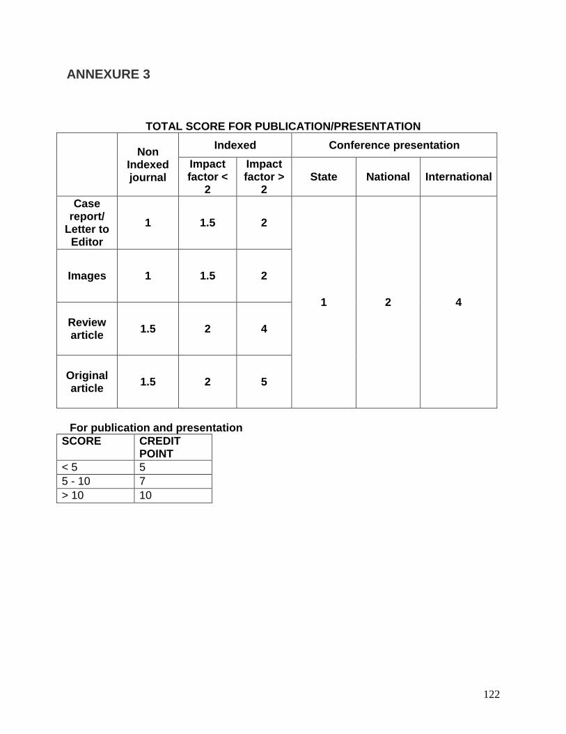
122
ANNEXURE 3
TOTAL SCORE FOR PUBLICATION/PRESENTATION
Non
Indexed journal
Indexed Conference presentation Impact factor <
2
Impact factor >
2 State National International
Case report/
Letter to Editor
1 1.5 2
1 2 4
Images 1 1.5 2
Review article 1.5 2 4
Original article 1.5 2 5
For publication and presentation
SCORE CREDIT POINT
< 5 5 5 - 10 7 > 10 10
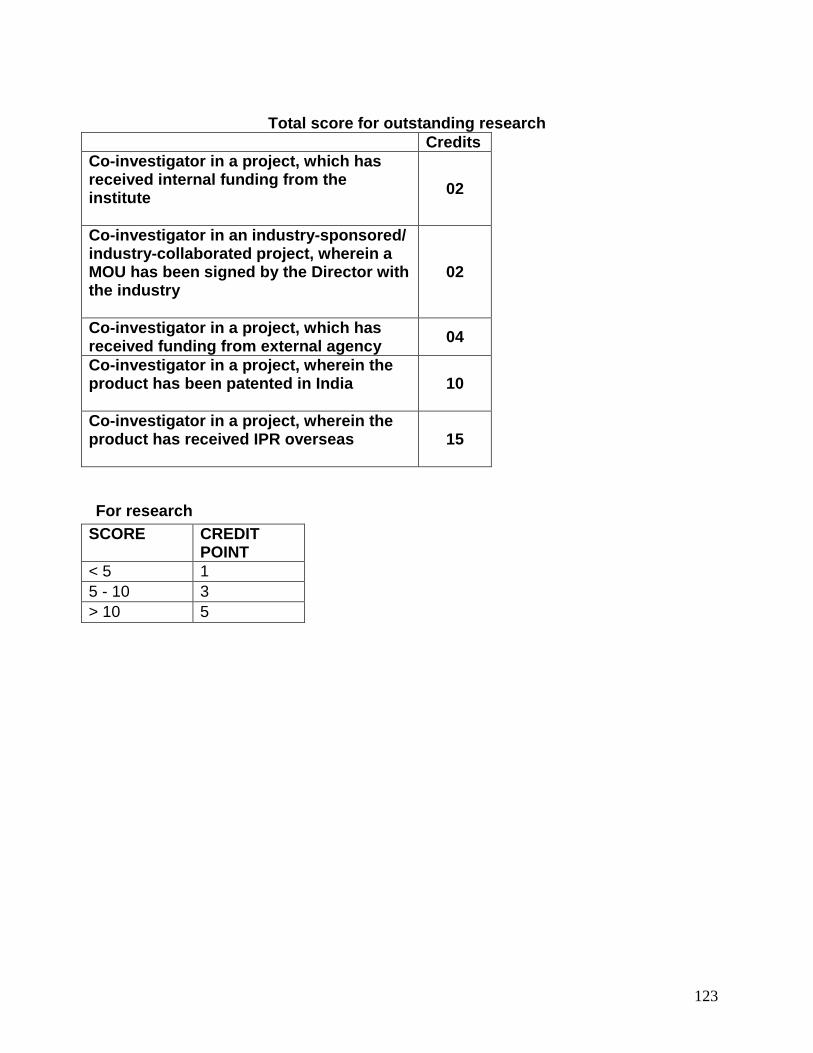
123
Total score for outstanding research
Credits Co-investigator in a project, which has received internal funding from the institute
02
Co-investigator in an industry-sponsored/ industry-collaborated project, wherein a MOU has been signed by the Director with the industry
02
Co-investigator in a project, which has received funding from external agency 04
Co-investigator in a project, wherein the product has been patented in India
10
Co-investigator in a project, wherein the product has received IPR overseas
15
For research SCORE CREDIT
POINT < 5 1 5 - 10 3 > 10 5