Embed Size (px)
Citation preview
Med Buccale Chir Buccale 2017;23:123–130© The authors, 2017DOI: 10.1051/mbcb/2017004
www.mbcb-journal.org M BC BMédecine BuccaleChirurgie Buccale
Original Article
Maxillary restoration with complete maxillary prosthesissupported by implants with immediate loading: clinicalretrospective study of 48 casesVictor Fau1,*, Dany Diep2, Hervé Bénateau2, Alexis Veyssière2, Fabrice Clipet1,Patrick Limbour1
1 Odontology and Oral Surgery Department, CHU, Rennes, France2 Maxillofacial Surgery and Oral Surgery Department, CHU, Caen, France
(Received: 30 November 2016, accepted: 28 February 2017)
Keywords:immediate loading /dental implant /edentulous jaw /osseointegration
* Correspondence: victor.f
This is an Open Access article dunrestricted use, distribution,
Abstract -- Introduction: In recent years, the immediate loading procedure for the rehabilitation of edentulous jawshas gained popularity among patients and practitioners. The purpose of this study was to evaluate implant andprosthetic success rates for the rehabilitation of edentulous maxillae using tilted distal implants and immediateloading of prostheses, after 2 years. Material and method: Patients included in the study received a completeprosthesis of the upper arch, attached to 4, 5 or 6 implants. The provisional prosthesis was fixed the same day theimplants were placed. After a period of osseointegration, the provisional prosthesis was replaced by a definitiveprosthesis with a titanium framework made using computer-aided design/computer-assisted manufacturing (CAD/CAM) technology. Judgment criteria were the implant success rate, the provisional prosthesis success rate, and thedefinitive prosthetic success rate. Results: Two hundred and forty-two implants were placed in 48 patients. Fiveimplants were lost, resulting in an implant survival at 2 years of 97.9%. Two provisional fixed prosthesis had to betemporarily replaced by a removable solution, resulting in a provisional prosthesis success rate of 95.8%. Thedefinitive prosthesis success rate was 100%. Discussion: Use of a provisional prosthesis during the osseointegrationperiod is essential, both for the preparation of the final prosthesis as well as for proper management of potentialimplant failures. Implant failures are more easily managed if at least five implants were originally placed.
Introduction
Immediate loading of supra-implanted total prosthesis,defined in 2014 by the Fifth Consensus Conference of the ITI(International Team for Implantology) as the occlusion withina maximum period of 7 days after the placement of the implants[1], has become a standard solution for the rehabilitation ofthe toothless arcades, both in the mandible and in the maxilla.It provides the advantage of offering quick, comfortableaesthetic and functional restoration to the patient. It alsoensures strong retention between implants, limiting theamplitude of the micromovements to the bone–implantinterface below the critical threshold for osseointegration[2] and decreasing the constraints transferred to thesurrounding bone [3].
The primary stability of the implant is considered the mostimportant factor for osseointegration success in immediateloading protocols [4,5]. The length, diameter [6], and the
istributed under the terms of the Creative Commons Aand reproduction in any medium, provided the origin
roughness of the implants [7,8], as well as the associateddrilling [9] are elements for achieving high insertion torque,which means good primary stability.
In the maxilla, a minimum of four implants is essential forthe viability of the reconstruction. In the posterior region,where bone quality and quantity are not always optimal,surgical alternatives have been proposed: short implants [10],onlay bone graft, sinus floor elevation, or implants in specificanatomical areas such as the zygomatic bone [11], thepterygoid region [12], or the maxillary tuber [13]. Each of thesetechniques have their own advantages and disadvantages, butthey all have limitations and require advanced surgicalexpertise. During the 2000s, many studies have demonstratedthe interest of implant angulation in distal position [14–16].Placed at the level of the anterior wall of the sinus, where thebone is generally dense, these implants can be longer, whichprovides them with better bone anchoring. Their tilt allows toincrease the distance between the pillars implant mesial anddistal abutments, thus decreasing the length of the prostheticcantilever to achieve better distribution of occlusal forces [17].
ttribution License (http://creativecommons.org/licenses/by/4.0), which permitsal work is properly cited.
123
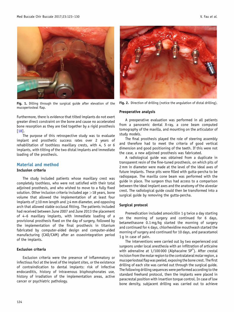
Fig. 1. Dilling through the surgical guide after elevation of themucoperiosteal flap.
Fig. 2. Direction of drilling (notice the angulation of distal drilling).
Med Buccale Chir Buccale 2017;23:123–130 V. Fau et al.
Furthermore, there is evidence that tilted implants do not exertgreater direct constraint on the bone and cause no acceleratedbone resorption as they are tied together by a rigid prosthesis[18].
The purpose of this retrospective study was to evaluateimplant and prosthetic success rates over 2 years ofrehabilitation of toothless maxillary crests, with 4, 5 or 6implants, with tilting of the two distal implants and immediateloading of the prosthesis.
Material and methodInclusion criteria
The study included patients whose maxillary crest wascompletely toothless, who were not satisfied with their totaladjoined prosthesis, and who wished to move to a fully fixedsolution. Other inclusion criteria included age>18 years, bonevolume that allowed the implementation of at least fourimplants of≥10mm length and ≥4mm diameter, and opposingarch that allowed stable occlusal fitting. The patients includedhad received between June 2007 and June 2013 the placementof 4–6 maxillary implants, with immediate loading of aprovisional prosthesis fixed on the day of surgery, followed bythe implementation of the final prosthesis in titaniumfabricated by computer-aided design and computer-aidedmanufacturing (CAD/CAM) after an osseointegration periodof the implants.
Exclusion criteria
Exclusion criteria were the presence of inflammatory orinfectious foci at the level of the implant sites, or the existenceof contraindication to dental implants: risk of infectiveendocarditis, history of intravenous bisphosphonates use,history of irradiation of the implementation areas, activecancer or psychiatric pathology.
124
Preoperative analysis
A preoperative evaluation was performed in all patientsfrom a panoramic dental X-ray, a cone beam computedtomography of the maxilla, and mounting on the articulator ofstudy models.
The final prosthesis played the role of steering assemblyand therefore had to meet the criteria of good verticaldimension and good positioning of the teeth. If this were notthe case, a new adjoined prosthesis was fabricated.
A radiological guide was obtained from a duplicate intransparent resin of the fine-tuned prosthesis, on which pits of2mm in diameter were made at the level of the ideal axes offuture implants. These pits were filled with gutta-percha to beradiopaque. The maxilla cone beam was performed with theguide in place. The surgeon thus had access to a comparisonbetween the ideal implant axes and the anatomy of the alveolarcrest. The radiological guide could then be transformed into asurgical guide by removing the gutta-percha.
Surgical protocol
Premedication included amoxicillin 1 g twice a day startingon the morning of surgery and continued for 6 days,betamethasone 0.1mg/kg started the morning of surgeryand continued for 4 days, chlorhexidine mouthwash started themorning of surgery and continued for 10 days, and paracetamol1 g in case of pain.
The interventions were carried out by two experienced oralsurgeons under local anesthesia with an infiltration of articainewith adrenaline at 1/100 000 (Alphacaine SP®). After crestalincision fromthemolar regionto the contralateralmolar region, amucoperiostealflapwaspeeled, exposing thebonecrest. Thefirstdrilling of each site was carried out through the surgical guide.The followingdrilling sequenceswere performedaccording to thestandard freehand protocol, then the implants were placed inparacrestal position with insertion torque control. In case of lowbone density, subjacent drilling was carried out to achieve
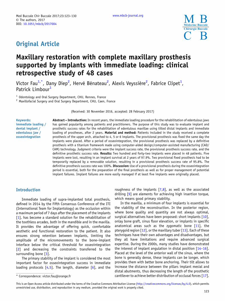
Fig. 3. Screwing of Multi-Unit Abutments® on the implants.
Fig. 4. Screwing of Provisional Copings® on Multi-Unit Abutments®
then flap suture.
Fig. 5. Prosthesis with cappings.
Fig. 6. Provisional copings® unified to prosthesis.
Med Buccale Chir Buccale 2017;23:123–130 V. Fau et al.
insertion torque of at least 30N/cm. The posterior implants weretilted in the distal direction, following the anterior sinuswall andemergingmore in as posterior a position as possible on the crest.
Multi-Unit Abutment® (Nobel Biocare) abutments werescrewed at 15 N/cm on the implants. These abutments werestraight on the vertical implants and angulated at 17° or 30° onthe tilted implants, to parallelize the set. Provisional cylinders(provisional Coping Multi-Unit®; Nobel Biocare) were thenscrewed on the abutments and the mucosa was repositionedand sutured using Vicryl 3.0 slow absorption wire (Figs. 1–4).
All implants placed were NobelSpeedy® Replace RP (NobelBiocare) of 4mm diameter and 10–15mm length. Those areconical implants with internal three-grooved connectivity andrough TiUnite surface achieved by anodic oxidation.
Prosthetic protocol
At this point, the adjoined final prosthesis was transformedinto immediate loading prosthesis through the flaps at the levelof implant emergences. Then it was positioned in the mouth and
attached to the cylinders byfilling the operculawith a self-curingresin (GC Unifast Trad®). The cylinders were then unscrewed andtheprosthesiswas sent to the laboratory for rework. The cylinderswere trimmed at the top of the occlusal surface of the resin, theartificial palate and the vestibular band were removed, and ametal reinforcement increased the rigidity of the prosthesis.
In the end, the prosthesis was screwed in the mouth, andocclusion, aesthetics, and phonation were controlled (Figs. 5–9,,,,). A panoramic X-ray was obtained to check the position ofthe implants as well as the adaptation of prosthesis on theabutment (Fig. 10). A soft diet was recommended for 3 months.
The final prosthesis was fabricated after clinical andradiological validation of implant osseointegration, within adelay of 6–18 months after surgery. A model was made from aplaster model. The steering assembly was carried out and fittedin, inspired by the provisional prosthesis whose function andaesthetics were already validated by several months of use. Thetitanium superstructure was then manufactured by CAD/CAMand tested “naked” in the mouth before assembly of thecosmetic part in acrylic resin.
125
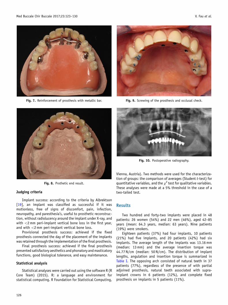
Fig. 7. Reinforcement of prosthesis with metallic bar.
Fig. 8. Prothetic end result.
Fig. 10. Postoperative radiography.
Fig. 9. Screwing of the prosthesis and occlusal check.
Med Buccale Chir Buccale 2017;23:123–130 V. Fau et al.
Judging criteria
Implant success: according to the criteria by Albrektson[19], an implant was classified as successful if it wasmotionless, free of signs of discomfort, pain, infection,neuropathy, and paresthesia’s, useful to prosthetic reconstruc-tion, without radiolucency around the implant under X-ray, andwith <2mm peri-implant vertical bone loss in the first year,and with <2mm peri-implant vertical bone loss.
Provisional prosthesis success: achieved if the fixedprosthesis connected the day of the placement of the implantswas retained through the implementation of the final prosthesis.
Final prosthesis success: achieved if the final prosthesispresented satisfactory aesthetics and phonatory andmasticatoryfunctions, good biological tolerance, and easy maintenance.
Statistical analysis
Statistical analyses were carried out using the software R (RCore Team) (2015). R: a language and environment forstatistical computing. R Foundation for Statistical Computing,
126
Vienna, Austria). Two methods were used for the characteriza-tion of groups: the comparison of averages (Student t-test) forquantitative variables, and the x2 test for qualitative variables.These analyses were made at a 5% threshold in the case of atwo-tailed test.
Results
Two hundred and forty-two implants were placed in 48patients: 26 women (54%) and 22 men (46%), aged 42–85years (mean: 64.3 years, median: 63 years). Nine patients(19%) were smokers.
Eighteen patients (37%) had four implants, 10 patients(21%) had five implants, and 20 patients (42%) had siximplants. The average length of the implants was 13.16mm(median: 13mm) and the average insertion torque was44.77 N/cm (median: 50 N/cm). The distribution of implantlengths, angulation and insertion torque is summarized inTable I. The opposing arch consisted of natural teeth in 37patients (77%), regardless of the presence of with partialadjoined prosthesis, natural teeth associated with supra-implant crowns in 6 patients (12%), and complete fixedprosthesis on implants in 5 patients (11%).
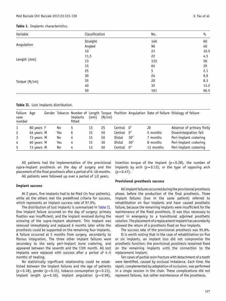
Table I. Implants characteristics.
Variable Classification No. %
AngulationStraight 146 60Angled 96 40
Length (mm)
10 23 10.511.5 22 4.513 133 5615 64 29
Torque (N/cm)
25 5 2.130 24 9.935 20 8.340 32 13.250 161 66.5
Table II. Lost implants distribution.
Failurecasenumber
Age Gender Tobacco Number ofimplantsfitted
Length(mm)
Torque(N/cm)
Position Angulation Date of failure Etiology of failure
1 80 years F No 5 13 25 Central 0° J0 Absence of primary fixity2 64 years M Yes 6 15 50 Central 0° 5 months Osseointegration fail3 73 years M No 4 15 50 Distal 30° 7 months Peri-implant cratering4 60 years M Yes 4 15 30 Distal 30° 8 months Peri-implant cratering5 73 years M No 4 13 50 Central 0° 13 months Peri-implant cratering
Med Buccale Chir Buccale 2017;23:123–130 V. Fau et al.
All patients had the implementation of the provisionalsupra-implant prosthesis on the day of surgery and theplacement of the final prosthesis after a period of 6–18 months.
All patients were followed up over a period of ≥2 years.
Implant success
At 2 years, five implants had to be filed (in four patients),while all the others met the predefined criteria for success,which represents an implant success rate of 97.9%.
The distribution of lost implants is summarized in Table II.One implant failure occurred on the day of surgery: primaryfixation was insufficient, and the implant revolved during thescrewing of the supra-implant abutment. This implant wasremoved immediately and replaced 6 months later while theprosthesis could be supported on the remaining four implants.A failure occurred at 5 months from surgery, secondarily tofibrous integration. The three other implant failures weresecondary to the early peri-implant bone cratering, andappeared between the seventh and the 13th month. All lostimplants were replaced with success after a period of 4–5months of healing.
No statistically significant relationship could be estab-lished between the implant failures and the age of patients(p = 0.18), gender (p = 0.15), tobacco consumption (p = 0.23),implant length (p = 0.10), implant angulation (p = 0.99),
insertion torque of the implant (p = 0.28), the number ofimplants by arch (p = 0.13), or the type of opposing arch(p = 0.47).
Provisional prosthesis success
All implant failuresoccurredduring theprovisionalprosthesisphase, before the production of the final prosthesis. Threeimplant failures (two in the same patient) referred torehabilitation on four implants and have caused prostheticfailure, because the remaining implants were insufficient for themaintenance of the fixed prosthesis. It was thus necessary toresort in emergency to a transitional adjoined prostheticsolution. Theplacementofa replacement implanthassecondarilyallowed the return of a prosthesis fixed on four implants.
The success rate of the provisional prosthesis was 95.8%.It is worth noting that in the case of rehabilitation on five
or six implants, an implant loss did not compromise theprosthetic function: the provisional prosthesis remained fixedon the remaining implants until the connection to thereplacement implant.
Ten cases of partial resin fracture with detachment of a toothwere identified, caused by occlusal imbalance. Each time, therepair, complemented by adaptation of occlusion, was performedin a single session in the chair. These complications did notrepresent failures, but rather maintenance of the prosthesis.
127
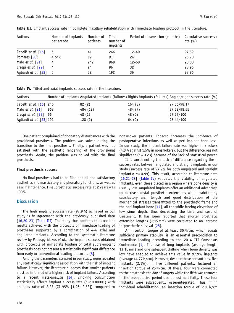
Table III. Implant success rate in complete maxillary rehabilitation with immediate loading protocol in the literature.
Authors Number of implantsper arcade
Number ofpatients
Totalnumber ofimplants
Period of observation (months) Cumulative success rate (%)
Capelli et al. [16] 6 41 246 12–40 97.59Pomares [20] 4 or 6 19 91 24 96.70Malo et al. [21] 4 242 968 12–60 98.00Crespi et al. [22] 4 24 96 32 98.96Agliardi et al. [23] 6 32 192 36 98.96
Table IV. Tilted and axial implants success rate in the literature.
Authors Number of implants Angulated implants (failures) Rights implants (failures) Angled/right success rate (%)
Capelli et al. [16] 246 82 (2) 164 (3) 97.56/98.17Malo et al. [21] 968 484 (12) 484 (7) 97.52/98.55Crespi et al. [22] 96 48 (1) 48 (0) 97.97/100Agliardi et al. [23] 192 128 (2) 64 (0) 98.44/100
Med Buccale Chir Buccale 2017;23:123–130 V. Fau et al.
One patient complained of phonatory disturbances with theprovisional prosthesis. The problem was solved during thetransition to the final prosthesis. Finally, a patient was notsatisfied with the aesthetic rendering of the provisionalprosthesis. Again, the problem was solved with the finalprosthesis.
Final prosthesis success
No final prosthesis had to be filed and all had satisfactoryaesthetics and masticatory and phonatory functions, as well aseasy maintenance. Final prosthetic success rate at 2 years was100%.
Discussion
The high implant success rate (97.9%) achieved in ourstudy is in agreement with the previously published data[16,20–23] (Table III). The study thus confirms the excellentresults achieved with the protocols of immediate loading ofprostheses supported by a combination of 4–6 axial andangulated implants. According to the systematic literaturereview by Papaspyridakos et al., the implant success obtainedwith protocols of immediate loading of total supra-implantprosthesis does not present a statistically significant differencefrom early or conventional loading protocols [5].
Among the parameters assessed in our study, none revealedany statistically significant association with the risk of implantfailure. However, the literature suggests that smoker patientsmust be informed of a higher risk of implant failure. Accordingto a recent meta-analysis [24], smoking consumptionstatistically affects implant success rate (p< 0.00001) withan odds ratio of 2.23 (CI 95% [1.96; 2.53]) compared to
128
nonsmoker patients. Tobacco increases the incidence ofpostoperative infections as well as peri-implant bone loss.In our study, the implant failure rate was higher in smokers(4.3% against 1.5% in nonsmokers), but the difference was notsignificant (p = 0.23) because of the lack of statistical power.
It is worth noting the lack of difference regarding the nsuccess rates between angulated and straight implants in ourstudy (success rate of 97.9% for both angulated and straightimplants; p = 0.99). This result, according to literature data[16,21–23] (Table IV) validates the viability of angulatedimplants, even those placed in a region where bone density isusually low. Angulated implants offer an additional advantageto decrease distal prosthetic extensions while maintainingsatisfactory arch length and good distribution of themechanical stresses transmitted to the prosthetic frame andthe peri-implant bone [17], all the while freeing elevations oflow sinus depth, thus decreasing the time and cost oftreatment. It has been reported that shorter prostheticextension lengths (<15mm) were correlated to an increasein prosthetic survival [25].
An insertion torque of at least 30 N/cm, which equalssufficient primary stability, is an essential precondition toimmediate loading according to the 2014 ITI ConsensusConference [1]. The use of long implants (average length13.16mm) and one subjacent drilling when bone density waslow have enabled to achieve this value in 97.9% implants(average 44.77 N/cm). However, despite these precautions, fiveimplants (2.1%), in five different patients, featured aninsertion torque of 25 N/cm. Of these, four were connectedto the prosthesis the day of surgery while the fifth was removedin the preoperative period due almost null fixity. These fourimplants were subsequently osseointegrated. Thus, if inindividual rehabilitation, an insertion torque of <30 N/cm
Med Buccale Chir Buccale 2017;23:123–130 V. Fau et al.
may be a contraindication to immediate implant loading,presumably that is not the case in total rehabilitation, insofaras the implant is connected to other implants with higherprimary fixity through the immediate prosthesis, which playsthe role of rigid restraints and allows to decrease implantmicromovements [2,3].
It is interesting to note that in our study, the majority ofimplant failures occurred in the first months following theintervention. A literature review by Patzelt et al. [26] onrehabilitation by prostheses on stilts regarding 4804 implantsshows that 74% implant failures have occurred during the first12 months. This is a percentage close to the 80% failure ratebefore 12 months found in our study. Similarly, Malo et al. [21]shows 89% implant failures in 13 months following surgery.These figures show that the osseointegration period in the firstpostoperative months is the most conducive period to theimplant failure. The use of a provisional prosthesis during thisperiod will allow for simple adaptation in case an implantshould be replaced. Provided that at least four residual implantspersist, the maintenance of the fixed prosthesis can be ensuredthrough its connection to the replacement implant, placed 4–6months after the removal of the lost implant. This situation wasfound twice in our study. The observation is not valid in thecase where only four implants are initially in place, as the threeresidual implants are insufficient to maintain the fixedprosthesis in sustainable conditions. In this case, it wouldbe fitting to use an adjoined prosthesis during the time to placethe replacement implant. This situation was found twice in ourstudy. Particularly painful for the patient, it is a strongargument in favor of placing at least five implants. Althoughthe number of implants in maxillary reconstruction is rarelydiscussed in the literature, this argument is found in theliterature revue by Lambert et al. [27].
In any case, if an implant failure occurs while the finalprosthesis is in place, it must be removed and the titaniumframe must be fully rebuilt, causing significant delays andfinancial cost.
As in the literature [20–22], most prosthetic complicationsidentified in our study are represented by the resin fractures onthe provisional prosthesis, which occurred in 10 patients(20.8% cases). They bear witness to the importance of goodocclusal control insofar as the absence of periodontal aroundthe implants strongly decreases proprioceptive abilities, andtherefore detection of occlusal overload.
The occlusal scheme applied was that of bilaterallybalanced occlusion with simultaneous anterior and posteriorcontacts in centered and excursion relationship. Although nostudy proves the superiority of this technique, a majority ofauthors agree that this scheme allows to minimize the most thetraction forces applied to implants and to spread thecompressive forces over the whole of the arch [28]. To steerthe most the masticatory forces in the axis of the implants,lateral stress and intercuspation interference must bedecreased by limiting the heights and intercuspation slopesas well as the depth of the fossae [29].
The success rate of the final prosthesis achieved in thestudy remains 100%, as in the majority of published studies[16,20,22].
Conclusion
Prosthetic implant rehabilitation of toothless maxillaryarches by a combination of axial and tilted implants withimmediate loading of the prosthesis on stilts shows excellentresults after 2 years of service when done by an experiencedsurgical team. The use of a provisional prosthesis is an essentialpath to ensure the success of the treatment.
Conflicts of interest: The authors declare that theyhave no conflicts of interest in relation to this article.
References
1. Gallucci GO, Benic GI, Eckert SE, Papaspyridakos P, Schimmel M,Schrott A, et al. Consensus statements and clinical recommen-dations for implant loading protocols. Int J Oral MaxillofacImplants 2014;29(Suppl):287–290.
2. Tarnow DP, Emtiaz S, Classi A. Immediate loading of threadedimplants at stage 1 surgery in edentulous arches: ten consecutivecase reports with 1- to 5-year data. Int J Oral Maxillofac Implant1997;12:319–324.
3. Bergkvist G, Simonsson K, Rydberg K, Johansson F, Dérand T. Afinite element analysis of stress distribution in bone tissuesurrounding uncoupled or splinted dental implants. Clin ImplantDent Relat Res 2008;10:40–46.
4. Javed F, Romanos GE. The role of primary stability for successfulimmediate loading of dental implants. A literature review. J Dent2010;38:612–620.
5. Papaspyridakos P, Chen C-J, Chuang S-K, Weber H-P. Implantloading protocols for edentulous patients with fixed prostheses: asystematic review and meta-analysis. Int J Oral MaxillofacImplants 2014;29(Suppl):256–270.
6. Ding X, Liao S-H, Zhu X-H, Zhang X-H, Zhang L. Effect of diameterand length on stress distribution of the alveolar crest aroundimmediate loading implants. Clin Implant Dent Relat Res2009;11:279–287.
7. Gotfredsen K, Wennerberg A, Johansson C, Skovgaard LT,Hjørting-Hansen E. Anchorage of TiO2-blasted, HA-coated, andmachined implants: an experimental study with rabbits. J BiomedMater Res 1995;29:1223–1231.
8. Cochran DL, Schenk RK, Lussi A, Higginbottom FL, Buser D. Boneresponse to unloaded and loaded titanium implants with asandblasted and acid-etched surface: a histometric study in thecanine mandible. J Biomed Mater Res 1998;40:1–11.
9. Degidi M, Daprile G, Piattelli A. Influence of underpreparation onprimary stability of implants inserted in poor quality bone sites:an in vitro study. J Oral Maxillofac Surg Off J Am Assoc OralMaxillofac Surg 2015;73:1084–1088.
10. Renouard F, Nisand D. Short implants in the severely resorbedmaxilla: a 2-year retrospective clinical study. Clin Implant DentRelat Res 2005;7(Suppl 1):104–110.
11. Chrcanovic BR, Abreu MHNG. Survival and complications ofzygomatic implants: a systematic review. Oral Maxillofac Surg2013;17:81–93.
129
Med Buccale Chir Buccale 2017;23:123–130 V. Fau et al.
12. Balshi TJ, Wolfinger GJ, Balshi SF. Analysis of 356 pterygomax-illary implants in edentulous arches for fixed prosthesisanchorage. Int J Oral Maxillofac Implants 1999;14:398–406.
13. Lopes LF dT P, da Silva VF, Santiago JF, Panzarini SR, Pellizzer EP.Placement of dental implants in the maxillary tuberosity: asystematic review. Int J Oral Maxillofac Surg 2015;44:229–238.
14. Aparicio C, Perales P, Rangert B. Tilted implants as an alternativeto maxillary sinus grafting: a clinical, radiologic, and perioteststudy. Clin Implant Dent Relat Res 2001;3:39–49.
15. Krekmanov L, Kahn M, Rangert B, Lindström H. Tilting of posteriormandibular and maxillary implants for improved prosthesissupport. Int J Oral Maxillofac Implants 2000;15:405–414.
16. Capelli M, Zuffetti F, Del Fabbro M, Testori T. Immediaterehabilitation of the completely edentulous jaw with fixedprostheses supported by either upright or tilted implants: amulticenter clinical study. Int J Oral Maxillofac Implants2007;22:639–644.
17. Bevilacqua M, Tealdo T, Menini M, Pera F, Mossolov A, Drago C,et al. The influence of cantilever length and implant inclinationon stress distribution in maxillary implant-supported fixeddentures. J Prosthet Dent 2011;105:5–13.
18. Del Fabbro M, Ceresoli V. The fate of marginal bone around axialvs. tilted implants: a systematic review. Eur J Oral Implantol2014;7(Suppl 2):171–189.
19. Albrektsson T, Jansson T, Lekholm U. Osseointegrated dentalimplants. Dent Clin North Am 1986;30:151–174.
20. Pomares C.A retrospective clinical study of edentulous patientsrehabilitated according to the “all on four” or the “all on six”immediate function concept. Eur J Oral Implantol 2009;2:55–60.
130
21. Maló P, de Ara�ujo Nobre M, Lopes A, Francischone C, Rigolizzo M.“All-on-4” immediate-function concept for completely edentu-lous maxillae: a clinical report on the medium (3 years) and long-term (5 years) outcomes. Clin Implant Dent Relat Res 2012;14(Suppl 1):e139–e150.
22. Crespi R, Vinci R, Capparé P, Romanos GE, Gherlone E. A clinicalstudy of edentulous patients rehabilitated according to the “allon four” immediate function protocol. Int J Oral MaxillofacImplants 2012;27:428–434.
23. Agliardi EL, Pozzi A, Stappert CFJ, Benzi R, Romeo D, Gherlone E.Immediate fixed rehabilitation of the edentulous maxilla: aprospective clinical and radiological study after 3 years ofloading. Clin Implant Dent Relat Res 2014;16:292–302.
24. Chrcanovic BR, Albrektsson T, Wennerberg A. Smoking and dentalimplants: a systematic review and meta-analysis. J Dent2015;43:487–498.
25. Shackleton JL, Carr L, Slabbert JC, Becker PJ. Survival of fixedimplant-supported prostheses related to cantilever lengths. JProsthet Dent 1994;71:23–26.
26. Patzelt SBM, Bahat O, Reynolds MA, Strub JR. The all-on-fourtreatment concept: a systematic review. Clin Implant Dent RelatRes 2014;16:836–855.
27. Lambert FE, Weber H-P, Susarla SM, Belser UC, Gallucci GO.Descriptive analysis of implant and prosthodontic survival rateswith fixed implant-supported rehabilitations in the edentulousmaxilla. J Periodontol 2009;80:1220–1230.
28. Bocklage R. Biomechanical aspects of monoblock implant bridgesfor the edentulous maxilla and mandible: concepts of occlusionand articulation. Implant Dent 2004;13:49–53.
29. Nikolopoulou F, Ktena-Agapitou P. Rationale for choices ofocclusal schemes for complete dentures supported by implants. JOral Implantol 2006;32:200–203.


![INDEX [microdentsystem.com] · 2015-11-24 · INDEX PRESENTATION. INTRODUCTION MULTIPLE PROSTHESIS. REMOVABLE AND IMMEDIATE PROSTHESIS. SINGLE PROSTHESIS CEMENTED PROSTHESIS. Microdent](https://img.dokumen.tips/doc/110x75/5facd9ee77a5ed547a36b19c/index-2015-11-24-index-presentation-introduction-multiple-prosthesis-removable.jpg)