Embed Size (px)
Citation preview
INdAM Workshop on
Mathematical and Numerical Modeling
of the Cardiovascular System
Istituto Nazionale di Alta Matematica (INdAM)Citta Universitaria
P.le Aldo Moro 5, Roma
April 16–19, 2018
supported byIstituto Nazionale di Alta Matematica (INdAM)
Dipartimento di Matematica, Universita degli Studi di PaviaDepartment of Mathematical Analysis, Modelling, and Application, SISSA
Dipartimento di Matematica, Politecnico di Milano
organized byLuca F. Pavarino1, Gianluigi Rozza2, Simone Scacchi3, Christian Vergara4
1 Dipartimento di Matematica, Universita degli Studi di Pavia, Pavia, Italy2 Department of Mathematical Analysis, Modelling, and Application,
SISSA, Trieste, Italy3 Dipartimento di Matematica, Universita degli Studi di Milano, Milan,
Italy4 MOX-Dipartimento di Matematica, Politecnico di Milano, Milan, Italy

PROGRAM
Monday, April 16, 2018
14:00-14:15 Opening and Welcome
Chair: L. F. Pavarino14:15-15:00 P. Colli Franzone (Universita degli Studi di Pavia, Italy), L. F.
Pavarino, S. Scacchi: Stability of re-entrant cardiac dynamics: in-fluences of mechano-electric feedbacks and of infarct scar geometricproperties
15:00-15:45 R. Krause (Universita della Svizzera Italiana, Switzerland), M.Favino, M. Nestola, S. Pozzi, P. Zulian, A. Auricchio: Fast SolutionMethods for Coupled Problems in Cardiac Simulation
15:45-16:10 Coffee break
16:10-16:30 C. Cherubini (Universita Campus Bio-Medico di Roma, Italy), S.Filippi, A. Gizzi, A. Loppini, R. Ruiz-Baier: On Stress-drivenAnisotropic Electrical Diffusion in Cardiac Electro-Mechanics
16:30-16:50 C. Pierre (Universite de Pau, France), C. Douanla-Lontsi: Efficiencyof high order schemes for the monodomain model
16:50-17:10 L. Gerardo Giorda (Basque Center for Applied Mathematics, Spain),N. Cusimano: Combining tissue anisotropy and heterogeneity in car-diac electrophysiology: a space-fractional Monodomain model
17:10-17:30 M. K. Gobbert (University of Maryland, USA), C. Barajas, S. Khu-vis: Challenges and Opportunities for the Simulations of CalciumWaves on Modern Multi-Core and Many-Core Parallel ComputingPlatforms
17:30-17:50 R. Spiteri (University of Saskatchewan, Canada), J. Cervi: High-Order Operator-Splitting Methods for the Bidomain and MonodomainModels
17:50-18:10 M. Landajuela (Politecnico di Milano, Italy), C. Vergara, A. Gerbi,L. Dede, L. Formaggia, A. Quarteroni: Numerical approximation ofthe electromechanical coupling in the left ventricle with inclusion ofthe Purkinje network

Tuesday, April 17, 2018
Chair: G. Plank09:00-09:45 A. F. Frangi (University of Sheffield, UK), R. Attar, M. Pereanez, J.
M. Pozo, A. Sarrami-Foroushani, L. Zhang, A. Gooya, T. Lassila, Z.A. Taylor: Towards computational imaging phenomics and in silicoclinical trials in cardiovascular science: vision and progress so far
09:45-10:30 K.-A. Mardal (University of Oslo & Simula, Norway): Mathematicalmodeling of the glymphatic system
10:30-10:50 Coffee break
10:50-11:10 C. Corsi (Universita degli Studi di Bologna, Italy), A. Masci: Medicalimaging for cardiac simulation studies
11:10-11:30 E. Votta (Politecnico di Milano, Italy), F. Piatti, O. Pappalardo, A.Caimi, M. Selmi, G. Rossini, F. Sturla, A. Redaelli: Clinically drivennumerical models in cardiovascular surgery
11:30-11:50 T. Weber (Otto von Guericke University Magdeburg, Germany), E.Scholz, F. Langkamp, H. A. Katus, S. Sager: Applicability of thePREMAP algorithm to accelerate activation mapping of scar-relatedventricular reentry tachycardia
11:50-12:10 F. Caforio (INRIA & Universite Paris-Saclay, France), S. Imperiale,D. Chapelle: Modelling of impulsive source in a prestressed soft tissue
12:10-12:30 S. Pozzi (Politecnico di Milano, Italy), F. Piatti, A. Camporeale,N. Cobo Gomez, G. Di Giovine, S. Castelvecchio, L. Menicanti, A.Greiser, E. Votta, A. Redaelli, M. Lombardi: Towards the exhaustiveanalysis of left ventricle dysfunctions in ischemic cardiomiopathy: in-tegrating wall kinetics, scar transmurality and wall shear stress
12:30-14:00 Lunch break
Chair: R. Krause14:15-14:45 W. A. Wall (Technische Universitaet Muenchen, Germany): TBA
14:45-15:30 M. Behr (RWTH Aachen University, Germany), S. Hassler, L. Pauli:Towards Predictive Simulation of Artificial Blood Pumps
15:30-15:45 Coffee break
Wednesday, April 18, 2018
Chair: Y. Coudiere09:00-09:45 D. Chapelle (INRIA & Universite Paris-Saclay, France): Multi-scale
modeling of chemo-mechanical coupling in muscle contraction and ap-plications to cardiac modeling
09:45-10:30 L. Dede (Politecnico di Milano, Italy), A. Gerbi, A. Quarteroni:Mathematical and Numerical Modeling of Cardiac Electromechanics:Numerical Coupling and Large-Scale Simulation
10:30-10:50 Coffee break
10:50-11:10 C. Cavaterra (Universita degli Studi di Milano, Italy), E. Beretta, M.C. Cerutti, A. Manzoni, L. Ratti: An inverse problem arising fromcardiac electrophysiology
11:10-11:30 A. Quaglino (Universita della Svizzera Italiana, Switzerland), S. Pez-zuto, R. Krause: Enabling uncertainty quantification for the mon-odomain equation via multifidelity techniques
11:30-11:50 A. Manzoni (Politecnico di Milano, Italy), S. Pagani, A. Quarteroni:Reduced Order Modeling for Uncertainty Quantification of the cardiacfunction
11:50-12:10 Z. Zainib (SISSA, Italy), F. Ballarin, G. Rozza: Reduced order mod-elling for cardiovascular haemodynamics: optimal flow control, dataassimilation and geometrical reconstruction
12:10-12:30 F. Regazzoni (Politecnico di Milano, Italy), L. Dede, A. Quarteroni:Active contraction of cardiac cells: a reduced model of force generation
12:30-12:50 L. Fassina (Universita degli Studi di Pavia, Italy), M. Cornacchione,M. E. Mognaschi, G. Magenes, F. Naro: Ergotropic effect in cardiactissue after electromagnetic and β-adrenergic stimulus
12:50-14:15 Lunch break

Chair: C. Vergara14:15-14:45 M. Domanin (Universita degli Studi di Milano, Italy), C. Vergara:
Computational fluid dynamic modeling for the choice of the closuretechnique in carotid surgery
14:45-15:15 E. Macchi (Universita degli Studi di Parma, Italy), L. Magnani, M.Miragoli, E. Musso, S. Rossi: Arrhythmia vulnerability by single pre-mature stimulation in normal ventricular myocardium
15:15-15:45 D. Catanzariti (Ospedale S. Maria del Carmine, Rovereto, Italy):Learning by doing (pacing, burning and mapping)
15:45-16:15 M. Miragoli (Universita degli Studi di Parma, Italy), G. Rozzi, F. P.Lo Muzio, S. Rossi, L. Fassina, S. Strozzi, G. Faggian, G. B. Luciani:Real-time video evaluation of the right ventricle kinematics duringcardiac surgery: novelties, implications and future perspectives
16:15-16:40 Coffee break
16:40-17:10 R. Scrofani (Ospedale L. Sacco, Milano, Italy), F. Nicolo, G. Cagnoni,C. Antona: Cardiac Surgery and Biomedical Engineering
17:10-17:40 N. Salvarani (Humanitas Clinical and Research Center, Milano,Italy), P. Carullo, M. Miragoli: Anode Break Excitation in Hyper-trophic Cardiomyocytes: An Investigative Tool for Predicting Modu-lation of Cardiac Excitability
17:40-18:00 C. Contarino (Universita degli Studi di Trento, Italy), E. F. Toro,A. Louveau, S. Da Mesquita, D. Raper, I. Smirnov, N. Agarwal, J.Kipnis: A global, multi-scale mathematical model of the murine fluidsystems: application to idiopathic intracranial hypertension
18:00-18:20 F. Scardulla (Universita degli Studi di Palermo, Italy), S. Hu, L.D’Acquisto, S. Pasta, L. Barrett, P. Blanos, L. Yan: Systolic bloodpressure detection using a multi-wavelength Opto-Electronic patchsensor at peripheral circulation
20:00: Social Dinner
Thursday, April 19, 2018
Chair: G. Rozza09:00-09:45 A. V. Panfilov (Ghent University, Belgium), M. P. Nash, L. D. Weise:
Mechano-electric feedback and initiation of cardiac arrhythmias
09:45-10:30 Y. Coudiere (Universite de Bordeaux & IHU Lyric, France), A. Davi-dovic, C. Poignard: Modeling the propagation of cardiac action po-tential in hearts with structural heterogeneities
10:30-10:50 Coffee break
10:50-11:10 L. Gastaldi (Universita degli Studi di Brescia, Italy), D. Boffi, L.Heltai, M. Annese: A distributed Lagrange multiplier formulation forthe finite element discretization of FSI
11:10-11:30 S. Pasta (Fondazione Ri.MED, Palermo, Italy), G. Gentile, G. M.Raffa, D. Bellavia, M. Pilato: In Silico Shear and Intramural Stressesare Linked to Aortic Valve Morphology in Dilated Ascending Aorta
11:30-11:50 A. This (INRIA Paris, France), L. Boilevin-Kayl, H. G. Morales, O.Bonnefous, M. A. Fernandez, J.-F. Gerbeau: Modeling isovolumicphases using a pressure corrected RIS model
11:50-12:10 M. Fedele (Politecnico di Milano, Italy), E. Faggiano, L. Dede1, A.Quarteroni: Patient-specific simulations of the hemodynamics througha moving aortic valve with the resistive immersed implicit surfacesmethod
12:10-12:30 S. Zonca (Politecnico di Milano, Italy), L. Formaggia, C. Vergara:Fluid-Structure Interaction via an XFEM/DG Approach for ValveDynamics
12:30-12:50 V. Meschini (SISSA, Italy), R. Mittal, R. Verzicco: Flow-Induced Mi-tral Leaflet Motion in Hypertrophic Cardiomyopathy
12:50-14:15 Lunch break
Chair: L. Dede14:15-15:00 G. Plank (Medical University of Graz, Austria): Personalizing models
of total heart function
15:00-15:45 L. Teresi (Universita degli Studi Roma Tre, Italy), V. Varano, P.Piras, S. Gabriele, I. Dryden, P. Nardinocchi, P. E. Puddu: ShapeAnalysis for 3D Cardiac Imaging
15:45-16:10 Coffee break
16:10-16:30 S. Severi (Universita degli Studi di Bologna, Italy), A. Fabbri, M.Paci, R. Wilders, Y. Lutz, A. Loewe: Computational Modeling of theCardiac Pacemaking in Humans
16:30-16:50 A. Gizzi (Universita Campus Bio-Medico di Roma, Italy), D. Bianchi,M. Marino, G. Vairo, S. Filippi: Multi-Scale Computational Modelingof FSI in Aorta Physiopathology: A Quantitative Risk AssessmentStudy
16:50-17:10 D. E. Hurtado (Pontificia Universidad Catolica de Chile, Chile), J.Jilberto: Non-conforming finite-element schemes for cardiac electro-physiology
17:10-17:30 I. van Herck (Simula, Norway), B. H. Bentzen, V. Seutin, J. T. Koivu-maki, M. M. Maleckar, N. V. Marrion, A. G. Edwards: Developmentin silico model of SK channel gating, calcium sensitivity and druginteraction
17:30-17:50 E. M. Wuelfers (University of Freiburg, Germany), G. Seemann: Es-timating ion current fractions in mathematical models of non-selectivechannels
17:50-18:10 C. Mahapatra (IIT Bombay, India), R. Manchanda: A computationalmodel of action potential in the mouse detrusor smooth muscle cell
18:10-18:20 Closing
ABSTRACTS
Towards Predictive Simulation of Artificial Blood Pumps
Marek Behr1, Stefan Hassler1, Lutz Pauli11 Chair for Computational Analysis of Technical Systems, RWTH
Aachen University, Aachen, Germany
Modeling and computational analysis play an increasingly important role in bioengi-neering, particularly in the design of implantable ventricular assist devices (VAD)and other blood-handling devices. Numerical simulation of blood flow and asso-ciated physiological phenomena has the potential to shorten the design cycle andgive the designers important insights into causes of blood damage and subopti-mal performance. A set of modeling techniques is presented which are based onstabilized space-time finite element formulation of the Navier-Stokes equations. Al-ternate methods that represent the rotating components in an averaged sense usinga rotating frame of reference will be discussed [1]. In order to obtain quantita-tive hemolysis prediction, cumulative tensor-based measures of strain experiencedby individual blood cells must be developed; red blood cells under shear can bemodeled as deforming droplets, and their deformation tracked throughout the flowvolume [2]. The methods are applied to a simplified rotary blood pump, which iscurrently a subject of an inter-laboratory round-robin study.
REFERENCES[1] L. Pauli, J. Both and M. Behr. Stabilized Finite Element Method for Flows withMultiple Reference Frames. Int. J. Numer. Meth. Fluids, (2015) 78: 657–669.[2] L. Gesenhues, L. Pauli and M. Behr. Strain-Based Blood Damage Estimationfor Computational Design of Ventricular Assist Devices. Int J Artif Organs, (2016)39: 166–170.

Modelling of impulsive source in a prestressed soft tissue
Federica Caforio1,2, Sebastien Imperiale1,2, Dominique Chapelle1,2
1 Inria, Universite Paris-Saclay, France2 LMS, Ecole Polytechnique, CNRS, Universite Paris-Saclay, France
Shear acoustic waves remotely induced by the acoustic radiation force (ARF) of afocused ultrasound beam generated by piezoelectric probes have been increasinglyused in biomedical applications, e.g. in transient elastography techniques [Bercoffet al., 2004; Sarvazyan et al., 1998]. By measuring the velocity of propagation ofgenerated shear waves in biological tissues and fluids, it is possible to locally assessbiomechanical properties which are highly sensitive to structural changes corre-sponding to physiological and pathological processes. Recent experimental studiesshow the applicability of transient elastography in the cardiac setting [Correia etal., 2017; Pernot et al., 2011]. In this context, the wave propagation induced bythe ARF is superposed with the nonlinear mechanics associated with the heartdeformation during the cardiac cycle.
The aim of this work is to mathematically justify an original expression of theexcitation induced by the ARF in nonlinear solids, based on energy considerationsand asymptotic analysis. In soft media, such as biological tissues, the propagationvelocity of shear waves (110 ms−1) is much smaller than the velocity of pressurewaves (1500 ms−1). The approach we propose consists in consid- ering a family ofproblems, parametrised by a small parameter ε related to the velocity ratio betweenthe two wave propagation phenomena, the high frequency of the piezoelectric sourceterm and the viscosity. In order to derive a simplified model for the expression ofARF, we investigate the limit behaviour of the solution for ε→ 0.
By formal asymptotic analysis – an asymptotic expansion of the solution is used– we show that the leading order term of the expansion is the underlying nonlinearcardiac mechanics, the electrical activation of the heart being the correspondingsource term. Then, two corrector terms are computed. The first consists in a fast-oscillating pressure wave excited by the probes, and we show that it is the solutionof a scalar quasi-static Helmholtz equation at every time step. Additionally, itsexistence and properties are rigorously analysed in the linearised case. On theother hand, the second corrector term is an elastic field with prescribed divergence,having as source term a function of the first corrector. This field corresponds to theshear acoustic wave induced by the ARF. As a by-product of our analysis we provethat, in prestressed media, the presence of viscosity is essential to produce shearwaves with ARF, and that they are related to the nonlinear form of the expressionof volume deformation.
REFERENCES[1] J. Bercoff, M. Tanter and M. Fink. Supersonic shear imaging: a new tech-nique for soft tissue elasticity mapping. IEEE Trans. Ultrason., Ferroelect., Freq.Control, (2004) 51 (4): 396–409.[2] M. Correia, I. Podetti, O. Villemain, J. Baranger, M. Tanter and M. Pernot.Non-invasive myocardial shear wave elastography device for clinical applications incardiology. IRBM, (2017) 38 (6): 357–362.[3] M. Pernot, M. Couade, P. Mateo, B. Crozatier, R. Fischmeister and M. Tanter.Real-time assessment of myocardial contractility using shear wave imaging. J. Am.Coll. Cardiol., (2011) 58 (1): 65–72.[4] A. P. Sarvazyan, O. V. Rudenko, S. D. Swanson, J. B. Fowlkes and S. Y.Emelianov. Shear wave elasticity imaging: a new ultrasonic technology of medicaldiagnostics. Ultrasound Med. Biol., (1998) 24 (9): 1419–1435.
Learning by doing (pacing, burning and mapping)
Domenico Catanzariti11 Electrophysiology Laboratory, S. Maria del Carmine Hospital,
Division of Cardiology, Rovereto, Italy
Aim of my talk is to expose our electrophysiological laboratory results in some ofour fields of interest and theoretical problems encountered in our daily work. Thiscan serve the purpose of collecting different experiences and promoting dialoguebetween mathematical science and ”cardiological world”.
Tipically, during ventricular ablation we mapped global endocardial activationof left ventricle in order to obtain a detailed sequence of electrical activation alsoconsidering the rapid endocardial network of the so-called Purkinje fibers.
In our clinical experience, we evaluated intraprocedural Coronary sinus activa-tion patterns in patients with and without left bundle branch block undergoingelectroanatomic mapping system-guided cardiac resynchronization therapy deviceimplantation. Indeed, the implantation of the left ventricular (LV) lead in seg-ments with delayed electrical activation may improve response to cardiac resyn-chronization therapy (CRT). To this purpose, we examined the amount and re-gional distribution of LV electrical delay (LVED) in patients with or without leftbundle branch block (LBBB) in 60 patients who underwent electroanatomic map-ping system-guided CRT device implantation. Activation mapping of the coronarysinus (CS) branches was performed using an insulated guidewire. LVED was de-fined as the interval between the beginning of the QRS complex on the surfaceelectrocardiogram (ECG) and the local electrogram and expressed in millisecondsor as percentage of the total QRS duration (LVED%). Patients with LBBB showedhigher maximum LVED when compared to patients without LBBB. The maximumLVED was usually recorded in mid-basal anterolateral or inferolateral LV segments(traditional CRT targets), significantly more often in patients with LBBB than inpatients without LBBB (85% vs 59%; P = .02). The number of CS branches show-ing LVED >50% of the total QRS duration, >75% of the total QRS duration, and>85 ms was significantly higher in patients with LBBB than in patients withoutLBBB. Thus patients without LBBB showed lower LVED and more heterogeneouselectrical activation of the CS than did patients with LBBB. This finding may con-tribute to a lower rate of response to CRT of patients without LBBB and suggeststhe use of activation mapping to guide LV lead placement.
In order to reduce pacing-induced intra- and inter-ventricular desynchronizationand its detrimental effects, we performed his bundle pacing in 30 patients. Duringconventional right ventricular apical pacing (RVAP), LV ejection fraction decreased(50.1 ± 8.8% vs. 57.3 ± 8.5%, P < 0.001), mitral regurgitation increased (22.5 ±10.9% vs. 16.3 ± 12.4%; P = 0.018), and inter-ventricular delay worsened (33.4± 19.5 ms vs. 7.1 ± 4.7 ms, P = 0.003) in comparison with HBP. No asynchronywas revealed during His Bundle Pacing, while during RVAP the asynchrony indexwas significantly higher. Thus His-bundle pacing has long-term positive effectson inter- and intra-ventricular synchrony and ventricular contractile performancein comparison with RVAP. It prevents asynchronous pacing-induced LV ejectionfraction depression and mitral regurgitation.
Intuitive analysis of low voltage fractionated signals with a close spatial cluster-ing at the border zone between dense scar and normal voltage, during electricalmapping of left atrium and ablation procedures, appear to be effective in termi-nating some episodes of persistent and long-duration atrial fibrillation. Complexand in-depth studies of chaotic signals aimed at analyzing these signals and atdiscriminating different form of noises are desirable where there is theoretical dif-ficulty in extrapolating low amplitude and long duration signals incorporated incomplex electrograms with just a single repetitive characteristic of building up in
specific sites. Closer co-peration between mathematicians and interventional elec-trophysiologists may result in the creation of sophysticated mathematical modelswith formalization and forecasting capabilities.
An inverse problem arising from cardiac electrophysiology
Cecilia Cavaterra1, Elena Beretta2, Maria Cristina Cerutti2,Andrea Manzoni2, Luca Ratti2
1 Dipartimento di Matematica, Universita degli Studi di Milano,Milan, Italy
2 Dipartimento di Matematica, Politecnico di Milano, Milan, Italy
We consided a model describingtheevolution ofthe electric potential in the hearttissue. The goal is the determination of a small inhomogeneity inside the domainoccupied by the heart from observations of the potential on the boundary. Sucha problem is related to the detection of myocardial ischemic regions, characterizedby severely reduced blood perfusion and consequent lack of electric conductivity.Both theoretical analysis and numerical reconstruction techniques are developed.
Multi-scale modeling of chemo-mechanical coupling in musclecontraction and applications to cardiac modeling
Dominique Chapelle1
1 Inria & LMS, Ecole Polytechnique, CNRS, Universite Paris-Saclay,France
We propose a chemo-mechanical model of muscle contraction by which myosin heads- that can chemically bind to actin, thus creating so-called cross-bridges producingcontraction forces in sarcomeres at the subcellular level - are considered as spe-cial chemical entities having internal mechanical variables pertaining to the actualgeometric configuration. This provides a thermodynamical basis for modeling thecomplex interplay of chemical and mechanical phenomena at the sarcomere level.The resulting model is in the form of stochastic equations governing the dynamicsof these microscopic mechanical variables in a Langevin framework. Equivalently,Fokker-Planck equations can be derived to describe the evolution of the associatedprobability densities. Under certain assumptions, the corresponding moment equa-tions can be closed, thus directly providing access to macroscopic quantities thatcan be incorporated in the overall constitutive equations of the muscle tissue. Theunderlying thermodynamical framework also enables the derivation of compatiblenumerical schemes, in particular in terms of energy balances. These modeling anddiscretization ingredients can be integrated in a global model of the cardiac system,to represent physiological and pathological phenomena in various medical applica-tions.
On Stress-driven Anisotropic Electrical Diffusion in CardiacElectro-Mechanics
Christian Cherubini1,2, Simonetta Filippi1,2, Alessio Gizzi1, AlessandroLoppini1, Ricardo Ruiz-Baier3
1 Nonlinear Physics and Mathematical Modeling Unit, UniversitaCampus Bio-Medico di Roma, Rome, Italy
2 International Center for Relativistic Astrophysics Network,Pescara, Italy
3 Mathematical Institute, University of Oxford, Oxford, UK
We discuss a novel generalized theoretical framework aimed to describe reaction-diffusion processes within the cardiac tissue in the context of active deformablemedia, i.e. mechano-electric feedback (MEF). In detail, we couple the electricaldiffusivity tensor to the mechanical stress, taking inspiration from the physics ofclassical deformable dielectrics. The models analysis reveals that initially isotropicand homogeneous diffusivity tensors turn into inhomogeneous and anisotropic onesupon deformations. More in detail, numerical results obtained using a mixed-primalfinite element method clearly support relevant evidences of stress-driven diffusioneffects on anisotropy patterns, drifting, and conduction velocity of the resultingexcitation waves. Possible extensions of the model are finally discussed.
REFERENCES[1] C. Cherubini, S. Filippi, P. Nardinocchi and L. Teresi. An electromechanicalmodel of cardiac tissue: constitutive issues and electrophysiological effects. Prog.Biophys. Mol. Biol., (2008) 97: 562–573.[2] A. Karma. Physics of Cardiac Arrhythmogenesis. Annu. Rev. Condens. Mat-ter. Phys., (2013) 4: 313–337.[3] A. Gizzi, C. Cherubini, S. Filippi and A. Pandolfi. Theoretical and numericalmodeling of nonlinear electromechanics with applications to biological active media.Comm. Comput. Phys., (2015) 17: 93–126.[4] C. Cherubini, S. Filippi, A. Gizzi and R. Ruiz-Baier. A note on stress-drivenanisotropic diffusion and its role in active deformable media. J. Theor. Biol., (2017)430: 221–228.
Stability of re-entrant cardiac dynamics: influences of mechano-electricfeedbacks and of infarct scar geometric properties
Piero Colli Franzone1, Luca F. Pavarino1, Simone Scacchi21 Department of Mathematics, University of Pavia, Pavia, Italy2 Department of Mathematics, University of Milan, Milan, Italy
In this work, we investigate the influence of cardiac tissue deformation on re-entrantwave dynamics. We have developed a 3D strongly coupled electro-mechanical Bido-main model posed on an ideal monoventricular geometry, including fiber directionanisotropy and stretch-activated currents. The cardiac mechanical deformation in-fluences the bioelectrical activity with two main mechanical feedbacks: a) the geo-metric feedback (GEF) due to the deformation gradient and deformation rate, andb) the mechano-electric feedback (MEF) due to stretch-activated currents (SAC).We investigate the relative contribution of these two factors to the scroll wavestability by considering the full electro-mechanical model with both selective andnon-selective components of the stretch activated currents. Finally, by means ofthe Bidomain simulations we investigate also the role of repolarization propertiesand thickness of the border zones (BZs) superimposed on a necrotic scar volume inthe genesis of sustained re-entry pathways mimicking ventricular tachycardia.
A global, multi-scale mathematical model of the murine fluid systems:application to idiopathic intracranial hypertension
Christian Contarino1, Eleuterio F. Toro2, Antoine Louveau3, SandroDa Mesquita3, Daniel Raper3, Igor Smirnov3, Nivedita Agarwal4,
Jonathan Kipnis31 Department of Mathematics, University of Trento, Trento, Italy
2 Laboratory of Applied Mathematics, University of Trento,Trento, Italy
3 Center for Brain Immunology and Glia (BIG) and Department ofNeuroscience, University of Virginia, Charlottesville, VA, USA
4 Section of Radiology Ospedale Santa Maria del Carmine,Rovereto, Italy
Building upon the recent discovery of a meningeal lymphatics system for the mouse[1] and mathematical modelling capabilities developed for extracellular fluids inhumans [2-5], here we propose a holistic, multi-scale and closed-loop mathematicalmodel for the murine circulatory system coupled to the cerebrospinal fluid (CSF),and the CSF-draining lymphatic system. A validation of the mathematical modelfor the circulatory system is provided by comparing the theoretical model resultswith in-vivo pressure and MRI flow measurements. The mathematical model showshow the intracranial venous and CSF fluid compartments respond to the high pres-sure arterial cerebral blood inflow, in particular, by displacing CSF into the spinalsubarachnoid space. We will show how the dynamics of the cerebral fluid is in-fluenced by impairments of the venous system through the computational modeland measurements on a mouse model of Idiopathic Intracranial Hypertension, aneurological disorder characterized by an abnormal intracranial pressure increase.Preliminary theoretical results show that under ligation of the major intracranial-blood draining vessels, the intracranial pressure increases by more than 100%, andthis is broadly in agreement with experimental measurements.
REFERENCES[1] A. Louveau, I. Smirnov, T. J. Keyes, J. D. Eccles, S. J. Rouhani, J. D. Peske, N.C. Derecki, D. Castle, J. W. Mandell, K. S. Lee, T. H. Harris and J. Kipnis. Struc-tural and functional features of central nervous system lymphatic vessels. Nature,(2015) 523 (7560): 337–341.[2] L. O. Mueller and E. F. Toro. A global multiscale mathematical model for thehuman circulation with emphasis on the venous system. Int. J. Numer. Meth.Biomed. Eng., (2014) 30 (7): 681–725.[3] L. O. Mueller and E. F. Toro. Enhanced global mathematical model for studyingcerebral venous blood flow. J. Biomech., (2014) 47 (13): 3361–3372.[4] E. F. Toro, A. Linninger, Q. Zhang, L. O. Mueller and C. Contarino. Holisticmulti-fluid mathematical model for the central nervous system. (2017) In prepara-tion.[5] A. A. Linninger, C. Xu, K. Tangen and G. Hartung. Starling forces driveintracranial water exchange during normal and pathological states. Croatian Med.J., (2017) 58 (6): 384–394.
Medical imaging for cardiac simulation studies
Cristiana Corsi1, Alessandro Masci11 Department of Electrical, Electronic and Information
Engineering ”Guglielmo Marconi”, University of Bologna, Bologna,Italy
In medical imaging, a large number of applications are moving towards the use of3D geometrical models, including surgical planning, patient risk assessment andstratification. 3D anatomical models are increasingly being used in the precisionmedicine field, including simulations of biological processes and functions. Forsimulations of cardiac function in a personalized setting, a volumetric mesh definingthe computational domain of the problem is required. Many subsequent steps,including imaging, segmentation and meshing must be taken into account to achieveaccurate geometric representations from cardiac imaging. In this talk we will brieflydiscuss on the generation of patient-specific 3D geometries which serve as domaindefinitions in simulation studies.
Modeling the propagation of cardiac action potential in hearts withstructural heterogeneities
Yves Coudiere1,2,3, Andjela Davidovic2,3, Clair Poignard4
1 Universite de Bordeaux, Bordeaux, France2 INRIA Carmen, Bordeaux, France
3 IHU LIRYC, Bordeaux, France4 INRIA Monc, Bordeaux, France
The bidomain or monodomain equations model the propagation of the cardiac ac-tion potential (AP) at the tissue scale. They rely on the tissue being described as aregular, homogeneous, network of cardiomyocytes. Though, several pathologies canaffect the organisation of cells at various scales, e.g. in fibrosis (remodeling), fattyinfiltrations, border zone of an infarct scar, myostructural diseases in general. Suchregions with structural defects are hypothesized to play a role in arrhythmias. Iwill present an adaptation of the bidomain model to these situations. It is obtainedby multiscale analysis, assuming periodic alterations in the tissue. I’ll show howthe propagation velocity is modified by these structural defects, and how we usedthe model to build a computational heart model accounting for structural hetero-geneities from high-resolution MR images of a rat heart.
Mathematical and Numerical Modeling of Cardiac Electromechanics:Numerical Coupling and Large-Scale Simulation
Luca Dede1, Antonello Gerbi2, Alfio Quarteroni1,21 MOX-Dipartimento di Matematica, Politecnico di Milano, Milan,
Italy2 Institute of Mathematics, Ecole Polytechinque Federale de
Lausanne, Lausanne, Switzerland
The simulation of the whole cardiac function is a challenging task from severalstandpoints: mathematical, numerical and computational. This is the main con-sequence of the multiphysics nature of the cardiac function, which is the result ofthe concerted action of several core models, namely electrophysiology, cellular ac-tivity, passive and active mechanics of the tissue, valves dynamics, and blood flowdynamics. In addition, each of these models is intrinsically complex and featurewide ranges of spatial and temporal scales along the heartbeat. Such scales needto be suitably captured to correctly represent the mutual interactions of the heartcomponents and meaningfully characterize its multiscale nature.
In this talk, we consider the mathematical and numerical modeling of the leftventricle by integrating state-of-the art models for the electrophysiology of the tis-sue, mechanical activation at the cellular level, and the passive mechanical responseof the muscle, thus yielding a coupled electromechanical problem. We considerthe spatial approximation of the Partial Differential Equations therein involved bymeans of the Finite Element method and the time discretization by means of Back-ward Differentiation Formulas. We solve the coupled electromechanical problem byexploiting both monolithic and staggered approaches in combination with eitherimplicit of semi-implicit schemes. For the monolithic approach, we solve the large-scale discrete problem by means of the GMRES method with a newly proposed,physics-based preconditioner exploiting the coupling of core models through theblock structure of the linear system. We numerically verify, compare, and criti-cally discuss the accuracy of the monolithic and staggered schemes, as well as theircomputational efficiency and feasibility for simulating the whole cardiac cycle.
Finally, we present several numerical results of the electromechanics problem inthe human left ventricle obtained in the high performance computing framework.
Computational fluid dynamic modeling for the choice of the closuretechnique in carotid surgery
Maurizio Domanin1,2, Christian Vergara3
1 Department of Clinical Sciences and Community Universita degliStudi di Milano, Milan, Italy
2 Operative Unit of Vascular Surgery Fondazione I.R.C.C.S. Ca’Granda Ospedale Maggiore Policlinico di Milano, Milan, Italy
3 MOX, Dipartimento di Matematica, Politecnico di Milano, Milan,Italy
Hemodynamic is essential to acquire a thorough knowledge in vascular surgery,besides depth studies of the arterial anatomy, the pathophysiology of vascularatherosclerotic disease and surgical skilfulness. In fact, hemodynamic knowledgeis basilar to fully understand the development and growth of the abdominal aor-tic aneurysm, the clinical relevance of steno-occlusive lesion of peripheral arterialdisease of the lower limbs to diagnose severe carotid stenosis in cerebrovascularinsufficiency.Moreover, at the end of vascular intervention in our daily practicethe surgeon must always evaluate the hemodynamic results, to correct in advanceeventual technical defects or to forecast immediate and long term outcome of therevascularization procedure.
Thanks to the advancement of Computational Fluid-Dynamic (CFD) models wehave now the opportunity to control complex physiopathological issues, to explorealternative solutions and to forecast the results of vascular surgery.
Our research effort in these years has been, with the help of various collaborators,to transfer this knowledge from the bench to the bedside, starting from the field ofthe pure basic research and landing on the analysis of more practical aspects.
In this field, a typical case study research design is constituted by our workabout the use of different closure techniques, i.e. primary closure vs patch graft,after carotid surgery.
By means of CFD, we have analysed WSS related quantities (OSI and RRT) inthe two different closure techniques configurations in patients with severe carotidstenosis (> 70%) submitted to carotid endarterectomy. At the end of the studywe have discovered that both OSI and RRT values resulted higher when PG waspreferred to DS and also areas with disturbed flow resulted wider. The absolutehigher values computed by means of CFD were observed when PG was used indis-criminately regardless of carotid diameters. DS does not seem to create negativehemodynamic conditions with potential adverse effects on long-term outcomes, inparticular when CEA is performed at the CCA and/or the bulb or when ICA di-ameter is greater than 5.0 mm.
Then, performing the opposite intervention i.e. virtually removing/adding thepatch graft, we have analysed with CFD the results of the switched closure tech-nique of the arteriotomy. OSI and RRT values resulted generally higher in PGcases with respect to PC, especially for high carotids and/or when the arteriotomyis mainly at the bulb region. Thus, an elective use of patch should be consideredin order to prevent disturbed flows.
At least we have analysed, various geometric quantities of the carotid bifurcationswith the hemodynamic results and matching those values respect of the localiza-tion and the severity of new atherosclerotic lesions, responsible for restenosis andpotentially influencing the long-term outcomes of the surgical interventions.
In conclusion, we can argue that CFD analysis has now exceeded the limit of thepast decade, where it was considered just a powerful research tool not amenable forroutine clinical use, and currently can help the surgeon to improve and optimisethe results of vascular surgery.

Ergotropic effect in cardiac tissue after electromagnetic andβ-adrenergic stimulus
Lorenzo Fassina1,2, Marisa Cornacchione3, Maria Evelina Mognaschi1,Giovanni Magenes1,2, Fabio Naro4
1 Department of Electrical, Computer and Biomedical Engineering,University of Pavia, Pavia, Italy
2 Centre for Health Technologies (CHT), University of Pavia, Pavia,Italy
3 IRCCS SDN, Neaples, Italy4 Department of Anatomical, Histological, Forensic and
Orthopaedic Sciences, Sapienza University of Rome, Rome, Italy
A core idea of Tissue Engineering is to understand the relationships between struc-tures and functions in mammalian cells. This information is important during thegrowth of tissue substitutes in vitro; in other words, Tissue Engineering constructsare based not only on the use of growth factors, but also on the stimuli provided bythe structural context (e.g., the biomaterials with their biocompatibility and me-chanical properties) and provided by the biophysical context (e.g., the forces actingonto the plasma membrane, transmitted to the cytoskeleton, and biochemicallytransduced). In particular, a modulation of the cellular function is well attested bythe cardiomyocytes subjected to the mechanical forces induced by an electromag-netic field [1,2]. In addition, in the heart, the β-adrenergic receptors, associated toG proteins, play a fundamental role in regulating the cardiac function. In this work,we have studied the contraction movement of murine cardiomyocytes under electro-magnetic stimulation (magnetic induction field, 3 mT ; frequency, 75 Hz) and/orβ-adrenergic stimulation (isoproterenol, 10 µM), addressing, in particular, the er-gotropic effect (contraction energy). Via an image processing analysis, we havefound that the electromagnetic stimulation is able to counteract the β-adrenergicaction of isoproterenol.
REFERENCES[1] M. Cornacchione, M. Pellegrini, L. Fassina, M. E. Mognaschi, S. Di Siena, R.Gimmelli, P. Ambrosino, M. V. Soldovieri, M. Taglialatela, D. Gianfrilli, A. M.Isidori, A. Lenzi and F. Naro. β-Adrenergic response is counteracted by extremely-low-frequency pulsed electromagnetic fields in beating cardiomyocytes. J. Mol. CellCardiol., (2016) 98: 146–158.[2] M. E. Mognaschi, P. Di Barba, G. Magenes, A. Lenzi, F. Naro and L. Fassina.Field models and numerical dosimetry inside an extremely-low-frequency electro-magnetic bioreactor: the theoretical link between the electromagnetically inducedmechanical forces and the biological mechanisms of the cell tensegrity. Springer-plus, (2014) 3: 473.
Patient-specific simulations of the hemodynamics through a movingaortic valve with the resistive immersed implicit surfaces method
Marco Fedele1, Elena Faggiano1, Luca Dede1, Alfio Quarteroni11 MOX, Dipartimento di Matematica, Politecnico di Milano, Milan,
Italy
We present a full framework to simulate the hemodynamics in the aorta includingthe valve. The open and the closed position of the aortic valve and the lumenaorta are reconstructed directly from medical images allowing patient-specific sim-ulations. In the fluid dynamics problem, the aortic valve is modeled with a resistivepenalization term based on level set functions, enforcing the blood to adhere to thevalve leaflets. A reduced geometric 0D model is adopted to represent the dynamicsof the valve between its closed and open position. The global problem results in anew 3D-0D fluid-structure interaction model. At a discrete level, we adopt a sta-bilized finite element formulation for the fluid problem and a staggered approachfor the coupling between the 3D fluid and 0D valve models. This computationalframework, applied to a patient-specific geometry and data, allows to simulate thesharp pressure jump across the leaflets and the blood flow inside the aorta influ-enced by the movement of the valve. Possible applications of the method to othercardiovascular districts are briefly discussed.
Towards computational imaging phenomics and in silico clinical trialsin cardiovascular science: vision and progress so far
Alejandro F. Frangi1, Rahman Attar1, Marco Pereanez1, Jose M.
Pozo1, Ali Sarrami-Foroushani1, Le Zhang1, Ali Gooya1, Toni Lassila1,Zeike A. Taylor1
1 CISTIB Center for Computational Imaging & SimulationTechnologies in Biomedicine, Faculty of Engineering, The University
of Sheffield, Sheffield, UK
These are exciting times for medicine and engineering. Big data meets in silicomedicine. Phenomenological and mechanistic models converge in new ways. Newapproaches and tools to disease understanding promise to improve healthcare dra-matically in the years to come. This talk overviews our vision and progress to-wards two exciting avenues of in silico medicine in cardiovascular science, namely,computational imaging phenomics and in silico clinical trials. First, we discussthe challenges and opportunities in scaling up image analytics and image-basedmodelling at population scale to undertake deep in vivo and in silico phenotyp-ing. Computational imaging phenomics harnesses image phenotypes and links it tomultiple omics (imaging phenomics). We illustrate these concepts with our recentwork on cardiac MR analytics on population imaging coming from the UK Biobankand work on cardiac imaging genetics by other groups. Second, we discuss somechallenges and opportunities in in silico clinical trials introducing modelling andsimulation with increase explanatory and predictive capabilities. These conceptsare illustrated with past/recent work in virtual endovascular embolisation of cere-bral aneurysms. Finally, we introduce MULTI-X (www.multi-x.org) and some ofits underlying architectural design concepts and abstractions. MULTI-X is a cloud-based software platform streamlining and distributing computational analytics andsimulation workflows scaling up and addressing the massive computational prob-lems arising from the previous exemplars.

A distributed Lagrange multiplier formulation for the finite elementdiscretization of FSI
Lucia Gastaldi1, Daniele Boffi2, Luca Heltai3, Michele Annese1
1 Dipartimento di Ingegneria Civile, Architettura, Territorio,Ambiente e di Matematica, Universita degli Studi di Brescia,
Brescia, Italy2 Dipartimento di Matematica, Universita degli Studi di Pavia, Pavia,
Italy3 SISSA, Trieste, Italy
We provide a general variational framework for the finite element discretization offluid-structure interaction problems. This formulation originates from the so calledFinite Element Immersed Boundary Method (FE-IBM) and is suited to deal withimmersed structures which can be of codimension 0 or 1. The main idea consists inusing the Navier-Stokes equations all over the domain occupied by both the fluidand the structure and considering the effect of the presence of the structure asadditional forces acting on the fluid. In the case of thick structure, the FE-IBMcan be interpretd as a fictitious domain approach and a new formulation based onthe introduction of a Lagrange multiplier has been studied. The main constraintof such formulation consists in requiring that the velocity of the structure equalsthat of the fluid, and, consequently, only incompressible structures can be easilyconsidered. Recently, a new formulation has been introduced which can take intoaccount also compressible structures. The mathematical formulation of the problemfits into the framework of saddle point systems, thus requiring an accurate analysisin order to obtain the solvability of the problem at each time step. Some results onthe accuracy of the method are also avalaible in a simplified situation.
Combining tissue anisotropy and heterogeneity in cardiacelectrophysiology: a space-fractional Monodomain model
Luca Gerardo Giorda1, Nicole Cusimano1
1 BCAM–Basque Center for Applied Mathematics, Bilbao, Spain
Classical models of electrophysiology often feature extremely accurate descriptionsof the underlying cardiac fiber structure but do not typically account for the effectsthat high tissue heterogeneity has on electrical pulse propagation. In particular,experimental data point at peculiar features of the electrophysiological dynamics,such as wide action potential foot [1] and a marked dispersion of action potentialduration (APD) away from the stimulus source [2].
We combine structural anisotropy and tissue heterogeneity via a nonlocal mod-ification of the classical Monodomain model, obtained by considering a fractionalpower (of exponent 0 < s < 1) of its diffusive term: as the power s decreases, non-local effects are enhanced and increasing levels of heterogeneity are represented.Differently from available examples of space-fractional models for cardiac electro-physiology [2, 3] that are limited to 1D intervals or cartesian domains, we use anintegral representation of the nonlocal operator on bounded domains that does notrely on direct knowledge of its spectrum [4]. This formulation allows to handle bothsimple, cartesian domains, and irregular geometries within the same framework.
In this talk, we will describe the fractional Monodomain model, and discussits numerical approximation. We will also present some simulations, on cartesianand realistic geometries, capturing the characteristic features of both anisotropyand heterogeneity. Fibers direction triggers anisotropic propagation patterns, asin the case of classical models, while fractional powers of the diffusion term inMonodomain yield the physical behaviour expected in the presence of structuralmicro-heterogeneities.
REFERENCES[1] M.S. Spach, R.C. Barr. Effects of cardiac microstructure on propagating elec-trical waveforms. Circ. Res. 2000; 86: e23–e28.[2] A. Bueno-Orovio, D. Kay, V. Grau, B. Rodriguez, K. Burrage. Fractionaldiffusion models of cardiac electrical propagation: Role of structural heterogeneityin dispersion of repolarization. J. R. Soc. Interface, 2014; 11: 20140352.[3] N. Cusimano, A. Bueno-Orovio, I. Turner, K. Burrage. On the order of the frac-tional Laplacian in determining the spatio-temporal evolution of a space-fractionalmodel of cardiac electrophysiology. PLoS ONE. 2015; 10 (12): e0143938.[4] N. Cusimano, L. Gerardo-Giorda. A space-fractional Monodomain model forcardiac electro-physiology combining anisotropy and heterogeneity on realistic ge-ometries. J. Comp. Phys. 2018 (to appear).
Multi-Scale Computational Modeling of FSI in Aorta Physiopathology:A Quantitative Risk Assessment Study
Alessio Gizzi1, Daniele Bianchi2, Michele Marino3, Giuseppe Vairo2,Simonetta Filippi1,4
1 University Campus Bio-Medico of Rome, Department ofEngineering, Rome, Italy
2 University of Roma ”Tor Vergata”, Dept. of Civil Engineering &Computer Science, Rome, Italy
3 Leibniz University of Hannover, Institute of Continuum Mechanics,Hannover, Germany
4 International Center for Relativistic Astrophysics Network(I.C.R.A.Net), Rome, Italy
Preventive diagnostic and therapeutic strategies associated with cardiovascular dis-eases are still limited due to the lack of knowledge concerning the key mecha-nisms influencing and governing hemodynamic physio-pathological behaviors. Inthis framework, modeling approaches able to describe the mechanisms associatedwith blood-vessel interaction in vascular segments can furnish useful clinical indica-tions on dominant mechanical features affecting the onset and evolution of vasculardiseases.
In this contribution we discuss a flow-tissue multiscale modeling strategy foranalyzing aorta physiopathology. In particular, we account for the influence ofhistological and biochemical environment explicitly and perform a quantitative as-sessment of the risk indices based on the recent Three-Band Decomposition analysis,associated to the dynamics of the wall shear stress and to the vessel wall mechanicalstrains.
We assume blood as an incompressible Newtonian fluid, described via Navier-Stokes equations and formulated in a Eulerian framework. Boundary conditions aredefined by considering a Dirichlet-type inflow and a Neumann-type outflow deter-mined via 0-D Windkessel model. Accordingly, we obtain a realistic description ofthe downstream vasculature and outflow proximal pressure. The arterial tissue ismodeled via a structurally-motivated nonlinear multiscale constitutive rationale ac-counting for collagen hierarchical organization. The numerical scheme used for thesolution of the nonlinear fluid-structure-interaction problem is based on a staggeredexplicit time-marching technique enforcing compatibility and equilibrium relation-ships via an incremental multiphysics approach.
We discuss a number of computational analyses associated to patient-specificaneurysmatic geometries highlighting soundness and consistency of the adoptedmodeling approach in accurately describing tissue nonlinearities and blood dynam-ics. In addition, we provide useful insights into the etiology of mechano-driventissue remodeling mechanisms opening towards the definition of quantitative andreliable synthetic clinical risk indices.
REFERENCES[1] F. Maceri, M. Marino and G. Vairo. A unified multiscale mechanical model forsoft collagenous tissues with regular fiber arrangement. J. Biomech., (2010) 43:355–363.[2] A. Gizzi, M. Bernaschi, D. Bini, C. Cherubini, S. Filippi, S. Melchionna andS. Succi. Three-band decomposition analysis of wall shear stress in pulsatile flows.Phys. Rev. E, (2011) 83: 031902.[3] F. Maceri, M. Marino and G. Vairo. Age-dependent arterial mechanics via amultiscale elastic approach. Int. J. Comp. Met. Eng. Sci. Mech., (2013) 14:141–151.
[4] D. Bianchi, E. Monaldo, A. Gizzi, M. Marino, S. Filippi and G. Vairo. AFSI computational framework for vascular physiopathology: A novel flow-tissuemultiscale strategy. Med. Eng. Phys., (2017) 47: 25–37.
Challenges and Opportunities for the Simulations of Calcium Waves onModern Multi-Core and Many-Core Parallel Computing Platforms
Matthias K. Gobbert1, Carlos Barajas1, Samuel Khuvis11 Department of Mathematics and Statistics, University of
Maryland, Baltimore, MD, USA2 Ohio Supercomputer Center (OSC), Columbus, OH, USA
State-of-the-art distributed-memory computer clusters contain multi-core CPUswith 16 and more cores. The second-generation of the Intel Xeon Phi many-coreprocessor has more than 60 cores with 16 GB of high-performance on-chip memory.These processors are ideally suited for many applications, for instance in particularfor parameter studies of long-time simulations of calcium waves in a heart cell.
We contrast the performance of the second-generation Intel Xeon Phi, code-named Knights Landing (KNL), with 68 computational cores to the latest multi-core CPU Intel Skylake with 24 cores.
A special-purpose code solving a system of non-linear reaction-diffusion par-tial differential equations with several thousands of point sources modeled math-ematically by Dirac delta distributions serves as realistic test bed. The system isdiscretized in space by the finite volume method and advanced by fully implicittime-stepping, with a matrix-free implementation that allows the complex modelto have an extremely small memory footprint. The method is implemented inC with MPI and OpenMP for distributed- and shared-memory parallelism. Thesample application is a seven-variable model of calcium induced calcium release(CICR) that models the interplay between electrical excitation, calcium signaling,and mechanical contraction in a heart cell.
The results demonstrate the scale and speed of simulations possible on both plat-forms, but particularly with CPUs that are accessible also for individual researchlabs. Additionally, we demonstrate the use of the VTune performance analyzer toidentify bottlenecks in a parallel code.
Non-conforming finite-element schemes for cardiac electrophysiology
Daniel E. Hurtado1,2, Javiera Jilberto1
1 Department of Structural and Geotechnical Engineering,Pontificia Universidad Catolica de Chile, Chile
2 Institute for Biological and Medical Engineering, PontificiaUniversidad Catolica de Chile, Chile
The study of cardiac disease in the human heart has greatly benefitted from com-putational tools in the last decade. Computational models of the electrical activityof the heart have enabled in-silico studies of arrhythmogenesis, cardiac failure andtherapy design that are otherwise impossible to perform in-vivo. Despites theseadvances, the computational effort associated to whole heart simulations remainscostly, as accuracy of such simulations impose strict discretization demands bothin space and time. For example, in order to recover accurate conduction velocitiesand wavefront shapes in cardiac simulations, the mesh size in Q1 finite-elementformulations cannot exceed 0.1 mm [1]. In this contribution, we present a novelnon-conforming finite-element formulation for solving the cardiac electrophysiol-ogy equations, suitable for arbitrary cardiac domains [2], which we integrate withimplicit and semi-implicit time integration schemes. We show that the proposedspatial interpolation scheme results in more accurate wavefront shapes and lowermesh-dependence in the conduction velocity than traditional Q1 formulations, whileretaining the same number of global degrees of freedom. Our formulation enablescoarser discretizations of cardiac domains, that can be employed in simulationswithout significant loss of accuracy, at the same time they reduce the overall com-putational effort. We demonstrate the applicability of the proposed scheme in sim-ple and biventricular geometries, and study the effect of non-conforming schemesin improving the accuracy-efficiency trade-off of cardiac simulations.
REFERENCES[1] S. Pezzuto, J. Hake and J. Sundnes. (2016). Space-discretization error analysisand stabilization schemes for conduction velocity in cardiac electrophysiology. Int.J. Numer. Meth. Biomed. Eng., (2016) 32 (10): e02762.[2] D. E. Hurtado and G. Rojas. Non-conforming finite-element formulation forcardiac electrophysiology: an effective approach to reduce the computation time ofheart simulations without compromising accuracy. Comput. Mech., in press.
Fast Solution Methods for Coupled Problems in Cardiac Simulation
Rolf Krause1, Marco Favino1, Maria G. Nestola1, Sonia Pozzi1, PatrickZulian1, Angelo Auricchio2
1 Institute of Computational Science, Universita della SvizzeraItaliana, Lugano, Switzerland
2 Cardiology Division, Cardiocentro Ticino, Lugano, Switzerland,and Institute of Computational Science, Universita della Svizzera
Italiana, Lugano, Switzerland
For numerical simulation of electrical and biomechnical processes in the humanheart, large scale systems arising from the discretization of coupled and non-lineardifferential equations have to be solved. This includes electrophysiology, electro-mechanical activation, and fluid-structure interaction in hert valves. In this talk,we present different coupling and solution strategies for the arising non-linear anddiscrete large-scale systems. Our approaches are based on ideas from multileveland domain decomposition methods. Particular emphasis will be put on transferopertors betweeen different scales and between different discretizations, which arecrucial for the construction of efficient solution methods. We will discuss efficiencyand parallel scalability of our methods using different examples, including contactproblems, the electro-mechanical activation of the human heart, and fluid-structureinteraction in heart valves.
We will moreover shortly comment on personalization, e.g. parameter fittingand uncertainty quantification.
Numerical approximation of the electromechanical coupling in the leftventricle with inclusion of the Purkinje network
Mikel Landajuela1, Christian Vergara1, Antonello Gerbi2, Luca Dede1,
Luca Formaggia1, Alfio Quarteroni11 MOX, Dipartimento di Matematica, Politecnico di Milano, Milan,
Italy2 Chair of Modelling and Scientific Computing, Institute of
Mathematics, Ecole Polytechnique Federale de Lausanne (EPFL),Lausanne, Switzerland
In this talk, we consider the numerical approximation of the electromechanical cou-pling in the leftventricle with inclusion of the Purkinje network. The mathemati-cal model couples the 3D elastodynamicsand bidomain equations for the electro-physiology in the myocardium with the 1D monodomain equation in the Purkinjenetwork.For the numerical solution of the coupled problem, we consider a fixed-point iterative algorithm that enables a partitioned solution of the myocardiumand Purkinje network problems. Different levels of myocardium-network splittingare considered and analyzed. The results are compared with those obtained usingstandard strategies proposed in the literature to trigger the electrical activation.Finally, we present a physiological cardiac simulation,including the initiation of thesignal in the Purkinje network, and the systolic and diastolic phases.
Arrhythmia vulnerability by single premature stimulation in normalventricular myocardium
Emilio Macchi1, Luca Magnani1,3, Michele Miragoli2, Ezio Musso1,Stefano Rossi1
1 Dipartimento di Scienze Chimiche, della Vita e della SostenibilitaAmbientale, Universita degli Studi di Parma, Parma, Italy
2 Dipartimento di Medicina e Chirurgia, Universita degli Studi diParma, Parma, Italy
3 L’Institut de Rythmologie et Modelisation Cardiaque, Universitede Bordeaux, Bordeaux, France
Background. Single high-intensity premature stimuli when applied to the ventri-cles during ventricular drive of an ectopic site, as in Winfrees pinwheel experiment,usually induce reentry arrhythmias in the normal heart, while single low-intensitystimuli barely do.Objective. With a view to identify ventricular vulnerability to unidirectionalconduction block and reentry, we revisited the pinwheel experiment with reducedconstraints in the in situ rat heart.Methods. New features included single premature stimulation during normal sinusrhythm, stimulation and unipolar potential mapping from the same high-resolutionepicardial electrode array, and progressive increase in stimulus strength and pre-maturity from diastolic threshold until arrhythmia induction. Measurements wereperformed with 1-ms cathodal stimuli at multiple test sites in seven rats.Results and discussion. Stimulus induced virtual electrode polarization duringsinus beat recovery phase influenced premature ventricular responses. Specifically,gradual increase in stimulus strength and prematurity progressively induced make,break, and graded-response stimulation mechanisms.
Lower intensity premature stimuli induced one or more ventricular complexestermed repetitive ventricular response (RVR). This type of reentry was independenton stimulation strength, occurred in the absence of an apparent conduction block,and very rarely induced ventricular tachycardia.
Moreover, activation patterns during RVR were characterized by the surfacingwithin the paced ventricle of breakthrough points as during sinus rhythm. Accord-ingly, results of the present study suggest that a myocardial impulse, even initiatedby a low-intensity premature stimulus, can eventually encounter an appropriatesubstrate for unidirectional conduction block and reentrant excitation within thespecialized conducting system (macro-reentry).
Higher intensity stimuli induced unidirectional conduction block at the pacingsite that eventually initiated figure-eight or spiral wave reentry and tachycardia. Anovel finding of this study was that ventricular tachycardia is sustained by episodesof recurring scroll-like wave and focal activation couplets.

A computational model of action potential in the mouse detrusorsmooth muscle cell
Chitaranjan Mahapatra1, Rohit Manchanda1
1 Computational Neurophysiology Lab, Bio Sciences & BioEngineering Dept., Indian Institute of Technology Bombay, Mumbai,
India
Urinary incontinence (UI) is defined as the involuntary loss of urine that can bedemonstrated objectively and which constitutes a social or hygienic problem. Over-active Bladder is a type of UI, which is an associated with a strong prematuredesire to urinate and correlates with an overactive detrusor smooth muscle (DSM)cell. Spontaneous contractile activity is recorded in DSM strips of mouse, rat, pig,guinea pig and humans. Membrane electrical activity in the form of action poten-tials (AP) play an important role in initiating the DSM contraction by mediatinginflux of Ca2+ through voltage-gated Ca2+ channels. It is suggested that the spon-taneously evoked action potentials (sAPs) in DSM cells initiate and modulate thecontractions. Computational models can quantitatively analysis the interactionsamong various ion channels and allow the user to investigate the contribution ofeach ion channel to the overall observed cellular electrical behavior. In order tofurther our understanding of the underlying ionic mechanisms in sAP generation,we present here a biophysically detailed computational model of a single DSM cell.We constructed mathematical models for nine ion channels found in DSM cellsbased on published experimental data. After incorporating all ion channels, ourDSM model is capable of reproducing experimentally recorded spike-type sAPs ofvarying configurations. Our model, constrained heavily by physiological data, pro-vides a powerful tool to investigate the ionic mechanisms underlying the genesis ofDSM electrical activity, which can further shed light on urinary bladder functionand dysfunction.
Reduced Order Modeling for Uncertainty Quantification of the cardiacfunction
Andrea Manzoni1, Stefano Pagani2, Alfio Quarteroni11 MOX, Dipartimento di Matematica, Politecnico di Milano, Milan,
Italy2 Chair of Modelling and Scientific Computing, Institute of
Mathematics, Ecole Polytechnique Federale de Lausanne (EPFL),Lausanne, Switzerland
The mathematical and numerical modeling of the cardiovascular system requiresa huge amount of data when trying to reproduce both physiological and patho-logical behaviors. Often partially missing, these data are inevitably hampered byuncertainty, e.g., in (i) the computational domain, (ii) physical parameters (e.g.vascular material properties) and (iii) boundary conditions, among others. Theseare the main reasons behind the very rapid growth of applications of uncertaintyquantification (UQ) methods to cardiovascular problems in the past decade, in viewof both model calibration and personalization - that is, the adaptation of modelinputs to subject-specific conditions.
Reduced-order models (ROMs) such as the reduced basis (RB) method, areemerging methodologies in the UQ framework since they are aimed at reducingthe computational complexity entailed by the repeated solution of PDEs withoutaffecting their accuracy. In this talk we show how to take advantage of ROMtechniques to treat forward and backward propagation of uncertainty, for the lattercase focusing on statistical inversion methods within a Bayesian framework, inrelevant problems dealing with cardiac electrophysiology and electromechanics.
Mathematical modeling of the glymphatic system
Kent-Andre Mardal11 University of Oslo, Simula Research Laboratory, Oslo, Norway
The newly proposed glymphatic system offers a potential explanation for how thebrain (which mostly lack a lymphatic system) clears waste. As malfunctioningwaste clearance seems to be a main problem in diseases such as Alzheimer, whereaccumulation of amyloid-beta plaques is one of the hallmark features, an under-standing of this process may have huge potential. The glymphatic system remainscontroversial. It is a biomechanical theory that links the transport between thecerebrospinal fluid, the peri- and paravascular spaces that surrounds the blood ves-sels and the extracellular matrix and has as such been the subject of subject ofmany recent modeling efforts. In this talk we present an overview of mathematicalmodels for the glymphatic system and include our own results in this type of mod-eling.
Flow-Induced Mitral Leaflet Motion in Hypertrophic Cardiomyopathy
Valentina Meschini1, Rajat Mittal2, Roberto Verzicco3
1 SISSA, Trieste, Italy2 Johns Hopkins University, Baltimore, USA
3 DII, Universita di Roma ”Tor Vergata”, Rome, Italy
Hypertrophic cardiomyopathy (HCM) is considered the cause of sudden cardiacdeath in developed countries. Clinically it is found to be related to the thickeningof the intra-ventricular septum combined with elongated mitral leaflets [1]. Duringsystole the low pressure, induced by the abnormal velocities in the narrowed aorticchannel, can attract one or both the mitral leaflets causing the aortic obstructionand sometimes instantaneous death. In this paper a fluid structure interactionmodel for the flow in the left ventricle with a native mitral valve, essentially theone presented in [2], is employed to investigate the physio-pathology of HCM. Theproblem is studied using direct numerical simulations of the Navier-Stokes equationswith a two-way coupled structural solver based on interaction potential approachfor the structure dynamics. Simulations are performed for two different degrees ofhypertrophy, and two values of pumping efficiency. The leaflets dynamics and theventricle deformation resulting from the echocardiography of patients affected byHCM are well captured by the simulations. Moreover, the procedures of leafletsplication and septum myectomy are simulated in order to get insights into theefficiency and reliability of such surgery.
REFERENCES[1] B. J. Maron, S. R. Ommen, C. Semsarian, P. Spirito, I. Olivotto and M. S.Maron. Hypertrophic Cardiomyopathy. Present and Future, With TranslationInto Contemporary Cardiovascular Medicine. J. Am. Coll. Cardiol., (2014) 64:83–89.[2] V. Meschini, M. D. de Tullio, G. Querzoli and R. Verzicco. Effects of naturaland prosthetic mitral valves on the flow structure in healthy and pathological leftventricles. J. Fluid Mech., (2018) 834: 271–307.
Real-time video evaluation of the right ventricle kinematics duringcardiac surgery: novelties, implications and future perspectives
Michele Miragoli1, Giacomo Rozzi1,2, Francesco P. Lo Muzio1, Stefano
Rossi1, Lorenzo Fassina3, Serena Strozzi1, Giuseppe Faggian2, GiovanniB. Luciani2
1 Dipartimento di Medicina e Chirurgia, Universita degli Studi diParma, Parma, Italy
2 Dipartimento Scienze Chirurgiche Odontostomatologiche eMaterno-Infantili, Universita degli Studi di Verona, Verona, Italy
3 Dipartimento di Ingegneria Industriale e dell’Informazione,Universita degli Studi di Pavia, Pavia, Italy
Survival after repair of congenital heart disease (CHD) in infancy incidence hasdramatically improved, with over 90% of children surviving into adulthood nowa-days. It is estimated that the population of adults with CHD has since overcomethe one of children in Western World countries. Amongst the complex congeni-tal heart lesions associated with the best survival into late adulthood is repairedTetralogy of Fallot (TOF). Unfortunately, a growing proportion of adult with op-erated TOF show the long-term sequelae chronic pulmonary regurgitation, whichfollows the relief of right ventricular outflow tract in infancy, namely right ventric-ular volume overload and dysfunction. The ideal timing to recommend surgicalreintervention to insert a pulmonary valve for pulmonary regurgitation after TOFrepair are controversial, due to intrinsic limitations in estimating RV dysfunctionand its recovery. Nowadays, evaluation of RV function is limited to pre and post-operative imaging analysis using MRI, but the gold-standard technique to assesscardiac function during surgery (transesophageal echocardiography) is inadequatefor RV assessment.
Here proposed is an innovative method for intraoperative RV function monitoringusing video-kinematic evaluation of the beating heart. This method has been previ-ously shown to describe global RV parameters such as kinetic energy, force, contrac-tility and displacement (Fassina L, Rozzi G et al. Sci Rep. 2017 Apr 11;7:46143)before and after the cardiac operations. Furthermore, the contraction/relaxationtrajectories on RV can be monitored as index of preserved cardiac cycle before andafter surgical interventions. In six TOF patients with chronic pulmonary regurgita-tion undergoing surgical pulmonary valve implantation the average kinetic energy,contraction velocity and force decreased significantly. The trajectories were pre-served before and after pulmonary valve implantation. Echocardiographic follow-upevaluation of patients after surgery confirmed the data displayed in the operatingroom. A case study is also here reported whereby an intra-operative tachycardiaafter pulmonary valve implantation for TOF drastically modified RV kinematic pa-rameters. Similar conditions have been simulated in in-vivo female heart rats atthe open chest, showing that cardiac kinematics following the triggered heart rateis frequency dependent.
The results herein suggest that acute reduction of chronic RV volume overload inTOF patients allows recovery requires of normal RV mechanics with less force andenergy expenditure. Real-time evaluation in the cardiac theater offers laboratoryinformation which might guide physicians in pharmacological intervention. More-over, real-time evaluation of the RV during cardiac surgery in TOF patients mayprovide for laboratory indices, which may display prognostic relevance in terms offunctional recovery.
Mechano-electric feedback and initiation of cardiac arrhythmias
Alexander V. Panfilov1, Martyn P. Nash2, Louis D. Weise1
1 Department of Physics and Astronomy, Ghent University, Ghent,Belgium
2 Bioengineering Institute and Department of Engineering Science,The University of Auckland, Auckland, New Zealand
The heart beat is controlled by electrical excitation waves which propagate throughthe heart and initiate cardiac contraction. Contraction of the heart also affects theprocess of wave propagation resulting in a complex global feedback phenomenonknown as mechano-electrical feedback (MEF). MEF has been studied in electro-physiology for well over a century and may have both pro-arrhythmic and arrhyth-mogenic consequences. Some time ago we have developed an approach to study thephenomenon of MEF as coupled reaction-diffusion-mechanics system, which com-bines the parabolic reaction-diffusion equations with the elliptic equations of finiteelasticity. For electrophysiological tissue properties we use models of cardiac tissueeither low dimensional of the FitzHugh-Nagumo type, or detailed ionic model forhuman ventricular cells developed in our group. For representation of mechanicswe either use a finite element approach, or a discrete mass-lattice framework.
We report the results of our studies on the various mechanisms of initiation ofcardiac arrhythmias due to MEF caused by stretch activated channels, discuss otherMEF mechanisms and unsolved questions.
In Silico Shear and Intramural Stresses are Linked to Aortic ValveMorphology in Dilated Ascending Aorta
Salvatore Pasta1,2, Giovanni Gentile3, Giuseppe M. Raffa2, DiegoBellavia2, Michele Pilato2
1 Fondazione RiMED, Palermo, Italy2 Department for the Treatment and Study of CardiothoracicDiseases and Cardiothoracic Transplantation, IRCCS-ISMETT,
Palermo, Italy3 Radiology Unit, Department of Diagnostic and Therapeutic
Services, IRCCS-ISMETT, Palermo, Italy.
The reported prevalence of dilatation of the ascending aorta among individuals withbicuspid aortic valve (namely ”BAV aortopathy”) ranges from 20 to 84%. Fatalcomplications related to the presence of an ATAA are aortic rupture and acute dis-section, which are both considered cardiovascular emergencies with high morbidityand mortality. To improve the clinical-decision making process related to the in-terventions of patients with dilatation of ascending aorta, we adopted extensivelyfluid-solid interaction analysis to assess both the intramural and wall shear stresseson a cohort of n.150 patients with BAV aortopathy. Correlation between clinicaldata (ie, age and aortic diameter) revealed that wall shear stress and intramu-rals stress are linked to the aortic valve morphology (ie, tricuspid vs bicuspid aorticvalve) and the pattern of aortic dilatation (ie, aortic root vs ascending aortic dilata-tion). We conclude that valve-mediated haemodynamic and structural parametersmay be used to identify which regions of aortic wall are at greater stress and enablethe development of a personalized approach for the diagnosis and management ofaortic dilatation beyond traditional guidelines.
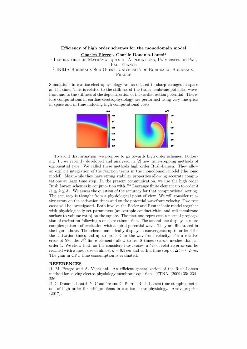
Efficiency of high order schemes for the monodomain model
Charles Pierre1, Charlie Douanla-Lontsi21 Laboratoire de Mathematiques et Applications, Universite de Pau,
Pau, France2 INRIA Bordeaux Sud Ouest, Universite de Bordeaux, Bordeaux,
France
Simulations in cardiac-electrophysiology are associated to sharp changes in spaceand in time. This is related to the stiffness of the transmembrane potential wave-front and to the stiffness of the depolarization of the cardiac action potential. There-fore computations in cardiac-electrophysiology are performed using very fine gridsin space and in time inducing high computational costs.
To avoid that situation, we propose to go towards high order schemes. Follow-ing [1], we recently developed and analyzed in [2] new time-stepping methods ofexponential type. We called these methods high order Rush-Larsen. They allowan explicit integration of the reaction terms in the monodomain model (the ionicmodel). Meanwhile they have strong stability properties allowing accurate compu-tations at large time step. In the present communication, we use the high orderRush Larsen schemes in conjunc- tion with P k Lagrange finite element up to order 3(1 ≤ k ≤ 3). We assess the question of the accuracy for that computational setting.The accuracy is thought from a physiological point of view. We will consider rela-tive errors on the activation times and on the potential wavefront velocity. Two testcases will be investigated. Both involve the Beeler and Reuter ionic model togetherwith physiologically set parameters (anisotropic conductivities and cell membranesurface to volume ratio) on the square. The first one represents a normal propaga-tion of excitation following a one site stimulation. The second one displays a morecomplex pattern of excitation with a spiral potential wave. They are illustrated inthe figure above. The scheme numerically displays a convergence up to order 4 forthe activation times and up to order 3 for the wavefront velocity. For a relativeerror of 5%, the P 3 finite elements allow to use 8 times coarser meshes than atorder 1. We show that, on the considered test cases, a 5% of relative error can bereached with a mesh size of almost h = 0.1 cm and with a time step of ∆t = 0.2ms.The gain in CPU time consumption is evaluated.
REFERENCES[1] M. Perego and A. Veneziani. An efficient generalization of the Rush-Larsenmethod for solving electro-physiology membrane equations. ETNA, (2009) 35: 234–256.[2] C. Douanla-Lontsi, Y. Coudiere and C. Pierre. Rush-Larsen time-stepping meth-ods of high order for stiff problems in cardiac electrophysiology. Arxiv preprint(2017).
Personalizing models of total heart function
Gernot Plank1
1 Medical University of Graz, Gottfried Schatz Center, Division ofBiophysics, Graz, Austria
Advances in numerical techniques and the ever increasing computational powerhave rendered the execution of forward models of total heart function feasible.Using such models based on clinical images and parameterized to reflect a givenpatient’s physiology are a highly promising approach to the comprehensive quanti-tative characterization of cardiovascular function in a given patient. Such modelsare anticipated to play a pivotal role in future precision medicine as a method tostratify diseases, optimize therapeutic procedures, predict outcomes and thus bet-ter inform clinical decision making. Key challenges to be addressed are two-fold.Expensive computational models must be made efficient enough to be compatiblewith clinical time frames and generic models must be specialized based on clinicaldata, which requires complex parameterization and data assimilation procedures.
Towards the exhaustive analysis of left ventricle dysfunctions inischemic cardiomiopathy: integrating wall kinetics, scar transmurality
and wall shear stress
S. Pozzi1, F. Piatti2, A. Camporeale3, N. Cobo Gomez4, G. DiGiovine5, S. Castelvecchio3, L. Menicanti3, A. Greiser6, E. Votta2, A.
Redaelli2, M. Lombardi31 Department of Mathematics, Politecnico di Milano, Milan, Italy
2 Department of Electronics, Information and Bioengineering,Politecnico di Milano, Milan, Italy
3 Multimodality Cardiac Imaging, IRCCS Policlinico San Donato,Milan, Italy
4 Division of Cardiology, Hospital Universitario Nuestra Senora deValme, Sevilla, Spain
5 Division of Cardiology, Azienda Ospedaliera-UniversitariaMaggiore della Carita, Novara, Italy
6 Siemens Healthcare GmbH, Erlangen, Germany
Post-ischemic dilated cardiomyopathy (DCM) is a complex clinical condition char-acterized by adverse left ventricular (LV) remodeling, with impairment of LV sys-tolic and diastolic function. Different features of DCM, like LV function and loca-tion and extension of the ischemic scar, can be assessed through different cardiacmagnetic resonance modalities. Recently, it was proposed to characterize in vivointraventricular flow patterns through 4D Flow MRI sequences. However, the in-formation yielded by these modalities is typically not integrated, thus limiting thepotential for comprehensive understanding of DCM pathophysiology. The purposeof this work was to integrate standard MRI and 4D Flow analysis. In particular,LV hemodynamics were described from 4D Flow in terms of wall shear stress anddiastolic vortices. Study population included 19 patients (all males, age = 66± 10years) with post-ischemic DCM and severe LV dysfunction (EF< 35%). Prelim-inary results confirmed the potential of 4D Flow in describing LV impairments,aiming at elucidating the fluid-dynamic determinants of post-ischemic DCM.
Enabling uncertainty quantification for the monodomain equation viamultifidelity techniques
Alessio Quaglino1, Simone Pezzuto1, Rolf Krause1
1 Center for Computational Medicine in Cardiology, Institute ofComputational Science, Universita della Svizzera Italiana, Lugano,
Switzerland
In cardiac electrophysiology, there are numerous sources of uncertainty, both inthe parameters and in the modeling aspects. For instance, the de facto standardmonodomain model has several parameters with large uncertainties: location andtiming of source currents, microstructure organization (fibers and sheets), anatomy,electrodes location, etc. In particular, the conductivities are not known exactly.This motivates modeling them as spatially-correlated random fields via, e.g., atruncated Karhunen-Love expansion, yielding a solution that is also a random field.
While sampling a random field can be done very efficiently via the pivotedCholesky decomposition [1], computing the activation map from the monodomainequation is a computational demanding task. A single patient-tailored simulationcan take several CPU hours even on a large cluster. This makes uncertainty quan-tification (UQ) unfeasible, unless modeling reduction strategies are employed. Onesuch strategy is represented by multifidelity methods [2]. The central idea of thisapproach is to build a hierarchy of increasingly faster low-fidelity models, whichmight be inaccurate but exhibit some degree of correlation with the high-fidelityone. It is the control of the correlation, rather than of the error estimates, thatensures that low-fidelity models provide useful information on the statistics of thehigh-fidelity quantity-of-interest.
A key ingredient of the multifidelity approach is the choice of low-fidelity mod-els. Typical strategies are projection-based or data-fit surrogates [2], which howeverneed to be trained anew for each patient and may become inefficient for a large di-mensionality (> 10) of the input [3], as in the case under consideration. Instead, amore physics-based approach is to take advantage of the natural hierarchy of avail-able models. These include different cellular models for the monodomain equation,the time-independent eikonal equation [4], and the 1D geodesic point activation [3].By exploiting the statistical correlations in this hierarchy, we observed a reductionof the computational cost by at least two orders of magnitude, enabling to performa full analysis within a reasonable time frame. Moreover, we incorporate Bayesiantechniques [5], which provide confidence intervals and full probability distributionsat selected points, thus augmenting the information provided by standard frequen-tist approaches [2].
REFERENCES[1] H. Harbrecht, M. Peters and R. Schneider. On the low-rank approximation bythe pivoted cholesky decomposition. Appl. Numer. Math., (2012) 62 (4): 428–440.[2] B. Peherstorfer, K. Willcox and M. Gunzburger. Survey of multifidelity methodsin uncertainty propagation, inference, and optimization. Technical Report, (2016).[3] A. Quaglino, S. Pezzuto, P. S. Koutsourelakis, A. Auricchio and R. Krause.Fast uncertainty quantification in patient-specific cardiac electrophysiology meetingclinical time constraints. Under review.[4] P. Colli Franzone and L. Guerri. Spreading of excitation in 3-d models of theanisotropic cardiac tissue. i. validation of the eikonal model. Math. Biosci., (1993)113 (2): 145–209.[5] P-S. Koutsourelakis. Accurate uncertainty quantification using inaccurate computa-tional models. SIAM J. Sci. Comput., (2009) 31 (5): 3274–3300.
Active contraction of cardiac cells: a reduced model of force generation
Francesco Regazzoni1, Luca Dede1, Alfio Quarteroni1,2
1 MOX - Dipartimento di Matematica, Politecnico di Milano, Milan,Italy
2 Institute of Mathematics, Ecole Polytechnique Federale deLausanne, Lausanne, Switzerland
The primary role of the heart as a pump is made possible by the ability of car-diac cells to contract. The mathematical modelling of the complex phenomenabehind the active contraction of cardiomyocytes is crucial for understanding heartfunctionality, since it represents the natural bridge between electrophysiology andmechanics [1, 2]. The most detailed models of muscle contraction, based on theHuxleys model of crossbridge cycling [3], are Markov Chain models describing theattachment-detachment of actin and myosin filaments. The centrality of the phe-nomenon of cooperativity between adjacent binding sites prevents applying themean-field hypothesis in order to reduce the complexity of the model [4]; therefore,the time-consuming Monte Carlo method is typically employed in simulations, hin-dering the applicability of such models to the simulation of the full organ [5]. In ourwork we propose a reduced ODE model for the mechanical activation of cardiac my-ofilaments, which is based on explicit spatial representation of nearest-neighbourinteractions. Our reduced model is derived, by combining different techniques,starting from the cooperative Markov Chain model presented in [6] and later mod-ified in [7], under the assumption of conditional independence of specific sets ofevents. This physically motivated assumption allows to drastically reduce the num-ber of degrees of freedom, thus reducing the computational cost by more than 10000 times [8]. Thanks to its computational efficiency, our reduced model is suitablefor multiscale full organ simulations. We show through numerical tests that ourmodel is able to reproduce physiological behaviours observed under various exper-imental settings, including the steady-state relationships between calcium, lengthand force, isosarcometric and shortening twitches, and force redevelopment after asudden active force drop.
REFERENCES[1] R. Chabiniok, V. Y. Wang, M. Hadjicharalambous, L. Asner et al. Multiphysicsand multiscale modelling, datamodel fusion and integration of organ physiology inthe clinic: ventricular cardiac mechanics. Interface Focus, (2016) 6 (2): 20150083.[2] M. Fink, S. A. Niederer, E. M. Cherry et al. Cardiac cell modelling: observationsfrom the heart of the cardiac physiome project. Prog. Biophys. Mol. Biol., (2011)104 (1): 2–21.[3] A. F. Huxely. Muscle structure and theories of contraction. Prog. Biophys.Biophys. Chem., (1957): 255–318.[4] J. J. Rice and P. P. de Tombe. Approaches to modeling crossbridges and calcium-dependent activation in cardiac muscle. Prog. Biophys. Mol. Biol., (2004) 85 (2):179–195.[5] J. Hussan, P. P. de Tombe and J. J. Rice. A spatially detailed myofilamentmodel as a basis for large-scale biological simulations. IBM J. Res. Dev., (2006) 50(6): 583–600.[6] J. J. Rice, G. Stolovitzky, Y. Tu and P. P. de Tombe. Ising model of cardiacthin filament activation with nearest-neighbor cooperative interactions. Biophys.J., (2003) 84 (2): 897–909.[7] T. Washio, J. Okada, S. Sugiura and T. Hisada. Approximation for cooperativeinteractions of a spatially-detailed cardiac sarcomere model. Cell. Mol. Bioeng.,(2012) 5 (1): 113–126.

[8] F. Regazzoni, L. Dede and A. Quarteroni. Active contraction of cardiac cells: amodel for sarcomere dynamics with cooperative interactions. MOX Report 48/2017(2017).
Anode Break Excitation in Hypertrophic Cardiomyocytes: AnInvestigative Tool for Predicting Modulation of Cardiac Excitability
Nicolo Salvarani1, Pierluigi Carullo1,3, Michele Miragoli1,2,31 Humanitas Clinical and Research Center, Rozzano, Italy
2 Department of Medicine and Surgery, University of Parma, Parma,Italy
3 Institute of Genetics and Biomedical Research, Milan Unit,National Research Council, Milan, Italy
Excitation at the closure of the anode is one of the two ways by which actionpotentials in single cells can be electrically elicit. Despite the fact that ”anodebreak” excitation has long been observed in cardiac myocytes (the phenomenonof anodal excitation of cardiac muscle has been described by Cranefield et al., inthe late-1950s), very little is known about its mechanism and implications. Anodalstimulation of cardiac tissue has been explained for long time using bidomain mod-els of cardiac tissue (Henriquez et. al. ”A planar slab bidomain model for cardiactissue”. Ann. Biomed. Eng. 1990) until Ranjan et al. demonstrated that anodebreak excitation has an active cellular basis (”Mechanism of anode break stimula-tion in the heart”. Biophys. J. 1998). Indeed, they described that the activationof a hyperpolarization-activated inward current (If) provides the current necessaryto drive the potential to more depolarized levels, and the time-dependent block ofinwardly rectifying K+ current (IK1) aids the process by increasing membrane resis-tance. Furthermore, a very important property of anode excitation, first describedby Dekker et al. in 1970 (”Direct current make and break thresholds for pacemakerelectrodes on the canine ventricle”. Circ. Res. 1970), is represent by ”supernor-mal excitability” and consists in an increase of the excitability in the refractoryheart due to anode break excitation as compared with its diastolic values. Conse-quently, if a stimulus falls in the vulnerable period of a spontaneous cycle, the riskof triggering an arrhythmic event is higher with anodal than cathodal stimulations(Merx et al. ”Effects of unipolar cathodal and bipolar stimulation on vulnerabilityof ischemic ventricles to fibrillation”. 1975). Even supernormal excitability hasbeen explained using bidomain models of cardiac tissue relying solely on passivecardiac tissue properties. The characterization of the fundamental basis of anodalstimulation at cellular level will advance the understanding of either the excitabilityin the heart and the mechanism of potential clinically important properties (e.g.”supernormal excitability”) of anodal excitation in the ventricular myocardium.
In this work, we tested the hypothesis that anode break excitation might un-mask in hypertrophic heart alterations in cardiac electrical excitability in both thediastolic and refractory period. Experiments were performed on single ventricu-lar cardiomyocytes from healthy and hypertrophic mice derived from transverseaortic constriction (TAC). Cathodal and anodal excitability in diastole and duringrefractory period were recorded using dedicated whole cell patch clamp protocolsin order to obtain strength-duration and strength-interval curves. The comparisonof strength-duration curves, as a global index of excitability, showed that the ra-tio between anodal and cathodal rheobase was much higher in healthy than TACcardiomyocytes. We finally measured strength-interval curves to investigate an-odal excitability during refractory period and found not only that supernormalexcitability was present in both healthy and hypertrophic cardiomyocytes but itwas significantly decreased in TAC cardiomyocytes.

These findings demonstrate an increased excitability in pathologic mice followinganodal stimulations both in the diastole and refractory period. Of interest, cathodalstrength-duration and strength interval curves did not show significant differencesamong hypertrophic and healthy cardiomyocytes. Furthermore, we have provedthat the phenomenon of ”supernormal excitability”, which was observed only in thetissue, is intrinsic to the mechanism of cellular excitability (the anodal ”strength-interval” curve in single mice ventricular myocytes was measured for the first timein this research). These findings taken together open the intriguing possibility ofmaking anode break a tool both to unmask potential arrhythmogenic ionic mech-anisms in pathological hearts and study, for the first time, action potentials in therefractory period.
Systolic blood pressure detection using a multi-wavelengthOpto-Electronic patch sensor at peripheral circulation
Francesco Scardulla1, Sijung Hu2, Leonardo D’Acquisto1, SalvatorePasta4, Laura Barrett3, Panagiotis Blanos2, Liangwen Yan5
1 DIID - Dipartimento dell’Innovazione Industriale e Digitale,Universita degli Studi di Palermo, Palermo, Italy
2 School of Electronic, Electrical and System Engineering,Loughborough University, Loughborough, UK
3 National Centre for Sport and Exercise Medicine, School ofSport, Exercise and Health Sciences, Loughborough University,
Loughborough, UK4 Fondazione Ri.MED, Palermo, Italy
5 School of Mechatronic Engineering and Automation, ShanghaiUniversity, Shanghai, China
Through the use of a multi-wavelength opto-electronic patch sensor (MOEPS)(1) based on PPG technology, we sought to determine the systolic blood pres-sure (SBP), detecting the changes in the morphology of the MOEPS signal. Thepressure acquisitions were performed both with gold standard, using an electronicsphygmomanometer, and the MOEPS sensor (experimental procedure), on sub-jects from a multiethnic cohort (aged 29±6). More specifically, the MOEPS sensorwas applied together with a manual inflatable cuff placed around the left upperarm, that was inflated going slightly above the level of the systolic pressure withincreases of +10 mmHg and subsequently deflated, by 10 mmHg until reachingfull deflation. MOEPS signals were captured using four wavelength illuminationsources (i.e., green 525 nm, orange 595 nm, red 650 nm and IR 870 nm) on threedifferent measuring sites, which are forefinger, radial artery and wrist. The MatLabalgorithm analyzed each wavelength of every sensor position taking into accountdifferent parameters. It provided approximations on the instant at which the SBPwas reached and therefore the loss of the signal occurred since the vessel was com-pletely blocked, as well as the instant in which it came back again below the systolicpressure and therefore there was a resumption of the signal. It also provided a cor-relation between diastolic pressure as the starting point in which the signal startsreducing. The general pattern described by green light recorded the best perfor-mance for every site. Capillary site has provided the most accurate values for allwavelengths. In addition, better results were achieved in the inflation phase thanwith the deflating of the cuff, since some delay was observed in the recovery ofthe bloodstream in the artery and therefore in regeneration of the signal. Furtherstudies will improve the clinical relevance on patients cohort diagnosed with hyper-or hypotension.
REFERENCES[1] A. Alzahrani, S. Hu , V. Azorin-Peris , L. Barrett, D. Esliger, M. Hayes, S.Akbare, J. Achart and S. Kuoch. A Multi-Channel Opto-Electronic Sensor toAccurately Monitor Heart Rate against Motion Artefact during Exercise. Sensors,(2015) 15: 25681–25702.
Cardiac Surgery and Biomedical Engineering
Roberto Scrofani1, Francesca Nicolo1, Giovanni Cagnoni1, CarloAntona1
1 Cardiovascular Department, ”Luigi Sacco Hospital”, Milan, Italy
Nowadays, even if the role of mathematical consultant in medical team is discussed,we must be aware that the potential benefit of this collaboration will allow to tacklethe large and complex medical problems. Cardiothoracic surgeons have been inven-tors, developers and modifiers of a wide range of technologies from medical devices,such as heart mechanical or biological valves, bypass machines, blood vessel substi-tutes and novel imaging techniques. But it is not just the individual pratictioneror researcher who can contribute to the process of bringing new technology to theclinic. In the deep evolution obtained in cardiac surgery, mathematics has been thelink between the measured data and the primary structured information on whichthe realization of an idea was possible.
In medical engineering and in heart surgery in particular it is of increasing im-portance to recognize the essentials out of a huge amount of information about avery complex situations and to draw the right conclusions. Mathematical modelsof the heart and circulation offers many interesting options both in diagnosis andtherapy planning of heart surgery:
• Electophysiological models allow to– optimize radio-frequency (RF) ablation for atrial fibrillation;– adapt stimulation sequences for heart pacemaker and cardiac resyn-
chronization.• Elastomechanical models allow to
– optimize preoperative surgical cuts in open heart surgery for treat-ment of aneurysms and for reconstruction of ventricle in endoventri-coloplasty;
– optimize preoperative surgical techniques in valvuloplasty therapy;– predict the trans-catheter percutaneous valve implantation preopera-
tive results;– test new surgical techniques;– self-education in young training surgeons.
• Computational Fluid dynamical models allows to– better planning the behavior of stenoses of the coronary arteries;– understand the behavior of stenoses and/or the insufficiency of the
heart valves;– optimize the behavior of different kind of anastomoses between coro-
nary arteries and arterial or vein grafts in bypass surgery.• Circulation models allows to
– control extracorporeal circulation during heart surgery;– study the fluid structure interaction in the artificial heart and ventric-
ular assisted devices;– optimize the fluid dynamic effect of the vascular graft thoracic aortic
aneurysm surgery.
Today the process of bringing technological advances to our patients has becomecomplex and more structured. What seems to be most needed is to extend the areasof collaboration among different specialty. Knowledge transfer and ”team work”for multidisciplinary approach could give us new ways to treat some of the mostdaily diseases.

Computational Modeling of the Cardiac Pacemaking in Humans
Stefano Severi1, Alan Fabbri1, Michelangelo Paci2, Ronald Wilders3,Yannick Lutz4, Axel Loewe4
1 Laboratory of Cellular and Molecular Engineering ”SilvioCavalcanti”, Department of Electrical, Electronic and InformationEngineering ”Guglielmo Marconi”, University of Bologna, Cesena,
Italy2 BioMediTech Institute and Faculty of Biomedical Sciences and
Engineering, Tampere University of Technology, Tampere, Finland3 Department of Medical Biology, Academic Medical Center,
University of Amsterdam, Amsterdam, The Netherlands4 Institute of Biomedical Engineering, Karlsruhe Institute of
Technology, Karlsruhe, Germany
The sinoatrial node (SAN) is the normal pacemaker of the mammalian heart. Overseveral decades, a large amount of data on the ionic mechanisms underlying thespontaneous electrical activity of SAN pacemaker cells has been obtained, mostlyin experiments on single cells isolated from rabbit SAN. This wealth of data hasallowed the development of mathematical models of the electrical activity of rabbitSAN pacemaker cells. However, the translation of animal data/models to humansis not straightforward. Even less so for SAN pacemaker cells than working myocar-dial cells given the big difference in their main output (i.e. pacing rate) betweenhuman and laboratory animals. The development of a comprehensive model of theelectrical activity of a human SAN pacemaker cell strictly based on and constrainedby the available electrophysiological data will be presented. We started from theSeveri-DiFrancesco rabbit SAN model, which integrates the two principal mecha-nisms that determine the beating rate: the ”membrane clock” and ”calcium clock”.Several current formulations were updated based on available measurements. A setof parameters, for which no specific data were available, were automatically opti-mized to reproduce the measured AP and calcium transient data. The model wasthen validated by assessing the effects of several mutations affecting heart rate andrate modulation. Moreover, two recent applications of the model will be presented:i) We used our SAN AP computational model to assess the effects of the inclu-sion of the small conductance K+ current (ISK) on the biomarkers that describethe AP waveform and calcium transient; ii) We analysed the effect of altered elec-trolyte levels (as systematically occurring in hemodialysis patients) on pacemakingto investigate the possible mechanisms of the bradycardic sudden cardiac deathspointed out by two recent human studies using implantable loop recorders.
High-Order Operator-Splitting Methods for the Bidomain andMonodomain Models
Raymond Spiteri1, Jessica Cervi2
1 Department of Computer Science, University of Saskatchewan,Saskatoon, Canada
2 Department of Mathematics and Statistics, University ofSaskatchewan, Saskatoon, Canada
The bidomain and monodomain models are among the most widely used mathemat-ical models to describe cardiac electrophysiology. They take the form of multi-scalereaction-diffusion partial differential equations that couple the dynamic behaviouron the cellular scale with that on the tissue scale. The systems of differential equa-tions associated with these models are large and strongly non-linear, but they alsohave a distinct structure due to their multi-scale nature. Operator-splitting meth-ods attempt to take advantage of this structure to efficiently produce numericalsolutions.
The focus of this presentation is on operator-splitting methods with order higherthan two. Such methods require backward time integration and historically havebeen considered unstable for solving deterministic parabolic systems. The stabilityand performance of operator-splitting methods of up to order four to solve thebidomain and monodomain models are demonstrated on several examples arisingin the field of cardiovascular modeling.
Shape Analysis for 3D Cardiac Imaging
Luciano Teresi1, V. Varano1, P. Piras3, S. Gabriele1, I. Dryden2, P.Nardinocchi3, P. E. Puddu3 Torromeo 3
1 Universita Roma Tre, Rome, Italy2 University of Nottingham, Nottingham, UK3 Sapienza, Universita di Roma, Rome, Italy
Keywords: shape analysis, heart function, differential geometry, parallel transport.
Shape analysis encompasses many powerful tools used to compare shapes and toquantify their differences. Here we present a biological application aimed at com-paring the motion of the cardiac revolution of different subjects, using raw datafrom 3D Speckle-Tracking Echocardiography; our goal is comparing the motion ofthe same organ, the human heart, from different individuals, and to assess thedifferences in the cardiac revolution by filtering out inter-individual shape differ-ences. This method is under scrutiny, and is being applied to a database of 200echocardiographic data acquired at the University Hospital Umberto I, at SapienzaUniversity of Rome.
The use of the shape analysis to compare both the deformation and the shapeof motion itself (and not merely shapes) requires advanced tools from differentialgeometry and statistics. In particular, we present an appropriate definition ofparallel transport along the geodesics of the Size-and-Shape Space that enablesshape analysis with the possibility of comparing not only different shapes but alsodifferent motions.
REFERENCES[1] V. Varano et al. The Decomposition of Deformation: new metrics to enhanceshape analysis in medical imaging. Med. Image Anal., (2018) 46: 35–56.[2] V.Varano et al.The TPS Direct Transport: a new method for transportingdeformations in the Size-and- shape Space. Int. J. Comput. Vis., accepted (2017).[3] S. Durrleman et al.Deformetrica - A software for the statistical analysis of 2Dand 3D shape data. www.deformetrica.org (2014).[4] P. Piras et al.Left Atrial trajectory impairment in Hypertrophic Cardiomyopathydisclosed by Geometric Morphometrics and Parallel Transport. Sci. Rep., (2016)6: 34906.[5] A. Madeo et al.A New 4D Trajectory-Based Approach Unveils Abnormal LVRevolution Dynamics in Hypertrophic Cardiomyopathy. PLOS ONE, (2015) 10(4): e0122376.
Modeling isovolumic phases using a pressure corrected RIS model
Alexandre This1,2,3, Ludovic Boilevin-Kayl2,3, Hernan G. Morales1,Odile Bonnefous1, Miguel A. Fernandez2,3, Jean-Frederic Gerbeau2,3
1 Medisys, Philips Research, France2 INRIA Paris, Paris, France
3 Sorbonne Universites, UPMC Univ Paris 6, Paris, France
Introduction. Modeling often trade computational complexity against phys- icalaccuracy. In the case of the heart modeling, the physics involves a complex mixbetween chemical activity, electri- cal activity, mechanics and fluid dynamics. How-ever, multi- physics simulations are computationally expensive. Alternative to fullycoupled multi-physics simulation is to only solve a subset of the physical mecha-nism. For instance, Image-driven CFD is a way to model the blood flow inside theheart by using patient-specific images as boundary conditions of the fluid problem.
Although very attractive, those methods introduce artificial difficulties when try-ing to resolve multi-physics phenomenon. For example, modeling the LV isovolumicphases turns into a challenging task due to two main reasons. Generally speaking,when an incompressible fluid is enclosed in a cavity under Dirichlet boundary con-ditions, the pressure inside the cavity can only be determined up to a constant.Secondly, the Dirichlet boundary condition imposed on the cavity surface may vio-late the incompressibility constraint. This issue arises for example when the imageframerate is too low to properly recover all the cardiac phases([3], [2]).
The contribution of this work is to propose a strategy that builds upon a sim-plified valve model (RIS, [1], [4]) in order to properly resolve isovolumic phases.Material and Methods. A fluid cavity enclosed by a solid surface and twovalves is considered. Valves are modeled using RIS (i.e. a penalization methodon an immersed surface with duplicate degrees of freedom to correctly capture thepressure jump, [1], [4]) and no-slip boundary condition is applied on the other solidsurfaces. When both valves are closed and thanks to the incompressibility of thefluid, one can derive a simplified expression of the cavity pressure which relatesthe inside of the cavity with the cavity wall velocity, the penalization term and theexterior pressure. This explains why realistic isovolumic phases cannot be obtainedin the given setting. A pressure correction term is designed in order to counteractthose contributions and allows the cavity pressure to reach a given pressure value,which we assume to be either given by measurements or by an electro-mechanicalsimulation.
The method is validated on an academic toy problem for various boundary con-ditions and domain motions. Additionally, the method was also experimented ona 3D heart simulation where the motion of the ventricle was given by an electro-mechanical simulation ([4]) for which atrium, ventricular and aortic pressure timeseries were also provided. The goal was to allow modeling of isovolumic phases evenif the given ventricular motion was subject to the issues described in the introduc-tion (i.e the ventricular volume was changing even during what should have beenisovolumic phase).Results. A reasonable agreement between the theoretical pressure and the averagecavity pressure for the tested boundary condition on the toy model. In particu-lar, it was possible to correct the cavity pressure in complex situations involvingimportant gaps with exterior pressures and a changing cavity volume induced byinhomogeneous valve and wall motion for example. The correction term was appliedand cavity pressures could be set to arbitrary values.
Similar results were obtained on the complex 3D LV simulation where the theo-retical ventricular pressure was reasonably close to the average ventricular pressure.It has therefore been possible to obtain physiological ventricular pressures for acomplete cardiac cycle even during isovolumic phases.

Discussion. he presented correction term benefits from the fact that the RISmodel is penalizing the fluid on the valve surface and not strictly enforcing Dirichletboundary condition. First, this means that, unlike a cavity enclosed under fullDirichlet BC, the inner pressure is defined by the outer pressure. Additionally, thismeans that the incompressibility constraint of the fluid can be satisfied even whenthe enclosed cavity undergoes volume change by allowing small leaks through thevalve.
However, as the penalization factor goes to infinity, the RIS model leads to asingular problem equivalent to imposing Dirichlet BC everywhere. This means thatthe intraventricular pressure cannot be computed nor corrected properly anymorein this case. Therefore a trade-of on the penalization factor has to be chosen.
REFERENCES[1] M. Astorino, J. Hamers, S. C. Shadden and J.-F. Gerbeau. A robust and efficientvalve model based on resistive immersed surfaces. Int. J. Numer. Meth. Biomed.Eng., (2012) 28 (9): 937–959.[2] A. M. Bavo, A. M Pouch, J. Degroote, J. Vierendeels, J. H. Gorman, R. C.Gorman and P. Segers. Patient-specific CFD models for intraventricular flow anal-ysis from 3D ultrasound imaging: Comparison of three clinical cases. J. Biomech.,(2016) 50: 144–150.[3] C. Chnafa, S. Mendez and F. Nicoud. Image-based large-eddy simulation in arealistic left heart. Comput. Fluids, (2014) 94: 173–187.[4] M. Fedele, E. Faggiano, L. Dede and A. Quarteroni. A patient-specific aorticvalve model based on moving resistive immersed implicit surfaces. Biomech. Mod.Mechanobiol., (2017) 16 (5): 1779–1803.

Development in silico model of SK channel gating, calcium sensitivityand drug interaction
Ilse van Herck1, Bo H. Bentzen2,3, Vincent Seutin4, Jussi T.Koivumaki5, Mary M. Maleckar1,6, Neil V. Marrion7, Andrew G.
Edwards1,81 Simula Research Laboratory, Oslo, Norway
2 Acesion Pharma, Copenhagen, Denmark3 Biomedical Institute, University of Copenhagen, Copenhagen,
Denmark4 Neurophysiology unit, GIGA Neurosciences, University of Liege,
Liege, Belgium5 A. I. Virtanen Institute of Molecular Sciences, Kuopio, Finland
6 Allen Institute for Cell Science, Seattle, WA, USA7 School of Physiology, Pharmacology and Neuroscience, University
of Bristol, Bristol, UK8 Physiology and Cell Biology, University of Oslo, Oslo, Norway
Computational models are increasingly used in clinical and pharmaceutical set-tings, for example in guiding surgery and arrhythmia drug testing initiative suchas CiPa. Biological targets linked to disease and treatments are often not includedin available computational models, or lack important mechanistic detail. One suchexample is the small conductance calcium-activated potassium (SK) channel, whichis active in the late repolarization phase of the atrial action potential and thoughtto contribute to arrhythmogenic substrate in atrial fibrillation (AF). In vitro and invivo experiments support involvement of the SK current in AF arrhythmogenesis,specifically in initiation and disease progression.
An initial Markov state model of the SK2 channel [Hirschberg et al. 1998], basedon single-channel patch clamp data, utilized 4 closed and 2 open states to captureSK channel gating dynamics. We extend this model to reflect new inside-out patchclamp data in which we have observed a strong temperature sensitivity of calcium-dependent SK activation, with a shift in EC50 towards the diastolic calcium range.New patch clamp experiments at the single channel level are also included in thepresence of known inhibitor apamin and novel SK modulator, AP14145. These datahave allowed us to develop a model of channel gating that more closely replicatesin vivo behavior and incorporates two modes of drug interaction. We anticipatethis will pave the way for further work investigating myocyte- and tissue-level sim-ulations assessing the impact of SK channels and pharmacological modulation onhuman atrial arrhythmogenesis.

Clinically driven numerical models in cardiovascular surgery
Emiliano Votta1, Filippo Piatti1, Omar Pappalardo1,2, AlessandroCaimi1, Matteo Selmi1,2, Giovanni Rossini1, Francesco Sturla1,2,
Alberto Redaelli11 Department of Electronics, Information and Bioengineering,
Politecnico di Milano, Milan, Italy2 Division of Cardiovascular Surgery, Universita degli Studi di
Verona, Verona, Italy3 3D and Computer Simulation Laboratory, IRCCS Policlinico San
Donato, San Donato Milanese, Italy
Current imaging technologies allow or accessing extensive patient-specific anatom-ical and functional information. In clinical practice, this information is often onlypartially exploited when performing diagnoses, prognoses or when planning clinicalsolutions.
In principle, this information can be further exploited by feeding state-of-the-artnumerical models to quantify the physical behaviour of tissues and organs on apatient-specific basis. Such analysis may provide insight into pathological condi-tions and highlight physics-based reasons for the changes in the clinical scenario aswell as for the effects of different therapies.
However, despite this great potential, the application of this approach in thereal clinical setting is limited because the most advanced and detailed modellingapproaches, i.e., the most realistic and reliable ones, typically are not user-friendlynor sufficiently time-efficient.
On this basis, the search for an effective trade-off between models sophisticationand clinical applicability becomes crucial. This aspect will be discussed in lightof some of the most recent computational modelling activities carried out by theBiomechanics Research Group at Politecnico di Milano in collaboration with severalclinical centres.
Applicability of the PREMAP algorithm to accelerate activationmapping of scar-related ventricular reentry tachycardia
Tobias Weber1, Eberhard Scholz2, Felix Langkamp2, Hugo A. Katus2,Sebastian Sager1
1 Institute of Optimization, Faculty of Mathematics, Otto vonGuericke University Magdeburg, Magdeburg, Germany
2 Department of Cardiology, Angiology, and Pneumology,University Hospital Heidelberg, Heidelberg, Germany
Background. We published a mapping algorithm (PREMAP) that based on lin-ear regression analysis and mathematical optimization significantly speeds up thelocalization of focal cardiac arrhythmias (Heart Rhythm 2017;14:875-882). Whenintegrated into 3D electroanatomical mapping systems this algorithm could guidethe operator to the source of focal activity by continuously optimizing the searchpath. In the current study we extend the algorithm to scar related ventriculartachycardia (VT).Methods and Results. 3D electroanatomical maps of 20 patients that had un-dergone mapping and ablation of scar related left ventricular tachycardia were usedfor analysis. To increase the quality of our models, anatomical obstacles such asthe mitral anulus and scarred area were defined as non- or slowly-conducting zones.Reentry tachycardia was simulated by randomly defining exit sites along the bor-der of the scar. Within this simulation environment, the algorithm was given thetask to reconstruct the VT and locate the exit site with a minimal number of localactivation time measurements (LAT). For this purpose, the PREMAP algorithmperforms linear regression analyses thereby (1) predicting the exit site of the tachy-cardia (2) and positioning the next measurement at the position with maximumadditive value. Within 400 simulated VTs, the algorithm reconstructed the VT cir-cuit including the exit site with a mean number of 10.0± 5.6 LAT measurements.Conclusion. Taken together we show that an algorithm based on regression anal-ysis and mathematical optimization is able to efficiently reconstruct activation andestimate the exit site of scar-related ventricular tachycardia. The mapping per-formance was not attenuated by the increased complexity of the geometries. Thisalgorithm might significantly accelerate the mapping procedure by guiding the op-erator to the optimallocation for the next LAT measurement, reducing the numberof points with redundant information and predicting the exit site with high accu-racy from an early stage of the mapping procedure.
Estimating ion current fractions in mathematical models ofnon-selective channels
Eike M. Wuelfers1, Gunnar Seemann1
1 Institute for Experimental Cardiovascular Medicine, UniversityHeart Center Freiburg-Bad Krozingen, Medical Center, University
of Freiburg, and Faculty of Medicine, University of Freiburg,Freiburg, Germany.
In many mathematical models of non-selective channels, the total channel currentis formulated using Ohm’s law in the form ICh,Ohm = gCh · GCh · (Vm − ECh),depending on the transmembrane voltage Vm and the channel’s equilibrium po-tential ECh. The channel conductivity gCh combines single-channel conductivity,channel density, optionally modulated by a gating mechanism GCh modeled basedon patch-clamp data.
However, the formulation based on Ohm’s law can only describe the total elec-tric current, the sum of all ion currents passing through the channel. In someelectrophsiological models, however, intracellular ion concentrations are not con-stant but part of the model state. Then, state trajectories could become inaccuratewhen not accounting for the ions passing through non-selective channels. Here wesuggest an approach to transform a model formulation based on Ohm’s law suchthat partial ion currents can be accounted for.
The Goldman-Hodgkin-Katz (GHK) current equation states that the ion currentof species X through a membrane pore is
IX =VmF
2
RTPXz
2X
[X]i − [X]e exp(−zXVmF/RT )
1− exp(−zXVmF/RT ),
with [X]i and [X]e intra- and extracellular concentrations, and PX and zX per-meability and valence of X, respectively [1]. With ICh,GHK =
∑X IX for all ions
X that can pass through the channel, we can then choose unknown parameters inICh,GHK , such that ICh,GHK ≈ ICh,Ohm, meaning the total current through thechannel would follow a similar trajectory as predicted by Ohm’s law, but we wouldalso have formulations for the partial ion currents of every species.
In the instance of Channelrhodopsin-2 (ChR2), a light-gated non-selective cationchannel, PK and PCa have been measured relative to PNa, and the ratio PH/PNa
has been calculated based on the equilibrium potential using the GHK voltageequation (e.g., [2]). Considering those four cations only, we are left with one un-
known, PNa. Solving gChR2 = limVm→0mVdICh,GHK
dVmin order to match the Ohmic
relationship in slope at 0 mV, we find
PNa = 2gChR2RT
F 2
([Na+]i + [Na+]e +
PH
PNa([H+]i + [H+]e)+
PK
PNa([K+]i + [K+]e) + 4
PCa
PNa([Ca2+]i + [Ca2+]e)
)−1.
We integrated the ChR2 model by Williams et al. [4] into the human ventricularAP model by ten Tusscher & Panfilov [5] Fig. 1 shows the resulting ion currentscompared to the Ohmic relationship. The contribution ratio of the different cationsto the total current in our model also correlates well with measured data at multipleion concentrations [3].
Using both variants, we further investigated two scenarios: Pacing by opticalstimulation, and long-term illumination, a case particularly interesting in light ofrecent investigations of using ChR2 to terminate atrial fibrillation [6]. Fig. 2shows ion concentration trajectories for those scenarios using the unaltered formu-lation from Williams et al. (bulk current, no direct influence on ion concentrations,gChR2 = 0.12ı,mS/cm2) and our GHK-based modification. When optically pacing,

the bulk model leads to a linearly declining intracellular potassium concentration,counteracting the charge imbalance caused by the unspecific ChR2 current. Forcontinuous illumination, our model suggests an increase in intracellular sodiumthat is not predicted by the bulk model.
First results show that considering ion species for non-specific channels can havean effect on intracellular ion concentrations in models. In the concrete example ofChR2, pacing using an unspecific bulk current model would only in a long term leadto unphysiological concentrations that could affect the cellular electrophysiology in asignificant way. However, for continuous illumination the bulk current results couldbe misleading (e.g., in our optical defibrillation model, not predicting a sodiumoverload). Our method can be applied to any non-selective channel given sufficientexperimental data.
REFERENCES[1] B. Hille. Ionic channels of excitable membranes. (1992) Second edition. Sun-derland, Massachusetts: Sinauer Associates Inc.[2] J. Y. Lin, M. Z. Lin, P. Steinbach and R. Y. Tsien. Characterization of engi-neered channelrhodopsin variants with improved properties and kinetics. Biophys.J., (2009) 96 (5): 1803–1814.[3] F. Schneider, D. Gradmann and P. Hegemann. Ion selectivity and competitionin channelrhodopsins. Biophys. J., (2013) 105 (1): 91–100.[4] J. C. Williams, J. Xu, Z. Lu, A. Klimas, X. Chen, C. M. Ambrosi, I. S. Cohenand E. Entcheva. Computational Optogenetics: Empirically-Derived Voltage- andLight-Sensitive Channelrhodopsin-2 Model. PLoS Comput. Biol., (2013) 9 (9):e1003220.[5] K. H. W. J. ten Tusscher and A. V. Panfilov. Alternans and spiral breakup in ahuman ventricular tissue model. Am. J. Physiol. Heart Circ. Physiol., (2006) 291(3): H1088–H1100.[6] T. Bruegmann, T. Beiert, C. C. Vogt, J. W. Schrickel and P. Sasse. Optogenetictermination of atrial fibrillation in mice. Cardiovasc. Res. (2017).
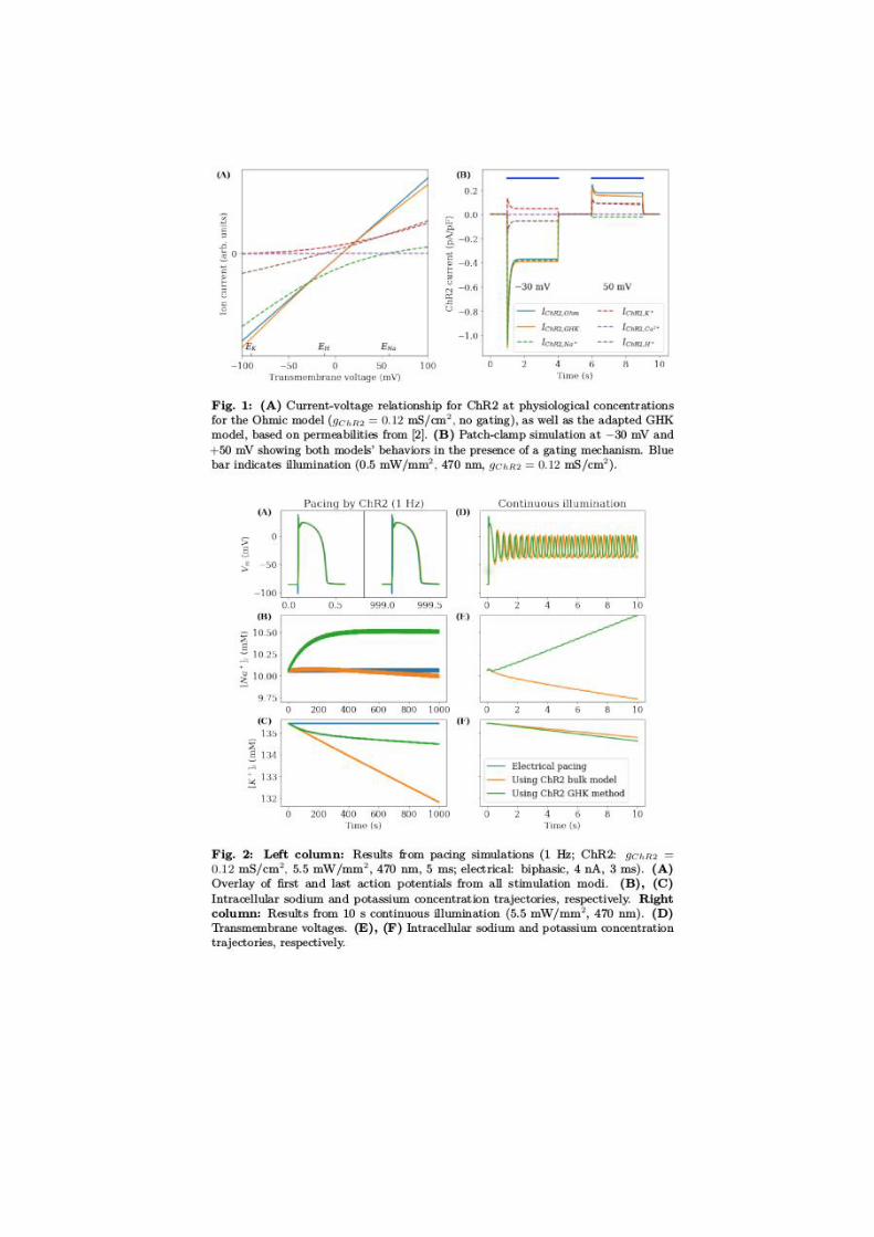

Reduced order modelling for cardiovascular haemodynamics: optimalflow control, data assimilation and geometrical reconstruction
Zakia Zainib1, Francesco Ballarin1, Gianluigi Rozza1
1 mathLab, Mathematics Area, SISSA International School forAdvanced Studies, Trieste, Italy
Bypass grafting is a commonly used procedure to restore blood flow in obstructedcardiovascular system, caused by diseases such as arterial stenosis or aortic coarcta-tion [2, 4, 3]. We present a complete pipeline incorporating real-patient geometries(reconstructed from clinical images), numerical simulations [8], and optimal control[5] on physical parameters while investigating the haemodynamics in grafts, andtheir corresponding arteries [7]. Furthermore, by exploiting reduced-order methods[1, 6] in our framework, we not only lessen the computational cost but also profi-ciently bridge the gap between numerical simulations and medical measurements,thanks to flow control. In the end, some examples illustrating the application onreal-patient geometries for coronary artery bypass graft (CABG) will be discussed.
REFERENCES[1] F. Ballarin, E. Faggiano, S. Ippolito, A. Manzoni, A. Quarteroni, G. Rozza, andR. Scrofani. Fast simulations of patient-specific haemodynamics of coronary arterybypass grafts based on a POD-Galerkin method and a vascular shape parametriza-tion. J. Comput. Phys., (2016) 315: 609–628.[2] F. Ballarin, E. Faggiano, A. Manzoni, A. Quarteroni, G. Rozza, S. Ippolito, C.Antona and R. Scrofani. Numerical modeling of hemodynamics scenarios of patient-specific coronary artery bypass grafts. Biomech. Mod. Mechanobiol., (2017) 16 (4):1373–1399.[3] Z. Chen, F. Ballarin, G. Rozza, A. M. Crean, L. Jimenez-Juan and P. Trive-rio. Non-invasive assessment of aortic coarctation severity using computationalfluid dynamics: a feasibility study. 20th Annual Scientific Sessions, Society forCardiovascular Magnetic Resonance, Washington, DC, Feb. 1-4 2017.[4] L. Formaggia, A. Quarteroni and A. Veneziani. Cardiovascular Mathematics:Modeling and Simulation of the Circulatory System. Springer-Verlag Italia, Milano,2009.[5] M. D. Gunzburger. Perspectives in Flow Control and Optimization, volume 5.SIAM, Philadelphia, 2003.[6] J. S. Hesthaven, G. Rozza and B. Stamm. Certified Reduced Basis Methodsfor Parametrized Partial Differential Equations. Springer Briefs in Mathematics.Springer, Switzerland, 1 edition, 2015.[7] A. L. Marsden. Optimization in cardiovascular modeling. Annual Rev. FluidMech., (2014) 46 (1): 519–546.[8] A. Veneziani, F. Auricchio, A. Reali, G. Rozza, M. Conti, S. Morganti and A.Lefieux. Computational methods in cardiovascular mechanics. CRC press, 2018(In press).
Fluid-Structure Interaction via an XFEM/DG Approach for ValveDynamics
Stefano Zonca1, Luca Formaggia1, Christian Vergara1
1 MOX, Dipartimento di Matematica, Politecnico di Milano, Milan,Italy
We present a numerical method that relies on unfitted and overlapping meshes tosimulate a three-dimensional (3D) immersed elastic structure that undergoes largedisplacements interacting with an incompressible fluid. The idea is to discretizeindependently the fluid and solid domains so that the solid mesh overlaps the fluidone. The fluid mesh is kept fixed, while the solid one is free to move, resulting inunfitted meshes at the fluid structure interface. The proposed method is based onthe Extended Finite Element Method (XFEM), that is able to treat discontinuitiesof the numerical solution within the same element, coupled with a DiscontinuousGalerkin (DG) mortaring, in order to couple the numerical solution at the fluid-structure interface. Due to the unfitted nature of the meshes, this method avoidsadaptation or remeshing procedures of the fluid mesh when the structure moves.This framework is applied in the study of the interaction arising between blood andleaflets of the aortic valve.